cellular/molecular ceramidase regulates synaptic vesicle ... · tified slug-a-bed (slab) in a...
TRANSCRIPT

Cellular/Molecular
Ceramidase Regulates Synaptic Vesicle Exocytosis andTrafficking
Jeffrey Rohrbough,1 Emma Rushton,1 Laura Palanker,2 Elvin Woodruff,1 Heinrich J. G. Matthies,1 Usha Acharya,3
Jairaj K. Acharya,3 and Kendal Broadie1
1Department of Biological Sciences, Vanderbilt Kennedy Center, Vanderbilt Brain Institute, Vanderbilt University, Nashville, Tennessee 37235-1634,2Department of Biology, University of Utah, Salt Lake City, Utah 84112, and 3Regulation of Cell Growth Laboratory, National Cancer Institute-Frederick,Frederick, Maryland 21702
A screen for Drosophila synaptic dysfunction mutants identified slug-a-bed (slab). The slab gene encodes ceramidase, a central enzyme insphingolipid metabolism and regulation. Sphingolipids are major constituents of lipid rafts, membrane domains with roles in vesicletrafficking, and signaling pathways. Null slab mutants arrest as fully developed embryos with severely reduced movement. The SLABprotein is widely expressed in different tissues but enriched in neurons at all stages of development. Targeted neuronal expression of slabrescues mutant lethality, demonstrating the essential neuronal function of the protein. C5-ceramide applied to living preparations israpidly accumulated at neuromuscular junction (NMJ) synapses dependent on the SLAB expression level, indicating that synapticsphingolipid trafficking and distribution is regulated by SLAB function. Evoked synaptic currents at slab mutant NMJs are reduced by50 –70%, whereas postsynaptic glutamate-gated currents are normal, demonstrating a specific presynaptic impairment. Hypertonicsaline-evoked synaptic vesicle fusion is similarly impaired by 50 –70%, demonstrating a loss of readily releasable vesicles. In addition,FM1-43 dye uptake is reduced in slab mutant presynaptic terminals, indicating a smaller cycling vesicle pool. Ultrastructural analyses ofmutants reveal a normal vesicle distribution clustered and docked at active zones, but fewer vesicles in reserve regions, and a twofold tothreefold increased incidence of vesicles linked together and tethered at the plasma membrane. These results indicate that SLAB cerami-dase function controls presynaptic terminal sphingolipid composition to regulate vesicle fusion and trafficking, and thus the strengthand reliability of synaptic transmission.
Key words: Drosophila; synaptic transmission; synapse; presynaptic; ceramidase; ceramide; sphingolipids; lipid regulation
IntroductionMembrane lipid composition and lipid-based signaling are criti-cal to neuronal function. Synaptic membranes are heteroge-neously distributed into specialized domains, ensuring specificlipid and protein interactions required for regulated neurotrans-mission. Whereas neuronal plasma membrane (PM) and vesiclelipids consist predominantly of phospholipids and cholesterol,sphingolipids, including sphingomyelin, ceramide, and relatedglycosphingolipids, serve essential structural, modulatory, andsignaling roles in surface and internal membranes (Merrill et al.,1997; van Meer and Holthuis, 2000; Hoekstra et al., 2003). Sphin-golipids have compact head groups and relatively saturated acyltails, promoting lipid order and packing, and self-aggregate withcholesterol into lipid raft domains (Brown and London, 2000;Lai, 2003; van Blitterswijk et al., 2003). Lipid rafts are platforms
for protein targeting and sorting, vesicle endocytosis, fusion andtrafficking, actin cytoskeleton regulation, and diverse cellular sig-naling pathways (Brown and London, 2000; Maekawa et al.,2003; Helms and Zurzolo, 2004).
Ceramide is particularly important in membrane domain for-mation and vesicle trafficking. Ceramide is synthesized in theendoplasmic reticulum (ER) and transported to the PM and gen-erated in the PM by sphingomyelin hydrolysis (van Blitterswijk etal., 2003; Riezman and van Meer, 2004). In the outer PM leaftet,ceramide aggregates into lateral domains in association with ste-rols and other sphingolipids, aiding raft stabilization (Venkatara-man and Futerman, 2000; van Blitterswijk et al., 2003). Cerami-dase, a central enzyme in sphingolipid metabolism and signaling,cleaves ceramide to produce the motile second messenger sphin-gosine. Neutral/alkaline ceramidases (Cdases) share the greatestconservation among disparate organisms. Mammalian neutralCdases are secreted but localized to the PM by O-glycosylation ofa serine–threonine-rich mucin domain absent in invertebratehomologs (Tani et al., 2003). Perturbing Cdase activity disruptscholesterol and sphingolipid distribution (Pagano et al., 2000a)and raft protein sorting (McMaster, 2001; Watanabe et al., 2002)and modulates endocytic pathways in the Drosophila retina(Acharya et al., 2003, 2004).
Lipid rafts have increasingly recognized roles in synaptic do-
Received March 27, 2004; revised July 11, 2004; accepted July 13, 2004.This work was supported by National Institutes of Health Grant GM54544 to K.B. We gratefully acknowledge the
Bloomington Drosophila Stock Center and the Iowa Hybridoma Bank for essential genetic and antibody reagents,respectively. We are particularly grateful to T. Fergestad and B. Aravamudan for technical advice and feedback andto C. Rodesch and Y. Zhang for technical assistance.
Correspondence should be addressed to Dr. Kendal S. Broadie, Department of Biological Sciences, VanderbiltUniversity, VU Station B, Box 351634, Nashville, TN 37235-1634. E-mail: [email protected].
DOI:10.1523/JNEUROSCI.1146-04.2004Copyright © 2004 Society for Neuroscience 0270-6474/04/247789-15$15.00/0
The Journal of Neuroscience, September 8, 2004 • 24(36):7789 –7803 • 7789
main organization and signaling processes (Martin, 2000;Paratcha and Ibanez, 2002; Tsui-Pierchala et al., 2002). Lipidtopology is relevant for synaptic morphological specializationand the extreme membrane structural changes accompanyingsynaptic vesicle (SV) endocytosis and fusion (van Blitterswijk etal., 2003). Rafts localize and functionally modulate certain neu-ronal ion channels and neurotransmitter receptors (Bruses et al.,2001; Suzuki et al., 2001; Tsui-Pierchala et al., 2002; Eroglu et al.,2003; Hering et al., 2003; Taverna et al., 2004) and regulatepostsynaptic morphology (Hering et al., 2003). In particular, raftlipid and protein interactions potentially regulate neurotrans-mitter release. Raft and raft-like domains localize essential com-ponents of the vesicular exocytic machinery, including syntaxin,SNAP-25 (soluble N-ethylmaleimide-sensitive factor attachmentprotein), and synaptobrevin (Lafont et al., 1999; Chamberlain etal., 2001; Lang et al., 2001; Chamberlain and Gould, 2002). SVrecycling may also entail recruitment of endocytic machinery topreassembled domains concentrated in sphingolipids, choles-terol, and SV proteins (Martin, 2000; Mitter et al., 2003). Addi-tionally, multiple proteins regulating SV pool organization andmobilization, including F-actin, synapsin, and synaptophysin,bind or interact directly with SV lipids (Benfenati et al., 1992;Greengard et al., 1993, 1994; Ceccaldi et al., 1995; Thiele et al.,2000; Bloom et al., 2003; Sankaranarayanan et al., 2003).
The Drosophila neuromuscular junction (NMJ) is a well stud-ied model for investigating SV trafficking and transmitter releasemechanisms (Richmond and Broadie, 2002; Kidokoro, 2003).The homology between Drosophila and vertebrate raft composi-tion and function (Rietveld et al., 1999) predicts that genetic andfunctional analysis in this system will provide insight into theroles of sphingolipids and rafts in synaptic regulation. We iden-tified slug-a-bed (slab) in a forward screen for novel synapticdysfunction mutants. The slab gene encodes a long-chain Cdase(Yoshimura et al., 2002) essential in the nervous system. Null slabmutant embryos characteristically arrest partially hatched fromthe egg case, thus appearing disinclined to get moving (hence“slug-a-bed”; see Romeo and Juliet: scene V). Mutant NMJs haveimpaired presynaptic transmitter release and a reduced cyclingSV pool. Ultrastructurally, slab terminals have normally clusteredand docked SVs at active zones (AZs), but fewer SVs overall, andincreased tethering of vesicles together and to the PM, indicatingspecific defects in SV fusion and trafficking. These results revealan essential role for SLAB Cdase in regulating sphingolipid-dependent SV fusion and trafficking processes underlyingneurotransmission.
Materials and MethodsGenetics and Drosophila stocks. The slab1 mutation was generated in anethyl methanesulfonate (EMS) screen of an isogenized rucuca (ru, h, th,st, cu, sr, e, ca) third chromosome (Featherstone et al., 2000). For map-ping and functional analyses, deficiencies in the 93–100 region were ob-tained from the Third Chromosome Deficiency kit (Bloomington Dro-sophila Stock Center, Bloomington, IN). Df(3R)20 was a gift from Zhi-Chun Lai (Pennsylvania State University, University Park, PA). Otherslab alleles were generated by local hop P-element mutagenesis (Grigli-atti, 1998), using P{lacW}l(3)j8B9 j8B9 (Bloomington Drosophila StockCenter). The j8B9 flies were crossed to �2–3 CyO/Bc, and male progenycontaining both transposon and transposase were crossed singly to w;Ly/TM6 Tb virgins (66 crosses). Male progeny lacking the �2–3 CyOchromosome were mated singly to w; slab1/TM6 Sb, Tb virgins (309crosses). Seven new independent P-element insertion lines were identi-fied based on failure to complement slab1. An adjacent sequence wascloned by plasmid rescue (O’Kane, 1998) and identified by a BLAST(Basic Local Alignment Search Tool) search of the Drosophila genome
database (Adams et al., 2000). The slab2 mutation contains an 855 bpdeletion spanning exons 4 and 5 of slab (CG1471). The slab3 mutationdeletes the slab and CG2224 genes, and portions of adjacent genes aralarand PH4alphaEFB (Adams et al., 2000). Other slab alleles included largerdeletions, in each case with the P element 5� (downstream) segmentretaining its original position in l(3)j8B9 and the 3� (upstream) segmentadjacent to upstream genomic DNA.
To map deletions, homozygous slab mutant embryos were selected bythe absence of the green fluorescent protein (GFP) balancer TM3,P{GAL4-Kr.C}DC2, P{UAS-GFP.S65T}DC10, Sb. Single-embryo PCRwas performed on four homozygous mutant embryos and two balancedembryos for each allele. The primer pair CGGCAATGAGTGTGATC-TAC and GTTGCGCATTAAGTGATGACC, which generate an 824 basepair fragment from the coding region of CG1471, was used for screening.These primers produced no band for any single homozygous embryos.Control primers specific for a region of genomic scaffold AE003678 pro-duced a band in each case. To identify the slab1 mutation, homozygousslab1 embryos were selected by the absence of the GFP balancer, and RNAwas prepared using TriZol (Invitrogen, San Diego, CA). The cDNA wasprepared using the Ominiscript kit (Qiagen, Chatsworth, CA), amplifiedby PCR using Platinum Pfx (Invitrogen), and the resulting DNA, as wellas control cDNA from parental rucuca flies, was sequenced. Homozygousand hemizygous slab1, slab2, and slab3 alleles were used for characteriza-tion of mutant morphological, functional, and ultrastructural pheno-types and, in all cases, selected at late embryonic stages by the absence ofGFP. Controls included wild-type (Oregon R) and slab1 heterozygotesbalanced over TM3 Sb Kr-GFP (indicated in the text as slab1/TM3).
Bioinformatics. Entrez-PubMed searches were performed at the Na-tional Center for Biotechnology Information website (http://ww-w.ncbi.nlm.nih.gov/pubmed). BLASTP searches were performed usingsearch queries for Drosophila CG1471, human acid Cdase (Farber’s dis-ease, AAC50907), and the C. elegans conjectural proteins 04586 andQ09551. Ceramidase sequences were analyzed using CLUSTALW at theCenter for Molecular and Biomolecular Informatics website.
Generation and transformation of transgenic animals. A construct con-taining the full-length genomic slab (CG1471) sequence was generatedby PCR of Oregon R genomic DNA with the Expand Long Templatesystem (Roche, Indianapolis, IN). The construct includes 3553 basesupstream of the translation start and the complete genomic coding re-gion. The fragment was cut with EagI and XhoI and ligated into pUAST(http://flybase.bio.indiana.edu). Constructs of UAS-slab and UAS-slabfused to the coding sequence of enhanced GFP (eGFP) were generated byPCR of the genomic coding region. For the UAS-slab-eGFP construct,the stop codon was omitted from the fragment. These fragments weresimilarly cut and ligated into pUAST-GFP. All three constructs weretransformed into DH10B electrocompetent cells (Invitrogen) and iso-lated in concentrated and purified form for microinjection with a trans-posase (�2–3). Embryos (w1118) were injected for transformation usingstandard techniques, and progeny were screened for w� expression toidentify stable insertions of the transgene.
The third chromosomes carrying the full-length slab-eGFP transgeneand either the slab1 or slab2 mutations were made by recombination. Thepresence of the slab mutation was confirmed by PCR and sequencing.UAS-slab and UAS-slab-eGFP were driven using P{GAL4-da.G32}UH1(UH1 gal4) (Wodarz et al., 1995) and elav gal4 (Luo et al., 1994). Geno-types of rescued flies were elav gal4; UAS-slab, slab1/slab2; and UH1 gal4,slab2/UAS-slab, slab1.
Apoptosis assays. Wild-type and slab1/slab3 mutant embryos were se-lected at early stage 16 [�13 hr after egg laying (AEL) at 25°C]. Embryoswere treated for 5 min at room temperature with 5 �g/ml acridine orange(Molecular Probes, Eugene, OR) in PBS, 1:1 with heptane, mounted inhalocarbon oil (Abrams et al., 1993), and oriented on their sides. Z-seriesconfocal images were made with a LSM 510 microscope (Zeiss,Oberkochen, Germany), using a 488 nm argon excitation laser. Eachseries sectioned approximately halfway through the embryo, using iden-tical slice thickness and number for each embryo. Bright spots indicatingapoptotic cells were counted and summed in 17 slices for each embryo.
Immunohistology. Immunocytochemistry of mature control and mu-tant embryos was performed as described previously (Featherstone et al.,
7790 • J. Neurosci., September 8, 2004 • 24(36):7789 –7803 Rohrbough et al. • Ceramidase Regulates Neurotransmission

2002). Central neurons, peripheral nerves, and presynaptic NMJ termi-nals were visualized with Texas Red- or FITC-conjugated anti-horseradish peroxidase (HRP; 1:200; Molecular Probes). Presynapticvesicle staining was examined with anti-synaptotagmin I (1:200) andAlexa-conjugated secondary antibody (1:500). Glutamate receptor clus-ters were visualized with anti-GluRIIA antibody (1:20; Iowa HybridomaBank, University of Iowa, Iowa City, IA) and Alexa 488-conjugated sec-ondary antibody (1:500; Molecular Probes). Anti-SLAB staining wasdone in mature embryos and third instar larvae using rabbit polyclonalantibodies against the N-terminal (Ab11.3) or C-terminal (Ab10.3) halfof the protein or mouse monoclonal antibodies, at dilutions of 1:200 –1:500, and visualized with the appropriate Alexa-conjugated secondaryantibody. Confocal images of the CNS, neuromusculature, and addi-tional tissues were acquired and processed with Adobe Photoshop soft-ware. NMJ presynaptic terminal areas were measured at muscles 12 and13 in anti-HRP-stained wild-type and mutant embryonic preparations.Terminal regions were outlined, and area measurements were made us-ing NIH Image (total pixels); areas for one to two NMJs in five animalswere averaged for each genotype.
Western analysis. Sixty embryos or 10 fly heads were collected from theappropriately identified genotypes and homogenized in sample buffercontaining 2� complete protease inhibitors. Extract from 60 embryos ortwo fly heads were electrophoresed using 5–20% SDS-PAGE gradientgels and transferred to polyvinylidene difluoride. Ceramidase antibodystaining (1:1000 dilution) was performed in PBS–Tween– 4% powderedmilk. Blots were developed by using alkaline phosphatase-conjugatedsecondary antibodies with 5-bromo-4-chloro-3-indolyl phosphate andnitroblue tetrazolium as substrates.
Fluorescent ceramide staining and analyses. Staged embryos (22–24 hrAEL) or newly hatched larvae, or mature third instar larvae, were dis-sected in low [Ca 2�] recording saline (see below) and transferred toSchneiders Drosophila medium (Invitrogen, Gaithersburg, MD). Prepa-rations were incubated for 30 – 40 min at 18°C with 10 �M BODIPY FlC5-ceramide (C5-Cer) complexed to BSA (Molecular Probes) in Schnei-ders medium, rinsed several times with fresh medium, and then washedfor 1–2 hr, with several medium changes, in the dark at room tempera-ture. Transgenic Fl-slab animals were processed in parallel with slab3/TM3 heterozygotes and/or wild-type larvae in the same chamber in eachexperiment. Confocal images of BODIPY C5-Cer fluorescence were col-lected using a 40� or 63� water immersion lens, using identical confocalsettings for each set of preparations to allow direct comparison of fluo-rescence among genotypes. Emitted fluorescence was acquired with a 560nm long-pass filter. Emission spectra of BODIPY-labeled sphingolipidsis shifted from green to red wavelengths (�617 nm emission peak) withincreasing membrane concentration, allowing regions of accumulatedBODIPY-lipid to be distinguished (Pagano et al., 1991, 2000a,b). Toconfirm synaptic localization of fluorescent sphigolipid, preparationswere fixed (4% paraformaldehyde for 30 min at room temperature) afterlive imaging, stained with rabbit anti-Discs large (DLG; 1:500) and Alexa488-conjugated secondary antibodies (Beumer et al., 2002), andmounted between two coverslips in Vectashield (Vector Laboratories,Burlingame, CA). The same NMJs were reimaged (40� oil objective) tocompare synaptic C5-Cer and DLG localization. The synaptic C5-Cerfluorescence level was quantified using NIH Image software. Contiguousregions of stained synaptic terminals (NMJ 4) were outlined, and meanbrightness (1–256 grayscale) was measured for the outlined region. Sev-eral NMJs were averaged for each preparation, and the average value ineach experiment was normalized to that of Fl-slab.
Electrophysiology. Physiological recordings were made at the NMJ ofstaged embryos and newly hatched larvae, as described previously(Broadie and Bate, 1993). Unhatched embryos were dechorionated inbleach and removed manually from the vitelline membrane case. Ani-mals were transferred to recording saline, secured at the head and tail tosylgard-coated coverslips using surgical histoacryl glue, dissected opendorsally, and glued flat. Preparations were exposed to collagenase (1.0mg/ml, type IV; Sigma, St. Louis, MO) in 0.2 mM Ca 2�-containing salinefor 1–2 min and washed with fresh recording saline. The bath recordingsaline composition contained (in mM): 135 NaCl, 5 KCl, 1.8 CaCl2, 4MgCl2, 5 TES, 36 sucrose, and 2 NaOH, pH 7.2.
Standard whole-cell patch-clamp recordings at a holding potential of�60 mV were made from muscle 6 in anterior abdominal segments(A2–A4). Excitatory junctional currents (EJCs) were evoked by periph-eral nerve stimulation using a glass suction electrode, or by central stim-ulation using a 3 M KCl-filled sharp electrode placed in the center of theventral nerve cord (Yoshihara et al., 2000). Both stimulation techniquesproduced similar EJC amplitudes, but nerve stimulation was used in themajority of recordings because central stimulation tended to promotesegmental muscle contraction. Glutamate (100 mM in dH20, pH 9) wasiontophoretically applied from sharp microelectrodes positioned in themiddle of the muscle 6/7 NMJ (Broadie and Bate, 1993). Brief glutamateapplications of 2–10 msec elicited currents with highly reproducible peakamplitudes and �500 msec duration. Hyperosmotic (HO) saline con-sisted of normal recording saline with 850 mM of added sucrose (Ara-vamudan et al., 1999; Fergestad and Broadie, 2001). HO saline was pres-sure ejected (2 sec) from an unpolished patch pipette onto the muscle 6/7NMJ using low (1–2 psi) pressure to reduce movement artifacts andnonsynaptic muscle conductance. The ejected stream was observed visu-ally to encompass the entire NMJ. The number of discrete current peaksin the response and the synaptic charge (measured as area under thecurrent trace relative to baseline, in units of pAS) were quantified for 4sec beginning with the start of the application.
Postsynaptic currents were filtered (1–2 kHz) and acquired to disk (5kHz sampling) using hardware and computer interface from Axon Instru-ments (Foster City, CA) and analyzed with PClamp version 6 or version 8software. Representative data traces were exported and prepared for displayusing Igor or standard spreadsheet and graphics software.
FM1-43 dye imaging. Hatching-stage (22–24 hr AEL) control and mu-tant embryos were dissected in Ca 2�-free recording saline. One controland one mutant embryo were always prepared on the same coverslip toensure identical processing and imaging conditions (Fergestad andBroadie, 2001). Confocal settings underwent little or no variation be-tween experiments. Embryos were exposed to 10 �M FM1-43 (MolecularProbes) in 90 mM K � saline containing 1.8 mM Ca 2� (Fergestad andBroadie, 2001) for 2.5 min to load synaptic terminals, then washed inCa 2�-free recording saline (2 mM K �) for a minimum of 5 min toremove background fluorescence. Confocal fluorescence images wereacquired with a 63� water lens. In each animal, one to two loaded muscle12 and 13 NMJs were imaged (segments A2–A4) and then re-imagedafter a second high K � treatment to destain terminals. Successive imagesfor each NMJ were exported into Adobe Photoshop and aligned preciselyas layers and quantified as a multi-image stack in NIH Image. The densityslice function, adjusted to twofold to threefold over background, wasused to outline the loaded terminal boutons (�1 �m), and the identicalregion was superimposed onto the aligned destained terminals. Meansynaptic loading intensity was measured for each loaded and destainedNMJ 12/13 pair. Background fluorescence from a nonsynaptic region ofmuscle surface was subtracted from loaded and destained synaptic levels,and values from multiple NMJs were averaged for each animal. Normal-ized, background-subtracted pixel intensities are reported.
Electron microscopy and ultrastructural analysis. Mutant (slab1/slab3)and control (wild-type or slab/TM3) embryos from timed egg lays werecollected, fixed, sectioned, and visualized in parallel using standardtransmission electron microscopy techniques, as reported previously(Broadie et al., 1995; Featherstone et al., 2001). Briefly, embryos weredechorionated, removed from the vitelline membrane, and the anteriorand posterior ends were excised in 5% glutaraldehyde in 0.05 M PBS.Embryos were then transferred to 2.5% glutaraldeyde in 0.05 M PBS for 1hr, washed three times in PBS, transferred to 1% OsO4 in distilled water(dH2O) for 1 hr, and washed three times in dH2O. Preparations werethen stained en bloc in 1% aqueous uranyl acetate for 1 hr, washed threetimes in dH2O, dehydrated in an ethanol series (30 –100%), passedthrough propylene oxide, transferred to a 1:1 araldite:propylene oxidemixture, and removed and embedded in Epon 812 epoxy resin. Ultrathinserial sections (50 – 60 nm) were obtained on a UCT Ultracut microtome(Leica, Nussloch, Germany), transferred to formvar-coated grids, andexamined on a Phillips CM12 transmission electron microscope. Synap-tic boutons were serially sectioned, and profiles for each identified bou-ton were quantified in the section where an electron-dense AZ and T-bar
Rohrbough et al. • Ceramidase Regulates Neurotransmission J. Neurosci., September 8, 2004 • 24(36):7789 –7803 • 7791

structure were most prominent. Only sectionscontaining a single AZ were quantified. SVs inthe “clustered” pool were defined as thosewithin 250 nm of an AZ. Docked vesicles weredefined as those within 0.5 vesicle diameter(�20 nm) of the electron-dense PM at the AZ.Measurements and quantification were madeusing Image J. Docked, clustered, and total ves-icles were scored for each profile, as well as vesi-cles linked together or tethered to the PM outsidethe clustered zone, by electron-dense material.Linked and tethered vesicles were also examinedin one to two serial sections adjacent to the prin-ciple AZ-containing section. Mean quantified pa-rameters were statistically compared using theMann–Whitney test, and presentation imageswere processed in Adobe Photoshop.
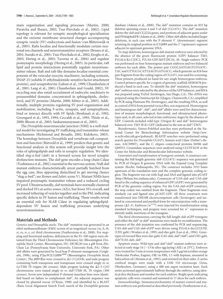
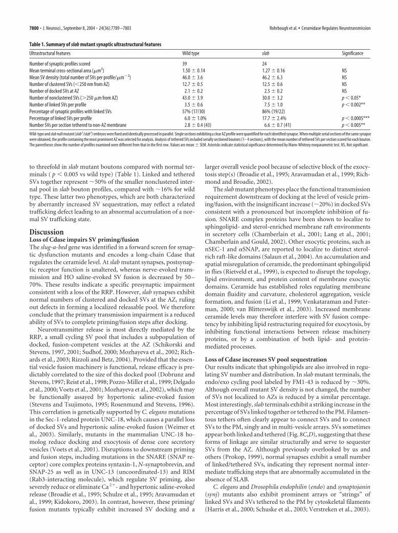
ResultsIdentification and characterization ofthe slug-a-bed Cdase geneAn EMS mutagenesis screen of the thirdchromosome (comprising �40% of theDrosophila genome) was performed to iden-tify novel molecules essential for neurotrans-mission. Mutant lines resulting in late em-bryonic or early postembryonic lethalitywere sequentially screened for normal grossanatomy, impaired movement, correctlypatterned neuromusculature, and finallyfor synaptic dysfunction using whole-cellpatch-clamp recordings at the embryonicNMJ (Broadie and Bate, 1993). The EMS-induced slab1 mutation was isolated on anisogenized rucuca (ru, h, th, st, cu, sr, e, ca)chromosome and mapped by recombina-tion with wild type (Oregon R) to a regiondistal to the e locus. Genomic deficiencymapping showed that slab1 fails to comple-ment Df(3R)tll-g (99F1–2;100B5) but com-plements Df(3R)20 (100A1–2;100B1–2),placing the slab locus in 99F1–2;100A1–2 or100B1–2;100B5 (Fig. 1A).
All available lethal P-element insertionsin the region complement slab1. To createadditional mutant alleles and to facilitatecloning the gene, P{lacW}l(3)j8B9 j8B9
(j8B9) was mobilized in a P-element localhop mutagenesis. New insertions werecomplementation tested for lethality againstslab1 to isolate seven new slab mutant alleles.All insertion alleles retain a 5� (distal) P-element sequence adjacent to prolyl-4-hy-droxylase-� EFB (PH4�EFB), the originalinsertion site of j8B9 (Fig. 1B). Six insertion alleles have a 3� (proxi-mal) P-element sequence adjacent to genes proximal to j8B9 (Fig. 1Band data not shown). PCR on single homozygous embryos withCG1471-specific primers verified that the CG1471 sequence was de-leted in three of these lines. The slab2 deletion allele has an intragenicdeletion of 855 bases removing parts of exons 4 and 5; slab2 retains itsoriginal insertion site both 5� and 3� of the j8B9 P-element and has anew insertion in CG1471 (Fig. 1B). A second larger deletion allele,slab3, removes CG1471 and CG2224 and portions of the adjacentgenes aralar and PH4�EFB (Fig. 1B). CG1471 maps to 99F5–99F6
(The FlyBase Consortium, 2003), consistent with the deficiencymapping data. The slab1 (EMS) mutation results in a single pointmutation of valine 264 to methionine in exon 4 of CG1471 (Fig.1B,C).
BLAST searches against sequenced genomes revealed CG1471to be a recently identified Drosophila Cdase (Renault et al., 2002;Yoshimura et al., 2002), with demonstrated functional Cdase ac-tivity when expressed in Drosophila S2 cultured cells (Yoshimuraet al., 2002). Comparison among different organisms revealsthree major groups of Cdases; the best characterized and mosthighly conserved are “long” Cdases (670 –761 amino acids),
Figure 1. Genomic mapping and identification of the slug-a-bed ceramidase gene and mutants. A, Genomic map showing theright tip of chromosome 3R. Genomic regions deleted by complementing and noncomplementing deficiency (Df) lines are repre-sented below. B, Molecular organization of the slab locus, schematizing genomic scaffold AE003774. The boxes represent knownand predicted genes in the region, with slab indicated by the shaded box. The deleted sequence in the slab3 mutation (line andtriangles) is indicated above; the right triangle is the original point of insertion of j8B9. The expanded region below shows theintron/exon structure of slab to scale. The site of the slab1 point mutation (arrow) and the region deleted in the slab2 mutation areindicated. C, CLUSTALW alignment of five related Cdases, with overall homology to D.mel SLAB Cdase indated by the percentagesat right: human mitochondrial acid (H.sap), mouse alkaline/neutral (M.mus), Arabidopsis neutral (A.thal), and Dictyosteliumpredicted gene “random slug cDNA25 protein” (D.disc) Cdases. The slab1 point mutation occurs in a highly conserved valine(arrow). D, Phylogenetic trees (midpoint rooted) showing relationship of Cdases. al, Alkaline; n, neutral; a, acid; conj, conjectural(predicted from a genome sequence annotation). In addition to Cdases listed in C, related proteins include rat neutral (R.ratt),Dermatophilus congolensis alkaline (D.cong), and Pseudomonas alkaline (P.aer) Cdases. D.mel bwa is the gene brainwashing, andH.sap acid Cdase is implicated in Farber’s disease.
7792 • J. Neurosci., September 8, 2004 • 24(36):7789 –7803 Rohrbough et al. • Ceramidase Regulates Neurotransmission
which share 23–39% amino acid homology and function pre-dominantly in alkaline/neutral environments (Fig. 1D). The slabgene encodes a long Cdase that functions predominantly as analkaline/neutral form in vitro but reportedly also has significant
activity at acidic pH (Yoshimura et al., 2002).The valine altered by the slab1 mutation is ahighly conserved residue in human, mouse,Drosophila, and plant Cdases (Fig. 1C).Other Cdase classes consist of “short” acidicand alkaline forms (264–395 amino acids)(Fig. 1D), which lack homology or commonmotifs between members of other short orlong classes. In Drosophila, the only addi-tional Cdase is a short alkaline form encodedby the brainwashing gene (Boquet et al.,2000).
To confirm that lethality is attributableto mutations in slab, we rescued lethalitywith a slab construct consisting of a full-length slab genomic coding sequence in-cluding 3553 bases of endogenous up-stream regulatory sequence (Fl-slab). Asingle copy of the Fl-slab transgene inslab1/slab2 and slab1/slab3 mutants is suffi-cient to confer adult viability at percent-ages near expected for full rescue. In addi-tion, the genomic slab coding sequencewas cloned into a pUAS-T vector, and thisconstruct transformed into Drosophila. Asingle copy of UAS-slab expressed underthe control of either a ubiquitous (UH1-GAL4) (Wodarz et al., 1995) or a nervoussystem-specific GAL4 (elav-GAL4) (Luo etal., 1994) driver is sufficient to fully rescueslab1/slab2 flies to adulthood. In contrast,slab expression targeted to muscle using amyosin heavy chain GAL4 fails to rescueslab1/slab2 embryonic lethality. Thesestudies demonstrate that mutation of theCdase encoded by slab is the sole cause ofmutant lethality and demonstrate an es-sential requirement for slab Cdase withinthe nervous system.
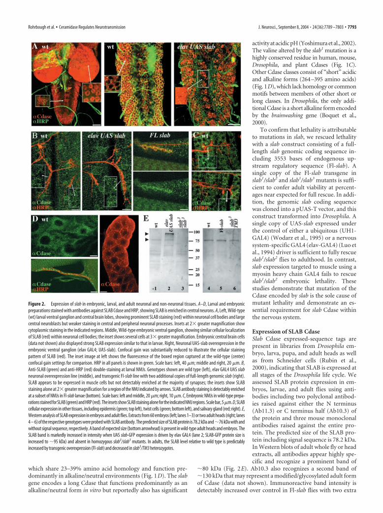
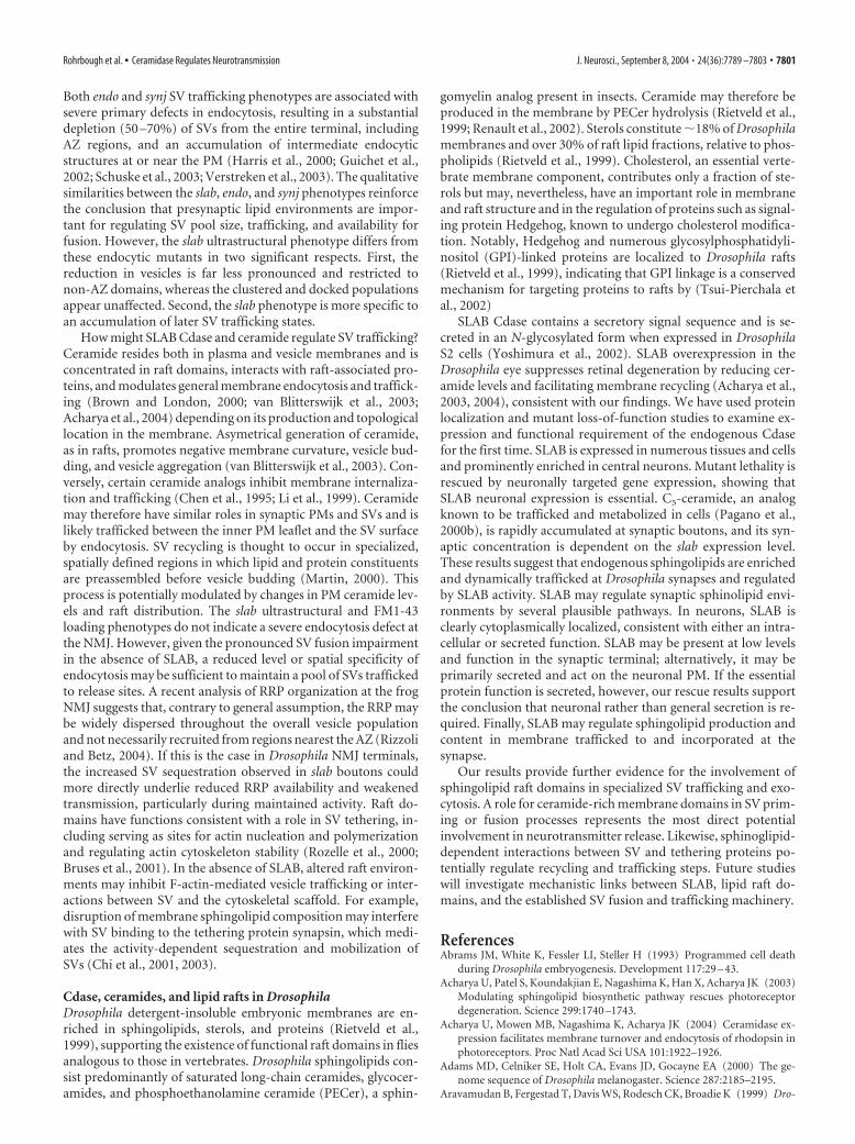
Expression of SLAB CdaseSlab Cdase expressed-sequence tags arepresent in libraries from Drosophila em-bryo, larva, pupa, and adult heads as wellas from Schneider cells (Rubin et al.,2000), indicating that SLAB is expressed atall stages of the Drosophila life cycle. Weassessed SLAB protein expression in em-bryos, larvae, and adult flies using anti-bodies including two polyclonal antibod-ies raised against either the N terminus(Ab11.3) or C terminus half (Ab10.3) ofthe protein and three mouse monoclonalantibodies raised against the entire pro-tein. The predicted size of the SLAB pro-tein including signal sequence is 78.2 kDa.In Western blots of adult whole fly or headextracts, all antibodies appear highly spe-cific and recognize a prominent band of
�80 kDa (Fig. 2E). Ab10.3 also recognizes a second band of�130 kDa that may represent a modified/glycosylated adult formof Cdase (data not shown). Immunoreactive band intensity isdetectably increased over control in Fl-slab flies with two extra
Figure 2. Expression of slab in embryonic, larval, and adult neuronal and non-neuronal tissues. A–D, Larval and embryonicpreparations stained with antibodies against SLAB Cdase and HRP, showing SLAB is enriched in central neurons. A, Left, Wild-type(wt) larval ventral ganglion and central brain lobes, showing prominent SLAB staining (red) within neuronal cell bodies and largecentral neuroblasts but weaker staining in central and peripheral neuronal processes. Insets at 2� greater magnification showcytoplasmic staining in the indicated regions. Middle, Wild-type embryonic ventral ganglion, showing similar cellular localizationof SLAB (red) within neuronal cell bodies; the inset shows several cells at 3� greater magnification. Embryonic central brain cells(data not shown) also displayed strong SLAB expression similar to that in larvae. Right, Neuronal UAS-slab overexpression in theembryonic ventral ganglion (elav GAL4; UAS-slab). Confocal gain was substantially reduced to illustrate the cellular stainingpattern of SLAB (red). The inset image at left shows the fluorescence of the boxed region captured at the wild-type (center)confocal gain settings for comparison. HRP in all panels is shown in green. Scale bars: left, 40 �m; middle and right, 20 �m. B,Anti-SLAB (green) and anti-HRP (red) double-staining at larval NMJs. Genotypes shown are wild type (left), elav GAL4 UAS slabneuronal overexpression line (middle), and transgenic Fl-slab line with two additional copies of full-length genomic slab (right).SLAB appears to be expressed in muscle cells but not detectably enriched at the majority of synapses; the insets show SLABstaining alone at 2� greater magnification for a region of the NMJ indicated by arrows. SLAB antibody staining is detectably enrichedat a subset of NMJs in Fl-slab larvae (bottom). Scale bars: left and middle, 20 �m; right, 10 �m. C, Embryonic NMJs in wild-type prepa-rationsstainedforSLAB(green)andHRP(red).TheinsetsshowSLABstainingalonefortheindicatedNMJregions.Scalebar,5�m.D,SLABcellular expression in other tissues, including epidermis (green; top left), twist cells (green; bottom left), and salivary gland (red; right). E,Western analysis of SLAB expression in embryos and adult flies. Extracts from 60 embryos (left; lanes 1–3) or two adult heads (right; lanes4 – 6) of the respective genotypes were probed with SLAB antibody. The predicted size of SLAB protein is 78.2 kDa and�76 kDa with andwithout signal sequence, respectively. A band of expected size (bottom arrowhead) is present in wild-type adult heads and embryos. TheSLAB band is markedly increased in intensity when UAS slab-GFP expression is driven by elav GAL4 (lane 2; SLAB-GFP protein size isincreased to �95 kDa) and absent in homozygous slab3/slab3 mutants. In adults, the SLAB level relative to wild type is predictablyincreased by transgenic overexpression (Fl-slab) and decreased in slab3/TM3 heterozygotes.
Rohrbough et al. • Ceramidase Regulates Neurotransmission J. Neurosci., September 8, 2004 • 24(36):7789 –7803 • 7793
copies of slab and decreased in slab3/TM3 heterozygotes with asingle copy (Fig. 2E). Anti-SLAB signal is greatly increased whenUAS slab expression is driven by a ubiquitous or neuronal-specific GAL4 driver (data not shown). Western blots of wild-type embryonic extracts (30 – 60 embryos) revealed the expected�80 kDa SLAB band. The SLAB signal is strongly increased byneuronally driven expression of SLAB-GFP and cannot be de-tected in slab3/slab3 mutants, indicating that this allele representsa protein null, as predicted (Fig. 2E).
The SLAB protein is strongly enriched in neurons, includingcentral neurons in the brain and ventral nerve cord of embryosand larvae (Fig. 2A). The protein is clearly concentrated withinthe cytoplasm of neuronal cell bodies and weakly expressed orabsent from neuronal processes within the central neuropil andin peripheral axons (Fig. 2A). Neuronally driven slab expressiongreatly increases the staining intensity in central neurons, but thecytoplasmic localization remains similar to that of endogenousSLAB in wild-type neurons (Fig. 2A, middle and right). SLABprotein is also evident at a lower level in muscle cells (Fig. 2B,C).In Fl-slab preparations, a few NMJs exhibit clearly enriched syn-aptic SLAB localization (Fig. 2B, right). However, SLAB stainingis not detectably enriched at most NMJs, even when neuronallyoverexpressed (Fig. 2B, left and middle). We also examined aSLAB-GFP protein driven by neuronal elav-GAL4 (data notshown). Consistent with antibody staining results, neuronallyexpressed SLAB-GFP is present at high levels within neuronal cellbodies and some central axons. SLAB-GFP also appears cytosolic,and its cellular localization is indistinguishable from the intracel-lular pattern of anti-SLAB staining. The SLAB protein is widelyexpressed and also present in other tissues in situ, including epi-dermis, salivary gland, and adult muscle precursor (twist) cells(Broadie and Bate, 1991) (Fig. 2D). In these cells, as in neurons,the SLAB protein appears cytoplasmic with no clear subcellularlocalization.
Null slab mutants show no detectable increase in cell deathCeramide derivatives are known signaling molecules in pro-grammed cell death and induce apoptosis in some cell lines (Mer-rill et al., 1997; Dbaibo and Hannun, 1998; Spiegel et al., 1998;Acharya et al., 2003). During embryogenesis, apoptosis is a nor-mal process for programmed removal of unneeded cells andsculpting of tissues (Abrams et al., 1993). In flies, increased em-bryonic expression of the death-inducing signal Reaper triggersceramide generation and activation of caspases, effectors of celldeath (Pronk et al., 1996; Bose et al., 1998). A block of Sph-1-phosphate hydroylsis in sply mutants results in increased embry-onic apoptosis (Herr et al., 2003). The Reaper and ceramide path-way is modulated in part by stress-activated protein kinase/Junkinase, a target of ceramide signaling (Dbaibo and Hannun,1998). To address the possibility that ceramide-mediated celldeath contributes to the mutant phenotype, we assayed apoptosisin slab null mutant (slab3/slab3) and wild-type embryos.
Apoptotic profiles were quantitatively examined in mid-stageembryos (stage 16; �13 hr AEL) by comparison of numbers ofacridine orange-stained cells in confocal sections. There was noevidence for increased cell death in slab mutant embryos; 532 50 and 492 126 acridine orange-stained cells were counted inwild-type and slab preparations, respectively (mean SD; n 5for both genotypes). The absence of detectably increased celldeath in slab Cdase mutants is likely attributable to cellularcheckpoints preventing unwanted cell death. For example, Dro-sophila caspase activity is inhibited by binding of DIAP1 (inhib-itor of apoptosis 1); this inhibition is relieved by several proteins
including Reaper, which thus regulates apoptosis both upstreamand downstream of ceramide generation (Holley et al., 2002).Additionally, Bcl-2 can protect mammalian cells from ceramide-induced apoptosis (Dbaibo and Hannun, 1998). The Drosophilahomologs buffy and death executioner Bcl-2 likely perform similarprotective functions in the fly. We conclude that slab mutantlethality is unlikely to be a result of decreased neuronal viabilityor cell maintenance.
SLAB Cdase regulates synaptic sphingolipid leveland distributionDrosophila raft lipid content has been investigated in extracted em-bryonic membranes (Rietveld et al., 1999); however, no known spe-cific probes or markers exist for endogenous Drosophila sphingolip-ids or raft domains. Fluorescent sphingolipid analogs have beenwidely used to study sphingolipid trafficking and metabolism inmammalian in vitro systems (Putz and Schwarzmann, 1995; Paganoand Chen, 1998; Pagano et al., 2000b). Ceramide analogs with afluorophore attached to a shortened fatty acid chain are rapidly in-corporated into the PM, internalized and concentrated in sphingo-lipid-rich domains within the Golgi, and subsequently metabolizedto higher fluorescent sphingolipids and glycosphingolipids andtrafficked via vesicles back to the surface membrane (Lipsky andPagano, 1985; Pagano and Sleight, 1985; Pagano et al., 1989, 1991,2000b; Putz and Schwarzmann, 1995).
We assessed the distribution and cellular trafficking of BODIPYFl C5-ceramide (C5-Cer) (Pagano and Chen, 1998; Pagano et al.,2000b) at living NMJ synaptic membranes and asked whether local-ization of fluorescent ceramide over a 1–2 hr time course is depen-dent on the SLAB expression level. Dissected third instar larvae andmature embryos (22–24 AEL) were incubated in C5-Cer (5–10 �M)in Schneider’s medium for 30–45 min, washed at room temperaturefor 1–2 hr to remove excess surface labeling, and imaged on theconfocal microscope. Significant fluorescence is present in manycells, consistent with expected surface incorporation and internaltrafficking of the lipid. In muscle cells, for example, C5-Cer is dif-fusely incorporated throughout the PM as well as accumulatedwithin internal organelle membranes, including the ER membranesurrounding nuclei (Fig. 3 and data not shown). Little or no fluores-cence is found in the CNS and ventral ganglion, presumably becauseof the glial sheath barrier.
C5-Cer is concentrated to NMJ synapses, most prominently atlarge (�3 �m diameter) “type Ib” synaptic boutons, which aresurrounded by multi-layered postsynaptic subsynaptic reticular(SSR) membranes (Fig. 3A,B,D). As confirmation of synapticlocalization, we compared C5-Cer distribution with that of anti-DLG, a synaptic marker associated primarily with the SSR of typeI boutons. After labeling and imaging of C5-Cer at living NMJs,preparations were fixed and stained with anti-DLG, and the iden-tical DLG-stained NMJs were then re-imaged, confirming thatC5-Cer fluorescence is consistently enriched at synaptic boutons(Fig. 3A). In addition, C5-Cer is strongly accumulated at manysmall type II boutons that do not have associated postsynapticSSR membranes (Fig. 3B). C5-Cer is also clearly concentrated atembryonic synaptic boutons before SSR development (Fig. 3D).These results provide evidence that ceramide, the substrate forSLAB, is characteristically enriched and localized in embryonicsynapses at the time of essential requirement (see below) as wellas in mature larval stages. To address whether altered SLAB ex-pression modifies the synaptic sphingolipid environment in vivo,we compared and quantified C5-Cer levels at type I larval boutonsbetween transgenic Fl-slab larvae and slab3/TM3 heterozgotes(Fig. 3D,E). Mean synaptic fluorescence incorporated at Fl-slab
7794 • J. Neurosci., September 8, 2004 • 24(36):7789 –7803 Rohrbough et al. • Ceramidase Regulates Neurotransmission

NMJs is significantly increased compared with slab3/TM3 NMJs( p � 0.02) (Fig. 3E). Because C5-Cer was acutely applied andimaged (within �2 hr), this result indicates that the localizedendogenous synaptic sphingolipid environment is regulated bySLAB function and altered by the level of SLAB activity.
Essential requirement for SLAB Cdase in neuronal functionThe slab mutant lethal phenotype is highly penetrant in all alleliccombinations of slab mutants (slab 1, slab 2, slab 3 homozygotesand transheterozygote combinations) (Fig. 1B), with �99% ofmutant animals dying either during or very shortly after hatch-ing. Mutant embryos appear anatomically and morphologically
normal, including a normally segmented epidermis, tracheal sys-tem, gut, nervous system, and musculature (data not shown andFig. 4). Mutants move spontaneously and in response to touchbefore hatching, although both spontaneous and evoked move-ment are less vigorous than in control embryos, initiate hatching(21–22 hr AEL at 25°C), and protrude their heads from the eggcase. The majority of mutant animals exhibit only weak headmovements for a few minutes and fail to move further. A minor-ity briefly generate peristaltic body contractions sufficient toemerge partly or completely from the egg case; a few succeed inhatching completely but rarely move more than a few bodylengths. When moved to an aqueous environment before or im-mediately after hatching, most mutant animals maintain sponta-neous and touch-evoked movements for 1–2 hr. However, per-sistence of coordinated whole-body peristaltic locomotionbehavior typical of normal animals is never observed. Heterozy-
Figure 3. Fluorescent C5-ceramide is trafficked to NMJ membranes, and synaptic accumu-lation is dependent on the level of SLAB expression. A, Left, The fluorescent ceramide analogBODIPY FL C5-ceramide (C5-Cer) is incorporated into neuronal and muscle membranes andstrongly localized to living larval NMJ boutons within 1–2 hr after a 30 min incubation. Shownis the NMJ at muscle 4 in a living larval preparation. Right, The identical NMJ imaged afterfixation and staining with an antibody against DLG, which localizes to type Ib synaptic boutons.Scale bar, 10 �m. B, Wild-type NMJ after incubation in C5-Cer, showing fluorescence concen-trated at both large type Ib boutons (I; arrowheads) and small type II boutons (II; arrows).Labeling of the presynaptic axonal membrane and type II boutons lacking postsynaptic SSRindicates that sphingolipid-enriched environments are present presynaptically as well aspostsynaptically. Scale bar, 20 �m. C, C5-Cer accumulated at embryonic synapses (NMJ 12/13,Fl-slab embryo). Synaptic boutons (arrowhead) and presynaptic nerve (n) are indicated. Scalebar, 2 �m. D, Comparison of NMJ C5-Cer fluorescence in Fl-slab larva with two additional copiesof genomic slab (left), with that in slab3/TM3 heterozygote with a single copy of slab. Bothimages shown are Z-series of 5 �m total thickness for comparison of synaptic accumulation andare representative of quantified fluorescence differences in eight paired experiments. Scale bar,10 �m. E, Quantified mean NMJ synaptic C5-Cer fluorescence (muscle 4) in slab3/TM3 larvae(filled bar), normalized to that in Fl-slab larvae in eight paired experiments. *Significant reduc-tion in mean fluorescence relative to Fl-slab ( p � 0.02).
Figure 4. Null slab mutant NMJs have normal presynaptic terminal morphology andpostsynaptic glutamate receptor localization. A, NMJ terminals on identified muscles (7, 6, 13,12) within a single ventral hemisegment in mature (20 –22 hr AEL) wild-type (wt; left) andslab1/slab3 null mutant (right) embryos, visualized with anti-HRP antibody staining. These NMJterminals are the subject of synaptic electrophysiology, SV dye imaging, and ultrastructuralstudies (see Figs. 5– 8). Scale bar, 5 �m. B, Localization and distribution of anti-synaptogmin I,a SV protein, showing normal morphology and differentiation of NMJ synaptic boutons in slabnull mutants. C, NMJs of wt and slab null mutants double-stained against HRP (�HRP; red) andthe GluRIIA postsynaptic glutamate receptor (�GluR; green). The left pair of panels shows NMJ6/7; the right pair shows NMJ 12/13. GluR puncta are closely opposed to presynaptic boutons.GluR puncta size and appearance are indistinguishable between control and slab NMJs, and slabGluR functional responses show no postsynaptic impairment (see Fig. 5C). Scale bar, 2.5 �m.
Rohrbough et al. • Ceramidase Regulates Neurotransmission J. Neurosci., September 8, 2004 • 24(36):7789 –7803 • 7795

gous slab1/TM3 balanced siblings display wild-type movementand behavior as well as physiological transmission properties (seebelow).
Examination of the nervous system and somatic muscles withthe light microscope reveals no obvious anatomical defects in slabmutants. NMJ morphology and differentiation was examined at aconfocal level with presynaptic and postsynaptic antibody mark-ers to investigate possible defects in synaptic morphogenesis ormolecular differentiation. In mature slab mutant embryos(22–24 hr AEL), NMJs are correctly and stereotypically formed,with presynaptic terminal elaboration, branching, and synapticbouton formation indistinguishable from control synapses (Fig.4A,B; see also Fig. 7). We measured areas of NMJ presynapticterminals in anti-HRP-stained embryos and confirmed that NMJarbor size is not significantly altered in slab mutants ( p � 0.5;slab1/slab1 vs wild type; muscles 12 and 13 NMJs; n 5 animals).Mutant NMJs also exhibited normal levels and distribution ofantibody staining against SV proteins such as Syt1 (Fig. 4B).Likewise, staining against postsynaptic glutamate receptors(GluRs; GluRIIA subunit) (Petersen et al., 1997; Saitoe et al.,1997) reveals GluR punctae closely opposed to presynaptic bou-tons at both slab and control NMJs (Fig. 4C). GluR cluster ap-pearance and number do not differ detectably between controland mutant NMJs; furthermore, direct assays of GluR receptorfunction show no impairment in slab mutants (see below), sug-gesting that the postsynaptic receptor field is normally formedand aggregated. These studies indicate that the severe movementimpairment of slab mutants is likely associated with impairedneuromuscular function.
Impaired neurotransmission at slab mutant NMJs isattributable to a presynaptic defectElectrophysiological assays were performed in slab mutants atmature embryonic stages (20 –21 hr AEL) and in larvae immedi-ately after hatching (22–23 hr AEL). Direct electrical stimulationof the muscle or application of glutamate to the postsynapticmuscle membrane results in visible muscle contraction in slabmutants similar to that observed for control (wild-type or slab/TM3 heterozygotes) animals, indicating that mutants retainmuscle function. In control animals, electrical stimulation of theventral ganglion or peripheral motor nerves evokes strong seg-mental muscle contraction and usually patterned waves of peri-staltic muscular activity. In contrast, such stimulation evokesonly weak, local muscle contraction in slab mutants, consistentwith behavioral movement defects indicating a specific defect inthe nervous system or NMJ synaptic transmission.
Whole-cell patch-clamp recordings were made from voltage-clamped (�60 mV) embryonic muscle (m6) (Broadie and Bate,1993). Spontaneous EJC events are recorded at nearly all slabmutant NMJs, indicating the presence of functional glutamater-gic transmission. However, EJCs evoked by direct electrical stim-ulation of the motor nerve are substantially impaired at slabNMJs. At control NMJs, nerve-evoked EJCs are robust and repro-ducible, with mean amplitudes of �1000 pA (963 81 pA, wildtype, n 4; 987 59 pA, slab1/TM3, n 7) at basal stimulationfrequencies of 0.2– 0.5 Hz (Fig. 5A). In contrast, slab mutantNMJs display significantly reduced, low-fidelity evoked trans-mission. Evoked EJC amplitudes display considerable variationand are reduced, on average, by 50 –70% overall in different slaballeles [slab1/slab1 (327 92 pA; n 7; p � 0.01) vs slab1/TM3;slab 1/slab2 (490 59 pA; n 6; p � 0.01) vs slab1/TM3] (Fig.5A). The incidence of transmission failures and near-failures atslab NMJs is progressively increased at higher (5–10 Hz) stimu-
lation frequencies, suggesting a reduced efficacy of presynaptictransmitter release in response to evoked nerve activity (Fig. 5B).The decreased mutant EJC amplitudes are not attributable to afailure in evoked action potential induction or propagation, be-
Figure 5. Impaired neurotransmission at slab mutant NMJs is attributable to a presynapticdefect. A, Representative nerve-evoked (arrow) EJCs from control (slab1/TM3; top traces) andhomozygous slab1 mutant NMJs (bottom traces; examples from 2 animals are shown). In con-trols, EJCs are robust (�1 nA) and reproducible. In slab mutants, EJC amplitude and fidelity arereduced and include transmission failures and near-failures. The mean EJC amplitudes are plot-ted at right for controls (wild-type, slab1/TM3), slab1/slab1 mutants (*p � 0.01 vs slab1/TM3)and slab1/slab2 mutants (*p � 0.01 vs slab1/TM3). The two slab alleles are not significantlydifferent ( p � 0.13). B, Higher-frequency (10 Hz) evoked EJC trains illustrate well maintainedtransmission under elevated demand at control NMJs. In contrast, slab mutant NMJs showseverely reduced amplitudes and erratic transmission, including failures (marked with sym-bols). C, Postsynaptic current responses elicited by direct iontophoretic application of gluta-mate. Representative traces (left) show glutamate-gated currents at control and slab NMJs. Nodifference in mean glutamate current amplitudes (plotted at right) is observed between con-trols (wild-type, slab1/TM3) and slab mutant (slab1/slab1, slab1/slab2) alleles.
7796 • J. Neurosci., September 8, 2004 • 24(36):7789 –7803 Rohrbough et al. • Ceramidase Regulates Neurotransmission
cause this direct nerve stimulation technique is readily able tosupport release by electronic presynaptic depolarization (Broadieand Bate, 1993; Auld et al., 1995). Thus, although a level of endo-genously generated transmission and weakened evoked trans-mission persists at slab mutant synapses, normal coupling oftransmission to presynaptic depolarization is greatly reduced,with transmission fidelity increasingly compromised at higherlevels of activity.
As noted above, direct visualization of postsynaptic GluRfields at slab mutant NMJs indicates that receptors appear local-ized and clustered similarly to wild type. We directly tested GluRfunction by recording postsynaptic current responses evoked bybrief (2–10 msec) iontophoretic application of glutamate directlyto the muscle 6/7 synapse (Fig. 5C). Glutamate application pro-duced visible muscle contraction in all recorded slab prepara-tions, and robust glutamate current responses were recorded atall slab mutant NMJs, with current amplitudes indistinguishablefrom controls [e.g., 2516 381 pA (slab 1/slab1) vs 2292 235 pA(slab1/TM3); p � 0.3] (Fig. 5C). Strong postsynaptic function waspresent regardless of the level of presynaptic function suggestedby endogenous transmission present at individual NMJs. Theseresults indicate that the postsynaptic GluR field is normally dif-ferentiated and functional and that slab mutants are impairedspecifically in presynaptic function.
Reduction in fusion of readily releasable SVs at slab synapsesThe preceding analyses demonstrate a defect in the coupling ofpresynaptic depolarization to neurotransmitter release in slabmutant synapses. Several potential mechanisms, including a re-duced presynaptic Ca 2� signal, a reduction in the readily releas-able pool (RRP) of SVs, or an impairment in the SV fusion mech-anism, could explain the release defect. To differentiate betweenthese possibilities, we next assayed fusion evoked by HO saline,which specifically triggers the release of the RRP independently ofCa 2� influx (Rosenmund and Stevens, 1996). Whole-cellpostsynaptic currents were recorded in response to brief (2 sec)focal applications of HO saline to the m6/7 NMJ (Aravamudan etal., 1999; Fergestad et al., 2001).
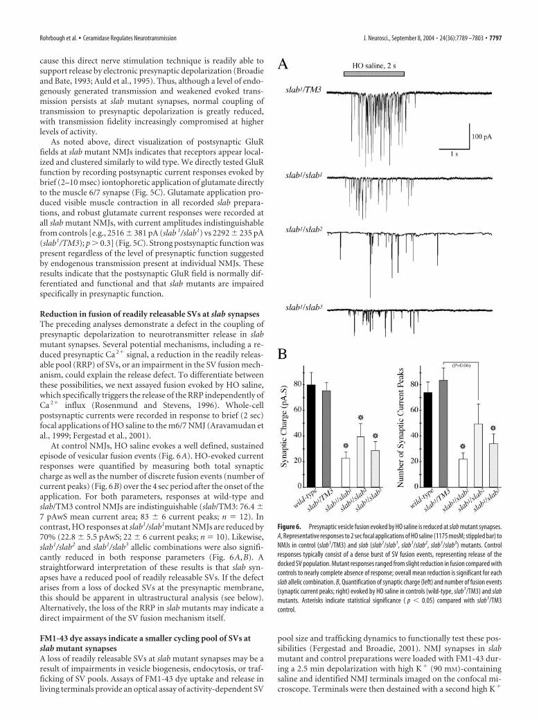
At control NMJs, HO saline evokes a well defined, sustainedepisode of vesicular fusion events (Fig. 6A). HO-evoked currentresponses were quantified by measuring both total synapticcharge as well as the number of discrete fusion events (number ofcurrent peaks) (Fig. 6B) over the 4 sec period after the onset of theapplication. For both parameters, responses at wild-type andslab/TM3 control NMJs are indistinguishable (slab/TM3: 76.4 7 pAwS mean current area; 83 6 current peaks; n 12). Incontrast, HO responses at slab1/slab1mutant NMJs are reduced by70% (22.8 5.5 pAwS; 22 6 current peaks; n 10). Likewise,slab1/slab2 and slab1/slab3 allelic combinations were also signifi-cantly reduced in both response parameters (Fig. 6A,B). Astraightforward interpretation of these results is that slab syn-apses have a reduced pool of readily releasable SVs. If the defectarises from a loss of docked SVs at the presynaptic membrane,this should be apparent in ultrastructural analysis (see below).Alternatively, the loss of the RRP in slab mutants may indicate adirect impairment of the SV fusion mechanism itself.
FM1-43 dye assays indicate a smaller cycling pool of SVs atslab mutant synapsesA loss of readily releasable SVs at slab mutant synapses may be aresult of impairments in vesicle biogenesis, endocytosis, or traf-ficking of SV pools. Assays of FM1-43 dye uptake and release inliving terminals provide an optical assay of activity-dependent SV
pool size and trafficking dynamics to functionally test these pos-sibilities (Fergestad and Broadie, 2001). NMJ synapses in slabmutant and control preparations were loaded with FM1-43 dur-ing a 2.5 min depolarization with high K� (90 mM)-containingsaline and identified NMJ terminals imaged on the confocal mi-croscope. Terminals were then destained with a second high K�
Figure 6. Presynaptic vesicle fusion evoked by HO saline is reduced at slab mutant synapses.A, Representative responses to 2 sec focal applications of HO saline (1175 mosM; stippled bar) toNMJs in control (slab1/TM3) and slab (slab1/slab1, slab1/slab2, slab1/slab3) mutants. Controlresponses typically consist of a dense burst of SV fusion events, representing release of thedocked SV population. Mutant responses ranged from slight reduction in fusion compared withcontrols to nearly complete absence of response; overall mean reduction is significant for eachslab allelic combination. B, Quantification of synaptic charge (left) and number of fusion events(synaptic current peaks; right) evoked by HO saline in controls (wild-type, slab1/TM3) and slabmutants. Asterisks indicate statistical significance ( p � 0.05) compared with slab1/TM3control.
Rohrbough et al. • Ceramidase Regulates Neurotransmission J. Neurosci., September 8, 2004 • 24(36):7789 –7803 • 7797

depolarization of varying duration (30 secto 2.5 min) and re-imaged. These condi-tions reliably and consistently load andunload the “endo/exo” cycling SV pool atembryonic NMJs (Fergestad and Broadie,2001), similar to studies at larval NMJs(Kuromi and Kidokoro, 1998, 2000).
In loaded terminals, dye fluorescence istypically localized to the periphery of larger(�2 �m diameter) embryonic boutons (Fig.7A,B), showing that SVs are concentrated incortical regions underlying the presynapticPM, as in mature larval boutons. Dye is alsoincorporated throughout the interior ofsome boutons, in particular smaller boutons(Fig. 7A,B), although usually at less inten-sity, corresponding to a lower SV density ininterior regions. The pattern of dye uptakesuggests that most or all of the SV populationin embryonic NMJ terminals may partici-pate in active endo/exo cycling, differingfrom larval NMJs, which contain a large re-serve pool that is not cycled under theseconditions (Kuromi and Kidokoro, 1999,2000).
A 2.5 min loading protocol appears tonearly maximally load the cycling SV pool inboth controls and slab mutants. The spatialpattern and distribution of loaded dyewithin normal and slab mutant boutons ap-pears qualitatively similar (Fig. 7A,B). How-ever, slab terminals display significantly re-duced quantified mean loaded fluorescencecompared with control values (69 4% ofcontrol; p � 0.0001) (Fig. 7C), indicating areduction in the size of the actively cyclingmutant SV pool. A 1–2.5 min 90 mM K�
depolarization effectively destains �80% ofloaded fluorescence in both control and mu-tant terminals (84 3% control vs 77 5%slab; p � 0.4), showing that slab synapses arecapable of cycling and releasing the smallerSV pool in a similar period of time (Fig. 7C).No significant difference in the unloadedfractions between control and slab NMJs wasmeasured for 30 sec, 1 min, or 2.5 min depo-larizing stimuli, suggesting a cycling poolturnover time of �30 sec when challengedwith 90 mM K�. To target possible differ-ences in the rate of SV pool release, we fur-ther assayed destaining using more moder-ate depolarizing conditions. As shown inFigure 7D, fractional release with 30 mM K�
for either 1 or 2 min is comparable betweenslab and wild-type NMJs. Therefore, duringsustained high K�-mediated depolarization,slab mutant synapses are capable of traffick-ing and releasing SVs similarly to controlsynapses. However, the actively cycling slabSV pool is reduced in size, consistent with areduced ability to maintain neurotransmit-ter release during repetitive stimulation.
Figure 7. FM1-43 imaging indicates a reduced cycling SV pool size in slab mutant synapses. FM1-43 dye (10 �M) was loadedinto NMJ boutons with 2.5 min depolarizing stimulation with saline containing 90 mM K �. After imaging of loaded fluorescenceat identified NMJ 12/13 terminals, dye was unloaded with 1–2.5 min stimulation with 90 or 30 mM K � saline to maximally orpartially destain the terminal. A, Representative images of loaded and unloaded (1 min, 90 mM K �) wild-type control (left) andslab1 (right) NMJs. The insets show individual large boutons at each terminal at twofold increased magnification. B, Representa-tive images of loaded and partially destained NMJs (1 min, 30 mM K �) in wild-type and slab1 NMJs. C, Quantified mean fluores-cence after loaded (solid bars) and remaining after subsequent destaining with 90 mM K � (stippled bars; 1 and 2.5 min unloadingexperiments are pooled) in wild-type control (Con) and slab1 mutants (left). Values are normalized to mean control loadedfluorescence (100%). Mean dye loading of slab NMJs is significantly reduced (*p � 0.0001) compared with control. The right plotshows the percentage of loaded fluorescence released by destaining stimulation (% Released) for the same experiments. Controland slab mutant 90 mM K � percentage unloading shows no significant difference. D, Percentage release of loaded fluorescence by1 and 2 min stimulation with 30 mM K � saline; 90 mM K � unloading is shown for comparison (rightmost pair of bars). Unloadingfor slab mutants is not significantly different from that in controls for each destaining condition, indicating cycling of the mutantSV pool under these conditions is not detectably altered.
7798 • J. Neurosci., September 8, 2004 • 24(36):7789 –7803 Rohrbough et al. • Ceramidase Regulates Neurotransmission

Electron microscopy reveals aberrant SV trafficking in slabmutant synapsesUltrastructural analyses of embryonic NMJ terminals were un-dertaken to assess the hypothesis that loss of SLAB Cdase func-tion alters SV pool size or distribution. Ultrastructural featuresexamined included PM appearance and integrity; bouton cross-sectional area; AZ regions, defined by electron-dense PM andsynaptic cleft; electron-dense “T-bar” bodies at AZ fusion sites;SV morphology and diameter; number of SVs clustered anddocked at the AZ; and number/distributions of SVs within thebouton interior (Fig. 8, Table 1). As in previous studies (Ara-vamudan et al., 1999; Featherstone et al., 2001), clustered SVswere defined as those within 250 nm of an AZ, and “docked” SVsas those within �0.5 SV diameter (�20 nm) of the AZ membranein the section containing the most prominent T-bar structure.
In all fundamental respects, synaptic ultrastructural differen-tiation at the EM level appears unaltered at slab mutant presyn-
aptic terminals (Fig. 8A). Membrane in-tegrity is preserved, and terminalorganelles, including SVs, maintain nor-mal morphology and size. Mutant termi-nals exhibit electron-dense AZ mem-branes and T-bar densities, and SVsclustered at the AZ. The clustered popula-tion at the AZ contains the same numberof SVs in control and mutant synaptic pro-files (13 1 SV/AZ for both wild type andslab) (Table 1). Surprisingly, we found thatslab AZs also exhibited normal or greaternumbers of docked SVs (2.5 0.2) com-pared with wild-type AZs (2.1 0.2) (Ta-ble 1). This result indicates that the slabrelease impairment is not attributable todeficiencies in localizing SVs to releasesites or in docking of RRP SVs to the mem-brane, but rather a downstream defect inSV priming/fusion. One possible explana-tion for this phenotype would be a dis-rupted assembly or maintenance the mu-tant AZ. We therefore quantified thelength of mutant AZ membranes andcross-sectional areas of presynaptic T-barstructures, which are known to serve a fu-sogenic function. There were no signifi-cant differences between wild-type andslab mutants in either of these features.Thus, slab mutants must be specifically de-fective in SV priming/fusion immediatelyupstream of neurotransmitter release.
In addition, slab mutant terminals ex-hibit several significant differences in thesize and distribution of the internal non-clustered SV pool, suggesting defects invesicle trafficking. First, although overallSV density throughout the bouton is notsignificantly reduced, slab boutons have,on average, �30% fewer SVs within thenonclustered population removed fromthe AZ ( p � 0.05 vs wild type) (Table 1).This reduction in SV number closelymatches the reduction in mutant FM1-43uptake, supporting the conclusion thatslab synapses have a smaller functional cy-
cling pool. Second, slab boutons contain increased numbers ofSVs linked together in multi-vesicle clusters or chains (Fig. 8C,D;Table 1). These vesicles appear either in direct contact or to beconnected by electron-dense globular or filamentous structuresand are located both within the bouton interior as well as near thePM. Similar SV–SV linkages are present in normal terminals butare increased twofold to threefold in slab boutons. Third, slabmutant boutons exhibit increased numbers of SVs tethered byfilamentous structures to the PM. Tethered vesicles resembledocked vesicles but are not localized to AZs and are typicallylocated at a greater distance (�1 vesicle diameter) from the mem-brane. To confirm that tethered SVs were not associated withnearby AZs in adjacent sections, we scored and counted tetheredSVs in serial sections in a subset of boutons (two to four sections/bouton in 32 boutons). This analysis indicated that tethered SVstended to be localized to PM regions removed from an AZ. Theincidence of both linked and tethered SVs is increased by twofold
Figure 8. slab mutant terminals have normal clustered and docked RRP but a reduction in nonclustered SVs and an accumu-lation of linked and tethered RP SVs. Electron micrographs of synaptic ultrastructure at the NMJ of fully developed (20 –22 hr AEL)wild-type control (left; A–D) and slab1/slab3 (slab; right; A–D) mutant embryos. A, Representative profiles of wild-type (left) andslab (right) boutons, each displaying an electron-dense presynaptic T-bar and electron-dense PM marking the AZ (large arrow-heads). SVs are clustered near the AZ and distributed throughout the bouton interior and periphery. Similar profiles were used forall quantitative analyses. cs, Cisternae; m, mitochondria. Scale bar, 100 nm. B, Detailed images of wild-type (1, 2) and slab (3, 4 )AZ regions, showing the population of SVs clustered near the AZ and docked SVs (arrowheads) at the PM. C, Detailed imagesillustrating SVs tethered by filamentous structures (arrows) to the non-AZ PM. Linked SVs, defined as those either in direct contactor connected by electron-dense structures, are present among the SV visible in slab panels (3, 4, arrowheads). D, Linked SVs inwild-type and slab terminals; arrowheads indicate linking structures or point of contact between linked SVs. Linked/tethered SVsin slab boutons often appear as arrays or rows aligned near the PM as in panels 3 and 4. Significantly increased numbers andpercentages of linked and tethered SVs are present in slab mutant boutons (see Table 1). Scale bars, A-D (in right panels), 100 nm.
Rohrbough et al. • Ceramidase Regulates Neurotransmission J. Neurosci., September 8, 2004 • 24(36):7789 –7803 • 7799
to threefold in slab mutant boutons compared with normal ter-minals ( p � 0.005 vs wild type) (Table 1). Linked and tetheredSVs together represent �50% of the smaller nonclustered inter-nal pool in slab bouton profiles, compared with �16% for wildtype. These latter two phenotypes, which are both characterizedby aberrantly increased SV sequestration, may reflect a relatedtrafficking defect leading to an abnormal accumulation of a nor-mal SV trafficking state.
DiscussionLoss of Cdase impairs SV priming/fusionThe slug-a-bed gene was identified in a forward screen for synap-tic dysfunction mutants and encodes a long-chain Cdase thatregulates the ceramide level. At slab mutant synapses, postsynap-tic receptor function is unaltered, whereas nerve-evoked trans-mission and HO saline-evoked SV fusion is decreased by 50 –70%. These results indicate a specific presynaptic impairmentconsistent with a loss of the RRP. However, slab synapses exhibitnormal numbers of clustered and docked SVs at the AZ, rulingout defects in forming a localized releasable pool. We thereforeconclude that the primary transmission impairment is a reducedability of SVs to complete priming/fusion steps after docking.
Neurotransmitter release is most directly mediated by theRRP, a small cycling SV pool that includes a subpopulation ofdocked, fusion-competent vesicles at the AZ (Schikorski andStevens, 1997, 2001; Sudhof, 2000; Mozhayeva et al., 2002; Rich-ards et al., 2003; Rizzoli and Betz, 2004). Provided that the essen-tial vesicle fusion machinery is functional, release efficacy is pre-dictably correlated to the size of this docked pool (Dobrunz andStevens, 1997; Reist et al., 1998; Pozzo-Miller et al., 1999; Delgadoet al., 2000; Voets et al., 2001; Mozhayeva et al., 2002), which maybe functionally assayed by hypertonic saline-evoked fusion(Stevens and Tsujimoto, 1995; Rosenmund and Stevens, 1996).This correlation is genetically supported by C. elegans mutationsin the Sec-1-related protein UNC-18, which causes a parallel lossof docked SVs and hypertonic saline-evoked fusion (Weimer etal., 2003). Similarly, mutants in the mammalian UNC-18 ho-molog reduce docking and exocytosis of dense core secretoryvesicles (Voets et al., 2001). Disruptions to downstream primingand fusion steps, including mutations in the SNARE (SNAP re-ceptor) core complex proteins syntaxin-1, N-synaptobrevin, andSNAP-25 as well as in UNC-13 (uncoordinated-13) and RIM(Rab3-interacting molecule), which regulate SV priming, alsoseverely reduce or eliminate Ca 2�- and hypertonic saline-evokedrelease (Broadie et al., 1995; Schulze et al., 1995; Aravamudan etal., 1999; Kidokoro, 2003). In contrast, however, these priming/fusion mutants typically exhibit increased SV docking and a
larger overall vesicle pool because of selective block of the exocy-tosis step(s) (Broadie et al., 1995; Aravamudan et al., 1999; Rich-mond and Broadie, 2002).
The slab mutant phenotypes place the functional transmissionrequirement downstream of docking at the level of vesicle prim-ing/fusion, with the insignificant increase (�20%) in docked SVsconsistent with a pronounced but incomplete inhibition of fu-sion. SNARE complex proteins have been shown to localize tosphingolipid- and sterol-enriched membrane raft environmentsin secretory cells (Chamberlain et al., 2001; Lang et al., 2001;Chamberlain and Gould, 2002). Other exocytic proteins, such asnSEC-1 and �SNAP, are reported to localize to distinct sterol-rich raft-like domains (Salaun et al., 2004). An accumulation andspatial misregulation of ceramide, the predominant sphingolipidin flies (Rietveld et al., 1999), is expected to disrupt the topology,lipid environment, and protein content of membrane exocyticdomains. Ceramide has established roles regulating membranedomain fluidity and curvature, cholesterol aggregation, vesicleformation, and fusion (Li et al., 1999; Venkataraman and Futer-man, 2000; van Blitterswijk et al., 2003). Increased membraneceramide levels may therefore interfere with SV fusion compe-tency by inhibiting lipid restructuring required for exocytosis, byinhibiting functional interactions between release machineryproteins, or by a combination of both lipid- and protein-mediated processes.
Loss of Cdase increases SV pool sequestrationOur results indicate that sphingolipids are also involved in regu-lating SV number and distribution. In slab mutant terminals, theendo/exo cycling pool labeled by FM1-43 is reduced by �30%.Although overall mutant SV density is not changed, the numberof SVs not localized to AZs is reduced by a similar percentage.Most interestingly, slab terminals exhibit a striking increase in thepercentage of SVs linked together or tethered to the PM. Filamen-tous tethers often clearly appear to connect SVs and to connectSVs to the PM, singly and in multi-vesicle arrays. SVs sometimesappear both linked and tethered (Fig. 8C,D), suggesting that theseforms of linkage are similar structurally and serve to sequesterSVs from the AZ. Although previously overlooked by us andothers (Prokop, 1999), normal synapses exhibit a small numberof linked/tethered SVs, indicating they represent normal inter-mediate trafficking steps that are abnormally accumulated in theabsence of SLAB.
C. elegans and Drosophila endophilin (endo) and synaptojanin(synj) mutants also exhibit prominent arrays or “strings” oflinked SVs and SVs tethered to the PM by cytoskeletal filaments(Harris et al., 2000; Schuske et al., 2003; Verstreken et al., 2003).
Table 1. Summary of slab mutant synaptic ultrastructural features
Ultrastructural features Wild type slab Significance
Number of synaptic profiles scored 39 24Mean terminal cross-sectional area (�m2) 1.50 0.14 1.27 0.16 NSMean SV density (total number of SVs per profile/�m�2) 46.0 3.6 46.2 6.3 NSNumber of clustered SVs (�250 nm from AZ) 12.7 0.5 12.5 0.6 NSNumber of docked SVs at AZ 2.1 0.2 2.5 0.2 NSNumber of nonclustered SVs (�250 �m from AZ) 43.0 3.9 30.0 3.2 p � 0.05*Number of linked SVs per profile 3.5 0.6 7.5 1.0 p � 0.002**Percentage of synaptic profiles with linked SVs 57% (17/30) 86% (19/22)Percentage of linked SVs per profile 6.0 1.0% 17.7 2.4% p � 0.0005***Number SVs per section tethered to non-AZ membrane 2.8 0.4 (43) 6.6 0.7 (41) p � 0.005**
Wild-type and slab null mutant (slab1/slab3) embryos were fixed and identically processed in parallel. Single sections exhibiting a clear AZ profile were quantified for each identified synapse. When multiple serial sections of the same synapsewere obtained, the profile containing the most prominent AZ was selected for analysis. Analysis of tethered SVs included serially sectioned boutons (1– 4 sections), with the mean number of tethered SVs per section scored for each bouton.The parentheses show the number of profiles examined were different from that in the first row. Values are mean SEM. Asterisks indicate statistical significance determined by Mann-Whitney nonparametric test. NS, Not significant.
7800 • J. Neurosci., September 8, 2004 • 24(36):7789 –7803 Rohrbough et al. • Ceramidase Regulates Neurotransmission
Both endo and synj SV trafficking phenotypes are associated withsevere primary defects in endocytosis, resulting in a substantialdepletion (50 –70%) of SVs from the entire terminal, includingAZ regions, and an accumulation of intermediate endocyticstructures at or near the PM (Harris et al., 2000; Guichet et al.,2002; Schuske et al., 2003; Verstreken et al., 2003). The qualitativesimilarities between the slab, endo, and synj phenotypes reinforcethe conclusion that presynaptic lipid environments are impor-tant for regulating SV pool size, trafficking, and availability forfusion. However, the slab ultrastructural phenotype differs fromthese endocytic mutants in two significant respects. First, thereduction in vesicles is far less pronounced and restricted tonon-AZ domains, whereas the clustered and docked populationsappear unaffected. Second, the slab phenotype is more specific toan accumulation of later SV trafficking states.
How might SLAB Cdase and ceramide regulate SV trafficking?Ceramide resides both in plasma and vesicle membranes and isconcentrated in raft domains, interacts with raft-associated pro-teins, and modulates general membrane endocytosis and traffick-ing (Brown and London, 2000; van Blitterswijk et al., 2003;Acharya et al., 2004) depending on its production and topologicallocation in the membrane. Asymetrical generation of ceramide,as in rafts, promotes negative membrane curvature, vesicle bud-ding, and vesicle aggregation (van Blitterswijk et al., 2003). Con-versely, certain ceramide analogs inhibit membrane internaliza-tion and trafficking (Chen et al., 1995; Li et al., 1999). Ceramidemay therefore have similar roles in synaptic PMs and SVs and islikely trafficked between the inner PM leaflet and the SV surfaceby endocytosis. SV recycling is thought to occur in specialized,spatially defined regions in which lipid and protein constituentsare preassembled before vesicle budding (Martin, 2000). Thisprocess is potentially modulated by changes in PM ceramide lev-els and raft distribution. The slab ultrastructural and FM1-43loading phenotypes do not indicate a severe endocytosis defect atthe NMJ. However, given the pronounced SV fusion impairmentin the absence of SLAB, a reduced level or spatial specificity ofendocytosis may be sufficient to maintain a pool of SVs traffickedto release sites. A recent analysis of RRP organization at the frogNMJ suggests that, contrary to general assumption, the RRP maybe widely dispersed throughout the overall vesicle populationand not necessarily recruited from regions nearest the AZ (Rizzoliand Betz, 2004). If this is the case in Drosophila NMJ terminals,the increased SV sequestration observed in slab boutons couldmore directly underlie reduced RRP availability and weakenedtransmission, particularly during maintained activity. Raft do-mains have functions consistent with a role in SV tethering, in-cluding serving as sites for actin nucleation and polymerizationand regulating actin cytoskeleton stability (Rozelle et al., 2000;Bruses et al., 2001). In the absence of SLAB, altered raft environ-ments may inhibit F-actin-mediated vesicle trafficking or inter-actions between SV and the cytoskeletal scaffold. For example,disruption of membrane sphingolipid composition may interferewith SV binding to the tethering protein synapsin, which medi-ates the activity-dependent sequestration and mobilization ofSVs (Chi et al., 2001, 2003).
Cdase, ceramides, and lipid rafts in DrosophilaDrosophila detergent-insoluble embryonic membranes are en-riched in sphingolipids, sterols, and proteins (Rietveld et al.,1999), supporting the existence of functional raft domains in fliesanalogous to those in vertebrates. Drosophila sphingolipids con-sist predominantly of saturated long-chain ceramides, glycocer-amides, and phosphoethanolamine ceramide (PECer), a sphin-
gomyelin analog present in insects. Ceramide may therefore beproduced in the membrane by PECer hydrolysis (Rietveld et al.,1999; Renault et al., 2002). Sterols constitute �18% of Drosophilamembranes and over 30% of raft lipid fractions, relative to phos-pholipids (Rietveld et al., 1999). Cholesterol, an essential verte-brate membrane component, contributes only a fraction of ste-rols but may, nevertheless, have an important role in membraneand raft structure and in the regulation of proteins such as signal-ing protein Hedgehog, known to undergo cholesterol modifica-tion. Notably, Hedgehog and numerous glycosylphosphatidyli-nositol (GPI)-linked proteins are localized to Drosophila rafts(Rietveld et al., 1999), indicating that GPI linkage is a conservedmechanism for targeting proteins to rafts by (Tsui-Pierchala etal., 2002)
SLAB Cdase contains a secretory signal sequence and is se-creted in an N-glycosylated form when expressed in DrosophilaS2 cells (Yoshimura et al., 2002). SLAB overexpression in theDrosophila eye suppresses retinal degeneration by reducing cer-amide levels and facilitating membrane recycling (Acharya et al.,2003, 2004), consistent with our findings. We have used proteinlocalization and mutant loss-of-function studies to examine ex-pression and functional requirement of the endogenous Cdasefor the first time. SLAB is expressed in numerous tissues and cellsand prominently enriched in central neurons. Mutant lethality isrescued by neuronally targeted gene expression, showing thatSLAB neuronal expression is essential. C5-ceramide, an analogknown to be trafficked and metabolized in cells (Pagano et al.,2000b), is rapidly accumulated at synaptic boutons, and its syn-aptic concentration is dependent on the slab expression level.These results suggest that endogenous sphingolipids are enrichedand dynamically trafficked at Drosophila synapses and regulatedby SLAB activity. SLAB may regulate synaptic sphinolipid envi-ronments by several plausible pathways. In neurons, SLAB isclearly cytoplasmically localized, consistent with either an intra-cellular or secreted function. SLAB may be present at low levelsand function in the synaptic terminal; alternatively, it may beprimarily secreted and act on the neuronal PM. If the essentialprotein function is secreted, however, our rescue results supportthe conclusion that neuronal rather than general secretion is re-quired. Finally, SLAB may regulate sphingolipid production andcontent in membrane trafficked to and incorporated at thesynapse.
Our results provide further evidence for the involvement ofsphingolipid raft domains in specialized SV trafficking and exo-cytosis. A role for ceramide-rich membrane domains in SV prim-ing or fusion processes represents the most direct potentialinvolvement in neurotransmitter release. Likewise, sphinoglipid-dependent interactions between SV and tethering proteins po-tentially regulate recycling and trafficking steps. Future studieswill investigate mechanistic links between SLAB, lipid raft do-mains, and the established SV fusion and trafficking machinery.
ReferencesAbrams JM, White K, Fessler LI, Steller H (1993) Programmed cell death
during Drosophila embryogenesis. Development 117:29 – 43.Acharya U, Patel S, Koundakjian E, Nagashima K, Han X, Acharya JK (2003)
Modulating sphingolipid biosynthetic pathway rescues photoreceptordegeneration. Science 299:1740 –1743.
Acharya U, Mowen MB, Nagashima K, Acharya JK (2004) Ceramidase ex-pression facilitates membrane turnover and endocytosis of rhodopsin inphotoreceptors. Proc Natl Acad Sci USA 101:1922–1926.
Adams MD, Celniker SE, Holt CA, Evans JD, Gocayne EA (2000) The ge-nome sequence of Drosophila melanogaster. Science 287:2185–2195.
Aravamudan B, Fergestad T, Davis WS, Rodesch CK, Broadie K (1999) Dro-
Rohrbough et al. • Ceramidase Regulates Neurotransmission J. Neurosci., September 8, 2004 • 24(36):7789 –7803 • 7801
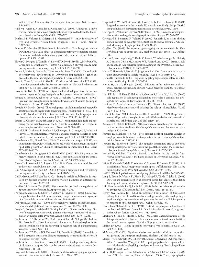
sophila Unc-13 is essential for synaptic transmission. Nat Neurosci2:965–971.
Auld VJ, Fetter RD, Broadie K, Goodman CS (1995) Gliotactin, a noveltransmembrane protein on peripheral glia, is required to form the blood-nerve barrier in Drosophila. Cell 81:757–767.
Benfenati F, Valtorta F, Chieregatti E, Greengard P (1992) Interaction offree and synaptic vesicle-bound synapsin I with F-actin. Neuron8:377–386.
Beumer K, Matthies HJ, Bradshaw A, Broadie K (2002) Integrins regulateDLG/FAS2 via a CaM kinase II-dependent pathway to mediate synapseelaboration and stabilization during postembryonic development. Devel-opment 129:3381–3391.
Bloom O, Evergren E, Tomilin N, Kjaerulff O, Low P, Brodin L, Pieribone VA,Greengard P, Shupliakov O (2003) Colocalization of synapsin and actinduring synaptic vesicle recycling. J Cell Biol 161:737–747.
Boquet I, Hitier R, Dumas M, Chaminade M, Preat T (2000) Central brainpostembryonic development in Drosophila: implication of genes ex-pressed at the interhemispheric junction. J Neurobiol 42:33– 48.
Bose R, Chen P, Loconti A, Grullich C, Abrams JM, Kolesnick RN (1998)Ceramide generation by the Reaper protein is not blocked by the caspaseinhibitor, p35. J Biol Chem 273:28852–28859.
Broadie K, Bate M (1993) Activity-dependent development of the neuro-muscular synapse during Drosophila embryogenesis. Neuron 11:607–619.
Broadie K, Prokop A, Bellen HJ, O’Kane CJ, Schulze KL, Sweeney ST (1995)Syntaxin and synaptobrevin function downstream of vesicle docking inDrosophila. Neuron 15:663– 673.
Broadie KS, Bate M (1991) The development of adult muscles in Drosophila:ablation of identified muscle precursor cells. Development 113:103–118.
Brown DA, London E (2000) Structure and function of sphingolipid- andcholesterol-rich membrane rafts. J Biol Chem 275:17221–17224.
Bruses JL, Chauvet N, Rutishauser U (2001) Membrane lipid rafts are nec-essary for the maintenance of the (�)7 nicotinic acetylcholine receptor insomatic spines of ciliary neurons. J Neurosci 21:504 –512.
Ceccaldi PE, Grohovaz F, Benfenati F, Chieregatti E, Greengard P, Valtorta F(1995) Dephosphorylated synapsin I anchors synaptic vesicles to actincytoskeleton: an analysis by videomicroscopy. J Cell Biol 128:905–912.
Chamberlain LH, Gould GW (2002) The vesicle- and target-SNARE pro-teins that mediate Glut4 vesicle fusion are localized in detergent-insolublelipid rafts present on distinct intracellular membranes. J Biol Chem277:49750 – 49754.
Chamberlain LH, Burgoyne RD, Gould GW (2001) SNARE proteins arehighly enriched in lipid rafts in PC12 cells: implications for the spatialcontrol of exocytosis. Proc Natl Acad Sci USA 98:5619 –5624.
Chen CS, Rosenwald AG, Pagano RE (1995) Ceramide as a modulator ofendocytosis. J Biol Chem 270:13291–13297.
Chi P, Greengard P, Ryan TA (2001) Synapsin dispersion and reclusteringduring synaptic activity. Nat Neurosci 4:1187–1193.
Chi P, Greengard P, Ryan TA (2003) Synaptic vesicle mobilization is regu-lated by distinct synapsin I phosphorylation pathways at different fre-quencies. Neuron 38:69 –78.
Dbaibo GS, Hannun YA (1998) Signal transduction and the regulation ofapoptosis: roles of ceramide. Apoptosis 3:317–334.
Delgado R, Maureira C, Oliva C, Kidokoro Y, Labarca P (2000) Size of ves-icle pools, rates of mobilization, and recycling at neuromuscular synapsesof a Drosophila mutant, shibire. Neuron 28:941–953.
Dobrunz LE, Stevens CF (1997) Heterogeneity of release probability, facili-tation, and depletion at central synapses. Neuron 18:995–1008.
Eroglu C, Brugger B, Wieland F, Sinning I (2003) Glutamate-binding affin-ity of Drosophila metabotropic glutamate receptor is modulated by asso-ciation with lipid rafts. Proc Natl Acad Sci USA 100:10219 –10224.
Featherstone DE, Rushton EM, Hilderbrand-Chae M, Phillips AM, JacksonFR, Broadie K (2000) Presynaptic glutamic acid decarboxylase is re-quired for induction of the postsynaptic receptor field at a glutamatergicsynapse. Neuron 27:71– 84.
Featherstone DE, Davis WS, Dubreuil RR, Broadie K (2001) Drosophila �-and �-spectrin mutations disrupt presynaptic neurotransmitter release.J Neurosci 21:4215– 4224.
Featherstone DE, Rushton E, Broadie K (2002) Developmental regulationof glutamate receptor field size by nonvesicular glutamate release. NatNeurosci 5:141–146.
Fergestad T, Broadie K (2001) Interaction of stoned and synaptotagmin insynaptic vesicle endocytosis. J Neurosci 21:1218 –1227.
Fergestad T, Wu MN, Schulze KL, Lloyd TE, Bellen HJ, Broadie K (2001)Targeted mutations in the syntaxin H3 domain specifically disrupt SNAREcomplex function in synaptic transmission. J Neurosci 21:9142–9150.
Greengard P, Valtorta F, Czernik AJ, Benfenati F (1993) Synaptic vesicle phos-phoproteins and regulation of synaptic function. Science 259:780–785.
Greengard P, Benfenati F, Valtorta F (1994) Synapsin I, an actin-bindingprotein regulating synaptic vesicle traffic in the nerve terminal. Adv Sec-ond Messenger Phosphoprotein Res 29:31– 45.
Grigliatti TA (1998) Transposons-gene tagging and mutagenesis. In: Dro-sophila: a practical approach, Ed 2 (Roberts DB, ed), pp 85–107. Oxford:IRL.
Guichet A, Wucherpfennig T, Dudu V, Etter S, Wilsch-Brauniger M, HellwigA, Gonzalez-Gaitan M, Huttner WB, Schmidt AA (2002) Essential roleof endophilin A in synaptic vesicle budding at the Drosophila neuromus-cular junction. EMBO J 21:1661–1672.
Harris T, Hartweig E, Horvitz R, Jorgensen E (2000) Mutations in synapto-janin disrupt synaptic vesicle recycling. J Cell Biol 150:589 –599.
Helms JB, Zurzolo C (2004) Lipids as targeting signals: lipid rafts and intra-cellular trafficking. Traffic 5:247–254.
Hering H, Lin CC, Sheng M (2003) Lipid rafts in the maintenance of syn-apses, dendritic spines, and surface AMPA receptor stability. J Neurosci23:3262–3271.
Herr DR, Fyrst H, Phan V, Heinecke K, Georges R, Harris GL, Saba JD (2003)Sply regulation of sphingolipid signaling molecules is essential for Dro-sophila development. Development 130:2443–2453.
Hoekstra D, Maier O, van der Wouden JM, Slimane TA, van ISC (2003)Membrane dynamics and cell polarity: the role of sphingolipids. J LipidRes 44:869 – 877.
Holley CL, Olson MR, Colon-Ramos DA, Kornbluth S (2002) Reaper elim-inates IAP proteins through stimulated IAP degradation and generalizedtranslational inhibition. Nat Cell Biol 4:439 – 444.
Kidokoro Y (2003) Roles of SNARE proteins and synaptotagmin I in synap-tic transmission: studies at the Drosophila neuromuscular synapse. Neu-rosignals 12:13–30.
Kuromi H, Kidokoro Y (1998) Two distinct pools of synaptic vesicles insingle presynaptic boutons in a temperature-sensitive Drosophila mutant,shibire. Neuron 20:917–925.
Kuromi H, Kidokoro Y (1999) The optically determined size of exo/endocycling vesicle pool correlates with the quantal content at the neuromus-cular junction of Drosophila larvae. J Neurosci 19:1557–1565.
Kuromi H, Kidokoro Y (2000) Tetanic stimulation recruits vesicles fromreserve pool via a cAMP-mediated process in Drosophila synapses. Neu-ron 27:133–143.
Lafont F, Verkade P, Galli T, Wimmer C, Louvard D, Simons K (1999) Raftassociation of SNAP receptors acting in apical trafficking in Madin-Darbycanine kidney cells. Proc Natl Acad Sci USA 96:3734 –3738.
Lai EC (2003) Lipid rafts make for slippery platforms. J Cell Biol 162:365–370.Lang T, Bruns D, Wenzel D, Riedel D, Holroyd P, Thiele C, Jahn R (2001)
SNAREs are concentrated in cholesterol-dependent clusters that definedocking and fusion sites for exocytosis. EMBO J 20:2202–2213.
Li R, Blanchette-Mackie EJ, Ladisch S (1999) Induction of endocytic vesiclesby exogenous C(6)-ceramide. J Biol Chem 274:21121–21127.
Lipsky NG, Pagano RE (1985) Intracellular translocation of fluorescentsphingolipids in cultured fibroblasts: endogenously synthesized sphingo-myelin and glucocerebroside analogues pass through the Golgi apparatusen route to the plasma membrane. J Cell Biol 100:27–34.
Luo L, Liao YJ, Jan LY, Jan YN (1994) Distinct morphogenetic functions ofsimilar small GTPases: Drosophila Drac1 is involved in axonal outgrowthand myoblast fusion. Genes Dev 8:1787–1802.
Maekawa S, Iino S, Miyata S (2003) Molecular characterization of thedetergent-insoluble cholesterol-rich membrane microdomain (raft) ofthe central nervous system. Biochim Biophys Acta 1610:261–270.
Martin TF (2000) Racing lipid rafts for synaptic-vesicle formation. Nat CellBiol 2:E9 –E11.
McMaster CR (2001) Lipid metabolism and vesicle trafficking: more thanjust greasing the transport machinery. Biochem Cell Biol 79:681– 692.
Merrill Jr AH, Schmelz EM, Dillehay DL, Spiegel S, Shayman JA, Schroeder JJ,Riley RT, Voss KA, Wang E (1997) Sphingolipids—the enigmatic lipidclass: biochemistry, physiology, and pathophysiology. Toxicol Appl Phar-macol 142:208 –225.
Mitter D, Reisinger C, Hinz B, Hollmann S, Yelamanchili SV, Treiber-Held S,Ohm TG, Herrmann A, Ahnert-Hilger G (2003) The synaptophysin/
7802 • J. Neurosci., September 8, 2004 • 24(36):7789 –7803 Rohrbough et al. • Ceramidase Regulates Neurotransmission

synaptobrevin interaction critically depends on the cholesterol content.J Neurochem 84:35– 42.
Mozhayeva MG, Sara Y, Liu X, Kavalali ET (2002) Development of vesiclepools during maturation of hippocampal synapses. J Neurosci22:654 – 665.
O’Kane C (1998) Enhancer traps. In: Drosophila: a practical approach, Ed 2(Roberts DB, ed), pp 131–178. Oxford: IRL.
Pagano RE, Chen CS (1998) Use of BODIPY-labeled sphingolipids to studymembrane traffic along the endocytic pathway. Ann NY Acad Sci845:152–160.
Pagano RE, Sleight RG (1985) Defining lipid transport pathways in animalcells. Science 229:1051–1057.
Pagano RE, Sepanski MA, Martin OC (1989) Molecular trapping of a fluo-rescent ceramide analogue at the Golgi apparatus of fixed cells: interac-tion with endogenous lipids provides a trans-Golgi marker for both lightand electron microscopy. J Cell Biol 109:2067–2079.
Pagano RE, Martin OC, Kang HC, Haugland RP (1991) A novel fluorescentceramide analogue for studying membrane traffic in animal cells: accu-mulation at the Golgi apparatus results in altered spectral properties of thesphingolipid precursor. J Cell Biol 113:1267–1279.
Pagano RE, Puri V, Dominguez M, Marks DL (2000a) Membrane traffic insphingolipid storage diseases. Traffic 1:807– 815.
Pagano RE, Watanabe R, Wheatley C, Dominguez M (2000b) Applicationsof BODIPY-sphingolipid analogs to study lipid traffic and metabolism incells. Methods Enzymol 312:523–534.
Paratcha G, Ibanez CF (2002) Lipid rafts and the control of neurotrophicfactor signaling in the nervous system: variations on a theme. Curr OpinNeurobiol 12:542–549.
Petersen SA, Fetter RD, Noordermeer JN, Goodman CS, DiAntonio A(1997) Genetic analysis of glutamate receptors in Drosophila reveals aretrograde signal regulating presynaptic transmitter release. Neuron19:1237–1248.
Pozzo-Miller LD, Gottschalk W, Zhang L, McDermott K, Du J, Gopalakrish-nan R, Oho C, Sheng ZH, Lu B (1999) Impairments in high-frequencytransmission, synaptic vesicle docking, and synaptic protein distributionin the hippocampus of BDNF knockout mice. J Neurosci 19:4972– 4983.
Prokop A (1999) Integrating bits and pieces: synapse structure and forma-tion in Drosophila embryos. Cell Tissue Res 297:169 –186.
Pronk GJ, Ramer K, Amiri P, Williams LT (1996) Requirement of an ICE-like protease for induction of apoptosis and ceramide generation byREAPER. Science 271:808 – 810.
Putz U, Schwarzmann G (1995) Golgi staining by two fluorescent ceramideanalogues in cultured fibroblasts requires metabolism. Eur J Cell Biol68:113–121.
Reist NE, Buchanan J, Li J, DiAntonio A, Buxton EM, Schwarz TL (1998)Morphologically docked synaptic vesicles are reduced in synaptotagminmutants of Drosophila. J Neurosci 18:7662–7673.
Renault AD, Starz-Gaiano M, Lehmann R (2002) Metabolism of sphin-gosine 1-phosphate and lysophosphatidic acid: a genome wide analysis ofgene expression in Drosophila. Mech Dev 119 [Suppl 1]:S293–S301.
Richards DA, Guatimosim C, Rizzoli SO, Betz WJ (2003) Synaptic vesiclepools at the frog neuromuscular junction. Neuron 39:529 –541.
Richmond JE, Broadie KS (2002) The synaptic vesicle cycle: exocytosis andendocytosis in Drosophila and C. elegans. Curr Opin Neurobiol12:499 –507.
Rietveld A, Neutz S, Simons K, Eaton S (1999) Association of sterol- andglycosylphosphatidylinositol-linked proteins with Drosophila raft lipidmicrodomains. J Biol Chem 274:12049 –12054.
Riezman H, van Meer G (2004) Lipid pickup and delivery. Nat Cell Biol6:15–16.
Rizzoli SO, Betz WJ (2004) The structural organization of the readily releas-able pool of synaptic vesicles. Science 303:2037–2039.
Rosenmund C, Stevens CF (1996) Definition of the readily releasable poolof vesicles at hippocampal synapses. Neuron 16:1197–1207.
Rozelle AL, Machesky LM, Yamamoto M, Driessens MH, Insall RH, RothMG, Luby-Phelps K, Marriott G, Hall A, Yin HL (2000) Phosphatidyl-inositol 4,5-bisphosphate induces actin-based movement of raft-enrichedvesicles through WASP-Arp2/3. Curr Biol 10:311–320.
Rubin GM, Hong L, Brokstein P, Evans-Holm M, Frise E, Stapleton M, Har-vey DA (2000) A Drosophila complementary DNA resource. Science287:2222–2224.
Saitoe M, Tanaka S, Takata K, Kidokoro Y (1997) Neural activity affectsdistribution of glutamate receptors during neuromuscular junction for-mation in Drosophila embryos. Dev Biol 184:48 – 60.
Salaun C, James DJ, Chamberlain LH (2004) Lipid rafts and the regulationof exocytosis. Traffic 5:255–264.
Sankaranarayanan S, Atluri PP, Ryan TA (2003) Actin has a molecular scaf-folding, not propulsive, role in presynaptic function. Nat Neurosci6:127–135.
Schikorski T, Stevens CF (1997) Quantitative ultrastructural analysis of hip-pocampal excitatory synapses. J Neurosci 17:5858 –5867.
Schikorski T, Stevens CF (2001) Morphological correlates of functionallydefined synaptic vesicle populations. Nat Neurosci 4:391–395.
Schulze KL, Broadie K, Perin MS, Bellen HJ (1995) Genetic and electrophys-iological studies of Drosophila syntaxin-1A demonstrate its role in non-neuronal secretion and neurotransmission. Cell 80:311–320.
Schuske KR, Richmond JE, Matthies DS, Davis WS, Runz S, Rube DA, van derBliek AM, Jorgensen EM (2003) Endophilin is required for synaptic ves-icle endocytosis by localizing synaptojanin. Neuron 40:749 –762.
Spiegel S, Cuvillier O, Edsall LC, Kohama T, Menzeleev R, Olah Z, Olivera A,Pirianov G, Thomas DM, Tu Z, Van Brocklyn JR, Wang F (1998)Sphingosine-1-phosphate in cell growth and cell death. Ann NY Acad Sci845:11–18.
Stevens CF, Tsujimoto T (1995) Estimates for the pool size of releasablequanta at a single central synapse and for the time required to refill thepool. Proc Natl Acad Sci USA 92:846 – 849.
Sudhof TC (2000) The synaptic vesicle cycle revisited. Neuron 28:317–320.Suzuki T, Ito J, Takagi H, Saitoh F, Nawa H, Shimizu H (2001) Biochemical
evidence for localization of AMPA-type glutamate receptor subunits inthe dendritic raft. Brain Res Mol Brain Res 89:20 –28.
Tani M, Iida H, Ito M (2003) O-glycosylation of mucin-like domain retainsthe neutral ceramidase on the plasma membranes as a type II integralmembrane protein. J Biol Chem 278:10523–10530.
Taverna E, Saba E, Rowe J, Francolini M, Clementi F, Rosa P (2004) Role oflipid microdomains in P/Q-type calcium channel (Cav2.1) clustering andfunction in presynaptic membranes. J Biol Chem 279:5127–5134.
The Flybase Consortium (2003) The Flybase database of the Drosophila ge-nome projects and community literature. Nucleic Acids Res 31:172–175.
Thiele C, Hannah MJ, Fahrenholz F, Huttner WB (2000) Cholesterol bindsto synaptophysin and is required for biogenesis of synaptic vesicles. NatCell Biol 2:42– 49.
Tsui-Pierchala BA, Encinas M, Milbrandt J, Johnson Jr EM (2002) Lipidrafts in neuronal signaling and function. Trends Neurosci 25:412– 417.
van Blitterswijk WJ, van der Luit AH, Veldman RJ, Verheij M, Borst J (2003)Ceramide: second messenger or modulator of membrane structure anddynamics? Biochem J 369:199 –211.
van Meer G, Holthuis JC (2000) Sphingolipid transport in eukaryotic cells.Biochim Biophys Acta 1486:145–170.
Venkataraman K, Futerman AH (2000) Ceramide as a second messenger:sticky solutions to sticky problems. Trends Cell Biol 10:408 – 412.
Verstreken P, Koh TW, Schulze KL, Zhai RG, Hiesinger PR, Zhou Y, MehtaSQ, Cao Y, Roos J, Bellen HJ (2003) Synaptojanin is recruited by en-dophilin to promote synaptic vesicle uncoating. Neuron 40:733–748.
Voets T, Toonen RF, Brian EC, de Wit H, Moser T, Rettig J, Sudhof TC, NeherE, Verhage M (2001) Munc18 –1 promotes large dense-core vesicledocking. Neuron 31:581–591.
Watanabe R, Funato K, Venkataraman K, Futerman AH, Riezman H (2002)Sphingolipids are required for the stable membrane association ofglycosylphosphatidylinositol-anchored proteins in yeast. J Biol Chem277:49538 – 49544.
Weimer RM, Richmond JE, Davis WS, Hadwiger G, Nonet ML, JorgensenEM (2003) Defects in synaptic vesicle docking in unc-18 mutants. NatNeurosci 6:1023–1030.
Wodarz A, Hinz U, Engelbert M, Knust E (1995) Expression of crumbsconfers apical character on plasma membrane domains of ectodermalepithelia of Drosophila. Cell 82:67–76.
Yoshihara M, Suzuki K, Kidokoro Y (2000) Two independent pathways me-diated by cAMP and protein kinase A enhance spontaneous transmitterrelease at Drosophila neuromuscular junctions. J Neurosci 20:8315– 8322.
Yoshimura Y, Okino N, Tani M, Ito M (2002) Molecular cloning and char-acterization of a secretory neutral ceramidase of Drosophila melanogaster.J Biochem (Tokyo) 132:229 –236.
Rohrbough et al. • Ceramidase Regulates Neurotransmission J. Neurosci., September 8, 2004 • 24(36):7789 –7803 • 7803