cellulose nanofi brils: from strong materials to bioactive ...ojrojas/pdf/2013_18.pdf · cellulose...
TRANSCRIPT
J. Renew. Mater., Vol. 1, No. 3, July 2013 © 2013 Scrivener Publishing LLC 195
Cellulose Nanofi brils: From Strong Materials to Bioactive Surfaces**
Yanxia Zhang1, Tiina Nypelö1*, Carlos Salas1, Julio Arboleda1, Ingrid C. Hoeger1*, Orlando J. Rojas1,2,*
1 Departments of Forest Biomaterials and Chemical and Biomolecular Engineering, North Carolina State University, Raleigh, North Carolina 27695, United States2 School of Chemical Technology, Department of Forest Products Technology, Aalto University, FI-00076 Aalto, Espoo, Finland
Received March 08, 2013; Accepted May 05, 2013; Published online May 22, 2013
ABSTRACT: Cellulose nanofi brils (CNF), also known as nanofi brillar cellulose (NFC), are an advanced biomaterial made mainly from renewable forest and agricultural resources that have demonstrated exceptional performance in composites. In addition, they have been utilized in barrier coatings, food, transparent fl exible fi lms and other applications. Research on CNF has advanced rapidly over the last decade and several of the fundamental questions about production and characterization of CNF have been addressed. An interesting shift in focus in the recent reported literature indicates increased efforts aimed at taking advantage of the unique properties of CNF. This includes its nanoscale dimensions, high surface area, unique morphology, low density and mechanical strength. In addition, CNF can be easily (chemically) modified and is readily available, renewable, and biodegradable. These facts are expected to materialize in a more widespread use of CNF. However, there is no clear indication of the most promising avenues for CNF deployment in commercial products. This review attempts to illustrate some exciting opportunities for CNF, specifi cally, in the development of aerogels, composites, bioactive materials and inorganic/organic hybrid materials.
KEYWORDS: Cellulose nanofi brils, CNF, nanofi brillar cellulose, NFC, cellulose materials, aerogels, hydrogels, composites, bioactive surfaces, inorganic/organic hybrids
1 INTRODUCTION
Cellulose, the most abundant biopolymer on earth,
has been studied thoroughly for different applications
including reinforcement of hydrophobic polymeric
matrices, biomedical implant and composite materi-
als, among others. Cellulose is composed of repeating
units of cellobiose consisting of anhydroglucose linked
together by β-1,4 linkages (Figure 1). Unlike starch
and other polysaccharides, the β-1,4 glucosidic bond
in cellulose causes the macromolecular chains to align
straight and for the hydroxyl groups at given posi-
tions to hydrogen bond within (intra-chain hydrogen
bonding) and between the polymer chains (inter-chain
hydrogen bonding). Hydrogen bonding in cellulose
affects solvency, swelling and key physical properties
of cellulosic structures (Figure 1).
Cellulose nanofi bril (CNF) is a material containing fi brils with length in the micrometer and width in the nanometric range, forming a network structure. CNF can be prepared by its liberation from the constituent fi ber matrix and microfi ber bundles [1–3]. The nanofi -brils contain both amorphous and crystalline cellulosic regions [4]. This fi brillar material should be differenti-ated from crystalline cellulose, referred to as cellulose nanocrystals (CNC) or nanowhiskers (Figure 1). CNCs are prepared from fi bers and fi brils via acid hydrolysis that degrades the amorphous regions, yielding highly crystalline nanoparticles [5].
CNF is prepared by mechanical methods via high shearing followed by homogenization at high pres-sure. The equipment commonly used to produce CNF includes high pressure homogenizers, refi ners, grinders, cryo-crushers, and microfl uidizer. A thorough descrip-tion of these techniques has already been reported in a review by Lavoine et al. [6]. Reduction of energy con-sumption has been a primary focus in the production of CNF; thus, several methods have been developed including chemical, mechanical or enzymatic fi ber pretreatment. A comprehensive description of these
*Corresponding authors: [email protected], [email protected],
**All authors contributed equally to this review
DOI: 10.7569/JRM.2013.634115

Yanxia Zhang et al.: Cellulose Nanofi brils (CNF) DOI: 10.7569/JRM.2013.634115
196 J. Renew. Mater., Vol. 1, No. 3, July 2013 © 2013 Scrivener Publishing LLC
pretreatments can be found elsewhere [2, 6–9]. Besides wood and plant-based CNF, the fi brils can be prepared by bottom-up approaches using biosynthesis by micro-organisms [10]. Under specifi c culturing condition, bac-terial cellulose (BC) is produced by the gram-negative bacteria Acetobacter xylinum (or Gluconacetobacter xyli-num), as extracellular cellulose, where the fi ber material is amenable to extraction [10].
Since the development of CNF from wood in the 1980s, along with the progress of cellulosic fi bril research, the nomenclature has changed. When the material was fi rst introduced it was referred to as microfi brillated cellulose (MFC) [1, 3]. Since then vary-ing approaches have been utilized to prepare the fi bril-lar material with different degrees of deconstruction, leading to materials with characteristic sizes in the micro and nano scales. Typically MFC consists of fi bers with fi brillated surfaces, whereas CNF is composed of fi brils or fi bril bundles that are separated as nanosized entities. However, micron-sized fi brils are loosely referred to as CNF. In addition to CNF the nanomate-rial can also be called nanofi brillar cellulose (NFC).
Applications taking advantage of physical, mechanical and chemical features of CNF are being explored in academia and industry. Some of the cur-rent applications of nanofi brillar cellulose are high-lighted schematically in Figure 2. Due to their surface properties, CNF fi brils are able to bind signifi cant amounts of water inside the fi bril network, forming a hydrogel. This phenomenon is benefi cial in applica-tions as a rheology modifi er [3] or in wound dressing [11]. However, the high water content of the material makes preparation of structures such as solid fi lms and coatings a challenge. Despite this limitation, CNF has been extensively explored in the preparation of transparent fi lms [12–14]. In addition to optical appli-cations, CNF offers potential in packaging efforts to replace oil-based materials due to the fact that the crystallinity and the network structure of CNF make it attractive in applications demanding barrier prop-erties. CNF can be used as a strength agent [15–17], can be combined with polyelectrolytes in fi ber pro-cessing [18–21] and can be used in the fabrication of composites [22–24]. Finally, CNF has great potential in
Figure 1 Schematic illustration of cellulose nanofi brils (CNF) obtained from wood fi bers after deconstruction of the highly
hierarchical, multidimensional structure in wood. The major molecular component of the respective building blocks in fi bers
is the cellulose polymer (note the repeating cellobiose unit of cellulose and the intra-chain bonds). The fi bril structures made
from cellulose can be disassembled to produce CNF, but if the amorphous regions of the fi brils are removed by acid hydrolysis,
cellulose nanocrystals are obtained (CNC). Note that nanocellulose from other sources may occur in different packing as dictated
by the (bio)synthesis in nonwood plants, microorganisms, algae and sea animals. Some reported sources in addition to bleached
softwood and hardwood pulps include cotton, ramie, hemp, flax, sisal, wheat, straw, palm, sugar beet pulp, bacterial cellulose,
Valonia algae, tunicates, etc.
0.2 μm
1 μm
200 nm
CNF
CNC

DOI: 10.7569/JRM.2013.634115 Yanxia Zhang et al.: Cellulose Nanofi brils (CNF)
J. Renew. Mater., Vol. 1, No. 3, July 2013 © 2013 Scrivener Publishing LLC 197
the biomedical fi eld. A detailed account of the role of CNF in the applications reported in the literature is a challenging task. In this review a few aspects related to the use of CNF are discussed, namely, in the pro-duction of aerogels, composites, bioactive materials and inorganic/organic hybrids (see Figure 2).
2 CNF AEROGELS
Aerogels are ultra lightweight materials that are
formed by the removal of liquid solvent from a gel
without collapsing the network structure [25–27].
These types of materials were fi rst introduced in 1931
[28] and are often referred to as solid foams (when air
bubbles are introduced in the gel), frozen smoke (com-
mon name for silica aerogels), cryogels (aerogels pro-
duced by freeze-casting), porous nanopapers (porous
thin materials obtained after drying cellulose nanofi -
bril gels) and xerogels (obtained from air- drying).
Aerogels can be inorganic, inorganic-organic and
organic. Most commonly they are prepared by sol-gel
polymerization of inorganic metal oxides [29].
Interest in aerogels stems from their unique proper-ties, which include low density (0.004–0.5 g cm3), high porosity (above 80%), high specifi c surface area, low thermal conductivity and low dielectric permittivity [25–27]. Aerogel formation, usually in aqueous medium, can be induced by a chemical (reaction) or physical (pH, temperature) crosslinking. Drying the wet gels is criti-cal for most of the aerogel properties; the most common procedures used include supercritical carbon dioxide drying [29], freeze-casting [30], and vacuum-drying [31]. Inorganic aerogels are usually very brittle; therefore hybrid aerogels have been developed with organic poly-mers in order to improve their elastic properties.
Cellulose is an attractive polymer for the produc-tion of bio-based aerogels due to its biodegradability, biocompatibility, availability, renewability and capac-ity for chemical modifi cation [17, 32]. The high surface area of CNF makes it a good candidate for the produc-tion of aerogels.
Figure 2 Current and potential applications of CNF and BC, including composites, organic-inorganic hybrids, gels, foams,
aerogels/xerogels, coatings and nanopaper, bioactive and wound dressing materials, bioconversion, etc.
0
00.0
0.1
0.2
0.3
0.4
0.5
0.6 33.3–100%
CNF %
16.7%0%
10 20 30 40 50 60 70 80 90 100
–100
–80
–60
–40
–20
0
20 40 60Time (min)
Time (min)0
–80
–60
–40
–20
0
20
40
1 2 3 4 5 6
80
BSA
Δf3
(Hz)
Δf3
(Hz)
100
Repel BSA
lgGBioconversion
Bioactive materials
Hydrogels & Aerogels
Organic-inorganic hybrids
Composites(reinforced materials)
CNF & BC
Nanopaper
Strain (%)
Str
ess
(Mpa
)
10083.367.75033.316.70
Yanxia Zhang et al.: Cellulose Nanofi brils (CNF) DOI: 10.7569/JRM.2013.634115
198 J. Renew. Mater., Vol. 1, No. 3, July 2013 © 2013 Scrivener Publishing LLC
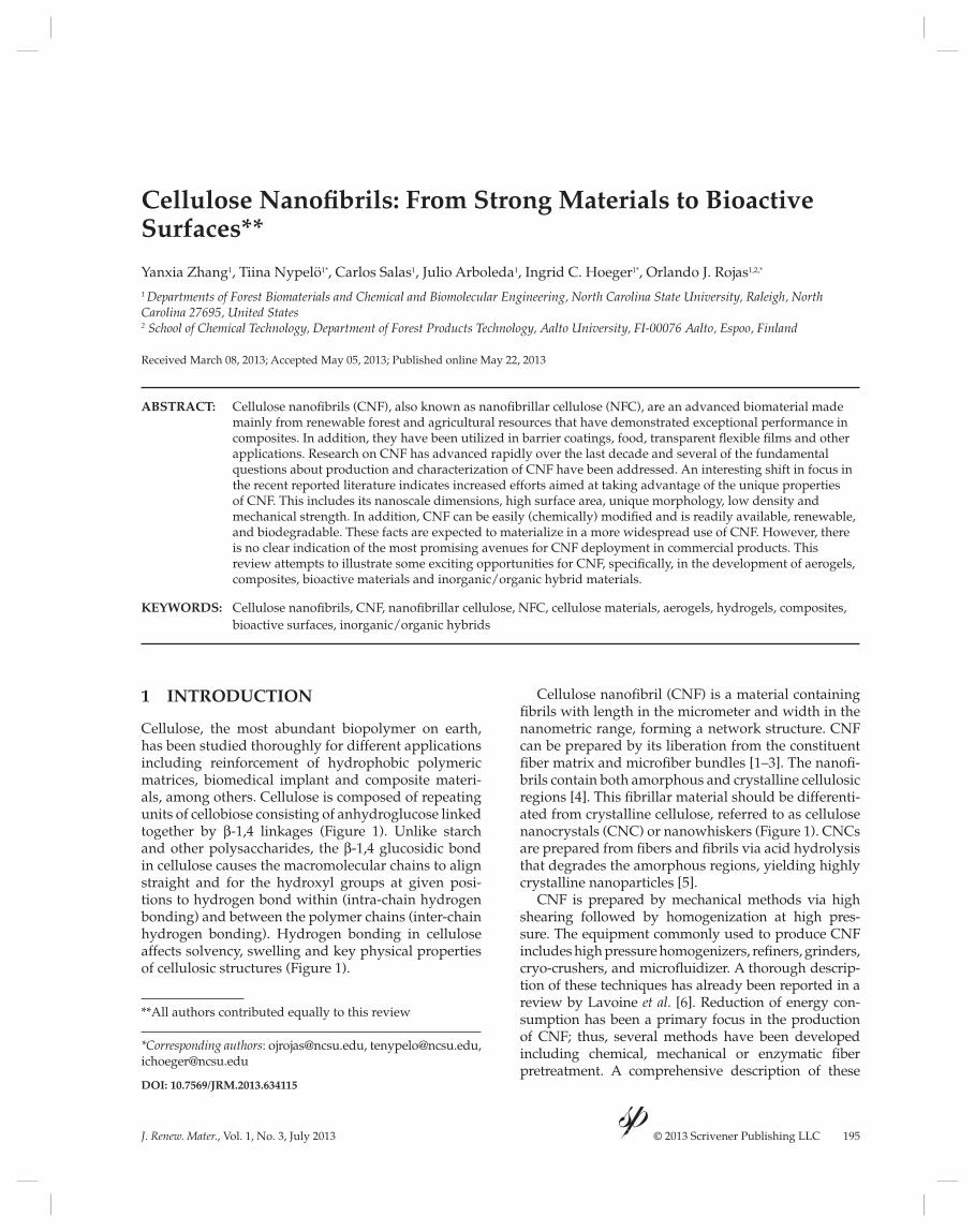
CNF aerogels can be functionalized chemically for different applications by post-treatment of formed aerogels or by modifi cation of precursor nanofi bers prior aerogel production. For example, titanium diox-ide (TiO2) nanoparticles [33] and silanes [34–36] have been used to make superhydrophobic materials via post-treatment. As a result, wetting of the aerogel is prevented while oil can be adsorbed into the pores of the aerogel (Figure 3). The oil could be removed by exposure to organic solvents enabling multiple uses of the aerogel. Modifi cation of precursor nanofi bers with particle coating is a facile way to prepare a material of interest as oil-adsorbent for sustainable applications. Additionally, deposition of titanium dioxide on CNF aerogels makes them a photosensitive hybrid mate-rial [37]; in fact, the ability of TiO2 nanoparticles to respond to UV light can switch the wetting behavior of the aerogel from hydrophilic to hydrophobic [37].
CNF-based aerogels have also been studied as envi-ronmentally friendly alternatives to replace superab-sorbent polymers (SAP). CNF aerogels absorb water at high rates; however they typically have half the water loading capacity of conventional SAPs. Aerogels also have weaker mechanical stability in the wet state rela-tive to that when dry. Two approaches have been used to improve this property: (1) addition of macroscopic fi bers as reinforcement [38], and (2) crosslinking to obtain aerogels with fast shape recovery upon water sorption [39]. Conductivity of these aerogels can be affected by polyaniline–dodecyl benzene sulfonic acid treatment [31], and incorporation of magnetic domains can be used to generate magnetic proper-ties allowing large stimuli-responsive deformation [40]. Modifi cation with 2,2,6,6-Tetramethylpiperidine 1-oxyl (TEMPO) produces aerogels with excellent
catalytic effi ciency for the azide-alkyne Huisgen cycloadition reaction, suggesting that fi brillar aerogels are also suitable supports for catalysts [41].
In addition to the aerogel structures described above, paper-like fi lms of CNF, nanopaper, prepared via removal of water by supercritical drying, liquid CO2 evaporation or freeze-drying can be considered as CNF aerogels. These systems are porous mem-branes with potential use in fuel cells, catalysis, liquid purifi cation and fi ltration, tissue engineering, protein immobilization, protein separation, protective cloth-ing, etc. [42]. Overall, there is a tremendous potential in tailoring the functions of CNF aerogels upon chemi-cal modifi cations, making this an active area of current research.
3 CNF-BASED NANOCOMPOSITES
Historically natural fi bers have been used in compos-
ites primarily for reinforcement. Several applications
of natural fi bers in the manufacture of composites
for the automotive [43–46] and other industries are
available [47–49]. In automotive applications natural
fi bers offer several advantages over glass fi ber: they
have lower cost, are lightweight, biodegradable, and
less abrasive to processing equipment. However the
recyclability, moisture repellence/sensitivity, durabil-
ity and fl ame retardancy are still some issues that need
to be addressed for implementation on a large scale
[43, 50]. Fiber reinforced composites have been used
in a variety of interior car parts such as door trim pan-
els, door liners, headliners, cabin linings and seatbacks
[43, 45]. However, car exterior components present
obvious challenges.
Compatibility of the fi bers with polymers is one of the key issues that have been largely investigated. The main challenge appears to be the poor adhesion and dispersibility of the fi bers in the hydrophobic polymer matrices due to the dissimilar nature of the compo-nents. In fact, several physical and chemical modifi -cations to improve fi ber-matrix adhesion have been suggested [51–55], including silane coupling [56]. The inherent properties of the source natural fi bers, such as, structure, mechanical and thermal properties, can have a large impact in the manufacture process and their suitability for composites [55, 57–60].
Cellulose has been proven to have great potential as reinforcing material in thermoplastics and elastomers [61]. Improved adhesion between cellulosic fi bers and polypropylene was reported when the fi bers have been accomplished upon pretreatment with silane coupling agents in methanol/water solution and also with maleic anhydride modifi ed polypropylene (MAPP) [62]; however, this procedure leads to thermal
Figure 3 Superhydrophobic and oleophilic CNF aerogel
produced by loading it with titanium dioxide nanoparticles.
The hydrophobic and oleophilic properties are indicated
by the fact that water (colored with reactive blue dye) and
glycerol droplets are not absorbed by the CNF-TiO2-coated
aerogels. However, paraffi n oil and mineral oil droplets
are readily absorbed (a). The TiO2-coated aerogels fl oat on
water (b). Reprinted with permission from [33]. Copyright
(2011) American Chemical Society.
WaterParaffin oil
Mineral oilGlycerol
(b)(a)

DOI: 10.7569/JRM.2013.634115 Yanxia Zhang et al.: Cellulose Nanofi brils (CNF)
J. Renew. Mater., Vol. 1, No. 3, July 2013 © 2013 Scrivener Publishing LLC 199
degradation of the cellulosic fi bers during process-ing. The nature of the adhesion between cellulose and polypropylene in composites has been studied [63] by treating the cellulosic fi bers fi rst with toluene in a Soxhlet system during 24 h and then dried, fol-lowed by immersion in a solution of 5% maleic anhy-dride modifi ed polypropylene in toluene. The dried fi bers and polypropylene were then mixed in a mix-ing extruder for further processing by injection mold-ing. The results indicated highly hydrophobic fi bers upon pretreatment (contact angle ~140o) that led to an improved interfacial adhesion resulting in increased composite mechanical properties (tensile modulus and strength at yield). Additionally, a pre-activation of the MAPP at 180°C allows to increase the reactivity of the polymer that reacts with the hydroxyl groups at the surface of cellulose forming strong, ester cova-lent bonds that improve adhesion with polypropylene [63]. Another way to increase the hydrophobicity of fi bers is by graft copolymerization of methyl acrylate; in this case the composites display improved mechani-cal properties [64].
As with natural fi bers, surface modifi cation of CNF is critical for incorporation in composite material. Several reports have dealt in detail with the devel-opment of nanocomposites [23, 65–67], the inclusion of nanofi bers in composites [24, 50, 68–72], and the resultant barrier properties of the nanocomposites [6]. Approaches as those used in the case of cellulose fi bers can also be applied for CNF. For example, acety-lation, silane coupling (silylation), graft copolymeriza-tion, treatment with isocyanate, triazine or physical treatments including electrical discharge (corona and cold plasma) and mercerization [51, 54, 55]. Areas of research in cellulose nanofi ber composites include applications in food packaging, automotive industry, construction materials, furniture production, trans-parent fi lms for displays, nanopaper, and fi ltration membranes. The production of such nanocomposites includes simple methods such as casting aqueous CNF dispersions using water soluble matrix materials; casting fi lms from CNF dispersions mixed with poly-mer latex dispersions; surface modifi cation of CNF followed by mixing in a solvent with the polymeric matrix to cast fi lms; dispersion of dry CNF into the hydrophobic matrix, etc. [73]. Following are examples of the application of CNF in composites.
3.1 CNF in Packaging Films
CNF barrier properties are important in packag-
ing applications, a topic that has received extensive
attention in the literature [6, 17, 74–78]. For exam-
ple, comparison of the barrier properties of cellulose
nanocrystals and microfi brillated cellulose from sisal
fi bers showed that water diffusion was higher for
fi lms made from CNCs compared to CNF [79]. Mass
transport properties of CNF fi lms have been evaluated
where fi lms were produced with glycerol as plasticizer
[76]. Glycerol reduces the water uptake at low humid-
ity whilst the opposite effect occurred at higher humid-
ity; the diffusion coeffi cient was higher for plasticized
samples, despite the porosity of the fi lms they showed
high oxygen barrier properties. Nanocomposite fi lms
of TEMPO-oxidized nanocellulose/montmorrillonite
presented high tensile strength and low oxygen per-
meability at 0 and 50% relative humidity, these prop-
erties were ascribed to the hydrogen bonding between
the TEMPO oxidized cellulose and the montmorril-
lonite [80]. Pulp characteristics and treatments used to
produce the cellulose microfi brils play an important
role in the resultant barrier properties [6].
3.2 Optically Transparent Composites
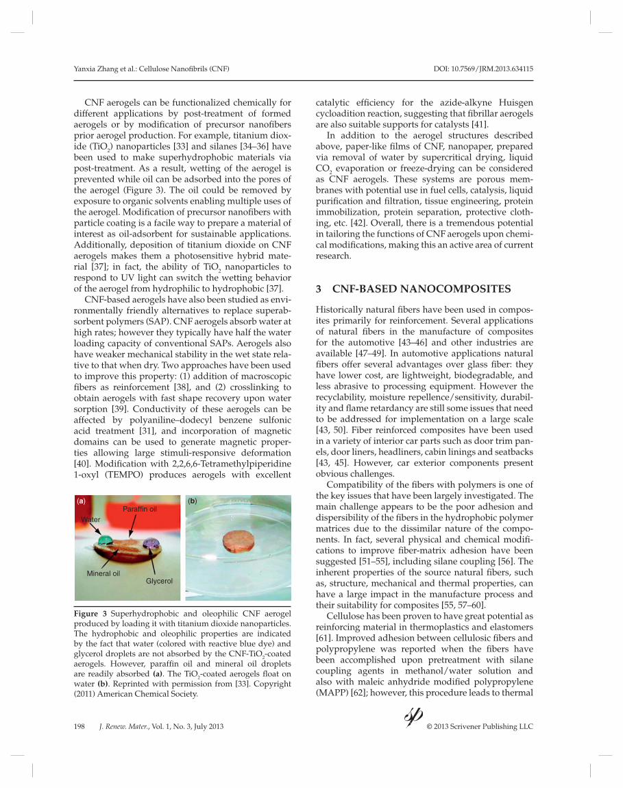
CNF has been studied for possible applications in
fl exible display manufacture [12]. Films (~ 60 μm
thickness) from Douglas fi r CNF obtained by grinding
method gave low light transmittance as a dried sheet,
but transparency increased after polishing the fi lm
[13]. BC has also been used to prepare transparent
cellulose nanocomposites manufactured by using dif-
ferent resins, epoxy, acrylic and phenol formaldehyde
(Figure 4a) [12]. The fi ber content in the BC nano-
composites (up to 70% CNF content) have very low
thermal expansion coeffi cient. Compared to the neat
epoxy resins the BC-epoxy nanocomposite displayed
10% less light transmittance (Figure 4b). Transparent
fi lms (~ 20 μm thick) have been produced by suction
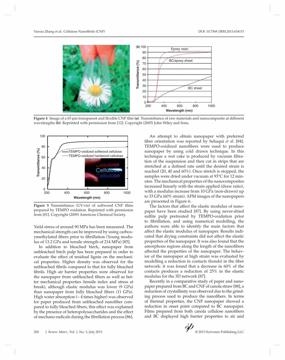
fi ltration of TEMPO-oxidized hardwood and softwood
CNF (Figure 5) [81]. Other approaches to produce
transparent fi lms include acetylation of cellulose fi brils
and further graft polymerization with hydroxyethyl
methacrylate (HEMA) to produce a continuous
hydrogel matrix of poly(2-hydroxyethyl methacrylate
(PHEMA) [82]. Electrically conductive transparent
paper was made by incorporation of fi broin solution
into BC membrane followed by adsorption of multi-
walled carbon nanotubes [83].
3.3 Nanopaper
Nanopaper has been a subject of recent studies due to
its properties, such as low thermal expansion coeffi -
cient, optical transparency and good mechanical prop-
erties combined with a simple preparation procedure
(fi ltration from aqueous suspensions) [84]. The typical
preparation of nanopaper is carried out by vacuum
fi ltration of CNF suspensions [85] to produce fi lms
of different porosities and thickness (~ 70–90 nm).
Yanxia Zhang et al.: Cellulose Nanofi brils (CNF) DOI: 10.7569/JRM.2013.634115
200 J. Renew. Mater., Vol. 1, No. 3, July 2013 © 2013 Scrivener Publishing LLC
Yield stress of around 90 MPa has been measured. The
mechanical strength can be improved by using carbox-
ymethylated fi bers prior to fi brillation (Young modu-
lus of 13.2 GPa and tensile strength of 214 MPa) [85].
In addition to bleached birch, nanopaper from unbleached birch pulp has been prepared in order to evaluate the effect of residual lignin on the mechani-cal properties. Higher density was observed for the unbleached fi brils compared to that for fully bleached fi brils. High air barrier properties were observed for the nanopaper from unbleached fi bers as well as bet-ter mechanical properties (tensile index and stress at break), although elastic modulus was lower (9 GPa) than nanopaper from fully bleached fi bers (11 GPa). High water absorption (~ 4 times higher) was observed for paper produced from unbleached nanofi ber com-pared to fully bleached fi bers, this effect was explained by the presence of heteropolysaccharides and the effect of mechano-radicals during the fi brillation process [86].
An attempt to obtain nanopaper with preferred fi ber orientation was reported by Sehaqui et al. [84]. TEMPO-oxidized nanofi bers were used to produce nanopaper by using cold drawn technique. In this technique a wet cake is produced by vacuum fi ltra-tion of the suspension and then cut in strips that are stretched at a defi ned rate until the desired strain is reached (20, 40 and 60%). Once stretch is stopped, the samples were dried under vacuum at 93°C for 12 min-utes. The mechanical properties of the nanocomposites increased linearly with the strain applied (draw ratio), with a modulus increase from 10 GPa (non-drawn) up to 33 GPa (60% strain). AFM images of the nanopapers are presented in Figure 6.
The factors that affect the elastic modulus of nano-paper have been studied [87]. By using never-dried sulfi te pulp pretreated by TEMPO-oxidation prior to fi brillation, and using numerical modelling, the authors were able to identify the main factors that affect the elastic modulus of nanopaper. Results indi-cated that drying constraints did not affect the elastic properties of the nanopaper. It was also found that the amorphous regions along the length of the nanofi bers affected the properties of the nanopaper. The behav-ior of the nanopaper at high strain was evaluated by modelling a reduction in contacts (bonds) in the fi ber network: it was found that a decrease in 60% of the contacts produces a reduction of 25% in the elastic modulus for the 3D network [87].
Recently in a comparative study of paper and nano-paper prepared from BC and CNF of canola straw [88], a reduction of crystallinity was observed due to the grind-ing process used to produce the nanofi bers. In terms of thermal properties, the CNF nanopaper showed a reduction in onset point compared to BC nanopaper. Films prepared from both canola cellulose nanofi bers and BC displayed high barrier properties to air and
Figure 4 Image of a 65 μm transparent and fl exible CNF fi lm (a). Transmittance of raw materials and nanocomposite at different
wavelengths (b). Reprinted with permission from [12]. Copyright (2005) John Wiley and Sons.
Wavelength (nm)
Tran
smit
tan
ce (
%)
Epoxy resin
BC/epoxy sheet
BC sheet
2000
10
20
30
40
50
60
70
80
90
400
100
600 800 1000
(a) (b)
Figure 5 Transmittance (UV-vis) of softwood CNF fi lms
prepared by TEMPO oxidation. Reprinted with permission
from [81]. Copyright (2009) American Chemical Society.
2000
20
40
60
80
100
400 600
Wavelength (nm)
Tran
smit
tan
ce (
%)
800 1000
TEMPO-oxidized softwood celluloseTEMPO-oxidized hardwood cellulose
DOI: 10.7569/JRM.2013.634115 Yanxia Zhang et al.: Cellulose Nanofi brils (CNF)
J. Renew. Mater., Vol. 1, No. 3, July 2013 © 2013 Scrivener Publishing LLC 201
good mechanical strength, with Young modulus of 13.6 and 17.3 GPa for CNF and BC nanopaper, respectively.
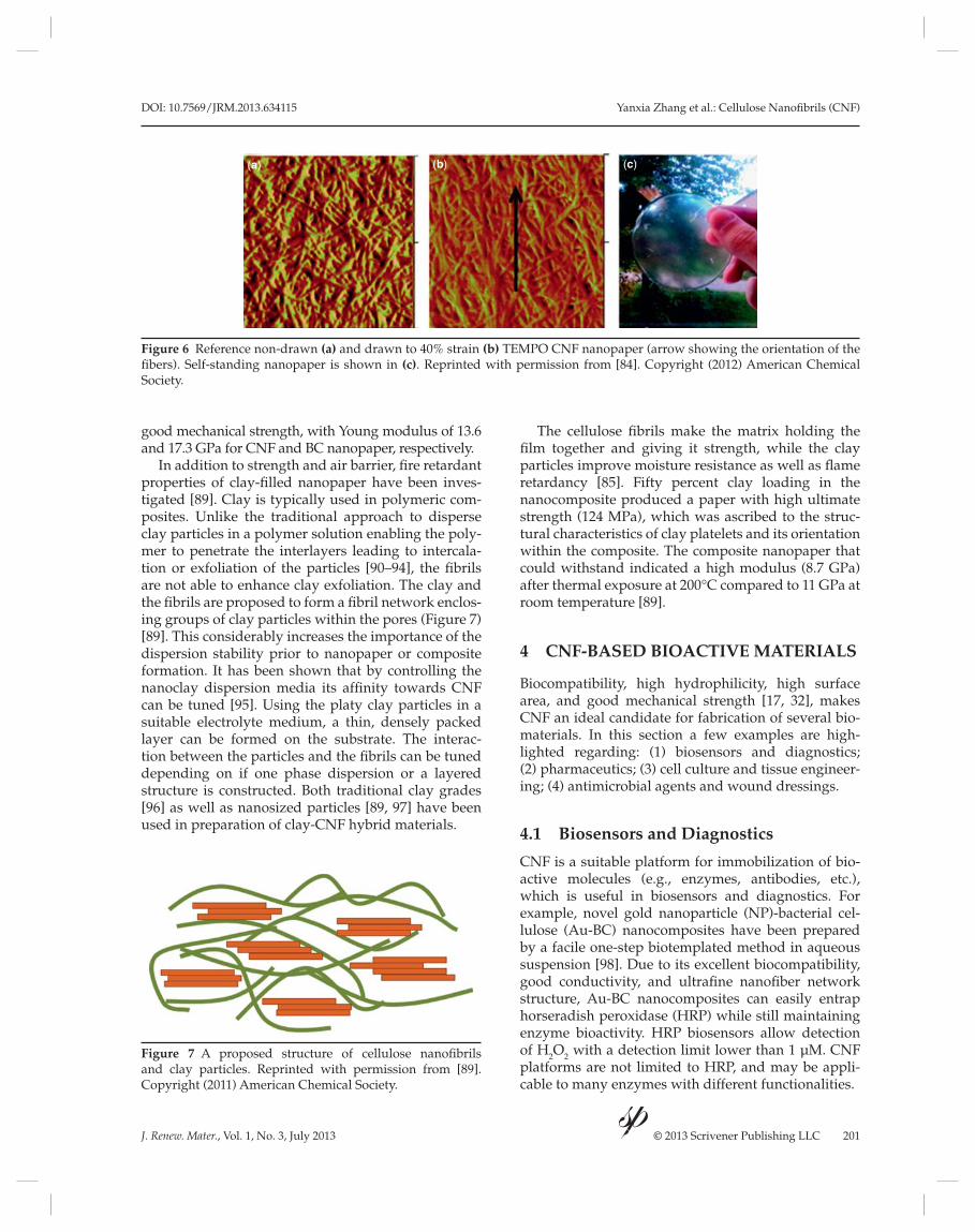
In addition to strength and air barrier, fi re retardant properties of clay-fi lled nanopaper have been inves-tigated [89]. Clay is typically used in polymeric com-posites. Unlike the traditional approach to disperse clay particles in a polymer solution enabling the poly-mer to penetrate the interlayers leading to intercala-tion or exfoliation of the particles [90–94], the fi brils are not able to enhance clay exfoliation. The clay and the fi brils are proposed to form a fi bril network enclos-ing groups of clay particles within the pores (Figure 7) [89]. This considerably increases the importance of the dispersion stability prior to nanopaper or composite formation. It has been shown that by controlling the nanoclay dispersion media its affi nity towards CNF can be tuned [95]. Using the platy clay particles in a suitable electrolyte medium, a thin, densely packed layer can be formed on the substrate. The interac-tion between the particles and the fi brils can be tuned depending on if one phase dispersion or a layered structure is constructed. Both traditional clay grades [96] as well as nanosized particles [89, 97] have been used in preparation of clay-CNF hybrid materials.
The cellulose fi brils make the matrix holding the fi lm together and giving it strength, while the clay particles improve moisture resistance as well as fl ame retardancy [85]. Fifty percent clay loading in the nanocomposite produced a paper with high ultimate strength (124 MPa), which was ascribed to the struc-tural characteristics of clay platelets and its orientation within the composite. The composite nanopaper that could withstand indicated a high modulus (8.7 GPa) after thermal exposure at 200°C compared to 11 GPa at room temperature [89].
4 CNF-BASED BIOACTIVE MATERIALS
Biocompatibility, high hydrophilicity, high surface
area, and good mechanical strength [17, 32], makes
CNF an ideal candidate for fabrication of several bio-
materials. In this section a few examples are high-
lighted regarding: (1) biosensors and diagnostics;
(2) pharmaceutics; (3) cell culture and tissue engineer-
ing; (4) antimicrobial agents and wound dressings.
4.1 Biosensors and Diagnostics
CNF is a suitable platform for immobilization of bio-
active molecules (e.g., enzymes, antibodies, etc.),
which is useful in biosensors and diagnostics. For
example, novel gold nanoparticle (NP)-bacterial cel-
lulose (Au-BC) nanocomposites have been prepared
by a facile one-step biotemplated method in aqueous
suspension [98]. Due to its excellent biocompatibility,
good conductivity, and ultrafi ne nanofi ber network
structure, Au-BC nanocomposites can easily entrap
horseradish peroxidase (HRP) while still maintaining
enzyme bioactivity. HRP biosensors allow detection
of H2O
2 with a detection limit lower than 1 μM. CNF
platforms are not limited to HRP, and may be appli-
cable to many enzymes with different functionalities.
Figure 6 Reference non-drawn (a) and drawn to 40% strain (b) TEMPO CNF nanopaper (arrow showing the orientation of the
fi bers). Self-standing nanopaper is shown in (c). Reprinted with permission from [84]. Copyright (2012) American Chemical
Society.
(a) (b) (c)
Figure 7 A proposed structure of cellulose nanofi brils
and clay particles. Reprinted with permission from [89].
Copyright (2011) American Chemical Society.
Yanxia Zhang et al.: Cellulose Nanofi brils (CNF) DOI: 10.7569/JRM.2013.634115
202 J. Renew. Mater., Vol. 1, No. 3, July 2013 © 2013 Scrivener Publishing LLC
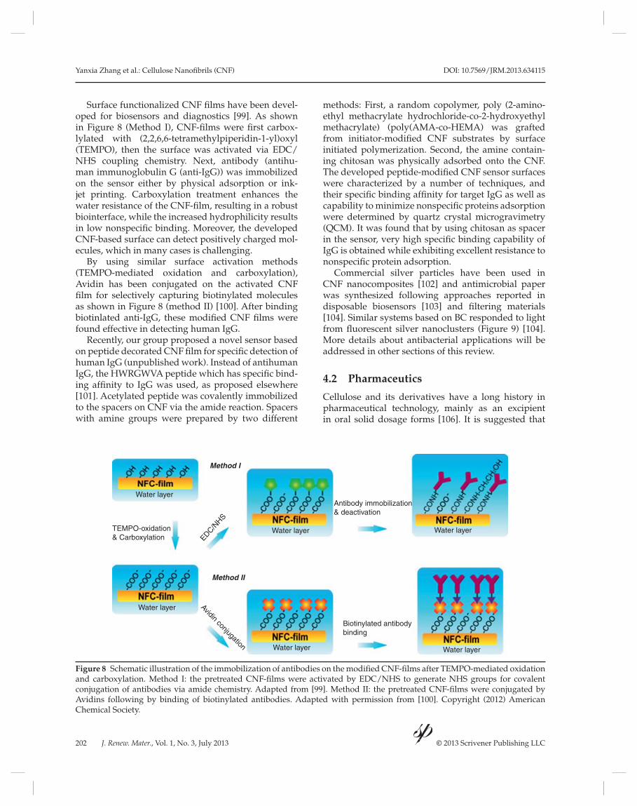
Surface functionalized CNF fi lms have been devel-oped for biosensors and diagnostics [99]. As shown in Figure 8 (Method I), CNF-fi lms were fi rst carbox-lylated with (2,2,6,6-tetramethylpiperidin-1-yl)oxyl (TEMPO), then the surface was activated via EDC/NHS coupling chemistry. Next, antibody (antihu-man immunoglobulin G (anti-IgG)) was immobilized on the sensor either by physical adsorption or ink-jet printing. Carboxylation treatment enhances the water resistance of the CNF-fi lm, resulting in a robust biointerface, while the increased hydrophilicity results in low nonspecifi c binding. Moreover, the developed CNF-based surface can detect positively charged mol-ecules, which in many cases is challenging.
By using similar surface activation methods (TEMPO-mediated oxidation and carboxylation), Avidin has been conjugated on the activated CNF fi lm for selectively capturing biotinylated molecules as shown in Figure 8 (method II) [100]. After binding biotinlated anti-IgG, these modifi ed CNF fi lms were found effective in detecting human IgG.
Recently, our group proposed a novel sensor based on peptide decorated CNF fi lm for specifi c detection of human IgG (unpublished work). Instead of antihuman IgG, the HWRGWVA peptide which has specifi c bind-ing affi nity to IgG was used, as proposed elsewhere [101]. Acetylated peptide was covalently immobilized to the spacers on CNF via the amide reaction. Spacers with amine groups were prepared by two different
methods: First, a random copolymer, poly (2-amino-ethyl methacrylate hydrochloride-co-2-hydroxyethyl methacrylate) (poly(AMA-co-HEMA) was grafted from initiator-modifi ed CNF substrates by surface initiated polymerization. Second, the amine contain-ing chitosan was physically adsorbed onto the CNF. The developed peptide-modifi ed CNF sensor surfaces were characterized by a number of techniques, and their specifi c binding affi nity for target IgG as well as capability to minimize nonspecifi c proteins adsorption were determined by quartz crystal microgravimetry (QCM). It was found that by using chitosan as spacer in the sensor, very high specifi c binding capability of IgG is obtained while exhibiting excellent resistance to nonspecifi c protein adsorption.
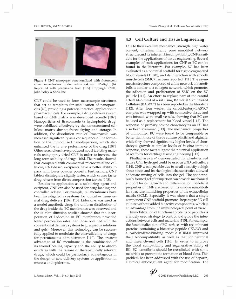
Commercial silver particles have been used in CNF nanocomposites [102] and antimicrobial paper was synthesized following approaches reported in disposable biosensors [103] and fi ltering materials [104]. Similar systems based on BC responded to light from fl uorescent silver nanoclusters (Figure 9) [104]. More details about antibacterial applications will be addressed in other sections of this review.
4.2 Pharmaceutics
Cellulose and its derivatives have a long history in
pharmaceutical technology, mainly as an excipient
in oral solid dosage forms [106]. It is suggested that
Figure 8 Schematic illustration of the immobilization of antibodies on the modifi ed CNF-fi lms after TEMPO-mediated oxidation
and carboxylation. Method I: the pretreated CNF-fi lms were activated by EDC/NHS to generate NHS groups for covalent
conjugation of antibodies via amide chemistry. Adapted from [99]. Method II: the pretreated CNF-fi lms were conjugated by
Avidins following by binding of biotinylated antibodies. Adapted with permission from [100]. Copyright (2012) American
Chemical Society.
Method I
Method II
Water layer
Water layer Water layer
Water layerWater layer
Water layer
TEMPO-oxidation& Carboxylation
Antibody immobilization& deactivation
EDC/NHS
Biotinylated antibodybinding
Avidin conjugation
DOI: 10.7569/JRM.2013.634115 Yanxia Zhang et al.: Cellulose Nanofi brils (CNF)
J. Renew. Mater., Vol. 1, No. 3, July 2013 © 2013 Scrivener Publishing LLC 203
CNF could be used to form macroscopic structures
that act as templates for stabilization of nanoparti-
cles [40], providing a potential practical application in
pharmaceuticals. For example, a drug delivery system
based on CNF matrix was developed recently [107].
Nanoparticles of Itraconazole (a hydrophobic drug)
were stabilized effectively by the nanostructured cel-
lulose matrix during freeze-drying and storage. In
addition, the dissolution rate of Itraconazole was
increased signifi cantly as a consequence of the forma-
tion of the immobilized nanodispersion, which also
enhanced the in vivo performance of the drug [107].
Other researchers have produced novel tableting mate-
rials using spray-dried CNF in order to increase the
long-term stability of drugs [108]. The results showed
that compared with commercial microcrystalline cel-
lulose, CNF-based excipients have a better ability to
pack with lower powder porosity. Furthermore, CNF
tablets disintegrate slightly faster, which causes faster
drug release from direct compression tablets [108].
Besides its application as a stabilizing agent and excipient, CNF can also be used for drug loading and controlled release. For example, BC membranes have been investigated as systems for topical or transder-mal drug delivery [109, 110]. Lidocaine was used as a model anesthetic drug; the uniform distribution of the drug inside the BC membranes was observed and the in vitro diffusion studies showed that the incor-poration of Lidocaine in BC membranes provided lower permeation rates than those obtained with the conventional delivery systems (e.g., aqueous solutions and gels). Moreover, this technology can be success-fully applied to modulate the bioavailability of drugs for percutaneous administration [110]. The greatest advantage of BC membrane is the combination of its wound healing capacity and the ability to absorb exudates with the release of therapeutically relevant drugs, which could be particularly advantageous in the design of new delivery systems or application in mucosa and epidermis.
4.3 Cell Culture and Tissue Engineering
Due to their excellent mechanical strength, high water
content, ultrafi ne, highly pure nanofi bril network
structure and its inherent biocompatibility, CNF is suit-
able for the applications of tissue engineering. Several
examples of such applications for CNF or BC can be
found in the literature. For example, BC has been
evaluated as a potential scaffold for tissue- engineered
blood vessels (TEBV), and its interaction with smooth
muscle cells (SMC) has been reported [111]. The asym-
metric structure composed of a fi ne network of nanofi -
brils is similar to a collagen network, which promotes
the adhesion and proliferation of SMC on the BC
pellicle [111]. An effort to replace part of the carotid
artery (4–6 mm) of a rat using BActerial SYnthesized
Cellulose (BASYC®) has been reported in the literature
[112]. After four weeks, the carotid-artery-BASYC®
complex was wrapped up with connective tissue and
was infused with small vessels, showing that BC can
be used as a replacement for blood vessel [112]. The
response of primary bovine chondrocytes on BC has
also been examined [113]. The mechanical properties
of unmodifi ed BC were found to be comparable or
better than those of tissue culture plastic and alginate,
while they showed signifi cantly higher levels of chon-
drocyte growth at similar levels of in vitro immune
response; these facts suggest the potential application
of scaffolds for cartilage tissue engineering [113].
Bhattacharya et al. demonstrated that plant-derived native CNF hydrogel could be used as a 3D cell culture [114]. CNF was injectable due to small viscosity at high shear stress and its rheological characteristics allowed adequate mixing of cells into the gel. The spontane-ously formed gel after injection can provide mechanical support for cell growth and differentiation. Benefi cial properties of CNF are based on its unique nanofi bril-lar structure mimicking properties of the extracellular matrix (ECM). Especially, it was shown that a single component CNF scaffold promotes hepatocyte 3D cell culture without added bioactive components, which is an advantage from the immunological point of view.
Immobilization of functional proteins or peptides is a widely used strategy to control and guide the inter-actions between cells and materials [115]. For example, the functionalization of BC surfaces with recombinant proteins containing a bioactive peptide (IKVAV) and a carbohydrate-binding module (CBM3) improved their biocompatibility, as well as that for neuronal and mesenchymal cells [116]. In order to improve the blood compatibility and regenerative ability of BC, BC nanofi brils should be crosslinked with some materials to prevent the formation of blood clots. This problem has been addressed with the use of heparin, a typical anticoagulant agent for modifi cation. The
Figure 9 CNF nanopaper functionalized with fl uorescent
silver nanoclusters under white (a) and UV-light (b). Reprinted with permission from [105]. Copyright (2011)
John Wiley & Sons, Inc.
(a) (b)

Yanxia Zhang et al.: Cellulose Nanofi brils (CNF) DOI: 10.7569/JRM.2013.634115
204 J. Renew. Mater., Vol. 1, No. 3, July 2013 © 2013 Scrivener Publishing LLC
cell compatibility results indicated that the modifi ed BC nanofi bers were suitable for cell proliferation and ingrowth [117]. To mimic the natural collagen fi bers, a natural peptide, ε-polylysine (PLL) was introduced to the surfaces of BC nanofi bers via crosslinking method [118]. The BC/PLL nanofi bers proved to have similar shape and molecular structure compared with natu-ral collagen nanofi bers. After immersion in CaCl2 and simulated body fl uid, the BC/PLL nanofi bers served as nanotemplates to induce the formation of nano-sized platelet-like, calcium-defi cient, B-type carbon-ated hydroxyapatite (HAp). The fi nal mineralized BC/PLL nanofi bers exhibited similar architecture and composition of natural bone, suggesting that BC/PLL nanofi bers are promising biomaterials in bone tissue engineering.
CNF can also be used to form a nanocomposite material for cell culture [119]. For example, nanocom-posites of BC networks and calcium-defi cient HAp have been produced by introducing the mineral phase into the bacteria culture medium during formation of cellulose fi brils. The biocompatibility and cell viabil-ity were confi rmed by HEK cell seeding, making this nanocomposite potentially viable in biomedical appli-cations. In another report, biocomposite scaffolds consisting of poly(3-hydroxubutyrate-co-4-hydrox-ubutyrate) (P(3HB-co-4HB)) and BC were prepared, which combined their respective advantages [120]. The resultant scaffolds exhibited 3D network struc-ture composing of multi-distribution of pore size and proved to be capable of forming cell adhesion and proliferation.
4.4 Antibacterial Agent and Wound Dressing
Another application of CNF in the biomedical fi eld is
their use as antibacterial agent and wound dressing.
A widely used method is to integrate silver nanopar-
ticles to CNF because silver metal and its compounds
have been known for their strong inhibitory and bac-
tericidal effects as well as a broad spectrum of antimi-
crobial activities [121, 122]. The assembly of the CNF
and silver nanoparticle (AgNP) composites can be
carried out through electrostatic interaction of AgNP
(aqueous colloids) onto CNF using polyelectrolytes
as linkers [102]. Their antibacterial activity against
S. aureus (gram-positive bacteria) and K. pneumoni-aestrains (gram-negative bacteria) was reported [102].
Another strategy is through magnetic interaction
[123]. Here, the unique, ultrafine 3D structure of the
BC matrix can provide plenty of sites for heterogenous
nucleation of magnetite. The resultant magnetic Ag
nanocomposite possesses a high antimicrobial activ-
ity against the model microbes E. coli (gram-positive
bacteria) and Bacillus subtilis (gram-negative bacteria).
The combination of magnetic nanoparticles with sil-
ver nanoparticles in the nanostructure of BC makes it
an interesting approach to develop recoverable and
reusable antibacterial agents [123]. Finally, the func-
tionalization of native CNF using silver nanoclusters
(AgNC) to form a novel type of functional nanocel-
lulose/nanocluster composite was found to exhibit
a pronounced antibacterial activity [105]. Instead of
preformed particles, silver nitrate was used to pre-
cipitate silver by reducing the ions to metallic nano-
particles using sodium borohydride. The CNF/AgNC
composite retained the appealing properties of both
components. As observed in Figure 10, the bacteria
grew freely all around the unmodifi ed CNF fi lm, indi-
cating no antibacterial activity. In contrast, a large halo
free of bacteria was observed around the CNF/AgNC
fi lms due to the release of silver ions or AgNC, which
prevented bacterial growth. These results suggested
this CNF/AgNC composite may fi nd application in
wound-healing pads.
BC is an interesting material for using as a wound dressing since it can control wound exudates and can provide moist environment to a wound result-ing in better wound healing [124]. Biofi ll® is a com-mercial product of BC used as a temporary substitute for human skin in cases of second- and third-degree burns [112, 125]. In order to prevent wound infection AgNP have been incorporated in the BC membranes [11, 126, 127]. Maneerung et al. impregnated AgNP into bacterial cellulose through chemical reduction [11]. The modifi ed BC exhibited a strong antimicrobial activity against S. aureus and E. coli, which are bacte-ria found in the contaminated wounds. Especially, the
Figure 10 Antibacterial properties of CNF fi lms
functionalized with silver nanoclusters. Reprinted with
permission from [105]. Copyright (2011) John Wiley &
Sons, Inc.

DOI: 10.7569/JRM.2013.634115 Yanxia Zhang et al.: Cellulose Nanofi brils (CNF)
J. Renew. Mater., Vol. 1, No. 3, July 2013 © 2013 Scrivener Publishing LLC 205
impregnation, instead of coating the BC with AgNP, improved the antimicrobial activity of the BC and low-ered the possibility of normal human tissue damage.
Besides AgNP, quaternary ammonium compound is another group of well-known antibacterial agents. Furthermore, cellulose has been covalently grafted with a quaternary ammonium compound octadecyldi-methyl (3-trimethoxysilylpropyl) ammonium chloride (ODDMAC) by a simple adsorption-curing process [128]. The resultant fi lms showed very good antibacte-rial capacity even at very low concentrations of anti-microbial agent immobilized on the surface. Because ODDMAC is covalently immobilized onto the mate-rial surface, this fi lm exhibits long-term antimicrobial activity. Moreover, due to the large surface area to vol-ume ratio of the cellulose fi lms and the fact that each fi bril is modifi ed with ODDMAC, these nonporous fi lms could be very effective as antimicrobial separa-tion fi lters.
There is great potential for the use of CNFs in a range of applications. So far their applications in aerogels, composites and the biomedical fi eld have been dis-cussed. Some examples of inorganic/organic hybrids have been mention in these sections: TiO2 (aerogels), clay (nanocomposite section), and silver (bioactive materials). In the next section of this review other important CNF-inorganic hybrids will be introduced.
5 CELLULOSE NANOFIBRIL HYBRIDS
Hybrid materials combine organic and inorganic com-
ponents aiming for synergistic effects. An increasing
number of studies presenting the combination of cel-
lulosic nanofi brils with inorganic particles are being
reported. The inorganic phase is typically added to
improve the barrier and optical properties, as well as
to enable preparation of responsive materials with the
incorporation of functional particles.
5.1 Silica
Silica particles are extensively used in a variety of
applications for optical effects as well as for provid-
ing durability, for example, in surface fi nishes. Mainly
for these reasons silica nanoparticles have been used
for creating hybrid structures with cellulose fi brils. For
instance, a hybrid structure of bacterial cellulose with
silica nanoparticles was prepared using tetraethoxysi-
lane as a precursor. High loading of up to 66% was
achieved with the particles attached to the fi brils [129].
Another approach for preparing a bacterial cellulose- silica network by growing the fi brils in a silica dispersion using the bacteria has been presented [12]. The bacterium (Ga. Xylinus) was noted for being
more active in acid conditions leading to loadings as high as 50%. The concentration of the silica solution was found to affect the amount of silica in the structure formed. Bacterial growth in a silica matrix is a simple alternate approach to enable silica particles to pene-trate into a preformed cellulose matrix. However, the loading could not be increased over ca. 10%. The fi bril layer in the silica hybrid structures provides strength whereas the silica brings the functionality. However, with high silica loadings the strength of the matrix is compromised. It can be predicted that hybrid materi-als combining cellulose nanomaterials and silica will make their way into (electronic) displays and related technologies.
5.2 Responsive Hybrid Materials
There is a growing interest in hybrid materials able
to respond to external stimuli. Here we present a
few examples of the most common types of respon-
sive materials involving the incorporation of UV
active and magnetic particles into the CNF network,
enabling preparation of responsive cellulosic prod-
ucts. Needless to say, such materials have promising
potential in packaging and sensing.
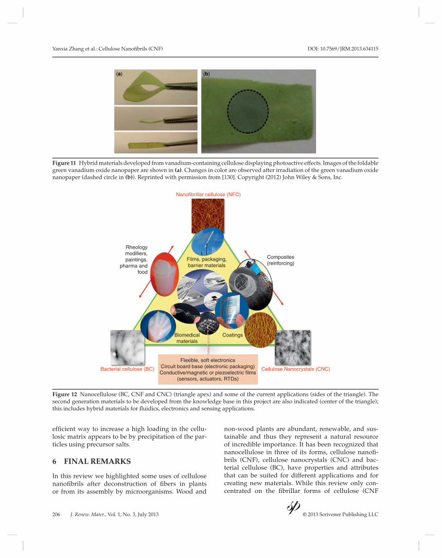
In a study leading to preparation of conductive BC network with photoswitchability, the photosensitivity was created by introducing vanadium nanoparticles on a cellulosic self-standing fi lm [130]. Addition of these particles altered the fi lm, i.e., changed the color of the nanopaper to green. This effect was further altered by photoactivation via irradiating the sample leading to color change to purple (Figure 11). The change in color was due to changes in the oxidation stage of the vanadium. Quite importantly to sensing materials the effect is reversible and the fi lm can be used multiple times. In addition to vanadium, a photosensitive CNF hybrid material has been prepared by deposition of titanium dioxide fi lm on CNF aerogels [37], already discussed in the aerogels section of this review.
Magnetic cellulose materials offer solutions for separation techniques and fi ltering as well as in intelligent packaging. Producing cellulosic fi brils with magnetic characteristics offers opportunities in many applications where magnetic components are already used, such as biomedical and separation techniques [131, 132]; the organic phase can be used to provide functionality to the magnetic domains. For example, as discussed earlier, embedding magnetic domains into a cellulose aerogel can be used to pre-pare super-adsorbent structures that can be triggered with applied magnetic fi elds [40]. In the case of cellu-lose matrix structured as BC the hybrid material can be deployed as a magnetic scaffold [133]. The most
Yanxia Zhang et al.: Cellulose Nanofi brils (CNF) DOI: 10.7569/JRM.2013.634115
206 J. Renew. Mater., Vol. 1, No. 3, July 2013 © 2013 Scrivener Publishing LLC
effi cient way to increase a high loading in the cellu-losic matrix appears to be by precipitation of the par-ticles using precursor salts.
6 FINAL REMARKS
In this review we highlighted some uses of cellulose
nanofi brils after deconstruction of fi bers in plants
or from its assembly by microorganisms. Wood and
non-wood plants are abundant, renewable, and sus-
tainable and thus they represent a natural resource
of incredible importance. It has been recognized that
nanocellulose in three of its forms, cellulose nanofi -
brils (CNF), cellulose nanocrystals (CNC) and bac-
terial cellulose (BC), have properties and attributes
that can be suited for different applications and for
creating new materials. While this review only con-
centrated on the fi brillar forms of cellulose (CNF
Figure 11 Hybrid materials developed from vanadium-containing cellulose displaying photoactive effects. Images of the foldable
green vanadium oxide nanopaper are shown in (a). Changes in color are observed after irradiation of the green vanadium oxide
nanopaper (dashed circle in (b)). Reprinted with permission from [130]. Copyright (2012) John Wiley & Sons, Inc.
(a) (b)
Figure 12 Nanocellulose (BC, CNF and CNC) (triangle apex) and some of the current applications (sides of the triangle). The
second generation materials to be developed from the knowledge base in this project are also indicated (center of the triangle);
this includes hybrid materials for fl uidics, electronics and sensing applications.
Nanofibrillar cellulose (NFC)
Bacterial cellulose (BC) Cellulose Nanocrystals (CNC)
Films, packaging,barrier materials
Composites(reinforcing)
Rheologymodifiers,paintings,
pharma andfood
Biomedicalmaterials
Coatings
Flexible, soft electronicsCircuit board base (electronic packaging)Conductive/magnetic or piezoelectric films
(sensors, actuators, RTDs)

DOI: 10.7569/JRM.2013.634115 Yanxia Zhang et al.: Cellulose Nanofi brils (CNF)
J. Renew. Mater., Vol. 1, No. 3, July 2013 © 2013 Scrivener Publishing LLC 207
and BC), together with CNC these materials offer a
number of opportunities for developments in differ-
ent fi elds, as illustrated in Figure 12. A major part of
research activities has concentrated on the utilization
of CNF in traditional high volume, low cost products
such as paper, packaging, paints, composites and food
(some of which were not covered in this review). In
these areas of application, removal of water in nano-
cellulose dispersions is a challenge that demands the
development of methods for surface functionalization
of fi brils to improve dewatering. Such efforts seem to
progress at a relatively slow pace. There is also an
opportunity to address the critical need to innova-
tively develop new platforms that will lead to another
generation of nanocellulose-based products. This
can be achieved by using the power of nanoscience
along with a full understanding of the behavior (e.g.,
structural assembly) of different types of nanocellu-
lose; by characterizing and exploiting their surface
chemistry and interactions with other materials at the
nanoscale; and by exploring several of its properties
(structural, physical, chemical, thermal, piezoelectric,
etc.) (Figure 12).
REFERENCES
1. F.W. Herrick, R.L. Casebier, J.K. Hamilton, and K.R. Sandberg,
Microfi brillated cellulose: Morphology and accessibility,
in Applied Polymer Symposia, A. Sarko, (Ed.), pp. 797–813,
Proceedings of the Ninth Cellulose Conference. vol. 37,
Wiley, New York City (1983).
2. M. P ääkkö, M. Ankerfors, H. Kosonen, A. Nykänen,
S. Ahola, M. Österberg, J. Ruokolainen, J. Laine,
P.T. Larsson, O. Ikkala, and T. Lindström, Enzymatic
hydrolysis combined with mechanical shearing and
high-pressure homogenization for nanoscale cellulose
fi brils and strong gels. Biomacromolecules 8, 1934 (2007).
3. A.F. Turbak, F.W. Snyder, and K.R. Sandberg,
Microfi brillated cellulose, a new cellulose prod-
uct: Properties, uses and commercial potential, in
Applied Polymer Symposia, A. Sarko, (Ed.), pp. 815–827,
Proceedings of the Ninth Cellulose Conference. vol. 37,
Wiley, New York City (1983).
4. C. A ulin, S. Ahola, P. Josefsson, T. Nishino, Y. Hirose,
M. Österberg, and L. Wågberg, Nanoscale cellulose
fi lms with different crystallinities and mesostructures-
their surface properties and interaction with water.
Langmuir 25, 7675 (2009).
5. Y. H abibi, L.A. Lucia, and O.J. Rojas, Cellulose nano-
crystals: Chemistry, self-assembly, and applications.
Chem. Rev. 110, 3479 (2010).
6. N. L avoine, I. Desloges, A. Dufresne, and J. Bras,
Microfi brillated cellulose – its barrier properties and
applications in cellulosic materials: A review. Carbohydr. Polym. 90, 735 (2012).
7. A. I sogai, T. Saito, and H. Fukuzumi, TEMPO-oxidized
cellulose nanofi bers. Nanoscale 3, 71 (2011).
8. K. S pence, R. Venditti, O. Rojas, Y. Habibi, and
J. Pawlak, A comparative study of energy consumption
and physical properties of microfi brillated cellulose
produced by different processing methods. Cellulose 18,
1097 (2011).
9. L. W ågberg, G. Decher, M. Norgren, T. Lindström,
M. Ankerfors, and K. Axnäs, The build-up of polyelec-
trolyte multilayers of microfi brillated cellulose and cat-
ionic polyelectrolytes. Langmuir 24, 784 (2008).
10. D. Klemm, B. Heublein, H.P. Fink, and A. Bohn,
Cellulose: Fascinating biopolymer and sustainable raw
material. Angew. Chem. 44, 3358 (2005).
11. T. Maneerung, S. Tokura, and R. Rujiravanit,
Impregnation of silver nanoparticles into bacterial cel-
lulose for antimicrobial wound dressing. Carbohydr.
Polym. 72, 43 (2008).
12. H. Yano, J. Sugiyama, A.N. Nakagaito, M. Nogi,
T. Matsuura, M. Hikita, and K. Handa, Optically trans-
parent composites reinforced with networks of bacte-
rial nanofi bers. Adv. Mater. 17, 153 (2005).
13. M. Nogi, S. Iwamoto, A.N. Nakagaito, and H. Yano,
Optically transparent nanofi ber paper. Adv. Mater. 21,
1595 (2009).
14. Y. Okahisa, A. Yoshida, S. Miyaguchi, and H. Yano,
Optically transparent wood-cellulose nanocomposite
as a base substrate for fl exible organic light-emitting
diode displays. Compos. Sci. Technol. 69, 1958 (2009).
15. T. Taipale, M. Österberg, A. Nykänen, J. Ruokolainen,
and J. Laine, Effect of microfi brillated cellulose and
fi nes on the drainage of kraft pulp suspension and
paper strength. Cellulose 17, 1005 (2010).
16. R. Guimond, B. Chabot, K.N. Law, and C. Daneault,
The use of cellulose nanofi bres in papermaking. J. Pulp Pap. Sci. 36, 55 (2010).
17. K. Syverud and P. Stenius, Strength and barrier proper-
ties of MFC fi lms. Cellulose 16, 75 (2009).
18. S. Ahola, M. Österberg, and J. Laine, Cellulose nano-
fi brils-adsorption with poly(amideamine) epichloro-
hydrin studied by QCM-D and application as a paper
strength additive. Cellulose 15, 303 (2008).
19. M. Eriksson, A. Torgnysdotter, and L. Wågberg, Surface
modifi cation of wood fi bers using the polyelectrolyte
multilayer technique: Effects on fi ber joint and paper
strength properties. Ind. Eng. Chem. Res. 45, 5279 (2006).
20. R. Lingström, L. Wågberg, and P.T. Larsson, Formation
of polyelectrolyte multilayers on fi bres: Infl uence on
wettability and fi bre/fi bre interaction. J. Colloid Interface Sci. 296, 396 (2006).
21. L. Wågberg, S. Forsberg, A. Johansson, and P. Juntti,
Engineering of fi bre surface properties by applica-
tion of the polyelectrolyte multilayer concept. Part I:
Modifi cation of paper strength. J. Pulp Pap. Sci. 28, 222
(2002).
22. A.N . Nakagaito and H. Yano, Novel high-strength
biocomposites based on microfi brillated cellulose hav-
ing nano-order-unit web-like network structure. Appl. Phys. A 80, 155 (2005).
23. M.A . Hubbe, O.J. Rojas, L.A. Lucia, and M. Sain,
Cellulosic nanocomposites: A review. BioResources 3,
929 (2008).
Yanxia Zhang et al.: Cellulose Nanofi brils (CNF) DOI: 10.7569/JRM.2013.634115
208 J. Renew. Mater., Vol. 1, No. 3, July 2013 © 2013 Scrivener Publishing LLC
24. S.J . Eichhorn, A. Dufresne, M. Aranguren, N.E. Marcovich,
J.R. Capadona, S.J. Rowan, C. Weder, W. Thielemans, M.
Roman, S. Renneckar, W. Gindl, S. Veigel, J. Keckes, H.
Yano, K. Abe, M. Nogi, A.N. Nakagaito, A. Mangalam,
J. Simonsen, A.S. Benight, A. Bismarck, L.A. Berglund,
and T. Peijs, Review: Current international research into
cellulose nanofi bres and nanocomposites. J. Mater. Sci. 45, 1 (2010).
25. C.B . Tan, B.M. Fung, J.K. Newman, and C. Vu, Organic
aerogels with very high impact strength. Adv. Mater. 13,
644 (2001).
26. N. Hüsing and U. Schubert, Aerogels airy materials:
Chemistry, structure, and properties. Angew. Chem. 37,
23 (1998).
27. P. Tingaut, T. Zimmermann, and G. Sebe, Cellulose
nanocrystals and microfi brillated cellulose as building
blocks for the design of hierarchical functional materi-
als. J. Mater. Chem. 22, 20105 (2012).
28. S.S . Kistler, Coherent expanded aerogels and jellies.
Nature 127, 741 (1931).
29. A.C . Pierre and G.M. Pajonk, Chemistry of aerogels
and their applications. Chem. Rev. 102, 4243 (2002).
30. W.L . Li, K. Lu, and J.Y. Walz, Freeze casting of porous
materials: Review of critical factors in microstructure
evolution. Int. Mater. Rev. 57, 37 (2012).
31. M. Pääkkö, J. Vapaavuori, R. Silvennoinen, H. Kosonen,
M. Ankerfors, T. Lindström, L.A. Berglund, and
O. Ikkala, Long and entangled native cellulose I nanofi b-
ers allow fl exible aerogels and hierarchically porous
templates for functionalities. Soft Matter 4, 2492 (2008).
32. H. Yano and S. Nakahara, Bio-composites produced
from plant microfi ber bundles with a nanometer unit
web-like network. J. Mater. Sci. 39, 1635 (2004).
33. J.T . Korhonen, M. Kettunen, R.H.A. Ras, and O. Ikkala,
Hydrophobic nanocellulose aerogels as fl oating, sus-
tainable, reusable, and recyclable oil absorbents. ACS Appl. Mater. Interfaces 3, 1813 (2011).
34. H. Jin, M. Kettunen, A. Laiho, H. Pynnönen, J. Paltakari,
A. Marmur, O. Ikkala, and R.H.A. Ras, Superhydrophobic
and superoleophobic nanocellulose aerogel membranes
as bioinspired cargo carriers on water and oil. Langmuir
27, 1930 (2011).
35. C. Aulin, J. Netrval, L. Wågberg, and T. Lindström,
Aerogels from nanofi brillated cellulose with tunable
oleophobicity. Soft Matter 6, 3298 (2010).
36. N.T . Cervin, C. Aulin, P.T. Larsson, and L. Wågberg,
Ultra porous nanocellulose aerogels as separation
medium for mixtures of oil/water liquids. Cellulose 19,
401 (2012).
37. M. Kettunen, R.J. Silvennoinen, N. Houbenov,
A. Nykänen, J. Ruokolainen, J. Sainio, V. Pore, M. Kemell,
M. Ankerfors, T. Lindström, M. Ritala, R.H.A. Ras, and
O. Ikkala, Photoswitchable superabsorbency based on
nanocellulose aerogels. Adv. Funct. Mater. 21, 510 (2011).
38. F.W . Brodin, K. Lund, H. Brelid, and H. Theliander,
Reinforced absorbent material: A cellulosic composite
of TEMPO-oxidized MFC and CTMP fi bres. Cellulose
19, 1413 (2012).
39. W. Zhang, Y. Zhang, C.H. Lu, and Y.L. Deng, Aerogels
from crosslinked cellulose nano/micro-fi brils and their
fast shape recovery property in water. J. Mater. Chem.
22, 11642 (2012).
40. R.T. Olsson, M.A.S.A. Samir, G. Salazar-Alvarez,
L. Belova, V. Ström, L.A. Berglund, O. Ikkala, J. Nogués,
and U.W. Gedde, Making fl exible magnetic aerogels
and stiff magnetic nanopaper using cellulose nanofi -
brils as templates. Nat. Nanotechnol. 5, 584 (2010).
41. H. Koga, A. Azetsu, E. Tokunaga, T. Saito, A. Isogai,
and T. Kitaoka, Topological loading of Cu(I) catalysts
onto crystalline cellulose nanofi brils for the Huisgen
click reaction. J. Mater. Chem. 22, 5538 (2012).
42. H. Sehaqui, Q. Zhou, O. Ikkala, and L.A. Berglund,
Strong and tough cellulose nanopaper with high spe-
cifi c surface area and porosity. Biomacromolecules 12,
3638 (2011).
43. J. Holbery and D. Houston, Natural-fi ber-reinforced
Polym. Compos. in automotive applications. JOM 58,
80 (2006).
44. A.M. Cunha, A.R. Campos, C. Cristovão, C. Vila,
V. Santos, and J.C. Parajó, Sustainable materials in auto-
motive applications. Plast. Rubber Compos. 35, 233 (2006).
45. A. Ashori, Wood–plastic composites as promising
green-composites for automotive industries. Bioresour. Technol. 99, 4661 (2008).
46. G. Koronis, A. Silva, and M. Fontul, Green composites:
A review of adequate materials for automotive applica-
tions. Composites Part B 44, 120 (2013).
47. D.J. Gardner, G.S. Oporto, R. Mills, and M.A.S.A. Samir,
Adhesion and surface issues in cellulose and nanocel-
lulose. J. Adhes. Sci. Technol. 22, 545 (2008).
48. K. Oksman, A.P. Mathew, and M. Sain, Novel bionano-
composites: Processing, properties and potential appli-
cations. Plast. Rubber Compos. 38, 396 (2009).
49. S. Iwamoto, A.N. Nakagaito, H. Yano, and M. Nogi,
Optically transparent composites reinforced with plant
fi ber-based nanofi bers. Appl. Phys. A81, 1109 (2005).
50. L.A. Berglund and T. Peijs, Cellulose biocomposites—
from bulk moldings to nanostructured systems. MRS Bulletin 35, 201 (2010).
51. A.K. Bledzki, S. Reihmane, and J. Gassan, Properties
and modifi cation methods for vegetable fi bers for natu-
ral fi ber composites. J. Appl. Polym. Sci. 59, 1329 (1996).
52. A.K . Bledzki and J. Gassan, Composites reinforced with
cellulose based fi bres. Prog. Polym. Sci. 24, 221 (1999).
53. D.N . Saheb and J.P. Jog, Natural fi ber polymer compos-
ites: A review. Adv. Polym. Technol. 18, 351 (1999).
54. J. George, M.S. Sreekala, and S. Thomas, A review on
interface modifi cation and characterization of natural
fi ber reinforced plastic composites. Polym. Eng. Sci. 41,
1471 (2001).
55. O. Faruk, A.K. Bledzki, H.-P. Fink, and M. Sain,
Biocomposites reinforced with natural fi bers: 2000–
2010. Prog. Polym. Sci. 37, 1552 (2012).
56. Y. Xie, C.A.S. Hill, Z. Xiao, H. Militz, and C. Mai, Silane
coupling agents used for natural fi ber/polymer com-
posites: A review. Composites Part A 41, 806 (2010).
57. M.- p. Ho, H. Wang, J.-H. Lee, C.-k. Ho, K.-t. Lau,
J. Leng, and D. Hui, Critical factors on manufacturing
processes of natural fi bre composites. Composites Part B
43, 3549 (2012).
DOI: 10.7569/JRM.2013.634115 Yanxia Zhang et al.: Cellulose Nanofi brils (CNF)
J. Renew. Mater., Vol. 1, No. 3, July 2013 © 2013 Scrivener Publishing LLC 209
58. U. Riedel and J. Nickel, Konstruktionswerkstoffe
aus nachwachsenden rohstoffen (bioverbunde).
Materialwiss. Werkstofftech. 32, 493 (2001).
59. A. Shalwan and B.F. Yousif, In state of art: Mechanical
and tribological behaviour of polymeric composites
based on natural fi bres. Mater. Design 48, 14 (2013).
60. A.K . Mohanty, M. Misra, and G. Hinrichsen, Biofi bres,
biodegradable polymers and biocomposites: An over-
view. Macromol. Mater. Eng. 276–277, 1 (2000).
61. P. Zadorecki and A.J. Michell, Future prospects for
wood cellulose as reinforcement in organic polymer
composites. Polym. Compos. 10, 69 (1989).
62. P. Bataille, L. Richard, and S. Sapiana, Effects of cellu-
lose fi bers in polypropylene composites. Polym. Compos.
10, 103 (1989).
63. J.M . Felix and P. Gatenholm, The nature of adhesion in
composites of modifi ed cellulose fi bers and polypro-
pylene. J. Appl. Polym. Sci. 42, 609 (1991).
64. S. Kalia, B. Kaith, S. Sharma, and B. Bhardwaj,
Mechanical properties of fl ax-g-poly(methyl acrylate)
reinforced phenolic composites. Fibers Polym. 9, 416
(2008).
65. M. Sain and K. Oksman, Introduction to cellulose nano-
composites. in Cellulose nanocomposites, ACS Symp.
Ser., p. 2 (2006).
66. A.N . Nakagaito and H. Yano, Nanocomposites based
on cellulose microfi bril, in Cellulose nanocomposites,
ACS Symp. Ser., p. 151 (2006).
67. A. Dufresne, Cellulose-Based Composites and
Nanocomposites, in Monomers, Polymers and Composites from Renewable Resources, M.N. Belgacem and
A. Gandini, (Eds.), p. 401 Elsevier, Oxford, UK (2008).
68. A.N . Nakagaito, M. Nogi, and H. Yano, Displays from
transparent fi lms of natural nanofi bers. MRS Bulletin
35, 214 (2010).
69. I. Siró and D. Plackett, Microfi brillated cellulose and
new nanocomposite materials: A review. Cellulose 17,
459 (2010).
70. A. Gandini, The irruption of polymers from renewable
resources on the scene of macromolecular science and
technology. Green Chem. 13, 1061 (2011).
71. R.J . Moon, A. Martini, J. Nairn, J. Simonsen, and
J. Youngblood, Cellulose nanomaterials review:
Structure, properties and nanocomposites. Chem. Soc. Rev. 40, 3941 (2011).
72. G. Siqueira, J. Bras, and A. Dufresne, Cellulosic biona-
nocomposites: A review of preparation, properties and
applications. Polym. 2, 728 (2010).
73. D. Klemm, F. Kramer, S. Moritz, T. Lindström,
M. Ankerfors, D. Gray, and A. Dorris, Nanocelluloses:
A new family of nature-based materials. Angew. Chem.
50, 5438 (2011).
74. G. Chinga-Carrasco, N. Kuznetsova, M. Garaeva,
I. Leirset, G. Galiullina, A. Kostochko, and K. Syverud,
Bleached and unbleached MFC nanobarriers: Properties
and hydrophobisation with hexamethyldisilazane. J. Nanopart. Res. 14, 1280 (2012).
75. C. Aulin, M. Gällstedt, and T. Lindström, Oxygen and
oil barrier properties of microfi brillated cellulose fi lms
and coatings. Cellulose 17, 559 (2010).
76. M. Minelli, M.G. Baschetti, F. Doghieri, M. Ankerfors,
T. Lindström, I. Siró, and D. Plackett, Investigation of
mass transport properties of microfi brillated cellulose
(MFC) fi lms. J. Membr. Sci. 358, 67 (2010).
77. G. Rodionova, M. Lenes, O. Eriksen, and O. Gregersen,
Surface chemical modifi cation of microfi brillated cellu-
lose: Improvement of barrier properties for packaging
applications. Cellulose 18, 127 (2011).
78. K.L . Spence, R.A. Venditti, O.J. Rojas, J.J. Pawlak, and
M.A. Hubbe, Water vapor barrier properties of coated
and fi lled microfi brillated cellulose composite fi lms.
BioResources 6, 4370 (2011).
79. S. Belbekhouche, J. Bras, G. Siqueira, C. Chappey,
L. Lebrun, B. Khelifi , S. Marais, and A. Dufresne, Water
sorption behavior and gas barrier properties of cellu-
lose whiskers and microfi brils fi lms. Carbohydr. Polym.
83, 1740 (2011).
80. C.- N. Wu, T. Saito, S. Fujisawa, H. Fukuzumi, and
A. Isogai, Ultrastrong and high gas-barrier nanocellulose/
clay-layered composites. Biomacromolecules 13, 1927 (2012).
81. H. Fukuzumi, T. Saito, T. Iwata, Y. Kumamoto, and
A. Isogai, Transparent and high gas barrier fi lms of cel-
lulose nanofi bers prepared by TEMPO-mediated oxi-
dation. Biomacromolecules 10, 162 (2009).
82. Y. Dahman and T. Oktem, Optically transparent nano-
composites reinforced with modifi ed biocellulose
nanofi bers. J. Appl. Polym. Sci. 126, E188 (2012).
83. R. Jung, H.-S. Kim, Y. Kim, S.-M. Kwon, H.S. Lee, and
H.-J. Jin, Electrically conductive transparent papers
using multiwalled carbon nanotubes. J. Polym. Sci. B 46,
1235 (2008).
84. H. Sehaqui, N. Ezekiel Mushi, S. Morimune,
M. Salajkova, T. Nishino, and L.A. Berglund, Cellulose
nanofi ber orientation in nanopaper and nanocompos-
ites by cold drawing. ACS Appl. Mater. Interfaces 4, 1043
(2012).
85. M. Henriksson, L.A. Berglund, P. Isaksson, T. Lindström,
and T. Nishino, Cellulose nanopaper structures of high
toughness. Biomacromolecules 9, 1579 (2008).
86. A. Ferrer, E. Quintana, I. Filpponen, I. Solala, T. Vidal,
A. Rodríguez, J. Laine, and O. Rojas, Effect of residual
lignin and heteropolysaccharides in nanofi brillar cellu-
lose and nanopaper from wood fi bers. Cellulose 19, 2179
(2012).
87. A. Kulachenko, T. Denoyelle, S. Galland, and
S. Lindström, Elastic properties of cellulose nanopaper.
Cellulose 19, 793 (2012).
88. H. Yousefi , M. Faezipour, S. Hedjazi, M.M. Mousavi,
Y. Azusa, and A.H. Heidari, Comparative study of paper
and nanopaper properties prepared from bacterial cel-
lulose nanofi bers and fi bers/ground cellulose nanofi -
bers of canola straw. Ind. Crops Prod. 43, 732 (2013).
89. A. Liu, A. Walther, O. Ikkala, L. Belova, and L.A. Berglund,
Clay nanopaper with tough cellulose nanofi ber matrix for
fi re retardancy and gas barrier functions. Biomacromolecules
12, 633 (2011).
90. W.X . Liu, Y.H. Ni, and H.N. Xiao, Montmorillonite
intercalated with polyaminoamide-epichlorohydrin:
Preparation, characterization, and sorption behavior. J. Colloid Interface Sci. 275, 584 (2004).

Yanxia Zhang et al.: Cellulose Nanofi brils (CNF) DOI: 10.7569/JRM.2013.634115
210 J. Renew. Mater., Vol. 1, No. 3, July 2013 © 2013 Scrivener Publishing LLC
91. Q. Sun, F.J. Schork, and Y. Deng, Water-based polymer/
clay nanocomposite suspension for improving water
and moisture barrier in coating. Compos. Sci. Technol. 67,
1823 (2007).
92. G. Choudalakis and A.D. Gotsis, Permeability of poly-
mer/clay nanocomposites: A review. Eur. Polym. J. 45,
967 (2009).
93. N. Bitinis, M. Hernandez, R. Verdejo, J.M. Kenny, and
M.A. Lopez-Manchado, Recent advances in clay/poly-
mer nanocomposites. Adv. Mater. 23, 5229 (2011).
94. T.V . Duncan, Applications of nanotechnology in food
packaging and food safety: Barrier materials, antimi-
crobials and sensors. J. Colloid Interface Sci. 363, 1 (2011).
95. T. Nypelö, H. Pynnönen, M. Österberg, J. Paltakari, and
J. Laine, Interactions between inorganic nanoparticles
and cellulose nanofi brils. Cellulose 19, 779 (2012).
96. K. Mörseburg and G. Chinga-Carrasco, Assessing the
combined benefi ts of clay and nanofi brillated cellulose
in layered TMP-based sheets. Cellulose 16, 795 (2009).
97. H. Sehaqui, Q. Zhou, and L.A. Berglund, Nanostructured
biocomposites of high toughness-a wood cellulose
nanofi ber network in ductile hydroxyethylcellulose
matrix. Soft Matter 7, 7342 (2011).
98. T. Zhang, W. Wang, D. Zhang, X. Zhang, Y. Ma, Y. Zhou,
and L. Qi, Biotemplated synthesis of gold nanoparticle-
bacteria cellulose nanofi ber nanocomposites and their
application in biosensing. Adv. Funct. Mater. 20, 1152
(2010).
99. H. Orelma, I. Filpponen, L.-S. Johansson, M. Österberg,
O.J. Rojas, and J. Laine, Surface functionalized nanofi -
brillar cellulose (CNF) fi lm as a platform for immuno-
assays and diagnostics. Biointerphases 7, 61 (2012).
100. H. Orelma, L.-S. Johansson, I. Filpponen, O.J. Rojas, and
J. Laine, Generic method for attaching biomolecules via
avidin-biotin complexes immobilized on fi lms of regen-
erated and nanofi brillar cellulose. Biomacromolecules 13,
2802 (2012).
101. H. Yang, P.V. Gurgel, and R.G. Carbonell, Hexamer
peptide affi nity resins that bind the Fc region of human
immunoglobulin G. J. Pept. Res. 66, 120 (2005).
102. N. C.T. Martins, C.S.R. Freire, R.J.B. Pinto, S.C.M. Fernandes,
C.P. Neto, A.J.D. Silvestre, J. Causio, G. Baldi, P. Sadocco,
and T. Trindade, Electrostatic assembly of Ag nanoparticles
onto nanofi brillated cellulose for antibacterial paper prod-
ucts. Cellulose 19, 1425 (2012).
103. R. Pelton, Bioactive paper provides a low-cost platform
for diagnostics. TrAc- Trends Anal. Chem. 28, 925 (2009).
104. T. A. Dankovich and D.G. Gray, Bactericidal paper
impregnated with silver nanoparticles for point-of-use
water treatment. Environ. Sci. Technol. 45, 1992 (2011).
105. I. Díez, P. Eronen, M. Österberg, M.B. Linder, O. Ikkala,
and R.H.A. Ras, Functionalization of nanofi brillated
cellulose with silver nanoclusters: Fluorescence and
antibacterial activity. Macromol. Biosci. 11, 1185 (2011).
106. M. C. Gohel and P.D. Jogani, A review of co-processed
directly compressible excipients. J. Pharm. Pharm. Sci. 8,
76 (2005).
107. H. Valo, M. Kovalainen, P. Laaksonen, M. Häkkinen,
S. Auriola, L. Peltonen, M. Linder, K. Järvinen,
J. Hirvonen, and T. Laaksonen, Immobilization of
protein-coated drug nanoparticles in nanofi brillar
cellulose matrices-enhanced stability and release. J. Control. Release 156, 390 (2011).
108. R. Kolakovic, L. Peltonen, T. Laaksonen, K. Putkisto,
A. Laukkanen, and J. Hirvonen, Spray-dried cellulose
nanofi bers as novel tablet excipient. AAPS Pharmscitech
12, 1366 (2011).
109. E. Trovatti, N.H.C.S. Silva, I.F. Duarte, C.F. Rosado,
I.F. Almeida, P. Costa, C.S.R. Freire, A.J.D. Silvestre,
and C.P. Neto, Biocellulose membranes as supports for
dermal release of lidocaine. Biomacromolecules 12, 4162
(2011).
110. E. Trovatti, C.S.R. Freire, P.C. Pinto, I.F. Almeida,
P. Costa, A.J.D. Silvestre, C. Pascoal Neto, and C.
Rosado, Bacterial cellulose membranes applied in topi-
cal and transdermal delivery of lidocaine hydrochlo-
ride and ibuprofen: In vitro diffusion studies. Int. J. Pharm. 435, 83 (2012).
111. H. Bäckdahl, G. Helenius, A. Bodin, U. Nannmark, B.R.
Johansson, B. Risberg, and P. Gatenholm, Mechanical
properties of bacterial cellulose and interactions with
smooth muscle cells. Biomaterials 27, 2141 (2006).
112. D. Klemm, D. Schumann, U. Udhardt, and S. Marsch,
Bacterial synthesized cellulose - artifi cial blood vessels
for microsurgery. Prog. Polym. Sci. 26, 1561 (2001).
113. A. Svensson, E. Nicklasson, T. Harrah, B. Panilaitis,
D.L. Kaplan, M. Brittberg, and P. Gatenholm, Bacterial
cellulose as a potential scaffold for tissue engineering of
cartilage. Biomaterials 26, 419 (2005).
114. M. Bhattacharya, M.M. Malinen, P. Lauren, Y.-R. Lou,
S.W. Kuisma, L. Kanninen, M. Lille, A. Corlu, C.
GuGuen-Guillouzo, O. Ikkala, A. Laukkanen, A. Urtti,
and M. Yliperttula, Nanofi brillar cellulose hydro-
gel promotes three-dimensional liver cell culture.
J. Control. Release 164, 291 (2012).
115. Q. Yu, Y. Zhang, H. Wang, J. Brash, and H. Chen, Anti-
fouling bioactive surfaces. Acta Biomater. 7, 1550 (2011).
116. R. Pértile, S. Moreira, F. Andrade, L. Domingues, and
M. Gama, Bacterial cellulose modifi ed using recombi-
nant proteins to improve neuronal and mesenchymal
cell adhesion. Biotechnol. Prog. 28, 526 (2012).
117. J. Wang, Y. Wan, and Y. Huang, Immobilisation of hepa-
rin on bacterial cellulose-chitosan nano-fi bres surfaces
via the cross-linking technique. IET Nanobiotechnol. 6,
52 (2012).
118. C. Gao, Y. Wan, X. Lei, J. Qu, T. Yan, and K. Dai,
Polylysine coated bacterial cellulose nanofi bers as novel
templates for bone-like apatite deposition. Cellulose 18,
1555 (2011).
119. C. J. Grande, F.G. Torres, C.M. Gomez, and M. Carmen
Bañó, Nanocomposites of bacterial cellulose/hydroxy-
apatite for biomedical applications. Acta Biomater. 5,
1605 (2009).
120. Z. Cai, C. Hou, and G. Yang, Poly(3-hydroxubutyrate-
co-4-hydroxubutyrate)/bacterial cellulose composite
porous scaffold: Preparation, characterization and bio-
compatibility evaluation. Carbohydr. Polym. 87, 1073
(2012).
DOI: 10.7569/JRM.2013.634115 Yanxia Zhang et al.: Cellulose Nanofi brils (CNF)
J. Renew. Mater., Vol. 1, No. 3, July 2013 © 2013 Scrivener Publishing LLC 211
121. V. Sambhy, M.M. MacBride, B.R. Peterson, and A. Sen,
Silver bromide nanoparticle/polymer composites: Dual
action tunable antimicrobial materials. J. Am. Chem. Soc.
128, 9798 (2006).
122. S. Berndt, F. Wesarg, C. Wiegand, D. Kralisch, and
F.A. Muller, Antimicrobial porous hybrids consisting
of bacterial nanocellulose and silver nanoparticles.
Cellulose 20, 771 (2013).
123. M. Sureshkumar, D.Y. Siswanto, and C.-K. Lee,
Magnetic antimicrobial nanocomposite based on bacte-
rial cellulose and silver nanoparticles. J. Mater. Chem.
20, 6948 (2010).
124. W. Czaja, A. Krystynowicz, S. Bielecki, and R.M. Brown,
Microbial cellulose - the natural power to heal wounds.
Biomaterials 27, 145 (2006).
125. J. D. Fontana, A.M. Desouza, C.K. Fontana, I.L. Torriani,
J.C. Moreschi, B.J. Gallotti, S.J. Desouza, G.P. Narcisco,
J.A. Bichara, and L.F.X. Farah, Acetobacter cellulose
pellicle as a temporary skin substitute. Appl. Biochem. Biotechnol. 24–25, 253 (1990).
126. R. Jung, Y. Kim, H.-S. Kim, and H.-J. Jin, Antimicrobial
properties of hydrated cellulose membranes with silver
nanoparticles. J. Biomater. Sci. 20, 311 (2009).
127. W. Hu, S. Chen, X. Li, S. Shi, W. Shen, X. Zhang, and
H. Wang, In situ synthesis of silver chloride
nanoparticles into bacterial cellulose membranes.
Mater. Sci. Eng. C 29, 1216 (2009).
128. M. Andresen, P. Stenstad, T. Møretrø, S. Langsrud,
K. Syverud, L.-S. Johansson, and P. Stenius, Nonleaching
antimicrobial fi lms prepared from surface-modifi ed
microfi brillated cellulose. Biomacromolecules 8, 2149
(2007).
129. H. S. Barud, R.M.N. Assuncao, M.A.U. Martines,
J. Dexpert-Ghys, R.F.C. Marques, Y. Messaddeq,
and S.J.L. Ribeiro, Bacterial cellulose-silica organic-
inorganic hybrids. J. Sol-Gel Sci. Technol. 46, 363 (2008).
130. J. Gutierrez, S.C.M. Fernandes, I. Mondragon, and
A. Tercjak, Conductive photoswitchable vana-
dium oxide nanopaper based on bacterial cellulose.
ChemSusChem 5, 2323 (2012).
131. L. H. Reddy, J.L. Arias, J. Nicolas, and P. Couvreur,
Magnetic nanoparticles: Design and characterization,
toxicity and biocompatibility, pharmaceutical and bio-
medical applications. Chem. Rev. 112, 5818 (2012).
132. J. Gao, H. Gu, and B. Xu, Multifunctional magnetic
nanoparticles: Design, synthesis, and biomedical appli-
cations. Acc. Chem. Res. 42, 1097 (2009).
133. S. Vitta, M. Drillon, and A. Derory, Magnetically
responsive bacterial cellulose: Synthesis and magnetic
studies. J. Appl. Phys. 108, 053905 (2010).