central nervous system and peripheral nervous system anatomy and physiology assessment diagnostic...
TRANSCRIPT

Central Nervous System andPeripheral Nervous System
Anatomy and PhysiologyAssessment
Diagnostic Tests.

Objectives• Name the two structural divisions of the nervous
system and give the functions of each
• List the parts of the neuron and describe the function of each part
• Explain the anatomic location and functions of the cerebrum, brainstem, cerebellum, spinal cord, peripheral nerves, and cerebrospinal fluid

Objectives
• Discuss the parts of the peripheral nervous system and how the system works with the central nervous system
• List the 12 cranial nerves and the areas they serve

Objectives• Identify the significant subjective and objective data
related to the nervous system that should be obtained from a patient during assessment
• Differentiate between normal and common abnormal findings
• List the physiologic changes that occur in the nervous system with aging
• List common laboratory and diagnostic examinations for evaluation of neurologic disorders

General Overview
• The nervous system is responsible for communication and control within the body
• It interprets or processes information received and sends it to the appropriate area of the brain or spinal cord where the response is generated
• It works in conjunction with the endocrine system to maintain homeostasis


Overview of Anatomy and Physiology
• Structural divisions– Central nervous system (CNS)• Brain and spinal cord – interpret incoming sensory
information, and issues instructions based on past experience
– Peripheral nervous system• Somatic nervous system (conscious control) – sends
messages from the CNS to skeletal muscles (voluntary muscles)
Overview of Anatomy and Physiology
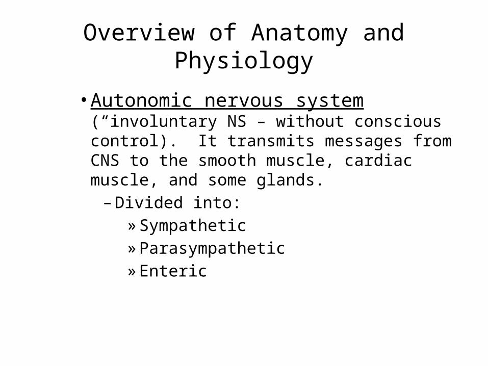
• Autonomic nervous system (“involuntary NS – without conscious control). It transmits messages from CNS to the smooth muscle, cardiac muscle, and some glands.– Divided into:» Sympathetic » Parasympathetic» Enteric

Overview of Anatomy and Physiology
• Cells of the nervous system– Neuron – 2 categories:• Neuron – transmitter cells; carry messages from the
brain and spinal cord• Glial or neuroglial cells- supports and protects neurons
while producing CSF
– Neuron = the basic unit of the nervous system• Composed of 3 parts:– Cell body– Axon– dendrites

Overview of Anatomy and Physiology

Axons and Dendrites
• Branch off the main cell body
• Axons conduct impulses away from the cell body
• Dendrites convey impulses toward the cell body

Overview of Anatomy and Physiology
– Neuromuscular junction• The area of contact between the ends of a large
myelinated nerve fiber and a fiber of skeletal muscle
• SYNAPSE: the “gap” or space between each neuron. Nerve impulses are transmitted across this “gap” or synapse by the action of “neurotransmitters”. – Neurotransmitters• chemicals• Act to make sure that the neurological impulse passess
from the nerve to the muscle

Neurotransmitters
• Functions– Acetylcholine: impulse transmission
– Norepinephrine: Maintaining arousal (from deep sleep), dreaming, mood regulation
– Dopamine: motor function; emotional responses
– Serotonin: induces sleep; controls temp.; mood control; affects sensory perception

Overview of Anatomy and Physiology• Neuron coverings• Many neurons have a white, waxy, fatty material called
“myelin”
• Myelin increases the rate of transmission of impulses and protects and insulates the fibers
• Axons leaving the CNS are wrapped in layers of myelin with indentations called the nodes of Ranvier
• These nodes further increase the rate of transmission because the impulse can jump from node to node

Overview of Anatomy and Physiology

Overview of Anatomy and Physiology
• In the peripheral nervous system, the myelin is protected by Schwann cells– Outer layer of Schwann cells protected by another
layer called “neurilemma” which helps to regenerate injured axonsRegeneration can only happen in the PNS


Cerebrospinal Fluid (CSF)
• Bathes the structures of the CNS
• Composed of water, glucose, sodium chloride, and protein
• Acts as a shock absorber for the brain and spinal cord

CENTRAL NERVOUS SYSTEM
Central Nervous System
• One of two divisions of the nervous system• Composed of the brain and spinal cord• Functions somewhat like a computer but more
complex• Cranium protects the brain• Vertebrae protect the spinal cord
Overview of Anatomy and Physiology
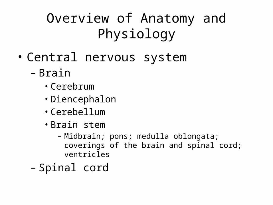
• Central nervous system– Brain• Cerebrum• Diencephalon• Cerebellum• Brain stem
– Midbrain; pons; medulla oblongata; coverings of the brain and spinal cord; ventricles
– Spinal cord

Central Nervous System
• Brain – Specialized cells (in the brain’s mass of convoluted
soft, gray or white tissue) coordinate and regulate the functions of the CNS
– One of the largest organs in the body• Approx 3 lbs
– 4 parts: cerebrum, diencephalon, cerebellum, brainstem

Cerebrum• Largest part of the brain• Divided into: left and right hemispheres
Each hemisphere controls the opposite side of the body: the right hemisphere controls the left side of the body, and the left hemisphere controls the right side of the body
• Outer portion = cerebral cortex (“gray” matter)– Arranged in folds called gyri (convolutions)– Grooves called sulci (fissures)

Cerebrum
• Largest part of the brain
• Divides the two hemispheres into 4 lobes named for the bones over them: frontal, parietal, temporal, and occipital lobes
• Corpus Callosum = the connecting bridge between the two hemispheres

Cerebrum
• Function: controls initiation of movement on the opposite side of the body– Specific areas of the cerebral cortex are associated
with specific functions
*NOTE: see box 14-1 p. 654
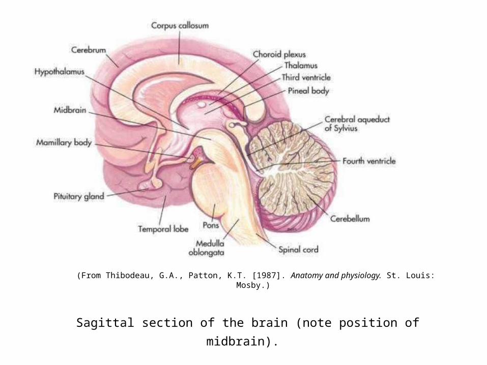
Sagittal section of the brain (note position of midbrain).
(From Thibodeau, G.A., Patton, K.T. [1987]. Anatomy and physiology. St. Louis: Mosby.)

Youtube.com Animation


Figure 28-1
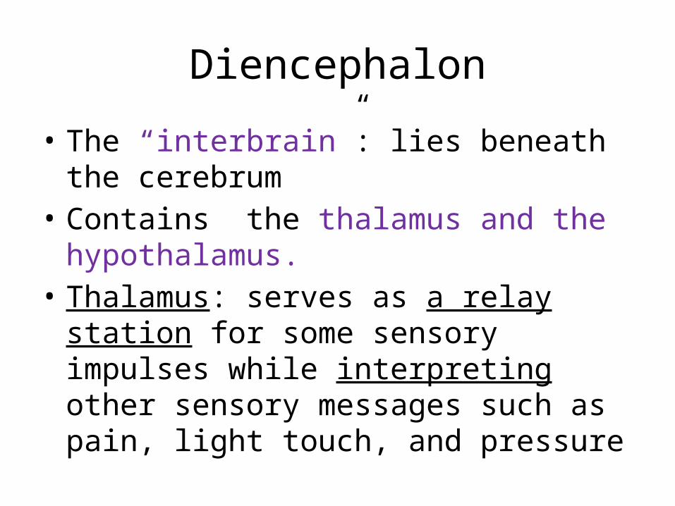
Diencephalon
• The “interbrain”: lies beneath the cerebrum• Contains the thalamus and the hypothalamus.• Thalamus: serves as a relay station for some
sensory impulses while interpreting other sensory messages such as pain, light touch, and pressure

Diencephalon
• Hypothalamus: • Vital role in temperature, fluid balance, appetite,
and emotions such as fear, pleasure, and pain.
• Sympathetic and parasympathetic nervous systems are under the control of the hypothalamus
• Pituitary gland also under control of hypothalamus

Cerebellum (“little brain”)
• Lies posterior and inferior to the cerebrum• The 2nd largest portion of the brain• Also contains 2 hemispheres
• Uses information received from the cerebrum, muscles, joints, and inner ear to coordinate movement, balance, and posture

Cerebellum (“little brain”)
• Unlike the cerebrum, the right side of the cerebellum controls the right side of the body, and the left side of the cerebellum controls the left side of the body

Brainstem
• Located at the base of the brain • Contains the midbrain, pons, and medulla
oblongata• These structures connect the spinal cord and
the cerebrum• Carries all nerve fibers between the spinal
cord and cerebrum
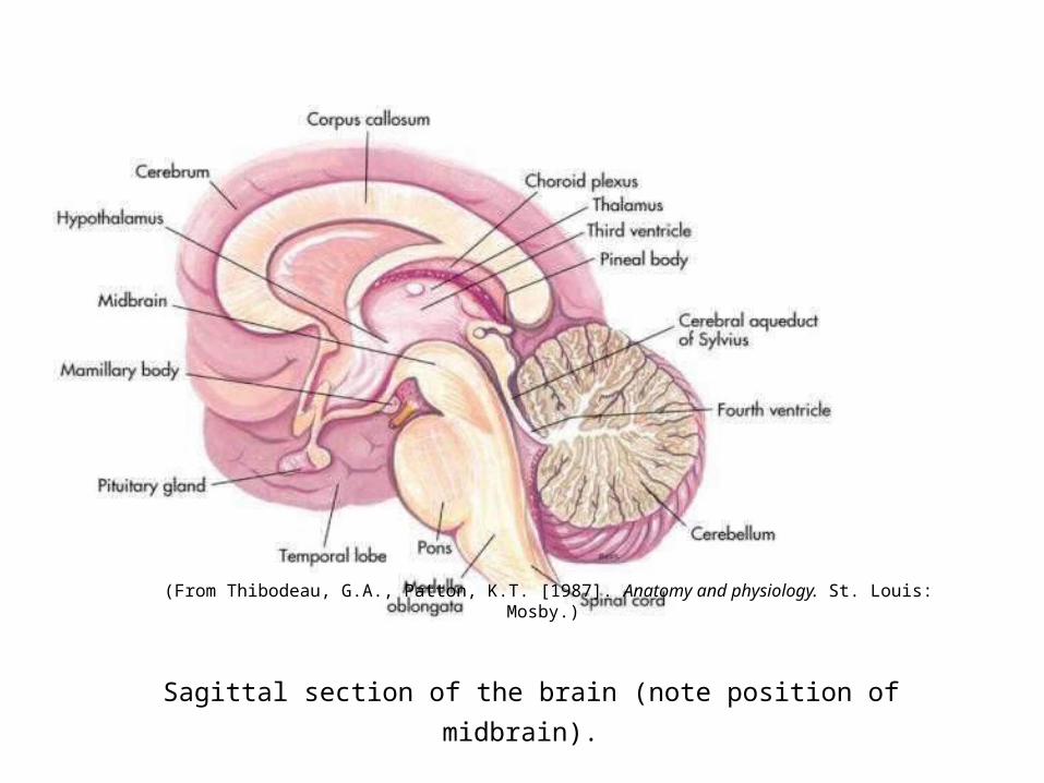
Sagittal section of the brain (note position of midbrain).
(From Thibodeau, G.A., Patton, K.T. [1987]. Anatomy and physiology. St. Louis: Mosby.)

Brainstem
• Midbrain: responsible for motor movement, relay of impulses, auditory and visual reflexes– The origin of cranial nerves III and IV
• Pons (“bridge”): responsible for sending impulses to the structures superior and inferior to it– Contains a respiratory center– The origin of cranial nerves V through VIII

Brainstem
• Medulla Oblongata: controls heartbeat, rhythm of breathing, swallowing, coughing, sneezing, vomiting , hiccups

Protective Structure of the Brain
• Meninges- three layers that surround both the brain and spinal cord1. Dura mater – outermost membrane2. Arachnoid membrane – 2nd layer3. Pia mater – innermost membrane; provides
oxygen and nourishment to the nervous tissue• These layers bathe the spinal cord and brain in
CSF fluid

Ventricles• 4 spaces or cavities located in the brain• CSF flows into the subarachnoid spaces around the
brain and spinal cord and cushions them
• CSF contains protein, glucose, urea, salts
• Also contains substances that form a protective barrier (the blood-brain barrier) that prevents harmful substances from entering the brain or spinal cord

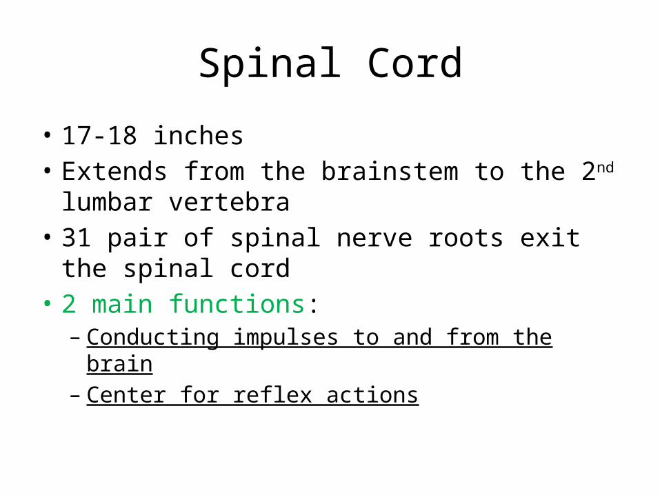
Spinal Cord
• 17-18 inches• Extends from the brainstem to the 2nd lumbar
vertebra• 31 pair of spinal nerve roots exit the spinal cord• 2 main functions:– Conducting impulses to and from the brain– Center for reflex actions


Disks• Vertebrae separated by disks which serve as shock
absorbers for the vertebral column
• Composed of– Anulus fibrosus: ring of tissue; encircles nucleus pulposus – Nucleus pulposus: saclike structure with a gelatinous
filling that has a high water content
• As we age, nucleus pulposus loses much of its water; less effective as a shock absorber

Pyramids and Pyramidal Tracts
• Pyramidal Tracts carry motor information from the CNS neurons to the PNS neurons
• In the medulla of the brainstem, information from one side of the brain “crosses over” and goes down the pyramidal tracts to affect the other side of the body.
• The area where the “crossing over” occurs is the Pyramids

Spinal Cord
• Reflexive Action• Sensory neuron sends information to the cord• A central neuron (located within the cord) interprets
the impulse• Motor neuron sends the message back to the organ or
muscle involvedA message is sent, interpreted and acted upon without
having traveled to the brain– E.g. knee jerk


Ganglia and Nuclei
• Ganglia are information “hubs” of the PNS– Eg. Dorsal root ganglia contain the cell bodies of
sensory neurons• Nuclei are information “hubs” of the CNS– Eg. Basal Ganglia in the brain contains neurons
connecting the cerebral cortex, thalamus, and brainstem. (incorrectly named; should be “Basal Nuclei)

Peripheral Nervous System

Overview of Anatomy and Physiology
• Peripheral nervous system– Spinal nerves (31 pairs)• Mixed nerves: transmit sensory information to the
spinal cord through afferent neurons and • motor information sent to the CNS to the various areas
of the body through efferent neurons
• Named according to the corresponding vertebra– E.g. C1, C2, T12, L3

Overview of Anatomy and Physiology
• Cranial Nerves (12 pairs)• Attach to the posterior surface of the brain, mainly the
brainstem• Conduct impulses between the head, neck, and brain
except the vagus nerve (X) which also serves the organs in the thoracic and abdominal cavities
• More detail: p. 657 Table14-1 Cranial Nerves
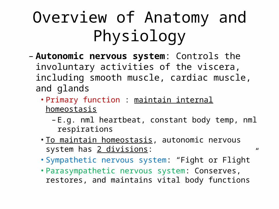
Overview of Anatomy and Physiology
– Autonomic nervous system: Controls the involuntary activities of the viscera, including smooth muscle, cardiac muscle, and glands• Primary function : maintain internal homeostasis– E.g. nml heartbeat, constant body temp, nml
respirations• To maintain homeostasis, autonomic nervous system
has 2 divisions:• Sympathetic nervous system: “Fight or Flight”• Parasympathetic nervous system: Conserves, restores,
and maintains vital body functions

Sympathetic vs Parasympathetic
• These 2 divisions are antagonistic to each other: one slows action; one accelerates action
• These systems function simultaneously but have the ability to dominate when the need arises

Overview of Anatomy and Physiology
• Effects of Normal Aging on the Nervous System– Loss of brain weight– Loss of neurons– Reduction in cerebral blood flow– Decrease in brain metabolism and oxygen
utilization– Decreased blood supply to spinal cord causes
decreased reflexes

Prevention of Neurological Problems
• Modifying lifestyle factors– Reducing risk for cardiac disease– Stop smoking– Avoid drug/alcohol– Safe use of motor vehicles: seat belts, appropriate
speed, helmets for motorcycle riding

Prevention of Neurological Problems
• Modifying Lifestyle factors (cont.)
– Safe swimming practices– Safe handling and storage of firearms– Prompt treatment of infections: sinus, ear– Sexually safe practices

Laboratory and Diagnostic Examinations

Laboratory and Diagnostic Examinations• For the neurologically impaired person:• Urine:– Culture: to rule out infection– Drug screens: to rule out drug use– Glucosuria
• Blood:– Arterial blood gases: monitor the oxygen content
of the blood– Routine lab work

Laboratory and Diagnostic Examinations
• Cerebrospinal fluid: – Checked and cultured if indicated for: infection, protein
(elevated with degenerative diseases and brain tumor)
• Radiographs: – Skull and vertebra: can detect fractures– Used less frequently than CT
• Computed tomography (CT) • Noninvasive examination of the specific levels of the
spinal cord to be visualized, bony vertebrae, and the spinal nerves
Laboratory and Diagnostic Examinations
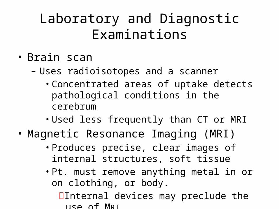
• Brain scan– Uses radioisotopes and a scanner• Concentrated areas of uptake detects pathological
conditions in the cerebrum• Used less frequently than CT or MRI
• Magnetic Resonance Imaging (MRI) • Produces precise, clear images of internal structures,
soft tissue• Pt. must remove anything metal in or on clothing, or
body.Internal devices may preclude the use of MRI

Laboratory and Diagnostic Examinations
• Positron Emmission Tomography Scan (PET)• Injection of radioactive flourine shows glucose
metabolism in the area that is questioned. • Color composite picture is obtained • Level of glucose metabolism can be translated into
indications of a pathologic state

Laboratory and Diagnostic Examinations
• Lumbar puncture: – To obtain CSF for examination, to relieve pressure, or to
introduce dye or medication
Contraindicated in pts. with increased intracranial pressure due to the risk of the medulla oblongata herniating into the foramen magnum during withdrawal of CSF fluid

Figure 14-5
Position and angle of the needle when lumbar puncture is performed.
(From Elkin, M.K., Perry, A.G., Potter, P.A. [2004]. Nursing interventions and clinical skills. [3rd ed.]. St. Louis: Mosby.)

Figure 27-8

Laboratory and Diagnostic Examinations
• Electroencephalogram– EEG– Used to provide evidence of focal or generalized
disturbances of brain function by measuring the electrical activity of the brain
• Angiogram– A procedure used to visualize the cerebral arterial
system by using radiopaque material

Laboratory and Diagnostic Examinations
• Carotid duplex study: – Combines ultrasound and pulsed Doppler technology– Graph measures velocity of blood flow in the carotid
arteries
• Electromyogram (EMG)– Measures the contraction of a muscle in response to an
electrical stimulus– Provides evidence of lower motor neuron disease
• Echoencephalogram– Uses ultrasound to depict intracranial structures

Miscellaneous Slides

NEUROLOGICAL ASSESSMENT
• Cerebral function• Cranial nerve function• Motor function• Sensory function• Reflexes

Assessment of the Neurological System
• History– Comprehensive– From patient; family members/significant other– Symptoms experienced– Pt. understanding and perception of what is
happening– Pp. 691, 692
Assessment of the Neurological System
• Mental status– Orientation, mood, behavior, general knowledge– Short and long-term memory– Attention span and ability to concentrate– Be specific in documentation

Assessment of the Neurological System
• Level of consciousness– The earliest and most sensitive indicator that
something is changing• E.g. A decreasing LOC is the earliest sign of increased
intracranial pressure
– 2 components: arousal (wakefulness); awareness– Observations are recorded in terms of behavior
and signs (not a general “disoriented”)
Assessment of the Neurological System
• Language and speech– Aphasia: language function is defective or absent• Due to injury to frontal lobe and part of temporal lobe• Includes speech, reading, writing, and understanding

Assessment of the Neurological System
– Dysarthria• Difficult, poorly articulated speech• Usually results from interference in the control over the
muscles of speech• Cause is usually damage to a central or peripheral
nerve

Assessment of the Neurological System
• Cranial nerve function– See handouts
• Motor function– Assess gait, stance, muscle tone, coordination,
involuntary movements, and muscle stretch reflexes
– Note bilateral responses

Assessment of the Neurological System
• Motor function cont.– Mobility: • Paralysis = loss of function• Paresis = lesser degree of movement deficit• Flaccid = weak, soft, flabby or lacking normal muscle
tone• Spastic = involuntary sudden movement or muscle
contraction

Assessment of the Neurological System
• Sensory and Perceptual Status– Pain, touch, temperature and proprioception– Proprioception: the sensation pertaining to
spatial-position and muscular-activity stiumuli• Gives one the ability to know the position of the body
without looking at it and to know objects by the sense of touch
– Unilateral neglect: perceptually unaware of and inattentive to one side of the body
– Hemianopia: blindness or defective vision in half of the visual field

Assessment of the Neurological System
• Glasgow coma scale• A quick, practical, standardized system for assessing the
degree of consciousness impairment in the critically ill patient, and
• For predicting the duration and ultimate outcome of coma, particularly with head injuries
• Consists of an assessment of 3 parts:– Eye opening– Best motor response– Best verbal response

