cerebral palsy
DESCRIPTION
Cerebral Palsy. Health Surveillance Christopher Ewing R1 University of Calgary Pediatrics. What is Cerebral Palsy?. Most common cause of chronic neurological impairment in children Permanent, non-progressive brain injury Motor movement and posture impairments as well as disturbances in: - PowerPoint PPT PresentationTRANSCRIPT

Cerebral PalsyHealth Surveillance
Christopher Ewing R1University of CalgaryPediatrics

What is Cerebral Palsy? Most common cause of chronic
neurological impairment in children Permanent, non-progressive brain injury Motor movement and posture impairments
as well as disturbances in: Sensation and perception Cognition and communication Gastrointestinal function and nutrition Social and behavioural Epilepsy

April 2006
Cerebral palsy (CP) describes a group of permanent disorders of the development of movement and posture, causing activity limitation, that are attributed to non-progressive disturbances that occurred in the developing fetal or infant brain. The motor disorders of cerebral palsy are often accompanied by disturbances of sensation, perception, cognition, communication, and behaviour; by epilepsy, and by secondary musculoskeletal problems.

Epidemiology Most common cause of childhood
disability in Western society. Incidence approximately 2-3/1000 births Incidence has not declined despite
improvements in perinatal and obstetrical care.
However more premature and VLBW children with severe disabilities are surviving.

Etiology Often can only be described in 50% of cases Complex and multifactorial May result from structural abnormalities of
the brain or be acquired Prenatal/perinatal/postnatal injury due to:
Vascular insufficiency Toxins Infections Prematurity Birth asphyxia

Differential Diagnosis Progressive:
Glutaric aciduria type 1 Arginase deficiency Sjogren-Larsson syndrome Metachromic leukodystrophy Lesch-Nyhan syndrome Chiari type 1 malformation Dandy-Walker malformation Angelman syndrome Gillespie syndrome Ataxia-Telangectasia Hexoaminidase A and B
deficiency Serotendinosus
xanthomatosis
Non-progressive Intellectual
disability Deprivation Malnutrition Isolated non-motor
and motor handicaps Spina bifida Myopathies
Risk Factors
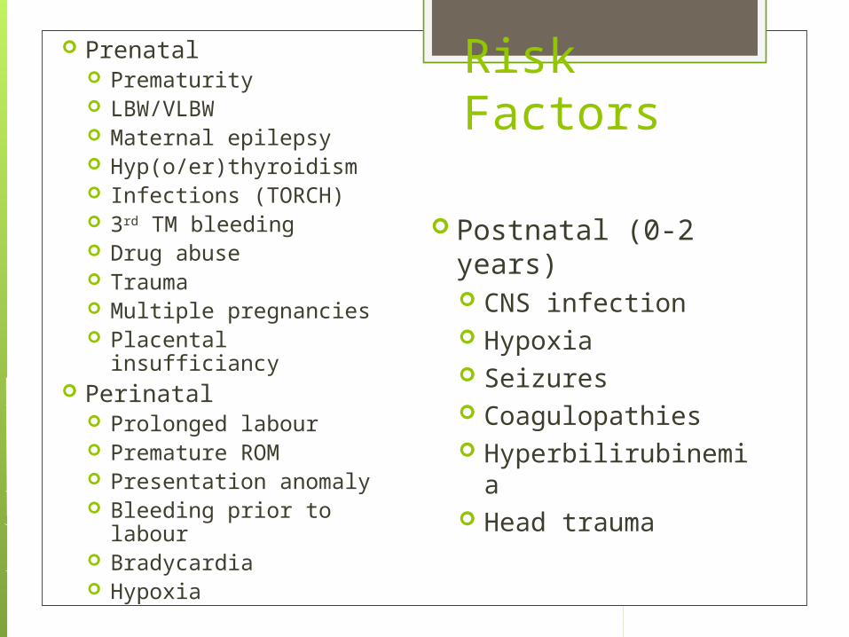
Prenatal Prematurity LBW/VLBW Maternal epilepsy Hyp(o/er)thyroidism Infections (TORCH) 3rd TM bleeding Drug abuse Trauma Multiple pregnancies Placental insufficiancy
Perinatal Prolonged labour Premature ROM Presentation anomaly Bleeding prior to labour Bradycardia Hypoxia
Postnatal (0-2 years) CNS infection Hypoxia Seizures Coagulopathies Hyperbilirubinemia Head trauma

CNS Pathology CP lesions often occur in regions sensitive to
blood supply disturbance. Hypoxic-ischemic-encephalopathy (HIE)

Motor Primary impairments:
Tone (spasticity, dystonia) Balance Strength Loss of selectivity Sensation
Secondary impairments Contractures Deformities
Tertiary impairments (adaptive mechanisms)

Contractures & Deformities Contractures
Upper extremity: pronator, wrist and finger flexors, thumb adductor
Lower extremity: hip adductor/flexor, knee flexor, ankle plantar flexors
Deformities Spine: scoliosis, kyphosis Hip: subluxation, dislocation Femur/tibia: internal/external torsion Foot: equinus, valgus, varus

Natural History Brain lesion is incurable but non-progressive Clinical picture changes as child grows and
develops due to growth and maturation of the CNS and MSK.
Primary movement disorder not evident early on in infancy, although some abnormality may be present
Primitive reflexes persist, and advanced postural reactions fail to develop.
Early intervention can minimize functional effects of neurological impairments.

Early Signs Many infants appear normal Abnormal behaviour
Excessive docility/irritability Poor eye contact or sleep
Oromotor problems Frequent vomiting Poor sucking Tongue retraction or thrusting Grimacing
Poor mobility Poor head control Hand preference, fisting prior to age 2 Abnormal tone, asymmetric posture

Major Deficits in CP Loss of selective motor control, and
dependence on primitive reflex patterns for movement
Abnormal muscle tone influenced by posture, position, and movement
Imbalance between agonist and antagonist muscles that may lead to contractures and deformities
Impaired body balance mechanisms Sensory loss

Associated Problems Seizures Visual impairments Intellectual impairment Learning disabilities Hearing Communication Oromotor dysfunction Gastrointestinal Nutrition Dental Respiratory dysfunction Bladder and bowel Social and emotional
Multidisciplinary Care Pediatrician Physiatrist Developmental pediatrician Orthopedic surgeon Neurologist Speech therapy Physical therapy Occupational therapy Orthotist Dietician Ophthalmologist Audiologist Teacher Social Worker Psychologist

Traditional Classifications Spastic
Hemiplegic Diplegic Quadriplegic
Dyskinetic Choreoathetoid Dystonic
Ataxic

Traditional Classifications

Modern Principles of classification
Motor abnormalities Nature and typology of motor disorder Functional motor abilities
Associated impairments Anatomical distribution Radiological findings Causation and timing

Health Surveillance
Regular primary and specialized care required to optimize the various and potentially progressive and debilitating health issues in cerebral palsy

History Developmental milestones Gross motor function:
head control, sitting, crawling, walking, running
Fine motor function and ADL’s: feeding, dressing, toilet care, hand function
Speech and sensory Pain Social and psychological needs Caregiver burden, financial and community
supports
History Associated impairment surveillance
Seizure frequency and management Bowel and bladder function Communication needs Oromotor function drooling Weight gain Respiratory function Pressure sores School function

Physical Examination Neurological
Mental status, cranial nerves vision, hearing, strength, control, reflexes, tone, involuntary movements, sensory
Musculoskeletal Range of motion, contractures,
deformities, posture Cardiorespiratory
Abdominal Skin breakdown

Musculoskeletal Exam Back
Spinal deformities (scoliosis, kyphosis, lordosis) Pelvis
Obliquity, stability and sitting balance Hip
rotation, flexion/adduction contractures Knee
Patella position, popliteal angle, contractures Foot and ankle
Gastroc/soleus, tibial torsion, equinus/varus/valgus Upper extremity
ROM, grasp, pronator contracture/spasticity, function

Functional Examination Communication Sitting Balance Mobility Gait Running, jumping Hand function Ability to perform ADL’s

Gait Assessments Computerized gait
analysis Kinematics Dynamic EMG Kinetics Energetics
Observation Normal gait Balance Propulsion
Video recording


GMFCS 1. Walks without
restrictions 2. Walks without assistive
devices but limitations in community
3. Walks with assistive devices
4. Transported or uses powered mobility
5. Severely limited, dependent on wheelchair

MACS 1. Handles objects easily and
successfully. 2. Handles most objects but with
somewhat reduced quality and/or speed of achievement.
3. Handles objects with difficulty; needs help to prepare and/or modify activities.
4. Handles a limited selection of easily managed objects in adapted situations.
5. Does not handle objects and has severely limited ability to perform even simple actions.


Imaging
X-ray Cranial ultrasound Cranial CT Cranial MRI EEG

Treatment Goals Infancy
Optimize physical development, nutrition, and exercise
Childhood Maximize independent mobility and nutrition
Preschooler Maximize independent mobility, function,
and minimize deformity Adolescence
Education, vocation and community integration

Treatment Principles Communication Activities of daily living Mobility Ambulation Psychosocial

Management Principles Consider the natural history Appreciate significance of sensation/perception Recognize limitation of treatments Focus on function and comfort, not deformity Provide functional mobility Establish appropriate priorities and shift with
age Emphasize child’s assets Maintain family health Avoid management fads Protect the child’s childhood and play
experience

Rehabilitation Physiotherapy Occupational therapy Bracing Assistive devices Adaptive technology Sports and recreation Environment modification

Rehabilitation Infancy
Educate the family, provide optimal sensorineural development with positioning, stimulation, and exercise.
Childhood Achieve independent mobility,
involvement in self care and ADL’s School age/adolescence
School integration, social, and vocational skills

Bracing Increases function Prevents deformity Keeps joint in functional position Stabilize the trunk and extremities Facilitate selective motor control Decrease spasticity Protect extremity from injury in the postoperative phase

AFO Ankle-Foot Orthotic
Most common brace used in CP Crucial in spastic hemi/diplegia
for mobility Maintains foot in plantigrade Provides ground contact and
foot clearance May prevent contractures if
worn at night Hinged, flexible, or solid

Mobility Aids Transfer aids and lifts Standers Walkers Crutches Canes Wheelchairs
Seating system

Spasticity Part of upper motor
neuron syndrome Increase in physiologic
tone as a resistance to passive movement
Velocity dependent Results in difficulty
with movement, posture, and function
May develop into contractures and deformities

Spasticity Treatment Rehabilitation Botox injection Systemic oral medications
Baclofen, benzos, dantrolene, tizanidine, clonidine
Intrathecal baclofen Surgery


Spasticity Problem Patterns Hemiplegic
Rectus femoris – stiff knee gait Gastrocnemius – pes equinus Arm flexor-pronators – forearm pronation Wrist/digit flexors - thumb in palm, wrist/digit flexion
Diplegic Hip adductor-flexors – hip subluxation Hamstring - knee flexion Gastrocnemius - pes equinus
Quadriplegic Hip adductor-flexors – hip subluxation Hamstring – sacral sitting

Hemiplegic 20% of spastic CP Involvement of unilateral arm and leg Upper extremity involvement > lower
extremity Usually few associated problems, may have
seizures, learning or behavioural issues Common MSK problems:
Forearm pronation, wrist flexion and ulnar deviation, thumb in palm, finger flexion, poor grasp, pes equinus, hemiplegic gait (foot drop, intoeing, knee stiff/flexion)

Diplegic 50% of spastic CP Gross motor involvement of lower extremities
and minor fine motor involvement of upper. Usually normal mental function and
communication Primary problem is walking Common MSK problems:
Hip flexion/IR/adduction and scissoring, ankle equinus, jump/crouch gait, risk of hip instability

Quadriplegic
30% of spastic CP Involvement of neck, trunk, and all four
extremities Severe motor impairment and other signs
and symptoms of CNS dysfunction Cognitive, seizures, speech and swallowing
Frequent comorbid medical associations Higher mortality Common MSK problems
Hip dislocation, spinal and postural problems

Dyskinetic Hyperkinetic/Choreoathetoid
Purposeless, often massive involuntary movements
Poor selective motor control/coordination Dystonic
Abnormal shifts in muscle tone in stereotyped pattern
MSK issues Ambulation dependent on severity, clumsy and
unsteady gait, motor planning and execution, scoliosis, degenerative hip disease

Spine Scoliosis
Most common spinal deformity in CP Progressive, causes difficulty with sitting
and respiratory function Related to severity of CP neuromuscular
involvement Hyperlordosis Hyperkyphosis Pelvic obliquity

Hip Surveillance Hip dislocation is a common concern in
cerebral palsy and can be debilitating Second most common MSK deformity in
children (second to equinus) Directly related to GMFCS Common in spastic CP Progressive instability due to:
Muscle imbalance, persistent primitive reflexes, faulty posture, absence of weight bearing.

Hip Surveillance


Surgery Corrective casting Tendon lengthening Split transfer Simple tenotomy Angular osteotomy Hip surgery Rotational osteotomy Arthrodesis Spinal surgery

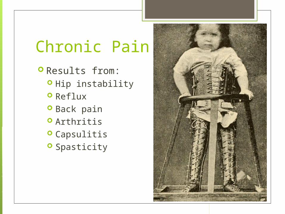
Chronic Pain Results from:
Hip instability Reflux Back pain Arthritis Capsulitis Spasticity

Issues in Adulthood Pain Fractures Contractures Overuse syndromes Pressure sores Scoliosis Sexuality Nutrition Social and vocational

References The HELP Guide To Cerebral Palsy, Second
Edition. Berker, Yalcın. Merrill Corporation, Washington, USA, 2010
Proposed definition and classification of cerebral palsy, April 2005. Paneth et al, Developmental Medicine & Child Neurology 2005, 47: 571-576
Definition and Classification of Cerebral Palsy. Rosenbaum, Paneth, Leviton, Goldstein, Bax. Developmental Medicine & Child Neurology 2006.

References The updated European Consensus 2009 on
the use of Botulinum toxin for children with cerebral palsy. Heinen et al. European Journal of Pediatric Neurology 2010, 14: 45-66
A classification system for hip disease in cerebral palsy. Robin et al. Developmental Medicine & Child Neurology 2009, 51: 183-192
Consensus statement on Hip Surveillance for Children with Cerebral Palsy: Australian Standards of Care 2008. Wynter, Gibson, Kentish, Love, Thomason, Graham.

Appendices and Pictures

Thomas’ Test


