cerebral palsy
DESCRIPTION
CPTRANSCRIPT

Diagnosis, Treatment, and Prevention of Cerebral Palsy in Near-Term/Term Infants
T. Michael O’Shea, MD, MPHWake Forest University School of Medicine
AbstractCerebral palsy is the most prevalent cause of persisting motor function impairment. In a majorityof cases, the predominant motor abnormality is spasticity; other forms of cerebral palsy includedyskinetic (dystonia or choreoathetosis) and ataxic cerebral palsy. The care of individuals withcerebral palsy should include the provision of a primary care medical home for care coordinationand suppor and diagnostic evaluations. Current strategies to decrease the risk of cerebral palsyinclude interventions to prolong pregnancy (e.g., 17α-progesterone), limiting the number ofmultiple gestations related to assisted reproductive technology, antenatal steroids for mothersexpected to deliver prematurely, caffeine for extremely low birth weight neonates, and inducedhypothermia for a subgroup of neonates diagnosed with hypoxic-ischemic encephalopathy.
Keywordscerebral palsy; function impairment; neuroimaging classification; postnatal prevention
IntroductionAmong the variety of disorders that severely impair motor function in young children,cerebral palsy is the most prevalent. In birth cohorts from developed countries, theprevalence is 1-2/1000 live births(1). The prevalence rises dramatically with decreasinggestational age at birth such that among extremely low gestational age newborns (i.e.,gestational age < 28 weeks), the prevalence is about 100 per 1000 of surviving infant, a 100-fold higher risk than infants born at term. As a function of all live births, the prevalence hasbeen remarkably stable for decades, but this has not been the case among very low birthweight and very preterm infants, among whom prevalence increased after the introduction ofneonatal intensive care and has begun to decrease in the past decade(1).
In addition to motor manifestations, children with cerebral palsy frequently exhibit cognitiveand sensory impairments, epilepsy, and nutritional deficiencies. Except in the mildest cases,cerebral palsy has a substantial impact on families’ well being and societal health care costs.
As a result of both laboratory-based and clinical research over the past several decades, afew perinatal interventions have been identified which probably are effective for loweringthe risk of cerebral palsy. Successes from clinical research on cerebral palsy has beenenhanced by efforts to increase the reliability (and thereby validity) of the diagnosis andclassification of this disorder. These efforts have improved the efficiency of observational
Address for correspondence: Michael O’Shea, Department of Pediatrics, Wake Forest University School of Medicine, Medical CenterBlvd, Winston-Salem, NC 27157, Phone: (336)-716-2529, FAX: (336)-716-2525, e-mail address: [email protected] of support: none to report
NIH Public AccessAuthor ManuscriptClin Obstet Gynecol. Author manuscript; available in PMC 2011 March 9.
Published in final edited form as:Clin Obstet Gynecol. 2008 December ; 51(4): 816–828. doi:10.1097/GRF.0b013e3181870ba7.
NIH
-PA Author Manuscript
NIH
-PA Author Manuscript
NIH
-PA Author Manuscript
and experimental epidemiologic studies related to prevention, as well as intervention trialsfor individuals with cerebral palsy.
In this manuscript I will begin by reviewing research on the diagnosis and classification ofcerebral palsy, including efforts to standardize diagnostic criteria, quantify severity in termsof motor impairment and impact on quality of life, and classify children with regard tolocation and type of neurological abnormality, associated non-motor impairments, andneuroimaging findings. I will then discuss the care of individuals particularly, children, withcerebral palsy, with a primary focus on evidence from randomized trials. In concluding Iwill discuss preventive interventions that appear likely to have an impact on the risk ofcerebral palsy or that are currently under study in clinical trials. While some of theseinterventions are not applicable to obstetric care, it is likely that all will be of interest toobstetricians.
Diagnosis of Cerebral PalsyThe diagnosis of cerebral palsy is based on a clinical assessment, and not on laboratorytesting or neuroimaging. A recent international working group offered the followingdefinition for cerebral palsy: “Cerebral palsy is a group of permanent disorders of thedevelopment of movement and posture, causing activity limitation, that are attributed tonon-progressive disturbances that occurred in the developing fetal or infant brain.”(2) Thisdefinition allows for heterogeneity of clinical manifestations (“a group of ... disorders”) andemphasizes that impaired movement and posture due to a disturbance in the brain is theinvariant clinical manifestation. While the underlying abnormality of the brain is presumedto be permanent and non-progressive, there is overwhelming evidence that clinicalmanifestations and severity of functional impairment often change over time. Thus from apatient’s perspective, the term “permanent” may connate an unduly pessimistic prognosis. Incontrast to earlier definitions of cerebral palsy, that proposed by the aforementionedinternational working group included an explicit criterion for the lower limit of abnormalitythat must be exceeded in order to diagnose CP, i.e., “activity limitation”.
On the other hand, this working group did not offer a readily operational approach to theascertainment of activity limitation. The approach that appears to be most widely used isbased on a standardized measure of motor function, the Gross Motor Function ClassificationSystem (GMFCS)(3), which will be discussed below in greater detail. The GMFCS has beenincluded in several recent randomized clinical trials as part of the definition of severity, andin these trials children with mild cerebral palsy without functional impairment were notconsidered to have the outcome of interest. The Western Australian Cerebral Palsy Registergrades severity using the GMFCS, which allows for researchers to include or exclude caseswithout functional impairment. In an international study involving prematurely bornchildren with cerebral palsy, “disabling”, as opposed to “non-disabling” cerebral palsy wasascertained with greater reliability, suggesting that the exclusion of cases without functionalimpairment may improve the validity of observational epidemiologic studies as well asrandomized trials in which cerebral palsy is an outcome. Conversely, clinicians shouldregard with caution those studies of cerebral palsy in which information is lacking about theseverity of cerebral palsy, as assessed with a reliable measure.
In clinical practice, the diagnosis of cerebral palsy is typically based on observations orparent reports of attained motor milestones, such as sitting, pulling to stand, and walking,and evaluation of posture, deep tendon reflexes, and muscle tone. Particularly among infantsborn prematurely, neurological abnormalities, observed in the early months of life, may notbe associated with motor impairment and may resolve during the first one or two years oflife. One such abnormality, transient dystonia, was described in early studies of premature
O’Shea Page 2
Clin Obstet Gynecol. Author manuscript; available in PMC 2011 March 9.
NIH
-PA Author Manuscript
NIH
-PA Author Manuscript
NIH
-PA Author Manuscript
infants and refers to abnormal neurological signs (e.g., hyperextension of the trunk) that areno longer present after one year of age. Because the diagnosis of cerebral palsy depends inpart on neurological findings that are subject to inter-examiner variation, with regard to boththe method used to elicit the neurological finding as well as the interpretation of the finding,and because neurological abnormalities may be transient, many clinicians avoid basing thediagnosis on a single aspect of the parent’s report or the clinician’s examination andtypically will make a definitive diagnosis only after repeated examination(s).
Because cerebral palsy occurs in only 1-2 / 1000 live births, prospective ascertainment ofcases has been limited primarily to cohorts of high risk infants, such as low birth weightbabies, very preterm infants, and infants enrolled in clinical trials. For population-basedstudies, registries of children with cerebral palsy have been used, such as those establishedin Sweden, Western Australia, Metropolitan Atlanta, and Europe. Children listed in theseregistries are identified from records of human services providers and in some studies thediagnosis is confirmed by examination of medical records or of the child. These populationstudies have been particularly informative about trends in prevalence over time.
In many recent studies of high risk infants(4-6), painstaking efforts were directed atstandardizing the definition of cerebral palsy used for prospective identification. With someminor differences between studies, each of these studies base the definition on acombination of delayed motor development (either observed or parent-reported), deeptendon reflexes, posture, and muscle tone (see Figure 1). For more details the reader isdirected to reference(7).
For both clinical as well as research purposes, cerebral palsy has often been classifiedaccording to the nature of the movement disorder (spasticity, ataxia, dystonia, and athetosis)and the anatomic, or topographic distribution of the motor abnormalities. The predominantabnormality among children born prematurely is spasticity, referring to a velocity dependentincrease in muscle tone. Spasticity can be assessed clinically as the joint angle at which anincrease muscle tone (resistance to stretch) is encountered. In a much smaller proportion ofcases, referred to as having dyskinetic cerebral palsy, the predominant abnormality is eitherdystonia or choreo-athetosis. Dystonia refers to hypertonia and reduced activity;choreoathetosis, to irregular, spasmodic, involuntary movements of the limbs or facialmuscles. With ataxic cerebral palsy there is a loss of orderly muscular coordination, so thatmovements are performed with abnormal force, rhythm, and accuracy.
A traditional classification of children with spastic cerebral palsy includes spastic diplegia(bilateral spasticity with leg involvement greater than arm), spastic hemiplegia (unilateralspasticity), or quadriplegia (bilateral spasticity with arm involvement equal to or greater thanleg). In a large population-based study of very low birth weight children with cerebral palsy,25% of children with spastic CP had hemiplegia, 37.5% had quadriplegia, and 37.5% haddiplegia. Children with hemiplegia almost always develop independent ambulation, whereasa majority of those with quadriplegia do not. There is some evidence that the profile of riskfactors differs for each of these topgraphic types of cerebral palsy. It should be noted,however, that the inter-rater reliability of examiners’ topographic classification is not high;thus the Surveillance for Cerebral Palsy in Europe (SCPE), a multi-center researchcollaboration, has proposed an alternative classification that includes unilateral spasticcerebral palsy, bilateral spastic cerebral palsy, dystonic cerebral palsy, choreo-athetoidcerebral palsy, and ataxia (see Figure 2). Currently no method of topographic classificationhas been broadly agreed upon.
More agreement has been achieved on classification of cerebral palsy in terms of thefunctional severity. More than a decade ago, Palisano and his colleagues developed the
O’Shea Page 3
Clin Obstet Gynecol. Author manuscript; available in PMC 2011 March 9.
NIH
-PA Author Manuscript
NIH
-PA Author Manuscript
NIH
-PA Author Manuscript
GMFCS, which defines five levels of gross motor function, which have been shown tocorrespond to five distinct “trajectories” of motor development. For example, amongchildren younger than 2 years, those at level II can “maintain floor sitting but may need touse their hands for support to maintain balance,” whereas those at level IV have head controlbut trunk support is required for floor sitting”. The GMFCS correlates strongly with theWorld Health Organization International Classification of Impairments, Disabilities andHandicap code, but is considerably less time-consuming and can be derived from medicalrecords. The inter-rater reliability of the GMFCS is high, as is the stability over time. TheGMFCS level of children, as reported by parents, agrees well with that assigned by aphysician; and the level self-reported by adults agrees well with that assigned by a physicaltherapist..
Other assessments have been developed for fine motor function, including the ABILHAND-Kids, the Bimanual Fine Motor Function (BFMF) Classification, and the Manual AbilityClassification System. These assessments have not been studied as extensively as theGMFCS, but in one study the BFMF correlated strongly with the GMFCS, and in another, ahigh level of agreement about the MACS was found between pairs of therapists. Thus acomprehensive description of an individual with cerebral palsy would include both theGMFCS as well as a measure of upper extremity (fine motor) function.
In a large proportion of individuals with cerebral palsy, the motor impairment isaccompanied by secondary musculoskeletal problems, epilepsy, and disturbances ofsensation, perception, cognition, communication, and behavior(8). Among childrenregistered in the SCPE Collaboration, 31% have severe intellectual disability, 11% havesevere visual disability, and 21% have epilepsy. The likelihood of associated impairmentsvaries according to topographic type. Less than one half of children with leg dominantbilateral spastic cerebral palsy have mental retardation, which is present in more than onehalf of children with other forms of spastic cerebral palsy. Severe visual impairment ispresent in more than one half of children with four leg dominant spastic CP but only lessthan 10% of children with two leg dominant spastic cerebral palsy.
Quality of life has been defined as “an individual’s perception of their position in life in thecontext of the culture and value systems in which they live, and in relation to their goals,expectations, standards and concerns.” Using validated instruments which allow children toself-report on their quality of life, researchers have found that most children with cerebralpalsy report similar quality of life to children not affected with this disorder and that qualityof life is not worse with greater levels of functional impairment(9). In contrast, healthrelated quality of life, which measures life satisfaction in areas such as self-care, mobility,and communication is influenced by the severity of impairment.
Neuroimaging classificationUntil recently, neuroimaging studies of preterm infants with cerebral palsy were basedlargely on neonatal cranial ultrasound. The most widely used classification system, derivedfrom computed tomography but applied readily to ultrasound findings, was described byPapile et al. In this approach to classification, grade 1 refers to hemorrhage limited to thesubependymal germinal matrix region, grade 2 refers to hemorrhage in the cerebralventricule(s), grade 3 refers to hemorrhage in the ventricle(s) with ventricular enlargement,and grade 4 refers to hemorrhage in the periventricular cerebrum. More recent approaches toclassification, justified by extensive research, emphasize echolucency in the periventricularcerebrum and moderate-to-severe ventricular enlargement as most predictive of subsequentcerebral palsy(10). Very low birth weight infants with either of these abnormalities have agreater than 50% risk of developing cerebral palsy. For more details see reference (11)
O’Shea Page 4
Clin Obstet Gynecol. Author manuscript; available in PMC 2011 March 9.
NIH
-PA Author Manuscript
NIH
-PA Author Manuscript
NIH
-PA Author Manuscript
Most children who are born at term and subsequently develop cerebral palsy do not undergoneuroimaging studies as neonates, but rather, after the diagnosis is suspected. Magneticresonance imaging (MRI) is recommended for children with neurological findingssuggestive of cerebral palsy in order to determine if a brain abnormality is present. In astudy of 273 children with cerebral palsy who were born after 35 weeks gestation andunderwent neuroimaging, one third of infants who underwent neuroimaging had normalstudies(12). The most frequent observed abnormality was focal infarction, which wasobserved in 22% of the children and 45% of those with hemiplegia. The next most commonfinding was brain malformation, including schizencephaly, hydrocephalus, polymigrogyria,lissenencephaly, agenesis of the cropus callosum, holoprosencephaly, septooptic dysplasia,and cerebellar anomalies. In 12% of children neuroimaging revealed periventricularleukomalacia, a finding often is associated with prematurity. Of note is that only 5% ofchildren had neuroimaging findings considered specific to hypoxia-ischemia(12).
Care of the child with cerebral palsyInitial assessments (see Figure 3) for children diagnosed with cerebral palsy includeneuroimaging if the etiology has not been established and metabolic and genetic studies ifclinical history and findings on neuroimaging do not determine a specific etiology or if thereare aspects of the history or physical examination that suggestive of a metabolic or geneticetiology (including a brain malformation). In addition, testing for prothromboticabnormalities of coagulation should be considered in individuals with hemiplegia. Allchildren with cerebral palsy should be screened for mental retardation, ophthalmologic andhearing impairment, and speech and language disorders, and nutrition and growth should bemonitored(13).
The American Academy of Pediatrics has emphasized the importance of a primary care“medical home” for children with chronic illnesses, including cerebral palsy(8). The roles ofthe medical home include care coordination to minimize redundant services and assurecomprehensive services, monitoring of response to treatments and impact of the child’sillness on the family, interpreting findings to the family, orchestrating comanagment withspecialists and specialty teams, and advocating for the patient with payers and providerssuch as the public schools(8). (Beginning in 1975, a series of United States public laws hasspecified that all handicapped children have available to them a free appropriate educationdesigned to meet their unique needs. The most recent of these is Public Law 108-446, theIndividuals with Disabilities Education Improvement Act of 2004.)
As discussed above, a large majority of children with cerebral palsy have spasticity. Activemanagement of spasticity is needed to prevent painful contractures and deformities andpromote optimal function. This management generally is provided by multidisciplinaryteams that include physical therapists, orthopedic surgeons, and physiatrists. Physicaltherapy, which is uniformly utilized for cerebral palsy has not been subjected to randomizedtrials, but is widely accepted as a component of standard management. Table 1 describes theresults of systematic reviews for five therapies used in the management of spasticity inpatients with cerebral palsy. Those provided by the Cochrane Collaboration can be assessedat http://www.mrw.interscience.wiley.com/cochrane. For each of these therapies systematicreviews have provided evidence of benefit. Botulinum toxin type A for management ofspasticity and casting for management of equinus deformity of the foot are probably themost widely available and used. Dorsal rhizotomy is a more invasive approach to themanagement of spastic diplegia and is reserved for the more difficult cases.
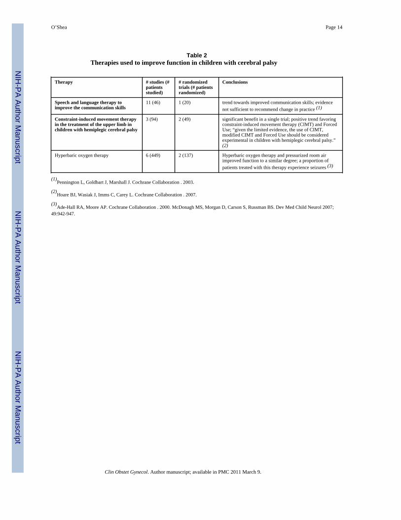
Other therapies to improve function in cerebral palsy are listed in Table 2. Therapies specificfor hemiplegia, such as constraint-induced movement therapy (CIMT) and hand-arm
O’Shea Page 5
Clin Obstet Gynecol. Author manuscript; available in PMC 2011 March 9.
NIH
-PA Author Manuscript
NIH
-PA Author Manuscript
NIH
-PA Author Manuscript

bimanual intensive therapy (HABIT), have been studied in a relatively small number ofindividuals. With CIMT, the non-involved upper extremity is restrained and the involvedextremity is engaged in targeted practice. With HABIT, structured tasks requiring bimanualfunction are practiced in the context of play and functional activities. While more study iswarranted, these appear to hold promise of benefit. More research on the effects ofhyperbaric oxygen therapy is required, as there is considerable uncertainty about itsefficiency and adverse effects, and this treatment costs approximately $400 per 90-minutesession.
PrognosisAs discussed above, prognosis for independent ambulation depends in large part on the typeof motor impairment. Ambulation status, intelligence quotient, quality of speech, and handfunction together are predictive of employment status. For example, in a study of adults withcerebral palsy, intelligence quotient ≥ 80, and understandable speech, who were ambulatoryand independent of the need for “significant assistance”, 90% were employed in a“competitive job” (i.e., one which could be filled by a similarly qualified person without adisability).
Mortality also is strongly associated with both the level of functional impairment as well asassociated non-motor impairments. In one study of over 2014 individuals with cerebralpalsy, the strongest predictor of mortality was intellectual disability. For example, amongthose with profound intellectual disability (i.e., IQ < 20), only one half survived intoadulthood; whereas among those with IQ > 35, 92% survived to adulthood. More generally,mortality risk increases incrementally with increasing number of impairments, includingintellectual, limb function, hearing, and vision. In a recent population-based study, theshortest life expectancy was observed among those individuals who were unable to lift theirhead in prone, who had a life expectancy of 20 years.
Antenatal approaches to preventionTo an alarming extent, cerebral palsy is mistakenly attributed to acts of omission orcommission by obstetricians. Among the most important lines of research during the pasttwo decades is the investigation of the role of perinatal infection and inflammation inacquired brain damage in both term and preterm fetuses and newborns(7;14), and the role ofprothrombotic factors and other causes of neonatal stroke in the pathogenesis of congenitalhemiplegia(15). Although epidemiologic studies indicate that less than 10% of cerebralpalsy results from intrapartum hypoxia, three quarters of United States obstetricians reportbeing the subject of a litigation event, most frequently related to their allegedly causingcerebral palsy by not preventing or treating fetal hypoxia(16). In a study of 46 malpracticecases, investigators concluded that “the severity of the patient’s disability, not theoccurrence of an adverse event or an adverse event due to negligence, was predictive ofpayment to the plaintiff”(17).
Many assumptions about the effectiveness of obstetric care for preventing cerebral palsygenerally are based on weak or no evidence(16). Published checklists provide a moreevidence-based approach to attribution(18;19). A intrapartum event as a cause of cerebralpalsy is more likely if significant fetal acidosis (such as pH < 7.0) and neonatalencephalopathy are observed, and is more likely with spastic quadriplegia or dyskineticcerebral palsy. While these events might be the result of intrapartum hypoxia, they mightalso be the result of fetal infection. Thus there is almost always considerable uncertainty asto the cause of cerebral palsy, particularly among infants born at or near term.
O’Shea Page 6
Clin Obstet Gynecol. Author manuscript; available in PMC 2011 March 9.
NIH
-PA Author Manuscript
NIH
-PA Author Manuscript
NIH
-PA Author Manuscript
Nonetheless, because approximately one-half of all new cases of cerebral palsy arise fromthe group of neonates born prematurely, it is possible that interventions which either prolonggestation or decrease the risk of preterm delivery will also decrease the risk of cerebralpalsy. Specific approaches to reduce the rate of preterm birth, that are supported by a highlevel of evidence, include limiting the number of embryos transferred with in vitrofertilization, smoking cessation during pregnancy, screening for and treatment ofasymptomatic bacteriuria during pregnancy, antiplatelet drugs to prevent preeclampsia, 17α-progesterone caproate, and cervical cerclage for women with previous preterm birth andshort cervix (i.e., < 2.5 centimeters)(20). In addition, interventions which have been shownto prolong pregnancy include calcium channel blockers and an oxytocin antagonist(atosiban) for women with preterm labor and erythromycin for women with prematurerupture of the membranes(20). In addition to these measures, the results of a meta-analysisof four trials suggests that treatment of mothers expected to deliver before 36 weeksgestation with glucocorticoids (eg. β-methasone) reduces the risk of cerebral palsy. In theAustralasian Collaborative Trial of Magnesium Sulphate, magnesium sulfate treatment ofmothers at risk for preterm birth before 30 weeks gestation reduced the risk of substantialgross motor dysfunction, and in a multicenter randomized trial in France a trend towards areduction in cerebral white matter damage was observed. More information about thepossible beneficial of antenatal magnesium sulfate is expected soon from the NationalInstitutes of Health Maternal Fetal Medicine Network trial (Benefits of AntenatalMagnesium Sulfate).
Postnatal approaches to preventionMost term or near-term infants who develop cerebral palsy have uneventful neonatalcourses. A notable exception are those who have neonatal encephalopathy. One of thepresumed causes of neonatal encephalopathy is intrapartum cerebral hypoxia and ischemia,which in severe cases could result in permanent brain damage manifesting as cerebral palsy.In such infants, hypothermia, either selectively applied to the head or total body, appears todecrease the risk of neurodevelopmental impairments, including cerebral palsy(5). Whilethis intervention appears to be effective, it is applicable to only a small proportion ofchildren who subsequently develop cerebral palsy.
In preterm infants, caffeine is the only therapy that has been shown in a multicenter trial todecrease the risk of cerebral palsy. In this trial, extremely low birth weight infants wererandomized to caffeine or placebo in the first days of life. While the results might apply onlyto a small proportion of preterm infants, it is this subgroup that is at highest risk of cerebralpalsy. Postnatal steroids, given to premature infants to decrease lung inflammation anddecrease the risk of bronchopulmonary dysplasia, increase the risk of cerebral palsy. Thuslimiting the use of this treatment can be expected to reduce the risk of cerebral palsy. Thegoal of recently completed trial by the National Institutes of Health Neonatal ResearchNetwork was to study the effect of aggressive phototherapy for hyperbilirubinemia onneurodevelopmental outcome. The results of this trial may be relevant to the prevention ofcerebral palsy in extremely low birth weight infants.
SummaryCerebral palsy is relatively common cause of persistent motor impairment and is associatedwith a variety of developmental disabilities. In developing countries, approximately one halfof cases occur in prematurely born infants, and in this group the prevalence of cerebral palsyin developed countries appears to be decreasing over the past decade. In full term infants,the prevalence has been remarkably stable despite the advent of continuous fetal heart ratemonitoring. The care of children with cerebral palsy requires a multidisciplinary,
O’Shea Page 7
Clin Obstet Gynecol. Author manuscript; available in PMC 2011 March 9.
NIH
-PA Author Manuscript
NIH
-PA Author Manuscript
NIH
-PA Author Manuscript

comprehensive, and coordinated approach. In developed countries, the self-reported qualityof life for adolescents with cerebral palsy, except for severe cases, is similar to that ofadolescents without cerebral palsy. Interventions that hold promise for reducing theprevalence of cerebral palsy include interventions to decrease the risk of premature birth,antenatal steroids given to mothers expected to deliver prematurely, treatment of motherswho are expected to deliver prior to 30 weeks gestation with magnesium sulfate,hypothermia for neonates diagnosed with hypoxic-ischemic encephalopathy, and caffeinefor extremely low birth weight infants.
Reference List(1). Paneth N, Hong T, Korzeniewski S. The descriptive epidemiology of cerebral palsy. Clinics in
Perinatology 2006;33(2):251. + [PubMed: 16765723](2). Rosenbaum P. A report: the definition and classification of cerebral palsy April 2006 (vol 49, pg 8,
2007). Developmental Medicine and Child Neurology 2007;49(6):480.(3). Rosenbaum P, Walter SD, Hanna SE, Palisano R, Russell DJ, Raina P, et al. Prognosis for gross
motor function in cerebral palsy. JAMA 2002;288(11):1357–1363. [PubMed: 12234229](4). Surveillance of Cerebral Palsy in Europe (SCPE). Surveillance of cerebral palsy in Europe: a
collaboration of cerebral palsy surveys and registers. Dev Med Child Neurol 2000;42:816–824.[PubMed: 11132255]
(5). Shankaran S, Laptook AR, Ehrenkranz RA, Tyson JE, McDonald SA, Donovan EF, et al. Whole-body hypothermia for neonates with hypoxic-ischemic encephalopathy. New England Journal ofMedicine 2005;353(15):1574–1584. [PubMed: 16221780]
(6). Kuban KCK, O’Shea M, Allred E, Leviton A, Gilmore H, DuPlessis A, et al. Video and CD-ROMas a training tool for performing neurologic examinations of 1-year-old children in a multicenterepidemiologic study. Journal of Child Neurology 2005;20(10):829–831. [PubMed: 16417880]
(7). O’Shea TM. Cerebral palsy in very preterm infants: New epidemiological insights. MentalRetardation and Developmental Disabilities Research Reviews 2002;8(3):135–145. [PubMed:12216057]
(8). Cooley WC. Providing a primary care medical home for children and youth with cerebral.Pediatrics 2004;114(4):1106–1113. [PubMed: 15466117]
(9). Dickinson HO, Parkinson KN, Ravens-Sieberer U, Schirripa G, Thyen U, Arnaud C, et al. Self-reported quality of life of 8-12-year-old children with cerebral palsy: a cross-sectional Europeanstudy. Lancet 2007;369(9580):2171–2178. [PubMed: 17604799]
(10). Leviton A, Kuban K, Paneth N. Intraventricular haemorrhage grading scheme: time to abandon?Acta Paediatrica 2007;96(9):1254–1256. [PubMed: 17590196]
(11). Hintz SR, O’Shea M. Neuroimaging and neurodevelopmental outcomes in preterm infants.Semin Perinatol 2008;32(1):11–19. [PubMed: 18249235]
(12). Wu YW, Shah SJ, Newman TB, Najjar DV, Croen LA. Cerebral palsy in a term population: Riskfactors and neuroimaging findings. Annals of Neurology 2006;60:S115.
(13). Ashwal S, Russman BS, Blasco PA, Miller G, Sandler A, Shevell M, et al. Practice parameter:Diagnostic assessment of the child with cerebral palsy - Report of the Quality StandardsSubcommittee of the American Academy of Neurology and the Practice Committee of the ChildNeurology Society. Neurology 2004;62(6):851–863. [PubMed: 15037681]
(14). Nelson KB. The epidemiology of cerebral palsy interm infants. Mental Retardation andDevelopmental Disabilities Research Reviews 2002;8(3):146–150. [PubMed: 12216058]
(15). Hunt RW, Inder TE. Perinatal and neonatal ischaemic stroke: A review. Thrombosis Research2006;118(1):39–48. [PubMed: 16709474]
(16). Hankins GDV, MacLennan AH, Speer ME, Strunk A, Nelson K. Obstetric litigation isasphyxiating our maternity services. Obstetrics and Gynecology 2006;107(6):1382–1385.[PubMed: 16738167]
(17). Brennan TA, Sox CM, Burstin HR. Relation between negligent adverse events and the outcomesof medical-malpractice litigation. New England Journal of Medicine 1996;335(26):1963–1967.[PubMed: 8960477]
O’Shea Page 8
Clin Obstet Gynecol. Author manuscript; available in PMC 2011 March 9.
NIH
-PA Author Manuscript
NIH
-PA Author Manuscript
NIH
-PA Author Manuscript

(18). Di Tommaso M, Tranquilli AL. A checklist to identify the origin of cerebral palsy. Journal ofMaternal-Fetal and Neonatal Medicine 2006;15:281–286. [PubMed: 15280116]
(19). MacLennan A. A template for defining a causal relation between acute intrapartum events andcerebral palsy: international consensus statement. British Medical Journal 1999;319(7216):1054–1059. [PubMed: 10521205]
(20). Iams JD, Romero R, Culhane JF, Goldenberg RL. Preterm birth 2 - Primary, secondary, andtertiary interventions to reduce the morbidity and mortality of preterm birth. Lancet2008;371(9607):164–175. [PubMed: 18191687]
O’Shea Page 9
Clin Obstet Gynecol. Author manuscript; available in PMC 2011 March 9.
NIH
-PA Author Manuscript
NIH
-PA Author Manuscript
NIH
-PA Author Manuscript

Figure 1.Decision tree for inclusion/exclusion of cases of cerebral palsy on SCPE register.Reproduced with permission from Surveillance of Cerebral Palsy in Europe (SCPE).Surveillance of cerebral palsy in Europe: a collaboration of cerebral palsy surveys andregisters. Dev Med Child Neurol 2000; 42: 816-824
O’Shea Page 10
Clin Obstet Gynecol. Author manuscript; available in PMC 2011 March 9.
NIH
-PA Author Manuscript
NIH
-PA Author Manuscript
NIH
-PA Author Manuscript
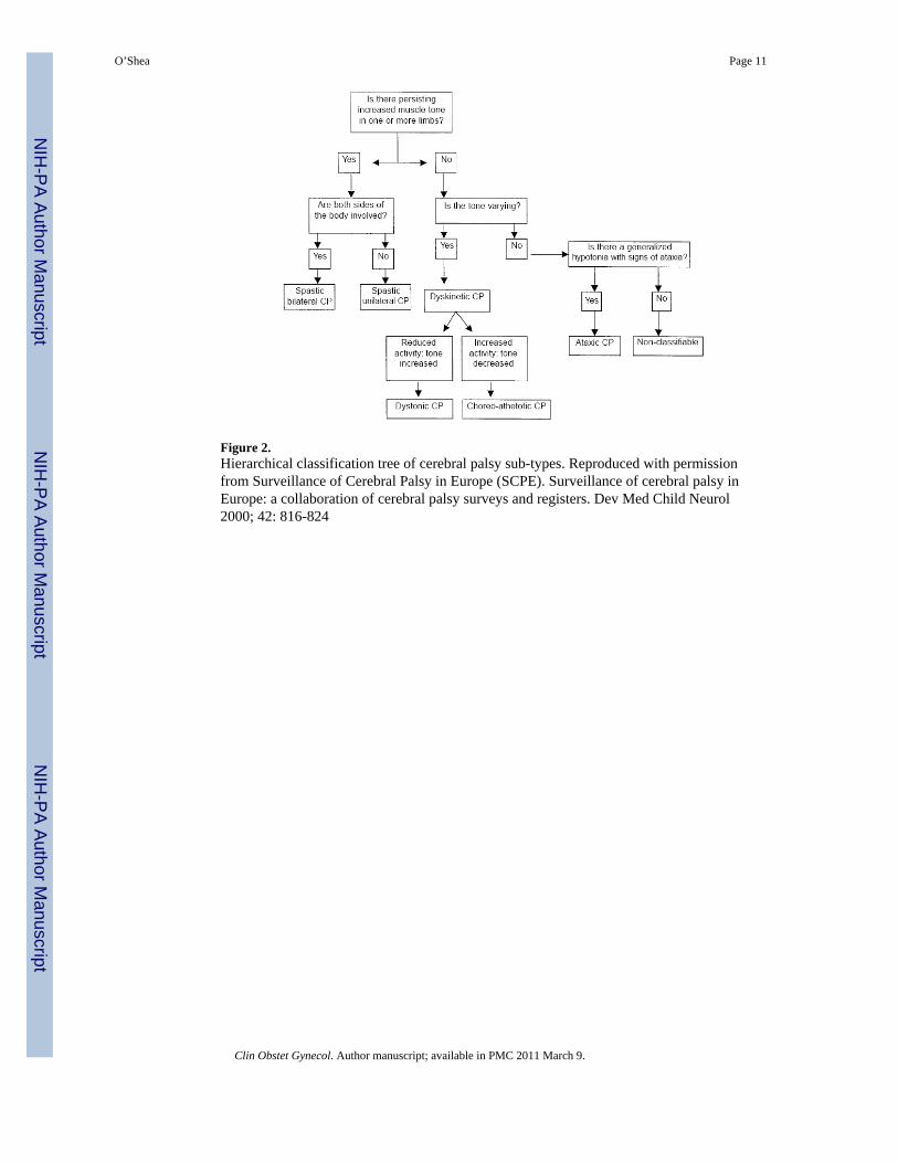
Figure 2.Hierarchical classification tree of cerebral palsy sub-types. Reproduced with permissionfrom Surveillance of Cerebral Palsy in Europe (SCPE). Surveillance of cerebral palsy inEurope: a collaboration of cerebral palsy surveys and registers. Dev Med Child Neurol2000; 42: 816-824
O’Shea Page 11
Clin Obstet Gynecol. Author manuscript; available in PMC 2011 March 9.
NIH
-PA Author Manuscript
NIH
-PA Author Manuscript
NIH
-PA Author Manuscript
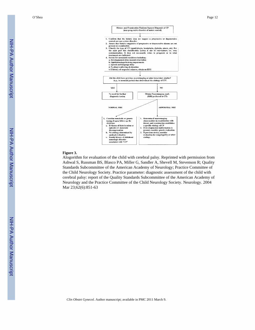
Figure 3.Alogorithm for evaluation of the child with cerebral palsy. Reprinted with permission fromAshwal S, Russman BS, Blasco PA, Miller G, Sandler A, Shevell M, Stevenson R; QualityStandards Subcommittee of the American Academy of Neurology; Practice Committee ofthe Child Neurology Society. Practice parameter: diagnostic assessment of the child withcerebral palsy: report of the Quality Standards Subcommittee of the American Academy ofNeurology and the Practice Committee of the Child Neurology Society. Neurology. 2004Mar 23;62(6):851-63
O’Shea Page 12
Clin Obstet Gynecol. Author manuscript; available in PMC 2011 March 9.
NIH
-PA Author Manuscript
NIH
-PA Author Manuscript
NIH
-PA Author Manuscript

NIH
-PA Author Manuscript
NIH
-PA Author Manuscript
NIH
-PA Author Manuscript
O’Shea Page 13
Table 1
Therapies used to manage spasticity in cerebral palsy
Therapy # studies (#patientsstudied)
# randomizedtrials (#patientsrandomized)
Conclusions
Botulinum toxin type A in thetreatment of lower limbspasticity
3 (52) 3 (52) no strong controlled evidence found to support or refute the useof Botulinum toxin type A for the treatment of leg spasticity(1)
Botulinum toxin type A fortreatment of spastic equinus foot
Not provided 4 (183) Botulinum toxin type A superior to placebo forimprovement of gait (2)
Botulinum toxin A as an adjunctto treatment in the managementof the upper limb in childrenwith spastic cerebral palsy
2 (44) 2 (44) one of the two randomized trials reported promising results insupport of reduced muscle tone following Botulinum toxin Ainjections; evidence not sufficient to support or refute the useof this therapy as an adjunct to managing the upper limb inchildren with spastic cerebral palsy (3)
selective’ dorsal rhizotomy plusphysiotherapy
3 (90) 3 (90) selective’ dorsal rhizotomy (SDR) plus physiotherapy reducesspasticity in children with spastic diplegia slightly improvesgross motor function (4)
casting for equinus 21 (473) 9 (238) No randomized trials available comparing protocols of castingin current use with no treatment; no strong and consistentevidence that combining casting and Botulinum toxin A issuperior to using either intervention along; no evidencethat the order of these two treatments affects outcome (5)
(1)Ade-Hall RA, Moore AP. Cochrane Collaboration . 2000.
(2)Cardoso ES, Rodrigues BM, Barroso M, Menezes CJ, Lucena RS, Nora DB et al. Ped Neurol 2006; 34:106-109.
(3)Wasiak J, Hoare B, Wallen M. Cochrane Collaboration . 2004.
(4)McLaughlin J, Bjornson K, Temkin N, Steinbok P, Wright V, Reiner A et al. Dev Med Child Neurol 2002; 44:17-25.
(5)Blackmore AM, Boettcher-Hunt E, Jordan M, Chan MDY. Dev Med Child Neurol 2007; 49:781-790.
Clin Obstet Gynecol. Author manuscript; available in PMC 2011 March 9.
NIH
-PA Author Manuscript
NIH
-PA Author Manuscript
NIH
-PA Author Manuscript
O’Shea Page 14
Table 2Therapies used to improve function in children with cerebral palsy
Therapy # studies (#patientsstudied)
# randomizedtrials (# patientsrandomized)
Conclusions
Speech and language therapy toimprove the communication skills
11 (46) 1 (20) trend towards improved communication skills; evidencenot sufficient to recommend change in practice (1)
Constraint-induced movement therapyin the treatment of the upper limb inchildren with hemiplegic cerebral palsy
3 (94) 2 (49) significant benefit in a single trial; positive trend favoringconstraint-induced movement therapy (CIMT) and ForcedUse; “given the limited evidence, the use of CIMT,modified CIMT and Forced Use should be consideredexperimental in children with hemiplegic cerebral palsy.”(2)
Hyperbaric oxygen therapy 6 (449) 2 (137) Hyperbaric oxygen therapy and pressurized room airimproved function to a similar degree; a proportion ofpatients treated with this therapy experience seizures (3)
(1)Pennington L, Goldbart J, Marshall J. Cochrane Collaboration . 2003.
(2)Hoare BJ, Wasiak J, Imms C, Carey L. Cochrane Collaboration . 2007.
(3)Ade-Hall RA, Moore AP. Cochrane Collaboration . 2000. McDonagh MS, Morgan D, Carson S, Russman BS. Dev Med Child Neurol 2007;
49:942-947.
Clin Obstet Gynecol. Author manuscript; available in PMC 2011 March 9.