cerebrovascular consequences of obstructive...
TRANSCRIPT
Cerebrovascular Consequences of Obstructive Sleep ApneaDavid J. Durgan, PhD; Robert M. Bryan, Jr, PhD
O bstructive sleep apnea (OSA) is defined by interruptedbreathing during sleep due to airway obstruction with
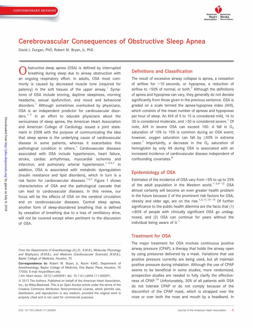
an ongoing respiratory effort. In adults, OSA most com-monly is caused by decreased muscle tone (required forpatency) in the soft tissues of the upper airway.1 Symp-toms of OSA include snoring, daytime sleepiness, morningheadache, sexual dysfunction, and mood and behavioraldisorders.1 Although sometimes overlooked by physicians,OSA is an independent predictor for cardiovascular disor-ders.1–7 In an effort to educate physicians about theseriousness of sleep apnea, the American Heart Associationand American College of Cardiology issued a joint state-ment in 2008 with the purpose of communicating the ideathat sleep apnea is the underlying cause of cardiovasculardisease in some patients, whereas it exacerbates thispathological condition in others.1 Cardiovascular diseasesassociated with OSA include hypertension, heart failure,stroke, cardiac arrhythmias, myocardial ischemia andinfarction, and pulmonary arterial hypertension.1–3,5,7 Inaddition, OSA is associated with metabolic dysregulation(insulin resistance and lipid disorders), which in turn is arisk factor for cardiovascular diseases.1,3,7 Figure 1 showscharacteristics of OSA and the pathological cascade thatcan lead to cardiovascular diseases. In this review, ourfocus will be the effects of OSA on the cerebral circulationand on cerebrovascular diseases. Central sleep apnea,another form of sleep-disordered breathing that is definedby cessation of breathing due to a loss of ventilatory drive,will not be covered except when pertinent to the discussionof OSA.
Definitions and ClassificationThe result of excessive airway collapse is apnea, a cessationof airflow for >10 seconds, or hypopnea, a reduction ofairflow to <50% of normal, or both.7 Although the definitionsof apnea and hypopnea can vary, they generally do not deviatesignificantly from those given in the previous sentence. OSA isgraded on a scale termed the apnea-hypopnea index (AHI),which consists of the mean number of apneas and hypopneasper hour of sleep. An AHI of 5 to 15 is considered mild, 16 to30 is considered moderate, and >30 is considered severe.1 Ofnote, AHI in severe OSA can exceed 100. A fall in O2
saturation of 10% to 15% is common during an OSA event;however, oxygen saturation can fall by ≥50% in extremecases.1 Importantly, a decrease in the O2 saturation ofhemoglobin by only 4% during OSA is associated with anincreased incidence of cardiovascular disease independent ofconfounding covariates.8
Epidemiology of OSAEstimates of the incidence of OSA vary from <5% to up to 25%of the adult population in the Western world.1–3,9–12 OSAalmost certainly will become an even greater health problemin the future because 2 of the prominent risk factors for OSA,obesity and older age, are on the rise.1,3,11,13–15 Of furthersignificance to the public health dilemma are the facts that (1)�85% of people with clinically significant OSA go undiag-nosed, and (2) OSA can continue for years without theindividual being aware of it.1
Treatment for OSAThe major treatment for OSA involves continuous positiveairway pressure (CPAP), a therapy that holds the airway openby using pressures delivered by a mask. Variations that usepositive pressure currently are being used, but all maintainpositive pressure during inhalation. Although the use of CPAPseems to be beneficial in some studies, more randomized,prospective studies are needed to fully clarify the effective-ness of CPAP.16 Unfortunately, 30% of all patients with OSAdo not tolerate CPAP or do not comply because of thediscomfort of the CPAP mask, which is strapped over thenose or over both the nose and mouth by a headband. In
From the Departments of Anesthesiology (D.J.D., R.M.B.), Molecular Physiologyand Biophysics (R.M.B.), and Medicine (Cardiovascular Sciences) (R.M.B.),Baylor College of Medicine, Houston, TX.
Correspondence to: Robert M. Bryan, Jr, Room 434D, Department ofAnesthesiology, Baylor College of Medicine, One Baylor Plaza, Houston, TX77030. E-mail [email protected] Am Heart Assoc. 2012;1:e000091 doi: 10.1161/JAHA.111.000091.
ª 2012 The Authors. Published on behalf of the American Heart Association,Inc., by Wiley-Blackwell. This is an Open Access article under the terms of theCreative Commons Attribution Noncommercial License, which permits use,distribution, and reproduction in any medium, provided the original work isproperly cited and is not used for commercial purposes.
DOI: 10.1161/JAHA.111.000091 Journal of the American Heart Association 1
CONTEMPORARY REVIEWS
by guest on July 8, 2018http://jaha.ahajournals.org/
Dow
nloaded from
asymptomatic or minimally symptomatic patients, lack ofcompliance is even greater.16 This lack of compliance iscompounded by the estimate that 93% of women and 82% ofmen with moderate to severe sleep apnea syndrome have notbeen clinically diagnosed.17 Other therapies, such as man-dibular devices, weight reduction, and surgery, have limitedeffectiveness in milder forms of OSA but generally are noteffective in more severe OSA.16 Pharmacological therapieshave been disappointing, even though several dozen drugshave been evaluated for the treatment of OSA.16
OSA and Cerebrovascular Disease
OSA and StrokeThe incidence of OSA in patients who have had a stroke ortransient ischemic attack is greater than that of the generalpopulation.18 Patients presenting with stroke or transientischemic attack were 3 to 4 times more likely to have OSAthan were matched control subjects.19,20 Regardless of sex,between 60% and 80% of patients with stroke and transientischemic attack had an AHI >10.20–22 Furthermore, theincidence of obstructive apnea was �7-fold greater than theincidence of apneas that were predominantly central innature.21,22 Palomaki23 compared 177 consecutive malepatients with brain infarctions to age-matched controls. Theodds ratio for snoring and brain infarction was 2.13 (95%confidence interval [CI], 1.29 to 3.52). (Odds ratio is definedas the odds of an event occurring in one group divided by theodds of the same event occurring in another group.) If sleepapnea (reported by subject or partner), daytime sleepiness,and obesity were also present, the odds ratio increased to 8.0(95% CI, 1.07 to 356.1). Palomaki concluded that snoringcould be a risk factor for ischemic stroke, likely because ofthe higher prevalence of OSA among snorers. Although theaforementioned studies are revealing and provocative, they
did not determine definitively whether OSA was presentbefore stroke or was the result of stroke.
Cross-sectional and longitudinal studies involving strokeand OSA confirm and amplify the findings of the previouslydiscussed studies.18 Redline et al24 monitored 5422 partic-ipants from the Sleep Heart Health Study over a median of8.7 years. Participants were excluded if they had a historyof stroke at the baseline examination (1995–1998) or ifthey had been treated for OSA. A significant positiveassociation between AHI and ischemic stroke in men(P=0.016) was observed. For men in the highest quartile(AHI >19), the hazard ratio was 2.86 (95% CI, 1.1 to 7.4)after age, body mass index, smoking status, systolic bloodpressure, use of antihypertensive medications, diabetesmellitus, and race were incorporated into the model. (Thehazard ratio is similar to the odds ratio except it involvesnot only the occurrence but also the rates at which anevent occurs.) The stroke risk was estimated to increase by6% for each 1-unit increase in AHI between 5 and 25. Incontrast to men, the incidence for stroke was not signif-icantly associated with AHI quartiles in women. Still, anincreased risk was observed in women when AHI was >25.Redline et al pointed out that modest to moderate associ-ations between AHI and stroke in women could have beenoverlooked in the existing data set.
Shahar et al25 examined 6424 individuals from the SleepHeart Health Study (age ≥40 years). When the upper AHIquartile was compared to the lower AHI quartile, the adjustedrelative odds ratio for stroke was 1.58 (95% CI, 1.02 to 2.46)after accounting for age, race, sex, smoking status, self-reported diabetes mellitus, total cholesterol, and high-densitylipoprotein cholesterol (P=0.03). However, when a model wasused that added the number of cigarettes smoked per day,self-reported hypertension, systolic blood pressure, use ofantihypertensive medications, and body mass index asadditional covariates, the model approached but did notreach significance (P=0.06; adjusted relative odds ratio 1.55;95% CI, 0.96 to 2.50). Central apneas were relativelyinfrequent in the Sleep Heart Health Study, and removal ofsubjects whose apneic events were central in nature did notalter the findings.
Similar results were reported in other cross-sectional andlongitudinal studies.26–29 However, central and obstructiveapneas were not always separated, and composite endpoints(cardiovascular events and stroke27,28 or stroke anddeath27,28) were used. Capampangan and colleagues30 usedthe data available in the study by Yaggi et al28 to separate thecomposite endpoints and calculated a relative risk of 5.16 forthe association of OSA with the endpoint (stroke andtransient ischemia attack).
Studies involving OSA and small-vessel cerebrovasculardisease, including white matter lesions and lacunar infarcts,
Hypoxia/Reoxygenation
Hypercapnia
Arousal from sleep
Sleep Deprivation
Negative Intrathoracic Pressure
Oxidative Stress
Inflammation
Sympathetic Activation
Hypercoagulation
Endothelial Dysfunction
Metabolic Dysregulation
Cerebrovascular Disease
Other Cardiovascular Diseases
Effects of OSA
Pathological Cascade
Disease
Figure 1. The effects of OSA lead to a pathological cascade that isresponsible for cerebrovascular and other cardiovascular diseases.Adapted from Somers et al.1
DOI: 10.1161/JAHA.111.000091 Journal of the American Heart Association 2
Cerebrovascular Effects of Sleep Apnea Durgan and BryanCONTEMPORARY
REVIE
WS
by guest on July 8, 2018http://jaha.ahajournals.org/
Dow
nloaded from
are limited. Silent brain infarcts, which lack stroke-likesymptoms and predominantly involve small-vessel cerebro-vascular disease,31 were frequent in patients with sleepapnea.32–34 Silent brain infarction occurred in 25% of patientswith moderate to severe OSA (AHI=45) but occurred in only6.7% and 7.7% of control subjects (AHI=3) or patients with mildOSA (AHI=11), respectively.33 Harbison et al35 reported thatwhite matter disease severity correlated with AHI in patientswith stroke. Because of the methods of assessing apneas,neither Harbison et al35 nor Eguchi et al32 were able separateobstructive apnea from central apnea. Others found that small-vessel cerebrovascular disease either was not associated withOSA36 or was best correlated with central sleep apnea.37
Any relationship between OSA (or sleep-disordered breath-ing) and small-vessel cerebrovascular disease is complicatedby the fact that the risk factors for small-vessel cerebrovas-cular disease (hypertension, diabetes mellitus, atherosclero-sis, and atrial fibrillation) are associated with OSA.1,5,7,38–42
Thus, any relationship between OSA and small-vessel cere-brovascular disease could be indirect, through the aforemen-tioned risk factors.
In summary, determining if OSA is an underlying cause ofstroke and other cerebrovascular disorders has been aformidable challenge. In studies conducted after the occur-rence of stroke, it can be determined only that OSA is moreprevalent after stroke but not whether OSA preceded thestroke or was responsible for the stroke. Nevertheless,arguments can be made that preexisting cerebrovasculardisease in patients with OSA or snoring (an indicator ofOSA) preceded the stroke.20,23,35 The best evidence thatOSA is an underlying cause of stroke has been derived fromcross-sectional and longitudinal studies of the generalpopulation. The studies are complicated by confoundingcomorbidities that might be both the result of OSA on onehand and a risk factor for stroke on the other.1,5,7,38,39
However, even after adjustment for these and otherconfounding comorbidities, OSA was often independentlyassociated with stroke.24,28,29 When the literature isconsidered as a whole, the evidence is compelling thatOSA is an independent risk factor for stroke.
The Effect of OSA on the Outcome After StrokeOSA not only is a risk factor for stroke but also exacerbatesthe damage produced by a stroke once it has occurred andincreases the risk for a subsequent stroke.1,7,39,43–46 OSA inpatients with stroke increased the length of hospital stay andthe likelihood of death after 6 months, with the mostpredictive measure for death being the length of apnea.47
Sahlin et al48 monitored 132 stroke patients over 10 years.Patients with an AHI <15 served as controls; 17% wereclassified as having OSA; and 21% were classified as having
central sleep apnea. Two patients with mixed OSA and centralsleep apnea were not included in the analysis. OSA was asignificant risk factor for death (adjusted hazard ratio, 1.76;95% CI, 1.05 to 2.95; P=0.03) independent of age, sex, bodymass index, current smoking status, hypertension, diabetesmellitus, atrial fibrillation, cognitive impairment, or depen-dency on caregivers. Interestingly, central sleep apnea wasnot related to increased rate of death, although AHIs forcentral and obstructive apnea were similar (33 versus 28,respectively). In addition to contributing to a worse outcomeafter stroke, sleep apnea (predominantly OSA) was reportedto be an independent risk factor for the recurrence ofstroke.43
Although it is clear that OSA exacerbates the damageproduced by a stroke once it has occurred, it is possible that“preconditioning” resulting from repeated episodes of hypoxiaprevents the insult from being even worse. Repeated episodesof systemic hypoxia protect the rodent brain from subsequentischemic damage.49 If this observation in rodents can betranslated to humans, then preconditioning resulting fromrepeated hypoxias accompanying OSA could limit the damageof stroke. However, even if preconditioning occurs in thehuman brain, the existing evidence suggests that thedetrimental effects of OSA outweigh any protection fromhypoxic preconditioning.
CPAP Therapy and StrokeAlthough some encouraging studies indicate beneficial effectsof CPAP treatment in patients with stroke and OSA, the resultshave not been consistent, and more studies will be required todetermine any benefits conclusively.1,50 CPAP therapy mightbe particularly important during the acute stages after strokein patients with OSA. As a result of stroke, brain regions withcompromised rates of blood flow can be affected negatively byongoing OSA. First, regions with compromised blood flow arefurther jeopardized by the hypoxia that occurs during OSA.Second, cerebral vessels in healthy tissues more effectivelydilate during hypercapnia than do vessels within the compro-mised regions. During an episode of OSA when hypercapniaoccurs, healthy tissue can “steal” blood from regions withinadequate cerebrovascular reserve. This phenomenon iscalled the “Reversed Robin Hood Syndrome.”51–53 Thus, CPAPtherapy in OSA patients could be especially important duringthe acute phase after stroke by reducing or preventing apneasand possibly preventing further damage to brain regions atrisk because of compromised blood flow.54,55
Martinez-Garcia et al56 studied the effects of CPAP therapyin OSA patients after ischemic stroke. Two months after theacute stroke, patients were assessed for the presence ofsleep apnea. Only patients with AHI ≥20 were enrolled, andCPAP therapy was prescribed for all patients. The patients
DOI: 10.1161/JAHA.111.000091 Journal of the American Heart Association 3
Cerebrovascular Effects of Sleep Apnea Durgan and BryanCONTEMPORARY
REVIE
WS
by guest on July 8, 2018http://jaha.ahajournals.org/
Dow
nloaded from
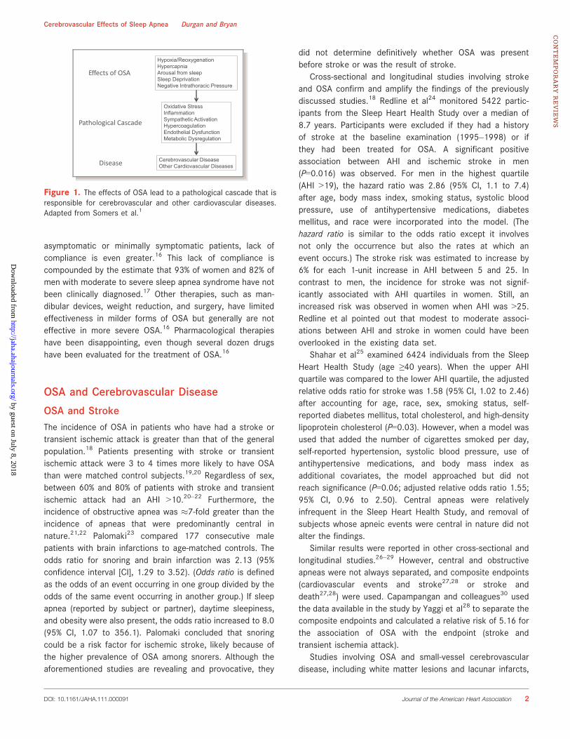
were divided into 1 of 2 groups: those who tolerated CPAPtherapy for the duration of the study (15 patients) and thosewho could not tolerate and discontinued CPAP therapy after 1month (36 patients). The incidence of new cerebrovascular orischemic coronary events was determined during thesubsequent 18 months. The incidence of new vascular eventswas 5-fold greater in patients who discontinued CPAP than inCPAP users after adjustment for other vascular risk factorsand neurological indexes (Figure 2). Martinez-Garcia et alconcluded that CPAP treatment in patients with mild to severesleep apnea can offer protection against new vascular eventsafter ischemic stroke. Wessendorf et al57 reported animproved sense of well-being and decreased blood pressureafter 10 days in stroke patients (n=74) who adhered to CPAPtherapy compared to those who did not (n=36). Ryan et al57a
randomized stroke patients with OSA (AHI ≥15) to a CPAP(n=22) group or control (n=22) group. CPAP treatment for4 weeks improved functional and motor outcomes but notneurocognitive outcomes. The authors concluded that CPAPtreatment provided a “significant, although modest, beneficialeffect” on stroke-related outcomes in patients with OSA.However, another study found no beneficial effects of 8 weeksor 6 months of CPAP therapy in stroke patients with AHI≥30.58 However, in that study, patients in the treatment grouphad limited compliance with CPAP, with the average use beingonly 1.4 hours per night. Depending on the study, compliancewith CPAP therapy among patients who had a stroke wassimilar to or lower than compliance in patients who did nothave a stroke.57–61
OSA, Dementia, and Cognitive ImpairmentPublished studies have reported an association between OSAand dementia in the elderly,62,63 with the severity of thedementia being related to the severity of the sleep-disorderedbreathing.64,65 In one study, Yaffe et al66 monitored 298
women without dementia (mean of 82 years of age) for�5 years. Women with sleep-disordered breathing were morelikely to develop mild cognitive impairment or dementia evenafter adjustment for other risk factors. The vast majority ofthe dementia and cognitive impairment in individuals >80years of age is mixed, as opposed to pure Alzheimer’s diseaseor vascular in nature.67 Given that OSA might be an underlyingcause of stroke and that strokes can be a major component inbrain atrophy and confusion in older patients,68 it stands toreason that OSA, though not necessarily the underlying causeof the dementia, is certainly a factor that would exacerbatethe dementia.67
Ayalon et al69 assessed cognitive function in younger (25to 44 years) and older (45 to 59 years) subjects with andwithout OSA. Older subjects with OSA showed cognitivedecline when compared to older subjects without OSA,younger subjects with OSA, or younger subjects without OSA.Thus, cognitive decline, which normally occurs with aging, canbe accelerated in individuals with OSA.
Studies indicate that patients with OSA and Alzheimer’sdisease could have positive benefits from CPAP therapy.Modest but statistically significant cognitive improvementswere reported after 3 weeks of CPAP therapy in one study.70
CPAP treatment for 13 months showed positive benefits inmood, sleep quality, and cognition compared to a group ofpatients not using CPAP treatment.71 Although definitiveconclusions are hampered by the limited sample sizes, thefindings are still provocative and should motivate futurestudies. As such, CPAP therapy might provide lastingimprovements, or at least stabilization, in the quality of lifeof individuals with dementia.
If the studies are taken as a whole, a convincing argumentcan be made that OSA accelerates cognitive decline and theonset and severity of dementia,62–66 although not all studiesfound a significant association between OSA and cognitivefunction in the elderly.72,73 That the incidence of OSAincrease with age14 adds to the seriousness of the clinicalsituation in the elderly.
Effects of OSA on the Cerebral CirculationThe cerebrovascular system must accommodate the distinctanatomic structure and physiological function of the brain.As a consequence, it is different in many respects from theperipheral vascular system. These differences presentunique challenges and vulnerabilities for the cerebrovascularsystem in maintaining a homeostatic environment in whichthe brain can operate. The O2 demand by the brain requiresan uninterrupted supply of O2 by the blood. Disruptions inO2 delivery for relatively short periods of time areaccompanied by significant pathological consequences. Inthis review, we will discuss only a few aspects of the
CPAP Without CPAP
Vascula
rE
vents
(%)
0
10
20
30
40
*
Figure 2. Incidence of vascular events over an 18-month periodafter stroke in CPAP users (n=15) and those who discontinued theuse of CPAP (n=36). All patients in the study had an AHI ≥20.*P=0.03. Created with data from Martinez-Garcia et al.56
DOI: 10.1161/JAHA.111.000091 Journal of the American Heart Association 4
Cerebrovascular Effects of Sleep Apnea Durgan and BryanCONTEMPORARY
REVIE
WS
by guest on July 8, 2018http://jaha.ahajournals.org/
Dow
nloaded from

cerebral circulation as they pertain to OSA. For in-depthdiscussion of the cerebral circulation, see the referencedreviews.74–78
Cerebral Blood Flow During an Episode of ApneaTranscranial Doppler ultrasonography (TCD) measurements ofblood velocity in cerebral arteries have been an important toolin assessing cerebral blood flow (CBF) in humans. TCD, whichmeasures the velocity of red blood cells in vessels (usually themiddle cerebral artery), does not provide a quantitativemeasure of CBF. Instead, it provides an index that is directlyproportional to the rate of blood flow, provided the diameterof the vessel remains constant throughout the time ofmeasurement. Several studies indicate that CBF velocity(CBFV) in the middle cerebral artery can provide a reasonableindex of CBF with different conditions.79–81 Although TCD hasits limitations, it is a powerful and noninvasive tool that hasbeen used in humans to provide meaningful insight intochanges in CBF during OSA.
CBFV has been reported to increase steadily in the middlecerebral artery during an episode of OSA. The magnitude ofthe CBFV increase varies greatly but can exceed 200% of thebaseline velocity (Figure 3).5,82–84 On termination of apnea,CBFV often, but not always, decreases below baseline beforerecovering after �1 minute.5,82,84
Several known, and likely some unknown, vasoactiveinfluences impinge on the cerebral circulation during OSA.During an episode of OSA, arterial PO2 and pH decrease, andPCO2 increases.1,7 Blood gasses and pH changes in these
directions act in concert to dilate cerebral arteries andarterioles.85,86 The increase in arterial PCO2 (hypercapnia)during an episode of OSA has been more of a focus on CBFregulation than the concomitant hypoxia.83,87,88
Arterial blood pressure has been reported to increaseprogressively during the apnea and to fall below baseline afterthe apnea.5,82,83 If autoregulation is compromised (see later)or the changes in blood pressure are sufficiently rapid thatautoregulation, even if intact, does not have time to adapt,then all or a portion of the changes in TCD velocity could berelated to the changes in blood pressure.5 Indeed, Balfors andFranklin82 reported a linear correlation between percentchange in mean arterial blood pressure and percent change inCBFV during and after an episode of apnea. It must be notedthat during an episode of OSA, blood pressure mostlyincreases, PO2 and pH decrease, and PCO2 increases. There-fore, it is not surprising that changes in the CBFV alsocorrelate with arterial PCO2.
83,88
Several publications in the literature present CBFV andO2 saturation data during a single or multiple OSAevent(s).82,84,89 The timing of maximal O2 desaturations oftenoccurred after breathing had resumed. For example, Balforsand Franklin82 noted that maximum O2 desaturation (asassessed by pulse oximetry) occurred �20 seconds aftertermination of the apnea. Although the delay was noted in thepreviously discussed studies, no explanations were providedfor the unexpected timing of the O2 desaturations. Wesuggest that the timing of the O2 desaturations shown inthese publications represents a delay in the output of themeasuring devices. The delay time for pulse oximetry, whichdepends on manufacturer, sampling interval, and probeplacement, can be ≥30 seconds.90,91
Contrary to the majority of studies that show an increase inCBFV during OSA, Netzer et al92 reported that CBFV decreasedduring 76% of obstructive hypopneas and 80% of obstructiveapneas observed. The reason for this disparity in the literatureis not known. However, blood pressure was notmeasured in thestudy by Netzer et al.92 As discussed previously, changes inarterial blood pressure can be an important consideration inCBF changes during an episode of OSA.
Other changes that potentially could affect CBF during anepisode of OSA include vasoactive substances in blood orstimulation of vasoactive central neurons.93,94 In addition, thesensory, sympathetic, or parasympathetic nervous systeminnervates cerebral arteries95 and could influence CBF duringan episode of OSA. In general, however, activation of thesympathetic nervous system has little effect on the cerebralcirculation except during some pathological states.95,96
Although none of the systems that innervate cerebral vesselshas been investigated in OSA, they must be considered, giventhe complexity and the number of systems involved with theresponse to OSA.
CB
FV (%
of B
asel
ine)
100
150
200
250
300
MAB
P
160
165
170
175
180
185
1 min
APNEA
Figure 3. Changes in blood flow velocity in the middle cerebralartery (CBFV; top) and mean arterial blood pressure (MABP; bottom)during an episode of apnea (shaded in light blue). Created with datafrom Klingelhofer et al.83
DOI: 10.1161/JAHA.111.000091 Journal of the American Heart Association 5
Cerebrovascular Effects of Sleep Apnea Durgan and BryanCONTEMPORARY
REVIE
WS
by guest on July 8, 2018http://jaha.ahajournals.org/
Dow
nloaded from
In healthy awake subjects, the CBFV increases observedduring voluntary apnea and the CBFV decreases observedafter the apnea were driven largely by arterial PCO2 and to alesser extent by changes in arterial pressure.87 Althoughthese studies in healthy subjects could provide insight intoCBF during OSA, it must be pointed out that the mechanismsin healthy subjects might not totally reflect the mechanisms inindividuals with OSA. The increase in CBFV during voluntaryapnea was reduced in patients with OSA compared to controlsubjects. Significantly, the response to the voluntary apneareturned to normal after 1 day or 1 month of CPAPtherapy.97,98
Impaired cerebrovascular control in individuals with OSAcan affect the energy state of the brain during an episodeof apnea. When O2 desaturations were >10% during OSA,brain adenosine triphosphate decreased, and inorganicphosphate increased.99 With different protocols, comparableor even greater levels of hypoxia did not significantly alter theenergy state in normal subjects or in animal models.100–102
During nonpathological conditions, an increase in CBF canaccommodate the O2 requirement for adenosine triphosphateproduction through the respiratory chain. Rae et al99 con-cluded that “transient hypoxia experienced during sleep mayimpair brain function more than previously thought.”
Effects of OSA on Resting CBFAlthough they are limited, studies indicate that resting CBF ischronically altered in individuals with OSA. Meyer andcolleagues103,104 reported that CBF in patients with OSAwas decreased during both sleep and wakefulness comparedto volunteers without OSA. These studies, using stable xenonand xenon-133 computerized tomography, are significant inthat they are the only studies, to our knowledge, in whichrates for CBF (mL / 100 g per minute) have been actuallyquantified in patients with OSA. CBF was decreased in thehemispheric, brainstem, and cerebellar gray regions ofpatients with OSA during wakefulness.103,104 Additionally,during sleep, patients with OSA exhibited significant reduc-tions in flow to the frontal and occipital cortex, pons, and thecerebellum.103 These decreases occurred even though end-tidal CO2 was significantly greater in patients with OSA duringwakefulness (2 mm Hg) and sleep (4 mm Hg) than in controlsubjects.103
TCD studies support the idea that resting CBF is decreasedin individuals with OSA.105 This decrease in CBFV persisted inwakefulness, non–rapid eye movement sleep, and rapid eyemovement sleep in patients with OSA, despite their havingend-tidal CO2 values greater than those of control subjects forall states.106 Hajak et al107 extended these findings bymeasuring CBFV during the 4 different stages of non–rapideye movement sleep. CBFV was decreased in patients with
OSA during sleep stages 1 to 3 after falling asleep comparedto control subjects. However, during all stages of rapid eyemovement sleep and in most sleep stage twos after the firstoccurrence, CBFV was greater in patients with OSA than incontrols. Overall, patients with OSA and control subjectsexhibited similar patterns of fluctuations in nocturnal brainperfusion; however, the amplitude of these changes wasgreater in control subjects during non–rapid eye movementsleep and was greater for patients with OSA during rapid eyemovement transitions.107
When the data are taken as a whole, it can be concludedthat CBF is decreased in the resting state with OSA. Althoughthere are some variations between studies, particularly duringsleep, existing studies consistently report alterations inresting CBF. The resting state reflects the background uponwhich CBF is regulated, and deviations from the resting stateof healthy individuals are indicative of cerebrovasculardysfunction. Of note, moderate decreases in CBF can havesignificant adverse consequences, including deficits in mem-ory, spatial learning, and attention, which are describedcommonly in patients with severe OSA.108–111 Consistentwith this idea is a recent study that shows disruption ofneurovascular coupling in the sensory cortex in mice afterintermittent hypoxia, a model of sleep apnea.112
Effects of OSA on Cerebral AutoregulationAutoregulation helps to maintain a homeostatic environmentin which the brain can operate by maintaining a constant CBFin the face of changes in arterial blood pressures over a rangebetween 60 and 160 mm Hg.96 Unlike many peripheraltissues in which the major resistance to flow resides in thearterioles, the resistance in the cerebrovascular systemoccurs along the entire vascular tree, including not only thesmaller arteries and arterioles but also the larger cerebralarteries.109,113,114 The vascular tree in a healthy brain, inwhich large arteries contribute significantly to the resistance,not only serves to regulate CBF but also protects arteriolesand capillaries from excessive pressures.
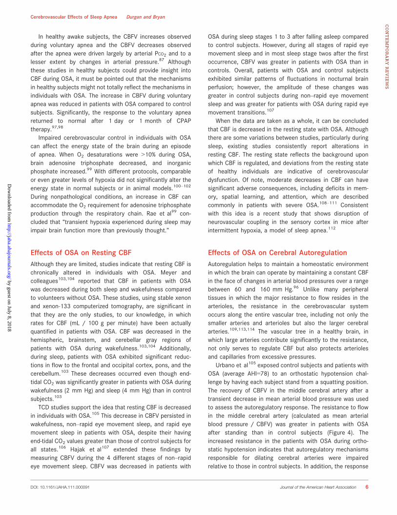
Urbano et al105 exposed control subjects and patients withOSA (average AHI=78) to an orthostatic hypotension chal-lenge by having each subject stand from a squatting position.The recovery of CBFV in the middle cerebral artery after atransient decrease in mean arterial blood pressure was usedto assess the autoregulatory response. The resistance to flowin the middle cerebral artery (calculated as mean arterialblood pressure / CBFV) was greater in patients with OSAafter standing than in control subjects (Figure 4). Theincreased resistance in the patients with OSA during ortho-static hypotension indicates that autoregulatory mechanismsresponsible for dilating cerebral arteries were impairedrelative to those in control subjects. In addition, the response
DOI: 10.1161/JAHA.111.000091 Journal of the American Heart Association 6
Cerebrovascular Effects of Sleep Apnea Durgan and BryanCONTEMPORARY
REVIE
WS
by guest on July 8, 2018http://jaha.ahajournals.org/
Dow
nloaded from
of the cerebral circulation to the hypotensive challenge wasless dynamic in patients with OSA during the initial phase ofthe hypotensive challenge, being only 62% of that in controlsubjects (slopes in Figure 4). Nasr et al115 studied theautoregulation in awake patients with moderate OSA (averageAHI=23) and matched controls by computing an autoregula-tory index consisting of a moving correlation coefficientbetween mean CBFV and mean arterial blood pressure. Notonly was autoregulation impaired in awake patients with OSA,but also the severity of the impairment strongly correlatedwith OSA severity.115 Finally, a recent study showed that themechanism for myogenic tone, the intrinsic property ofarteries to constrict when pressurized, was altered in ex vivomiddle cerebral arteries from rats after they had beensubjected to intermittent hypoxia.116 An impairment ofcerebral autoregulation likely contributes to the increasedincidence of stroke, as well as the poor outcome after stroke,in patients with OSA.
Effects of OSA on Cerebrovascular Response toHypoxia and HypercapniaRepeated exposures to hypoxia in individuals with OSA couldlead to hypoxic/ischemic brain injury, especially if cerebro-vascular control is impaired. The increase in CBFV in the
middle cerebral artery during hypoxic challenges was atten-uated significantly by 42%117 and 36%118 in patients withsevere OSA as compared to controls. The CBFV response tohypoxia correlated with the AHI and nocturnal O2 desatura-tion.117 Interestingly, significant improvements in the CBFVresponse to hypoxia occurred after 4 to 6 weeks117 or12 weeks118 of CPAP treatment. Finally, the dilator responseto hypoxia in isolated pressurized middle cerebral arteriesfrom rats after 2 weeks of intermittent hypoxia wascompletely abolished.119
Studies investigating the cerebrovascular response tohypercapnia in patients with OSA have yielded conflictingresults. Studies performed by Urbano et al and Fosteret al105,120 exposed patients with severe OSA to repetitivehypercapnic conditions while measuring CBFV by TCD. Bothstudies reported no difference in the hypercapnic response ascompared to control patients. Reichmuth et al118 reportedthat the increase in CBFV when end-tidal CO2 was increasedby 5 and 10 mm Hg was attenuated by 22% in individuals withOSA compared to controls. Similarly, Morgan et al121
reported that the cerebrovascular response to hypercapniawas blunted in individuals with OSA, with the severity of theresponse positively correlating with the degree of oxygendesaturation during OSA. Using methods to quantitativelymeasure CBF, Meyer et al103,104 reported that the increase inCBF with the addition of 5% CO2 to the ventilator gas wasblunted in patients with OSA during sleep and wakefulnesscompared to control subjects. Given the limited number ofstudies and the conflicting results, any conclusion about thecerebrovascular response to hypercapnia is premature. Unlikethe autoregulatory response and the cerebrovascularresponses to hypoxia, the effects of OSA on the responseto hypercapnia must await further investigations.
Mechanisms of Injury to the CerebralCirculation in OSA
Physical MechanismsSeveral events take place during an episode of apnea, inaddition to hypoxia/reoxygenation, that have detrimentaleffects on the cerebral circulation. There are repeated swingsin blood pressure with each episode of apnea, consisting notonly of increased blood pressure during the episode but also aperiod of hypotension after the episode.82,83 At the end of anepisode of OSA, blood pressure was reported to sharplyincrease by �35 mm Hg on average; however, in someindividuals, the systolic pressure surge exceeds100 mm Hg.1,122 These surges in blood pressure can besufficiently rapid that autoregulation, even if it were intact,might not have time to fully accommodate.5 Pressure surgeswill expose smaller arteries, arterioles, and capillaries to
Time (sec)
0 1 2 3 4 5
Resis
tance
MA
BP
(mm
Hg)
/C
BF
V(c
m/s
)
0.6
0.8
1.0
1.2
1.4
1.6
Control
OSA (AHI=78)
slope = -0.21
slope = -0.13
Figure 4. Resistances to blood flow through the middle cerebralartery in control subjects (n=26) and patients with OSA (n=78) afteran orthostatic hypotension challenge, obtained by having individualsstand from a squatting position.105 The patients with OSA had amean AHI of 78. The resistance was calculated by dividing meanarterial blood pressure (MABP) by blood flow velocity in the middlecerebral artery (CBFV). The increased resistance in the patients withOSA during orthostatic hypotension indicates that autoregulatorymechanisms responsible for dilating cerebral arteries were impairedrelative to the control subjects. The response of the cerebralcirculation to the hypotensive challenge was less dynamic in patientswith OSA during the initial phase of the hypotensive challenge. Thatis, the rate of change in resistance in patients with OSA was only 62%of that in control subjects (1009[�0.13/�0.21]). Created with datafrom Urbano et al.105
DOI: 10.1161/JAHA.111.000091 Journal of the American Heart Association 7
Cerebrovascular Effects of Sleep Apnea Durgan and BryanCONTEMPORARY
REVIE
WS
by guest on July 8, 2018http://jaha.ahajournals.org/
Dow
nloaded from
damaging pressures, resulting in endothelial damage anddisruption of the blood–brain barrier. At the end of an episodein which the blood pressure sharply increased, intracranialpressure was reported to often exceed 50 mm Hg.123 In asimilar manner, the sharp decreases in blood pressure ontermination of OSA82,83 can leave the brain vulnerable toischemia, especially in regions with poor cerebrovascularreserve.39 This would be especially true for areas withoutgood collateral circulation, including border zone areas andterminal arterial territories. Consistent with this idea is thefact that OSA has been associated with lacunar infarcts,small-vessel disease, and leukoaraiosis.33,35,124
Although the carotid artery is outside the cranial vault, thecarotid artery must be included when CBF is consideredbecause the majority of blood flow to the brain is via thecarotid arteries. The carotid arteries are a primary site fordevelopment of atherosclerosis (carotid artery disease), a riskfactor for stroke.109 Vibrations in the carotid arteries fromsnoring, common in patients with OSA, can produce endo-thelial damage and atherosclerosis.125,126 Vascular dysfunc-tion in the carotid artery will put more stress on the vascularsystem in the brain, rendering it more vulnerable to otherpathological consequences of OSA.
Cellular and Molecular MechanismsThere is a paucity of studies involving pathological mecha-nisms in the cerebral circulation in OSA. Given the moreextensive literature involving vascular effects outside of thebrain (see reviews1,3,7,9,16,127–131), it is tempting to extrapo-late these findings directly to the cerebrovascular circulation.However, extrapolation must be conducted with cautionbecause (1) the distinct anatomic structures and physiologicalfunction of the brain are uniquely affected by diseaseprocesses and (2) the cerebrovascular circulation can bemore sensitive to the pathological processes.109
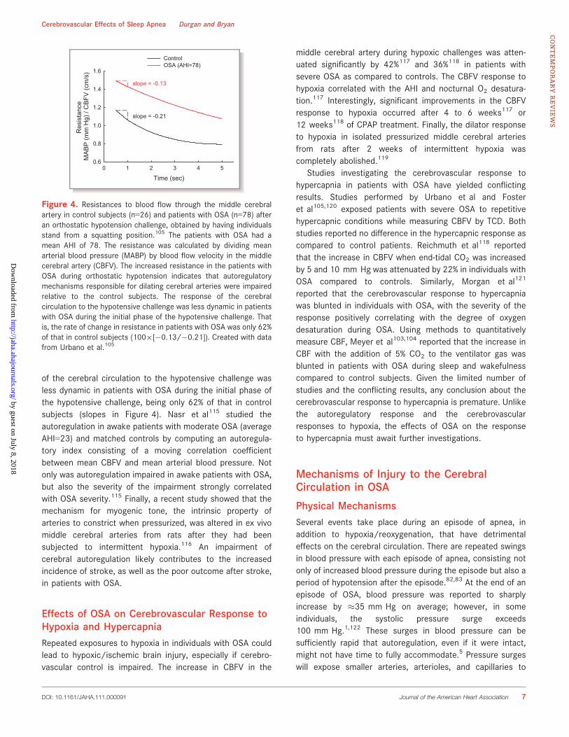
OSA initiates a pathological cascade through multiple andoften redundant pathways affecting the cardiovascular system(Figure 1).1 This cascade includes oxidative stress, inflamma-tion, sympathetic activation, hypercoagulation, endothelialdysfunction, increased platelet aggregation, and metabolicdysregulation.9,26,38,39,108 As with the peripheral circulation,the cerebral circulation is affected adversely by the patho-logical cascade, which impinges to a great extent on theendothelium. The pathological impact on the endothelium,commonly referred to as endothelial “dysfunction,” is char-acterized by oxidative stress and decreased production ofnitric oxide (NO), although the full extent of endothelialdysfunction is much more involved.9 Notably, dysfunction ofcerebrovascular endothelium occurs sooner and to a greaterextent in the progression of cardiovascular diseases than thatof endothelium in the periphery.109
The sympathetic nervous system is activated acutelyduring OSA as a result of (1) chemoreceptor discharges withchanges in blood gasses and pH, (2) arousals from sleep, and(3) sleep deprivation.1,7 In addition to the acute activationduring a single episode, chronic intermittent hypoxia or OSAalters the sensitivity of the chemoreceptors and barorecep-tors, as well as altering their central integration by nuclei inthe brainstem and hypothalamus.3,132–136 The carotid chemo-receptors increase baseline activity and become moresensitive to hypoxia through alterations in neurotransmittersystems, including endothelin-1 (ET-1) and angiotensinII.135,137–139 The mechanism for altered chemoreceptorfunction involves reactive oxygen species (ROS), resultingfrom transcriptional genes that upregulate hypoxia-induciblefactor-1 and downregulate hypoxia-inducible factor-2.135
One consequence of increased sympathetic activity on thekidney is stimulation of the renin–angiotensin system and thesubsequent increase in angiotensin II. Angiotensin II canenhance oxidative stress, produce inflammation, disruptblood–brain barrier integrity, initiate vessel remodeling, andimpair increases in nutritive blood flow.109,140–142 Throughstimulation of angiotensin I receptors on the endothelium,angiotensin II enhances the production of superoxide byactivating NADPH oxidase.143 Although angiotensin II hassimilar effects in all endothelial cells, cerebral vessels havegreater NADPH oxidase activity than do peripheral vesselsand are particularly sensitive to the effects of the superoxidegenerated.109,144–146 The pathway described in this paragraphis shown in Figure 5 (black lines). Although it is not knownwhether this particular pathway affects the cerebral circula-tion after chronic OSA, other pathological states negatively
Endothelin System
HIF
Damage to Cerebrovascular EndotheliumImpaired Neurovascular Coupling
Chronic Intermittent HypoxiaOSA
Chemoreceptor Activation
Renin-Angiotensin System
Sympathetic Activation
Angiotensin II
Superoxide
NADPH Oxidase
Damage to Peripheral Endothelium
?
?
Figure 5. Proposed pathway for activation of NADPH oxidase,generation of superoxide, and upregulation of the endothelin systemin the peripheral circulation after OSA and chronic intermittenthypoxia (black lines). An alternative pathway that could be respon-sible for generation of superoxide in the cerebral circulation (redlines).112 Dashed red lines (and “?”), which lack any experimentalevidence, represent hypothetical pathways. HIF indicates hypoxiainducible factor.
DOI: 10.1161/JAHA.111.000091 Journal of the American Heart Association 8
Cerebrovascular Effects of Sleep Apnea Durgan and BryanCONTEMPORARY
REVIE
WS
by guest on July 8, 2018http://jaha.ahajournals.org/
Dow
nloaded from
impact the cerebral circulation through activation of therenin–angiotensin system, upregulation of NADPH oxidase,and generation of superoxide.109 In addition to NADPHoxidase being a major source, superoxide also can begenerated in the vascular system by mitochondria, cycloox-ygenase, lipoxygenases, hydroxylases, and xanthine oxidase.
Superoxide is involved in the generation of other ROS andreactive nitrogen species. One of these reactive nitrogenspecies, peroxynitrite, is producedwhen superoxide reacts withNO. Peroxynitrite uncouples endothelial NO synthase, wherebyO2 becomes the terminal electron acceptor in the oxidation-reduction reaction to create superoxide in place of the normalend product, NO.1,7,109 The net result of an increased oxidativestate includes reduced NO bioavailability, damage to lipidmembranes, and alterations in protein structure and function.In addition to the increased production of ROS, reducedglutathione, a component of the antioxidant system forprotecting the brain, was reported to be significantly reducedwith reoccurring obstructive apneas over 1 hour, whereasreduced glutathione in the periphery was not affected.147
Although studies of endothelial dysfunction in cerebralarteries are limited, intermittent hypoxia, a model for sleepapnea, produced endothelial dysfunction in rodents.112,119
After only 2 weeks of intermittent hypoxia in rats, dilations toacetylcholine in isolated middle cerebral arteries were atten-uated by �90%.119 Activation of cholinergic receptors on theendothelium stimulate the synthesis of NO by endothelial NOsynthase. Once generated in the endothelium, NO diffuses tothe vascular smooth muscle and activates guanylyl cyclase, theinitial step in a dilatory pathway. Phillips et al119 demonstratedthat intermittent hypoxia did not alter the dilations whenguanylyl cyclase was activated directly by sodium nitroprus-side. Thus, attenuated dilations after intermittent hypoxia werethe result of reduced NO (ie, endothelial dysfunction) becausethe vascular smooth muscle still had the ability to dilate.
In a recent study, Capone et al112 amplified and extendedthe studies by Phillips et al.119 Increases in cortical blood flowin response to dilators requiring intact endothelium wereattenuated by �40% after 14 and 35 days of intermittenthypoxia in mice. However, dilations in response to adenosine,which acts directly on the vascular smooth muscle, were notaffected by intermittent hypoxia. ROS was enhanced signifi-cantly in both cerebral arterioles and neurons after intermittenthypoxia, as compared to tissues from control mice.112
Significantly, an acute application of a superoxide dismutasemimetic to decrease the oxidative stress completely restoredthe blood flow response to acetylcholine.112 The source of theROS after the intermittent hypoxia was predominantly, if notexclusively, from superoxide generated by NADPH oxidase,because an inhibitor of NADPH oxidase or mice lackingfunctional NADPH oxidase-2 showed neither endothelialdysfunction nor enhanced tissue ROS after 35 days of
intermittent hypoxia. The studies by Capone et al112 comple-ment previous studies demonstrating a crucial role for brainNADPH oxidase in hypersomnolence and the decline incognitive function after intermittent hypoxia or sleep fragmen-tation.148–150 Thus, intermittent hypoxia produces oxidativestress and decreases the bioavailability of endothelium-derivedNO through superoxide derived from NADPH oxidase-2.
Crossland et al151 developed a model of OSA in rats inwhich apnea was elicited in unanesthetized freely ranging ratsby remotely inflating a balloon implanted in the trachea. Theballoon was inflated for 10 seconds 30 times per hour during8 hours of the sleep cycle every day for 28 days. Dilations inisolated pressurized middle cerebral arteries, elicited bystimulating endothelial P2Y2 receptors by adenosine triphos-phate, were attenuated by �40% compared to a control groupthat was surgically prepared but did not undergo apnea.Although the dilation in response to adenosine triphosphatewas the result of multiple endothelial-dependent mechanismsof dilation,152–155 the attenuated response was due tosuppression of the NO component.151 Thus, there is continu-ity with the findings from different animal models for OSA,intermittent hypoxia, and apnea.
ET-1 is a potent vasoconstrictor that is involved withpathological processes, including OSA. The primary, but notexclusive, source for ET-1 in the vascular system is endothelialcells156,157; however, during pathological states, the vascularsmooth muscle also can synthesize ET-1.156,157 Stimulation ofendothelin receptor type B on the endothelium producesdilation of vessels through generation of NO, whereas stim-ulation of primarily endothelin receptor type A, but sometimesendothelin receptor type B, on the vascular smooth muscleproduces potent and sustained constrictions.156,158 Duringnormal physiological states, NO inhibits transcription andrelease of ET-1, reduces the interaction of ET-1 with endothelinreceptors on smooth muscle, and interferes with secondmessenger signaling in the endothelin signaling path-way.159,160 However, during pathological states such as OSA,reduced NO bioavailability, as a result of the oxidative stress,allows the endothelin pathway to upregulate. The endothelinsystem further adds to the oxidative stress by enhancing thepathological cascade that generates ROS.161–164
The endothelin system is upregulated in animal mod-els of OSA and likely is upregulated with OSA inhumans, although plasma ET-1 levels might not beincreased.1,3,9,16,38,112,127,165–168 Intermittent hypoxia increasedET-1 in cerebral vessels of mice by �2 orders of magnitudeand increased mRNA for endothelin-converting enzyme-1 andendothelin receptor type A but not mRNA for endothelinreceptor type B.112 Furthermore, the increase in ET-1expression occurred in both the endothelium and vascularsmooth muscle. The attenuated cerebrovascular responses toacetylcholine with intermittent hypoxia were restored after
DOI: 10.1161/JAHA.111.000091 Journal of the American Heart Association 9
Cerebrovascular Effects of Sleep Apnea Durgan and BryanCONTEMPORARY
REVIE
WS
by guest on July 8, 2018http://jaha.ahajournals.org/
Dow
nloaded from
acutely blocking endothelin type A receptors.112 Administra-tion of the endothelin type A blocker also reduced ROS incerebral vessels and neurons after intermittent hypoxia to thelevels in control mice.112 It is interesting to note that eitherinhibiting NADPH oxidase-2 or blocking endothelin type Areceptors reduced ROS and restored the cerebrovascularresponses to endothelial-mediated dilations. Upregulation ofNADPH oxidase often is associated with angiotensin II;however, with intermittent hypoxia, it seems that ET-1replaces the role of angiotensin II in regulating NADPHoxidase. This alternative pathway is shown in Figure 5 (redlines). It is possible that the endothelin system is upregulatedduring chronic intermittent hypoxia by a hypoxia-induciblefactor.169 However, at this time, we do not know if inhibitionof NADPH oxidase or scavenging ROS prevents upregulationof the endothelin system in the context of intermittenthypoxia. Answers to these and other significant questions areneeded for a clear understanding of the relationships amongNADPH oxidase, ROS, and the endothelin system.
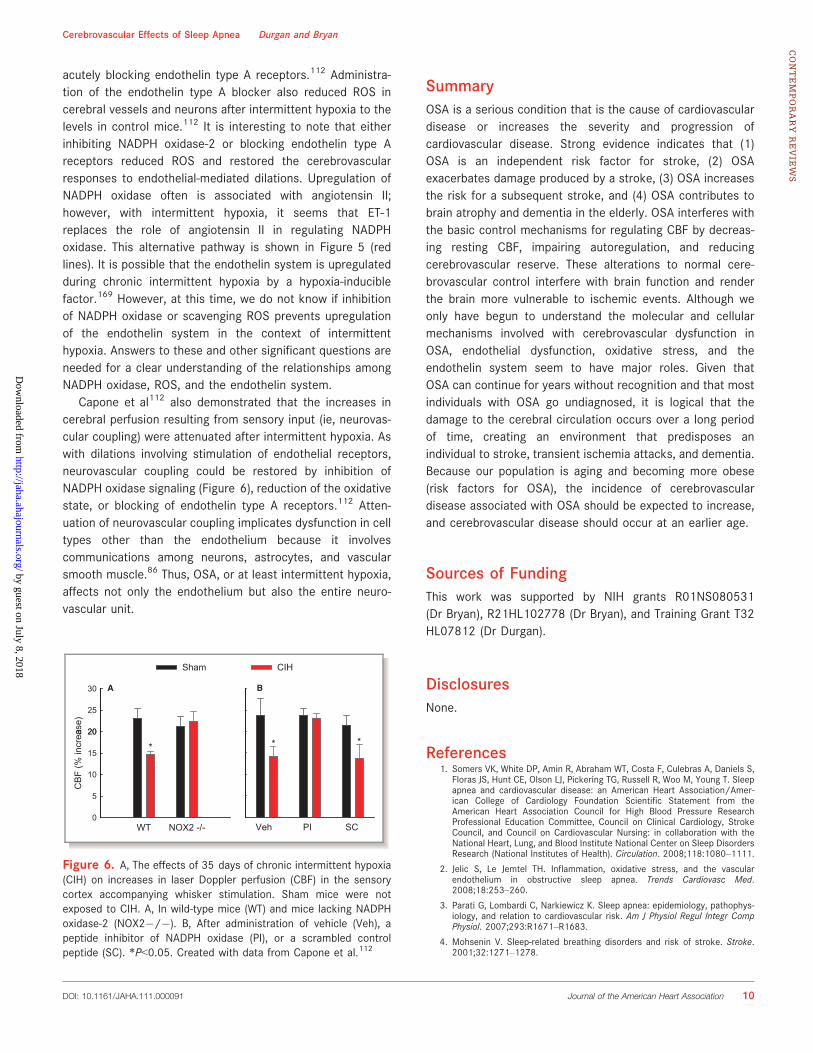
Capone et al112 also demonstrated that the increases incerebral perfusion resulting from sensory input (ie, neurovas-cular coupling) were attenuated after intermittent hypoxia. Aswith dilations involving stimulation of endothelial receptors,neurovascular coupling could be restored by inhibition ofNADPH oxidase signaling (Figure 6), reduction of the oxidativestate, or blocking of endothelin type A receptors.112 Atten-uation of neurovascular coupling implicates dysfunction in celltypes other than the endothelium because it involvescommunications among neurons, astrocytes, and vascularsmooth muscle.86 Thus, OSA, or at least intermittent hypoxia,affects not only the endothelium but also the entire neuro-vascular unit.
SummaryOSA is a serious condition that is the cause of cardiovasculardisease or increases the severity and progression ofcardiovascular disease. Strong evidence indicates that (1)OSA is an independent risk factor for stroke, (2) OSAexacerbates damage produced by a stroke, (3) OSA increasesthe risk for a subsequent stroke, and (4) OSA contributes tobrain atrophy and dementia in the elderly. OSA interferes withthe basic control mechanisms for regulating CBF by decreas-ing resting CBF, impairing autoregulation, and reducingcerebrovascular reserve. These alterations to normal cere-brovascular control interfere with brain function and renderthe brain more vulnerable to ischemic events. Although weonly have begun to understand the molecular and cellularmechanisms involved with cerebrovascular dysfunction inOSA, endothelial dysfunction, oxidative stress, and theendothelin system seem to have major roles. Given thatOSA can continue for years without recognition and that mostindividuals with OSA go undiagnosed, it is logical that thedamage to the cerebral circulation occurs over a long periodof time, creating an environment that predisposes anindividual to stroke, transient ischemia attacks, and dementia.Because our population is aging and becoming more obese(risk factors for OSA), the incidence of cerebrovasculardisease associated with OSA should be expected to increase,and cerebrovascular disease should occur at an earlier age.
Sources of FundingThis work was supported by NIH grants R01NS080531(Dr Bryan), R21HL102778 (Dr Bryan), and Training Grant T32HL07812 (Dr Durgan).
DisclosuresNone.
References1. Somers VK, White DP, Amin R, Abraham WT, Costa F, Culebras A, Daniels S,
Floras JS, Hunt CE, Olson LJ, Pickering TG, Russell R, Woo M, Young T. Sleepapnea and cardiovascular disease: an American Heart Association/Amer-ican College of Cardiology Foundation Scientific Statement from theAmerican Heart Association Council for High Blood Pressure ResearchProfessional Education Committee, Council on Clinical Cardiology, StrokeCouncil, and Council on Cardiovascular Nursing: in collaboration with theNational Heart, Lung, and Blood Institute National Center on Sleep DisordersResearch (National Institutes of Health). Circulation. 2008;118:1080–1111.
2. Jelic S, Le Jemtel TH. Inflammation, oxidative stress, and the vascularendothelium in obstructive sleep apnea. Trends Cardiovasc Med.2008;18:253–260.
3. Parati G, Lombardi C, Narkiewicz K. Sleep apnea: epidemiology, pathophys-iology, and relation to cardiovascular risk. Am J Physiol Regul Integr CompPhysiol. 2007;293:R1671–R1683.
4. Mohsenin V. Sleep-related breathing disorders and risk of stroke. Stroke.2001;32:1271–1278.
30
Sham CIH
BA
ase)
20
25
(% in
crea
15
20
* * * *
CB
F
5
10
WT NOX2 -/-0
Veh PI SC
Figure 6. A, The effects of 35 days of chronic intermittent hypoxia(CIH) on increases in laser Doppler perfusion (CBF) in the sensorycortex accompanying whisker stimulation. Sham mice were notexposed to CIH. A, In wild-type mice (WT) and mice lacking NADPHoxidase-2 (NOX2�/�). B, After administration of vehicle (Veh), apeptide inhibitor of NADPH oxidase (PI), or a scrambled controlpeptide (SC). *P<0.05. Created with data from Capone et al.112
DOI: 10.1161/JAHA.111.000091 Journal of the American Heart Association 10
Cerebrovascular Effects of Sleep Apnea Durgan and BryanCONTEMPORARY
REVIE
WS
by guest on July 8, 2018http://jaha.ahajournals.org/
Dow
nloaded from
5. Yaggi H, Mohsenin V. Obstructive sleep apnoea and stroke. Lancet Neurol.2004;3:333–342.
6. Partinen M, Palomaki H. Snoring and cerebral infarction. Lancet. 1985;2:1325–1326.
7. Dempsey JA, Veasey SC, Morgan BJ, O’Donnell CP. Pathophysiology of sleepapnea. Physiol Rev. 2010;90:47–112.
8. Punjabi NM, Newman AB, Young TB, Resnick HE, Sanders MH. Sleep-disordered breathing and cardiovascular disease: an outcome-based defi-nition of hypopneas. Am J Respir Crit Care Med. 2008;177:1150–1155.
9. Atkeson A, Yeh SY, Malhotra A, Jelic S. Endothelial function in obstructivesleep apnea. Prog Cardiovasc Dis. 2009;51:351–362.
10. Tufik S, Santos-Silva R, Taddei JA, Bittencourt LR. Obstructive sleep apneasyndrome in the Sao Paulo Epidemiologic Sleep Study. Sleep Med.2010;11:441–446.
11. Young T, Peppard PE, Gottlieb DJ. Epidemiology of obstructive sleep apnea:a population health perspective. Am J Respir Crit Care Med. 2002;165:1217–1239.
12. Young T, Palta M, Dempsey J, Skatrud J, Weber S, Badr S. The occurrence ofsleep-disordered breathing among middle-aged adults. N Engl J Med.1993;328:1230–1235.
13. Russell T, Duntley S. Sleep disordered breathing in the elderly. Am J Med.2011;124:1123–1126.
14. Hoch CC, Reynolds CF III, Monk TH, Buysse DJ, Yeager AL, Houck PR,Kupfer DJ. Comparison of sleep-disordered breathing among healthyelderly in the seventh, eighth, and ninth decades of life. Sleep. 1990;13:502–511.
15. Ancoli-Israel S, Kripke DF, Klauber MR, Mason WJ, Fell R, Kaplan O. Sleep-disordered breathing in community-dwelling elderly.Sleep. 1991;14:486–495.
16. Kohler M, Stradling JR. Mechanisms of vascular damage in obstructive sleepapnea. Nat Rev Cardiol. 2010;7:677–685.
17. Young T, Evans L, Finn L, Palta M. Estimation of the clinically diagnosedproportion of sleep apnea syndrome in middle-aged men and women. Sleep.1997;20:705–706.
18. Dyken ME, Im KB. Obstructive sleep apnea and stroke. Chest. 2009;136:1668–1677.
19. Dyken ME, Somers VK, Yamada T, Ren ZY, Zimmerman MB. Investigating therelationship between stroke and obstructive sleep apnea. Stroke.1996;27:401–407.
20. Bassetti C, Aldrich MS. Sleep apnea in acute cerebrovascular diseases: finalreport on 128 patients. Sleep. 1999;22:217–223.
21. Turkington PM, Bamford J, Wanklyn P, Elliott MW. Prevalence and predictorsof upper airway obstruction in the first 24 hours after acute stroke. Stroke.2002;33:2037–2042.
22. Johnson KG, Johnson DC. Frequency of sleep apnea in stroke and TIApatients: a meta-analysis. J Clin Sleep Med. 2010;6:131–137.
23. Palomaki H. Snoring and the risk of ischemic brain infarction. Stroke.1991;22:1021–1025.
24. Redline S, Yenokyan G, Gottlieb DJ, Shahar E, O’Connor GT, Resnick HE,Diener-West M, Sanders MH, Wolf PA, Geraghty EM, Ali T, Lebowitz M,Punjabi NM. Obstructive sleep apnea-hypopnea and incident stroke:the sleep heart health study. Am J Respir Crit Care Med. 2010;182:269–277.
25. Shahar E, Whitney CW, Redline S, Lee ET, Newman AB, Javier NF, O’ConnorGT, Boland LL, Schwartz JE, Samet JM. Sleep-disordered breathing andcardiovascular disease: cross-sectional results of the Sleep Heart HealthStudy. Am J Respir Crit Care Med. 2001;163:19–25.
26. Arzt M, Young T, Finn L, Skatrud JB, Bradley TD. Association of sleep-disordered breathing and the occurrence of stroke. Am J Respir Crit CareMed. 2005;172:1447–1451.
27. Marin JM, Carrizo SJ, Vicente E, Agusti AG. Long-term cardiovascularoutcomes in men with obstructive sleep apnoea-hypopnoea with or withouttreatment with continuous positive airway pressure: an observational study.Lancet. 2005;365:1046–1053.
28. Yaggi HK, Concato J, Kernan WN, Lichtman JH, Brass LM, Mohsenin V.Obstructive sleep apnea as a risk factor for stroke and death. N Engl J Med.2005;353:2034–2041.
29. Munoz R, Duran-Cantolla J, Martinez-Vila E, Gallego J, Rubio R, Aizpuru F, DeLa Torre G. Severe sleep apnea and risk of ischemic stroke in the elderly.Stroke. 2006;37:2317–2321.
30. Capampangan DJ, Wellik KE, Parish JM, Aguilar MI, Snyder CR,Wingerchuk D, Demaerschalk BM. Is obstructive sleep apnea anindependent risk factor for stroke? A critically appraised topic. Neurol-ogist. 2010;16:269–273.
31. Vermeer SE, Longstreth WT Jr, Koudstaal PJ. Silent brain infarcts: asystematic review. Lancet Neurol. 2007;6:611–619.
32. Eguchi K, Kario K, Hoshide S, Ishikawa J, Morinari M, Shimada K. Nocturnalhypoxia is associatedwith silent cerebrovascular disease in a high-risk Japanesecommunity-dwelling population. Am J Hypertens. 2005;18:1489–1495.
33. Minoguchi K, Yokoe T, Tazaki T, Minoguchi H, Oda N, Tanaka A, YamamotoM, Ohta S, O’Donnell CP, Adachi M. Silent brain infarction and plateletactivation in obstructive sleep apnea. Am J Respir Crit Care Med.2007;175:612–617.
34. Nishibayashi M, Miyamoto M, Miyamoto T, Suzuki K, Hirata K. Correlationbetween severity of obstructive sleep apnea and prevalence of silentcerebrovascular lesions. J Clin Sleep Med. 2008;4:242–247.
35. Harbison J, Gibson GJ, Birchall D, Zammit-Maempel I, Ford GA. White matterdisease and sleep-disordered breathing after acute stroke. Neurology.2003;61:959–963.
36. Davies CW, Crosby JH, Mullins RL, Traill ZC, Anslow P, Davies RJ, StradlingJR. Case control study of cerebrovascular damage defined by magneticresonance imaging in patients with OSA and normal matched controlsubjects. Sleep. 2001;24:715–720.
37. Robbins J, Redline S, Ervin A, Walsleben JA, Ding J, Nieto FJ. Associations ofsleep-disordered breathing and cerebral changes on MRI. J Clin Sleep Med.2005;1:159–165.
38. Bagai K. Obstructive sleep apnea, stroke, and cardiovascular diseases.Neurologist. 2010;16:329–339.
39. Culebras A. Sleep and stroke. Semin Neurol. 2009;29:438–445.
40. van Norden AG, de Laat KF, Gons RA, van Uden IW, van Dijk EJ, vanOudheusden LJ, Esselink RA, Bloem BR, van Engelen BG, Zwarts MJ,Tendolkar I, Olde-Rikkert MG, van der Vlugt MJ, Zwiers MP, Norris DG, deLeeuw FE. Causes and consequences of cerebral small vessel disease: theRUN DMC study: a prospective cohort study: study rationale and protocol.BMC Neurol. 2011;11:29.
41. Pantoni L. Cerebral small vessel disease: from pathogenesis and clinicalcharacteristics to therapeutic challenges. Lancet Neurol. 2010;9:689–701.
42. Behrouz R, Malek AR, Torbey MT. Small vessel cerebrovascular disease: thepast, present, and future. Stroke Res Treat. 2012;2012:839151.
43. Dziewas R, Humpert M, Hopmann B, Kloska SP, Ludemann P, Ritter M,Dittrich R, Ringelstein EB, Young P, Nabavi DG. Increased prevalence ofsleep apnea in patients with recurring ischemic stroke compared with firststroke victims. J Neurol. 2005;252:1394–1398.
44. Cherkassky T, Oksenberg A, Froom P, Ring H. Sleep-related breathingdisorders and rehabilitation outcome of stroke patients: a prospective study.Am J Phys Med Rehabil. 2003;82:452–455.
45. Good DC, Henkle JQ, Gelber D, Welsh J, Verhulst S. Sleep-disorderedbreathing and poor functional outcome after stroke. Stroke. 1996;27:252–259.
46. Kaneko Y, Hajek VE, Zivanovic V, Raboud J, Bradley TD. Relationship of sleepapnea to functional capacity and length of hospitalization following stroke.Sleep. 2003;26:293–297.
47. Turkington PM, Allgar V, Bamford J, Wanklyn P, Elliott MW. Effect of upperairway obstruction in acute stroke on functional outcome at 6 months.Thorax. 2004;59:367–371.
48. Sahlin C, Sandberg O, Gustafson Y, Bucht G, Carlberg B, Stenlund H,Franklin KA. Obstructive sleep apnea is a risk factor for death in patientswith stroke: a 10-year follow-up. Arch Intern Med. 2008;168:297–301.
49. StoweAM,Altay T, Freie AB, Gidday JM. Repetitive hypoxia extends endogenousneurovascular protection for stroke. Ann Neurol. 2011;69:975–985.
50. Parra O, Sanchez-Armengol A, Bonnin M, Arboix A, Campos-Rodriguez F,Perez-Ronchel J, Duran-Cantolla J, De La Torre G, Gonzalez Marcos JR, de laPena M, Carmen JM, Masa F, Casado I, Luz AM, Macarron JL. Early treatmentof obstructive apnoea and stroke outcome: a randomised controlled trial.Eur Respir J. 2011;37:1128–1136.
51. Alexandrov AV, Nguyen HT, Rubiera M, Alexandrov AW, Zhao L, Heliopoulos I,Robinson A, DeWolfe J, Tsivgoulis G. Prevalence and risk factors associatedwith reversed Robin Hood syndrome in acute ischemic stroke. Stroke.2009;40:2738–2742.
52. Alexandrov AV, Sharma VK, Lao AY, Tsivgoulis G, Malkoff MD, AlexandrovAW. Reversed Robin Hood syndrome in acute ischemic stroke patients.Stroke. 2007;38:3045–3048.
53. Palazzo P, Balucani C, Barlinn K, Tsivgoulis G, Zhang Y, Zhao L, DeWolfe J,Toaldo B, Stamboulis E, Vernieri F, Rossini PM, Alexandrov AV. Associationof reversed Robin Hood syndrome with risk of stroke recurrence. Neurology.2010;75:2003–2008.
54. Barlinn K, Balucani C, Palazzo P, Zhao L, Sisson A, Alexandrov AV.Noninvasive ventilatory correction as an adjunct to an experimental
DOI: 10.1161/JAHA.111.000091 Journal of the American Heart Association 11
Cerebrovascular Effects of Sleep Apnea Durgan and BryanCONTEMPORARY
REVIE
WS
by guest on July 8, 2018http://jaha.ahajournals.org/
Dow
nloaded from
systemic reperfusion therapy in acute ischemic stroke. Stroke Res Treat.2010;2010:108253.
55. Barlinn K, Alexandrov AV. Sleep-disordered breathing and arterial blood flowsteal represent linked therapeutic targets in cerebral ischaemia. Int J Stroke.2011;6:40–41.
56. Martinez-Garcia MA, Galiano-Blancart R, Roman-Sanchez P, Soler-Cataluna JJ,Cabero-Salt L, Salcedo-Maiques E. Continuous positive airway pressuretreatment in sleep apnea prevents new vascular events after ischemicstroke. Chest. 2005;128:2123–2129.
57. Wessendorf TE, Wang YM, Thilmann AF, Sorgenfrei U, Konietzko N, TeschlerH. Treatment of obstructive sleep apnoea with nasal continuous positiveairway pressure in stroke. Eur Respir J. 2001;18:623–629.
57a. Ryan CM, Bayley M, Green R, Murray BJ, Bradley TD. Influence of continuouspositive airway pressure on outcomes of rehabilitation in stroke patientswith obstructive sleep apnea. Stroke. 2011;42:1062–1067.
58. Hsu CY, Vennelle M, Li HY, Engleman HM, Dennis MS, Douglas NJ. Sleep-disordered breathing after stroke: a randomised controlled trial ofcontinuous positive airway pressure. J Neurol Neurosurg Psychiatry.2006;77:1143–1149.
59. Sandberg O, Franklin KA, Bucht G, Eriksson S, Gustafson Y. Nasalcontinuous positive airway pressure in stroke patients with sleep apnoea:a randomized treatment study. Eur Respir J. 2001;18:630–634.
60. Palombini L, Guilleminault C. Stroke and treatment with nasal CPAP. EurJ Neurol. 2006;13:198–200.
61. Minnerup J, Ritter MA, Wersching H, Kemmling A, Okegwo A, Schmidt A,Schilling M, Ringelstein EB, Schabitz WR, Young P, Dziewas R. Continuouspositive airway pressure ventilation for acute ischemic stroke: a randomizedfeasibility study. Stroke. 2012;43:1137–1139.
62. Reynolds CF III, Kupfer DJ, Taska LS, Hoch CC, Sewitch DE, Restifo K,Spiker DG, Zimmer B, Marin RS, Nelson J. Sleep apnea in Alzheimer’sdementia: correlation with mental deterioration. J Clin Psychiatry.1985;46:257–261.
63. Blackwell T, Yaffe K, Ancoli-Israel S, Redline S, Ensrud KE, Stefanick ML,Laffan A, Stone KL. Associations between sleep architecture and sleep-disordered breathing and cognition in older community-dwelling men: theOsteoporotic Fractures in Men Sleep Study. J Am Geriatr Soc. 2011;59:2217–2225.
64. Ancoli-Israel S, Klauber MR, Butters N, Parker L, Kripke DF. Dementia ininstitutionalized elderly: relation to sleep apnea. J Am Geriatr Soc.1991;39:258–263.
65. Erkinjuntti T, Partinen M, Sulkava R, Telakivi T, Salmi T, Tilvis R. Sleepapnea in multiinfarct dementia and Alzheimer’s disease. Sleep.1987;10:419–425.
66. Yaffe K, Laffan AM, Harrison SL, Redline S, Spira AP, Ensrud KE, Ancoli-IsraelS, Stone KL. Sleep-disordered breathing, hypoxia, and risk of mild cognitiveimpairment and dementia in older women. JAMA. 2011;306:613–619.
67. Fotuhi M, Hachinski V, Whitehouse PJ. Changing perspectives regarding late-life dementia. Nat Rev Neurol. 2009;5:649–658.
68. Hachinski VC, Lassen NA, Marshall J. Multi-infarct dementia: a cause ofmental deterioration in the elderly. Lancet. 1974;2:207–210.
69. Ayalon L, Ancoli-Israel S, Drummond SP. Obstructive sleep apnea and age: adouble insult to brain function? Am J Respir Crit Care Med. 2010;182:413–419.
70. Ancoli-Israel S, Palmer BW, Cooke JR, Corey-Bloom J, Fiorentino L, NatarajanL, Liu L, Ayalon L, He F, Loredo JS. Cognitive effects of treating obstructivesleep apnea in Alzheimer’s disease: a randomized controlled study. J AmGeriatr Soc. 2008;56:2076–2081.
71. Cooke JR, Ayalon L, Palmer BW, Loredo JS, Corey-Bloom J, Natarajan L,Liu L, Ancoli-Israel S. Sustained use of CPAP slows deterioration ofcognition, sleep, and mood in patients with Alzheimer’s disease andobstructive sleep apnea: a preliminary study. J Clin Sleep Med.2009;5:305–309.
72. Sforza E, Roche F, Thomas-Anterion C, Kerleroux J, Beauchet O, Celle S,Maudoux D, Pichot V, Laurent B, Barthelemy JC. Cognitive function andsleep related breathing disorders in a healthy elderly population: theSYNAPSE study. Sleep. 2010;33:515–521.
73. Foley DJ, Masaki K, White L, Larkin EK, Monjan A, Redline S. Sleep-disordered breathing and cognitive impairment in elderly Japanese-Americanmen. Sleep. 2003;26:596–599.
74. Cipolla MJ. The cerebral circulation. In: Granger DN, Granger JP, eds.Colloquium Series on Integrated Systems Physiology: From Molecule toFunction. San Francisco, CA: Morgan & Claypool; 2009:1–59.
75. Faraci FM, Heistad DD. Regulation of the cerebral circulation: role ofendothelium and potassium channels. Physiol Rev. 1998;78:53–97.
76. Iadecola C, Nedergaard M. Glial regulation of the cerebral microvasculature.Nat Neurosci. 2007;10:1369–1376.
77. Iadecola C, Davisson RL. Hypertension and cerebrovascular dysfunction.Cell Metab. 2008;7:476–484.
78. Edvinsson L, Krause DN. Cerebral Blood Flow and Metabolism. Philadelphia,PA: Lippincott Williams & Wilkins; 2002.
79. Serrador JM, Picot PA, Rutt BK, Shoemaker JK, Bondar RL. MRI measures ofmiddle cerebral artery diameter in conscious humans during simulatedorthostasis. Stroke. 2000;31:1672–1678.
80. Giller CA, Bowman G, Dyer H, Mootz L, Krippner W. Cerebral arterialdiameters during changes in blood pressure and carbon dioxide duringcraniotomy. Neurosurgery. 1993;32:737–741.
81. Larsen FS, Olsen KS, Hansen BA, Paulson OB, Knudsen GM. TranscranialDoppler is valid for determination of the lower limit of cerebral blood flowautoregulation. Stroke. 1994;25:1985–1988.
82. Balfors EM, Franklin KA. Impairment of cerebral perfusion during obstructivesleep apneas. Am J Respir Crit Care Med. 1994;150:1587–1591.
83. Klingelhofer J, Hajak G, Sander D, Schulz-Varszegi M, Ruther E, Conrad B.Assessment of intracranial hemodynamics in sleep apnea syndrome. Stroke.1992;23:1427–1433.
84. Siebler M, Nachtmann A. Cerebral hemodynamics in obstructive sleepapnea. Chest. 1993;103:1118–1119.
85. Hurn PD, Traystman RJ. Changes in arterial gas tension. In: Edvinsson L,Krause DN, eds. Cerebral Blood Flow and Metabolism. 2nd ed. Philadelphia,PA: Lippincott Williams & Wilkins; 2002:384–394.
86. Faraci FM. Breathe, breathe in the air. Hypertension. 2012;60:22–24.
87. Przybylowski T, Bangash MF, Reichmuth K, Morgan BJ, Skatrud JB, DempseyJA. Mechanisms of the cerebrovascular response to apnoea in humans. JPhysiol. 2003;548:323–332.
88. Siebler M, Daffertshofer M, Hennerici M, Freund HJ. Cerebral blood flowvelocity alterations during obstructive sleep apnea syndrome. Neurology.1990;40:1461–1462.
89. Furtner M, Staudacher M, Frauscher B, Brandauer E, Esnaola YRM,Gschliesser V, Poewe W, Schmidauer C, Ritsch-Marte M, Hogl B. Cerebralvasoreactivity decreases overnight in severe obstructive sleep apneasyndrome: a study of cerebral hemodynamics. Sleep Med. 2009;10:875–881.
90. Warley AR, Mitchell JH, Stradling JR. Evaluation of the Ohmeda 3700 pulseoximeter. Thorax. 1987;42:892–896.
91. Netzer N, Eliasson AH, Netzer C, Kristo DA. Overnight pulse oximetryfor sleep-disordered breathing in adults: a review. Chest. 2001;120:625–633.
92. Netzer N, Werner P, Jochums I, Lehmann M, Strohl KP. Blood flow of themiddle cerebral artery with sleep-disordered breathing: correlation withobstructive hypopneas. Stroke. 1998;29:87–93.
93. Iadecola C, Nakai M, Arbit E, Reis DJ. Global cerebral vasodilatationelicited by focal electrical stimulation within the dorsal medullary reticu-lar formation in anesthetized rat. J Cereb Blood Flow Metab. 1983;3:270–279.
94. Iadecola C. Nitric oxide participates in the cerebrovasodilation elicited fromcerebellar fastigial nucleus. Am J Physiol. 1992;263:R1156–R1161.
95. Goadsby PJ, Edvinsson L. Neurovascular control of the cerebral circula-tion. In: Edvinsson L, Krause DN, eds. Cerebral Blood Flow andMetabolism. 2nd ed. Philadelphia, PA: Lippincott Williams & Wilkins;2002:172–188.
96. Chillon JM, Baumbach GL. Autoregulation: arterial and intracranial pressure.In: Edvinsson L, Krause DN, eds. Cerebral Blood Flow and Metabolism. 2nded. Philadelphia, PA: Lippincott Williams & Wilkins; 2002:395–412.
97. Diomedi M, Placidi F, Cupini LM, Bernardi G, Silvestrini M. Cerebralhemodynamic changes in sleep apnea syndrome and effect of continuouspositive airway pressure treatment. Neurology. 1998;51:1051–1056.
98. Placidi F, Diomedi M, Cupini LM, Bernardi G, Silvestrini M. Impairment ofdaytime cerebrovascular reactivity in patients with obstructive sleep apnoeasyndrome. J Sleep Res. 1998;7:288–292.
99. Rae C, Bartlett DJ, Yang Q, Walton D, Denotti A, Sachinwalla T, Grunstein RR.Dynamic changes in brain bioenergetics during obstructive sleep apnea.J Cereb Blood Flow Metab. 2009;29:1421–1428.
100. Vidyasagar R, Kauppinen RA. 31P magnetic resonance spectroscopy studyof the human visual cortex during stimulation in mild hypoxic hypoxia. ExpBrain Res. 2008;187:229–235.
101. Garde K, Rostrup E, Toft PB, Henriksen O. Cerebral energy metabolismduring hypoxaemia: a 31P and 1H magnetic resonance study. Acta PhysiolScand. 1995;154:185–191.
DOI: 10.1161/JAHA.111.000091 Journal of the American Heart Association 12
Cerebrovascular Effects of Sleep Apnea Durgan and BryanCONTEMPORARY
REVIE
WS
by guest on July 8, 2018http://jaha.ahajournals.org/
Dow
nloaded from
102. Allen K, Busza AL, Crockard HA, Gadian DG. Brain metabolism and bloodflow in acute cerebral hypoxia studied by NMR spectroscopy and hydrogenclearance. NMR Biomed. 1992;5:48–52.
103. Meyer JS, Ishikawa Y, Hata T, Karacan I. Cerebral blood flow in normal andabnormal sleep and dreaming. Brain Cogn. 1987;6:266–294.
104. Meyer JS, Sakai F, Karacan I, Derman S, Yamamoto M. Sleep apnea,narcolepsy, and dreaming: regional cerebral hemodynamics. Ann Neurol.1980;7:479–485.
105. Urbano F, Roux F, Schindler J, Mohsenin V. Impaired cerebral autoregulationin obstructive sleep apnea. J Appl Physiol. 2008;105:1852–1857.
106. Fischer AQ, Chaudhary BA, Taormina MA, Akhtar B. Intracranial hemody-namics in sleep apnea. Chest. 1992;102:1402–1406.
107. Hajak G, Klingelhofer J, Schulz-Varszegi M, Sander D, Ruther E. Sleep apneasyndrome and cerebral hemodynamics. Chest. 1996;110:670–679.
108. Li Y, Veasey S. Neurobiology and neuropathophysiology of obstructive sleepapnea. Neuromolecular Med. December 15, 2011. doi: 10.1007/s12017-011-8165-7. Available at http://www.springerlink.com/content/2301t35512436325/. Accessed August 6, 2012.
109. Faraci FM. Protecting against vascular disease in brain. Am J Physiol HeartCirc Physiol. 2011;300:H1566–H1582.
110. Marshall RS, Lazar RM. Pumps, aqueducts, and drought management:vascular physiology in vascular cognitive impairment. Stroke. 2011;42:221–226.
111. Hossmann KA. Viability thresholds and the penumbra of focal ischemia. AnnNeurol. 1994;36:557–565.
112. Capone C, Faraco G, Coleman C, Pickel V, Anrather J, Iadecola C. Endothelin1–dependent neurovascular dysfunction in chronic intermittent hypoxia.Hypertension. 2012;60:106–113.
113. Faraci FM, Heistad DD. Regulation of large cerebral arteries and cerebralmicrovascular pressure. Circ Res. 1990;66:8–17.
114. Faraci FM, Heistad DD. Regulation of cerebral blood vessels by humoral andendothelium-dependent mechanisms. Hypertension. 1991;17:917–922.
115. Nasr N, Traon AP, Czosnyka M, Tiberge M, Schmidt E, Larrue V. Cerebralautoregulation in patients with obstructive sleep apnea syndrome duringwakefulness. Eur J Neurol. 2009;16:386–391.
116. Jackson-Weaver O, Paredes DA, Gonzalez Bosc LV, Walker BR, Kanagy NL.Intermittent hypoxia in rats increases myogenic tone through loss ofhydrogen sulfide activation of large-conductance Ca(2+)-activated potas-sium channels. Circ Res. 2011;108:1439–1447.
117. Foster GE, Hanly PJ, Ostrowski M, Poulin MJ. Effects of continuouspositive airway pressure on cerebral vascular response to hypoxia inpatients with obstructive sleep apnea. Am J Respir Crit Care Med. 2007;175:720–725.
118. Reichmuth KJ, Dopp JM, Barczi SR, Skatrud JB, Wojdyla P, Hayes D Jr,Morgan BJ. Impaired vascular regulation in patients with obstructive sleepapnea: effects of continuous positive airway pressure treatment. Am J RespirCrit Care Med. 2009;180:1143–1150.
119. Phillips SA, Olson EB, Morgan BJ, Lombard JH. Chronic intermittent hypoxiaimpairs endothelium-dependent dilation in rat cerebral and skeletalmuscle resistance arteries. Am J Physiol Heart Circ Physiol. 2004;286:H388–H393.
120. Foster GE, Hanly PJ, Ostrowski M, Poulin MJ. Ventilatory and cerebro-vascular responses to hypercapnia in patients with obstructive sleepapnoea: effect of CPAP therapy. Respir Physiol Neurobiol. 2009;165:73–81.
121. Morgan BJ, Reichmuth KJ, Peppard PE, Finn L, Barczi SR, Young T, Nieto FJ.Effects of sleep-disordered breathing on cerebrovascular regulation:a population-based study. Am J Respir Crit Care Med. 2010;182:1445–1452.
122. Somers VK, Dyken ME, Clary MP, Abboud FM. Sympathetic neuralmechanisms in obstructive sleep apnea. J Clin Invest. 1995;96:1897–1904.
123. Jennum P, Borgesen SE. Intracranial pressure and obstructive sleep apnea.Chest. 1989;95:279–283.
124. Bonnin-Vilaplana M, Arboix A, Parra O, Garcia-Eroles L, Montserrat JM,Massons J. Sleep-related breathing disorders in acute lacunar stroke.J Neurol. 2009;256:2036–2042.
125. Cho JG, Witting PK, Verma M, Wu BJ, Shanu A, Kairaitis K, Amis TC, WheatleyJR. Tissue vibration induces carotid artery endothelial dysfunction: amechanism linking snoring and carotid atherosclerosis? Sleep. 2011;34:751–757.
126. Lee SA, Amis TC, Byth K, Larcos G, Kairaitis K, Robinson TD, Wheatley JR.Heavy snoring as a cause of carotid artery atherosclerosis. Sleep. 2008;31:1207–1213.
127. Ziegler MG, Milic M, Elayan H. Cardiovascular regulation in obstructive sleepapnea. Drug Discov Today Dis Models. 2011;8:155–160.
128. Parati G, Lombardi C, Hedner J, Bonsignore MR, Grote L, Tkacova R, Levy P,Riha R, Bassetti C, Narkiewicz K, Mancia G, McNicholas WT. Position paperon the management of patients with obstructive sleep apnea andhypertension: joint recommendations by the European Society of Hyper-tension, by the European Respiratory Society and by the members ofEuropean COST (COoperation in Scientific and Technological research)ACTION B26 on Obstructive Sleep Apnea. J Hypertens. 2012;30:633–646.
129. Arnardottir ES, Mackiewicz M, Gislason T, Teff KL, Pack AI. Molecularsignatures of obstructive sleep apnea in adults: a review and perspective.Sleep. 2009;32:447–470.
130. Kanagy NL. Vascular effects of intermittent hypoxia. ILAR J. 2009;50:282–288.
131. Pack AI, Gislason T. Obstructive sleep apnea and cardiovascular disease: aperspective and future directions. Prog Cardiovasc Dis. 2009;51:434–451.
132. Narkiewicz K, van de Borne PJ, Montano N, Dyken ME, Phillips BG, SomersVK. Contribution of tonic chemoreflex activation to sympathetic activity andblood pressure in patients with obstructive sleep apnea. Circulation. 1998;97:943–945.
133. Narkiewicz K, van de Borne PJ, Pesek CA, Dyken ME, Montano N, SomersVK. Selective potentiation of peripheral chemoreflex sensitivity in obstruc-tive sleep apnea. Circulation. 1999;99:1183–1189.
134. Gonzalez-Martin MC, Vega-Agapito MV, Conde SV, Castaneda J, BustamanteR, Olea E, Perez-Vizcaino F, Gonzalez C, Obeso A. Carotid body function andventilatory responses in intermittent hypoxia: evidence for anomalousbrainstem integration of arterial chemoreceptor input. J Cell Physiol.2011;226:1961–1969.
135. Prabhakar NR, Kumar GK, Peng YJ. Sympatho-adrenal activation by chronicintermittent hypoxia. J Appl Physiol. June 21, 2012. doi: 10.1152/japplphysiol.00444.2012. Available at http://0-jap.physiology.org.library.pcc.edu/content/early/recent. Accessed August 6, 2012.
136. Coleman CG, Wang G, Park L, Anrather J, Delagrammatikas GJ, Chan J, ZhouJ, Iadecola C, Pickel VM. Chronic intermittent hypoxia induces NMDAreceptor–dependent plasticity and suppresses nitric oxide signaling in themouse hypothalamic paraventricular nucleus. J Neurosci. 2010;30:12103–12112.
137. Peng YJ, Nanduri J, Zhang X, Wang N, Raghuraman G, Seagard J, Kumar GK,Prabhakar NR. Endothelin-1 mediates attenuated carotid baroreceptoractivity by intermittent hypoxia. J Appl Physiol. 2012;112:187–196.
138. Peng YJ, Raghuraman G, Khan SA, Kumar GK, Prabhakar NR. Angiotensin IIevokes sensory long-term facilitation of the carotid body via NADPHoxidase. J Appl Physiol. 2011;111:964–970.
139. Pawar A, Nanduri J, Yuan G, Khan SA, Wang N, Kumar GK, Prabhakar NR.Reactive oxygen species–dependent endothelin signaling is required foraugmented hypoxic sensory response of the neonatal carotid body byintermittent hypoxia. Am J Physiol Regul Integr Comp Physiol. 2009;296:R735–R742.
140. Kazama K, Anrather J, Zhou P, Girouard H, Frys K, Milner TA, Iadecola C.Angiotensin II impairs neurovascular coupling in neocortex through NADPHoxidase–derived radicals. Circ Res. 2004;95:1019–1026.
141. Girouard H, Park L, Anrather J, Zhou P, Iadecola C. Angiotensin IIattenuates endothelium-dependent responses in the cerebral microcircu-lation through nox-2–derived radicals. Arterioscler Thromb Vasc Biol.2006;26:826–832.
142. Girouard H, Park L, Anrather J, Zhou P, Iadecola C. Cerebrovascularnitrosative stress mediates neurovascular and endothelial dysfunctioninduced by angiotensin II. Arterioscler Thromb Vasc Biol. 2007;27:303–309.
143. Chrissobolis S, Miller AA, Drummond GR, Kemp-Harper BK, Sobey CG.Oxidative stress and endothelial dysfunction in cerebrovascular disease.Front Biosci. 2011;16:1733–1745.
144. Miller AA, Drummond GR, De Silva TM, Mast AE, Hickey H, Williams JP,Broughton BR, Sobey CG. NADPH oxidase activity is higher in cerebralversus systemic arteries of four animal species: role of Nox2. Am J PhysiolHeart Circ Physiol. 2009;296:H220–H225.
145. Miller AA, Drummond GR, Schmidt HHHW, Sobey CG. NADPH oxidaseactivity and function are profoundly greater in cerebral versus systemicarteries. Circ Res. 2005;97:1055–1062.
146. Chrissobolis S, Faraci FM. The role of oxidative stress and NADPH oxidase incerebrovascular disease. Trends Mol Med. 2008;14:495–502.
147. Almendros I, Farre R, Planas AM, Torres M, Bonsignore MR, Navajas D,Montserrat JM. Tissue oxygenation in brain, muscle, and fat in a rat model ofsleep apnea: differential effect of obstructive apneas and intermittenthypoxia. Sleep. 2011;34:1127–1133.
DOI: 10.1161/JAHA.111.000091 Journal of the American Heart Association 13
Cerebrovascular Effects of Sleep Apnea Durgan and BryanCONTEMPORARY
REVIE
WS
by guest on July 8, 2018http://jaha.ahajournals.org/
Dow
nloaded from
148. Nair D, Zhang SX, Ramesh V, Hakim F, Kaushal N, Wang Y, Gozal D. Sleepfragmentation induces cognitive deficits via nicotinamide adenine dinucle-otide phosphate oxidase–dependent pathways in mouse. Am J Respir CritCare Med. 2011;184:1305–1312.
149. Nair D, Dayyat EA, Zhang SX, Wang Y, Gozal D. Intermittent hypoxia–induced cognitive deficits are mediated by NADPH oxidase activity in amurine model of sleep apnea. PLoS One. 2011;6:e19847.
150. Zhan G, Serrano F, Fenik P, Hsu R, Kong L, Pratico D, Klann E, Veasey SC.NADPH oxidase mediates hypersomnolence and brain oxidative injury in amurine model of sleep apnea. Am J Respir Crit Care Med. 2005;172:921–929.
151. Crossland RF, Durgan DJ, Lloyd EE, Phillips SC, Marrelli SP, Bryan RM.Cerebrovascular consequences of obstructive sleep apnea. FASEB J.2012;26:899.
152. Bryan RM Jr. Purines, purine nucleotides, and pyrimidine nucleotides. In:Edvinsson L, Krause DN, eds. Cerebral Blood Flow and Metabolism. 2nd ed.Philadelphia, PA: Lippincott Williams & Wilkins; 2002:311–324.
153. Bryan RM Jr, You J, Golding EM, Marrelli SP. Endothelium-derivedhyperpolarizing factor: a cousin to nitric oxide and prostacyclin. Anesthe-siology. 2005;102:1261–1277.
154. Bryan RM Jr, Marrelli SP. Endothelium-dependent hyperpolarization: out of thedish and into the brain. J Cereb Blood Flow Metab. 2011;31:1173–1174.
155. You JP, Johnson TD, Marrelli SP, Mombouli JV, Bryan RM Jr. P2u-receptormediated release of endothelium-derived relaxing factor/nitric oxide andendothelium-derived hyperpolarizing factor from cerebrovascular endothe-lium in rats. Stroke. 1999;30:1125–1133.
156. Schneider MP, Boesen EI, Pollock DM. Contrasting actions of endothelin ET(A) and ET(B) receptors in cardiovascular disease. Annu Rev PharmacolToxicol. 2007;47:731–759.
157. D’Orleans-Juste P, Plante M, Honore JC, Carrier E, Labonte J. Synthesisand degradation of endothelin-1. Can J Physiol Pharmacol. 2003;81:503–510.
158. Davenport AP. International union of pharmacology: XXIX: update onendothelin receptor nomenclature. Pharmcaol Rev. 2002;54:219–226.
159. Stow LR, Jacobs ME, Wingo CS, Cain BD. Endothelin-1 gene regulation.FASEB J. 2011;25:16–28.
160. Bourque SL, Davidge ST, Adams MA. The interaction between endothelin-1and nitric oxide in the vasculature: new perspectives. Am J Physiol RegulIntegr Comp Physiol. 2011;300:R1288–R1295.
161. Boehm S, Kubista H. Fine tuning of sympathetic transmitter release viaionotropic and metabotropic presynaptic receptors. Pharmcaol Rev.2002;54:43–99.
162. Hansen Schwartz JAS, Edvinsson L. Endothelin. In: Edvinsson L, Krause DN,eds. Cerebral Blood Flow and Metabolism. 2nd ed. Philadelphia, PA:Lippincott Williams & Wilkins; 2002:339–353.
163. Watts SW. Endothelin receptors: what’s new and what do we need to know?Am J Physiol Regul Integr Comp Physiol. 2010;298:R254–R260.
164. Elmarakby AA, Loomis ED, Pollock JS, Pollock DM. NADPH oxidase inhibitionattenuates oxidative stress but not hypertension produced by chronic ET-1.Hypertension. 2005;45:283–287.
165. Durgan DJ, Crossland RF, Lloyd EE, Phillips SC, Marrelli SP, Bryan R.Increased sensitivity to endothelin-1 in posterior cerebral arteries fromobstructive sleep apnea rats. FASEB J. 2012;26:685.
166. Allahdadi KJ, Walker BR, Kanagy NL. Augmented endothelin vasoconstrictionin intermittent hypoxia–induced hypertension. Hypertension. 2005;45:705–709.
167. Allahdadi KJ, Cherng TW, Pai H, Silva AQ, Walker BR, Nelin LD, Kanagy NL.Endothelin type A receptor antagonist normalizes blood pressure in ratsexposed to eucapnic intermittent hypoxia. Am J Physiol Heart Circ Physiol.2008;295:H434–H440.
168. Kanagy NL, Walker BR, Nelin LD. Role of endothelin in intermittent hypoxia–induced hypertension. Hypertension. 2001;37:511–515.
169. Semenza GL. Hypoxia-inducible factors in physiology and medicine. Cell.2012;148:399–408.
Key Words: cerebrovascular circulation • cerebrovasculardisease • dementia • sleep apnea, obstructive • sleep apneasyndromes • stroke • ischemic attack, transient
DOI: 10.1161/JAHA.111.000091 Journal of the American Heart Association 14
Cerebrovascular Effects of Sleep Apnea Durgan and BryanCONTEMPORARY
REVIE
WS
by guest on July 8, 2018http://jaha.ahajournals.org/
Dow
nloaded from
David J. Durgan and Robert M. BryanCerebrovascular Consequences of Obstructive Sleep Apnea
Online ISSN: 2047-9980 Dallas, TX 75231
is published by the American Heart Association, 7272 Greenville Avenue,Journal of the American Heart AssociationThe doi: 10.1161/JAHA.111.000091
2012;1:e000091; originally published August 13, 2012;J Am Heart Assoc.
http://jaha.ahajournals.org/content/1/4/e000091World Wide Web at:
The online version of this article, along with updated information and services, is located on the
for more information. http://jaha.ahajournals.orgAccess publication. Visit the Journal at
is an online only OpenJournal of the American Heart AssociationSubscriptions, Permissions, and Reprints: The
by guest on July 8, 2018http://jaha.ahajournals.org/
Dow
nloaded from