cerebrovascular manifestations of neurosarcoidosis: an
TRANSCRIPT

REVIEW ARTICLE
Cerebrovascular Manifestations of Neurosarcoidosis:An Underrecognized Aspect of the Imaging Spectrum
X G. Bathla, X P. Watal, X S. Gupta, X P. Nagpal, X S. Mohan, and X T. Moritani
ABSTRACTSUMMARY: Involvement of the central nervous system by sarcoidosis, also referred to as neurosarcoidosis, is seen clinically in about 5%of patients with systemic disease. CNS involvement most frequently affects the leptomeninges and cranial nerves, though the ventricularsystem, brain parenchyma, and pachymeninges may also be involved. Even though the involvement of the intracranial vascular structuresis well-known on postmortem studies, there is scant literature on imaging manifestations secondary to the vessel wall involvement, beingconfined mostly to isolated case reports and small series. The authors present a review of various cerebrovascular manifestations ofneurosarcoidosis, along with a brief synopsis of the existing literature.
ABBREVIATIONS: ICH � intracerebral hemorrhage; NS � neurosarcoidosis
Sarcoidosis is a noninfectious, idiopathic, inflammatory disor-
der that most frequently involves the lungs, skin, and lymph
nodes.1 It is more common in African Americans and patients
with Scandinavian ancestry and is characterized pathologically by
formation of noncaseating granulomas.1 CNS imaging findings in
neurosarcoidosis (NS) are seen in about 10% of patients with sys-
temic sarcoidosis. This is less than the incidence of CNS involvement
on postmortem studies (15%–25%), but more than the incidence of
symptomatic CNS involvement (approximately 5%), implying that
most patients with NS remain asymptomatic.1-4
Common imaging manifestations of NS include meningitis,
parenchymal granulomas, white matter signal abnormalities,
ventriculitis, hydrocephalus, and spinal involvement.1,5 These
have been previously well-described.1,6 However, patients with
NS may rarely present with cerebrovascular manifestations such
as hemorrhage and stroke. These are thought to result from in-
volvement of the arterial and venous structures and are only spo-
radically described in the literature, mostly through case reports
and small case series. Although rare, these are of considerable
importance, given the associated morbidity and mortality.
Herein, the authors present a review of the existing literature on
the pathophysiology, pathology, and imaging findings of cerebro-
vascular manifestations of NS.
Evolution and PathogenesisThe pathogenesis of sarcoidosis remains elusive, though it is gen-
erally believed that it represents an exaggerated response by a
genetically predisposed individual to a specific-yet-unidentified
antigen, which may be environmental or occupational.1 The scat-
tered reports of occurrence of clusters of sarcoidosis (or sarcoid-
like disease) in different population groups (health care workers,
teachers, automobile manufacturers), including more recently in
the firefighters exposed to World Trade Center “dust” during the
collapse, again support some role for antigenic exposure.7
The offending antigens are likely airborne, which would ex-
plain the high incidence of lung involvement in sarcoidosis. Post-
mortem studies by Iwai et al8 demonstrated granulomatous dis-
semination by hematogenous and lymphatic routes as well as
through local extension in the heart and lung. In the brain how-
ever, perivascular and vessel wall granulomas were the dominant
feature.8-11 CNS involvement in systemic sarcoidosis is therefore
presumably hematogenous, given the preferential perivascular
distribution and paucity of a well-defined intracranial lymphatic
system.
Besides the vascular involvement, postmortem studies have
also documented prominent granulomatous involvement of
the perivascular connective tissue, especially along the basal
meninges, brain sulci, and the deep brain substance.8,12-14 The
involvement of the deep gray nuclei likely results from exten-
sion along the lenticulostriate perforators or Virchow-Robin
spaces.5,14,15 Over the cerebral convexities, the inflammation
From the Division of Neuroradiology (G.B., P.W., P.N.,T.M.), Department of Radiol-ogy, and Department of Pathology (S.G.), University of Iowa Hospitals and Clinics,Iowa City, Iowa; and Division of Neuroradiology (S.M.), Department of Radiology,University of Pennsylvania, Philadelphia, Pennsylvania.
Please address correspondence to Girish Bathla, FRCR, MMeD, 3960 JPP, Depart-ment of Radiology, University of Iowa Hospitals and Clinics, 200 Hawkins Dr, IowaCity, IA 52246; e-mail: [email protected]
Indicates open access to non-subscribers at www.ajnr.org
http://dx.doi.org/10.3174/ajnr.A5492
1194 Bathla Jul 2018 www.ajnr.org
again likely extends along the Virchow-Robin spaces into the
cortex.
Although both arterial and venous involvement may be seen
histopathologically, for unclear reasons, small arterial perforators
are most frequently affected,12,14 while large-vessel involvement is
the least common. Granulomatous phlebitis, on the contrary,
most commonly involves the paraventricular veins along the lat-
eral and third ventricles.12,14
Even though the pathophysiology remains unclear, the vascu-
lar effects are unlikely to be purely secondary to vessel wall infil-
tration. Some studies have previously demonstrated significantly
increased CNS endothelial immunoglobulin G binding in pa-
tients with NS.16 Similarly, patients with sarcoidosis have also
been shown to have impaired endothelial function and elevated
arterial stiffness, which tends to parallel disease activity.17 Addi-
tionally, elevated levels of endothelin-1, a compound with a
strong mitogenic potential for smooth-muscle cells and fibro-
blasts, have been noted during active disease.18 Finally, recent
studies have also noted widespread endothelial damage in pa-
tients with sarcoidosis, even in the absence of granuloma forma-
tion.19 Quite possibly, the cerebrovascular manifestations of NS
are multifactorial.
Vascular involvement may result in cerebral ischemia or
parenchymal or subarachnoid hemorrhage. Involvement of the
larger venous sinuses has been reported, manifesting as sinus
thrombosis with or without parenchymal hemorrhage. Involve-
ment of the bigger arterial vessels may result in large or even
recurrent strokes. For unclear reasons, the latter is exceptionally
rare. Most interesting, the entire circumference of a large vessel is
rarely involved by the granulomatous inflammation on postmor-
tem examination, unlike smaller vessels where the inflammation
is more likely to be circumferential and transmural.13
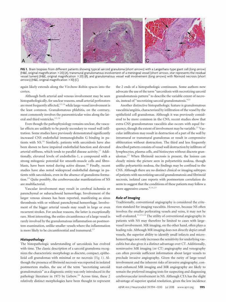
HistopathologyThe histopathologic understanding of sarcoidosis has evolved
with time. The classic description of a sarcoid granuloma recog-
nizes the characteristic morphology as discrete, compact, epithe-
lioid cell granulomas with minimal or no necrosis (Fig 1). Al-
though the presence of fibrinoid necrosis was reported in isolated
postmortem studies, the use of the term “necrotizing sarcoid
granulomatosis” as a diagnostic entity was only introduced in the
pathology literature in 1973 by Liebow.20 Across time, these 2
relatively distinct morphologies have been thought to represent
the 2 ends of a histopathologic continuum. Some authors now
advocate the use of the term “sarcoidosis with necrotizing sarcoid
granulomatosis pattern” to describe the variable extent of necro-
sis, instead of “necrotizing sarcoid granulomatosis.”21
Another distinctive histopathologic feature is granulomatous
vasculitis/angiitis, characterized by infiltration of the vessel by the
epithelioid cell granulomas. Although it was previously consid-
ered to be more common in the CNS, recent studies show that
extra-CNS granulomatous vasculitis also occurs with equal fre-
quency, though the extent of involvement may be variable.21 Vas-
cular infiltration may result in destruction of a part of the wall by
intramural or transmural granulomas or result in compressive
obliteration without destruction. The third and less frequently
described pattern consists of vessel wall destruction by infiltrate of
lymphocytes, plasma cells, and histiocytes without discrete gran-
ulomas.22 When fibrinoid necrosis is present, the lesions can
closely mimic the picture seen in polyarteritis nodosa, though
unlike polyarteritis nodosa, the findings may be confined to the
CNS. Although there are no distinct clinical or imaging subtypes
of patients with necrotizing sarcoid granulomatosis and fibrinoid
necrosis, isolated case reports with postmortem correlation do
seem to suggest that the conditions of these patients may follow a
more aggressive course.9,14,23
Role of ImagingTraditionally, conventional angiography is considered the crite-
rion standard for imaging vasculitis. However, because NS often
involves the smaller perforating vessels and veins, it may not be
well-evaluated.12,15,24 The utility of conventional angiography in
patients with NS may therefore be limited to cases with large-
vessel involvement. MR imaging, on the other hand, often plays a
leading role. Although MR imaging does not directly depict small
vessels, the superior ability to identify small infarcts and micro-
hemorrhages not only increases the sensitivity for underlying vas-
culitis but also gives it a distinct advantage over CT. Additionally,
noninvasive MR imaging (or CT) angiography and venography
can often provide sufficient information about larger vessels to
preclude invasive angiography. Given the rarity of large-vessel
involvement and the inherent risks of invasive angiography, con-
trast-enhanced MR imaging and MR angiography are likely to
remain the preferred imaging tests for suspecting and diagnosing
cerebrovascular involvement in NS. Although CTA has the slight
advantage of superior spatial resolution, given the low incidence
FIG 1. Brain biopsies from different patients showing typical sarcoid granuloma (short arrows) with a Langerhans-type giant cell (long arrow)(H&E, original magnification �20) (A), transmural granulomatous involvement of a meningeal vessel (short arrows, star represents the residualvessel lumen) (H&E, original magnification �20) (B), and granulomatous vessel wall involvement (long arrows) with fibrinoid necrosis (shortarrows) (H&E, original magnification �10) (C).
AJNR Am J Neuroradiol 39:1194 –1200 Jul 2018 www.ajnr.org 1195

of large-vessel vasculitis, it is unclear whether it adds any substan-
tial value over MRA, which can be combined with an MR imaging
study in a single session.
Ischemic LesionsIschemic insults in NS are intriguingly rare, despite the fre-
quent granulomatous vascular infiltration on postmortem
studies.3,4,12,15,25 The exact mechanism of infarction is likely mul-
tifactorial, resulting from a combination of small-vessel vasculitis,
large-vessel inflammation, or cardioembolic phenomenon sec-
ondary to sarcoid cardiomyopathy.2,26 Because perforating arter-
ies are most frequently involved, the infarcts are often small and
commonly involve the basal ganglia, thalamus, and brain stem,
though large-vessel and recurrent infarcts may also occur.25
In the pre-CT era, most of the infarcts were diagnosed on
postmortem studies. Commonly, these did not have a docu-
mented antemortem presentation.14,23,27-29 It is unclear whether
the infarcts were clinically silent due to their small size or were
overshadowed by the clinical manifestations of meningism, neu-
rologic deficits, and hydrocephalus known to accompany NS.
These predominantly involved the distribution of perforating ar-
teries. Most of the described patients were young and male and
had concurrent extracranial sarcoidosis.
At times, the ischemic insults in NS may be symptomatic and
present as acute stroke with or without a preceding transient isch-
emic attack, or even as recurrent TIAs without stroke.2,5,10,25,26
Table 1 lists the cases of acute stroke reported in patients with NS
in the English language literature.2-5,12,15,24-26,30-32 Like the asymp-
tomatic infarcts, most of the patients were young and nearly all
had coexisting extracranial disease. Also, unlike sarcoidosis,
which has a female predilection, cases with cerebrovascular find-
ings showed a male predilection.
Given the existing literature, one may therefore expect to see
both silent and symptomatic lesions on imaging, which is concor-
dant with the institutional experience of the authors. The infarcts
are often small and may be most apparent on diffusion-weighted
images as foci of restricted diffusion, often in the deep brain sub-
stance. T2-weighted images may not be very useful and any sur-
rounding parenchymal edema or white matter lesions tend to
make them even more inconspicuous. Occasionally, the infarcts
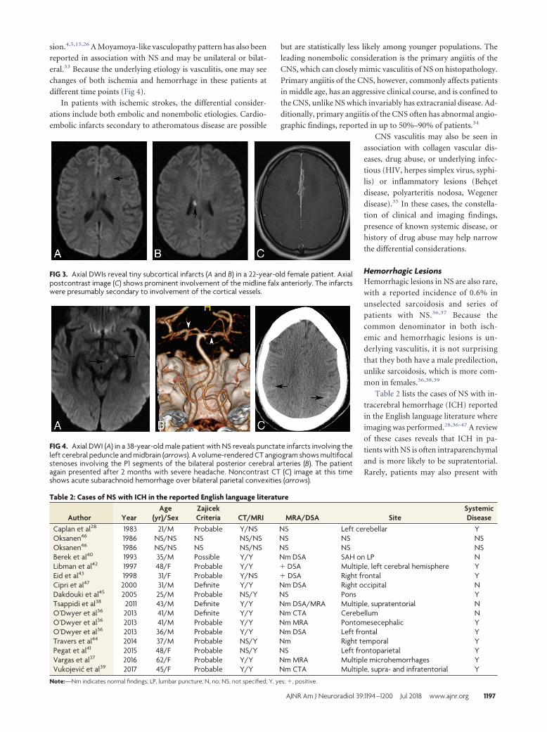
may be multiple, recurrent, or of varying ages (Fig 2).4,25 Rarely,
lesions may be more superficial and subcortical, occurring sec-
ondary to involvement of the cortical vessels from surrounding
meningeal inflammation (Fig 3). Although the presence of in-
farcts by itself is not specific for underlying NS, its occurrence,
especially in young patients with coexisting intracranial findings
of NS or known systemic sarcoidosis, may point to an underlying
granulomatous etiology. Rarely, an ischemic insult may be the
first manifestation of isolated NS without systemic involvement.2
In such cases, the diagnosis may be especially challenging and
eventually require histopathologic confirmation.
Large-vessel strokes in NS are exceptionally rare, as can be
expected from the less severe and noncircumferential vessel wall
involvement on pathology.13,25,26 MRA or conventional angiog-
raphy in such cases may show focal vascular stenosis or occlu-
FIG 2. Axial DWIs at the level of the corona radiata obtained at 2 different time points (A and B) show recurrent periventricular infarcts. Axialmagnified pre- (C) and postcontrast (D) images reveal subtle perivascular enhancement involving the bilateral basal ganglia (arrowheads, D).
Table 1: Cases of NS with stroke in the reported English language literaturea
Author YearAge
(yr)/SexZajicekCriteria CT/MRI DWI TIA Stroke
SystemicDisease
Brown et al12 1989 25/M Probable Y/NS NS Y Left IC and corona radiata YCorse and Stern26 1990 38/M Probable Y/Y NS Y Right IC YMichotte et al4 1991 29/M Probable Y/Y NS N Left BG, right frontal YZadra et al24 1996 30/M Probable Y/Y NS N Multiple, anterior circulation YDas et al30 1998 27/F Probable Y/NS NS N Left MCA infarct YBrisman et al3 2006 41/M Probable NS/Y Y N Multiple, anterior circulation YHodge et al2 2007 36/F Definite NS/Y Y N Left BG NNavi and DeAngelis31 2009 35/M Definite NS/Y N N Pons YNavi and DeAngelis31 2009 46/F Definite Y/Y NS N Pons YGonzález-Aramburu et al15 2012 27/M Probable Y/Y Y Y Right IC and thalamus YCampbell et al32 2015 26/M Probable Y/Y Y Y Left centrum semiovale YRaza and Schreck25 2017 73/F Probable NS/Y NS N Recurrent left MCA strokes YMacedo et al5 2016 62/M Possible Y/Y Y Y Multiple, anterior/posterior circulation Y
Note:—BG indicates basal ganglia; IC, internal capsule; N, no; NS, not specified; Y, yes.a On the basis of the Zajicek criteria,1 cases may be definite (neural tissue biopsy), probable (presence of CNS inflammation with evidence of systemic disease), or possible (clinicalpresentation consistent with NS with exclusion of alternate diagnosis).
1196 Bathla Jul 2018 www.ajnr.org
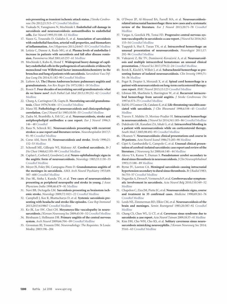
sion.4,5,15,26 A Moyamoya-like vasculopathy pattern has also been
reported in association with NS and may be unilateral or bilat-
eral.33 Because the underlying etiology is vasculitis, one may see
changes of both ischemia and hemorrhage in these patients at
different time points (Fig 4).
In patients with ischemic strokes, the differential consider-
ations include both embolic and nonembolic etiologies. Cardio-
embolic infarcts secondary to atheromatous disease are possible
but are statistically less likely among younger populations. The
leading nonembolic consideration is the primary angiitis of the
CNS, which can closely mimic vasculitis of NS on histopathology.
Primary angiitis of the CNS, however, commonly affects patients
in middle age, has an aggressive clinical course, and is confined to
the CNS, unlike NS which invariably has extracranial disease. Ad-
ditionally, primary angiitis of the CNS often has abnormal angio-
graphic findings, reported in up to 50%–90% of patients.34
CNS vasculitis may also be seen in
association with collagen vascular dis-
eases, drug abuse, or underlying infec-
tious (HIV, herpes simplex virus, syphi-
lis) or inflammatory lesions (Behcet
disease, polyarteritis nodosa, Wegener
disease).35 In these cases, the constella-
tion of clinical and imaging findings,
presence of known systemic disease, or
history of drug abuse may help narrow
the differential considerations.
Hemorrhagic LesionsHemorrhagic lesions in NS are also rare,
with a reported incidence of 0.6% in
unselected sarcoidosis and series of
patients with NS.36,37 Because the
common denominator in both isch-
emic and hemorrhagic lesions is un-
derlying vasculitis, it is not surprising
that they both have a male predilection,
unlike sarcoidosis, which is more com-
mon in females.36,38,39
Table 2 lists the cases of NS with in-
tracerebral hemorrhage (ICH) reported
in the English language literature where
imaging was performed.28,36-47 A review
of these cases reveals that ICH in pa-
tients with NS is often intraparenchymal
and is more likely to be supratentorial.
Rarely, patients may also present with
FIG 3. Axial DWIs reveal tiny subcortical infarcts (A and B) in a 22-year-old female patient. Axialpostcontrast image (C) shows prominent involvement of the midline falx anteriorly. The infarctswere presumably secondary to involvement of the cortical vessels.
FIG 4. Axial DWI (A) in a 38-year-old male patient with NS reveals punctate infarcts involving theleft cerebral peduncle and midbrain (arrows). A volume-rendered CT angiogram shows multifocalstenoses involving the P1 segments of the bilateral posterior cerebral arteries (B). The patientagain presented after 2 months with severe headache. Noncontrast CT (C) image at this timeshows acute subarachnoid hemorrhage over bilateral parietal convexities (arrows).
Table 2: Cases of NS with ICH in the reported English language literature
Author YearAge
(yr)/SexZajicekCriteria CT/MRI MRA/DSA Site
SystemicDisease
Caplan et al28 1983 21/M Probable Y/NS NS Left cerebellar YOksanen46 1986 NS/NS NS NS/NS NS NS NSOksanen46 1986 NS/NS NS NS/NS NS NS NSBerek et al40 1993 35/M Possible Y/Y Nm DSA SAH on LP NLibman et al42 1997 48/F Probable Y/Y � DSA Multiple, left cerebral hemisphere YEid et al43 1998 31/F Probable Y/NS � DSA Right frontal YCipri et al47 2000 31/M Definite Y/Y Nm DSA Right occipital NDakdouki et al45 2005 25/M Probable NS/Y NS Pons YTsappidi et al38 2011 43/M Definite Y/Y Nm DSA/MRA Multiple, supratentorial NO’Dwyer et al36 2013 41/M Definite Y/Y Nm CTA Cerebellum NO’Dwyer et al36 2013 41/M Probable Y/Y Nm MRA Pontomesecephalic YO’Dwyer et al36 2013 36/M Probable Y/Y Nm DSA Left frontal YTravers et al44 2014 37/M Probable NS/Y Nm Right temporal YPegat et al41 2015 48/F Probable NS/Y NS Left frontoparietal YVargas et al37 2016 62/F Probable Y/Y Nm MRA Multiple microhemorrhages YVukojevic et al39 2017 45/F Probable Y/Y Nm CTA Multiple, supra- and infratentorial Y
Note:—Nm indicates normal findings; LP, lumbar puncture; N, no; NS, not specified; Y, yes; �, positive.
AJNR Am J Neuroradiol 39:1194 –1200 Jul 2018 www.ajnr.org 1197

subarachnoid hemorrhage.40 For unclear reasons, patients with
symptomatic ICH seem to have a lobar predilection. This is some-
what counterintuitive because perivascular involvement in NS is
most prominent in the basal ganglia, brain stem, and spinal
cord.12-14 Isolated postmortem reports in patients with NS with
ICH have also documented hemorrhages in the basal ganglia and
posterior fossa compared with the cerebral hemispheres.14,28 Al-
though some of the apparent lobar predilection may be from a
reporting bias of larger symptomatic lesions, the reasons for rel-
ative sparing of the basal ganglia in the reported studies are un-
clear and unlikely to be entirely coincidental. Like some of the
recently reported cases37,39,42 and the authors’ personal experi-
ences, distinct peripheral distribution of the ICH with sparing of
the basal ganglia may be seen, though its significance is not clear
(Fig 5).
The overall heterogeneity of the lesion distribution may be
secondary to pathologically demonstrated “mixed” venous and
arterial involvement.36 As may be expected with predominantly
small-vessel involvement, hemorrhagic lesions in patients with
NS are often small and may be microhemorrhagic, though large
parenchymal hematomas do rarely occur.36,37,43 Similar to isch-
emic strokes, multiple or recurrent hemorrhages may also be
seen.37,39,42 Also analogous to cases with ischemic lesions, angio-
graphic studies (DSA/MRA/CTA) are often unyielding. As shown
in Table 2, of the 11 cases in which an
angiographic evaluation was performed,
positive findings were only noted in 2
cases.
The initial diagnosis of NS in patients
presenting with ICH can be very chal-
lenging, especially because almost half of
these patients may not have known sar-
coidosis.46 Although it has been previ-
ously reported that about 31% of these
patients have isolated NS, as noted in
Table 2, most of these patients do have
extra-CNS manifestations, though they
may not be symptomatic.36 Neverthe-
less, atypical locations of hemorrhage or
multiple smaller or microhemorrhagic
lesions, especially in the presence of other imaging signs of NS,
should raise the suspicion in the appropriate clinical setting.36,44
There is scarce literature on the impact of immunosuppressive
therapy on patients with ischemic lesions. However, cases with
hemorrhagic lesions do appear responsive to early aggressive an-
tigranulomatous therapy, an important consideration because
the microhemorrhages tend to increase with time and have been
anecdotally associated with cognitive decline.36,37 An increase in
intraparenchymal hemorrhages associated with steroid tapering
or discontinuation has also been reported in multiple recent cases,
highlighting the need for close follow-up while tapering immu-
nosuppressive therapy.37,39,44,45
The primary differential consideration of ICH in NS includes
vasculitis, especially primary angiitis of the CNS, as discussed pre-
viously. Similar to the NS patients with ischemic lesions, the an-
giographic studies in patients with ICH are also often negative,
though some cases may show large vessel vasculitis.36,42,43,46
Other differential considerations may include cerebral amyloid
angiopathy, hypertensive angiopathy, and cerebral autosomal
dominant arteriopathy with subcortical infarcts and leukoen-
cephalopathy, though these tend to affect older patients in the first
2 instances and often have a predilection for the temporal lobes
and external capsule in the latter.37
Dural Sinus InvolvementDural sinus thrombosis in NS is exceptionally rare, with only 5
cases reported in the literature.48-52 The underlying mechanism is
unclear and is presumably secondary to granulomatous menin-
geal involvement.51,52 Most patients present with symptoms of
raised intracranial pressure. Given the rare occurrence, there is
limited available imaging literature. The superior sagittal sinus is
most frequently involved. Byrne and Lawton49 reported a case of
meningeal sarcoidosis in a 32-year-old woman with an obstructed
straight sinus and bilateral transverse sinuses on angiography.
They also noted meningeal enhancement along the occluded si-
nuses without any change in appearance on follow-up imaging at
5 years. The patient had imaging findings of intracranial hyper-
tension, including an empty sella and erosion of the dorsum sella.
Leeds et al52 presented a case of superior sagittal sinus throm-
bosis, which they attributed to surrounding granulomatous in-
flammation. However, the images were presented as a part of
FIG 5. Axial SWI at the level of centrum semiovale (A) and basal ganglia (B) from 1 patient and atthe level of basal ganglia in the other patient (C). There are multiple scattered foci of susceptibilityinvolving bilateral subcortical white matter (arrows) with relative sparing of the basal ganglia inboth patients.
FIG 6. A and B, Axial postcontrast images through the posterior fossaobtained over an interval of 10 years. The prominent left sigmoid sinusenhancement remains essentially unchanged. The patient also hadsuperior sagittal sinus involvement (not shown).
1198 Bathla Jul 2018 www.ajnr.org

an imaging review article with no additional patient-specific de-
tails. Chapelon et al51 also reported a case of a 26-year-old man
with NS and superior sagittal sinus thrombosis in their review of
35 patients without much additional information.51 Akova et al48
presented a case report of possible NS in a 35-year-old man with
superior sagittal sinus thrombosis associated with meningeal sar-
coidosis and clinical features of pseudotumor cerebri.
More recently, Degardin et al50 presented a case of a 47-year-
old man with superior sagittal sinus thrombosis. They also noted
that the thrombosis persisted at 6-month follow-up despite satis-
factory anticoagulation. The patient also had large-vessel involve-
ment as well as multiple intraparenchymal hemorrhages of vary-
ing ages, which were attributed to granulomatous angiitis. We
also encountered a similar case of thrombosis in a 61-year-old
male patient involving the sagittal and left transverse sinuses over
a background of multiple intraparenchymal hemorrhages of vary-
ing ages. Our patient also showed a persistently enlarged and en-
hancing sinus with surrounding collaterals that remained un-
changed for 10 years (Fig 6). This feature contrasts with a bland
thrombus, which is usually dissolved with anticoagulation, leav-
ing behind a normal or attenuated vessel and a collateral network.
The persistent imaging findings are possibly from continued
granulomatous sinus involvement.
Cavernous Sinus InvolvementCavernous sinus involvement in NS is again exceedingly rare, with
only 2 prior reported cases.53,54 Chang et al53 reported a case of
left cavernous sinus involvement in a 54-year-old man with con-
current pulmonary sarcoidosis. The other reported case was by
Kim et al54 of a 40-year-old man with NS localized to the right
cavernous sinus without any additional CNS or extra-CNS in-
volvement. In both cases, the imaging findings were nonspecific
and showed a T1-isointense, T2-hyperintense, enhancing cavern-
ous sinus mass. We also encountered a case like that of Chang et al
with nodular thickening of the cavernous sinus, most apparent on
the contrast-enhanced images (Fig 7). Overall, the rarity of cav-
ernous sinus involvement, the scarcity of literature, and nonspe-
cific imaging findings preclude any meaningful conclusions about
the expected MR imaging findings.
The differential considerations in such cases are invariably
broad and include neoplasms, infective lesions, or inflammatory
conditions such as Tolosa-Hunt syndrome. Coexisting CNS or
extra-CNS findings of sarcoidosis may
be the only helpful clue in such cases.
CONCLUSIONSThe authors present a review of the var-
ious cerebrovascular manifestations that
may be encountered in patients with NS.
Although rare, these are of utmost im-
portance, not only because of the associ-
ated morbidity but also because patients
may present with isolated NS and pose
considerable diagnostic challenges, es-
pecially when the manifestations are
atypical. Vasculitic manifestations of NS
should be considered in patients with re-
current hemorrhages or infarcts, espe-
cially when the patients are younger and demonstrate additional
intracranial imaging findings that may support NS.
Disclosures: Suyash Mohan—UNRELATED: Grants/Grants Pending: Novocure, Ra-diological Society of North America, Guerbet, Comments: research grants*. *Moneypaid to the institution.
REFERENCES1. Bathla G, Singh AK, Policeni B, et al. Imaging of neurosarcoidosis:
common, uncommon, and rare. Clin Radiol 2016;71:96–106 CrossRefMedline
2. Hodge MH, Williams RL, Fukui MB. Neurosarcoidosis presentingas acute infarction on diffusion-weighted MR imaging: summary ofradiologic findings. AJNR Am J Neuroradiol 2007;28:84 – 86 Medline
3. Brisman JL, Hinduja A, McKinney JS, et al. Successful emergent an-gioplasty of neurosarcoid vasculitis presenting with strokes. SurgNeurol 2006;66:402– 04 CrossRef Medline
4. Michotte A, Dequenne P, Jacobovitz D, et al. Focal neurological deficitwith sudden onset as the first manifestation of sarcoidosis: a case re-port with MRI follow-up. Eur Neurol 1991;31:376–79 Medline
5. Macedo PJ, da Silveira VC, Ramos LT, et al. Isolated central nervoussystem vasculitis as a manifestation of neurosarcoidosis. J StrokeCerebrovas Dis 2016;25:e89 – e92 CrossRef Medline
6. Smith JK, Matheus MG, Castillo M, et al. Imaging manifestations ofneurosarcoidosis. AJR Am J Roentgenol 2004;182:289 –95 CrossRefMedline
7. Izbicki G, Chavko R, Banauch GI, et al. World Trade Center “sar-coid-like” granulomatous pulmonary disease in New York City FireDepartment rescue workers. Chest J 2007;131:1414 –23 CrossRefMedline
8. Iwai K, Takemura T, Kitalchi M, et al. Pathological studies on sar-coidosis autopsy, II: early change, mode of progression and deathpattern. Acta Pathol Jpn 1993;43:377– 85 Medline
9. Robert F. Sarcoidosis of the central nervous system: report of a caseand review of the literature. Arch Neurol 1962;7:442– 49 CrossRefMedline
10. Duffey P, Bates D. Transient focal neurological deficit in sarcoid-osis. Sarcoidosis Vasc Diffuse Lung Dis 1997;14:171–72 Medline
11. Camp WA, Frierson JG. Sarcoidosis of the central nervous system: acase with postmortem studies. Arch Neurol 1962;7:432– 41 CrossRefMedline
12. Brown MM, Thompson AJ, Wedzicha JA, et al. Sarcoidosis present-ing with stroke. Stroke 1989;20:400 – 05 CrossRef Medline
13. Reske-Nielsen E, Harmsen A. Periangiitis and panangiitis as a man-ifestation of sarcoidosis of the brain: report of a case. J Nerv MentDis 1962;135:399 – 412 CrossRef Medline
14. Herring AB, Urich H. Sarcoidosis of the central nervous system.J Neurol Sci 1969;9:405–22 CrossRef Medline
15. Gonzalez-Aramburu I, Ruiz-Perez E, Gomez-Roman J, et al. Sarcoid-
FIG 7. Coronal pre- (A) and postcontrast (B) images in a patient with NS show nodular thickeningand enhancement of the cavernous sinuses bilaterally. The patient had additional intracranialstigmata of NS (not shown), and the cavernous sinus involvement was presumed to be secondaryto NS.
AJNR Am J Neuroradiol 39:1194 –1200 Jul 2018 www.ajnr.org 1199
osis presenting as transient ischemic attack status. J Stroke Cerebro-vasc Dis 2012;21:515–17 CrossRef Medline
16. Tsukada N, Yanagisawa N, Mochizuki I. Endothelial cell damage insarcoidosis and neurosarcoidosis: autoantibodies to endothelialcells. Eur Neurol 1995;35:108 –12 Medline
17. Siasos G, Tousoulis D, Gialafos E, et al. Association of sarcoidosiswith endothelial function, arterial wall properties, and biomarkersof inflammation. Am J Hypertens 2011;24:647–53 CrossRef Medline
18. Letizia C, Danese A, Reale MG, et al. Plasma levels of endothelin-1increase in patients with sarcoidosis and fall after disease remis-sion. Panminerva Med 2001;43:257– 61 Medline
19. Mochizuki I, Kubo K, Hond T. Widespread heavy damage of capil-lary endothelial cells in the pathogenesis of sarcoidosis: evidence bymonoclonal von Willebrand factor immunohistochemistry in thebronchus and lung of patients with sarcoidosis. Sarcoidosis Vasc Dif-fuse Lung Dis 2014;31:182–90 CrossRef Medline
20. Liebow AA. The J Burns Amberson lecture: pulmonary angiitis andgranulomatosis. Am Rev Respir Dis 1973;108:1–18 Medline
21. Rosen Y. Four decades of necrotizing sarcoid granulomatosis: whatdo we know now? Arch Pathol Lab Med 2015;139:252– 62 CrossRefMedline
22. Churg A, Carrington CB, Gupta R. Necrotizing sarcoid granuloma-tosis. Chest 1979;76:406 –13 CrossRef Medline
23. Manz HJ. Pathobiology of neurosarcoidosis and clinicopathologiccorrelation. Can J Neurol Sci 1983;10:50 –55 CrossRef Medline
24. Zadra M, Brambilla A, Erli LC, et al. Neurosarcoidosis, stroke andantiphospholipid antibodies: a case report. Eur J Neurol 1996;3:146 – 48 CrossRef
25. Raza N, Schreck KC. Neurosarcoidosis presenting with recurrentstrokes: a case report and literature review. Neurohospitalist 2017;7:91–95 CrossRef Medline
26. Corse AM, Stern BJ. Neurosarcoidosis and stroke. Stroke 1990;21:152–53 Medline
27. Schonell ME, Gillespie WJ, Maloney AF. Cerebral sarcoidosis. Br JDis Chest 1968;62:195–99 CrossRef Medline
28. Caplan L, Corbett J, Goodwin J, et al. Neuro-ophthalmologic signs inthe angiitic form of neurosarcoidosis. Neurology 1983;33:1130 –35CrossRef Medline
29. Meyer JS, Foley JM, Campagna-Pinto D. Granulomatous angiitis ofthe meninges in sarcoidosis. AMA Arch Neurol Psychiatry 1953;69:587– 600 CrossRef Medline
30. Das SK, Sinha I, Kundu TN, et al. Two cases of neurosarcoidosispresenting as peripheral neuropathy and stroke in young. J AssocPhysicians India 1998;46:479 – 81 Medline
31. Navi BB, DeAngelis LM. Sarcoidosis presenting as brainstem isch-emic stroke. Neurology 2009;72:1021–22 CrossRef Medline
32. Campbell J, Kee R, Bhattacharya D, et al. Systemic sarcoidosis pre-senting with headache and stroke-like episodes. Case Rep Immunol2015;2015:619867 CrossRef Medline
33. Ko JK, Lee SW, Choi CH. Moyamoya-like vasculopathy in neuro-sarcoidosis. J Korean Neurosurg Soc 2009;45:50 –52 CrossRef Medline
34. Birnbaum J, Hellmann DB. Primary angiitis of the central nervoussystem. Arch Neurol 2009;66:704 – 09 CrossRef Medline
35. Grossman RI, Yousem DM. Neuroradiology: The Requisites. St Louis:Mosby; 2003:196 –204
36. O’Dwyer JP, Al-Moyeed BA, Farrell MA, et al. Neurosarcoidosis-related intracranial haemorrhage: three new cases and a systematicreview of the literature. Eur J Neurol 2013;20:71–78 CrossRefMedline
37. Vargas A, Gorelick PB, Testai FD. Progressive central nervous sys-tem vasculopathy in sarcoidosis: a case report. J Neurol Sci 2016;362:153–54 CrossRef Medline
38. Tsappidi S, Hui F, Turan TN, et al. Intracerebral hemorrhage: anunusual presentation of neurosarcoidosis. Neurologist 2011;17:292–96 CrossRef Medline
39. Vukojevic Z, Ilic TV, Dominovic-Kovacevic A, et al. Neurosarcoid-osis and multiple intracerebral hematomas: an unusual clinicalpresentation. J Neurol Sci 2017;379:22–24 CrossRef Medline
40. Berek K, Kiechl S, Willeit J, et al. Subarachnoid hemorrhage as pre-senting feature of isolated neurosarcoidosis. Clin Investig 1993;71:54 –56 Medline
41. Pegat B, Drapier S, Morandi X, et al. Spinal cord hemorrhage in apatient with neurosarcoidosis on long-term corticosteroid therapy:case report. BMC Neurol 2015;15:123 CrossRef Medline
42. Libman RB, Sharfstein S, Harrington W, et al. Recurrent intracere-bral hemorrhage from sarcoid angiitis. J Stroke Cerebrovasc Dis1997;6:373–75 CrossRef Medline
43. Eid H, O’Connor CR, Catalano E, et al. Life-threatening vasculitis asso-ciated with sarcoidosis. J Clin Rheumatol 1998;4:338–43 CrossRefMedline
44. Travers F, Maltete D, Morisse-Pradier H. Intracranial hemorrhagein neurosarcoidosis. J Neurol Sci 2014;341:185– 86 CrossRef Medline
45. Dakdouki GK, Kanafani ZA, Ishak G, et al. Intracerebral bleeding ina patient with neurosarcoidosis while on corticosteroid therapy.South Med J 2005;98:492–95 CrossRef Medline
46. Oksanen V. Neurosarcoidosis: clinical presentations and course in50 patients. Acta Neurol Scand 1986;73:283–90 Medline
47. Cipri S, Gambardella G, Campolo C, et al. Unusual clinical presen-tation of cerebral-isolated sarcoidosis: case report and review of theliterature. J Neurosurg Sci 2000;44:140 – 44 Medline
48. Akova YA, Kansu T, Duman S. Pseudotumor cerebri secondary todural sinus thrombosis in neurosarcoidosis. J Clin Neuroophthalmol1993;13:188 – 89 Medline
49. Byrne JV, Lawton CA. Meningeal sarcoidosis causing intracranialhypertension secondary to dural sinus thrombosis. Br J Radiol 1983;56:755–57 CrossRef Medline
50. Degardin A, Devos P, Vermersch P, et al. Cerebrovascular symptom-atic involvement in sarcoidosis. Acta Neurol Belg 2010;110:349 –52Medline
51. Chapelon C, Ziza JM, Piette JC, et al. Neurosarcoidosis: signs, courseand treatment in 35 confirmed cases. Medicine 1990;69:261–76CrossRef Medline
52. Leeds NE, Zimmerman RD, Elkin CM, et al. Neurosarcoidosis of thebrain and meninges. Semin Roentgenol 1985;20:387–92 CrossRefMedline
53. Chang CS, Chen WL, Li CT, et al. Cavernous sinus syndrome due tosarcoidosis: a case report. Acta Neurol Taiwan 2009;18:37–41 Medline
54. Kim DH, Cho WH, Cho KS, et al. Solitary cavernous sinus neuro-sarcoidosis mimicking neurosyphilis. J Korean Neurosurg Soc 2014;55:61– 63 CrossRef Medline
1200 Bathla Jul 2018 www.ajnr.org