cetogÉnesis en astrocitos
TRANSCRIPT

UNIVERSIDAD COMPLUTENSE DE MADRID
FACULTAD DE CIENCIAS BIOLÓGICAS
Departamento de Bioquímica y Biología Molecular I
CETOGÉNESIS EN ASTROCITOS: CARACTERIZACIÓN, REGULACIÓN Y POSIBLE
PAPEL CITOPROTECTOR
MEMORIA PARA OPTAR AL GRADO DE DOCTOR
PRESENTADA POR
Cristina Blázquez Ortiz
Bajo la dirección del doctor
Manuel Guzmán Pastor
Madrid, 2001 ISBN: 84-669-1680-6

UNIVERSIDAD COMPLUTENSE DE MADRIDUNIVERSIDAD COMPLUTENSE DE MADRID
FACULTAD DE CIENCIAS BIOLÓGICASFACULTAD DE CIENCIAS BIOLÓGICAS
DEPARTAMENTO DE BIOQUÍMICA Y BIOLOGÍA MOLECULAR IDEPARTAMENTO DE BIOQUÍMICA Y BIOLOGÍA MOLECULAR I
CETOGÉNESIS EN ASTROCITOS: CETOGÉNESIS EN ASTROCITOS:
CARACTERIZACIÓN, REGULACIÓN Y CARACTERIZACIÓN, REGULACIÓN Y
POSIBLE PAPEL CITOPROTECTORPOSIBLE PAPEL CITOPROTECTOR
TESIS DOCTORAL
Cristina Blázquez OrtizCristina Blázquez Ortiz
DIRECTOR: Dr. Manuel Guzmán Pastor Dr. Manuel Guzmán Pastor
Madrid, Abril de 2001

Este trabajo ha sido realizado en el Departamento de Bioquímica y Biología Molecular I de
la Facultad de Ciencias Biológicas de la Universidad Complutense de Madrid bajo la dirección del
Dr. Manuel Guzmán Pastor.

INDICE ABREVIATURAS
1. INTRODUCCIÓN
1.1 Astrocitos - Generalidades - Metabolismo en astrocitos
Degradación de glucosa Metabolismo del glucógeno Gluconeogénesis 1.2 Cetogénesis
- Generalidades - Carnitina palmitoiltransferasa I
Estructura e isoformas Regulación 1.3 Lipoapoptosis
- Generalidades - Ceramida - Serina palmitoiltransferasa
Estructura Regulación 1.4 Objetivos 1.5 Bibliografía
2. RESULTADOS Y DISCUSIÓN I CETOGÉNESIS EN ASTROCITOS: CARACTERIZACIÓN Y REGULACIÓN
- Role of carnitine palmitoyltransferase I in the control of ketogenesis in primary cultures of rat astrocytes.
(Journal of Neurochemistry 71: 1597-1606, 1998)
- The stimulation of ketogenesis by cannabinoids in cultured astrocytes defines carnitine palmitoyltransferase I as a new ceramide-activated enzime.
(Journal of Neurochemistry 72 : 1759-1768, 1999)
- The AMP-activated protein kinase is involved in the regulation of ketone body production by astrocytes.
(Journal of Neurochemistry 73: 1674-1682, 1999) Sumario

3. RESULTADOS Y DISCUSIÓN II CETOGÉNESIS EN ASTROCITOS: POSIBLE PAPEL CITOPROTECTOR
- De novo-synthesized ceramide signals apoptosis in astrocytes via extracellular signal-regulated kinase
(FASEB Journal, 14: 2315-2322, 2000)
- The AMP-activated protein kinase prevents ceramide synthesis de novo and apoptosis in astrocytes.
(FEBS Letters, 489: 149-153, 2001) Sumario
4. DISCUSIÓN GENERAL Y CONCLUSIONES
- Is there an astrocyte-neurone ketone body shuttle? (Trends in Endocrinology and Metabolism 12: 169-173, 2001) Sumario

ABREVIATURAS:
ACC Acetil-CoA carboxilasa ACC-265 Isoforma de ACC de 265 kDa ACC-280 Isoforma de ACC de 280 kDa AMPK Proteína quinasa activada por 5´-AMP cAMP AMP cíclico CAPK Proteína quinasa activada por ceramida CAPP Proteína fosfatasa activada por ceramida CPT-I Carnitina palmitoiltransferasa I CPT-II Carnitina palmitoiltransferasa II ERK Proteína quinasa activada por señales extracelulares GABA Ácido γ-aminobutírico GFAP Proteína glial fibrilar ácida HMG-CoA 3-hidroxi-3-metilglutaril-coA KSR Quinasa supresora de Ras L-CPT-I Isoforma hepática de CPT-I LDH Lactato deshidrogenasa M-CPT-I Isoforma muscular de CPT-I PKA Proteína quinasa dependiente de cAMP PKB Proteína quinasa B PKC Proteína quinasa C PPAR Receptor activado por proliferadores de peroxisomas SM Esfingomielina SMasa Esfingomielinasa SP Esfingosina SPT Serina palmitoiltransferasa

1. INTRODUCCIÓN1. INTRODUCCIÓN

Introducción
1
La Introducción de este trabajo no pretende ser una revisión exhaustiva de los distintos temas que se incluyen en ella, sino una puesta al día de los antecedentes para permitir al lector una mejor comprensión del enfoque del trabajo, diseño experimental y los resultados obtenidos.
1.1 Astrocitos
GENERALIDADES
Los astrocitos son las células no neuronales mayoritarias del sistema nervioso central, y se agrupan junto con los oligodendrocitos y la microglía en el término general de células gliales (Travis, 1994; Porter y McCarthy, 1997; Araque et al., 1999; Albright et al., 2000). Tanto las neuronas como la macroglía (astrocitos y oligodendrocitos) se originan a partir de precursores en las capas germinales del cerebro en desarrollo, concretamente en las zonas ventricular y subventricular. Las células de origen neurales multipotenciales están presentes en estas regiones durante el desarrollo. No obstante, hace unos años se demostró la existencia de precursores separados para neuronas y células gliales (Raff et al.,1983; Luskin et al., 1988; Mayer-Proschel et al.,1997; Rao et al.,1998; Shi et al., 1998).
Durante mucho tiempo se pensó que los progenitores que originan los linajes de neuronas y macroglía iban siendo segregados progresivamente durante el desarrollo, de manera que estas células madre estarían presentes en etapas tempranas de la histogénesis del cerebro (de Vellis y Carpenter, 1999). Sin embargo, la neurogénesis y la gliogénesis continúan a lo largo de la vida puesto que también se han aislado células precursoras multipotenciales en cerebro adulto. Se trata de células que poseen la capacidad de autorrenovarse y diferenciarse en neuronas, astrocitos y oligodendrocitos (Doetsch et al., 1999; Laywell et al., 2000). Así, se han identificado recientemente en el cerebro dos tipos celulares como precursores iniciales neurales. Ambos tipos celulares son células gliales: las células ependimales (Johansson et al., 1999) y los astrocitos de la zona subventricular (Doestch et al., 1999). Las células ependimales se diferencian mayoritariamente a células gliales, las cuales bordean la superficie
luminal de la zona ventricular en el cerebro adulto y provienen de un grupo de células de la zona ventricular (Barres, 1999) . Johansson et al. (1999), sin embargo, no fueron capaces de observar división de células ependimales ni in vivo ni in vitro. Por otro lado, los astrocitos de la zona subventricular también son células precursoras neurales multipotenciales y expresan la estructura, morfología y características antigénicas típicas de los astrocitos (Doestch et al., 1999; Magavi et al., 2000). Así, existe acuerdo en que al menos un grupo de células gliales son así mismo células progenitoras neurales, y difieren en cuanto a si las células progenitoras son células ependimales o astrocitos de la zona subventricular.
Además de éstos, se han identificado otros precursores celulares en el cerebro del adulto, como por ejemplo el precursor de oligodendrocitos. Así, en 1985, se demostró la existencia de un “progenitor celular O-2A”, que se puede diferenciar a oligodendrocitos o a astrocitos tipo 2: en cultivo y en presencia de suero se diferencia a astrocitos de tipo 2, mientras que en un medio químicamente definido lo hace a oligodendrocitos (Temple y Raff, 1985). Curiosamente, estas células progenitoras pueden incluso proliferar indefinidamente si las condiciones del medio son las adecuadas (Tang et al., 2001). Hoy en día, los astrocitos se detectan generalmente mediante la utilización de anticuerpos anti -GFAP (proteína glial fibrilar ácida), que es marcador específico de astrocitos. En concreto, los astrocitos tipo 1 expresan GFAP, glicoproteína Ran-2, no expresan gangliósido GD3 (=inmunorreactividad frente al anticuerpo A2B5), tienen aspecto poligonal y no derivan de las células O-2A. Por el contrario, los astrocitos tipo 2 expresan GFAP, no expresan Ran-2, expresan GD3, tienen forma estrellada y derivan de las células O-2A (Lee et al., 2000).
Más recientemente se ha identificado un tercer precursor glial con inmunorreactividad frente a A2B5 que difiere del precursor O-2A en su respuesta a factores de crecimiento y en su capacidad de diferenciación (Rao et al., 1998). Este precursor se denominó GRP, y puede dar lugar al diferenciarse a astrocitos tipo 1, astrocitos tipo 2 y oligodendrocitos (Lee et al., 2000).
El desarrollo del sistema nervioso central implica una interacción dinámica entre las neuronas y las células de soporte, incluyendo la macroglía y la microglía. En concreto, los

Introducción
2
astrocitos realizan un gran número de funciones que permiten un adecuado funcionamiento del cerebro y desempeñan un papel fundamental en el desarrollo de los patrones de organización anatómicos finales que caracterizan el cerebro adulto (Travis, 1994; Porter y McCarthy, 1997; Araque et al., 1999; Albright et al., 2000). Las células de astroglía son también responsables de la homeostasis de iones (especialmente K+) y aminoácidos (como glutamato y ácido γ-aminobutírico, GABA) en el medio extracelular, regulan el pH y participan en la recaptura de determinados neurotransmi-sores; además, la astroglía posee una notoria capacidad de regulación del volumen celular, pudiendo de esta forma influir indirectamente en el volumen extracelular. En el desarrollo, durante la formación del tubo neural, los astrocitos dirigen la migración de las neuronas y producen factores neurotróficos y de crecimiento esenciales para el desarrollo y supervivencia neuronal. Los astrocitos configuran el espacio sináptico, mantienen una comunicación bidireccional con las neuronas a través de los neurotransmisores y participan en la formación de sinapsis (Ullian et al., 2001). Además, y en el contexto de este trabajo, los astrocitos son células que proporcionan sustratos metabólicos esenciales para el metabolismo neuronal (Magistretti y Pellerin, 1996; Giaume et al., 1997) (Fig. 1). Todas estas funciones son posibles gracias a que los astrocitos poseen receptores de membrana para un gran número de neurotransmisores y neurohormonas, y están equipados con sistemas específicos de transducción de señales y de transportadores para la captura de los neurotransmisores.
METABOLISMO EN ASTROCITOS § Degradación de glucosa
La glucosa está considerada como el
principal nutriente para las células del sistema nervioso adulto (Wiesinger et al., 1997; Clarke y Sokoloff, 1999). Cada tipo de célula nerviosa utiliza sus propios sustratos; además, algunas células nerviosas son capaces de producir compuestos que actúan como fuente de energía para otros tipos celulares. Con respecto a esto, los astrocitos desempeñan un papel fundamental en el flujo de sustratos energéticos a las neuronas debido tanto a su localización estratégica como a su versatilidad metabólica (Magistretti y Pellerin, 1996; Deitmer 2000) (Fig.
1). La particular morfología de los astrocitos, con sus terminaciones alrededor de los capilares del parénquima cerebral, en los cuales se encuentra la fuente de glucosa, hace que los astrocitos sean la primera barrera celular que la glucosa ha de atravesar para penetrar en el cerebro. Esta situación privilegiada apoya el papel que desempeñan los astrocitos en la distribución de nutrientes desde la sangre hasta otras células cerebrales (Magistretti y Pellerin, 1999ab) (Fig. 1).
La utilización de la glucosa por los astrocitos implica mayoritariamente la producción de lactato y piruvato, los cuales no tienen que ser necesariamente metabolizados a través del ciclo de los ácidos tricarboxílicos, sino que, en su mayoría, son liberados al medio extracelular. La glucosa, además, puede incorporarse en lípidos, aminoácidos y glucógeno, y es precursor de algunos neurotransmisores como el GABA, el glutamato y la acetilcolina (Wiesinger et al., 1997; Hertz et al., 1999; Deitmer, 2000). Ciertos intermediarios metabólicos, bajo circunstancias particulares, pueden sustituir a la glucosa y actuar como sustratos alternativos del metabolismo energético cerebral (Magistretti y Pellerin, 1996, 1999ab). Así, el ayuno prolongado, la diabetes o la lactancia en neonatos producen un incremento en los niveles plasmáticos de los cuerpos cetónicos acetoacetato y 3-hidroxibutirato, que pueden ser utilizados por el cerebro como sustratos metabólicos y pueden preservar la integridad y excitabilidad neuronal, particularmente en el desarrollo (Robinson y Williamson, 1980; Edmond et al., 1985; Izumi et al., 1997).
El primer paso en la entrada de glucosa al interior de los astrocitos es el transporte a través de la membrana plasmática, proceso que es llevado a cabo por transportadores de tipo GLUT1 y GLUT5 (Tabernero et al., 1996; Vanucci et al., 1997, Vanucci y Vanucci, 2000). A continuación tiene lugar su fosforilación, siendo ésta a su vez una de las etapas limitantes en su metabolismo. Las células de astroglía expresan (mayoritariamente en la mitocondria) hexoquinasa 1, que transforma la glucosa en glucosa 6-fosfato. Ésta puede entrar en la ruta glicolítica, en la ruta de las pentosas fosfato, en la ruta del sorbitol, o bien almacenarse en forma de glucógeno (Wiesinger et al., 1997). La cantidad de glucosa metabolizada por la ruta de las pentosas fosfato en células de astroglía en cultivo es generalmente pequeña comparada con la

Introducción
3
metabolizada vía glicolítica. Además, la actividad glicolítica de células de astroglía en cultivo es predominantemente anaerobia y se considera el lactato como el principal producto metabólico (Wiesinger et al., 1997; Magistretti y Pellerin 1999ab). De hecho, el lactato mantiene la función sináptica en ausencia de glucosa en cortes de hipocampo (Schurr et al., 1988, 1999) y puede también mantener la función cognitiva en hipoglucemia (Maran et al., 1994; Magistretti y Pellerin, 1999ab) e hipoxia (Schurr y Rigor, 1998). El piruvato también mantiene la actividad sináptica y la morfología neuronal durante la carencia de glucosa o cuando se administra iodoacetato, un inhibidor de la glicolisis (Izumi et al., 1994; 1997). Así, la astroglía proporciona a las neuronas vecinas monocarboxilatos (lactato y piruvato) durante situaciones de elevado requerimiento energético o carencia de glucosa. La utilización de éstos como fuente de energía requiere metabolismo oxidativo, el cual queda comprometido en los casos de anoxia, pero puede tener lugar si la glicolisis está inhibida. Con bajos niveles de glucosa los astrocitos son capaces de mantener la actividad neuronal por un largo período de tiempo vía liberación de monocarboxilatos (Izumi et al., 1997; Schurr y Rigor, 1998).
Fig. 1. Representación esquemática de la relación citológica entre los astrocitos, las neuronas y los vasos sanguíneos. Abreviaturas: Glc, glucosa; NT, neurotransmisores.
Para poder ser utilizado como sustrato
celular, el lactato ha de transportarse a través de las membranas mediante transportadores
específicos. Se han identificado al menos dos transportadores de monocarboxilatos en cerebro con actividad frente a lactato, piruvato, y los cuerpos cetónicos 3-hidroxibutirato y acetoacetato: MCT1 (expresado en células endoteliales vasculares y astrocitos) y MCT2 (expresado fundamentalmente en neuronas de hipocampo y corticales) (Gerhart et al., 1997; Bröer et al., 1997; Pellerin et al., 1998). A continuación, el lactato ha de convertirse en piruvato, constituyendo así una fuente metabólica eficiente de ATP, puesto que una molécula de piruvato proporciona 15 moléculas de ATP tras su oxidación a través de la piruvato deshidrogenasa y el ciclo de los ácidos tricarboxílicos. La enzima responsable de esta conversión es la lactato deshidrogenasa (LDH), cuya expresión e isoformas varían en los distintos tejidos. Bittar et al. (1996) observaron que la isoforma LDH-5, que favorece la producción de lactato, se encuentra mayoritariamente en astroglía, mientras que la LDH-1, isoforma que favorece la utilización de lactato, se encuentra mayoritariamente en las neuronas. Estos datos apoyan la idea de que los astrocitos procesarían la glucosa vía glicolisis generando lactato, el cual, una vez liberado al medio y posteriormente capturado por las neuronas, sería transformado a piruvato, que tras entrar en el ciclo de los ácidos tricarboxílicos proporcionaría la energía necesaria para la célula (Magistretti y Pelllerin, 1999ab).
En relación con este proceso, Magistretti y colaboradores han observado que el glutamato, el principal neurotransmisor excitatorio del sistema nervioso central, estimula la captura de glucosa y la liberación de lactato por astrocitos. La captura del glutamato ocurre a través de transportadores, dos de los cuales son predominantemente, si no exclusivamente, específicos de células gliales. Se trata de los transportadores GLT-1 (=EAAT2) y GLAST (=EAAT1) (Robinson y Dowd, 1997; Araque et al., 1999). Este transporte de glutamato en astrocitos está dirigido por un gradiente electroquímico de Na+: una molécula de glutamato es cotransportada con tres iones Na+ hacia el interior celular y como consecuencia, un ión K+ sale al exterior. Esto conduce a un incremento de la concentración intracelular de Na+ , que es equilibrado a través de la Na+/K+-ATPasa. Este proceso consume ATP e implica la activación de la glicolisis y la producción de lactato (Pellerin y Magistretti, 1994, 1997; Takahashi et al., 1995; Magistretti y Pellerin,
Lactato
Lactato
K+
NT
Glc
Neuronas
Astrocito
CapilarGlc

Introducción
4
1999ab) (Fig. 2). Un gran número de evidencias experimentales apoya que este efecto del glutamato depende exclusivamente de sus transportadores y no de sus receptores (Pellerin y Magistretti, 1994, 1997; Takahashi et al., 1995).
De esta manera se consigue un acoplamiento entre actividad neuronal, liberación de glutamato, activación de la glicolisis en astrocitos y aporte de lactato como fuente energética para las neuronas (Fig. 2).
Fig. 2. Esquema de las rutas metabólicas de la glucosa en astrocitos y de la activación de la glicolisis por glutamato en condiciones de activación neuronal.
§ Metabolismo del glucógeno El glucógeno constituye la principal reserva
energética del cerebro y está mayoritariamente localizado en los astrocitos, aunque también está presente en células del plexo coroideo y ependimal, así como en algunas neuronas del tallo cerebral. El cerebro puede considerarse como un órgano almacenador de glucógeno, cuya función podría ser proveer unidades de glucosa durante la actividad fisiológica. De hecho, el recambio de glucógeno es extremadamente rápido en los astrocitos y se correlaciona con la actividad de las neuronas vecinas (Magistretti y Pellerin, 1996; Choi et al., 1999; Deitmer, 2000; Wender et al., 2000). La degradación de glucógeno astroglial resulta activada por diversos neurotransmisores (entre los que se encuentran péptido intestinal vasoactivo, glutamato, noradrenalina, serotonina e histamina) vía cAMP y Ca2+ (Magistretti y Pellerin, 1996;
Pellerin et al., 1997, Hamai et al., 1999). Una disminución de los niveles de glucosa del medio causa una pérdida en el glucógeno acumulado (Dringen y Hamprecht, 1992, 1993), mientras que la insulina y factores de crecimiento análogos incrementan los niveles de glucógeno almacenado en los astrocitos (Pellerin et al., 1997; Hamai et al., 1999).
El aspecto quizás más importante de la degradación del glucógeno es el destino de los residuos glicosílicos. Los residuos liberados no aparecen mayoritariamente como glucosa libre en el medio, sino que los astrocitos liberan principalmente ácido láctico (Dringen et al., 1993; Magistretti y Pellerin, 1996). Alternativamente, la glucosa procedente de la degradación del glucógeno puede metabolizarse vía ruta de las pentosas fosfato para generar NADPH y proteger a los astrocitos frente al estrés oxidativo (Dringen, 2000; Rahman et al., 2000). Aunque se ha detectado actividad, mRNA y proteína de glucosa
Glucosa Glucosa Glucosa-6-P
Glucógeno
Piruvato
Rutapentosasfosfato
Glicolisis
Lactato Lactato
Na+/K+
ATPasaATP
ADPK+
Glutamato
CicloTCA
CO2
K+
Na+Na+
GlutamatoGLUT 1/5
EAAT 1/2
MCT 1
Actividadneuronal

Introducción
5
6-fosfatasa en astrocitos (Forsyth et al., 1993), estudios posteriores no han sido capaces de reproducir estos hallazgos (Magistretti y Pellerin, 1999ab; Gotoh et al., 2000).
§ Gluconeogénesis
En los astrocitos también se da el
proceso de gluconeogénesis hasta glucosa 6-fosfato, y de hecho se ha descrito la presencia de tres enzimas implicadas exclusivamente en la ruta (piruvato carboxilasa, fosfoenolpiruvato carboxiquinasa y fructosa 1,6-bisfosfatasa) en estas células (Wiesinger et al., 1997). La gluconeogénesis en astrocitos podría actuar como mecanismo tamponador, controlando la osmolaridad del fluido extracelular y reciclando el lactato generado por las células vecinas, derivándolo hacia rutas como la síntesis de glucógeno (Schmoll et al., 1995; Wiesinger et al., 1997; Jitrapakdee y Wallace, 1999).
1.2 Cetogénesis
GENERALIDADES
La mayoría de los estudios sobre cetogénesis y oxidación de ácidos grasos realizados hasta el momento se han llevado a cabo en el hígado. La producción de cuerpos cetónicos por las mitocondrias del hígado es un proceso complejo y finamente regulado (Guzmán y Geelen, 1993; Zammit, 1994). En primer lugar, la célula debe capturar ácidos grasos. Aunque tradicionalmente se ha asumido que los ácidos grasos atraviesan la membrana plasmática siguiendo un proceso no saturable de difusión simple (Cooper et al., 1989), durante los últimos años se ha ido acumulando una cierta evidencia que apoya la existencia de un sistema saturable de transporte facilitado de ácidos grasos. En concreto, se ha identificado una proteína de membrana ligante de ácidos grasos de 40 kDa, que podría actuar como transportador de ácidos grasos y que podría constituir un primer punto de control de la entrada de ácidos grasos al interior de la célula (Stremmel et al., 1992; Glatz y van der Vusse, 1996; Hamilton, 1998).
El tráfico intracelular de ácidos grasos de cadena larga está mediado por proteínas ligantes de ácidos grasos (van Nieuwenhoven et al., 1996). Tras su activación a acil-CoA (Watkins, 1997), éstos se unen a proteínas
ligantes de acil-CoA, que podrían estar implicadas en la regulación del aporte de sustrato a las enzimas que utilizan acil-CoA (Gossett et al., 1996) .
La membrana mitocondrial interna es impermeable a los acil-CoA de cadena larga, por lo que, como se verá más adelante, existe un sistema de transporte dependiente de carnitina que transloca los acil-CoA a la matriz mitocondrial. En el interior de la mitocondria, los acil-CoA son escindidos en fragmentos de acetil-CoA a través de la ruta de la β-oxidación, los cuales a su vez pueden ser completamente oxidados a CO2 y H2O a través de la acción subsecuente del ciclo de los ácidos tricarboxílicos y la cadena respiratoria transportadora de electrones o bien ser convertidos en cuerpos cetónicos (Sugden et al., 1989). El principal punto de control de todo este proceso es la reacción catalizada por la carnitina palmitoiltransferasa I (CPT-I), pudiendo la 3-hidroxi-3-metilglutaril-CoA (HMG-CoA) sintasa mitocondrial desempeñar un papel regulador adicional (ver más adelante). Aunque la β-oxidación de ácidos grasos comprende dos etapas oxidativas (las reacciones catalizadas por la acil-CoA-deshidrogenasa y la 3-hidroxiacil-CoA-deshidrogenasa), que pueden ser reguladas por la relación entre las concentraciones de NADH y NAD+ en la mitocondria, y existe una serie de efectores que puedan actuar in vitro a distintos niveles en la β-oxidación (Eaton et al. 1996), no se considera que esta ruta posea un papel regulador importante en la cetogénesis (Zammit, 1994).
La formación de cuerpos cetónicos constituye el destino metabólico principal del acetil-CoA producido por la β-oxidación de ácidos grasos en el hígado. Así, la contribución del ciclo de los ácidos tricarboxílicos a la utilización de este acetil-CoA es bastante reducida (menos del 10%) en situaciones en las cuales la disponibilidad de glucosa es la adecuada, y prácticamente nula en estados catabólicos tales como el ayuno y la diabetes, en los que los ácidos grasos oxidados en el hígado se desvían prácticamente en su totalidad hacia la síntesis de cuerpos cetónicos (McGarry y Foster, 1980; Sugden et al., 1989; Brown, 1992; Zammit, 1994).
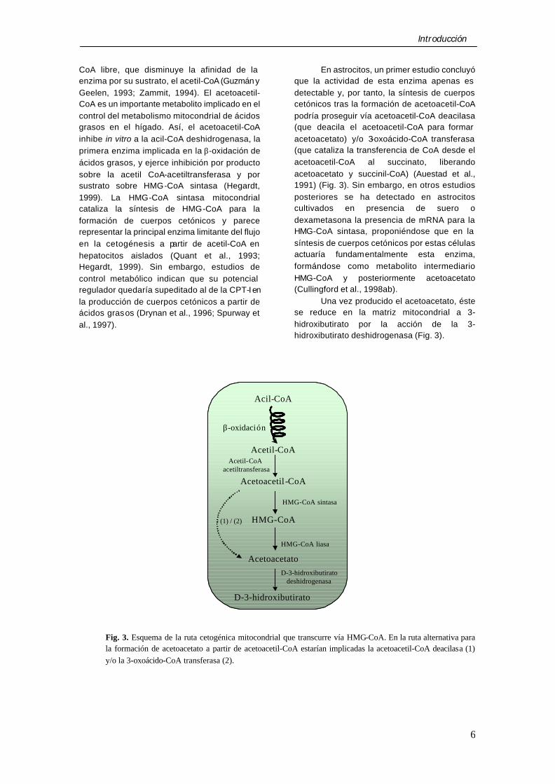
El primer paso de la formación de cuerpos cetónicos es la condensación de dos moléculas de acetil-CoA para generar acetoacetil-CoA, en una reacción catalizada por la acetil-CoA acetiltransferasa (Fig. 3), que puede ser inhibida por uno de sus productos, la
Introducción
6
CoA libre, que disminuye la afinidad de la enzima por su sustrato, el acetil-CoA (Guzmán y Geelen, 1993; Zammit, 1994). El acetoacetil-CoA es un importante metabolito implicado en el control del metabolismo mitocondrial de ácidos grasos en el hígado. Así, el acetoacetil-CoA inhibe in vitro a la acil-CoA deshidrogenasa, la primera enzima implicada en la β-oxidación de ácidos grasos, y ejerce inhibición por producto sobre la acetil CoA-acetiltransferasa y por sustrato sobre HMG-CoA sintasa (Hegardt, 1999). La HMG-CoA sintasa mitocondrial cataliza la síntesis de HMG-CoA para la formación de cuerpos cetónicos y parece representar la principal enzima limitante del flujo en la cetogénesis a partir de acetil-CoA en hepatocitos aislados (Quant et al., 1993; Hegardt, 1999). Sin embargo, estudios de control metabólico indican que su potencial regulador quedaría supeditado al de la CPT-I en la producción de cuerpos cetónicos a partir de ácidos grasos (Drynan et al., 1996; Spurway et al., 1997).
En astrocitos, un primer estudio concluyó que la actividad de esta enzima apenas es detectable y, por tanto, la síntesis de cuerpos cetónicos tras la formación de acetoacetil-CoA podría proseguir vía acetoacetil-CoA deacilasa (que deacila el acetoacetil-CoA para formar acetoacetato) y/o 3-oxoácido-CoA transferasa (que cataliza la transferencia de CoA desde el acetoacetil-CoA al succinato, liberando acetoacetato y succinil-CoA) (Auestad et al., 1991) (Fig. 3). Sin embargo, en otros estudios posteriores se ha detectado en astrocitos cultivados en presencia de suero o dexametasona la presencia de mRNA para la HMG-CoA sintasa, proponiéndose que en la síntesis de cuerpos cetónicos por estas células actuaría fundamentalmente esta enzima, formándose como metabolito intermediario HMG-CoA y posteriormente acetoacetato (Cullingford et al., 1998ab).
Una vez producido el acetoacetato, éste se reduce en la matriz mitocondrial a 3-hidroxibutirato por la acción de la 3-hidroxibutirato deshidrogenasa (Fig. 3).
Fig. 3. Esquema de la ruta cetogénica mitocondrial que transcurre vía HMG-CoA. En la ruta alternativa para la formación de acetoacetato a partir de acetoacetil-CoA estarían implicadas la acetoacetil-CoA deacilasa (1) y/o la 3-oxoácido-CoA transferasa (2).
Acetil-CoA
Acil-CoA
β-oxidación
Acetoacetil -CoA
Acetoacetato
D-3-hidroxibutirato
Acetil-CoA acetiltransferasa
D-3-hidroxibutiratodeshidrogenasa
HMG-CoA(1) / (2)
HMG-CoA sintasa
HMG-CoA liasa

Introducción
7
CARNITINA PALMITOILTRANSFERASA I
Como se ha mencionado anteriormente, los ácidos grasos de cadena larga no pueden atravesar la membrana mitocondrial interna. Así, el primer paso para su translocación a la matriz mitocondrial consiste en su activación en la membrana externa de la mitocondria por una acil-CoA sintetasa de cadena larga. A continuación, y previamente a la β-oxidación, los acil-CoA pasan al interior de la mitocondria por la acción del sistema de transporte dependiente de carnitina. Este sistema implica a tres proteínas, la CPT-I, la carnitina:acilcarnitina translocasa y la carnitina palmitoiltransferasa II (CPT-II), cada una de ellas con distinta localización submitocondrial (McGarry y Brown, 1997; Kerner y Hoppel, 2000). En una primera etapa, los acil-CoA son convertidos en acilcarnitinas por un proceso de transesterificación catalizado por la CPT-I, situada en la membrana mitocondrial externa; a continuación, el complejo acilcarnitina difundiría a través del espacio intermembrana o a través de los puntos de contacto entre las dos membranas mitocondriales (Kerner y Bieber, 1990; Fraser y Zammit, 1998; Hoppel et al., 1998) hasta llegar a la cara externa de la membrana mitocondrial interna, donde se translocaría al interior de la mitocondria por una reacción de intercambio catalizada por la translocasa de carnitina:acilcarnitina. En la matriz mitocondrial, las acilcarnitinas son entonces reconvertidas en sus respectivos acil-CoA por la acción de la CPT-II, enzima que se encuentra asociada a la cara interna de la membrana mitocondrial interna (McGarry y Brown, 1997; Kerner y Hoppel, 2000) (Fig. 4).
§ Estructura e isoformas
La caracterización de la CPT-I fue difícil por tratarse de una enzima estrechamente asociada a la membrana mitocondrial externa y por la pérdida de actividad catalítica que experimenta cuando se separa de su entorno nativo (Declercq et al., 1987; Woeltje et al., 1987; Zammit et al., 1989). Una vez desarrolladas las técnicas adecuadas para su caracterización, se aisló un clon que permitió predecir para el hígado de rata una proteína (L-CPT-I) de 773 aminoácidos y 88 kDa de peso molecular,
además de presentar una actividad inhibible por malonil-CoA y sensible al tratamiento con detergentes (Esser et al., 1993). Previamente el mismo grupo había clonado la CPT-II de hígado de rata (Woeltje et al., 1990) y unos años después se clonó la CPT-I de músculo de rata (M-CPT-I) (Esser et al., 1996).
De acuerdo con su papel fundamental en la oxidación mitocondrial de ácidos grasos, la CPT-I existe en al menos dos isoformas distintas. Las dos isoformas están codificadas por genes localizados en diferentes cromosomas y exhiben distinta distribución tisular y diferentes propiedades cinéticas y reguladoras (McGarry y Brown, 1997; Kerner y Hoppel, 1998; Jackson et al., 2000) (Tabla 1). Existen además isoformas de CPT-I generadas por procesamiento alternativo del mRNA de la CPT-I de músculo (Yu et al., 1998).
Estudios de topología han demostrado que la CPT-I se localiza en la membrana mitocondrial externa; está formada por dos fragmentos hidrofóbicos transmembranales, así como un corto dominio N-terminal y un largo dominio C-terminal situados hacia la cara citoplasmática de la membrana mitocondrial externa. Además, presenta un corto bucle de 27 aminoácidos que une los dominios transmembranales y que sobresale hacia el espacio intermembrana (Fraser et al., 1997; van der Leij et al., 1999). Se han realizado además experimentos con malonil-CoA y octanoil-CoA (inhibidor y sustrato de la CPT-I respectivamente) asociados a agarosa, para evitar que atraviesen la membrana mitocondrial externa, que indican que el dominio catalítico y regulador de la CPT-I están localizados hacia la cara citosólica (Fraser et al., 1997).

Introducción
8
Características L-CPT I M-CPT I CPT II
Peso molecular 88 kDa 88 kDa 67 kDa
IC50 para malonil-CoA
2.5 µM 0.03 µM --
Km para carnitina
30 µM 500 µM 120 µM
Cromosoma humano
11q13 22q13.3 1p32
Expresión
Hígado ++++ -- +
Músculo esq. (+) ++++ +
Corazón + +++ +
Riñón ++++ (+) +
Adiposo + +++ +
Cerebro ++++ -- +
Tabla 1. Características de las CPT mitocondriales . Los niveles relativos de expresión en cada órgano están basados en análisis por Northern blot (y en algunos casos por marcaje con [3H]etomoxir-CoA). (+) Sólo trazas en comparación con la isoforma alternativa. (-) Indetectable. Los datos de expresión en tejidos se refieren a rata. La Km para la carnitina de la CPT-II fue determinada tras solubilización de las mitocondrias en octilglucósido y por tanto no debe ser comparada directamente con los valores de CPT-I (McGarry y Brown, 1997).
Fig. 4. Translocación de ácidos grasos a la matriz mitocondrial. Abreviaturas: ACS, acil-CoA sintetasa; AG, ácido graso; Carn, carnitina; MME, membrana mitocondrial externa; MMI, membrana mitocondrial interna; T, translocasa de carnitina:acilcarnitina.
AG Acil-CoA
CoA-SHAcil-CoA
Acil-CarnCarn
Carn
Carn
ACS CPT-I
TCPT-II
MME
MMI
Matriz
Citoplasma
Acil-Carn
CoA-SH

Introducción
9
§ Regulación
La CPT-I cataliza el primer paso en la oxidación mitocondrial de los ácidos grasos y desempeña un papel fundamental en la regulación de todo el proceso. Los mecanismos por los cuales se controla la actividad CPT-I incluyen, entre otros, cambios en la concentración de malonil-CoA (regulación a corto plazo) y cambios en los niveles de enzima y en su sensibilidad a inhibición por malonil-CoA (regulación a largo plazo). También se ha propuesto la existencia de un mecanismo agudo de regulación de la actividad CPT-I independiente de los niveles de malonil-CoA..
Regulación por malonil-CoA
Hígado
La CPT-I es inhibida por malonil-CoA, el producto de la reacción catalizada por la acetil-CoA carboxilasa (ACC), que constituye a su vez la etapa limitante de la síntesis de ácidos grasos (McGarry et al., 1977; Geelen et al., 1980). De esta manera, se consigue un control coordinado de la síntesis y la oxidación de ácidos grasos en el hígado (Zammit, 1994). Los niveles de malonil-CoA en la célula son dependientes de la actividad ACC y, por tanto, se ven afectados notablemente por cambios a corto plazo que diferentes mecanismos, como pueden ser alteraciones en el estado hormonal y nutricional del animal, producen sobre dicha actividad enzimática (McGarry y Foster, 1980; Zammit, 1994). Especialmente determinante es el cociente insulina/glucagón en plasma: cuando este cociente es alto los niveles de malonil-CoA también lo son y la actividad CPT-I está inhibida, y viceversa (Zammit, 1996). Recientemente se ha propuesto que la malonil-CoA descarboxilasa, que descarboxila el malonil-CoA a acetil-CoA, podría determinar así mismo los niveles de malonil-CoA y, por tanto, la actividad CPT-I en hepatocitos (Dyck et al., 2000; Saha et al., 2000).
Músculo esquelético y corazón
Diferentes tejidos no lipogénicos, como
músculo esquelético y corazón, también contienen malonil-CoA, cuyos niveles oscilan en función del grado nutricional del organismo. Este hecho podría indicar un papel regulador del malonil-CoA en estos tejidos (Lopaschuk et al., 1994). La isoforma de CPT-I mayoritariamente
presente en músculo cardíaco y esquelético (M-CPT I) es mucho más sensible a malonil-CoA que la isoforma hepática (McGarry y Brown, 1997). Diversos estudios indican que los niveles de malonil-CoA en corazón dependen principalmente de la disponibilidad de acetil-CoA citosólico, el sustrato de la ACC. Puesto que además apenas existe ácido graso sintasa en músculo y corazón, el papel del malonil-CoA sería el de regulador de la CPT-I más que el de precursor para la síntesis de ácidos grasos (McGarry y Brown, 1997).
Por estudios de inmunofluorescencia se ha visto que la ACC-280 (isoforma de ACC de 280 kDa, expresada mayoritariamente en músculo y corazón) está asociada a la mitocondria, a diferencia de la ACC-265 (isoforma de ACC de 265 kDa, expresada predomi-nantemente en tejidos lipogénicos), que tiene localización citosólica. Esta asociación de la ACC-280 a la mitocondria apoya la hipótesis de su implicación en la regulación de la oxidación de ácidos grasos por la mitocondria a través de la inhibición de la CPT-I por el malonil-CoA sintetizado (Abu-Elheiga et al., 2000).
Célula β
La regulación de la CPT-I mediada por malonil-CoA parece tener un papel fisiológico importante en las células β del páncreas. Se ha propuesto que, tras un incremento de la concentración de glucosa en sangre, este azúcar es metabolizado por la célula β a través de la ruta glicolítica, de forma que parte del piruvato sería desviado hacia la síntesis de malonil-CoA. Este malonil-CoA inhibiría a la CPT-I, lo que provocaría una acumulación de acil-CoA de cadena larga en el citoplasma de la célula β. Los acil-CoA podrían actuar estimulando la exocitosis de los gránulos de insulina acumulados en estas células (Newgard y McGarry, 1995; Zammit, 1999).
Regulación a corto plazo independiente
de malonil-CoA
Durante más de dos décadas se ha considerado la inhibición de la CPT-I por malonil-CoA como el mecanismo principal de regulación de la cetogénesis hepática; sin embargo, observaciones más recientes indican que la CPT-I también está regulada a corto plazo por mecanismos independientes de malonil-CoA (Guzmán et al., 2000). Así,

Introducción
10
mediante el empleo de hepatocitos permeabilizados, se ha observado la existencia de cambios en la actividad CPT-I independientes de malonil-CoA (Guzmán y Geelen, 1993; Guzmán et al., 1994). Utilizando moduladores del estado de agregación y polimerización del citoesqueleto se propuso la posibilidad de un papel regulador de componentes del citoesqueleto sobre la actividad CPT-I, identificándose posteriormente los filamentos intermedios, y específicamente las citoqueratinas 8 y 18, como los principales candidatos a mediar esta regulación (Velasco et al., 1998ab). Además, el empleo de inhibidores específicos de proteína quinasas y fosfatasas, junto con experimentos de reconstitución y estudios de fosforilación de citoqueratinas en hepatocitos intactos, permitió implicar a la proteína quinasa dependiente de Ca2+-calmodulina de tipo II y a la proteína quinasa activada por AMP (AMPK) como enzimas que afectan al estado de agregación del citoesqueleto y, como consecuencia de ello, a la actividad CPT-I. Así, la activación de la Ca2+-calmodulina quinasa II o de la AMPK induce la fosforilación de las citoqueratinas 8 y 18, lo que supone la desagregación de los filamentos intermedios, con lo que se pierde la interacción entre la CPT-I y los componentes del citoesqueleto y la CPT-I se activa (Velasco et al.,1998ab). A su vez, la activación de las dos quinasas también regula la actividad CPT-I a través de un mecanismo dependiente de malonil-CoA, puesto que fosforilan e inactivan a la ACC produciendo un descenso en los niveles de malonil-CoA y, por tanto, la activación indirecta de la CPT-I (Guzmán et al., 2000).
Estudios llevados a cabo por Sleboda et al. (1999) también apoyan esta regulación de la CPT-I independiente de malonil-CoA por componentes celulares extramitocondriales. Así mismo, se ha observado en cardiomiocitos de rata neonata que la hipoxia produce una estimulación significativa en la actividad CPT-I por un mecanismo estable a nivel de regulación post-traduccional (Wang et al., 1998).
Regulación de la sensibilidad a malonil-
CoA
El malonil-CoA, por sí mismo, no sólo inhibe a la actividad CPT-I, sino que también podría determinar la sensibilidad de la CPT-I a inhibición por malonil-CoA. Así, en diferentes situaciones fisiopatológicas, la sensibilidad de la CPT-I al malonil-CoA se ve modificada. Por
ejemplo, los cambios inducidos por la dieta y el estado hormonal en el flujo a través de la ruta oxidativa de los ácidos grasos están normalmente acompañados de variaciones paralelas en la sensibilidad al malonil-CoA de la CPT-I de hígado. En estados cetóticos como el ayuno, la diabetes y el hipertiroidismo, la sensibilidad de la CPT-I a inhibición por malonil-CoA disminuye, mientras que en estados lipogénicos como el hipotiroidismo o la realimentación tras el ayuno ocurre lo contrario (Zammit, 1994). Este cambio en la sensibilidad a malonil-CoA sólo se ha observado en la isoforma hepática, no en la CPT-I de corazón ni en la de músculo.
Aún no se conoce el mecanismo por el cual se desensibiliza la CPT-I, pero se ha observado que la cardiolipina produce un incremento y recuperación en la sensibilidad al malonil-CoA en mitocondrias de hígado de rata en ayuno. Puesto que la cardiolipina es un componente minoritario de la membrana mitocondrial externa, se ha sugerido la posibilidad de que pueda formar un microambiente de membrana específico para la CPT-I (Kerner y Hoppel, 2000). De acuerdo con esto, preparaciones de membrana mitocondrial externa de ratas en ayuno, diabéticas o en estados cetóticos presentan una relación inversa entre la sensibilidad de la CPT-I a malonil-CoA y la fluidez de la membrana (Zammit et al., 1998), lo que se ha comprobado así mismo por experimentos de reconstitución (McGarry y Brown, 2000).
Regulación de los niveles de enzima
Existen bastantes estudios acerca de
cómo diversos factores nutricionales y hormonales pueden afectar a la expresión de las CPT de hígado de rata. Diferentes situaciones que estimulan la cetogénesis en hígado, como el ayuno, la diabetes, el hipertiroidismo o el tratamiento con agentes proliferadores de peroxisomas, inducen tanto un aumento en los niveles de proteína inmunorreactiva como de mRNA de CPT-I y CPT-II (Kolodziej et al., 1992; Park et al., 1995; McGarry y Brown, 1997; Jensen et al., 2000). Otra situación fisiológica que afecta de manera notable a la expresión de los genes de CPT-I y CPT-II es el desarrollo postnatal. En el hígado fetal, tanto los niveles de mRNA como la actividad CPT-I son muy bajos. Sin embargo, ambos aumentan bruscamente durante el primer día de vida extrauterina y permanecen elevados durante todo el periodo de lactancia hasta el

Introducción
11
destete (Thumelin et al., 1994; Asins et al., 1995), lo que se corresponde con el mayor porcentaje en grasas de la alimentación durante el periodo de lactancia.
Se están empezando a conocer los factores que regulan la transcripción de los genes de las isoformas de CPT-I. Así, se ha caracterizado el promotor del gen de la L-CPT-I, en el cual se han identificado dos sitios de unión a los factores de transcripción Sp1 y Sp3, así como también al factor nuclear Y (NF-Y) (Steffen et al., 1999). Por otro lado, la expresión de la M-CPT-I, está regulada a nivel transcripcional por la unión de receptores activados por proliferadores de peroxisomas (PPAR) a su promotor (Mascaró et al., 1998), mientras que la regulación transcripcional de la L-CPT-I parece ser independiente de PPAR (Louet et al., 2001).
1.3 Lipoapoptosis
GENERALIDADES
La apoptosis es un mecanismo esencial en el desarrollo del sistema nervioso de vertebrados que está implicado en procesos de remodelación tisular, embriogénesis, respuesta inmune y en determinadas situaciones patológicas como enfermedades autoinmunes y neurodegenerativas (Desagher y Martinou, 2000; Hengartner, 2000; Strasser et al., 2000). Se trata de un proceso de muerte celular caracterizado porque las células se encogen, se condensa la cromatina, la integridad (no la asimetría) de la membrana plasmática se mantiene hasta el último momento y se produce la activación de caspasas y la fragmentación internucleosómica del DNA. Aunque existen formas de muerte con características intermedias entre necrosis y apoptosis (Sperandio et al., 2000), todo ello diferencia a la primera de la segunda, que cursa con hinchamiento celular, degradación no específica del DNA, lisis de la célula e inflamación del tejido circundante.
Como se verá a lo largo de esta memoria, existen numerosos estudios de señalización celular sobre esfingolípidos (especialmente ceramida) como mediadores intracelulares de apoptosis (Kolesnick y Krönke, 1998; Hannun y Luberto, 2000; Radin, 2001). ¿Cómo podría influir la CPT-I en este proceso? Paumen et al. (1997) observaron que en líneas celulares hematopoyéticas la CPT-I podría
proteger de la apoptosis inducida por ácidos grasos a través de una inhibición de la síntesis de novo de esfingolípidos; así, observaron que la inhibición farmacológica de la CPT-I incrementaba la muerte celular inducida por palmitato y los niveles de ceramida sintetizada de novo a partir de este ácido graso. Posteriormente, Shimabukuro et al. (1998) descubrieron el mismo fenómeno en células β de páncreas, y acuñaron el término “lipoapoptosis” para definir este proceso de muerte celular vía síntesis de novo de ceramida a partir de ácidos grasos. Existen otros estudios que han demostrado que el palmitato está implicado en la inducción de apoptosis. Así, en cardiomiocitos de ratas neonatas la incubación en presencia de concentraciones fisiológicas de palmitato induce acumulación de ceramida, inhibición de la AMPK, inhibición de la CPT-I y, en último término, apoptosis (Hickson-Bick et al., 2000). En suma, a la vista de estos estudios se puede pensar que la CPT-I tendría un papel regulador del metabolismo lipídico y del destino celular puesto que, en función de su actividad, los ácidos grasos de cadena larga se dirigirán hacia la ruta de la β-oxidación o bien hacia la ruta de síntesis de ceramida.
CERAMIDA
Durante la última década se ha investigado intensamente la posibilidad de que los esfingolípidos sean mediadores importantes en la señalización celular. Los esfingolípidos están implicados en la regulación de la mayoría de los aspectos fundamentales de la biología celular, incluyendo procesos de crecimiento, diferenciación, arresto del ciclo celular, apoptosis y oncogénesis en células nerviosas y no nerviosas (Goswami y Dawson, 2000; Hannun y Luberto, 2000). En concreto, la ceramida es un segundo mensajero esfingolipídico que presenta una rápida capacidad de translocación de la superficie celular al interior de la membrana y que regula directa o indirectamente un gran número de enzimas. Se genera como respuesta a la estimulación de muy diversas vías implicadas en muerte celular, como por ejemplo aquellas ligadas a citoquinas proinflamatorias, agentes utilizados en quimioterapia, radiación ionizante y agentes oxidantes. No obstante, no debe olvidarse que la interpretación de algunos datos de la bibliografía es confusa debido a factores como la cinética variable de generación de

Introducción
12
ceramida, el alto número de enzimas implicadas en dicho proceso, la compartimentalización subcelular de la ceramida y el uso de análogos no fisiológicos de ceramida (Hoffmann y Dixit, 1998; Kolesnick y Hannun, 1999; Levade y Jaffrézou, 1999; Siskind y Colombini, 2000; Venkataraman y Futerman, 2000).
§ Generación de ceramida
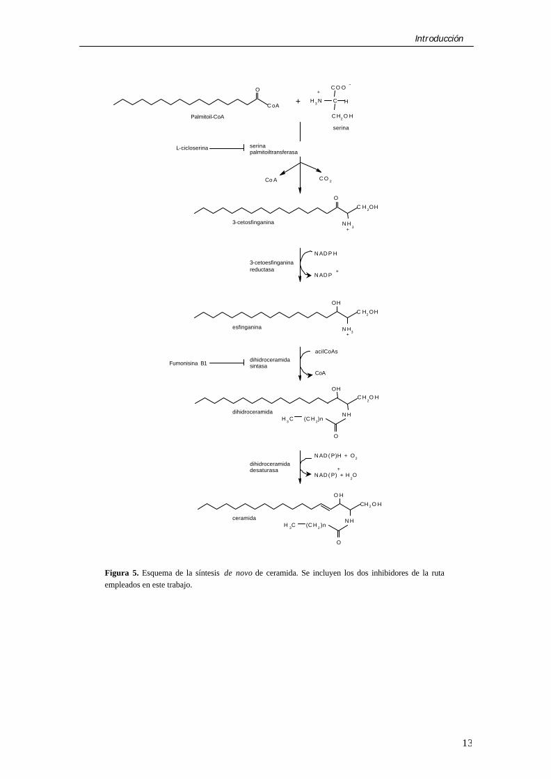
La ceramida puede formarse a través de diversas rutas. En el contexto de la presente memoria destaca sin duda la ruta de síntesis de novo (Fig. 5). Ésta comienza en el retículo endoplásmico con la condensación de palmitoil-CoA (o estearoil-CoA) y serina para formar 3-cetodihidroesfingosina en una reacción catalizada por la serina palmitoiltransferasa (SPT). A continuación se produce la reducción a esfinganina por una reductasa dependiente de NADPH. Por una reacción de acilación llevada a cabo por una aciltransferasa (la dihidroceramida sintasa) se forma la dihidroceramida y, por último, una desaturasa introduce un doble enlace en las posiciones 4 y 5 del esqueleto esfingoide de la molécula, formándose la N-acilesfingosina o ceramida (Kolter y Sandhoff, 1999; Luberto y Hannun, 1999).
La ceramida puede ser sintetizada alternativamente por acilación de la esfingosina por una aciltransferasa (Luberto y Hannun, 1999) que podría tratarse de una ceramidasa neutra que cataliza una reacción reversible de acilación-desacilación (Tani et al., 2000). Además, la ceramida constituye un punto intermedio en la formación de la mayoría de los esfingolípidos con importancia biológica (Fig. 6).
Se ha considerado hasta el momento que la generación de ceramida implicada en procesos apoptóticos tiene lugar por la hidrólisis de esfingomielina catalizada por esfingomieli-nasas. Estas enzimas hidrolizan el enlace fosfodiéster de la esfingomielina, que preferentemente se encuentra en la membrana plasmática de las células de mamíferos, produciendo ceramida y fosforilcolina (Kolesnick y Krönke, 1998; Levade y Jaffrézou, 1999). Existen diversas esfingomielinasas con características de regulación diferentes y con distinto pH óptimo. A grandes rasgos existen esfingomielinasas ácidas (pH óptimo 4.5-5.0), que se localizan en compartimentos celulares
ácidos como lisosomas y endosomas, y esfingomielinasas neutras (dependientes e independientes de Mg2+), que actúan en la membrana plasmática y abundan en los rafts lipídicos. Ambos tipos de esfingomielinasas responden generalmente a estímulos que incrementan los niveles de ceramida a tiempos cortos, del orden de segundos a minutos, por lo que se las principales responsables de la producción de ceramidas en las señales de transducción tempranas (Kolesnick y Krönke, 1998; Levade y Jaffrézou, 1999). Sin embargo, la cuestión de cuántas esfingomielinasas existen y cuál está implicada en cada pico de ceramida está aún sin resolver (Chatterjee et al., 1999; Levade y Jaffrézou, 1999; Bernardo et al., 2000; Fensome et al., 2000; Hofmann et al., 2000).
Por el contrario, la ruta de síntesis de novo de ceramidas ha ido ganando progresivamente importancia como forma alternativa de generar un pool apoptótico de ceramida, y compuestos como la L-cicloserina (un inhibidor de la SPT) y la fumonisina B1 (un inhibidor de la dihidroceramida sintasa) (Fig. 5) previenen la acumulación de ceramida y la muerte por apoptosis inducida por ácidos grasos en células hematopoyéticas y células β del páncreas (ver más arriba). Así mismo, la exposición a determinadas drogas utilizadas en quimioterapia como la doxorrubicina, la daunorrubicina y el etopósido podrían inducir apoptosis por un incremento en la síntesis de novo de ceramida (Bose et al., 1995; Perry et al., 2000) acompañado de la estimulación de la hidrólisis de esfingomielina (Jaffrézou et al., 1996; Andrieu-Abadie et al., 1999). Otros estudios han demostrado la importancia de la síntesis de novo de ceramida en la apoptosis producida por angiotensina A-II (Lehtonen et al., 1999) y el gen de la ataxia-talangiectasia (Liao et al., 1999), así como en la apoptosis que tiene lugar durante la diferenciación neuronal (Herget et al., 2000). Por todo ello se hará más adelante una especial mención a la enzima clave de la ruta, la SPT.
Introducción
13
Figura 5. Esquema de la síntesis de novo de ceramida. Se incluyen los dos inhibidores de la ruta empleados en este trabajo.
O
C oA
C H2 O H
C
C O O
HH 3 N
C O 2Co A
O
N H3
C H 2OH
N AD P H
N AD P
N H3
C H2 OH
O
OH
N H
C H2O H
OH
(C H 2)nCH 3
N AD ( P)H + O2
O
N H
CH 2 O H
O H
(C H 2 )nCH 3
N AD ( P) + H2O
+
+
+
-
serina
serinapalmitoiltransferasa
Palmitoil-CoA
+3-cetosfinganina
+
3-cetoesfinganinareductasa
+esfinganina
acilCoAs
CoA
dihidroceramidasintasa
dihidroceramida
dihidroceramidadesaturasa
ceramida
L-cicloserina
Fumonisina B1

Introducción
14
Figura 6. Esquema del papel central de la ceramida en el metabolismo de esfingolípidos.
§ Mecanismo de acción
La ceramida regula indirectamente la actividad de un gran número de enzimas y componentes de señalización, entre los que se incluyen quinasas, fosfatasas, fosfolipasas, factores de transcripción y caspasas. Sin embargo, los blancos primarios de la acción de la ceramida son aún poco conocidos.
Una de las posibles dianas de la ceramida es la proteína quinasa activada por ceramida (CAPK). La CAPK, caracterizada por el grupo de Kolesnick como quinasa supresora de Ras (KSR) (Zhang et al., 1997), es capaz de fosforilar y activar Raf-1, provocando así la activación de la cascada de proteína quinasas activadas por señales extracelulares (ERK) (Yao et al., 1995; Xing y Kolesnick, 2001). Se ha sugerido que KSR podría estar implicada en la apoptosis inducida por ceramida (Basu et al., 1998; Xing et al., 2000). Sin embargo, otros autores no han sido capaces de demostrar esto (Michaud et al., 1997; Müller et al., 1998). Huwiler et al. (1996) publicaron que Raf-1 podría ser una quinasa activada directamente por ceramida, lo que podría explicar cómo la ceramida activa la cascada ERK. Sin embargo, Müller et al. (1998) no fueron capaces de reproducir este efecto.
La proteína fosfatasa activada por
ceramida (CAPP), una fosfatasa de tipo 2A, también se ha identificado como posible diana de la ceramida (Dobrowsky et al., 1993). Aunque el grupo de Hannun ha trabajado afanosamente en esta fosfatasa (Chalfant et al., 1999; Hannun y Luberto, 2000), su papel señalizador es dudoso, aunque parece mediar la desfosforilación e inactivación de la proteína quinasa B (PKB) (Schubert et al., 2000). Sin embargo, esta relación entre ceramida, CAPP y PKB no es tan clara (Stratford et al., 2001).
Una tercera proteína, la proteína quinasa C ζ (PKCζ), puede unir directamente ceramida y resultar activada (Lozano et al., 1994; Galve-Roperh et al., 1997; Bourbon et al., 2000). El ácido araquidónico compite con la ceramida, inhibiendo así la activación que ésta ejerce sobre la PKCζ (Müller et al., 1995).
OH
CH2OH
NH
O
CH3-(CH2)n
OH
CH2OP
NH
O
CH3-(CH2)n
O
O
OO H
CH2OP
NH
O
CH3-(CH2)n
O
O
O
CH 2-CH2-N(CH 3)3
OH
CH2OH
NH3+
O
CCH3-(CH
2)n
O H
CH2O-glucosa
NH
O
CH3-(CH2)n
O
OH
CH2OP
NH3+
O
O
O
Acil-transferasa
ceramida
ceramida-1-fosfato
+
esfingomielina
+glucosilceramida
esfingosina (SP)
ácido graso
SMasaSM sintasa
Glicosiltransferasa
Glicosidasa
Ceramidasa
FosfatasaQuinasa
esfingosina-1-fosfato
SP quinasa
SP1-fosfatofosfatasa

Introducción
15
SERINA PALMITOILTRANSFERASA
La SPT es la enzima clave en la biosíntesis de ceramida; se encuentra situada en el retículo endoplásmico y cataliza la condensación de palmitoil-CoA y serina para generar 3-cetoesfinganina en una reacción dependiente de piridoxal 5´-fosfato (Dickson et al., 1999).
§ Estructura Se han estudiado numerosas
propiedades de esta enzima y, sin embargo, su total purificación no se ha conseguido aún debido a que presenta una actividad bastante baja. La caracterización de la SPT se realizó primeramente en levaduras; la enzima está codificada por dos genes, LCB1 y LCB2, que presentan un 22 % de homología y codifican dos proteínas (Lcb1p y Lcb2p, de 62 y 63 kDa respectivamente) con actividad 5-aminolevuli-nato sintasa, 2-amino-oxobutirato-CoA ligasa y 8-amino-7-oxononanoato sintasa; ambos genes son requeridos para que la enzima exhiba actividad. Además, se ha caracterizado un tercer gen, TSC3, requerido para el ensamblado adecuado de la SPT. Este gen codifica para una proteína de 14 kDa, que actuaría post-traduccionalmente y se asociaría a las dos subunidades estimulando la actividad SPT (Dickson et al., 1999; Gable et al., 2000).
Se ha llevado a cabo el aislamiento y caracterización en ratón y humano de los cDNA de LCB1 y LCB2; de la expresión de estos genes se obtuvieron dos proteínas de 53 kDa y 63 kDa respectivamente, que además no contenían péptido señal ni se glicosilaban. A diferencia de la SPT de levaduras, para que la enzima sea activa sólo es necesaria la expresión de la subunidad LCB2. Por Northern blot se determinó el tamaño de los mRNAs de LCB1 y LCB2 (2.9 kb y 2.3 kb, respectivamente) y su distribución tisular (Tabla 2). Parece que el residuo Lys 397 de la subunidad catalítica LCB2 es esencial para la actividad catalítica de la SPT (Weiss y Stoffel, 1997; Hanada et al., 1998; Dickson et al., 1999).
Subunidades Características.
mLCB1 mLCB2
Peso molecular 53 kDa 63 kDa
Km para serina 0.6 mM
Km para palmitoil-CoA
0.05 mM
Km para piridoxal fosfato
2.5 µM
Expresión
Hígado + +
Músculo +++ ++
Corazón +++ ++++
Riñón +++ ++++
Pulmón ++++ ++
Cerebro +++ ++
Tabla 2. Características de la SPT de ratón. n.d. no determinado. Los signos + indican de un modo cualitativo la abundancia de las dos subunidades en los distintos tejidos.
§ Regulación
Existen pocos estudios acerca de la
regulación de la SPT. La angiotensina II, al unirse al receptor AT2, induce apoptosis en células PC12 por un incremento en la actividad de la SPT y, por tanto, en los niveles de ceramida. Esta acumulación de ceramida y muerte por apoptosis es inhibida por fumonisina B1 y β-cloro-L-alanina (inhibidores de la ruta de síntesis de novo), así como por la toxina pertúsica (inhibidora del sistema de transducción acoplado al receptor AT2) (Lehtonen et al., 1999). El etopósido, una droga quimioterapeútica, es capaz de activar la ruta de síntesis de novo de esfingolípidos. Esta sustancia induce apoptosis en células leucémicas humanas produciendo un incremento en la formación de ceramida por un aumento en la actividad de la SPT a nivel post-transcripcional, probablemente por alguna modificación covalente o por regulación alostérica de la enzima, pero no por cambios en los niveles de mRNA de la misma (Perry et al., 2000). Este mismo tipo de regulación ha sido también observada por Herget et al. (2000) en el proceso de diferenciación neuronal por ácido

Introducción
16
retinoico. En el caso de la radiación ultravioleta, ésta incrementa la actividad de la SPT y la síntesis de esfingolípidos en queratinocitos por un aumento en los niveles de mRNA y proteína de las subunidades LCB1 y LCB2 (Farrell et al., 1998). De esta forma, diversos agentes actúan regulando la enzima clave de la ruta de síntesis de novo de esfingolípidos tanto a nivel transcripcional como post-traduccional.
Se han identificado una serie de inhibidores de la SPT, que constituyen herramientas muy útiles para el estudio y regulación de la ruta, la posible implicación de la enzima y el origen de los productos obtenidos. Estos inhibidores son por ejemplo la L-cicloserina y las β-haloalaninas, tales como la β-cloro-L-alanina y la β-fluoro-L-alanina, aunque estas últimas no son necesariamente específicas de la SPT y pueden afectar también a otras enzimas que utilizan piridoxal 5´-fosfato como cofactor. Se han descrito además diversos inhibidores con mayor especificidad como son las esfingofunginas, la ISP1/myriocina, la lipoxamicina y las viridiofunginas (Dickson et al., 1999; Kolter y Sandhoff, 1999).
1.4 Objetivos Como ya se ha comentado anteriormen-
te, está ampliamente aceptado que los astrocitos desempeñan un papel muy activo en la regulación del metabolismo de la glucosa en el cerebro (Magistretti y Pellerin, 1996; Wiesinger et al., 1997). Así, los astrocitos capturan y metabolizan glucosa con gran actividad. Uno de los principales productos del metabolismo de la glucosa es el lactato, que podría cederse a las neuronas y ser utilizado por éstas como sustrato de rutas biosintéticas y catabólicas. Además, el glucógeno constituye una importante fuente de energía para el cerebro; el glucógeno cerebral se almacena principalmente en los astrocitos, y su tasa de recambio es muy alta y está coordinada con la actividad sináptica.
Los estudios realizados hasta el momento sobre metabolismo de astrocitos se han centrado principalmente en el metabolismo de glucosa y muy pocos de ellos han profundizado en el metabolismo de ácidos grasos. La glucosa es la principal fuente de energía del cerebro, pero bajo determinadas circunstancias los cuerpos cetónicos procedentes de la oxidación hepática de ácidos
grasos pueden reemplazar a la glucosa como fuente energética (Zammit, 1994). Se cree que el hígado es el órgano que mayoritariamente aporta cuerpos cetónicos a los tejidos extrahepáticos. Sin embargo, los ácidos grasos pueden ser oxidados en el cerebro (Auestad et al., 1991; Edmond, 1992). Los astrocitos son precisamente las únicas células del cerebro que pueden oxidar ácidos grasos y que además muestran preferencia por éstos como combustible sobre la glucosa y los cuerpos cetónicos (Edmond et al., 1987; Edmond, 1992; Staub et al., 1995). Es más, los astrocitos en cultivo son capaces de producir cuerpos cetónicos a partir de ácidos grasos (Auestad et al.,1991) y leucina (Bixel y Hamprecht, 1995). Todo esto plantea la cuestión de si los astrocitos podrían proveer a las neuronas in vivo con cuerpos cetónicos como fuente de energía alternativa a la glucosa.
Puesto que aún no se conocen los mecanismos que controlan la ruta cetogénica en astrocitos, el primer objetivo global de este trabajo fue la caracterización de la ruta cetogénica en astrocitos, así como el estudio de los posibles mecanismos que la regulan, poniendo especial énfasis en la CPT-I, enzima reguladora de la oxidación de ácidos grasos en otros tejidos. Para ello se utilizó como modelo experimental cultivos primarios de astrocitos de rata, y como posibles mecanismos reguladores de la cetogénesis los mediados por la proteína quinasa dependiente de cAMP (PKA) y la AMPK, así como los dependientes de ceramida. Además, y a la vista de los últimos estudios que se han llevado a cabo sobre la apoptosis inducida por ácidos grasos en distintos tipos celulares, nos propusimos como segundo objetivo global el estudio de este proceso en nuestro modelo experimental, así como su posible regulación por la AMPK con el fin de desvelar un posible papel fisiológico.

Introducción
17
1.5 BIBLIOGRAFÍA
- Abu-Elheiga L., Brinkley W.R., Zhong L., Chirala S.S., Woldegiorgis G. y Wakil S.J. (2000) Proc. Natl. Acad. Sci. USA 97: 1444-1449.
- Albright T.D., Jessell T.M., Kandel E.R. y Posner M.I. (2000) Cell 100: S1-S55.
- Andrieu-Abadie N., Jaffrézou J.P., Hatem S., Laurent G., Levade T. y Mercadier J.J. (1999) FASEB J. 13: 1501-1510.
- Araque A., Parpura V., Sanzgiri R.P. y Haydon P.G. (1999) Trends Neurosci. 22: 208-215.
- Asins G., Serra D., Arias G. y Hegardt F.G. (1995) Biochem. J. 306: 379-384.
- Auestad N., Korsak R.A., Morrow J.W. y Edmond J. (1991) J. Neurochem. 56: 1376-1386.
- Barres B.A. (1999) Cell 97: 667-670. - Basu S., Bayoumy S., Zhang Y., Lozano J. y
Kolesnick R. (1998) J. Biol. Chem. 273: 30419-30427.
- Bernardo K., Krut O., Wiegmann K., Kreder D., Micheli M., Schäfer R., Sickman A., Schmidt W.E., Schröder J.M., Meyer H.E., Sandhoff K. y Krönke M. (2000) J. Biol. Chem. 275: 7641-7647.
- Bittar P.G., Charnay Y., Pellerin L., Bouras C. y Magistretti P.J. (1996) J. Cereb. Blood Flow Metab. 16: 1079-1089.
- Bixel M.G. y Hamprecht B. (1995) J. Neurochem. 65: 2450-2461.
- Bose R., Verheij M., Haimovitz-Friedman A., Scotto K., Fuks Z. y Kolesnick R. (1995) Cell 82: 405-414.
- Bourbon N.A., Yun J. y Kester M. (2000) J. Biol. Chem. 275: 35617-35623.
- Bröer S., Rahman B., Pellegri G., Pellerin L., Martin J.L., Verleysdonk S., Hamprecht B. y Magistretti P.J. (1997) J. Biol. Chem. 272: 30096-30102.
- Brown G.C. (1992) Biochem. J. 284: 1-13. - Chalfant C.E., Kishikawa K., Mumby M.C.,
Kamibayashi C., Bielawska A. y Hannun Y.A. (1999) J. Biol. Chem. 274: 20313-20317.
- Chatterjee S., Han H., Rollins S. y Cleveland T. (1999) J. Biol. Chem. 274: 37407-37412.
- Choi I.Y., Tkác I., Ugurbil K. y Gruetter R. (1999) J. Neurochem. 73: 1300-1308.
- Clarke D.D. y Sokoloff L. (1999) Basic Neurochemistry. Molecular, Cellular and Medical Aspects. Sexta edición Siegel G.J. et al. (eds. ), Lippincott, 1999, pp. 637-669.
- Cooper B., Noy N. y Zakim D. (1989) J. Lipid Res. 30: 1719-1726.
- Cullinford T.E., Bhakoo K.K. y Clark J.B. (1998a) J. Neurochem. 71: 1804-1812.
- Cullingford T.E., Dolphin C.T., Bhakdo K.K., Peuchen S., Canevari L. y Clark J.B. (1998b) Biochem. J. 329: 373-381.
- de Vellis J. y Carpenter E. (1999) Basic
Neurochemistry. Molecular, Cellular and Medical Aspects. Sexta edición Siegel G.J. et al. (eds. ), Lippincott, 1999, pp. 535-563.
- Declercq P.E., Falck J.R., Kuwajima M., Tyminski H., Foster D.W. y McGarry J.D. (1987) J. Biol. Chem. 262: 9812-9821.
- Deitmer J.W. (2000) BioEssays 22: 747-752. - Desagher S. y Martinou J.C. (2000) Trends
Cell Biol. 10: 369-377. - Dickson R.C., Lester R.L. y Nagiec M.M.
(1999) Meth. Enzimol. 311: 3-9. - Dobrowsky R.T., Kamibayashi C., Mumby
M.C. y Hannun Y.A. (1993) J. Biol. Chem. 268: 15523-15530.
- Doetsch F., Caillé I., Lim D.A., García-Verdugo J.M. y Alvarez-Buylla A. (1999) Cell 97: 703-716.
- Dringen R. (2000) Progress Neurobiol. 62: 649-671.
- Dringen R. y Hamprecht B. (1992) J. Neurochem. 58: 511-517.
- Dringen R. y Hamprecht B. (1993) Glia 8: 143-149.
- Dringen R., Gebhardt R. y Hamprecht B. (1993) Brain Res. 623: 208-214.
- Drynan L., Quant P.A. y Zammit V.A. (1996) Biochem. J. 317: 791-795.
- Dyck J.R., Berthiaume L.G., Thomas P.D., Kantor P.F., Barr R., Singh D., Hopkins T.A., Voilley N., Prentki M. y Lopaschuk G.D. (2000) Biochem. J. 350: 599-608.
- Eaton S., Bartlett K. y Pourfarzam M. (1996) Biochem. J . 320: 345-357.
- Edmond J. (1992) Can. J. Physiol. Pharmacol. 70: S118-S129.
- Edmond J., Auestad N., Robbins R.A. y Bergstrom J.D. (1985) Fed. Proc. 44: 2359-2364.
- Edmond J., Robbins R.A., Bergstrom J.D., Cole R.A. y de Vellis J. (1987) J. Neurosci. Res. 18: 551-561.
- Esser V., Britton C.H., Weis B.C., Foster D.W. y McGarry J.D. (1993) J. Biol. Chem. 268: 5817-5822.
- Esser V., Brown N.F., Cowan A.T., Foster D.W. y McGarry J.D. (1996) J. Biol. Chem. 271: 6972-6977.
- Farrell A.M., Uchida Y., Nagiec M.M., Harris I.R., Dickson R.S., Elias P.M. y Holleran W.M. (1998) J. Lipid Res. 39: 2031-2038.
- Fensome A.C., Rodrigues-Lima F., Josephs M., Paterson H.F. y Katan M. (2000) J. Biol. Chem. 275: 1128-1136.
- Forsyth R.J., Bartlett K., Burchell A., Scott H.M. y Eyre J.A. (1993) Biochem. J. 294: 145-151.
- Fraser F. y Zammit V.A. (1998) Biochem. J. 329: 225-229.

Introducción
18
- Fraser F., Corstorphine C.G. y Zammit V.A. (1997) Biochem. J. 323: 711-718.
- Gable K., Slife H., Bacikova D., Monaghan E. y Dunn T.M. (2000) J. Biol. Chem. 275: 7597-7603.
- Galve-Roperh I., Haro A. y Diaz-Laviada I. (1997) FEBS Lett. 415: 271-274.
- Geelen M.J.H., Harris R.A., Beynen A.C. y McCune S.A. (1980) Diabetes 29: 1006-1022.
- Gerhart D.Z., Enerson B.E., Zhadankina O.Y., Leino R.L. y Drewes L.R. (1997) Am. J. Physiol. 273: E207-E213.
- Giaume C., Tabernero A. y Medina J.M. (1997) Glia 21: 114-123.
- Glatz J.F.C. y Van der Vusse G.J. (1996) Prog. Lipid Res. 35: 243-282.
- Gossett R.E., Frolov A.A., Roths J.B., Behnke W.D., Kier A.B. y Schroeder (1996) Lipids 31: 895-918.
- Goswami R. y Dawson G. (2000) J. Neurosci. Res. 60: 141-149.
- Gotoh J., Itoh Y., Kuang T-Y., Cook M., Law M.J. y Sokoloff L. (2000) J. Neurochem. 74: 1400-1408.
- Guzmán M. y Geelen M.J.H. (1993) Biochim. Biophys. Acta 1167: 227-241.
- Guzmán M., Kolodziej M.P., Caldwell A., Costorphine C.G. y Zammit V.A. (1994) Biochem. J. 300: 693-699.
- Guzmán M., Velasco G. y Geelen M.J.H. (2000) Trends Endocrinol. Metab. 11: 49-53.
- Hamai M., Minokoshi Y. y Shimazu T. (1999) J. Neurochem. 73: 400-407.
- Hamilton J.A. (1998) J. Lipid Res. 39: 467-481.
- Hanada K., Hara T., Fukasawa M., Yamaji A., Umeda M. y Nishijima M. (1998) J. Biol. Chem. 273: 33787-33794.
- Hannun Y.A. y Luberto C. (2000) Trends Cell Biol. 10: 73-80.
- Hegardt F.G. (1999) Biochem. J. 338: 569-582.
- Hengartner M.O. (2000) Nature 407: 770-776. - Herget T., Esdar C., Oehrlein S.A., Heinrich
M., Schutze S., Maelicke A. y van Echten-Deckert G. (2000) J. Biol. Chem. 275: 30344-30354.
- Hertz L., Dringen R., Schousboe A. y Robinson S.R. (1999) J. Neurosci. Res. 57: 417-428.
- Hickson-Bick D.L.M., Buja L.M. y McMillin J.B. (2000) J. Mol. Cell Cardiol. 32: 511-519.
- Hofmann K. y Dixit V.M. (1998) Trends Biol. Sci. 23: 374-377.
- Hofmann K., Tomiuk S., Wolff G. y Stoffel W. (2000) Proc. Natl. Acad. Sci. USA 97: 5895-5900.
- Hoppel C.L., Kerner J., Turkaly P., Turkaly J. y Tandler B. (1998) J. Biol. Chem. 273: 23495-23503.
- Huwiler A., Brunner J., Hummel R., Vervoordeldonk M., Stabel S., van der Bosch
H. y Pfeilschifter J. (1996) Proc. Natl. Acad. Sci. USA 93: 6959-6963.
- Izumi Y., Benz A.M., Katsuki H. y Zorumski C.F. (1997) J. Neurosci. 17: 9448-9457.
- Izumi Y.,Benz A.M., Zorumski C.F. y Olney J.W. (1994) NeuroReport 5: 617-620.
- Jackson V.N., Cameron J.M., Fraser F., Zammit V.A. y Price N.T. (2000) J. Biol. Chem. 275: 19560-19566.
- Jaffrézou J.P., Levade T., Bettaieb A., Andrieu N., Bezombes C., Maestre N., Vermersch S., Rousse A. y Laurent G. (1996) EMBO J. 15: 2417-2424.
- Jensen M.S., Cook G.A., Song S. y Park E.A. (2000) J. Biol. Chem. 275: 34989-34997.
- Jitrapakdee S. y Wallace J.C. (1999) Biochem. J. 340: 1-16.
- Johansson C.B., Momma S., Clarke D.L., Risling M., Lendahl V. y Frisén J. (1999) Cell 96: 25-34.
- Kerner J. y Bieber L. (1990) Biochemistry 29: 4326-4334.
- Kerner J. y Hoppel C. (1998) Annu. Rev. Nutr. 18: 179-206.
- Kerner J. y Hoppel C. (2000) Biochim. Biophys. Acta 1486: 1-17.
- Kolesnick R. y Hannun Y.A. (1999) Trends Biol. Sci. 24: 224-225.
- Kolesnick R. y Krönke M. (1998) Annu. Rev. Physiol. 60: 643-665.
- Kolodziej M.P. y Zammit V.A. (1992) Biochem. J. 282: 415-421.
- Kolter T. y Sandhoff K. (1999) Angew. Chem. Int. Ed. 38: 1532-1568.
- Laywell E.D., Rakic P., Kukekov V.G., Holland E.C. y Steindler D.A. (2000) Proc. Natl. Acad. Sci. USA 97: 13883-13888.
- Lee J.C., Mayer-Proschel M. y Rao M.S. (2000) Glia 30: 105-121.
- Lehtonen J.Y.A., Horiuchi M., Daviet L., Akishita M. y Dzau V.J. (1999) J. Biol. Chem. 274: 16901-16906.
- Levade T. y Jaffrézou J.P. (1999) Biochim. Biophys. Acta 1438: 1-17.
- Liao W.C., Haimovitz-Friedman A., Persaud R.S., McLoughlin M., Ehleiter D., Zhang N., Gatei M., Lavin M., Kolesnick R. y Fuks Z. (1999) J. Biol. Chem. 274: 17908-17917.
- Lopaschuk G.D., Belke D.D., Gamble J., Hoi T. y Schönekess B.O. (1994) Biochim. Biophys. Acta 1213: 263-276.
- Louet J.F., Chatelain F., Decaux J.F., Park E.A., Kohl C., Pineau T., Girard J. y Pegorier J.P. (2001) Biochem. J. 354: 189-197.
- Lozano J., Berra E., Municio M.M., Diaz-Meco M.T., Domínguez I., Sanz L. y Mossat J. (1994) J. Biol. Chem. 269: 19200-19202.
- Luberto C. y Hannun Y.A. (1999) Lipids 34: S5-S10.
- Luskin M.B., Pearlman A.L. y Sanes J.R. (1988) Neuron 1: 635-647.
- Magavi S.S., Leavitt B.R. y Macklis J.D. (2000) Nature 405: 951-955.

Introducción
19
- Magistretti P.J. y Pellerin L. (1996) Cereb. Cortex 6: 50-61.
- Magistretti P.J. y Pellerin L. (1999a) Phil. Trans. R. Soc. Lond. B 354: 1155-1163.
- Magistretti P.J. y Pellerin L. (1999b) News Physiol. Sci. 14: 177-182.
- Maran A., Cranston I., Lomes J., McDonald M. y Amiel S. (1994) Lancet 343: 16-20.
- Mascaró C., Acosta E., Ortiz J.A., Marrero P.F., Hergardt F.G. y Haro D. (1998) J. Biol. Chem. 273: 8560-8563.
- Mayer-Proschel M., Kalyani A.J., Mujtaba T. y Rao M.S. (1997) Neuron 19: 773-785.
- McGarry J.D. y Brown N.F. (1997) Eur. J. Biochem. 244: 1-14.
- McGarry J.D. y Brown N.F. (2000) Biochem J. 349: 179-187.
- McGarry J.D. y Foster D.W. (1980) Annu. Rev. Biochem. 49: 395-420.
- McGarry J.D., Mannaerts G.P. y Foster D.W. (1977) J. Clin. Invest. 60: 265-270.
- Michaud N.R., Therrien M., Cacace A., Edsall L.C., Spiegel S., Rubin G.M. y Morrison D.K. (1997) Proc. Natl. Acad. Sci. USA 94: 12792-12796. Correction 95: 2714. (1998).
- Müller G., Ayoub M., Storz P., Rennecke J., Fabbro D. y Pfizenmaier K. (1995) EMBO J. 14: 1156-1165.
- Müller G., Storz P., Bourteele S., Döppler H., Pfizenmaier K., Mischale H., Philipp A., Kaiser C. y Kolch W. (1998) EMBO J. 17: 732-742.
- Newgard C.B. y McGarry J.D. (1995) Annu. Rev. Biochem. 64: 689-719.
- Park E.A., Mynatt R.L., Cook G.A. y Kashfi K. (1995) Biochem. J. 310: 853-858.
- Paumen M.B., Ishida Y., Muramatsu M., Yamamoto M. y Honjo T. (1997) J. Biol.. Chem. 272: 3324-3329.
- Pellerin L. y Magistretti P.J. (1994) Proc. Natl. Acad. Sci. USA 91: 10625-10629.
- Pellerin L. y Magistretti P.J. (1997) J. Neurochem. 69: 2132-2137.
- Pellerin L., Pellegri G., Martin J.L. y Magistretti P.J. (1998) Proc. Natl. Acad. Sci. USA 95: 3990-3995.
- Pellerin L., Stolz M., Sorg O., Martin J.C., Deschepper C.F. y Magistretti P.J. (1997)
Glia 21: 74-83. - Perry D.K., Canton J., Shah A.K., Meredith F.,
Uhlinger D.J. y Hannun Y.A. (2000) J. Biol. Chem. 275: 9078-9084.
- Porter J.T. y McCarthy K.D. (1997) Prog. Neurobiol. 51: 439-455.
- Quant P.A., Robin D., Robin P., Girard J. y Brand M.D. (1993) Biochim. Biophys. Acta 1156: 135-143.
- Radin N.S. (2001) Eur. J. Biochem. 268: 193-204.
- Raff M.C., Miller R.H. y Noble M. (1983) Nature 303: 390-396.
- Rahman B., Kussmaul L., Hamprecht B. y Dringen R. (2000) Neurosci. Lett. 290: 169-172.
- Rao M.S., Noble M. y Mayer-Proschel M. (1998) Proc. Natl. Acad. Sci. USA 95: 3996-4001.
- Robinson A.M. y Williamson D.H. (1980) Physiol. Rev. 60: 143-187.
- Robinson M.B. y Dowd L.A. (1997) Adv. Pharmacol. 37: 69-115.
- Saha A.K., Schwarsin A.J., Roduit R., Masse F., Kaushik V., Tornheim K., Prentki M. y Ruderman N.B. (2000) J. Biol. Chem. 275: 24279-24283.
- Schmoll D., Führmann E., Gebhardt R. y Hamprecht B. (1995) Eur. J. Biochem. 227: 308-315.
- Schubert K.M., Scheid M.P. y Duronio V. (2000) J. Biol. Chem. 275: 13330-13335.
- Schurr A. y Rigor B.M. (1998) Dev. Neurosci. 20: 348-357.
- Schurr A., Miller J.J., Payne R.S. y Rigor B.M. (1999) J. Neurosci. 19: 34-39.
- Schurr A., West C.A. y Rigor B.M. (1988) Science 240: 1326-1328.
- Shi J., Marinovick A. y Barres B.A. (1998) J. Neurosci. 18: 4627-4636.
- Shimabukuro M., Zhou Y.T., Levi M. y Unger R.M. (1998) Proc. Natl. Acad. Sci. USA 95: 2498-2502.
- Siskind L.J. y Colombini M. (2000) J. Biol. Chem. 275: 38640-38644.
- Sleboda J., Risan K.A., Spydevold O. y Bremer J. (1999) Biochim. Biophys. Acta 1436: 541-549.
- Sperandio S., de Belle I. y Bredesen D.E. (2000) Proc. Natl. Acad. Sci. USA 97: 14376-14381.
- Spurway T., Sherrat M.S.A., Rogson C.I. y Agius L. (1997) Biochem. J. 323: 119-122.
- Staub F., Winkler A., Peters J., Goerke U., Kempski O. y Baethmann A. (1995) Neurochem. Res. 20: 1449-1456.
- Steffen M.L., Harrison W.R., Elder F.F.B., Cook G.A. y Park E.A. (1999) Biochem. J. 340: 425-432.
- Strasser A., O´Connor L. y Dixit V.M. (2000) Annu. Rev. Biochem. 69: 217-245.
- Stratford S., deWald D.B. y Summers S.A. (2001) Biochem. J. 354: 359-368.
- Stremmel W., Kleinert H., Fischer B.A., Gunawan J., Klaasen-Schlhter C., Möeller K. y Wegener M. (1992) Biochem. Soc. Trans . 20: 814-817.
- Sugden M.C., Holness M.J. y Palmer T.N. (1989) Biochem. J. 263: 313-323.
- Tabernero A., Giaume C. y Medina J.M. (1996) Glia 16: 187-195.
- Takahashi S., Driscoll B.F., Law M.J. y Sokoloff L. (1995) Proc. Natl. Acad. Sci. USA 92: 4616-4620.

Introducción
20
- Tang D.G., Tokumoto Y.M., Apperly J.A., Lloyd A.C. y Raff M.C. (2001) Science 291: 868-871.
- Tani M., Okino N., Mori K., Tanigawa T., Izu H. e Ito M. (2000) J. Biol. Chem. 275: 11229-11234.
- Temple S. y Raff M. (1985) Nature 313: 223-225.
- Thumelin S., Esser V., Charvy D., Kolodziej M., Zammit V.A., McGarry J.D., Girard J. y Pegorier J.P. (1994) Biochem. J. 300: 583-587.
- Travis J. (1994) Science 266: 970-972. - Ullian E.M., Sapperstein S.K., Christopherson
K.S. y Barres B.A. (2001) Science 291: 657-661.
- van der Leij F.R., Kram A.M., Bartelds B., Roelofsen H., Smid G.B., Takens J., Zammit V.A. y Kuipers J.R.G. (1999) Biochem. J. 341: 777-784.
- van Nieuwenhoven F.A., van der Vusse G.J. y Glatz J.F.C. (1996) Lipids 31: S223-S227.
- Vanucci R.C. y Vanucci S.J. (2000) Semin. Perinatol. 24: 107-115.
- Vanucci S.J., Maher F. y Simpson I.A. (1997) Glia 21: 2-21.
- Velasco G., Geelen M.J.H., Gómez del Pulgar T. y Guzmán M. (1998b) J. Biol. Chem. 273: 21497-21504.
- Velasco G., Gómez del Pulgar T., Carling D. y Guzmán M. (1998a) FEBS Lett. 439: 317-320.
- Venkataraman K. y Futerman A.H. (2000) Trends Cell Biol. 10: 408-412.
- Wang D., Xia Y., Buja L.M. y McMillin J.B. (1998) Moll. Cell. Cardiol. 180: 163-170.
- Watkins P.A. (1997) Prog. Lipid Res. 36: 55-83.
- Weiss B. y Stoffel W. (1997) Eur. J. Biochem. 249: 239-247.
- Wender R., Brown A.M., Fern R., Swanson R.A., Farrell K. y Ransom B.R. (2000) J. Neurosci. 20: 6804-6810.
- Wiesinger H., Hamprecht B. y Dringen R. (1997) Glia 21: 22-34.
- Woeltje K.F., Esser V., Weis B.C., Son A., Cox W.F., McPhaul M.J., Slaughter C.A., Foster D.W. y McGarry J.D. (1990) J. Biol. Chem. 265: 10720-10725.
- Woeltje K.F., Kuwajima H., Foster D.W. y McGarry J.D. (1987) J. Biol. Chem. 262: 9822-9827.
- Xing H.R. y Kolesnick R. (2001) J. Biol. Chem. 276: 9733-9741.
- Xing H.R., Lozano J. y Kolesnick R. (2000) J. Biol. Chem. 275: 17276-17280.
- Yao B., Zhang Y., Delikat S., Mathias S., Basu S. y Kolesnick R. (1995) Nature 378: 307-310.
- Yu G.S., Lu Y.C. y Gulick T. (1998) Biochem. J. 334: 225-231.
- Zammit V.A. (1994) Diabetes Rev. 2: 132-155.
- Zammit V.A. (1996) Biochem. J. 314: 1-14. - Zammit V.A. (1999) Biochem. J. 343: 505-
515. - Zammit V.A., Corstorphine C.G., Kolodziej
M.P. y Fraser F. (1998) Lipids 33: 371-375. - Zammit V.A., Costorphine C.G. y Kolodziej
M.P. (1989) Biochem. J. 263: 89-95. - Zhang Y., Yao B., Delikat S., Bayoumy S., Lin
X., Basu S., McGinley M., Chang-Hui P., Lichenstein M. y Kolesnick R. (1997) Cell 89: 63-72.

2. RESULTADOS Y DISCUSIÓN I2. RESULTADOS Y DISCUSIÓN I CETOGÉNESIS EN ASTROCITOS: CETOGÉNESIS EN ASTROCITOS: CARACTERIZACIÓN Y REGULACIÓN CARACTERIZACIÓN Y REGULACIÓN

1
ROLE OF CARNITINE PALMITOYLTRANSFERASE I IN THE CONTROL OF KETOGENESIS IN PRIMARY CULTURES OF RAT ASTROCYTES
Cristina Blázquez, Cristina Sánchez, Guillermo Velasco and Manuel Guzmán Department of Biochemistry and Molecular Biology I, School of Biology, Complutense University, 28040-Madrid,
Spain
Abbreviations used: ACC, acetyl-CoA carboxylase; ACC-265, 265-KDa isoform of ACC; ACC-280, 280-KDa isoform of
ACC; CPT-I, carnitine palmitoyltransferase I; TDGA, tetradecylglycidic acid.
Correspondence: Dr. Manuel Guzmán, Department of Biochemistry and Molecular Biology I, School of Biology,
Complutense University, 28040-Madrid, Spain. Telephone: 34-1-3944668. Fax: 34-1-3944672. E-mail:
ABSTRACT: The role of carnitine palmitoyltransferase I
(CPT-I) in the control of ketogenesis was studied in
primary cultures of rat astrocytes. Ketone bodies were
the major product of [14C]palmitate oxidation by cultured
astrocytes, whereas CO2 made a minor contribution to
total oxidation product. Using tetradecylglycidate as a
specific, cell-permeable inhibitor of CPT-I, a flux control
coefficient of 0.77±0.07 was calculated for CPT-I over
the flux of [14C]palmitate to ketone bodies. CPT-I from
astrocytes was sensitive to malonyl-CoA (IC50 = 3.4±0.8
µM) and cross-reacted on Western blots with an
antibody raised against liver CPT-I. On the other hand,
astrocytes expressed significant acetyl-CoA carboxylase
(ACC) activity and consequently they contained
considerable amounts of malonyl-CoA. Western blot
analysis of ACC isoforms showed that ACC in
astrocytes -like in neurons, liver and white adipose
tissue- mostly comprised the 265-KDa isoform, whereas
the 280-KDa isoform -which was highly expressed in
skeletal muscle - showed much lower abundance.
Forskolin was used as a tool to study the modulation of
the ketogenic pathway in astrocytes. Thus, forskolin
decreased in parallel ACC activity and intracellular
malonyl-CoA levels, whereas it stimulated CPT-I activity
and [14C]palmitate oxidation to both ketone bodies and
CO2. Results show that in cultured astrocytes (i) CPT-I
exerts a very high degree of control over ketogenesis
from palmitate, (ii) the ACC/malonyl-CoA/CPT-I system
is similar to that of liver, and (iii) the ACC/malonyl-
CoA/CPT-I system is regulable by cAMP. Key words:
Astrocytes; Fatty acids; Ketone bodies; Carnitine
palmitoyltransferase I; Acetyl-CoA carboxylase;
Malonyl-CoA.
Running title: Regulation of ketogenesis in astrocytes. J.
Neurochem. 71, 1597- 1606 (1998).
It is currently well established that astrocytes
play an active role in the regulation of brain glucose
metabolism (Magistretti and Pellerin, 1996). Thus,
astrocytes take up and metabolize glucose at higher
rates than neurons (Magistretti and Pellerin, 1996;
Vanucci et al., 1997). One of the major products of
glucose metabolism in astrocytes is lactate, which may
be delivered to neurons and used by these cells as a
substrate for biosynthetic and catabolic pathways
(Magistretti and Pellerin, 1996; Wiesinger et al., 1997). In
addition, glycogen is the single largest energy reserve
of the brain, and is mainly localized in astrocytes
(Magistreti and Pellerin, 1996; Wiesinger et al., 1997);
glycogen turnover in the brain is extremely rapid and
finely coordinated with synaptic activity (Magistretti
and Pellerin, 1996). Accruing evidence shows that
glucose utilization by astrocytes is sensitive to a great
number of mediators such as neurotransmitters (e.g.
vasoactive intestinal peptide, norepinephrine) and
cytokines (e.g. TNFα, interleukin-1a) (Pellerin et al.,
1997).
Although the pathways for glucose metabolism
in astrocytes have received considerable attention,
fewer studies have focussed on fatty acid metabolism in
these cells. Glucose is the primary fuel for the brain, but
ketone bodies may replace glucose as the major source
of neuronal energy metabolism in patho-physiological

2
situations in which energy deprivation ensues (Zammit,
1994; Magistretti and Pellerin, 1996). The liver is
generally believed to be the major organ that supplies
extra-hepatic tissues with ketone bodies as a glucose-
replacing fuel (Zammit, 1994; McGarry and Brown, 1997).
However, numerous studies have demonstrated that
fatty acids are readily taken up and oxidized by the brain
(cf. Auestad et al., 1991; Edmond, 1992). Among the
different brain cell populations, astrocytes are the only
one that can oxidize fatty acids and that exhibit a
preference for fatty acids over glucose or ketone bodies
as their primary metabolic fuel (Edmond et al., 1987;
Edmond, 1992; Staub et al., 1995). Furthermore,
astrocytes in culture are actually able to produce ketone
bodies from fatty acids (Auestad et al., 1991; Edmond,
1992) as well as from branched-chain amino acids (Bixel
and Hamprecht, 1995). This raises the intriguing
question of whether astrocytes might provide neurons
with ketone bodies as a glucose-replacing fuel in vivo.
Ketone body production by liver mitochondria
is a complex, finely regulated process (Guzmán and
Geelen, 1993; Zammit, 1994). It is well established that
carnitine palmitoyltransferase I (CPT-I), the carnitine
palmitoyltransferase located in the the mitochondrial
outer membrane, catalyzes the pace-setting step of long-
chain fatty acid translocation into the mitochondrial
matrix and is a key regulatory site of long-chain fatty
acid oxidation overall (Guzmán and Geelen, 1993;
Zammit, 1994; McGarry and Brown, 1997). CPT-I is
subject to long-term regulation in response to
alterations in the nutritional and hormonal status of the
animal (Zammit, 1994; McGarry and Brown, 1997). Short -
term control of CPT-I activity involves inhibition by
malonyl-CoA, the product of the reaction catalyzed by
acetyl-CoA carboxylase (ACC) (McGarry and Foster,
1980; McGarry and Brown, 1997). Since the latter enzyme
is a key regulatory site of fatty acid synthesis de novo
(cf. Guzmán and Geelen, 1993; Zammit, 1994), malonyl-
CoA inhibition of CPT-I allows an elegant explanation
for the coordinate control of the partition of hepatic
fatty acids into esterification and oxidation. More recent
studies have also put forward the existence of a
malonyl-CoA-independent mechanism of acute control
of rat liver CPT-I activity. This novel mechanism
involves the modulation of interactions between
cytoskeletal components and the mitochondrial outer
membrane (Velasco et al., 1996; 1997ab).
The mechanisms that control the flux through
the ketogenic pathway in astrocytes are as yet
unknown. In addition, the potential patho-physiological
importance of astroglial ketogenesis could be clearly
reinforced by showing that it is a process subject to
regulation by extra - and intracellular modulators.
However, as far as we know, this possibility has not
been studied to date. The present work was therefore
undertaken to study the potential regulatory
mechanisms that control the rate of ketogenesis in
astrocytes in culture, with especial emphasis on CPT-I.
MATERIALS AND METHODS
Materials
[1-14C]palmitic acid, L-[Me-3H]carnitine, [1-14C]acetyl-CoA and [35S]-streptavidin were from
Amersham International (Amersham, Bucks, UK).
Tetradecylglycidic acid (TDGA) was kindly donated by
Dr. J.M. Lowenstein (Brandeis University, Waltham,
MA). The anti rat-liver CPT-I polyclonal antibody was
kindly given by Dr. V.A. Zammit (Hannah Research
Institute, Ayr, United Kingdom). The anti rat-liver ACC
polyclonal antibody was kindly given by Dr. Math J.H.
Geelen (Utrecht University, The Netherlands). Digitonin
was from Sigma Chemical Co. (St. Louis, MO, USA).
Forskolin and okadaic acid were from Calbiochem (San
Diego, CA, USA). Foetal calf serum and all plastic
material for cell cultures were from Nunc (Roskilde,
Denmark).
Astrocyte and neuron cultures
Cortical astrocytes were derived from 1-2 day-
old rats and cultured as described by Galve-Roperh et al.
(1997). Briefly, cells were seeded at a density of ca. 3x10⁴
cells/cm2 on plastic plates previously coated with 5
µg/ml L-polyornithine in water. Cells were cultured for 3
weeks in a mixture of DMEM medium and Ham's F12
medium (1:1, v/v) supplemented with 0.66% (w/v)
glucose, 5 µg/ml streptomycin, 5 U/ml penicillin and 10%
FCS. Cell cultures consisted of at least 95% astrocytes
as judged by immunocytochemical staining of glial
fibrilary acidic protein (Galve-Roperh et al., 1997). All the
animal protocols followed the guidelines of the Spanish
Ministry of Health.
For all the experimental determinations
performed (see below), 48 h before the experiment the
FCS-containing medium was removed and cells were

3
transferred to a chemically -defined, serum-free medium
consisting of DMEM/Ham's F12 (1:1, v/v) supplemented
with 25 µg/ml insulin, 50 µg/ml human transferrin, 20 nM
progesterone, 50 µM putrescine, 30 nM sodium selenite
and 1.0% (w/v) defatted and dialyzed bovine serum
albumin. Stock solutions of TDGA, forskolin and
okadaic acid were prepared 1000X in Me2SO. Then,
control incubations had the corresponding Me2SO
content. No significant influence of Me2SO on any of
the parameters determined was observed at the final
concentration used (0.1%, v/v). Experiments were
performed at a cell density of ca. 6x10⁴ cells (=7 mg
protein)/cm2.
Primary cultures of newborn -rat cortical
neurons were used in some experiments for comparative
purposes. Neurons were cultured essentially as
described by O’Malley et al. (1993). Briefly, cells were
seeded at a density of ca. 3x10⁵ cells/cm2 on plastic
plates previously coated with 50 µg/ml L-polylysine in
water. Cells were cultured in a mixture of DMEM and
Ham’s F12 medium (1:1, v/v) supplemented with 5 µg/ml
streptomycin, 5 U/ml penicillin and 15% FCS. After 24 h,
the FCS-containing medium was removed and cells were
further cultured in a chemically -defined, serum-free
medium consisting of DMEM and Ham’s F12 medium
(1:1, v/v) supplemented with 6 mg/ml glucose, 25 µg/ml
insulin, 100 µg/ml human transferrin, 20 nM
progesterone, 60 µM putrescine and 30 nM sodium
selenite. Cells were maintained in this medium for 6 days
and then used for the experiments.
Determination of the rate of [14C]palmitate oxidation
Astrocytes were incubated as described above
in 25-cm2 flasks. Reactions were started by the addition
of 0.15 mM (final concentration) albumin-bound [1-14C]palmitate (0.5 µCi) plus 0.5 mM (final concentration)
L-carnitine to the cell cultures, and stopped with 0.3 ml
of 2 M HClO 4 after 2 h (standard assay). At the same
time, 0.15 ml of benzethonium hydroxide (1 M in
methanol) were injected in a center well containing filter
paper. Samples were allowed to equilibrate for 12
additional h, and the center well (with the 14CO2 fixed as
bicarbonate) was transferred to vials for radioactive
counting (Sánchez et al., 1997). The cell precipitates were
neutralized with K2CO3 and used to quantitate ketone
bodies as acid -soluble products (Guzmán and Geelen,
1992). Total oxidation products were determined as the
sum of CO2 plus acid -soluble products .
Assay of CPT-I activity CPT-I activity was determined in digitonin -
permeabilized astrocytes as the TDGA -sensitive
incorporation of radiolabelled L-carnitine into
palmitoylcarnitine by an adaptation of the method
routinely used in our laboratory for cultured
hepatocytes. Thus, astrocytes (cultured in P6 plates)
were preincubated for 45 min in the absence or in the
presence of 20 µM TDGA, a specific irreversible
inhibitor of CPT-I (Guzmán and Geelen, 1992; McGarry
and Brown, 1997). The medium was then aspirated and
cells were washed twice with PBS. Cells were
subsequently permeabilized with 700 µl of a medium
containing 50 mM imidazole, pH 7.1, 70 mM KCl, 80 mM
sucrose, 1 mM EGTA, 2 mM MgCl2, 1 mM
dithioerythritol, 1mM KCN, 1 mM ATP and 0.1% (w/v)
defatted and dialyzed bovine serum albumin (medium
A), supplemented with 100 µl of 0.2 mg/ml digitonin
(Spurway et al., 1997). The medium was aspirated after 3
min and reactions were started by the addition of 700 µl
of medium A supplemented with 100 µl of 0.4 mM
palmitoyl-CoA plus 4 mM L-[Me-3H]carnitine (0.3 µCi ).
After 4 min (standard assay) at 37°C, reactions were
stopped with 0.8 ml of 2 M HCl and
[3H]palmitoylcarnitine product was extracted with n-
butanol (Guzmán and Geelen, 1992). When the inhibitory
effect of malonyl-CoA was tested (Fig. 5), cells were
permeabilized and subsequently preincubated for 3 min
with 700 µl of medium A containing the indicated
concentrations of malonyl-CoA before the enzyme
substrates were added.
Calculation of the flux control coefficient of CPT-I Flux control coefficients were calculated as the
ratio of the initial slope of the inhibition curve of
ketogenesis by TDGA to the initial slope of the
inhibition curve of CPT-I by TDGA (Drynan et al., 1996;
Spurway et al., 1997). Init ial slopes were calculated from
the straight lines fitted to data obtained at low (≤ 100
nM) concentrations of TDGA using linear least squares
regression.
Western blot analysis of CPT-I Astrocytes were cultured in P100 plates. The
medium was aspirated, and cells were washed twice with
PBS and scraped in 500 µl of a medium containing 50
mM Tris -HCl, pH 7.5, 5 mM EDTA, 1 mM EGTA, 10 mM

4
2-mercaptoethanol, 1 mM PMSF, 5 µg/ml leupeptin, 2
µg/ml aprotinin, 10 µg/ml soybean trypsin inhibitor and
10 µg/ml benzamidine. Cells were sonicated (2x5 s) on ice
and the particulate fraction was obtained after
centrifugation at 40,000 g for 60 min (Galve-Roperh et al.,
1997). Samples were subjected to SDS-PAGE using 1.0-
mm thick 10% polyacrylamide gels. Stacking and
resolv ing buffer pH values were adjusted to 6.8 and 8.8,
respectively. Proteins were transferred from SDS gels
onto nitrocellulose membranes. The blots were then
blocked with 5% fat-free dried milk in PBS supplemented
with 0.1% Tween 20. They were subsequently incubated
with the anti-CPT-I antibody (1:10,000) in PBS/Tween 20
for 2 h at 4°C, and washed thorougly. The blots were
then incubated with anti-sheep peroxidase-conjugated
secondary antibody (1:15,000) for 1 h at room
temperature, and finally subjected to luminography with
an ECL detection kit. For comparative purposes,
mitochondria from rat liver, skeletal muscle and white
adipose tissue -isolated according to Guzmán et al.
(1994)- as well as membrane fractions (40,000 g
supernatants) of newborn -rat cortical neurons were
used in parallel.
Assay of ACC activity ACC activity was determined in digitonin -
permeablized astrocytes as the incorporation of
radiolabelled acetyl-CoA into fatty acids in a reaction
coupled to the fatty acid synthase reaction by an
adaptation of the method routinely used in our
laboratory for cultured hepatocytes. This method avoids
the number of interferences inherent to the classical
bicarbonate-fixation assay of ACC activity (Bijleveld
and Geelen, 1987). Thus, astrocytes (cultured in P6
plates) were washed twice with PBS. Reactions were
subsequently started by the addition of 250 µl of PBS
plus 250 µl of assay mixture containing 126 mM Hepes,
pH 7.5, 20 mM NaCl, 4.2 mM MgCl2, 1.0 mM citrate, 20
mM NaHCO3, 4.0 mM ATP, 1.0 mM NADPH, 1.0 mM
dithioerythritol, 2.0% (w/v) bovine serum albumin
(defatted and dialyzed), 80 µM [1-14C]acetyl-CoA (0.05
µCi), 80 µM butyryl-CoA, 3.0 mU of purified rat liver
fatty acid synthase and 0.05 mg/ml digitonin. After 5 min
(standard assay) at 37°C, reactions were stopped with
0.25 ml of 10 M NaOH and fatty acids were extracted for
radioactive counting (Guzmán et al., 1995).
Western blot analysis of ACC isoforms
Astrocytes cultured in P100 plates were treated
exactly as described above for the Western blot analysis
of CPT-I. After sonication, samples were centrifuged at
20,000 g for 15 min. The resulting post-mitochondrial
supernatants were used for Western blot analysis of
ACC isoforms, which was performed by a previously
described procedure which involves
immunoprecipitation, incubation with [35S]streptavidin,
SDS-PAGE, immunoblotting, autoradiography, and
scanning of the autoradiograms (Guzmán et al., 1995). As
in the case of CPT-I, post-mitochondrial supernatants of
rat liver, skeletal muscle, white adipose tissue and
newborn -rat cortical neurons were used in parallel for
comparative purposes.
Other analytical procedures
Intracellular levels of malonyl-CoA were
determined in neutralized perchloric acid extracts by a
radioenzymatic method (Guzmán et al., 1995). The
activities of lactate dehydrogenase and glutamate
dehydrogenase were calculated by standard
spectrophotometric methods (Bergmeyer, 1985).
Statistical analysis Results shown represent the means±S.D. of the
number of experiments indicated in every case. Five-six
different replicates of the various conditions included in
each experiment were routinely performed. Statistical
analysis was performed by the Student t -test.
RESULTS
[14C]palmitate oxidation by cultured astrocytes
Astrocytes were incubated with [14C]palmitate
and the time-course of both acid -soluble product and
CO2 formation was determined. As shown in Fig. 1, acid -
soluble products made a major contribution (85-90%) to
total oxidation products, whereas CO2 made only a minor
contribution (10-15%). Two methods were used to
determine the nature of the acid -soluble products
present in the medium after incubation of cultured
astrocytes with [14C]palmitate in the conditions
described above: (i) 14C-labelled acid-soluble products
were characterized by anion-exchange chromatography
exactly as described by Lopes -Cardozo and Klein (1982).
(ii) The amount of 3-hydroxybutyrate and acetoacetate
in total acid -soluble products was also determined by

5
standard spectrophotometric methods using 3-
hydroxybutyrate dehydrogenase (Bergmeyer, 1995). In
line with Auestad et al. (1991), the two methods showed
that ketone bodies accounted for more than 90% of total
acid-soluble products (results not shown). Therefore, in
subsequent experiments ketogenesis was determined as
the rate of acid -soluble products. A reaction time of 2 h
was chosen for subsequent standard determinations.
Properties of CPT-I in cultured astrocytes
Experiments were carried out to design an
assay of CPT-I activity in permeabilized astrocytes.
Permeabilizing cells with digitonin opens the possibility
of investigating intracellular enzymes in a more or less
natural environment and alleviates the necessity of
preparing cellular fractions for enzyme assay (cf.
Bijleveld and Geelen, 1987; Guzmán and Geelen, 1988).
As mitochondria stay within the cell at the
concentrations of digitonin used in the present study
(cf. Guzmán and Geelen, 1988; 1992), CPT-I activity is
assayed in situ. The use of this procedure may be
especially important for CPT-I, since it has been shown
that the activity of rat liver CPT-I is affected by extra -
mitochondrial cell components that are lost upon
isolation of mitochondria (e.g. Guzmán et al., 1994).
In a first series of experiments, the optimal
concentration of digitonin fo r measuring CPT-I activity
in cultured astrocytes was determined. As shown in Fig.
2, exposure of astrocytes to digitonin at 0.025 mg/ml
produced a rapid and considerable permeabilization of
the plasma membrane, as determined by the release of
lactate dehydrogenase, a cytosolic marker enzyme. The
integrity of mitochondria was proved by the fact that
less than 3% of total glutamate dehydrogenase, a
mitochondrial-matrix marker enzyme, was liberated from
the permeabilized cells during the first 3 min of exposure
to digitonin. Carnitine palmitoyltransferase activity
followed the same behaviour as GDH activity (Fig. 2).
However, after 3 min of exposure of the cells to that
concentration of digitonin a low but significant release
of both glutamate dehydrogenase and CPT-I was
evident (Fig. 2). Higher digitonin concentrations
produced a serious damage of mitochondria, whereas
lower digitonin concentrations failed to significantly
permeabilize the plasma membrane (results not shown).
Therefore, astrocyte permeabilization was achieved in
subsequent experiments by exposing cells to 0.025
mg/ml digitonin for 3 min.
Fig. 3 shows CPT-I activity in the
permeabilized-cell procedure as a function of time of
assay. The time course of the reaction shows that
exposure of the enzyme to the substrates must be very
quick since no lag phase was observed. The reaction
was linear at least for up to 4 min, enzyme activity
slightly declining between 4 and 6 min. Further assays
were carried out for 4 min.
The assay of CPT-I activity in permeabilized
astrocytes was used to study the role of CPT-I in the
control of the flux through the ketogenic pathway.
TDGA is a specific, irreversible, cell-permeable inhibitor
of CPT-I that may be used to titrate CPT-I activity in
whole-cell systems (Guzmán and Geelen, 1992) and
therefore it allows determination of the flux control
coefficient of CPT-I over the ketogenic pathway
(Drynan et al., 1996; Spurway et al., 1997). As shown in
Fig. 4, the pattern of inhibition of CPT-I by increasing
concentrations of TDGA closely resembled that of the
rate of ketogenesis from [14C]palmitate. Thus, the value
of the flux control coefficient of CPT-I, calculated from
the initial slopes of the plots in Fig. 4, was very high
(0.77±0.07).
Fig. 5 shows the plot of CPT-I inhibition by
malonyl-CoA in digitonin -permeabilized astrocytes. As
in other types of cells (McGarry and Brown, 1997), CPT-I
from astrocytes was sensitive to malonyl-CoA. The IC50
value of malonyl-CoA inhibition was 3.4±0.8 µM, a value
similar to that displayed by the liver CPT-I isoform
(McGarry and Brown, 1997). To confirm that astrocytes
express the liver isoform of CPT-I, Western blot
analyses of CPT-I were conducted. A major band of 88
KDa, which corresponds to the molecular weight of rat
liver CPT-I (McGarry and Brown, 1997), was observed in
the blots. As shown in Fig. 6, the antibody raised
against rat liver CPT-I recognized the enzyme from
astrocytes and neurons. The specificity of the antibody
was proved by the fact that it recognized liver CPT-I but
not skeletal muscle CPT-I (Fig. 6). Adipose tissue
expresses low levels of the liver-type CPT-I (Brown et
al., 1997; McGarry and Brown, 1997), and therefore a
very weak band was evident in the luminograms (Fig. 6).
Properties of ACC in cultured astrocytes
ACC is the enzyme responsible for malonyl-
CoA synthesis, and therefore its activity determines that
of CPT-I (Zammit, 1994; McGarry and Brown, 1997).
Experiments were carried out to design an assay of ACC

6
activity in permeabilized astrocytes. Since ACC is
mostly cytosolic and it leaks from permeabilized cells at
the concentrations of digitonin used (Bijleveld and
Geelen, 1987), cell permeabilization and assay of enzyme
activity were simultaneously performed. Fig. 7 shows
the time course of ACC activity in the permeabilized-cell
assay. The reaction was linear for up to 5 min and -like in
the case of CPT-I- did not show any lag phase. Further
assays of ACC activity were carried out for 5 min.
The presence of significant ACC activity in
cultured astrocytes indicate that malonyl-CoA must be
present in these cells. This was demonstrated by
measuring intracellular malonyl-CoA levels in astrocyte
extracts. Thus, Table 1 shows that malonyl-CoA was
indeed present in astrocytes at significant levels. Two different ACC isoforms with distinct
kinetic/regulatory properties and tissue distribution
have been characterized to date, viz. a 265-KDa isoform
(ACC-265) and a 280-KDa isoform (ACC-280) (Bianchi et
al., 1990; Lopaschuk et al., 1994; Ha et al., 1996). Fig. 8
shows the separation pattern of the two ACC isoforms
from astrocytes. Astrocytes expressed mostly ACC-265
(90-95% of total ACC) together with a small amount of
ACC-280 (5-10% of total ACC). This ACC isoform
pattern was similar to that of neurons (Fig. 8). In line
with previous reports, ACC-265 was also the major ACC
isoform in liver (75-80% of total ACC) and adipose
tissue (95-100% of total ACC), whereas skeletal muscle
exclusively expressed ACC-280 (Bianchi et al., 1990;
Guzmán et al., 1995).
Modulation of the ACC/malonyl-CoA/CPT-I system by forskolin Fatty acid oxidation is modulated by the cAMP
signalling pathway in many cell types (Guzmán and
Geelen, 1993; Zammit, 1994). In addition, glucose uptake
and utilization by astrocytes in culture is sensitive to
changes in the intracellular concentration of cAMP
(Pellerin et al., 1997; Wiesinger et al., 1997). Hence,
forskolin, a cell-permeable activator of adenylyl cyclase,
was used as a tool to test whether ketone body
production by cultured astrocytes may be modulated by
an elevation of intracellular cAMP levels (Table 1).
Forskolin decreased in parallel ACC activity and the
intracellular levels of malonyl-CoA. This was
concomitant to a remarkable stimulation of [14C]palmitate
oxidation to both ketone bodies and CO2. Although cell
permeabilization leads to the leakage of cytosolic
metabolites -including malonyl-CoA- in the extracellular
medium (Guzmán and Geelen, 1988; Guzmán et al., 1994),
and this is removed before the assay of CPT-I activity is
performed, an increase in CPT-I activity was observed
after challenge of astrocytes to forskolin. Likewise,
okadaic acid, an inhibitor of protein phosphatases 1 and
2A which stimulates hepatic CPT-I (Guzmán and Geelen,
1992), produced a 53±16% stimulation of CPT-I in
cultured astrocytes when enzyme activity was
determined by the permeabilized-cell procedure.
DISCUSSION
Previous studies on metabolic regulation in
cultured astrocytes have mostly focussed on
carbohydrate (e.g. Magistretti and Pellerin, 1996;
Wiesinger et al., 1997) and amino acid metabolism (e.g.
Sonnewald et al., 1997; Tsacopulos et al., 1997). Instead,
the present study focussed on the regulation of ketone
body production by astrocytes. The general conclusion
that may be inferred from our data is that astrocytes
closely resemble hepatocytes (paradigmatic ketogenic
cells) in many aspects concerning ketone body
production. This makes astroglial ketogenesis a
metabolic pathway with potential physiological
relevance. The common properties of hepatic and
astroglial ketogenesis are discussed below.
Ketone body production in astrocytes compared to hepatocytes
Cultured astrocytes produce ketone bodies at
rates similar to those of hepatocytes (e.g. Guzmán and
Geelen, 1992; Guzmán et al., 1995). In addition, ketone
bodies constitute the bulk of the oxidation products in
astrocytes (the present report) and hepatocytes
(Zammit, 1994). Moreover, both astrocytes (Edmond,
1992) and hepatocytes (Zammit, 1994) exhibit a
preference for fatty acids, compared with glucose, as
their primary metabolic fuel. Some studies performed in
liver cells have put forward the notion of substrate
channeling between the enzymes of the β-oxidation
spiral and the ketogenic route as well as between
pyruvate oxidation and the tricarboxylic acid cycle, i.e.
acetyl-CoA produced by β-oxidation of fatty acids
would be preferentially diverted into the ketogenic
pathway, whereas that formed by pyruvate
dehydrogenase would be mostly employed by the
tricarboxylic acid cycle (cf. Zammit, 1994; Eaton et al.,

7
1996). Other studies have shown that in liver the
ketogenic pathway may serve as an effective drain of
acetyl-CoA, since the activity of the tricarboxylic acid
cycle in liver mitochondria under basal conditions is
relatively close to saturation (cf. Guzmán and Geelen,
1993; Zammit, 1994). The present data indicate that this
preferential diversion to the ketogenic pathway of the
excess of acetyl-CoA produced by fatty acid β-oxidation
could also occur in astrocytes.
Regulatory role of CPT-I in astrocytes compared to hepatocytes
The control exerted by CPT-I over the flux
through the ketogenic pathway is consistently high in
both astrocytes (the present report) and hepatocytes
(Drynan et al., 1996; Spurway et al., 1997). Regulation of
ketogenesis from long-chain fatty acids is a highly
integrated process that comprises a number of potential
pace-setting steps (Guzmán et al., 1993; Zammit, 1994;
Eaton et al., 1996). However, recent determination of flux
control coefficients of the enzymes involved in hepatic
long-chain fatty acid oxidation shows that CPT-I
catalyzes the pace-setting step of hepatic ketogenesis
under different substrate concentrations and patho-
physiological states (Drynan et al., 1996; Spurway et al.,
1997). In addition to CPT-I, mitochondrial 3-hydroxy -3-
methylglutaryl-CoA synthase has been suggested by
some authors to be a putative regulatory site of hepatic
ketogenesis from acetyl-CoA in situations such as the
administration of diets containing medium-chain
triacylglycerols (Pégorier et al., 1988), the suckling
period (Serra et al., 1993) or the starved-to-fed transition
(Casals et al., 1992). However, the role of this enzyme in
ketone body production by astrocytes is still unclear.
On one hand, careful studies conducted by Auestad et
al. (1991) with radiolabelled tracers in cultured
astrocytes have shown that acetoacetate is primarily
formed by the action of 3-oxoacid-CoA transferase
and/or acetoacetyl-CoA deacylase instead of by the
action of 3-hydroxy -3-methylglutaryl-CoA synthase. In
line with this observation, these authors showed that
the activity of mitochondrial 3-hydroxy -3-
methylglutaryl-CoA synthase in astrocytes was barely
detectable (Auestad et al., 1991). In this respect ketone
body production in astrocytes would differ from that in
hepatocytes in that hepatic ketogenesis mostly relies on
the 3-hydroxy -3-methylglutaryl-CoA cycle (Zammit,
1994). On the other hand, more recent studies have
provided evidence for the existence of the mRNA
encoding the three enzymes of the 3-hydroxy -3-
methylglutaryl-CoA cycle (including mitochondrial 3-
hydroxy -3-methylglutaryl-CoA synthase) in primary
cultures of newborn -rat cortical astrocytes (Cullingford
et al., 1998). Further research is therefore necessary to
clarify the possible involvement of mitochondrial 3-
hydroxy -3-methylglutaryl-CoA synthase in the control
of ketogenesis in astrocytes.
Properties of CPT-I in astrocytes compared to hepatocytes
CPT-I from rat liver and from rat astrocytes
seems to be the same enzyme. (i) Compared to the
muscle CPT-I isoform, which is hypersensitive to
malonyl-CoA (IC50 ≈ 0.03 µM), the liver CPT-I isoform
has a lower sensitivity to malonyl-CoA (IC50 in the low
µM range, i.e. like astrocyte CPT-I) (Brown et al., 1997;
McGarry and Brown, 1997). Likewise, the sensitivity of
CPT-I to the synthetic inhibitor TDGA is similar in
astrocytes and in hepatocytes (IC50 about 0.2 µM)
(Guzmán and Geelen, 1992; Drynan et al., 1996). (ii)
Relative to total cell protein, CPT-I activity expressed by
astrocytes (the present report ) is similar to that of liver
(e.g. Guzmán et al., 1992; Velasco et al., 1997ab), which
expresses values of CPT-I activity intermediate between
those of lipogenic tissues, which have low CPT-I
activity (e.g. white adipose tissue, lactating mammary
gland), and those of oxidative tissues, which have high
CPT-I activity (e.g. skeletal muscle, heart). (iii) The
short -term stimulation of hepatic (Guzmán and Geelen,
1993; Guzmán et al., 1994; Velasco et al., 1997ab) and
astroglial CPT-I (the present report) by extracellular
modulators seems to rely in part on a malonyl-CoA-
independent mechanism. It should be kept in mind out
that, in order to measure CPT-I activity, we have
permeabilized the plasma membrane and this has caused
the cytosol to leak out of the cell, leading to a large
dilution of cytosolic components including malonyl-
CoA (cf. Guzmán and Geelen, 1988; 1992). In addition,
the extracellular medium is removed from the astrocyte
cultures before CPT-I activity is determined. Thus,
although inhibition of CPT-I by malonyl-CoA is a well-
described property of the enzyme (McGarry and Foster,
1980; McGarry and Brown, 1997), a more stable
regulatory mechanism may be also involved in the short -
term control of CPT-I activity by cellular effectors. This
mechanism seems to rely on the modulation of the
interactions between mitochondria and cytoskeletal

8
components (Velasco et al., 1996; 1997ab). (iv) CPT-I
from rat hepatocytes (McGarry and Brown, 1997) and rat
astrocytes exhibit the same electrophoretic mobility (M r
= 88 KDa) and recognition by antibodies. The hepatic
CPT-I isoform is also expressed in the kidney, another
tissue with ketogenic capacity (McGarry and Brown,
1997).
Properties of ACC in astrocytes compared to hepatocytes
The properties of ACC, the malonyl-CoA-
synthesizing enzyme, are similar in astrocytes and
hepatocytes. (i) Relative to total cell protein, ACC
activity expressed by astrocytes (the present report) is
similar to that of hepatocytes (Bijleveld and Geelen,
1987; Velasco et al., 1997a); consequently, malonyl-CoA
levels are similar in astrocytes (the present report) and
hepatocytes (Guzmán and Geelen, 1992; Guzmán et al.,
1995). (ii) The ACC isoform pattern in astrocytes (the
present report) is similar to that of hepatocytes (Bianchi
et al., 1990; Guzmán et al., 1995) in that the two types of
cells express high levels of ACC-265 and low levels of
ACC-280. However, ACC-280 is more abundant in liver
than in astrocytes. ACC-280 is less sensitive than ACC-
265 to allosteric activation by citrate and is especially
abundant in tissues with a high fatty acid -oxidative
capacity (e.g. heart and skeletal muscle) (Lopaschuk et
al., 1994). These and other observations have led to the
suggestion that ACC-265 might synthesize malonyl-
CoA to be channeled through FAS to lipogenesis,
whereas ACC-280 could be involved in the production
of malonyl-CoA for CPT-I inhibition (Lopaschuk et al.,
1994; Ha et al., 1996). However, experiments performed
with eluates obtained after selective damage of the
periportal and the perivenous zone of the liver (Witters
et al., 1994) or with isolated periportal and perivenous
hepatocytes (Guzmán et al., 1995) have shown that no
differences in the ACC-265:ACC-280 ratio exist in the
two regions of the liver in a number of patho-
physiological situations such as starvation, refeeding
after starvation and cold exposure, in which the rate of
fatty acid oxidation changes. Alternatively, Winz et al.
(1994) have suggested a possible role of ACC-280 in
controlling the aggregation state of ACC. In any case,
the precise role of the two ACC isoforms is as yet
unknown.
Possible physiological role of ketogenesis in astrocytes
Astrocytes express a number of receptors (e.g.
β-adrenergic receptors, vasoactive intestinal peptide
receptors) that are coupled to activation of adenylyl
cyclase (Porter and McCarthy, 1997). An increase in the
intracellular concentration of cAMP in cultured
astrocytes has been shown to stimulate glycogen
mobilization, glycolysis, and lactate output to the
extracellular medium (Magistretti and Pellerin, 1996;
Pellerin et al., 1997). This process has been suggested to
play a role in the supply of lactate for neuronal oxidative
metabolism in situations in which synaptic activity is
enhanced (Magistretti and Pellerin, 1996; Izumi et al.,
1997). In this respect it is worth noting that a saturable,
specific, high-affinity transport system for
monocarboxylates -e.g. lactate- exists in neurons
(Dringen et al., 1993). In contrast, the mechanism of
lactate transport in astrocytes is still unclear, and both a
carrier-mediated mechanism (Tildon et al., 1993) and a
simple diffusion process (Dringen et al., 1995) have been
suggested to occur. Interestingly, Bittar et al. (1996)
have shown that lactate dehydrogenase-5, an isoform
that favors lactate production, is found in astrocytes,
whereas lactate dehydrogenase-1, an isoform that favors
lactate use, is found in neurons. In short, a net flux of
lactate from astroglial glycolytic production to neuronal
utilization is assumed to exist in brain during
physiological activation (Magistretti and Pellerin, 1996).
In line with the aforementioned observations,
the ACC/malonyl-CoA/CPT-I system in astrocytes -like
in hepatocytes (Guzmán and Geelen, 1993)- is sensitive
to acute changes in intracellular cAMP concentration. In
particular, forskolin was able to stimulate CPT-I and
ketogenesis in astrocytes. Although the extent to which
ketone body formation by astrocytes occurs in vivo
remains to be determined, it could be suggested that
ketone bodies potentially supplied from astrocytes to
neurons in situ may be a relevant source of carbon (i) for
neuronal oxidative metabolism in situations such as
prolonged fasting or enhanced synaptic activity, and (ii)
for neuronal biosynthetic processes during postnatal
develo pment (Lopes -Cardozo et al., 1986; Edmond,
1992). In addition, fatty acid uptake and utilization by
astrocytes may serve as a mechanism for handling
elevated concentrations of non-esterified fatty acids
occuring in brain in pathological situations such as
trauma, ischemia or octanoic acidemia (cf. Edmond,
1992).

9
We are aware nonetheless that astrocytes and
neurons express similar amounts and isoform patterns of
CPT-I and ACC. Then why the former are ketone body-
producing cells whereas the latter are ketone body-
consuming cells? A number of factors additional to
those studied in the present report may explain this
apparent dilemma. For example, the enzyme activities
expressed by neurons regarding fatty acid β-oxidation
as well as ketone body synthesis and utilization allow
these cells oxidize -rather than produce- ketone bodies
(cf. Edmond, 1992). In this respect it is interesting that
neuronal CPT-I has been suggested to be actively
involved in the remodeling of triglyceride and
phospholipid acyl moieties ra ther than in shuttling acyl
groups into mitochondrial oxidative metabolism (Arduini
et al., 1994). In addition, ACC activity (and therefore
malonyl-CoA levels) inside the cell is dependent e.g. on
the intracellular concentration of allosteric activators
(e.g. citrate) and inhibitors (e.g. long-chain acyl-CoA),
as well as on the activities of the protein kinases and
phosphatases involved the covalent modification of the
enzyme (cf. Bijleveld and Geelen, 1987; Zammit, 1994).
Moreover, irrespective of the mechanism by which long-
chain fatty acids enter the brain (cf. Magret et al., 1996),
the localization of astrocytes in the blood-brain barrier
most likely make these cells the primary site of fatty acid
entrance in the brain.
Acknowledgment: We are indebted to Mr. Andrés Daza for
expert technical assistance. This study was supported by grants
from Comisión Interministerial de Ciencia y Tecnología (SAF
96/0113), Fondo de Investigación Sanitaria (FIS 97/0039) and
Comunidad Autónoma de Madrid (CAM-6648).
REFERENCES
Arduini A., Denisova N., Virmani A., Avrova N., Federici G., and Arrigoni-
Martelli E. (1994) Evidence for the involvement of carnitine-
dependent long-chain acyltransferases in neuronal triglyceride
and phospholipid fatty acid turnover. J. Neurochem. 62, 1530-
1538.
Auestad N., Korsak R.A., Morrow J.W., and Edmond J. (1991) Fatty acid
oxidation and ketogenesis by astrocytes in primary culture. J. Neurochem. 56, 1376-1386.
Bergmeyer H.U. (1985) Methods of Enzymatic Analysis. Verlag Chemie,
Weinheim.
Bianchi A., Evans J.L., Iverson A.J., Nordlund A.C., Watts T.D., and Witters
L.A. (1990) Identification of an isozymic form of acetyl-CoA
carboxylase. J. Biol. Chem. 265, 1502-1509.
Bijleveld C., and Geelen M.J.H. (1987) Measurement of acetyl-CoA carboxylase
activity in isolated hepatocytes. Biochim. Biophys. Acta 918, 274-283.
Bittar P.G., Charnay Y., Pellerin L., Bouras C., and Magistretti P.J. (1996)
Selective distribution of lactate dehydrogenase isoenzymes in
neurons and astrocytes of human brain. J. Cereb. Blood Flow Metab. 16, 1079-1089.
Bixel M.G., and Hamprecht B. (1995) Generation of ketone bodies from leucine
by cultured astroglial cells. J. Neurochem. 65, 2450-2461.
Brown N.F., Hill J.K., Esser V., Kirkland J.L., Corkey B.E., Foster D.W., and
McGarry J.D. (1997) Mouse white adipocytes and 3T3 cells
display an anomalous pattern of carnitine palmitoyltransferase
(CPT) I isoform expression during differentiation. Inter-tissue and
inter-species expression of CPT I and CPT II enzymes. Biochem. J. 327, 225-231.
Casals N., Roca N., Guerrero M., Gil-Gómez G., Ayté J., Ciudad C., and Hegardt
F.G. (1992) Regulation of the expression of the mitochondrial 3-
hydroxy -3-methylglutaryl-CoA synthase gene. Its role in the
control of ketogenesis. Biochem. J. 283, 261-264.
Cullingford T.E., Dolphin C.T., Bhakoo K.K., Peuchen S., Canevari L., and
Clark J.B. (1998) Molecular cloning of rat mitochondrial 3-
hydroxy -3-methylglutaryl-CoA lyase and detection of the
corresponding mRNA and of those encoding the remaining
enzymes comprising the ketogenic 3-hydroxy -3-methylglutaryl-
CoA cycle in central nervous system of suckling rats. Biochem. J. 329, 373-381.
Dringen R., Wiesinger H., and Hamprecht B. (1993) Uptake of L-lactate by
cultured rat brain neurons. Neurosci. Lett. 163, 5-7.
Dringen R., Peters H., Wiesinger H., and Hamprecht B. (1995) Lactate
transport in cultured glial cells. Dev. Neurosci. 17, 63-69.
Drynan L., Quant P.A., and Zammit V.A. (1996) Flux control exerted by
mitochondrial outer membrane carrnitine palmitoyltransfe rase over
ß-oxidation, ketogenesis and tricarboxylic acid cycle activity in
hepatocytes isolated from rats in different metabolic states.
Biochem. J. 317, 791-795.
Eaton S., Bartlett K., and Pourfarzam M. (1996) Mammalian mitochondrial β-
oxidation. Biochem. J. 320, 345-357.
Edmond J. (1992) Energy metabolism in developing brain cells. Can. J. Physiol. Pharmacol. 70, S118-S129.
Edmond J., Robbins R.A., Bergstrom J.D., Cole RA, and de Vellis J. (1987)
Capacity for substrate utilization in oxidative metabolism by
neurons, astrocytes, and oligodendrocytes from developing brain
in primary culture. J. Neurosci. Res. 18, 551-561.
Galve-Roperh I., Haro A, and Díaz-Laviada I (1997) Induction of nerve growth
factor synthesis by sphingomyelinase and ceramide in primary
astrocyte cultures. Mol. Brain. Res. 52, 90-97.
Guzmán M., and Geelen M.J.H. (1988) Short-term regulation of carnitine
palmitoyltransferase activity in isolated rat hepatocytes. Biochem. Biophys. Res. Commun. 151, 781-787.
Guzmán M., and Geelen, M.J.H. (1992) Activity of carnitine
palmitoyltransferase in mitochondrial outer membranes and
peroxisomes in digitonin-permeabilized hepatocytes. Selective
modulation of mitochondrial enzyme activity by okadaic acid.
Biochem. J. 287, 487-492.
Guzmán M., and Geelen M.J.H. (1993) Regulation of fatty acid oxidation in
mammalian liver. Biochim. Biophys. Acta 1167, 227-241.
Guzmán M., Kolodziej M.P., Caldwell A., Costorphine C.G., and Zammit V.A.
(1994) Evidence against direct involvement of phosphorylation
in the activation of carnitine palmitoyltransferase by okadaic acid
in rat hepatocytes. Biochem. J. 300, 693-699.
Guzmán M., Bijleveld C., and Geelen, M.J.H. (1995) Flexibility of zonation of
fatty acid oxidation in rat liver. Biochem. J. 311, 853-860.
Ha J., Lee J.K., Kim K.S., Witters L.A., and Kim K.H. (1996) Cloning of human
acetyl-CoA carboxylase-β and its unique features. Proc. Natl. Acad. Sci. USA 93, 11466-11470.
Izumi Y., Benz A.M., Katsuki H., and Zorumski C.F. (1997) Endogenous
monocarboxylates sustain hyppocampal synaptic function and

10
morphological integrity during energy deprivation. J. Neurosci. 17, 9448-9457.
Lopaschuk G.D., Belke D.D., Gamble J., Itoi T., and Schönekess, B.O. (1994)
Regulation of fatty acid oxidation in the mammalian heart in health
and d isease. Biochim. Biophys. Acta 1213, 263-276.
Lopes-Cardozo M., and Klein W. (1982) Ketone-body utilization by
homogenates of adult rat brain. Neurochem. Res. 7, 687-703.
Lopes-Cardozo M., Larson O.M., and Schousboe A. (1986) Acetoacetate and
glucose as lipid precursors and energy substrates in primary
cultures of astrocytes and neurons from mouse cerebral cortex. J. Neurochem. 46, 773-778.
Magistretti P.J., and Pellerin L. (1996) Cellular bases of brain energy
metabolism and their relevance to functional b rain imaging. Cereb. Cortex 6, 50-61.
Magret V., Elkhalil L., Nazih-Sanderson F., Martin F., Bourré J.M., Fruchart J.C.,
and Delbart C. (1996) Entry of polyunsaturated fatty acids into
the brain: evidence that high-density lipoprotein-induced
methylation of phosphatidylethanolamine and phospholipase A2
are involved. Biochem. J. 316, 805-811.
McGarry J.D., and Brown N.F. (1997) The mitochondrial carnitine
palmitoyltransferase system. From concept to molecular analysis.
Eur. J. Biochem. 244. 1-14.
McGarry J.D., and Foster D.W. (1980). Regulation of hepatic fatty acid
oxidation and ketone body production. Annu. Rev. Biochem. 49, 395-420.
O’Malley E.K., Black I.B., and Dreyfus C.F. (1993) Local support cell promote
survival of substantia nigra dopaminergic neurons in culture. Exp. Neurol. 112, 40-48.
Pégorier J.P., Duée P.H., Herbin C., Laulan P.Y., Bladé C., Peret J., and Girard J.
(1988) Fatty acid metabolism in hepatocytes isolated from rats
adapted to high-fat diets containing long- or medium-chain
triacylglycerols. Biochem. J. 249, 801-806.
Pellerin L., Stolz M., Sorg O., Martin J.C., Deschepper C.F., and Magistretti P.J.
(1997) Regulation of energy metabolism by neurotransmitters in
astrocytes in primary culture and in an immortalized cell line. Glia
21, 74-83.
Porter J.T., and McCarthy K.D. (1997) Astrocyte neurotransmitter receptors in situ and in vivo. Prog. Neurobiol. 51, 439-455.
Sánchez C., Velasco G., and Guzmán M. (1997) ∆9-Tetrahydrocannabinol
stimulates glucose utilization in C6 glioma cells. Brain Res. 767, 64-71.
Serra D., Asins G., and Hegardt F.G. (1993) Ketogenic mitochondrial 3-
hydroxy -3-methylglutaryl-CoA synthase gene expression in
intestine and liver of suckling rats. Arch. Biochem. Biophys. 301, 445-448.
Sonnewald U., Westergaard N., and Schousboe A. (1997) Glutamate transport
and metabolism in astrocytes. Glia 21, 56-63.
Spurway T., Sherrat H.S.A., Pogson C.I., and Agius L. (1997) The flux control
coefficient of carnitine palmitoyltransferase I on palmitate ß-
oxidation in rat hepatocyte cultures. Biochem. J. 323, 119-122.
Staub F., Winkler A., Peters J., Goerke U., Kempski O., and Baethmann A.
(1995) Clearance and metabolism of arachidonic acid by C6 glioma
cells and astrocytes. Neurochem. Res. 20, 1449-1456.
Tildon J.T., McKenna M.C., Stevenson J., and Couto R. (1993) Transport of L -
lactate by cultured rat brain astrocytes. Neurochem. Res. 18, 177-
184.
Tsacopoulos M., Poitry-Yamate C.L., Poitry S., Porrottet P., and Veuthey A.L.
(1997) The nutritive function of glia is regulated by signals
released by neurons. Glia 21, 84-91.
Vanucci S.J., Maher F., and Simpson I.A. (1997) Glucose transporter proteins in
brain: delivery of glucose to neurons and glia. Glia 21, 2-21.
Velasco G., Sánchez C., Geelen M.J.H., and Guzmán M. (1996) Are cytoskeletal
components involved in the control of hepatic carnitine
palmitoyltransferase I activity? Biochem. Biophys. Res. Commun. 224, 754-759.
Velasco G., Geelen M.J.H., and Guzmán M. (1997a) Control of hepatic fatty acid
oxidation by 5’-AMP -activated protein kinase involves a
malonyl-CoA-dependent and a malonyl-CoA-independent
mechanism. Arch. Biochem. Biophys. 337, 169-175.
Velasco G., Guzmán M., Zammit V.A., and Geelen M.J.H. (1997b) Involvement
of Ca2+/calmodulin-dependent protein kinase II in the activation of
carnitine palmitoyltransferase I by okadaic acid in rat hepatocytes.
Biochem. J. 321, 211-216.
Wiesinger H., Hamprecht B., and Dringen R. (1997) Metabolic pathways for
glucose in astrocytes. Glia 21, 22-34.
Witters L.A., Gao G., Kemp B.E., and Quistorff B. (1994) Hepatic 5’-AMP -
activated protein kinase: zonal distribution and relationship to
acetyl-CoA carboxylase activity in varying nutritional states.
Arch. Biochem. Biophys. 308, 413-419.
Winz R., Hess D., Aebersold R., and Brownsey R.W. (1994) Unique structural
features and differential phosphorylation of the 280-kDa
component (isozyme) of rat liver acetyl-CoA carboxylase. J. Biol. Chem. 269, 14438-14445.
Zammit V.A. (1994) Regulation of ketone body metabolism. A cellular
perspective. Diabetes Rev 2, 132-155.

11
TABLE 1. Effect of forskolin on fatty acid oxidation in cultured astrocytes
Additions
Parameter None Forskolin
Rate of [14C]palmitate oxidation
Ketone bodies 57.4±8.8 101.0±9.2*
CO2 9.1±1.0 16.5±3.0*
Total oxidation product 66.5±7.9 117.5±12.7*
[Malonyl-CoA] 87±4 47±8*
ACC activity 0.24±0.04 0.13±0.02*
CPT-I activity 2.85±0.67 3.99±0.54**
Astrocytes were incubated in the absence or in the presence of 20 µM forskolin for 30 min. Then, cells were
used for measurement of [14C]palmitate oxidation, ACC and CPT-I activities, as well as malonyl-CoA concentration.
Rates of fatty acid oxidation are expressed as nmol [1-14C]palmitate oxidized/h per mg cell protein. Malonyl-CoA levels
are expressed as pmol/mg cell protein. Enzyme activities are expressed as nmol product/min per mg cell protein.
Results correspond to 6 different cell preparations. Significantly different from incubations with no additions: *P<0.01.
** P<0.05.

Time (h)
0 1 2 3 4 5 6 7
Pro
duct
acc
umul
atio
n
(nm
ol/m
g pr
otei
n)
0
100
200
300
400
500
Fig. 1
Time (min)
0 1 2 3 4 5 6
Enz
yme
rele
ase
(%)
0
25
50
75
Fig. 2
Time (min)
0 2 4 6
Pro
duct
acc
umla
tion
(nm
ol/m
g pr
otei
n)
0
5
10
15
20
Fig. 3
FIGURES

[TDGA] (µM)
0,0 0,5 1,0 10,0
CP
T-I a
ctiv
ity o
r
rate
of k
etog
enes
is (%
)
0
20
40
60
80
100
Fig. 4
[TDGA] (µM)
0,00 0,05 0,10
CP
T-I a
ctiv
ity o
r
rate
of k
etog
enes
is (%
)
0
50
60
70
80
90
100
[Malonyl-CoA] (µM)
0 5 10 100
CP
T-I a
ctiv
ity (
%)
0
25
50
75
100
Fig. 5
Fig. 6
CPT -I
Astro
cyte
s
Neuro
ns
Liver
WAT
Muscle
ACC280
265
Neuro
ns
Astro
cyte
s
WAT
Liver
Muscle
Fig. 8
Time (min)
0 2 4 6 8
Pro
duct
acc
umul
atio
n
(nm
ol/m
g pr
otei
n)
0
1
2
Fig. 7

15
LEGENDS TO FIGURES
FIG. 1. Time course of [14C]palmitate oxidation to acid-soluble products (o) and CO2 (•) in cultured astrocytes. Results correspond to 4
different cell preparations.
FIG. 2. Release of lactate dehydrogenase (o), glutamate dehydrogenase (•) and carnitine palmitoyltransferase (�) from astrocytes
permeabilized with 0.025 mg/ml digitonin. Results correspond to 4 different cell preparations.
FIG. 3. CPT-I activity in permeabilized astrocytes as a function of reaction time. Astrocytes were permeabilized by exposure to 0.025
mg/ml digitonin for 3 min and CPT-I activity was subsequently determined at the times indicated. Results correspond to 4 different cell
preparations.
FIG. 4. Effect of TDGA on CPT-I activity and on the rate of ketogenesis from [14C]palmitate. Astrocytes were exposed to the
indicated concentrations of TDGA for 30 min and then CPT-I activity (o) and the rate of ketogenesis from [14C]palmitate (•) were
monitored. Results correspond to 6 different cell preparations. The inset shows the data corresponding to 12 different experiments
performed at low TDGA concentrations that were used for the calculation of the flux control coefficient of CPT-I.
FIG. 5. Sensitivity of CPT-I to malonyl-CoA in permeabilized astrocytes. Permeabilized astrocytes were preincubated for 3 min in
medium A supplemented with the indicated concentrations of malonyl-CoA before reactions were started by the addition of palmitoyl-
CoA and L-[Me-3H]carnitine. Results correspond to 4 different cell preparations.
FIG. 6. Western blot analysis of CPT-I from cultured astrocytes. Total membrane fractions (10 µg total protein per lane) from
astrocytes and neurons, or isolated mitochondria (5 µg total protein per lane) from liver, white adipose tissue (WAT) and skeletal
muscle were subjected to Western blotting with an antibody raised against rat liver CPT-I. A representative luminogram is shown.
Essentially identical results were obtained in three other experiments.
FIG 7. ACC activity in permeabilized astrocytes as a function of reaction time. Astrocytes were permeabilized with 0.025 mg/ml
digitonin and ACC activity was determined at the times indicated. Results correspond to 4 different cell preparations.
FIG. 8. Western blot analysis of ACC isoforms from cultured astrocytes. Post -mitochondrial supernatants (1.0 mg total protein) of the
indicated tissues or cell types were used for immunoprecipitation with an antibody raised against rat liver ACC. A representative
autoradiogram is shown. Essentially identical results were obtained in three other experiments. WAT: white adipose tissue.

The Stimulation of Ketogenesis by Cannabinoids in CulturedAstrocytes Defines Carnitine Palmitoyltransferase I
as a New Ceramide-Activated Enzyme
Cristina Blazquez, Cristina Sa´nchez, Andre´s Daza, Ismael Galve-Roperh, and Manuel Guzma´n
Department of Biochemistry and Molecular Biology I, School of Biology, Complutense University, Madrid, Spain
Abstract: The effects of cannabinoids on ketogenesis inprimary cultures of rat astrocytes were studied. D9-Tet-rahydrocannabinol (THC), the major active component ofmarijuana, produced a malonyl-CoA-independent stimu-lation of carnitine palmitoyltransferase I (CPT-I) and ke-togenesis from [14C]palmitate. The THC-induced stimu-lation of ketogenesis was mimicked by the synthetic can-nabinoid HU-210 and was prevented by pertussis toxinand the CB1 cannabinoid receptor antagonist SR141716.Experiments performed with different cellular modulatorsindicated that the THC-induced stimulation of ketogene-sis was independent of cyclic AMP, Ca21, protein kinaseC, and mitogen-activated protein kinase (MAPK). Thepossible involvement of ceramide in the activation ofketogenesis by cannabinoids was subsequently studied.THC produced a CB1 receptor-dependent stimulation ofsphingomyelin breakdown that was concomitant to anelevation of intracellular ceramide levels. Addition of ex-ogenous sphingomyelinase to the astrocyte culture me-dium led to a MAPK-independent activation of ketogen-esis that was quantitatively similar and not additive to thatexerted by THC. Furthermore, ceramide activated CPT-Iin astrocyte mitochondria. Results thus indicate that can-nabinoids stimulate ketogenesis in astrocytes by a mech-anism that may rely on CB1 receptor activation, sphingo-myelin hydrolysis, and ceramide-mediated activation ofCPT-I. Key Words: Astrocytes—Ketone bodies—Carni-tine palmitoyltransferase I—Ceramide—Cannabinoids.J. Neurochem. 72, 1759–1768 (1999).
Cannabinoids, the active components of marijuana,exert a wide spectrum of effects such as alterations incognition and memory, analgesia, anticonvulsion, anti-inflammation, and alleviation of both intraocular pres-sure and emesis (Abood and Martin, 1992; Pertwee,1997a). It is currently established that cannabinoids exerttheir effects by binding to specific plasma membranereceptors (Howlett, 1995; Pertwee, 1997a). To date, twodifferent cannabinoid receptors have been characterizedand cloned from mammalian tissues: CB1 and CB2(Howlett, 1995; Pertwee, 1997a). The CB1 receptor ismainly distributed in the CNS, whereas the CB2 receptoris mostly expressed in cells of the immune system (Per-
twee, 1997a). The recent discovery of a family of en-dogenous ligands of cannabinoid receptors (Hillard andCampbell, 1997; Pertwee, 1997a) and the potential ther-apeutic applications of several cannabinoid ligands (Per-twee, 1997b) have focused much attention on cannabi-noids during the last few years.
Astrocytes, the major class of glial cells in the mam-malian brain, play a pivotal role in the regulation of brainenergy metabolism by providing neurons with anaple-rotic metabolites and substrates for generation of energy.Studies on metabolic regulation in cultured astrocyteshave mostly focused on carbohydrate metabolism (see,e.g. Magistretti and Pellerin, 1996; Wiesinger et al.,1997). However, ketone bodies may replace glucose asthe major source of neuronal energy metabolism inpathophysiological situations in which energy depriva-tion ensues (Edmond, 1992; Zammit, 1994). Althoughthe liver is generally believed to be the major organ thatsupplies extrahepatic tissues with ketone bodies (Zam-mit, 1994), astrocytes in culture are able to produce largeamounts of ketone bodies from fatty acids (Auestad etal., 1991; Blazquez et al., 1998) as well as frombranched-chain amino acids (Bixel and Hamprecht,1995). This raises the interesting possibility that astro-cytes may provide neurons with ketone bodies as aglucose-replacing fuel in situ.
The existence of a direct and specific action of canna-binoids on glial cells is supported by some recent obser-vations. Thus, astrocytes express the CB1 receptormRNA (Bouaboula et al., 1995a) and protein (Sa´nchez etal., 1998a). Astrocytes in culture have also been shownto bind and take up anandamide, a putative endogenousligand of the CB1 receptor (Di Marzo et al., 1994). In
Received October 15, 1998; revised manuscript received December8, 1998; accepted December 8, 1998.
Address correspondence and reprint requests to Dr. M. Guzma´n atDepartment of Biochemistry and Molecular Biology I, School of Bi-ology, Complutense University, 28040-Madrid, Spain.
Abbreviations used:cAMP, cyclic AMP; CPT-I, carnitine palmitoyl-transferase I; HMG-CoA, 3-hydroxy-3-methylglutaryl-CoA; MAPK, mi-togen-activated protein kinase; PMA, 4b-phorbol 12b-myristate 13a-acetate; THC,D9-tetrahydrocannabinol.
1759
Journal of NeurochemistryLippincott Williams & Wilkins, Inc., Philadelphia© 1999 International Society for Neurochemistry

astrocytoma cells, cannabinoids lead to the stimulation ofthe mitogen-activated protein kinase (MAPK) cascadeand to the induction of the immediate-early genekrox-24(Bouaboula et al., 1995a,b). It is thus conceivable thatcannabinoids may perturb the metabolic functions of theastroglia. In fact,D9-tetrahydrocannabinol (THC), themajor active component of marijuana, has been shown toaffect glucose metabolism in rat glioma cells in vitro(Sanchez et al., 1997) and in rat (Megulies and Hammer,1991) and human (Volkow et al., 1996) brain in vivo.These observations stress the importance of the study ofthe metabolic effects of cannabinoids in primary astro-cytes. In addition, the mechanism by which cannabinoidsexert their metabolic effects is as yet unknown. Thepresent study was therefore designed to address twoquestions by using ketogenesis as a model pathway: (a)determining whether cannabinoids directly exert meta-bolic effects on astrocytes and (b) unraveling the mech-anism by which cannabinoids may affect astroglial me-tabolism.
MATERIALS AND METHODS
MaterialsSR141716 and SR144528 were kindly given by Sanofi Re-
cherche (Montpellier, France). HU-210 was kindly given byProf. R. Mechoulam (Hebrew University, Jerusalem, Israel).Tetradecylglycidic acid was kindly given by Dr. J. M. Lowen-stein (Brandeis University, Waltham, MA, U.S.A.). All theradioisotopes, the [3H]cyclic AMP ([3H]cAMP) assay kit, theECL detection kit, and the p42/p44 MAPK assay componentswere from Amersham (Bucks, U.K.). THC and neutral sphin-gomyelinase (fromStaphylococcus aureus) were from Sigma(St. Louis, MO, U.S.A.). Forskolin, pertussis toxin, A23187,BAPTA acetoxymethyl ester (BAPTA/AM), 4b-phorbol 12b-myristate 13a-acetate (PMA), GF109203X, wortmannin,PD098059, SB203580, andN-acetylsphingosine were fromCalbiochem (San Diego, CA, U.S.A.). Fetal calf serum and allplastic material for cell cultures were from Nunc (Roskilde,Denmark).
Astrocyte culturesCortical astrocytes were derived from 1–2 day-old rats and
cultures exactly as previously described (Bla´zquez et al., 1998).For all the experimental determinations performed (see below),48 h before the experiment the serum-containing medium wasremoved, and the cells were transferred to a chemically defined,serum-free medium consisting of Dulbecco’s modified Eagle’smedium/Ham’s F12 (1:1, vol/vol) supplemented with 25mg/mlinsulin, 50mg/ml human transferrin, 20 nM progesterone, 50mM putrescine, 30 nM sodium selenite, and 1.0% (wt/vol)defatted and dialyzed bovine serum albumin.
Determination of rate of ketogenesisReactions were started by addition to the astrocyte cultures
of 0.15 mM (final concentration) albumin-bound [1-14C]-palmitic acid or [1-14C]octanoic acid (0.3mCi per flask) plus0.5 mM (final concentration)L-carnitine and stopped with 0.3ml of 2 M HClO4 after 2 h. Ketone bodies were extracted andquantified exactly as described before (Bla´zquez et al., 1998).In some experiments, the mass of ketone bodies was deter-mined by a standard spectrophotometric method (Bla´zquez etal., 1998).
Assay of carnitine palmitoyltransferase I(CPT-I) activity
CPT-I activity was routinely determined in digitonin-perme-abilized astrocytes as the tetradecylglycidate-sensitive incorpo-rations of radiolabeledL-carnitine into palmitoylcarnitine by amodification of the previously described procedure (Bla´zquezet al., 1998). Thus, cultured astrocytes were preincubated for 45min in the absence or in the presence of 20mM tetradecylgly-cidate, a specific irreversible inhibitor of CPT-I (Guzma´n andGeelen, 1992; McGarry and Brown, 1997). The different mod-ulators were subsequently added. After 20 min, the mediumwas aspirated, and cells were washed twice with phosphate-buffered saline. Reactions were started by addition of 800ml ofa medium containing 50 mM imidazole (pH 7.1), 70 mM KCl,80 mM sucrose, 1 mM EGTA, 2 mM MgCl2, 1 mM dithio-erythritol, 1 mM KCN, 1 mM ATP, 1 mg/ml defatted anddialyzed bovine serum albumin, 50mM palmitoyl-CoA, 0.5mM L-[methyl-3H]carnitine (1.0mCi), and 0.025 mg/ml digi-tonin. After 1 min at 37°C, reactions were stopped with 0.8 mlof 2 M HCl, and [3H]palmitoylcarnitine product was extractedwith n-butanol (Guzma´n and Geelen, 1992).
In some experiments (see Fig. 7), CPT-I activity was deter-mined in isolated mitochondria as the malonyl-CoA-sensitiveincorporation of radiolabeledL-carnitine into palmitoylcarni-tine by a previously described procedure (Guzma´n et al., 1994;Velasco et al., 1998b).
Assay of mitochondrial 3-hydroxy-3-methylglutaryl-CoA (HMG-CoA) synthase activity
Mitochondrial HMG-CoA synthase activity was determinedin astrocytes as the incorporation of radiolabeled acetyl-CoAinto HMG-CoA by an adaptation of the methods used in ourlaboratory for liver mitochondria (Guzma´n et al., 1995) anddigitonin-permeabilized hepatocytes (C. Sa´nchez and M. Guz-man, unpublished data). Thus, the medium of the astrocytes(cultures in P6 plates) was aspirated, and cells were washedtwice with phosphate-buffered saline. Cells were subsequentlypermeabilized with 700ml of a medium containing 50 mMimidazole (pH 7.1), 70 mM KCl, 80 mM sucrose, 1 mM EGTA,2 mM MgCl2, 1 mM dithioerythritol, 1 mM KCN, 1 mM ATP,and 1 mg/ml defatted and dialyzed bovine serum albumin(medium A), supplemented with 100ml of 0.2 mg/ml digitonin.Permeabilized cells were subsequently washed twice with 800ml of medium A. This procedure allows release and washing ofcytosolic enzymes, whereas mitochondria remain intact withinthe permeabilized cells (cf. Guzma´n and Geelen, 1992;Blazquez et al., 1998). Reactions were started by addition of250 ml of phosphate-buffered saline supplemented with 2%(vol/vol) Triton X-100, plus 250ml of assay mixture containing100 mM Tris-HCl (pH 8.0), 5 mM EDTA, 2 mM dithioeryth-ritol, 20 mM acetoacetyl-CoA, and 0.1 mM [1-14C]acetyl-CoA(0.2mCi). After 3 min at 37°C, reactions were stopped with 0.2ml of concentrated HCl, and the amount of [14C]HMG-CoAproduct was determined (Guzma´n et al., 1995).
Determination of sphingomyelin levelsAstrocytes were cultured in P6 plates. Forty-eight hours
before the experiment, cells were transferred to chemicallydefined medium (see above) supplemented with 1mCi of[methyl-3H]choline per well. Reactions were started by additionof the different modulators and were terminated by aspirationof the medium and addition of 1.0 ml of methanol. Lipids wereextracted and saponified, and sphingomyelin was extractedexactly as described before (Galve-Roperh et al., 1998).
J. Neurochem., Vol. 72, No. 4, 1999
1760 C. BLAZQUEZ ET AL.

Determination of ceramide levelsAstrocytes were cultured as described above for determina-
tion of sphingomyelin levels but using 1mCi of [9,10-3H]palmitate instead of radiolabeled choline. Lipids were ex-tracted and saponified (Galve-Roperh et al., 1998), and cer-amide was resolved by TLC on silica-gel G60 plates withchloroform/methanol/water (100:42:6, by volume) as the de-veloping system until the front reached two-thirds of the plate.The solvent was then evaporated, and plates were subsequentlychromatographed with chloroform/methanol/acetic acid (94:1:5, by volume) until the front reached the top of the plate.
Other analytical proceduresAcetyl-CoA carboxylase activity was determined in digito-
nin-permeabilized astrocytes as the incorporation of [1-14C]-acetyl-CoA into fatty acids in a reaction coupled to the fattyacid synthase reaction (Bla´zquez et al., 1998). Intracellularlevels of malonyl-CoA were determined in neutralized perchlo-ric acid extracts by a radioenzymatic method (Bla´zquez et al.,1998). Intracellular levels of cAMP were determined in astro-cyte lysates with a [3H]cAMP assay kit (Sa´nchez et al., 1997).p42/p44 MAPK activity was determined in astrocyte extracts asthe incorporation of32P from [g-32P]ATP into a MAPK sub-strate peptide (Galve-Roperh et al., 1997).
Statistical analysisResults shown represent mean6 SD values of the number of
experiments indicated in every case. Five or six replicates ofthe various conditions included in each experiment were rou-tinely performed. Statistical analysis was performed by Stu-dent’s t test. ANOVA was used when more than two sets ofdata were compared.
RESULTS
THC produces a malonyl-CoA-independentstimulation of ketogenesis
The effect of cannabinoids on ketone body productionwas studied in primary cultures of newborn rat astro-cytes. THC, the major active component of marijuana,
induced a significant dose-dependent stimulation of ke-togenesis from [14C]palmitate (Fig. 1). Half-maximalstimulation of ketogenesis was observed at;40 nMTHC (Fig. 1), which is in the range of the binding affinityof THC for cannabinoid receptors in whole-cell systems(Howlett, 1995). Because the maximal effect of THCoccurred at;1 mM (Fig. 1), further experiments wereconducted with this standard dose of THC.
The stimulatory effect of THC on ketogenesis frompalmitate was also evident when the mass of ketonebodies was determined by a spectrophotometric assay.Thus, levels of acetoacetate plus 3-hydroxybutyrate pro-duction from 0.15 mM palmitate were 2216 28 (incu-bations with no additions) and 3036 47 (incubationswith 1 mM THC) nmol of ketone bodies/h/mg of cellularprotein (n 5 4). These values are in line with thoseshown in Table 1 for [14C]palmitate oxidation, consid-ering that the complete oxidation of 1 mol of palmitateyields 4 mol of ketone bodies.
Unlike ketogenesis from [14C]palmitate, ketogenesisfrom [14C]octanoate was not significantly affected byTHC (Fig. 1). It is well established that long-chain fattyacids such as palmitate are transported into mitochondriaby a carnitine-dependent process, whereas medium-chainfatty acids such as octanoate may enter mitochondriaindependently of carnitine (Zammit, 1994). Therefore,this observation suggests that the target for THC actionmight be CPT-I, the key regulatory enzyme of the trans-port of long-chain fatty acids into mitochondrial oxida-tive metabolism in many cell types, including astrocytes(Guzman and Geelen, 1993; Zammit, 1994; McGarryand Brown, 1997; Bla´zquez et al., 1998). As shown inTable 1, CPT-I activity was enhanced on incubation of
FIG. 1. Dose-dependent effect of THC on ketogenesis in cul-tured astrocytes. Cells were incubated with different concentra-tions of THC for 20 min. Then either [14C]palmitate (V) or[14C]octanoate (v) was added, and the rate of ketogenesis wasdetermined. Results are expressed as percentages of incuba-tions with no additions and were obtained from four different cellpreparations. Data are mean 6 SD (bars) values. Log (-`) [THC]denotes incubations with no THC.
TABLE 1. Malonyl-CoA-independent stimulation ofketogenesis by THC in cultured astrocytes
Parameter
Addition
None 1mM THC
Ketogenesis from[14C]Palmitate 59.86 9.2 83.16 7.8a
[14C]Octanoate 93.36 15.7 95.26 9.3CPT-I activity 1.986 0.26 2.976 0.28a
Mitochondrial HMG-CoAsynthase activity 0.376 0.03 0.366 0.04
Acetyl-CoA carboxylase activity 0.236 0.04 0.236 0.05[Malonyl-CoA] 1086 19 1186 26
Astrocytes were incubated in the absence or presence of THC for 20min. Then cells were used for measurement of the rate of ketogenesisfrom 14C-fatty acids, enzyme activities, and malonyl-CoA concentra-tion. Rates of ketogenesis are expressed as nanomoles of14C-fatty acidoxidized per hour per milligram of cellular protein. Enzyme activitiesare expressed as nanomoles of product per minute per milligram ofcellular protein. Malonyl-CoA levels are expressed as picomoles permilligram of cellular protein. Results were obtained from six differentexperiments, except for mitochondrial HMG-CoA synthase activity,which was obtained from four different experiments. Data are mean6 SD values.
a p , 0.01, significantly different from incubations with no addi-tions.
J. Neurochem., Vol. 72, No. 4, 1999
1761REGULATION OF KETOGENESIS IN ASTROCYTES

astrocytes with THC. Moreover, the magnitude of thisstimulation was quantitatively similar to that exerted byTHC on ketogenesis from [14C]palmitate.
It is widely accepted that malonyl-CoA, the product ofthe reaction catalyzed by acetyl-CoA carboxylase, is thephysiological inhibitor of CPT-I (Zammit, 1994; Mc-Garry and Brown, 1997). However, THC was unable toaffect either acetyl-CoA carboxylase activity or intracel-lular malonyl-CoA levels in cultured astrocytes (Table1), indicating that the THC-induced stimulation of CPT-Ioccurs by a malonyl-CoA-independent mechanism.
Astrocytes express mitochondrial HMG-CoA syn-thase, an enzyme that has been suggested to play aregulatory role in ketogenesis (Cullingford et al.,1998a,b). However, THC was unable to affect mitochon-drial HMG-CoA synthase activity in astrocytes (Table 1).
The CB1 cannabinoid receptor is involved inregulation of ketogenesis
Most of the effects of cannabinoids in the CNS de-scribed so far are believed to be mediated by the CB1cannabinoid receptor (Howlett, 1995; Pertwee, 1997a).Hence, several experiments were performed to testwhether the THC-induced stimulation of ketogenesis inastrocytes was also dependent on this receptor. LikeTHC, the synthetic cannabinoid HU-210 was able tostimulate ketogenesis (Table 2). Moreover, the stimula-tory effect of THC on ketogenesis was abolished bytreatment of cells with pertussis toxin, which preventsthe dissociation of Gi/Go protein, and with SR141716, aselective CB1 receptor antagonist (Table 2). Neither per-tussis toxin nor SR141716 at the concentrations used hadany significant effect on ketogenesis (data not shown).
It has been reported that cerebellar granule cells andthe cerebellum express not only the CB1 receptor but alsothe CB2 receptor (Skaper et al., 1996). In addition,SR141716 at 1mM may also bind to the CB2 receptor(Showalter et al., 1996). However, as shown in Table 2,the selective CB2 antagonist SR144528 (Rinaldi-Car-mona et al., 1998) was unable to prevent the ketogenic
effect of THC, pointing to a lack of involvement of theCB2 receptor in this cannabinoid action.
The THC-induced stimulation of ketogenesis isindependent of cAMP, Ca21, protein kinase C,and MAPK
In a first attempt to elucidate the mechanism of thecannabinoid-induced stimulation of ketogenesis, the pos-sible involvement of cAMP was studied. It is widelyaccepted that the CB1 cannabinoid receptor is coupled toinhibition of adenylyl cyclase through Gi protein(Howlett, 1995). However, Jung et al. (1997) have ob-served that the CB1 receptor in cultured astrocytes seemsto be uncoupled from adenylyl cyclase. Hence, we de-termined the effect of THC on the intracellular concen-tration of cAMP. THC partially antagonized the forsko-lin-induced elevation of intracellular cAMP concentra-tion (Fig. 2), pointing to a Gi protein-mediated inhibitionof adenylyl cyclase by cannabinoids in our experimentalsystem. The effect of THC on the forskolin-inducedelevation of cAMP levels was not evident whenSR141716 was present in the culture medium (Fig. 2),indicating the involvement of the CB1 receptor. How-ever, THC per se was unable to affect basal cAMP levels(Fig. 2). In addition, forskolin—like THC—stimulatedketogenesis (Table 3). These observations therefore in-dicate that the THC-induced stimulation of ketogenesisis independent of cAMP.
Studies using transfected cells have shown that theCB1 receptor is coupled to inhibition of N-type Ca21
channels (Pan et al., 1996). In addition, the endogenouscannabinoid ligand anandamide inhibits intercellularCa21 signaling in cultured astrocytes (Venance et al.,1995). However, the THC-induced stimulation of keto-genesis in astrocytes was not affected either by the in-tracellular Ca21 chelator BAPTA/AM or by the Ca21
FIG. 2. Effect of THC on intracellular cAMP concentration incultured astrocytes. Cells were incubated with or without 1 mMSR141716 (SR) for 20 min, then with or without 1 mM THC for anadditional 20 min, and finally with or without 20 mM forskolin(FSK) for an additional 20 min. Results were obtained from fourdifferent experiments. Data are mean 6 SD (bars) values. *p, 0.01, significantly different from incubations with no additions.
TABLE 2. Involvement of the CB1 cannabinoid receptor instimulation of ketogenesis by THC in cultured astrocytes
AdditionKetogenesis from[14C]palmitate (%)
None 1006 111 mM THC 1396 11a
0.1 mM HU-210 1416 6a
1 mM THC 1 25 ng/ml pertussis toxin 1046 31 mM THC 1 1 mM SR141716 1036 81 mM THC 1 1 mM SR144528 1456 8a
Astrocytes were incubated with the additions indicated for 20 min.Then [14C]palmitate was added, and the rate of ketogenesis was deter-mined. Results are expressed as percentages of incubations with noadditions and were obtained from four different experiments. Data aremean6 SD values.
a p , 0.01, significantly different from incubations with no addi-tions.
J. Neurochem., Vol. 72, No. 4, 1999
1762 C. BLAZQUEZ ET AL.

ionophore A23187 (Table 3). In addition, neitherBAPTA/AM nor A23187 per se exerted any significanteffect on ketogenesis (Table 3). Hence, ketogenesis inastrocytes seems to be independent of changes in intra-cellular Ca21 concentration.
Protein kinase C might play a role in cannabinoidaction. Thus, cannabinoids have been shown to activatebrain protein kinase C in vitro (Hillard and Auchampach,1994). In addition, the CB2 cannabinoid receptor-medi-ated induction of thekrox-24 gene seems to rely onprotein kinase C activation (Bouaboula et al., 1996). Wetherefore studied whether protein kinase C may be in-volved in the THC-induced stimulation of ketogenesis inastrocytes. As shown in Table 3, acute exposure ofastrocytes to the protein kinase C activator PMA had noeffect on ketogenesis. Likewise, longer treatment (12 h)of astrocytes with PMA did not affect ketogenesis frompalmitate (data not shown). In addition, the protein ki-nase C inhibitor GF109203X did not prevent the THC-induced stimulation of ketogenesis. Protein kinase Cseems therefore to play no modulatory role in astroglialketogenesis.
Stimulation of the p42/p44 MAPK cascade occurs onactivation of the CB1 and CB2 cannabinoid receptors intransfected cells and astrocytoma cells (Bouaboula et al.,1995a,b, 1996). The possibility that cannabinoids stim-ulate MAPK in primary astrocytes was therefore tested.THC was observed to stimulate significantly p42/p44MAPK activity in astrocytes. Thus, relative values ofp42/p44 MAPK activity (n5 4) were 1006 20 (incu-bations with no additions), 3866 88 (incubations with 1mM THC for 20 min), and 1036 24 (incubations with 1mM SR141716 for 20 min followed by 20 min with 1mM
THC). However, the p42/p44 MAPK cascade-specificinhibitor PD098059 was unable to antagonize the THC-induced stimulation of ketogenesis (Table 3). Likewise,the p38 MAPK-specific inhibitor SB203580 did not pre-vent the effect of THC on ketogenesis (Table 3). More-over, although the CB1 receptor-induced stimulation ofthe MAPK cascade has been shown to be prevented bythe phosphoinositide 39-kinase inhibitor wortmannin(Bouaboula et al., 1997), this compound did not antag-onize the stimulation of ketogenesis evoked by THC(Table 3). Therefore, the THC-induced stimulation ofketogenesis seems to be independent of MAPK.
Because negative results were obtained with A23187,BAPTA/AM, PMA, GF109203X, PD098059, SB203580,and wortmannin, it might be argued that these agentswere not used at effective concentrations. However, bothour previous observations (Galve-Roperh et al., 1997;Sanchez et al., 1998b) and data from the literature (cf.Berridge, 1995; Hinterding et al., 1998) show that theconcentrations at which those compounds were used inthe present study are effective in the modulation ofmetabolism in various types of cells, including astro-cytes.
The THC-induced stimulation of ketogenesis maybe mediated by a ceramide-dependent activation ofCPT-I
Sphingomyelin hydrolysis and subsequent ceramidegeneration are key processes in the control of manyphysiological events related to signal transduction andcellular regulation (Hannun, 1996; Kolesnick andKronke, 1998). Hence, the possible effect of THC onsphingomyelin hydrolysis was tested. As shown in Fig.3, THC induced a dose-dependent stimulation of sphin-gomyelin hydrolysis. The half-maximal effect of THC
FIG. 3. Dose-dependent stimulation of sphingomyelin (SM) hy-drolysis by THC in cultured astrocytes. Cells were exposed for20 min to different concentrations of THC (V) or to 0.2 U/mlsphingomyelinase (M). Incubations were also carried out in thepresence of 1 mM SR141716 for 20 min followed by an additional20 min with 1 mM THC (v). Results are expressed as percent-ages of incubations with no additions and were obtained fromsix different experiments. Data are mean 6 SD (bars) values. Log(-`) [THC] denotes incubations with no THC. *p , 0.01, signifi-cantly different from incubations with no additions.
TABLE 3. Effect of different cellular effectors onketogenesis in cultured astrocytes
AdditionKetogenesis from[14C]palmitate (%)
None 1006 101 mM THC 1406 9a
20 mM forskolin 1766 16a
10 mM A23187 1026 950 mM BAPTA/AM 98 6 81 mM THC 1 10 mM A23187 1426 11a
1 mM THC 1 50 mM BAPTA/AM 140 6 10a
1 mM PMA 936 121 mM THC 1 2 mM GF109203X 1436 11a
1 mM THC 1 50 mM PD098059 1396 6a
1 mM THC 1 10 mM SB203580 1486 7a
1 mM THC 1 0.1 mM wortmannin 1446 14a
0.2 U/ml SMase 1456 12a
0.2 U/ml SMase1 50 mM PD098059 1416 10a
0.2 U/ml SMase1 10 mM SB203580 1506 7a
0.2 U/ml SMase1 1 mM THC 1536 9a
Astrocytes were incubated with the additions indicated for 20 min.Then [14C]palmitate was added, and the rate of ketogenesis was deter-mined. Results are expressed as percentages of incubations with noadditions and were obtained from four different experiments. Data aremean6 SD values. SMase, sphingomyelinase.
a p , 0.01, significantly different from incubation with no additions.
J. Neurochem., Vol. 72, No. 4, 1999
1763REGULATION OF KETOGENESIS IN ASTROCYTES

occurred at a concentration of;30 nM, i.e., in the samerange as the THC-induced stimulation of ketogenesis.The THC-evoked sphingomyelin breakdown was con-comitant with a remarkable elevation of the intracellularlevels of ceramide, the product of sphingomyelin hydro-lysis by sphingomyelinase (Fig. 4). Moreover, the effectof THC on sphingomyelin breakdown (Fig. 3) and cer-amide generation (Fig. 4) in astrocytes seemed to rely ona CB1 receptor-dependent process as it was prevented bySR141716.
Experiments were subsequently conducted to studywhether sphingomyelin breakdown may be linked to theTHC-induced stimulation of ketogenesis in astrocytes. Itis well known that exogenous sphingomyelinase cleavessphingomyelin molecules in the outer leaflet of theplasma membrane and that the ceramide moleculesformed may translocate through the plasma membrane togain access to the inside of the cell. Addition of exoge-nous sphingomyelinase is therefore a widely usedmethod to increase intracellular ceramide levels in vitro(cf. Hannun, 1996; Kolesnick and Kro¨nke, 1998). Sphin-gomyelin breakdown and intracellular ceramide accumu-lation were thus triggered by addition of exogenousneutral sphingomyelinase to the astrocyte incubation me-dium (Figs. 3 and 4). As shown in Fig. 5, exogenoussphingomyelinase was able to activate ketogenesis from[14C]palmitate in a dose-dependent manner. The sphin-gomyelinase-induced stimulation of ketogenesis was notprevented by either PD098059 or SB203580 and was notadditive to that exerted by THC, pointing to a commonmechanisms of action (Table 3).
The THC-induced stimulation of ketogenesis might betherefore dependent on sphingomyelin hydrolysis andceramide generation. In any case, this modulation ofketogenesis seems to be independent of MAPK and toinvolve a malonyl-CoA-independent stimulation of
CPT-I (see above). Hence, the possibility that CPT-I wasdirectly activated by ceramide was tested. As shown inFig. 6, the synthetic ceramideN-acetylsphingosine wasable to induce a dose-dependent stimulation of CPT-I inastrocyte mitochondria.
DISCUSSION
Data presented in this report indicate that cannabi-noids modulate ketogenesis in astrocytes by a mecha-nism that may rely on the cascade CB1 receptor activa-tion 3 sphingomyelin breakdown3 ceramide genera-tion 3 CPT-I activation3 stimulation of ketogenesis.The different steps of this novel mechanism of cannabi-noid action are discussed below.
Receptor dependency of the effects of cannabinoidsTHC stimulated ketogenesis in primary astrocytes at
doses similar to those found in plasma from humans who
FIG. 4. Stimulation of ceramide formation by THC in culturedastrocytes. Cells were incubated with or without 1 mM THC, 0.2U/ml sphingomyelinase (SMase), and 1 mM SR141716 as de-scribed in Fig. 4. Results are expressed as percentages of incu-bations with no additions and were obtained from four differentexperiments. Data are mean 6 SD (bars) values. *p , 0.01,significantly different from incubations with no additions.
FIG. 5. Dose-dependent stimulation of ketogenesis by sphingo-myelinase in cultured astrocytes. Cells were incubated with dif-ferent concentrations of sphingomyelinase for 20 min. Then[14C]palmitate was added, and the rate of ketogenesis was de-termined. Results are expressed as percentages of incubationswith no additions and were obtained from six different cell prep-arations. Data are mean 6 SD (bars) values.
FIG. 6. Dose-dependent stimulation of CPT-I by N-acetylsphin-gosine. Mitochondria from astrocytes were preincubated withdifferent concentrations of N-acetylsphingosine for 5 min, andthen CPT-I activity was determined. Results were obtained fromfour different experiments. Data are mean 6 SD (bars) values.
J. Neurochem., Vol. 72, No. 4, 1999
1764 C. BLAZQUEZ ET AL.

had smoked marijuana or from laboratory animals thatwere injected with THC (Abood and Martin, 1992). It iswell established that THC is able to cross the blood–brain barrier and gain access to the brain (Schou et al.,1977; Pertwee, 1997a,b). In addition, the effect of THCoccurred at concentrations close to its binding affinity forcannabinoid receptors in whole-cell systems (Howlett,1995). All this supports a possible physiological rele-vance of the metabolic effects of cannabinoids describedherein.
On the basis of the antagonism exerted by pertussistoxin and especially SR141716, the cannabinoid-inducedstimulation of ketogenesis in astrocytes seems to be aCB1 receptor-mediated process. This is in line with theobservations that primary rat astrocytes express CB1receptor mRNA (Bouaboula et al., 1995a) and protein(Sanchez et al., 1998a). Nevertheless, the existence in ratbrain of two different isoforms of the central cannabinoidreceptor, i.e., CB1 and CB1A, should be kept in mind.Both CB1 and CB1A bind SR141716 and are coupled toactivation of the MAPK cascade through a pertussistoxin-sensitive G protein (Rinaldi-Carmona et al., 1996).
Induction of sphingomyelin breakdown andceramide accumulation by cannabinoids
The sphingomyelin cycle has been shown to play apivotal role in the regulation of cell function in the CNS(Ariga et al., 1998). Thus, changes in the activity of thesphingomyelin cycle, which may in turn be related to theinduction of apoptotic cell death, have been shown tooccur during brain development as well as in neurode-generative disorders such as Alzheimer’s disease, Par-kinson’s disease, epilepsy, and ischemia/stroke (Ariga etal., 1998; Leist and Nicotera, 1998). Likewise, exposureof neural cells to physical, e.g., ultraviolet radiation,chemical, e.g., tumor necrosis factor-a, bacterial, e.g.,lipopolysaccharide, or viral, e.g., human immunodefi-ciency virus 1, stimuli may stimulate sphingomyelinbreakdown and therefore evoke changes in the cell sur-vival/cell death decision (Ariga et al., 1998; Kolesnickand Kronke, 1998).
The possibility that cannabinoids may control the ac-tivity of the sphingomyelin cycle (the present study) andthe MAPK cascade (Bouaboula et al., 1995a,b) pointstherefore to a general role of these compounds as mod-ulators of cell fate. In this respect, we have recentlyobserved that the cannabinoid-induced accumulation ofceramide in astrocytes leads to the activation of theMAPK cascade through Raf-1 (Sa´nchez et al., 1998b).The Raf-1/MAPK cascade is known to play an importantrole in the cell proliferation/cell death decision (Morri-son and Cutler, 1997; Ariga et al., 1998; Kolesnick andKronke, 1998). This is in line with the recent observa-tions that the cannabinoid-induced apoptosis of C6 gli-oma cells (Sa´nchez et al., 1998a) is prevented byPD098059 (C. Sa´nchez and M. Guzma´n, unpublisheddata). It is also noteworthy that oleoylethanolamide, anacylethanolamide that is coreleased with anandamide byneurons (Di Marzo et al., 1994; Pertwee, 1997a), is an
inhibitor of acid ceramidase and should be able thereforeto increase intracellular ceramide levels (McKay, 1997).Our research is currently focused on the mechanism ofcontrol of the sphingomyelin cycle by cannabinoids.
Stimulation of CPT-I by ceramideEvidence has accumulated during the last 2 decades
highlighting the physiological importance of malonyl-CoA, the product of the reaction catalyzed by acetyl-CoA carboxylase, in the regulation of CPT-I activity andlong-chain fatty acid oxidation in different tissues (Guz-man and Geelen, 1993; Zammit, 1994; McGarry andBrown, 1997). Because acetyl-CoA carboxylase is a keyregulatory enzyme of fatty acid synthesis de novo (cf.Guzman and Geelen, 1993; Zammit, 1994), malonyl-CoA inhibition of CPT-I allows an elegant explanationfor the coordinate control of fatty acid synthesis andoxidation. However, a malonyl-CoA-independent mech-anism of control of CPT-I activity was put forwardseveral years ago. Studies using permeabilized hepato-cytes showed that various agents exert acute effects onCPT-I activity in parallel with changes in the rate ofketogenesis from palmitate (Guzma´n and Geelen, 1992,1993). These malonyl-CoA-independent changes ofCPT-I activity have been shown to require extramito-chondrial cell components (Guzma´n et al., 1994) andseem to rely on the modulation of interactions betweenmitochondria and cytoskeletal components (Velasco etal., 1996, 1998a).
A second malonyl-CoA-independent mechanism ofcontrol of CPT-I activity is presented in this report. Thus,cannabinoids and exogenous sphingomyelinase maystimulate CPT-I by evoking sphingomyelin breakdownand intracellular ceramide accumulation. Although cer-amide has been reported to modulate MAPK cascades(Kolesnick and Kro¨nke, 1998), the stimulatory effect ofcannabinoids and sphingomyelinase on ketogenesis wasnot prevented by either PD098059 or SB203580. More-over, ceramide was able to stimulate CPT-I in isolatedmitochondria. Ceramide has been reported to regulate theactivity of several enzymes and signaling components(Kolesnick and Kro¨nke, 1998). However, the interpreta-tion of the published data is hampered by the fact thatmost of them come from studies in which intact cellswere treated for long intervals with synthetic ceramides.Hence, the observed effects of ceramide (a) may reflectan indirect action of ceramide-activated protein kinase(s)or phosphatase(s) on the enzyme under study and (b)may not reflect the rapid and transient elevation of in-tracellular ceramide levels resulting from sphingomyelinbreakdown induced by physiological mediators. Al-though the molecular mechanism by which ceramideactivates CPT-I is as yet unknown, as far as we knowCPT-I is the first enzyme described so far distinct froma protein kinase or a protein phosphatase that seems tobe directly activated by ceramide (cf. Kolesnick andKronke, 1998).
The notion that CPT-I is a ceramide-activated enzymeis interesting in view of the possible implication of this
J. Neurochem., Vol. 72, No. 4, 1999
1765REGULATION OF KETOGENESIS IN ASTROCYTES

enzyme in ceramide-mediated apoptosis. As palmitate isa precursor for ceramide synthesis de novo, pharmaco-logical inhibition of CPT-I leads to accumulation ofpalmitate in the cytoplasm, increased ceramide synthesis,and apoptosis (Paumen et al., 1997b). Likewise, expres-sion of high CPT-I activity may help cells to withstandpalmitate-induced apoptosis (Paumen et al., 1997b;Velasco et al., 1998b). Our data suggest the existence ofa regulatory loop in which elevated ceramide levelsoccurring on CPT-I inhibition might be a signal forCPT-I reactivation. The observation that CPT-I directlyinteracts with the antiapoptotic protein Bcl-2 in the mi-tochondrial outer membrane (Paumen et al., 1997a) andthe well-established role of mitochondria in the onset ofapoptosis (Kolesnick and Kro¨nke, 1998) point to a gen-eral role of CPT-I as a regulator of apoptosis.
Stimulation of ketogenesis by cannabinoidsThe activation of CPT-I by cannabinoids in astrocyte
cultures was concomitant with a quantitatively similarstimulation of ketogenesis from palmitate. This is con-sistent with recent determinations of flux control coeffi-cients of the enzymes involved in long-chain fatty acidoxidation, which show that CPT-I catalyzes the pace-setting step of ketogenesis in astrocytes and hepatocytes(cf. Blazquez et al., 1998). In addition to CPT-I, mito-chondrial HMG-CoA synthase has been suggested to bea putative important site of control of ketogenesis fromacetyl-CoA in astrocytes (Cullingford et al., 1998a,b)and hepatocytes (cf. Guzma´n and Geelen, 1993; Zammit,1994). However, in the present study the THC-evokedstimulation of ketogenesis from palmitate was not ac-companied by any change in mitochondrial HMG-CoAsynthase activity. In addition, THC had no effect onketogenesis from octanoate. Hence, the regulation ofastroglial ketogenesis by THC seems to be independentof HMG-CoA synthase.
Astrocytes play an active role in the regulation ofbrain energy metabolism (Magistretti and Pellerin,1996). Accruing evidence shows that glucose utilizationby astrocytes is sensitive to mediators such as neuro-transmitters and cytokines (Pellerin et al., 1997;Wiesinger et al., 1997). Although astrocytes possess anenzymatic equipment capable of synthesizing ketonebodies (Auestad et al., 1991), exhibit a preference forfatty acids over glucose as their primary metabolic fuel(Edmond, 1992), and produce ketone bodies as the bulkof their fatty acid oxidation product (Bla´zquez et al.,1998), the regulatory mechanisms of astroglial ketogen-esis are largely unknown. The acetyl-CoA-carboxylase/malonyl-CoA/CPT-I system seems to be involved in theregulation of ketogenesis by cAMP in astrocytes(Blazquez et al., 1998). The direct stimulation of CPT-Iby ceramide provides an additional regulatory mecha-nism of ketogenesis in astrocytes. Recent observationsindicate that ceramide also modulates glucose uptake andmetabolism in adipocytes (David et al., 1998; Wang etal., 1998) and astrocytes (Sa´nchez et al., 1998b). Cer-
amide may be therefore a general modulator of interme-diate metabolism.
As far as we know, this is the first time in which aconnection between stimulation of a signal transductionsystem and a metabolic response is established for theaction of cannabinoids. Nevertheless, we are aware thatthe metabolic implications of the present findings are notobvious. The endogenous cannabinoid system, a poten-tial novel neuromodulatory system (Hillard and Camp-bell, 1997), might regulate the amount of ketone bodiessupplied from astrocytes to neurons as a source of carbonfor neuronal biosynthetic processes, e.g., myelination,and/or oxidative metabolism, e.g., synaptic activity (cf.Edmond, 1992). In addition, the Ca21-evoked productionof brain endogenous cannabinoids (Hillard and Camp-bell, 1997) might be a buffering mechanism for handlingelevated concentrations of nonesterified fatty acids oc-curring in stroke/ischemia (cf. Edmond, 1992). It is clear,anyway, that further research is required to understandthe role of cannabinoids in the modulation of astroglialfunctions.
Acknowledgment: We are indebted to Ms. T. Go´mez delPulgar for expert assistance in isolation of mitochondria. Thisstudy was supported by grant SAF 96/0113 from the Comisio´nInterministerial de Ciencia y Tecnologı´a and grant FIS 97/0039from the Fondo de Investigacio´n Sanitaria.
REFERENCES
Abood M. E. and Martin B. R. (1992) Neurobiology of marijuanaabuse.Trends Pharmacol. Sci.13, 201–206.
Ariga T., Jarvis W. D., and Yu R. K. (1998) Role of sphingomyelin-mediated cell death in neurodegenerative diseases.J. Lipid Res.39, 1–16.
Auestad N., Korsak R. A., Morrow J. W., and Edmond J. (1991) Fattyacid oxidation and ketogenesis by astrocytes in primary culture.J. Neurochem.56, 1376–1386.
Berridge M. J. (1995) Capacitative calcium entry.Biochem. J.312,1–11.
Bixel M. G. and Hamprecht B. (1995) Generation of ketone bodiesfrom leucine by cultured astroglial cells.J. Neurochem.65,2450–2461.
Blazquez C., Sa´nchez C., Velasco G., and Guzma´n M. (1998) Role ofcarnitine palmitoyltransferase I in the control of ketogenesis inprimary cultures of rat astrocytes.J. Neurochem.71, 1597–1606.
Bouaboula M., Bourrie´ B., Rinaldi-Carmona M., Shire D., Le Fur G.,and Casellas P. (1995a) Stimulation of cannabinoid receptor CB1induceskrox-24 expression in human astrocytoma cells.J. Biol.Chem.270,13973–13980.
Bouaboula M., Poinot-Chazel C., Bourrie´ B., Canat X., Calandra B.,Rinaldi-Carmona M., Le Fur G., and Casellas P. (1995b) Activa-tion of mitogen-activated protein kinase by stimulation of thecentral cannabinoid receptor CB1.Biochem. J.312,637–641.
Bouaboula M., Poinot-Chazel C., Marchand J., Canat X., Bourrie´ B.,Rinaldi-Carmona M., Calandra B., Le Fur G., and Casellas P.(1996) Signaling pathway associated with stimulation of the CB2peripheral cannabinoid receptor. Involvement of both mitogen-activated protein kinase and induction of Krox-24 expression.Eur.J. Biochem.237,704–711.
Bouaboula M., Perrachon S., Milligan L., Canat X., Rinaldi-CarmonaM., Portier M., Barth F., Calandra B., Pecceu F., Lupker J.,Maffrand J. P., Le Fur G., and Casellas P. (1997) A selectiveinverse agonist for central cannabinoid receptor inhibits mitogen-activated protein kinase activation stimulated by insulin or insulin-
J. Neurochem., Vol. 72, No. 4, 1999
1766 C. BLAZQUEZ ET AL.

like growth factor 1. Evidence for a new model of receptor/ligandinteractions.J. Biol. Chem.272,22330–22339.
Cullingford T. E., Bhakoo K. K., and Clark J. B. (1998a) Hormonalregulation of the mRNA encoding the ketogenic enzyme mito-chondrial 3-hydroxy-3-methylglutaryl-CoA synthase in neonatalprimary cultures of cortical astrocytes and meningeal fibroblasts.J. Neurochem.71, 1804–1812.
Cullingford T. E., Dolphin C. T., Bhakoo K. K., Peuchen S., CanevariL., and Clark J. B. (1998b) Molecular cloning of rat mitochondrial3-hydroxy-3-methylglutaryl-CoA lyase and detection of the cor-responding mRNA and of those encoding the remaining enzymescomprising the ketogenic 3-hydroxy-3-methylglutaryl-CoA cyclein central nervous system of suckling rats.Biochem. J.329,373–381.
David T. S., Ortiz P. A., Smith T. R., and Turinsky J. (1998) Sphin-gomyelinase has an insulin-like effect on glucose transportertranslocation in adipocytes.Am. J. Physiol.274,R1446–R1453.
Di Marzo V., Fontana A., Cadas H., Schinelli S., Cimino G., SchwartzJ. C., and Piomelli D. (1994) Formation and inactivation ofendogenous cannabinoid anandamide.Nature372,686–691.
Edmond J. (1992) Energy metabolism in developing brain cells.Can.J. Physiol. Pharmacol.70 (Suppl.), S118–S129.
Galve-Roperh I., Haro A., and Dı´az-Laviada I. (1997) Induction ofnerve growth factor synthesis by sphingomyelinase and ceramidein primary astrocyte cultures.Mol. Brain Res.52, 90–97.
Galve-Roperh I., Sa´nchez C., Se´gui B., Haro A., Dı´az-Laviada I., andLevade T. (1998) Evidence for the lack of involvement of sphin-gomyelinase hydrolysis in the tumor necrosis factor-induced se-cretion of nerve growth factor in primary astrocyte cultures.J. Neurochem.71, 498–505.
Guzman M. and Geelen M. J. H. (1992) Activity of carnitine palmi-toyltransferase in mitochondrial outer membranes and peroxi-somes in digitonin-permeabilized hepatocytes. Selective modula-tion of mitochondrial enzyme activity by okadaic acid.Biochem.J. 287,487–492.
Guzman M. and Geelen M. J. H. (1993) Regulation of fatty acidoxidation in mammalian liver.Biochim. Biophys. Acta1167,227–241.
Guzman M., Kolodziej M. P., Caldwell A., Costorphine C. G., andZammit V. A. (1994) Evidence against direct involvement ofphosphorylation in the activation of carnitine palmitoyltransferaseby okadaic acid in rat hepatocytes.Biochem. J.300,693–699.
Guzman M., Bijleveld C., and Geelen M. J. H. (1995) Flexibility ofzonation of fatty acid oxidation in rat liver.Biochem. J.311,853–860.
Hannun Y. A. (1996) Functions of ceramide in coordinating cellularresponses to stress.Science274,1855–1859.
Hillard C. J. and Auchampach J. A. (1994) In vitro activation of brainprotein kinase C by the cannabinoids.Biochim. Biophys. Acta1220,163–170.
Hillard C. J. and Campbell W. B. (1997) Biochemistry and pharma-cology of arachidonoylethanolamide, a putative endogenous can-nabinoid.J. Lipid Res.38, 2383–2398.
Hinterding K., Alonso-Dı´az D., and Waldmann H. (1998) Organicsynthesis and biological signal transduction.Angew. Chem. Int.Ed. 37, 688–749.
Howlett A. C. (1995) Pharmacology of cannabinoid receptors.Annu.Rev. Pharmacol. Toxicol.35, 607–634.
Jung M., Calassi R., Rinaldi-Carmona M., Chardenot P., Le Fur G.,SoubrieP., and Oury-Donat F. (1997) Characterization of CB1receptors on rat neuronal cell cultures: binding and functionalstudies using the selective receptor antagonist SR 141716A.J. Neurochem.68, 402–409.
Kolesnick R. N. and Kro¨nke M. (1998) Regulation of ceramide pro-duction and apoptosis.Annu. Rev. Physiol.60, 643–665.
Leist M. and Nicotera P. (1998) Apoptosis, excitotoxicity, and neuro-pathology.Exp. Cell Res.239,183–201.
Magistretti P. J. and Pellerin L. (1996) Cellular bases of brain energymetabolism and their relevance to functional brain imaging.Cereb. Cortex6, 50–61.
McGarry J. D. and Brown N. F. (1997) The mitochondrial carnitinepalmitoyltransferase system. From concept to molecular analysis.Eur. J. Biochem.244,1–14.
McKay C. P. (1997) Ceramidase in signal transduction, inSphingolip-id-Mediated Signal Transduction(Hannun Y. A., ed), pp. 173–181. R. G. Landes Co., Austin, Texas.
Megulies J. E. and Hammer R. P. Jr. (1991)D9-Tetrahydrocannabinolalters cerebral metabolism in a biphasic, dose-dependent mannerin rat brain.Eur. J. Pharmacol.202,373–378.
Morrison D. K. and Cutler R. E. Jr. (1997) The complexity of Raf-1regulation.Curr. Opin. Cell Biol.9, 174–179.
Pan X., Ikeda S., and Lewis D. L. (1996) Rat brain cannabinoidreceptor modulates N-type Ca21 channels in a neuronal expres-sion system.Mol. Pharmacol.49, 707–714.
Paumen M. B., Ishida Y., Han H., Muramatsu M., Eguchi Y., Tsuji-moto Y., and Honjo T. (1997a) Direct interaction of the mito-chondrial membrane protein carnitine palmitoyltransferase I withBcl-2. Biochem. Biophys. Res. Commun.231,523–525.
Paumen M. B., Ishida Y., Muramatsu M., Yamamoto M., and Honjo T.(1997b) Inhibition of carnitine palmitoyltransferase I augmentssphingolipid synthesis and palmitate-induced apoptosis.J. Biol.Chem.272,3324–3329.
Pellerin L., Stolz M., Sorg O., Martin J. C., Deschepper C. F., andMagistretti P. J. (1997) Regulation of energy metabolism byneurotransmitters in astrocytes in primary culture and in an im-mortalized cell line.Glia 21, 74–83.
Pertwee R. G. (1997a) Pharmacology of cannabinoid CB1 and CB2
receptors.Pharmacol. Ther.74, 129–180.Pertwee R. G. (1997b) Cannabis and cannabinoids: pharmacology and
rationale for clinical use.Pharmaceut. Sci.3, 539–545.Rinaldi-Carmona M., Calandra B., Shire D., Bouaboula M., Oustric D.,
Barth F., Casellas P., Ferrara P., and Le Fur G. (1996) Charac-terization of two cloned human CB1 cannabinoid receptor iso-forms.J. Pharmacol. Exp. Ther.278,871–878.
Rinaldi-Carmona M., Barth F., Millan J., Derocq J. M., Casellas P.,Cogny C., Oustric D., Sarrau M., Bouaboula M., Calandra B.,Portier M., Shire D., Breliere J. C., and Le Fur G. (1998)SR144528, the first potent and selective antagonist of the CB2receptor.J. Pharmacol. Exp. Ther.284,644–650.
Sanchez C., Velasco G., and Guzma´n M. (1997) D9-Tetrahydrocan-nabinol stimulates glucose utilization in C6 glioma cells.BrainRes.767,64–71.
Sanchez C., Galve-Roperh I., Canova C., Brachet P., and Guzma´n M.(1998a) D9-Tetrahydrocannabinol induces apoptosis in C6 gliomacells.FEBS Lett.436,6–10.
Sanchez C., Galve-Roperh I., Rueda D., and Guzma´n M. (1998b)Involvement of sphingomyelin hydrolysis and the mitogen-acti-vated protein kinase cascade in theD9-tetrahydrocannabinol-in-duced stimulation of glucose metabolism in primary astrocytes.Mol. Pharmacol.54, 834–843.
Schou J., Prockop L. D., Dahlstrom G., and Rohde C. (1977) Penetra-tion of delta-9-tetrahydrocannabinol and 11-OH-delta-9-tetrahy-drocannabinol through the blood–brain barrier.Acta Pharmacol.Toxicol.41, 33–38.
Showalter V. M., Compton D. R., Martin B. R., and Abood M. E.(1996) Evaluation of binding in a transfected cell line expressinga peripheral cannabinoid receptor (CB2): identification of canna-binoid receptor subtype selective ligands.J. Pharmacol. Exp.Ther.278,989–999.
Skaper S. D., Buriani A., Del Toso R., Petrelli L., Romanello S., FacciL., and Leon A. (1996) The ALIAmide palmitoylethanolamideand cannabinoids, but not anandamide, are protective in a delayedpostglutamate paradigm of excitotoxic death in cerebellar granuleneurons.Proc. Natl. Acad. Sci. USA93, 3984–3989.
Velasco G., Sa´nchez C., Geelen M. J. H., and Guzma´n M. (1996) Arecytoskeletal components involved in the control of hepatic carni-tine palmitoyltransferase I activity?Biochem. Biophys. Res. Com-mun.224,754–759.
Velasco G., Geelen M. J. H., Go´mez del Pulgar T., and Guzma´n M.(1998a) Malonyl-CoA-independent acute control of hepatic car-nitine palmitoyltransferase I activity. Role of Ca21/calmodulin-
J. Neurochem., Vol. 72, No. 4, 1999
1767REGULATION OF KETOGENESIS IN ASTROCYTES

dependent protein kinase II and cytoskeletal components.J. Biol.Chem.273,21497–21504.
Velasco G., Passilly P., Guzma´n M., and Latruffe N. (1998b) Loss ofresponse of carnitine palmitoyltransferase I to okadaic acid intransformed hepatic cells.Biochem. Pharmacol.56, 1485–1488.
Venance L., Piomelli D., Glowinski J., and Giaume C. (1995) Inhibi-tion by anandamide of gap junctions and intracellular calciumsignaling in striatal astrocytes.Nature376,590–594.
Volkow N. D., Gillespie H., Mullani N., Tancredi L., Grant C., Val-entine A., and Hollister L. (1996) Brain glucose metabolism in
chronic marijuana users at baseline and during marijuana intoxi-cation.Psychiatry Res. Neuroimaging67, 29–38.
Wang C. N., O’Brien L., and Brindley D. N. (1998) Effects of cell-permeable ceramides and tumour necrosis factor-a on insulinsignaling and glucose uptake in 3T3-L1 adipocytes.Diabetes47,24–31.
Wiesinger H., Hamprecht B., and Dringer R. (1997) Metabolic path-ways for glucose in astrocytes.Glia 21, 22–34.
Zammit V. A. (1994) Regulation of ketone body metabolism. A cellularperspective.Diabetes Rev.2, 132–155.
J. Neurochem., Vol. 72, No. 4, 1999
1768 C. BLAZQUEZ ET AL.

The AMP-Activated Protein Kinase Is Involved in theRegulation of Ketone Body Production by Astrocytes
Cristina Blazquez, *Angela Woods, †Marı´a L. de Ceballos, *David Carling,and Manuel Guzma´n
Department of Biochemistry and Molecular Biology I, School of Biology, Complutense University, and†NeurodegenerationGroup, Cajal Institute, CSIC, Madrid, Spain; and*MRC Cellular Stress Group, MRC Clinical Sciences Centre, Imperial College
School of Medicine, London, England
Abstract: The possible role of the AMP-activated proteinkinase (AMPK), a highly conserved stress-activated ki-nase, in the regulation of ketone body production byastrocytes was studied. AMPK activity in rat cortical as-trocytes was three times higher than in rat cortical neu-rons. AMPK in astrocytes was shown to be functionallyactive. Thus, incubation of astrocytes with 5-aminoimi-dazole-4-carboxamide ribonucleoside (AICAR), a cell-permeable activator of AMPK, stimulated both ketogen-esis from palmitate and carnitine palmitoyltransferase I.This was concomitant to a decrease of intracellular mal-onyl-CoA levels and an inhibition of acetyl-CoA carbox-ylase/fatty acid synthesis and 3-hydroxy-3-methylglu-taryl-CoA reductase/cholesterol synthesis. Moreover, inmicrodialysis experiments AICAR was shown to stimulatebrain ketogenesis markedly. The effect of chemical hyp-oxia on AMPK and the ketogenic pathway was studiedsubsequently. Incubation of astrocytes with azide led to aremarkable drop of fatty acid b-oxidation. However, ac-tivation of AMPK during hypoxia compensated the de-pression of b-oxidation, thereby sustaining ketone bodyproduction. This effect seemed to rely on the cascadehypoxia3 increase of the AMP/ATP ratio3 AMPK stim-ulation 3 acetyl-CoA carboxylase inhibition 3 decreaseof malonyl-CoA concentration 3 carnitine palmitoyl-transferase I deinhibition 3 enhanced ketogenesis. Fur-thermore, incubation of neurons with azide blunted lac-tate oxidation, but not 3-hydroxybutyrate oxidation. Re-sults show that (a) AMPK plays an active role in theregulation of ketone body production by astrocytes, and(b) ketone bodies produced by astrocytes during hypoxiamight be a substrate for neuronal oxidative metabolism.Key Words: Astrocyte —Ketone bodies—Hypoxia—AMP-activated protein kinase —Carnitine palmitoyltrans-ferase I—Neuroglial interactions.J. Neurochem. 73, 1674–1682 (1999).
Astrocytes play an active role in the regulation ofbrain glucose metabolism. Thus, lactate produced byastrocytes may be delivered to and used by neurons as anefficient energy substrate, therefore contributing to main-tain synaptic transmission, particularly during periods of
enhanced neuronal activity (Tsacopoulos and Magis-tretti, 1996; Izumi et al., 1997). In addition, glycogen isthe single largest energy reserve of the brain, and islocalized mainly in astrocytes; glycogen turnover in thebrain is extremely rapid and finely coordinated withsynaptic activity (Tsacopoulos and Magistretti, 1996;Wiesinger et al., 1997). Accruing evidence shows thatglucose utilization by astrocytes is sensitive to modula-tion by a number of mediators, including neurotransmit-ters (Pellerin et al., 1997), cytokines (Yu et al., 1995),and xenobiotics (Sa´nchez et al., 1998).
Although studies on metabolic regulation in culturedastrocytes have focused mostly on carbohydrate metab-olism, ketone bodies may replace glucose as the majorsource of brain energy in pathophysiological situations inwhich glucose deprivation ensues (Edmond, 1992; Zam-mit, 1994). The liver is generally believed to be themajor organ that supplies extrahepatic tissues with ke-tone bodies (Zammit, 1994). However, astrocytes in cul-ture are able to produce ketone bodies from fatty acids(Auestad et al., 1991; Bla´zquez et al., 1998), as well asfrom branched-chain amino acids (Bixel and Hamprecht,1995), raising the interesting possibility that astrocytesmay provide neurons with ketone bodies as a glucose-replacing fuel in situ. Carnitine palmitoyltransferase I(CPT-I), the carnitine palmitoyltransferase located in themitochondrial outer membrane, catalyzes the pace-set-ting step of long-chain fatty acid translocation into themitochondrial matrix and is a key regulatory site ofketogenesis in hepatocytes (Guzma´n and Geelen, 1993;McGarry and Brown, 1997) and astrocytes (Bla´zquez et
Received April 13, 1999; revised manuscript received June 14, 1999;accepted June 14, 1999.
Address correspondence and reprint requests to Dr. M. Guzma´n atDepartment of Biochemistry and Molecular Biology I, School of Bi-ology, Complutense University, 28040 Madrid, Spain.
Abbreviations used:ACC, acetyl-CoA carboxylase; AICAR, 5-ami-noimidazole-4-carboxamide ribonucleoside; AMPK, AMP-activatedprotein kinase; CPT-I, carnitine palmitoyltransferase I; HMG-CoA,3-hydroxy-3-methylglutaryl-CoA; PBS, phosphate-buffered saline;PDH, pyruvate dehydrogenase.
1674
Journal of NeurochemistryLippincott Williams & Wilkins, Inc., Philadelphia© 1999 International Society for Neurochemistry

al., 1998). CPT-I is subject to inhibition by malonyl-CoA, the product of the reaction catalyzed by acetyl-CoA carboxylase (ACC) (McGarry and Brown, 1997).Because the latter is a key regulatory enzyme of fattyacid synthesis, malonyl-CoA inhibition of CPT-I allowsthe coordinate control of fatty acid synthesis and keto-genesis in hepatocytes (McGarry and Brown, 1997) andastrocytes (Bla´zquez et al., 1998).
Modulation of ACC activity is therefore essential forthe control of CPT-I and ketogenesis. Although severalprotein kinases can phosphorylate and inactivate purifiedACC in vitro, there is good evidence demonstrating thatin intact hepatocytes and in the liver in vivo the AMP-activated protein kinase (AMPK) is the major proteinkinase responsible for the inactivation of ACC by phos-phorylation and therefore for the activation of CPT-I andketogenesis (Hardie and Carling, 1997; Velasco et al.,1997a, 1998b). AMPK is activated by AMP and byphosphorylation by an upstream kinase, which is itselfactivated by AMP (Hardie et al., 1998). Once activated,AMPK phosphorylates and inactivates a number of reg-ulatory enzymes involved in biosynthetic pathways. TheAMPK cascade seems to have evolved to monitor theenergy status of the cell and to initiate appropriate ener-gy-conserving mechanisms in response to ATP depletionduring metabolic stress (Hardie et al., 1998; Ponticos etal., 1998). Although AMPK has been shown to bepresent in brain (Gao et al., 1996; Stapleton et al., 1996;Woods et al., 1996), its function in this organ is as yetunknown. The present study was undertaken therefore totest whether AMPK is involved in the response of astro-cytes to metabolic stress, with special emphasis on theregulation of ketone body production during hypoxia.
MATERIALS AND METHODS
MaterialsRadiochemicals were from Amersham (Buckinghamshire,
U.K.) except 3-[1-14C]hydroxybutyric acid, which was fromAmerican Radiolabeled Chemicals (St. Louis, MO, U.S.A.).Tetradecylglycidic acid was kindly donated by Dr. J. M. Lo-wenstein (Brandeis University, Waltham, MA, U.S.A.). 5-Ami-noimidazole-4-carboxamide ribonucleoside (AICAR) was fromSigma Chemical Co. (St. Louis, MO, U.S.A.). Fetal calf serumand all plastic material for cell cultures were from Nunc (Rosk-ilde, Denmark).
Cell culturesCortical astrocytes were derived from 24-h-old Wistar rats
and cultured exactly as described by Galve-Roperh et al.(1997). For all the experimental determinations performed (seebelow), 48 h before the experiment the serum-containing me-dium was removed and cells were transferred to a chemicallydefined, serum-free medium consisting of Dulbecco’s modifiedEagle’s medium/Ham’s F-12 (1:1, vol/vol) supplemented with5 mg/ml insulin, 50mg/ml human transferrin, 20 nM proges-terone, 50mM putrescine, 30 nM sodium selenite, and 1.0%(wt/vol) defatted and dialyzed bovine serum albumin.
Cortical neurons were obtained and cultured exactly as de-scribed before (Bla´zquez et al., 1998).
Rate of ketogenesisReactions were started by the addition to the astrocyte cul-
tures of 0.15 mM (final concentration) albumin-bound[1-14C]palmitate or [1-14C]octanoate (0.3mCi/flask) plus 0.5mM (final concentration)L-carnitine, and stopped with 0.3 mlof 2 M HClO4 after 1 h (standard assay). Ketone bodies wereextracted and quantified exactly as described before (Bla´zquezet al., 1998).
Rate of fatty acid and cholesterol synthesisAstrocytes were incubated with 4 mM [1-14C]acetate (1.0
mCi/flask). After 1 h, reactions were stopped with 0.2 ml of 10M NaOH, and samples were saponified in 0.3M NaOH inmethanol (45 min at 80°C). Cholesterol was extracted withlight petroleum ether and subsequent digitonin precipitation,whereas fatty acids were obtained after acidification of thesamples and extraction with light petroleum ether (Bijleveldand Geelen, 1987).
Rate of CO2 productionNeurons were incubated with a 2 mM concentration (0.5
mCi/flask) of either [U-14C]lactate or 3-[1-14C]hydroxybu-tyrate. After 1 h, reactions were stopped with 0.3 ml of 2MHClO4 and, at the same time, 0.15 ml of benzethonium hy-droxide (1M in methanol) was injected in a center well con-taining filter paper. Samples were allowed to equilibrate for anadditional 12 h, and the center wells (with the14CO2 fixed asbicarbonate) were transferred to vials for radioactive counting(Sanchez et al., 1998).
Microdialysis proceduresMale Wistar rats (350–450 g) were used throughout. The
microdialysis probe (CMA, Stockholm, Sweden; 4 mm lengthand 0.5 mm diameter, polystyrene, cutoff 100 kDa) was im-planted stereotactically into the striatum (coordinates: A 1.0from bregma; L 3.0; V26.5; with bregma and lambda hori-zontal) of rats anesthetized with Equithesin (3.5 ml/kg). Perfu-sion was carried out with 0.15 mM (final concentration) albu-min-bound [1-14C]palmitate (30 Ci/mol) in artificial cerebro-spinal fluid (in mM: 126.5 NaCl, 2.4 KCl, 0.5 NaH2PO4, 1.1CaCl2, 0.85 MgCl2, and 27.5 NaHCO3, pH 6.5) at a flow of 2ml/min. Body temperature was maintained at 37°C with aheating pad controlled by a rectal thermometer. Thirty-minute(60-ml) fractions were collected after a 30-min stabilizationperiod. Following collection of four baseline samples, the per-fusion fluid was switched to one supplemented with 0.5 mMAICAR, and four further samples were collected before return-ing to basal conditions (i.e., AICAR-free medium), in whichtwo more fractions were collected. Collection tubes containedHClO4 (0.4 M final concentration) to avoid degradation ofketone bodies and were immediately frozen at280°C untilanalyzed. The recovery of the probes was determined for eachexperiment, and the values obtained were corrected for thatrecovery. The average relative recovery was 316 4% (n5 6).Blanks were determined with the probe immersed in a vialcontaining distilled water, instead of being placed in the brain,and were subtracted from the values of ketogenesis experimen-tally determined.
AMPK activityAMPK activity was determined essentially as described be-
fore (Ponticos et al., 1998; Velasco et al., 1998b) with somemodifications. Thus, the medium of the cells was aspirated andcells were washed with ice-cold phosphate-buffered saline(PBS). Cells were then scraped in buffer A, consisting of (inmM) 50 Tris-HCl, pH 7.5, 50 NaF, 1 EDTA, 5 sodium pyro-
J. Neurochem., Vol. 73, No. 4, 1999
1675REGULATION OF KETOGENESIS IN ASTROCYTES

phosphate, and 1 dithiothreitol, together with 10% (vol/vol)glycerol. This medium was supplemented with the followingproteinase inhibitors (inmg/ml): 175 phenylmethylsulfonyl flu-oride, 5 leupeptin, 2 aprotinin, 10 soybean trypsin inhibitor, and10 benzamidine. Cell extracts were sonicated (23 5 s) on ice,and the soluble fraction was obtained after centrifugation at40,000g for 60 min. AMPK was assayed in buffer A supple-mented with (in mM): 0.2 AMP, 0.2 [g-32P]ATP (2mCi/assay),5 MgCl2, and 0.2 SAMS peptide. Negative controls wereperformed in the absence of cell extract, whereas endogenousphosphorylation was monitored by omitting the SAMS peptidefrom the incubations. After incubation for 15 min at 30°C,aliquots of the samples were spotted onto P81 papers. The P81papers were washed five times with 1% (vol/vol) H3PO4 andonce with acetone, and were finally transferred to vials forradioactive counting.
CPT-I activityAstrocytes were preincubated for 45 min in the absence or
presence of 10mM tetradecylglycidate, a specific irreversibleinhibitor of CPT-I (Guzma´n and Geelen, 1992; McGarry andBrown, 1997). The different modulators were added subse-quently. After the times indicated, the medium was aspiratedand cells were washed twice with PBS. CPT-I activity wasdetermined in digitonin-permeabilized astrocytes as the tetra-decylglycidate-sensitive incorporation of [methyl-3H]carnitineinto palmitoylcarnitine by two different methods. In method A(“one-step assay”), cell permeabilization and assay of enzymeactivity were performed simultaneously (Bla´zquez et al., 1999).In method B (“two-step assay”), cells were first permeabilizedand washed, and then enzyme activity was determined in thecell ghosts (Bla´zquez et al., 1998).
Other enzyme activitiesACC activity was determined in digitonin-permeabilized
astrocytes as the incorporation of [1-14C]acetyl-CoA into fattyacids in a reaction coupled to the fatty acid synthase reaction(Blazquez et al., 1998).
Mitochondrial 3-hydroxy-3-methylglutaryl-CoA (HMG-CoA) synthase activity was determined in digitonin/TritonX-100-permeabilized astrocytes as the incorporation of[1-14C]acetyl-CoA into HMG-CoA (Guzma´n et al., 1995;Blazquez et al., 1999).
HMG-CoA reductase activity was determined in digitonin-permeabilized astrocytes as the incorporation of [3-14C]HMG-CoA into mevalonate by a procedure based on that reported forpermeabilized hepatocytes (Geelen et al., 1991). Thus, themedium was aspirated and cells were washed twice with PBS.Reactions were started by the addition of 300ml of PBS plus100 ml of assay mixture containing (in mM) 50 imidazol, pH7.2, 1 EDTA, 1 EGTA, 150 KF, 120 glucose 6-phosphate, 10NADP1, 6 dithiothreitol, and 1 [3-14C]HMG-CoA (0.5 mCi),together with 2.5 U of glucose-6-phosphate dehydrogenase and30 mg of digitonin. After 6 min, reactions were stopped by theaddition of 20ml of concentrated HCl, and [14C]mevalonateproduct was extracted (Geelen et al., 1991).
Other analytical proceduresIntracellular levels of malonyl-CoA were determined in per-
chloric acid cell extracts by a radioenzymatic method (Guzma´net al., 1995).
Intracellular levels of ATP, ADP, and AMP were deter-mined in perchloric acid cell extracts by standard spectropho-tometric methods (Bergmeyer, 1985).
Statistical analysisResults shown represent the means6 SD of the number of
experiments indicated in every case. Five or six different rep-licates of the various conditions within every experiment wereperformed routinely. Statistical analysis was performed by theStudentt test.
RESULTS
Activation of AMPK stimulates ketogenesis incultured astrocytes
AMPK has been shown to be present in brain (Gao etal., 1996; Stapleton et al., 1996; Woods et al., 1996), andits cellular distribution within the brain has been reportedrecently (Turnley et al., 1999). In a first set of experi-ments, we determined AMPK activity in extracts ofcultured astrocytes and neurons. Rat cortical astrocyteswere shown to express three times more AMPK activitythan rat cortical neurons. Thus, values of AMPK activity(in nmol of phosphate incorporated into SAMS peptide/min/mg of cellular protein) were 0.446 0.07 (astro-cytes) and 0.156 0.04 (neurons) (n5 4, p , 0.01).
AICAR is a selective cell-permeable activator ofAMPK, which has been used widely to demonstrate theimplication of this kinase in the regulation of cellularprocesses (Hardie and Carling, 1997). The effect ofAICAR on lipid metabolism was studied therefore inastrocyte cultures. AICAR produced a time- and dose-dependent stimulation of ketogenesis from [14C]palmi-tate (Fig. 1). CPT-I catalyzes the pace-setting step oflong-chain fatty acid translocation into the mitochondrialmatrix in many cell types (Guzma´n and Geelen, 1993;Zammit, 1994; McGarry and Brown, 1997), includingastrocytes (Bla´zquez et al., 1998). Three series of obser-vations indicate that the AICAR-induced stimulation ofketogenesis from palmitate relies on the malonyl-CoA-dependent modulation of CPT-I activity:
(a) It is widely accepted that malonyl-CoA, the prod-uct of the reaction catalyzed by ACC, is the main phys-iological inhibitor of CPT-I (Zammit, 1994; McGarryand Brown, 1997). Thus, incubation of astrocytes withAICAR produced a depression of ACC activity, intracel-lular malonyl-CoA concentration, and rate of fatty acidsynthesis de novo (Table 1). This was accompanied byan inhibition of HMG-CoA reductase activity and cho-lesterol synthesis de novo (Table 1).
(b) CPT-I activity may not be solely dependent onintracellular malonyl-CoA levels. It has been shown re-cently that rat liver CPT-I is also regulated by a malonyl-CoA-independent mechanism that involves inhibitory in-teractions with cytoskeletal components (Velasco et al.,1998a). A number of studies have shown that assayingCPT-I activity in extensively washed permeabilizedcells, in which malonyl-CoA is removed from the me-dium (i.e., method B in the present report), allows de-tection of stable, malonyl-CoA-independent changes ofenzyme activity. In contrast, assaying CPT-I activity bya rapid procedure in which permeabilization of theplasma membrane and assay of enzyme activity are per-
J. Neurochem., Vol. 73, No. 4, 1999
1676 C. BLAZQUEZ ET AL.

formed simultaneously (i.e., method A in the presentreport) allows retention of both malonyl-CoA-dependentand malonyl-CoA-independent changes of enzyme activ-ity (e.g., Velasco et al., 1997a,b, 1998a). As shown inTable 1, CPT-I activity was enhanced by exposure ofastrocytes to AICAR when enzyme activity was deter-mined by method A. Moreover, the magnitude of thisstimulation was quantitatively similar to that exerted byAICAR on ketogenesis from [14C]palmitate. However,CPT-I activity was not affected by AICAR when enzymeactivity was monitored by method B (Table 1).
(c) It is well established that long-chain fatty acids likepalmitate are transported into mitochondria by a carni-tine-dependent process, whereas medium-chain fatty ac-ids like octanoate enter mitochondria independently ofcarnitine (Zammit, 1994). As shown in Fig. 1B, ketogen-esis from [14C]octanoate was unaffected by AICAR.Moreover, AICAR did not exert any significant effect onmitochondrial HMG-CoA synthase activity (Table 1),the putative regulatory enzyme of ketone body produc-
tion from acetyl-CoA in hepatocytes (cf. Guzma´n andGeelen, 1993; Zammit, 1994) and astrocytes (Culling-ford et al., 1998a,b).
It is thus conceivable that the AICAR-induced stimu-lation of ketogenesis from long-chain fatty acids in as-trocytes may be mediated by the AMPK-dependentphosphorylation and inactivation of ACC, thereby de-creasing intracellular malonyl-CoA levels and deinhibit-ing CPT-I.
Activation of AMPK stimulates ketogenesis in thebrain in vivo
Experiments were conducted subsequently to testwhether activation of AMPK in vivo leads to a stimula-tion of ketone body production by the brain. It has beenshown that astrocytes are the only brain cell populationthat can oxidize fatty acids to yield ketone bodies andthat exhibit a preference for fatty acids over glucose orketone bodies as their primary metabolic fuel (Edmondet al., 1987; Edmond, 1992). Brain ketone body produc-tion may be therefore a good reflection of astroglialketogenesis. Thus, microdialysis experiments were car-ried out in which [14C]palmitate was perfused into thebrain, and the production of ketone bodies was recorded.As shown in Fig. 2, AICAR produced a remarkablestimulation of brain ketone body production in vivo.Furthermore, when AICAR was removed from the per-fusion medium, the recovery of ketone bodies in theperfusate returned to basal levels.
TABLE 1. Effect of AICAR on lipid metabolism incultured astrocytes
Parameter
Additions
None AICAR
Rate of ketogenesisKetogenesis from palmitate 56.66 7.9 89.06 12.4a
Ketogenesis from octanoate 92.36 15.8 96.06 6.1Rate of fatty acid synthesis 62.16 5.7 13.16 3.4a
Rate of cholesterol synthesis 9.26 1.1 1.56 1.1a
ACC activity 0.236 0.04 0.036 0.02a
[Malonyl-CoA] 876 8 126 7a
HMG-CoA reductase activity 0.506 0.10 0.176 0.08a
CPT-I activityMethod A 3.356 0.60 5.636 0.81a
Method B 2.876 0.56 2.856 0.37HMG-CoA synthase activity 0.416 0.13 0.366 0.14
Astrocytes were incubated in the absence or presence of 0.5 mMAICAR for 30 min. Then cells were used for measurement of rates oflipid metabolism, enzyme activities, and malonyl-CoA concentration.Rates of ketogenesis are expressed as nmol of14C-fatty acid oxidized/h/mg of cellular protein. Rates of fatty acid and cholesterol synthesisare expressed as nmol of acetyl units into lipid/h/mg of cellular protein.Enzyme activities are expressed as nmol of product/min/mg of cellularprotein. Malonyl-CoA levels are expressed as pmol/mg of cellularprotein. Results were obtained from six different experiments.
Significantly different from incubations with no additions:a p, 0.01.
FIG. 1. AICAR stimulates ketogenesis in cultured astrocytes. A:Time course. Cells were incubated with [14C]palmitate in theabsence (E) or presence (F) of 0.5 mM AICAR for the timesindicated. B: Dose–response curves. Cells were incubated withdifferent concentrations of AICAR for 30 min. Then either[14C]palmitate (E) or [14C]octanoate (F) was added, and the rateof ketogenesis was determined after 1 h. Results are expressedas percentage of incubations with no additions. In both panels,results were obtained from six different experiments.
J. Neurochem., Vol. 73, No. 4, 1999
1677REGULATION OF KETOGENESIS IN ASTROCYTES

Activation of AMPK sustains ketogenesis inastrocytes during hypoxia
Because the nervous system has one of the highestrates of oxygen metabolism in the body and lacks tissueoxygen stores, it depends on a continuous exogenoussupply of oxygen (Tsacopoulos and Magistretti, 1996).Pathological conditions such as ischemia/hypoxia thatdiminish or arrest oxygen supply may lead therefore to arapid breakdown of brain energy homeostasis (Obreno-vitch, 1995; Swanson et al., 1997). As the brain may useketone bodies as a glucose-replacing fuel (Edmond,1992; Zammit, 1994), we tested whether astroglial keto-genesis might play a role in the delivery of substrates toneurons under hypoxia. Azide blocks the oxygen-requir-ing steps of energy metabolism by inhibition of therespiratory electron transport chain, and is thereforewidely used as an experimental model of hypoxia invitro—the so called “chemical hypoxia” (cf. Swanson etal., 1997).
Fatty acidb-oxidation comprises two oxidative steps(i.e., the reactions catalyzed by acyl-CoA dehydrogenaseand 3-hydroxyacyl-CoA dehydrogenase), and hence un-coupling of the respiratory chain by azide might lead toinhibition of b-oxidation by preventing reoxidation ofreduction equivalents (Eaton et al., 1996). Thus, incuba-tion of astrocytes with azide depressed intramitochon-drial fatty acid metabolism, as evidenced by the inhibi-tion of octanoate oxidation (Fig. 3A). However, ketogen-esis from palmitate was not affected significantly byazide (Fig. 3B).
Mitochondrial metabolism of palmitate and that ofoctanoate differ essentially in that the former, but not thelatter, requires CPT-I to enter mitochondria (Zammit,1994). In addition, pharmacological activation of AMPKenhances CPT-I activity in astrocytes (see above). MayAMPK stimulate CPT-I and therefore long-chain fatty
acid translocation into mitochondria during hypoxia? Asshown in Table 2, azide produced a slight decrease inATP concentration in astrocytes, which was concomitantto an increase in ADP and particularly AMP levels.AMPK activity was determined subsequently in astro-cyte extracts. Thus, hypoxia led to a marked stimulationof astroglial AMPK that was accompanied by a decreaseof ACC activity and intracellular malonyl-CoA levels(Table 2). Moreover, CPT-I activity was enhanced inazide-treated astrocytes when method A was used todetermine enzyme activity. In contrast, the use of methodB to determine CPT-I activity did not reveal any effect ofazide (Table 2). All these observations indicate thatduring hypoxia the stimulation of AMPK may lead to anactivation of long-chain fatty acid transport into mito-chondria.
Cultured neurons preferentially oxidize3-hydroxybutyrate over lactate during hypoxia
The sustained production of ketone bodies by astro-cytes during hypoxia might provide ketone bodies assubstrate for neuronal metabolism. However, it might be
FIG. 3. Differential effect of hypoxia on ketogenesis from oc-tanoate and palmitate in cultured astrocytes. A: Ketogenesisfrom octanoate. B: Ketogenesis from palmitate. Cells were in-cubated in the absence (E) or presence (F) of 1 mM NaN3 for 1 h.Then the 14C-fatty acid was added, and ketogenesis was deter-mined at the times indicated. Results were obtained from sixdifferent experiments.
FIG. 2. AICAR stimulates ketogenesis in the brain in vivo. Mi-crodialysis experiments with [14C]palmitate were performed asdescribed in Materials and Methods. [14C]Palmitate was per-fused for 2 h. Then [14C]palmitate supplemented with 0.5 mMAICAR was perfused for an additional 2 h, after which [14C]palmi-tate alone was perfused again for 1 h more. Results are ex-pressed as pmol of 14C-fatty acid oxidized/fraction and wereobtained from six different experiments.
J. Neurochem., Vol. 73, No. 4, 1999
1678 C. BLAZQUEZ ET AL.

argued that ketone body oxidative utilization by neuronsmay be depressed during hypoxia. The effect of azide onneuronal utilization of 3-hydroxybutyrate was comparedtherefore with that of lactate, a well known substrate thatmay sustain neuronal activity (Tsacopoulos and Magis-tretti, 1996; Izumi et al., 1997; Wiesinger et al., 1997).As shown in Fig. 4, under basal conditions neuronsoxidized 3-hydroxybutyrate at a much higher rate thanlactate. Furthermore, chemical hypoxia produced a sig-nificant (376 10%) inhibition of lactate utilization byneurons, whereas 3-hydroxybutyrate oxidation remainedunaffected (Fig. 4). Hence, cultured neurons seem to use3-hydroxybutyrate preferentially over lactate as fuel dur-ing hypoxia.
DISCUSSION
AMPK is actively involved in the regulation ofketogenesis in astrocytes
It is widely accepted that the AMPK cascade plays theoverall function of controlling metabolism in response tothe varying energy status of the cell. The AMPK cascadeis believed to respond to changes in the AMP/ATP ratiorather than to changes in AMP levels (Hawley et al.,1995; Hardie et al., 1998). The reaction catalyzed byadenylyl kinase (2ADP ATP 1 AMP) makes AMPand ATP concentrations tend to change in opposite di-rections. Because the equilibrium constant of that reac-tion is close to one, the AMP/ATP ratio in cells variesapproximately as the square of the ADP/ATP ratio (cf.
Hardie et al., 1998; the present report). AMP levels infully energized cells are very low, and AMPK is basi-cally inactive. However, when environmental stress en-sues and the ADP/ATP ratio increases, the AMP/ATPratio enhances much further, allowing the AMPK cas-cade a high sensitivity to small changes in AMP levels(Hardie et al., 1998). This occurs, for example, whenisolated hepatocytes are exposed to heat shock, arsenite,fructose, or cadmium ions (cf. Hardie et al., 1998).However, the precise role of AMPK in liver under thephysiological setting is as yet not obvious (Hardie andCarling, 1997). On the basis of in vitro and in vivoexperiments, a physiological role for AMPK in the reg-ulation of astroglial ketogenesis during hypoxia is pro-posed in Fig. 5. Thus, the inhibition of the respiratoryelectron transport chain during hypoxia may lead to theactivation of long-chain fatty acid transport into mito-chondria through the cascade increase of the AMP/ATPratio 3 AMPK stimulation3 ACC inhibition 3 de-crease of malonyl-CoA concentration3 CPT-I deinhi-bition. This might compensate the depression of intrami-tochondrial fatty acid oxidative metabolism induced byhypoxia, thereby allowing astroglial ketogenesis to bemaintained at high rates. Nevertheless, we are aware thatit may be difficult to conceive how an increased intra-mitochondrial concentration of fatty acyl-CoA couldlead to a sustained increase in the rate of fatty acyl-CoAoxidation when oxidation power is rate-limiting. In ad-dition, it should be kept in mind that carnitine has beenshown to induce a modest stimulation of octanoate oxi-dation, perhaps through a malonyl-CoA-sensitive carni-tine octanoyltransferase, which seems to be the sameprotein as the soluble peroxisomal carnitine palmitoyl-transferase (cf. Zammit, 1994; McGarry and Brown,1997).
The preferential expression of AMPK activity in as-trocytes compared with neurons observed in the present
FIG. 4. Differential effect of hypoxia on 3-hydroxybutyrate andlactate oxidation by cultured neurons. A: 3-Hydroxybutyrate ox-idation. B: Lactate oxidation. Cells were incubated in the ab-sence (open columns) or presence (hatched columns) of 1 mMNaN3 for 1 h. Then either 3-[14C]hydroxybutyrate or [14C]lactatewas added, and 14CO2 production was determined after 1 h.Results were obtained from six different experiments. Signifi-cantly different from the respective incubations with no addi-tions: * p , 0.01.
TABLE 2. Effect of chemical hypoxia on AMPK activityand ketogenesis in cultured astrocytes
Parameter
Additions
None Azide
Rate of ketogenesisKetogenesis from palmitate 53.26 8.8 49.16 5.9Ketogenesis from octanoate 93.86 11.6 42.06 5.8a
[ATP] 16.16 1.7 13.46 1.4b
[ADP] 2.1 6 0.5 3.46 0.7b
[AMP] 0.3 6 0.01 1.16 0.4a
[AMP]/[ATP](%) 1.9 6 0.5 8.26 1.7a
AMPK activity 0.426 0.08 0.876 0.14a
ACC activity 0.256 0.04 0.166 0.02a
[Malonyl-CoA] 846 7 496 13a
CPT-I activityMethod A 3.046 0.43 4.276 0.35a
Method B 2.696 0.31 2.546 0.60
Astrocytes were incubated in the absence or presence of 1 mM NaN3
for 1 h. Then cells were used for measurement of the rate of ketogen-esis, enzyme activities, and concentrations of adenine nucleotides andmalonyl-CoA. Rates of ketogenesis are expressed as nmol of14C-fattyacid oxidized/h/mg of cellular protein. Enzyme activities are expressedas nmol of product/min/mg of cellular protein. Nucleotide levels areexpressed as nmol/mg of cellular protein. Malonyl-CoA levels areexpressed as pmol/mg of cellular protein. Results were obtained fromsix different experiments.
Significantly different from incubations with no additions:a p, 0.01; b p , 0.05.
J. Neurochem., Vol. 73, No. 4, 1999
1679REGULATION OF KETOGENESIS IN ASTROCYTES

work may support a potential physiological role of thekinase in the control of astroglial ketogenesis. However,Turnley et al. (1999) have shown recently that in thebrain in vivo the mRNA levels of the AMPKa1 anda2catalytic subunits are higher in neurons than in astro-cytes. The reason for this discrepancy may be that cul-tured cells most likely differ in several respects fromcells in vivo. In particular, cultured astrocytes take onmany of the characteristics of activated astrocytes,whereas most astrocytes in vivo are quiescent (Ridet etal., 1997; Wu and Schwartz, 1998). This makes a directcomparison of AMPK activity in cultured astrocytes andneurons difficult to interpret. In addition, AMPK activitymay not necessarily be directly correlated with the levelof expression of AMPK. Thus, AMPK activity inside thecell may depend on the intracellular concentration ofallosteric modulators (AMP, ATP), the activity of theprotein kinases and phosphatases involved in the cova-lent modification of AMPK, etc. It is worth noting,however, that Turnley et al. (1999) have reported anup-regulation of AMPK expression, especially of thea2subunit, in astrocytes from dysmyelinated mice, whichexhibit a remarkable gliosis. This may be in line withdata in the present article, as in vivo hypoxia is likely tocause activation of astrocytes (cf. Ridet et al., 1997;Aschner, 1998).
The ACC/malonyl-CoA/CPT-I system regulatesketogenesis in astrocytes
Previous studies on metabolic regulation in culturedastrocytes have investigated mostly carbohydrate (e.g.,Tsacopoulos and Magistretti, 1996; Wiesinger et al.,1997) and amino acid metabolism (e.g., Sonnewald et al.,
1997; Tsacopoulos et al., 1997). The present study fo-cused instead on the regulation of ketone body produc-tion by astrocytes. Like hepatocytes (Zammit, 1994),astrocytes possess an enzymatic equipment capable ofsynthesizing large amounts of ketone bodies (Auestad etal., 1991; Blazquez et al., 1998). In addition, both astro-cytes (Edmond et al., 1987) and hepatocytes (Zammit,1994) exhibit a preference for fatty acids, compared withglucose, as their primary metabolic fuel. However, themechanisms that regulate ketogenesis in astrocytes re-main largely unknown. Ketogenesis is a highly inte-grated process that comprises a number of potentialpace-setting steps (Guzma´n and Geelen, 1993; Zammit,1994; Eaton et al., 1996). Recent determination of fluxcontrol coefficients of the enzymes involved in ketogen-esis shows that CPT-I catalyzes the pace-setting step ofketogenesis from long-chain fatty acids in both hepato-cytes (Drynan et al., 1996; Spurway et al., 1997) andastrocytes (Bla´zquez et al., 1998). Data in the presentreport showing that changes in the rate of ketogenesisupon activation of AMPK rely on parallel changes inCPT-I activity are in agreement with that notion.
In addition to CPT-I, mitochondrial HMG-CoA syn-thase has been suggested by some authors to be a puta-tive important site of control of ketogenesis from acetyl-CoA in astrocytes (Cullingford et al., 1998a,b) and hepa-tocytes (cf. Zammit, 1994; Eaton et al., 1996). However,in the present study, AICAR did not exert any significanteffect on ketogenesis from octanoate and mitochondrialHMG-CoA synthase activity. Likewise, the stimulationof ketogenesis by cyclic AMP-raising agents (Bla´zquezet al., 1998) and ceramide-generating compounds(Blazquez et al., 1999) was shown to be independent ofchanges in mitochondrial HMG-CoA synthase activity.The regulatory role of mitochondrial HMG-CoA syn-thase in astrocytes needs therefore to be demonstrated.
Evidence has accumulated during the last two decadeshighlighting the physiological importance of malonyl-CoA, the product of the reaction catalyzed by ACC, inthe regulation of CPT-I activity and ketogenesis in theliver (Guzman and Geelen, 1993; Zammit, 1994; Mc-Garry and Brown, 1997). As ACC is a key regulatoryenzyme of fatty acid synthesis (cf. Guzma´n and Geelen,1993; Zammit, 1994), malonyl-CoA inhibition of CPT-Iallows an elegant explanation for the coordinate controlof fatty acid synthesis and oxidation. In addition, amalonyl-CoA-independent mechanism of control of ratliver CPT-I activity was put forward several years ago.These malonyl-CoA-independent changes of hepaticCPT-I activity have been shown to rely on the modula-tion of interactions between mitochondria and cytoskel-etal components, most likely keratin intermediate fila-ments (Velasco et al., 1998a). Here we show that theACC/malonyl-CoA/CPT-I system is solely responsiblefor the regulation of ketogenesis by AMPK in astrocytes.It is conceivable that the very different composition ofintermediate filaments in hepatocytes (mostly cytokera-tins 8 and 18) and astrocytes (mostly glial fibrillary
FIG. 5. Proposed model for the role of AMPK in the regulation ofketone body production by astrocytes during hypoxia. Dashedlines indicate inhibition. LCFA, long-chain fatty acids; RETC,respiratory electron transport chain. See Results and Discussionfor further details.
J. Neurochem., Vol. 73, No. 4, 1999
1680 C. BLAZQUEZ ET AL.

acidic protein) may account for the different mode ofregulation of CPT-I in the two cell types.
Ketone bodies and lactate as neuronal fuelsduring hypoxia
Evidence has accumulated over the last few yearsindicating that glucose entering the brain is taken upmostly by astrocytes, that lactate is a major product ofglucose metabolism by astrocytes, and that lactate is agood substrate for neuronal oxidative metabolism. Inaddition, glutamate released from neurons during synap-tic activity and taken up by astrocytes triggers glucoseutilization and lactate production in the latter cells.Moreover, the zonal distribution of monocarboxylatetransporters and lactate dehydrogenase isoforms withinthe brain suggests that astrocytes are lactate “sources,”whereas neurons are lactate “sinks.” These observations,mostly obtained by Magistretti and co-workers, point tothe existence of an activity-dependent astrocyte–neuronlactate shuttle for sustaining neuronal energy metabolism(Magistretti and Pellerin, 1996; Tsacopoulos and Mag-istretti, 1996; Pellerin et al., 1998).
Glucose utilization by the brain under the physiolog-ical setting is nearly matched by CO2 production. How-ever, when the local glycolytic rate exceeds the localoxidative metabolic rate, lactate may accumulate in brain(Swanson and Benington, 1996; Swanson et al., 1997;Schurr and Rigor, 1998). Accumulation of lactate in thebrain is especially notable during pathological conditionsof ischemia/hypoxia. In those situations, the anaerobicmetabolism of glucose in astrocytes is stimulated, lead-ing therefore to an enhanced output of lactate that mightbe available for neuronal metabolism (Swanson et al.,1997; Schurr and Rigor, 1998). However, the remarkableinhibition of astroglial glycolysis at low pH on lactateaccumulation (Swanson et al., 1997), as well as theinhibition of lactate utilization by neurons on exposure toazide (the present report), may prevent to a certain extentthe use of lactate as a neuronal fuel during hypoxia.When energy production in neurons is arrested, trans-membrane ion gradients are lost and neuronal deathensues (cf. Cotrina et al., 1998; Rose et al., 1998).
May ketone bodies be a major neuronal fuel duringhypoxia? It may be argued that whereas lactate output byastrocytes is enhanced during hypoxia, ketone body pro-duction is not stimulated. However, we have not observedany significant depression of ketone body production byastrocytes when the pH of the culture medium was reduceddown to 6.6 (C. Bla´zquez and M. Guzma´n, unpublishedobservations). Furthermore, under the experimental condi-tions used herein, (a) 3-hydroxybutyrate was preferred overlactate as a substrate for neuronal oxidative metabolism,and (b) 3-hydroxybutyrate oxidation by neurons was notsignificantly blunted by hypoxia. It should be also kept inmind that after conversion to acetyl-CoA by the action oflactate dehydrogenase and pyruvate dehydrogenase (PDH),1 mol of lactate can provide 18 mol of ATP via oxidativephosphorylation, whereas after conversion to acetyl-CoAby the reactions catalyzed by 3-hydroxybutyrate dehydro-
genase, 3-oxoacid-CoA transferase, and 3-ketoacyl-CoAthiolase, 1 mol of 3-hydroxybutyrate may yield 26 mol ofATP via oxidative phosphorylation. The differential effectof azide on lactate and 3-hydroxybutyrate utilization byneurons may be due to the high sensitivity to hypoxia of thereaction catalyzed by PDH, because elevations of theNADH/NAD1 ratio exert a double inhibitory effect onPDH activity, viz., direct inhibition by a mass action effectand indirect inhibition via activation of PDH kinase, whichphosphorylates and inactivates PDH (cf. Sugden et al.,1989; Zaidan et al., 1998). Although the extent to whichketone body formation by astrocytes occurs in vivo remainsto be determined, and the role of lactate as a neuronal fuelis supported by solid experimental evidence (see above),our data indicate that ketone bodies may be used togetherwith lactate as substrate for neuronal oxidative metabolism.It is clear anyway that further research is required to un-derstand the role of astroglial ketogenesis in supportingneuronal function.
Acknowledgment: We are indebted to Mr. A. Daza forexpert technical assistance. This work was supported by grantsfrom Comision Interministerial de Ciencia y Tecnologı´a (SAF96/0113) and Fondo de Investigacio´n Sanitaria (FIS 97/0039).
REFERENCES
Aschner M. (1998) Astrocytic functions and physiological reactions toinjury: the potential to induce and/or exacerbate neuronal disfunc-tion—a forum position paper.Neurotoxicology19, 7–18.
Auestad N., Korsak R. A., Morrow J. W., and Edmond J. (1991) Fattyacid oxidation and ketogenesis by astrocytes in primary culture.J. Neurochem.56, 1376–1386.
Bergmeyer H. U. (1985)Methods of Enzymatic Analysis.Verlag Che-mie, Weinheim.
Bijleveld C. and Geelen M. J. H. (1987) Measurement of acetyl-CoAcarboxylase activity in isolated hepatocytes.Biochim. Biophys.Acta 918,274–283.
Bixel M. G. and Hamprecht B. (1995) Generation of ketone bodies fromleucine by cultured astroglial cells.J. Neurochem.65, 2450–2461.
Blazquez C., Sa´nchez C., Velasco G., and Guzma´n M. (1998) Role ofcarnitine palmitoyltransferase I in the control of ketogenesis inprimary cultures of rat astrocytes.J. Neurochem.71, 1597–1606.
Blazquez C., Sa´nchez C., Daza A., Galve-Roperh I., and Guzma´n M.(1999) The stimulation of ketogenesis by cannabinoids in astro-cytes defines carnitine palmitoyltransferase I as a new ceramide-activated enzyme.J. Neurochem.72, 1759–1768.
Cotrina M. L., Kang J., Lin J. H. C., Bueno E., Hansen T. W., He L., LiuY., and Nedergaard M. (1998) Astrocytic gap junctions remain openduring ischemic conditions.J. Neurosci.18, 2520–2537.
Cullingford T. E., Bhakoo K. K., and Clark J. B. (1998a) Hormonalregulation of the mRNA encoding the ketogenic enzyme mito-chondrial 3-hydroxy-3-methylglutaryl-CoA synthase in neonatalprimary cultures of cortical astrocytes and meningeal fibroblasts.J. Neurochem.71, 1804–1812.
Cullingford T. E., Dolphin C. T., Bhakoo K. K., Peuchen S., Canevari L.,and Clark J. B. (1998b) Molecular cloning of rat mitochondrial3-hydroxy-3-methylglutaryl-CoA lyase and detection of the corre-sponding mRNA and of those encoding the remaining enzymescomprising the ketogenic 3-hydroxy-3-methylglutaryl-CoA cycle incentral nervous system of suckling rats.Biochem. J.329,373–381.
Drynan L., Quant P. A., and Zammit V. A. (1996) Flux control exertedby mitochondrial outer membrane carnitine palmitoyltransferaseoverb-oxidation, ketogenesis and tricarboxylic acid cycle activityin hepatocytes isolated from rats in different metabolic states.Biochem. J.317,791–795.
J. Neurochem., Vol. 73, No. 4, 1999
1681REGULATION OF KETOGENESIS IN ASTROCYTES

Eaton S., Bartlett K., and Pourfarzam M. (1996) Mammalian mito-chondrialb-oxidation.Biochem. J.320,345–357.
Edmond J. (1992) Energy metabolism in developing brain cells.Can.J. Physiol. Pharmacol.70, S118–S129.
Edmond J., Robbins R. A., Bergstrom J. D., Cole R. A., and de VellisJ. (1987) Capacity for substrate utilization in oxidative metabo-lism by neurons, astrocytes, and oligodendrocytes from develop-ing brain in primary culture.J. Neurosci. Res.18, 551–561.
Galve-Roperh I., Haro A., and Dı´az-Laviada I. (1997) Induction of nervegrowth factor synthesis by sphingomyelinase and ceramide in pri-mary astrocyte cultures.Brain Res. Mol. Brain Res.52, 90–97.
Gao G., Fernandez C. S., Stapleton D., Auster A. S., Widmer J., DyckJ. R. B., Kemp B. E., and Witters L. A. (1996) Non-catalyticb-and g-subunit isoforms of the 59-AMP-activated protein kinase.J. Biol. Chem.271,8675–8681.
Geelen M. J. H., Papiez J. S., Girgis K., and Gibson D. M. (1991)In situmeasurement of HMG-CoA reductase activity in digitonin-permeabi-lized hepatocytes.Biochem. Biophys. Res. Commun.180,525–530.
Guzman M. and Geelen M. J. H. (1992) Activity of carnitine palmi-toyltransferase in mitochondrial outer membranes and peroxi-somes in digitonin-permeabilized hepatocytes. Selective modula-tion of mitochondrial enzyme activity by okadaic acid.Biochem.J. 287,487–492.
Guzman M. and Geelen M. J. H. (1993) Regulation of fatty acid oxidationin mammalian liver.Biochim. Biophys. Acta1167,227–241.
Guzman M., Bijleveld C., and Geelen M. J. H. (1995) Flexibility ofzonation of fatty acid oxidation in rat liver.Biochem. J.311,853–860.
Hardie D. G. and Carling D. (1997) The AMP-activated protein kinase.Fuel gauge of the mammalian cell?Eur. J. Biochem.246,259–273.
Hardie D. G., Carling D., and Carlson M. (1998) The AMP-activated/SNF1 protein kinase subfamily: metabolic sensors of the eukary-otic cell?Annu. Rev. Biochem.67, 821–855.
Hawley S. A., Selbert M. A., Goldstein E. G., Edelman A. M., CarlingD., and Hardie D. G. (1995) 59-AMP activates the AMP-activatedprotein kinase cascade, and Ca21/calmodulin activates the cal-modulin-dependent protein kinase I cascade, via three independentmechanisms.J. Biol. Chem.270,27186–27191.
Izumi Y., Benz A. M., Katsuki H., and Zorumski C. F. (1997) Endog-enous monocarboxylates sustain hippocampal synaptic functionand morphological integrity during energy deprivation.J. Neuro-sci. 17, 9448–9457.
Magistretti P. J. and Pellerin L. (1996) Cellular bases of brain energymetabolism and their relevance to functional brain imaging.Cereb. Cortex6, 50–61.
McGarry J. D. and Brown N. F. (1997) The mitochondrial carnitinepalmitoyltransferase system. From concept to molecular analysis.Eur. J. Biochem.244,1–14.
Obrenovitch T. P. (1995) The ischaemic penumbra: twenty years on.Cerebrovasc. Brain Metab. Rev.7, 297–323.
Pellerin L., Stolz M., Sorg O., Martin J. C., Deschepper C. F., andMagistretti P. J. (1997) Regulation of energy metabolism byneurotransmitters in astrocytes in primary culture and in an im-mortalized cell line.Glia 21, 74–83.
Pellerin L., Pellegri G., Bittar P. G., Charnay Y., Bouras C., MartinJ. L., Stella N., and Magistretti P. J. (1998) Evidence supportingthe existence of an activity-dependent astrocyte–neuron lactateshuttle.Dev. Neurosci.20, 291–299.
Ponticos M., Lu Q. L., Morgan J. E., Hardie D. G., Partridge T. A., andCarling D. (1998) Dual regulation of the AMP-activated proteinkinase provides a novel mechanism for the control of creatinekinase in skeletal muscle.EMBO J.17, 1688–1699.
Ridet J. L., Malhotra S. K., Privat A., and Gage F. H. (1997) Reactiveastrocytes: cellular and molecular cues to biological function.Trends Neurosci.20, 570–577.
Rose C. R., Waxman S. G., and Ransom B. R. (1998) Effects ofglucose deprivation, chemical hypoxia, and simulated ischemia onNa1 homeostasis in rat spinal cord astrocytes.J. Neurosci.18,3554–3562.
Sanchez C., Galve-Roperh I., Rueda D., and Guzma´n M. (1998) In-volvement of sphingomyelin hydrolysis and the mitogen-activatedprotein kinase cascade in theD9-tetrahydrocannabinol-inducedstimulation of glucose metabolism in primary astrocytes.Mol.Pharmacol.54, 834–843.
Schurr A. and Rigor B. M. (1998) Brain anaerobic lactate production:a suicide note or a survival kit?Dev. Neurosci.20, 348–357.
Sonnewald U., Westergaard N., and Schousboe A. (1997) Glutamatetransport and metabolism in astrocytes.Glia 21, 56–63.
Spurway T. D., Sherratt H. A., Pogson C. I., and Agius L. (1997) The fluxcontrol coefficient of carnitine palmitoyltransferase I on palmitateb-oxidation in rat hepatocyte cultures.Biochem. J.323,119–122.
Stapleton D., Mitchelhill K. I., Gao G., Widmer J., Michell B. J., TehT., House C. M., Fernandez C. S., Cox T., Witters L. A., andKemp B. E. (1996) Mammalian AMP-activated protein kinasesubfamily.J. Biol. Chem.271,611–614.
Sugden M. C., Holness M. J., and Palmer T. N. (1989) Fuel selectionand carbon flux during the starved-to-fed transition.Biochem. J.263,313–323.
Swanson R. A. and Benington J. H. (1996) Astrocyte glucose metab-olism under normal and pathological conditions in vitro.Dev.Neurosci.18, 515–521.
Swanson R. A., Farrell K., and Stein B. A. (1997) Astrocyte energetics,function, and death under conditions of incomplete ischemia: amechanism of glial death in the penumbra.Glia 21, 142–153.
Tsacopoulos M. and Magistretti P. J. (1996) Metabolic coupling be-tween glia and neurons.J. Neurosci.16, 877–885.
Tsacopoulos M., Poitry-Yamate C. L., Poitry S., Perrottet P., andVeuthey A. L. (1997) The nutritive function of glia is regulated bysignals released by neurons.Glia 21, 84–91.
Turnley A. M., Stapleton D., Mann R. J., Witters L. A., Kemp B. E.,and Bartlett P. F. (1999) Cellular distribution and developmentalexpression of AMP-activated protein kinase isoforms in mousecentral nervous system.J. Neurochem.72, 1707–1716.
Velasco G., Geelen M. J. H., and Guzma´n M. (1997a) Control ofhepatic fatty acid oxidation by 59-AMP-activated protein kinaseinvolves a malonyl-CoA-dependent and a malonyl-CoA-indepen-dent mechanism.Arch. Biochem. Biophys.337,169–175.
Velasco G., Guzma´n M., Zammit V. A., and Geelen M. J. H. (1997b)Involvement of Ca21/calmodulin-dependent protein kinase II inthe activation of carnitine palmitoyltransferase I by okadaic acidin rat hepatocytes.Biochem. J.321,211–216.
Velasco G., Geelen M. J. H., Go´mez del Pulgar T., and Guzma´n M.(1998a) Malonyl-CoA-independent acute control of hepatic car-nitine palmitoyltransferase I activity. Role of Ca21/calmodulin-dependent protein kinase II and cytoskeletal components.J. Biol.Chem.273,21497–21504.
Velasco G., Go´mez del Pulgar T., Carling D., and Guzma´n M. (1998b)Evidence that the AMP-activated protein kinase stimulates ratliver carnitine palmitoyltransferase I by phosphorylating cytoskel-etal components.FEBS Lett.439,317–320.
Wiesinger H., Hamprecht B., and Dringen R. (1997) Metabolic path-ways for glucose in astrocytes.Glia 21, 22–34.
Woods A., Cheung P. C. F., Smith F. C., Davison M. D., Scott J., BeriR. K., and Carling D. (1996) Characterization of AMP-activatedprotein kinaseb and g subunits. Assembly of the heterotrimercomplexin vitro. J. Biol. Chem.271,10282–10290.
Wu V. W. and Schwartz J. P. (1998) Cell culture models for reactivegliosis: new perspectives.J. Neurosci. Res.51, 675–681.
Yu N., Maciejewski-Lenoir D., Bloom F. E., and Magistretti P. J.(1995) Tumor necrosis factor-a and interleukin-1a enhance glu-cose utilization by astrocytes: involvement of phospholipase A2.Mol. Pharmacol.48, 550–558.
Zaidan E., Sheu K. F., and Sims N. R. (1998) The pyruvate dehydrogenasecomplex is partially inactivated during early recirculation followingshort-term forebrain ischemia in rats.J. Neurochem.70, 233–241.
Zammit V. A. (1994) Regulation of ketone body metabolism. A cellularperspective.Diabetes Rev.2, 132–155.
J. Neurochem., Vol. 73, No. 4, 1999
1682 C. BLAZQUEZ ET AL.

Resultados y Discusión I
SUMARIO Los astrocitos son las únicas células del cerebro capaces de
utilizar los ácidos grasos preferentemente sobre la glucosa y los cuerpos cetónicos como fuente energética. Es más, los astrocitos en cultivo producen cuerpos cetónicos a partir de ácidos grasos y leucina. Sin embargo, los mecanismos que controlan el flujo de la ruta cetogénica en astrocitos son aún desconocidos. Por ello, tratamos de caracterizar el proceso de formación de cuerpos cetónicos en astrocitos en cultivo, realizando un estudio exhaustivo de la CPT-I, puesto que se trata de una enzima reguladora clave de la ruta en hepatocitos. Se puso además especial énfasis en el estudio de algunos de los mecanismos de regulación implicados, concretamente los mediados por PKA, AMPK y ceramida. Por último, estudiamos la importancia de la cetogénesis astroglial en la hipoxia, situación en la cual el estado energético celular se ve comprometido, lo que puede implicar la activación de la cascada de la AMPK.
Los datos obtenidos muestran que los as trocitos producen una gran cantidad de cuerpos cetónicos, que constituyen prácticamente el total de los productos de oxidación de ácidos grasos. Además, la CPT-I cataliza la etapa limitante de la cetogénesis en astrocitos. Estas células expresan la isoforma hepática de CPT-I y un patrón de isoformas de ACC similar al del hígado. La ruta cetogénica en astrocitos es flexible, y se activa vía PKA y AMPK. Ambas quinasas inducen la fosforilación e inactivación de la ACC, de manera que al disminuir los niveles de malonil-CoA se estimularía la CPT-I y por tanto la cetogénesis. La activación de la AMPK en hipoxia podría permitir a los astrocitos suministrar a las neuronas vecinas cuerpos cetónicos como combustible. Por último, el segundo mensajero lipídico ceramida parece ejercer una activación directa de la CPT-I que es independiente del sistema ACC/malonil-CoA/CPT-I.

3. RESULTADOS Y DISCUSIÓN II3. RESULTADOS Y DISCUSIÓN II CETOGÉNESIS EN ASTROCITOS: CETOGÉNESIS EN ASTROCITOS: POSIBLE PAPEL CITOPROTECTORPOSIBLE PAPEL CITOPROTECTOR

De novo-synthesized ceramide signals apoptosis inastrocytes via extracellular signal-regulated kinase
CRISTINA BLAZQUEZ, ISMAEL GALVE-ROPERH, AND MANUEL GUZMAN1
Department of Biochemistry and Molecular Biology I, School of Biology, Complutense University,28040 Madrid, Spain
ABSTRACT Recent observations support the im-portance of ceramide synthesis de novo in the induc-tion of apoptosis. However, the downstream targetsof de novo-synthesized ceramide are unknown. Herewe show that palmitate incorporated into ceramideand induced apoptotic DNA fragmentation in astro-cytes. These effects of palmitate were exacerbatedwhen fatty acid breakdown was uncoupled and werenot evident in neurons, which show a very lowcapacity to take up and metabolize palmitate. Palmi-tate-induced apoptosis of astrocytes was preventedby L-cycloserine and fumonisin B1, two inhibitors ofceramide synthesis de novo, and by PD098059, aninhibitor of the extracellular signal-regulated kinase(ERK) cascade. Accordingly, palmitate activatedERK by a process that was dependent on ceramidesynthesis de novo and Raf-1, but independent ofkinase suppressor of Ras. Other potential targets ofceramide in the control of cell fate, namely, c-Junamino-terminal kinase, p38 mitogen-activated pro-tein kinase, and protein kinase B, were not signifi-cantly affected in astrocytes exposed to palmitate.Results show that the Raf-1/ERK cascade is theselective downstream target of de novo-synthesizedceramide in the induction of apoptosis in astrocytesand also highlight the importance of ceramide syn-thesis de novo in apoptosis of astrocytes, which mighthave pathophysiological relevance.—Blazquez, C.,Galve-Roperh, I., Guzman, M. De novo-synthesizedceramide signals apoptosis in astrocytes via extracel-lular signal-regulated kinase. FASEB J. 14, 2315–2322(2000)
Key Words: cell death z sphingolipids z mitogen-activated pro-tein kinases z neural cells
Ceramide plays an important role in the control ofcell fate in the central nervous system under differ-ent pathophysiological situations. Thus, elevations ofintracellular ceramide levels, which may in turn berelated to the induction of apoptotic cell death, havebeen shown to occur in neurodegenerative disorderssuch as Alzheimer’s disease, Parkinson’s disease,epilepsy and ischemia/stroke (1–3). Likewise, expo-sure of neural cells to physical (e.g., ultraviolet
radiation), chemical (e.g., tumor necrosis factor,TNF), bacterial (e.g., lipopolysaccharide), or viral(e.g., human immunodeficiency virus 1) stimuli mayincrease intracellular ceramide levels and thereforeevoke changes in the cell survival/death decision (4,5). Moreover, changes in ceramide metabolism exertimportant regulatory effects on neuronal growth anddevelopment (6).
In ceramide signaling pathways leading to apopto-sis, ceramide generation through sphingomyelin hy-drolysis by neutral and/or acid sphingomyelinase isusually considered the norm. The link betweenreceptor activation, sphingomyelinase activation,and ceramide generation is mostly supported bycomprehensive studies of the p55 TNF receptor, thep75 neurotrophin receptor, and CD95/Fas (4, 5).However, the de novo synthesis pathway has beengaining recognition as an alternative means of gen-erating a signaling pool of ceramide. Thus, com-pounds such as L-cycloserine, an inhibitor of serinepalmitoyltransferase, and fumonisin B1, an inhibitorof ceramide synthase, prevent ceramide accumula-tion and apoptotic death in hematopoietic (7) andpancreatic b cells (8) exposed to long-chain fattyacids, which are substrates for ceramide synthesis denovo. A significant contribution of the de novo path-way to ceramide generation and apoptosis has alsobeen reported in endothelial cells exposed to TNF, aparadigmatic example of ligands that are believed togenerate ceramide solely through sphingomyelinbreakdown (9), and in PC12 pheochromocytomacells exposed to angiotensin II (10). In addition, thechemotherapeutic drug daunorubicin may induceapoptosis by enhancing ceramide synthesis de novo(11) as well as by inducing sphingomyelin break-down (12, 13).
Despite these recent observations supporting theimportance of ceramide synthesis de novo in theinduction of apoptosis, the characterization of thedownstream targets linking de novo-synthesized cer-amide to apoptosis remains elusive. Moreover, al-
1 Correspondence: Department of Biochemistry and Molec-ular Biology I, School of Biology, Complutense University,28040 Madrid, Spain. E-mail: [email protected]
23150892-6638/00/0014-2315/$02.25 © FASEB

though ceramide may significantly contribute to celldeath in neurological disorders, the possible involve-ment of ceramide synthesis de novo in neural celldeath is as yet unknown. The present study wastherefore undertaken to address two questions: 1)Does de novo-synthesized ceramide induce apoptosisof neural cells? 2) If so, which may be downstreamtargets of de novo-synthesized ceramide leading toapoptosis?
MATERIALS AND METHODS
Cell culture
Cortical astrocytes were derived from 24 h Wistar rats andcultured in serum-containing medium as described before(14). For all the experimental determinations performed, theserum-containing medium was removed and cells were trans-ferred to a chemically defined, serum-free medium consistingof DMEM/Ham’s F12 (1:1, v/v) supplemented with 5 mg/mlinsulin, 50 mg/ml transferrin, 20 nM progesterone, 50 mMputrescine, 30 nM sodium selenite, and 1.0% (w/v) defattedand dialyzed bovine serum albumin. Cortical neurons from24 h rats were cultured exactly as described before (14).
Cell death
Cell viability was determined by trypan blue exclusion. Oligo-nucleosomal DNA fragmentation, a characteristic biochemi-cal feature of apoptotic cell death, was measured using anucleosome DNA enzyme-linked immunoabsorbent assay(Boehringer, Mannheim, Germany), which quantitativelyrecords histone-associated DNA fragments.
Ceramide and sphingomyelin syntheses
Cells were transferred to chemically defined medium. After24 h, reactions were started by the addition of 1 mCi ofL-[U-14C]serine per well together with the different modula-tors. Reactions were terminated at the times indicated byaspiration of the medium and addition of 1 ml methanol.Lipids were extracted and saponified, and ceramide andsphingomyelin were resolved by thin-layer chromatography inparallel with standards on silica-gel G60 plates with chloro-form:methanol:water (100:42:6, v/v/v) as the developingsystem until the front had reached two-thirds of the plate. Thesolvent was then evaporated and plates were subsequently runwith chloroform:methanol:acetic acid (94:1:5, v/v/v) untilthe front had reached the top of the plate (15).
Fatty acid uptake and metabolism
Cells were transferred to chemically defined medium. After24 h, reactions were started by the addition of 0.2 mM (1 mCi)albumin-bound [9,10-3H]palmitate together with the differ-ent modulators. At the times indicated, the medium wasseparated from the cells, and lipids were extracted from thetwo compartments and subsequently resolved by thin-layerchromatography together with standards. Fatty acid uptakewas calculated as the disappearance of [3H]palmitate fromthe extracellular medium. Nonesterified fatty acids and tria-cylglycerols were separated on silica-gel G60 plates withchloroform/diethyl ether/acetic acid (70:30:1, v/v/v) as thedeveloping system. Phosphatidylcholine was resolved on sil-
ica-gel H60 plates with chloroform/methanol/acetic acid/water (50/25/8/1, v/v/v/v) as developing system. Ceramideand sphingomyelin were resolved as described above.
Mitogen- and stress-activated protein kinase activities
Cells were washed and lysed, and supernatants were obtainedas described before (16). Extracellular signal-regulated kinase(ERK) activity was determined as the incorporation of[g-32P]ATP into a specific peptide substrate (16). The activityof c-Jun amino-terminal kinase (JNK) and p38 mitogen-activated protein kinase (MAPK) was monitored as the incor-poration of [g-32P]ATP into specific substrates (c-Jun 1–169and MAPKAP kinase-2 46–600, respectively) after sodiumdodecyl sulfate-polyacrylamide gel electrophoresis (SDS-PAGE), autoradiography, and radioactive counting of theexcised substrate bands according to manufacturer’s instruc-tions (Upstate Biotechnology, Lake Placid, N.Y.) (17).
Raf-1 activity
Raf-1 was immunoprecipitated from cell lysates as describedbefore (16). The kinase reaction was carried out for 30 min at30°C with 0.7 mg kinase-negative MEK1[97A] (Upstate Bio-technology) and 2 mCi [g-32P]ATP as substrates in assaybuffer containing 25 mM Tris-HCl, pH 7.4, 10 mM MgCl2, 0.5mM EDTA, 5 mM NaF, 1 mM NaVO4, 1 mM 4-nitrophe-nylphosphate, and proteinase inhibitors (17, 18). Reactionswere stopped with SDS sample buffer, and substrate phos-phorylation was determined in the excised bands after SDS-PAGE and autoradiography.
Kinase suppressor of Ras (KSR) activity
KSR was immunoprecipitated from cell lysates with an anti-KSR antibody (Santa Cruz Biotechnology, Santa Cruz, Calif.)bound to protein G-Sepharose. The kinase reaction wascarried out for 30 min at 30°C, with 0.3 mM synthetic Raf-1peptide (17, 18) and 2 mCi [g-32P]ATP as substrates in theassay buffer described above for Raf-1. Phosphorylated pep-tide was resolved by P81 phosphocellulose paper.
Protein kinase B (PKB) activity
PKB was immunoprecipitated from cell lysates with 2 mg ofanti-PKBa antibody bound to protein G-Sepharose (19). PKBactivity was determined as the incorporation of [g-32P]ATPinto a specific peptide substrate. Phosphorylated peptide wasresolved by P81 phosphocellulose paper (19).
Statistical analysis
Results shown represent the means 6 sd of the number ofexperiments indicated in every case. Five to six differentreplicates of the various conditions included in each experi-ment were routinely performed. Statistical analysis was per-formed by analysis of variance. A post hoc analysis was made bythe Student-Neuman-Keuls test.
RESULTS
Palmitate signals apoptosis of astrocytes viaceramide synthesis de novo
Astrocytes in primary culture were exposed to palmi-tate at a concentration physiologically relevant in
2316 Vol. 14 November 2000 BLAZQUEZ ET AL.The FASEB Journal

brain (20), and cell viability was determined atdifferent times. As shown in Fig. 1A, palmitate in-duced the death of astrocytes in a time-dependentfashion. This effect of palmitate was more pro-nounced when mitochondrial fatty acid oxidationwas blocked with tetradecylglycidic acid (TDGA), aspecific inhibitor of carnitine palmitoyltransferase I(CPT-I), the key regulatory enzyme of long-chainfatty acid translocation into mitochondria in astro-cytes (14). TDGA alone had no significant effect oncell viability.
The lipid second messenger ceramide is involvedin the induction of apoptosis in a number of patho-physiological situations (4, 5). Ceramide is mostlygenerated by degradation of sphingomyelin or by denovo synthesis. Because long-chain fatty acids arebiosynthetic precursors of ceramide, the possibilitythat intracellular ceramide accumulation resultingfrom enhanced ceramide synthesis mediates palmi-tate-induced astrocyte death was tested. Palmitate
notably increased ceramide synthesis in primary as-trocytes (Fig. 2A). By contrast, no significant effect ofpalmitate on serine incorporation into sphingomye-lin was evident (Fig. 2B). Coincubation of the cellswith palmitate and TDGA exacerbated the effect ofthe fatty acid on ceramide synthesis (Fig. 2A). Asimilar time course was observed in astrocytes forpalmitate-induced death (Fig. 1A) and palmitate-induced ceramide synthesis (Fig. 2A).
To obtain additional evidence that enhanced cer-amide synthesis in palmitate-treated astrocytes re-flected de novo ceramide formation, cells were incu-bated with two inhibitors of ceramide biosynthesis: 1)L-cycloserine, an inhibitor of serine palmitoyltrans-ferase, the first committed step of ceramide synthesis
Figure 1. Palmitate-induced death of astrocytes but not ofneurons. Cells were incubated for the times indicated with 0.2mM palmitate in the absence (E) or presence (F) of 20 mMTDGA, or with 20 mM TDGA alone (M). Results are expressedas percentage of incubations with no additions. A) Astrocytes(n56). B) Neurons (n54).
Figure 2. Palmitate-induced synthesis of ceramide but not ofsphingomyelin in astrocytes. Cells were incubated for thetimes indicated with L-[14C]serine, either alone (M), plus0.2 mM palmitate (E), or plus 0.2 mM palmitate and 20 mMTDGA (F). Results are expressed as percentage of incuba-tions with no additions and were obtained from 6 differentexperiments. A) Ceramide. B) Sphingomyelin.
2317CERAMIDE-INDUCED APOPTOSIS VIA ERK

de novo; 2) fumonisin B1, an inhibitor of ceramidesynthase, which catalyzes the condensation of sphin-ganine and acyl-CoA to generate dihydroceramide.As shown in Table 1, both L-cycloserine and fumo-nisin B1 were able to block both palmitate-inducedceramide synthesis and palmitate-induced astrocytedeath, even in the presence of TDGA.
Next, we tested whether astrocyte death occurredby a process of apoptosis, as expected for ceramide-mediated cell death. As shown in Fig. 3, treatment ofastrocytes with palmitate led to a significant increasein oligonucleosomal DNA fragmentation, a hallmarkof apoptosis. Again, the effect of palmitate was moreremarkable when TDGA was simultaneously presentin the incubations. Moreover, the apoptotic effect ofpalmitate was prevented by L-cycloserine.
De novo-synthesized ceramide signals apoptosis ofastrocytes via Raf-1/ERK
It is generally accepted that the ERK cascade pro-motes cell proliferation. However, recent investiga-tions have begun to define situations in which sus-tained ERK activation mediates antiproliferativeeffects (21, 22). We therefore studied the possibleinvolvement of ERK in fatty acid-induced apoptosisof astrocytes. PD098059, a selective inhibitor of theERK cascade, prevented the decrease in astrocyteviability elicited by palmitate, even in the presence ofTDGA. Thus, values of viability of primary astrocyteswere 103 6 10% after 48 h exposure to 25 mMPD098059; 101 6 11% after 48 h exposure to 0.2 mMpalmitate and 25 mM PD098059; and 99 6 7% after48 h exposure to 0.2 mM palmitate, 20 mM TDGA
and 25 mM PD098059 (n56; 100%: incubations withno additions). Likewise, palmitate-induced apoptoticDNA fragmentation in astrocytes was prevented byPD098059 (Fig. 3). The effect of palmitate on ERKactivity was subsequently determined. As shown inTable 2, palmitate was able to induce a sustainedactivation of ERK in astrocytes. The stimulatoryeffect of palmitate was more remarkable when TDGAwas simultaneously present in the medium. More-over, the palmitate-induced stimulation of ERK in
TABLE 1. Palmitate-induced ceramide synthesis and astrocytedeath: prevention by L-cycloserine and fumonisin B1a
AdditionsViable cells
(%)
L-[14C]serine into lipid
Ceramide(%)
Sphingomyelin(%)
None 100 6 8 100 6 23 100 6 12C16:0 68 6 8* 303 6 43* 105 6 11C16:0 1 TDGA 56 6 7* 475 6 56* 112 6 6L-Cycloserine 94 6 8 51 6 14* 14 6 5*C16:0 1 L-
cycloserine 94 6 4 53 6 9* 8 6 6*C16:0 1 TDGA 1
L-cycloserine 96 6 6 56 6 6* 11 6 4*Fumonisin B1 93 6 12 18 6 5* 15 6 8*C16:0 1 TDGA 1
fumonisin B1 89 6 6 13 6 2* 10 6 4*
a Astrocytes were incubated for 48 h in the absence or presenceof 0.2 mM palmitate (C16:0), 20 mM TDGA, 2 mM L-cycloserine,and/or 0.1 mM fumonisin B1. Results are expressed as percentage ofincubations with no additions, and were obtained from 6 differentexperiments.
* Significantly different (P,0.01) from the respective incuba-tions with no additions.
Figure 3. Palmitate-induced apoptotic DNA fragmentation inastrocytes: prevention by L-cycloserine and PD098059. Cellswere incubated for 48 h in the absence or presence of 0.2 mMpalmitate (C16:0), 20 mM TDGA, 2 mM L-cycloserine, and/or25 mM PD098059. Results are expressed as percentage ofincubations with no additions, and were obtained from 4different experiments. *Significantly different (P,0.01) fromincubations with no additions.
TABLE 2. Palmitate-induced ERK activation in astrocytes:prevention by L-cycloserine and PD098059a
Additions ERK activity (%)
None 100 6 18C16:0 226 6 35*C16:0 1 TDGA 250 6 19*PD098059 87 6 17C16:0 1 PD098059 90 6 14C16:0 1 TDGA 1 PD098059 83 6 8L-Cycloserine 92 6 5C16:0 1 L-cycloserine 111 6 10C16:0 1 TDGA 1 L-cycloserine 113 6 13
a Astrocytes were incubated for 48 h in the absence or presenceof 0.2 mM palmitate (C16:0), 20 mM TDGA, 2 mM L-cycloserine,and/or 25 mM PD098059. Results are expressed as percentage ofincubations with no additions, and were obtained from 4 differentexperiments.
* Significantly different (P,0.01) from the respective incuba-tions with no additions.
2318 Vol. 14 November 2000 BLAZQUEZ ET AL.The FASEB Journal

astrocytes was prevented by L-cycloserine and, asexpected, by PD098059.
It is widely accepted that Raf-1 represents a pivotalelement in the control of cell fate by the ERKcascade (23). Although the molecular link betweenceramide accumulation and Raf-1 activation is notwell understood and contradictory data have beenreported, Kolesnick and co-workers have shown thatKSR is a ceramide-activated protein kinase that mayphosphorylate and activate Raf-1 (18, 24). The effectof palmitate on Raf-1 and KSR activity in astrocyteswas therefore determined. Thus, cell incubation withpalmitate induced a significant increase of Raf-1kinase activity (Fig. 4A). This effect was exacerbatedby coincubation with TDGA. Palmitate-induced acti-vation of Raf-1 was prevented by L-cycloserine (Fig.4A), pointing to an involvement of de novo-synthe-sized ceramide. In contrast to Raf-1, KSR activity wasnot significantly affected by palmitate, either aloneor in combination with TDGA (Fig. 4B).
Other protein kinases distinct from ERK—namely,JNK, p38 MAPK, and PKB—have been proposed aspotential targets of ceramide in the control of cellfate (4, 5). The activity of those kinases was thereforedetermined. However, exposure of astrocytes to 0.2mM palmitate (with or without 20 mM TDGA) for48 h did not significantly affect JNK activity (n54),p38 MAPK activity (n53), and PKB activity (n54).
Neurons are resistant to the apoptotic action ofpalmitate
In contrast to what was observed in primary astro-cytes, the viability of cortical neurons in primaryculture was not reduced by palmitate along the 72 hexperimental period, even in the presence of TDGAin the medium (Fig. 1B). Likewise, palmitate wasunable to stimulate ceramide synthesis in neurons,either alone or in combination with TDGA. Thus,incorporation of L-[14C]serine into ceramide in neu-rons was 97 6 20% after 48 h exposure to 0.2 mMpalmitate and 98 6 13% after 48 h exposure to 0.2mM palmitate and 20 mM TDGA (n54; 100%: incu-bations with L-[14C]serine alone).
To test whether neurons, unlike astrocytes, possessthe ability to prevent exogenous fatty acids fromentering the ceramide synthesizing pathway, cellswere cultured in the presence of exogenous[3H]palmitate and the metabolic fate of the fattyacid was determined. As shown in Table 3, comparedto astrocytes, neurons had a very low capacity to takeup palmitate, and to incorporate the fatty acid intoglycerolipids (phosphatidylcholine and especiallytriacylglycerols) and sphingolipids (sphingomyelinand especially ceramide). Furthermore, TDGA sig-nificantly enhanced palmitate uptake and incorpo-ration into ceramide in astrocytes but not in neurons(Table 3).
If ERK activation mediates apoptosis induced by denovo-synthesized ceramide (see above), then ERKshould not be stimulated in palmitate-treated neu-rons. ERK activity was therefore determined in neu-rons, and no significant effect of palmitate wasobserved. Thus, ERK activity in neurons was 112 69% after 48 h exposure to 0.2 mM palmitate and97 6 6% after 48 h exposure to 0.2 mM palmitateand 20 mM TDGA (n54; 100%: incubations with noadditions). Likewise, exposure of neurons to 0.2 mMpalmitate (with or without 20 mM TDGA) for 48 h
Figure 4. Palmitate-induced activation of Raf-1 but not of KSRin astrocytes: prevention by L-cycloserine. Cells were incu-bated for 48 h in the absence or presence of 0.2 mM palmitate(C16:0), 20 mM TDGA, and/or 2 mM L-cycloserine. Resultsare expressed as percentage of incubations with no additionsand were obtained from 4 different experiments. A) Raf-1. B)KSR. Significantly different from incubations with no addi-tions: *P , 0.05; **P , 0.01.
2319CERAMIDE-INDUCED APOPTOSIS VIA ERK

did not significantly affect JNK activity (n54), p38MAPK activity (n53), and PKB activity (n54).
DISCUSSION
Importance of ceramide synthesis de novo inapoptosis of astrocytes
The hypothesis that ceramide acts as a second mes-senger in the induction of apoptosis has attractedmuch attention during recent years. The interpreta-tion of some of the published data is hampered,however, by factors such as the variable kinetics ofceramide generation, the high number of regulatoryenzymes involved in ceramide formation, the subcel-lular compartmentation of ceramide, and the use ofnonphysiological short-chain ceramide analogs (4, 5,25). Nevertheless, ample consensus supports thenotion that ceramide plays a pivotal role in thecontrol of neural cell death (1–3) and differentiation(6). Ceramide-induced apoptosis is usually ascribedto ceramide generation through sphingomyelin hy-drolysis by neutral and/or acid sphingomyelinase (4,5, 25). By contrast, the present report demonstratesthat ceramide synthesis de novo is important in deter-mining the apoptotic outcome of neural cells. Workby Bazan in the early 1970s demonstrated an en-hanced breakdown of cellular glycerolipids and aconcomitant accumulation of nonesterified fatty ac-ids, including palmitic acid, in a number of modelsof brain trauma/ischemia (26, 27). The breakdownof membrane phospholipids on trauma/ischemiaseems to be the result of Ca21-induced stimulation ofphospholipases and of uncoupling of phospholipidresynthesis due to energy depletion, and may beinvolved in irreversible damage of membrane struc-ture and function (28). In addition, nonesterifiedfatty acids exert various detrimental effects on brainstructure and function such as uncoupling of oxida-tive phosphorylation, disruption of plasma mem-
brane and mitochondrial ion fluxes, inhibition ofmembrane receptors, enzymes, and ion channels,and elevation of synaptic glutamate concentration(28). Both apoptotic and necrotic cell death occur inbrain trauma/ischemia (1–3). Our data indicate thatunder these situations, ceramide synthesized de novofrom nonesterified fatty acids may contribute to theapoptotic death of astrocytes.
Selective involvement of ERK in apoptosis ofastrocytes induced by de novo-synthesized ceramide
It is generally accepted that the activation of the ERKcascade leads to cell proliferation (21, 22). However,recent investigations have begun to define situationsin which ERK mediates cell cycle arrest (e.g., ref 29),antiproliferation (e.g., ref 30), as well as apoptotic(e.g., ref 31) and nonapoptotic death (e.g., ref 32) ina number of cells, including neural cells. Data in thepresent work show for the first time that the apopto-tic action of de novo-synthesized ceramide reliesselectively on ceramide-induced Raf-1/ERK activa-tion. This assumption is mostly based on the follow-ing observations: 1) PD098059 prevents palmitate-induced ERK activation and astrocyte death; 2)blockade of ceramide synthesis de novo with L-cy-closerine prevents palmitate-induced ceramide accu-mulation and Raf-1/ERK activation; 3) TDGA en-hances palmitate-induced Raf-1/ERK activation andastrocyte death; 4) other potential targets of cer-amide in the control of cell fate were not signifi-cantly affected in astrocytes exposed to palmitate; 5)unlike astrocytes, neurons are reluctant to palmitate-induced ERK activation and cell death.
Data also show that ceramide-induced activation ofRaf-1/ERK in astrocytes occurs independently ofKSR, a protein kinase that has been suggested to beinvolved in the stimulation of Raf-1 by the p55 TNFreceptor (18) and in ceramide-induced apoptosis(24). These authors reported that ceramide selec-tively induces the autophosphorylation of KSR,
TABLE 3. Palmitate uptake and incorporation into lipids in astrocytes and neuronsa
Parameter
Astrocytes Neurons
2TDGA 1TDGA 2TDGA 1TDGA
Fatty acid uptake 1060 6 145 1711 6 169† 291 6 32* 271 6 40*Fatty acid incorporation intoPhosphatidylcholine 110 6 26 149 6 41 24.7 6 7.2* 20.8 6 5.1*Triacylglycerols 254 6 73 282 6 24 10.1 6 2.2* 8.9 6 1.7*Ceramide 5.3 6 0.3 8.4 6 0.4† 0.4 6 0.1* 0.4 6 0.1*Sphingomyelin 10.4 6 1.4 12.2 6 1.7 1.6 6 0.4* 1.7 6 0.4*
a Astrocytes and neurons were incubated for 24 h with 0.2 mM [3H]palmitate in the absence or presence of 20 mM TDGA. Results areexpressed as nmol [3H]fatty acid taken up or incorporated into lipid per 24 h per mg cellular protein and were obtained from 6 differentexperiments.
* Significantly different (P,0.01) from the respective incubations of astrocytes.† Significantly different (P,0.01) from the respective incubations without TDGA.
2320 Vol. 14 November 2000 BLAZQUEZ ET AL.The FASEB Journal

thereby enhancing its capacity to phosphorylate andactivate Raf-1/ERK. Our observations are in line withthose of Huwiler et al. (33), however, who haveshown that ceramide directly binds to and activatesRaf-1. As a matter of fact, Raf-1 has a ceramidebinding motif (4), therefore linking the ceramidepathway with the ERK cascade in the control of cellfate. Nevertheless, others have reported that cer-amide binding to Raf-1 does not lead to Raf-1stimulation (34) and that the activation of Raf-1 byKSR is independent of the kinase activity of the latter(35). The precise role of KSR as a modulator of theERK cascade is still a matter of debate.
Differential sensitivity of astrocytes and neurons tofatty acid-induced apoptosis
The importance of ceramide and ERK in fatty acid-induced apoptosis of astrocytes is supported by theobservation that the absence of palmitate-inducedceramide synthesis and ERK activation in neuronsrenders these cells reluctant to apoptosis. Inherentdifferences in fatty acid uptake and metabolism areevident between astrocytes and neurons. Comparedto astrocytes, neurons show a very low capacity totake up palmitate and to divert the fatty acid toglycerolipid and sphingolipid synthesis. This differ-ent behavior of the two cell types is particularlyrelevant for triacylglycerol and ceramide biosynthe-ses, which were ; 25 and 15 times higher in astro-cytes than in neurons, respectively. Triacylglycerolsseem to be the major source of the nonesterifiedpalmitic acid released during ischemia (36).
TDGA, a specific inhibitor of CPT-I, exacerbatedthe effect of palmitate on ceramide accumulation,Raf-1/ERK activation and apoptotic death in astro-cytes. Evidence has accumulated during the last twodecades highlighting the physiological importanceof CPT-I in the control of mitochondrial fatty acidoxidation in many cell types, including astrocytes(14). CPT-I has been implicated in ceramide-medi-ated apoptosis (7). Because palmitate is a precursorfor ceramide synthesis de novo, it is conceivable thatinhibition of CPT-I leads to accumulation of palmi-tate in the cytoplasm, increased ceramide synthesis,and apoptosis. This is what actually occurs in astro-cytes treated with TDGA. Likewise, expression ofhigh CPT-I activity may help cells to withstand palmi-tate-induced apoptosis (7, 37). Neural cells havebeen shown to exhibit a high activity of sphingosineacylation to generate ceramide (38). In addition, therecently purified neutral ceramidase might also beinvolved in ceramide synthesis de novo owing to itsability to catalyze the reverse amidase reaction, i.e.,the condensation of the fatty acid with sphingosineto generate ceramide (39). However, the situationmay be more complex in that CPT-I in astrocytes is a
ceramide-activated enzyme (14), pointing to theexistence of a regulatory loop in which elevatedceramide levels occurring on CPT-I inhibition mightbe a signal for the reactivation of the enzyme. Theobservation that CPT-I directly interacts with theanti-apoptotic protein Bcl-2 in the mitochondrialouter membrane (40) and the well-established roleof mitochondria in the onset of apoptosis (4) pointto a general role of CPT-I as a regulator of apoptosis(41).
We are indebted to Dr. D. R. Alessi (Dundee University,U.K.) for kindly donating the anti-PKBa antibody and thePKB substrate peptide (GRPRTSSFAEG); to Dr. J. M. Lowen-stein (Brandeis University, Waltham, Mass.) for kindly donat-ing the TDGA; and to Dr. Math J. H. Geelen (UtrechtUniversity, The Netherlands) for continuous support andexpert advice. This work was supported by grants fromComision Interministerial de Ciencia y Tecnologıa (PM 98/0079) and Comunidad Autonoma de Madrid (CAM 08.5/0017/98).
REFERENCES
1. Ariga, T., Jarvis, W. D., and Yu, R. K. (1998) Role of sphingo-myelin-mediated cell death in neurodegenerative diseases. J.Lipid Res. 39, 1–16
2. Nicotera, P., Leist, M., and Manzo, L. (1999) Neuronal cell death:a demise with different shapes. Trends Pharmacol. Sci. 20, 46–51
3. Price, D. L. (1999) New order from neurological disorders.Nature (London) 399, A3–A5
4. Kolesnick, R. N., and Kronke, M. (1998) Regulation of ceramideproduction and apoptosis. Annu. Rev. Physiol. 60, 643–665
5. Hannun, Y. A., and Luberto, C. (2000) Ceramide in theeukaryotic stress response. Trends Cell Biol. 10, 73–80
6. Futerman, A. H., Boldin, S. A., Brann, A. B., Pelled, D.,Meivar-Levy, I., and Zisling, R. (1999) Regulation of sphingo-lipid and glycosphingolipid metabolism during neuronalgrowth and development. Biochem. Soc. Trans. 27, 432–437
7. Paumen, M. B., Ishida, Y., Muramatsu, M., Yamamoto, M., andHonjo, T. (1997) Inhibition of carnitine palmitoyltransferase Iaugments sphingolipid synthesis and palmitate-induced apopto-sis. J. Biol. Chem. 272, 3324–3329
8. Shimabukuro, M., Zhou, Y. T., Levi, M., and Unger, R. H.(1998) Fatty acid-induced b cell apoptosis: a link betweenobesity and diabetes. Proc. Natl. Acad. Sci. USA 95, 2498–2502
9. Xu, J., Yeh, C. H., Chen, S., He, L., Sensi, S. L., Canzoniero,L. M. T., Choi, D. W., and Hsu, C. Y. (1998) Involvement of denovo ceramide biosynthesis in tumor necrosis factor-a/cyclohex-imide-induced cerebral endothelial cell death. J. Biol. Chem. 273,16521–16526
10. Lehtonen, J. Y. A., Horiuchi, M., Daviet, L., Akishita, M., andDzau, V. J. (1999) Activation of the de novo biosynthesis ofsphingolipids mediates angiotensin II type 2 receptor-inducedapoptosis. J. Biol. Chem. 274, 16901–16906
11. Bose, R., Verheij, M., Haimovitz-Friedman, A., Scotto, K., Fuks,Z., and Kolesnick, R. (1995) Ceramide synthase mediates DNR-induced apoptosis: an alternative mechanism for generatingdeath signals. Cell 82, 405–414
12. Jaffrezou, J. P., Levade, T., Bettaıeb, A., Andrieu, N., Bezombes,C., Maestre, N., Vermersch, S., Rousse, A., and Laurent, G.(1996) Daunorubicin-induced apoptosis: triggering of ceramidegeneration through sphingomyelin hydrolysis. EMBO. J. 15,2417–2424
13. Andrieu-Abadie, N., Jaffrezou, J. P., Hatem, S., Laurent, G.,Levade, T., and Mercadier, J. J. (1999) L-carnitine preventsdoxorubicin-induced apoptosis of cardiac myocytes: role ofinhibition of ceramide generation. FASEB J. 13, 1501–1510
14. Blazquez, C., Sanchez, C., Velasco, G., and Guzman, M. (1998)Role of carnitine palmitoyltransferase I in the control of keto-
2321CERAMIDE-INDUCED APOPTOSIS VIA ERK

genesis in primary cultures of rat astrocytes. J. Neurochem. 71,1597–1606
15. Blazquez, C., Sanchez, C., Daza, A., Galve-Roperh, I., andGuzman, M. (1999) The stimulation of ketogenesis by cannabi-noids in astrocytes defines carnitine palmitoyltransferase I as anew ceramide-activated enzyme. J. Neurochem. 72, 1759–1768
16. Sanchez, C., Galve-Roperh, I., Rueda, D., and Guzman, M.(1998) Involvement of sphingomyelin hydrolysis and the mito-gen-activated protein kinase cascade in the D9-tetrahydrocan-nabinol-induced stimulation of glucose metabolism in primaryastrocytes. Mol. Pharmacol. 54, 834–843
17. Galve-Roperh, I., Sanchez, C., Cortes, M. L., Gomez del Pulgar,T., Izquierdo, M., and Guzman, M. (2000) Antitumoral action ofcannabinoids: involvement of sustained ceramide accumulationand extracellular signal-regulated kinase activation. Nature Med.6, 313–319
18. Zhang, Y., Yao, B., Delikat, S., Bayoumy, S., Lin, X. H., Basu, S.,McGinley, M., Chan-Hui, P. Y., Lichenstein, H., and Kolesnick,R. (1997) Kinase suppressor of Ras is ceramide-activated proteinkinase. Cell 89, 63–72
19. Gomez del Pulgar, T., Velasco, G., and Guzman, M. (2000) TheCB1 cannabinoid receptor is coupled to the activation of proteinkinase B/Akt. Biochem. J. 347, 369–373
20. Edmond, J. (1992) Energy metabolism in developing brain cells.Can. J. Physiol. Pharmacol. 70, S118–S129
21. Marshall, C. J. (1998) Taking the Rap. Nature (London) 392,553–554
22. Grewal, S. S., York, R. D., and Stork, P. J. S. (1999) Extracellular-signal-regulated-kinase signalling in neurons. Curr. Opin. Neuro-biol. 9, 544–553
23. Morrison, D. K., and Cutler, R. E., Jr. (1997) The complexity ofRaf-1 regulation. Curr. Opin. Cell Biol. 9, 174–179
24. Basu, S., Bayoumy, S., Zhang, Y., Lozano, J., and Kolesnick, R.(1998) BAD enables ceramide to signal apoptosis via Ras andRaf-1. J. Biol. Chem. 273, 30419–30426
25. Levade, T., and Jaffrezou, J. P. (1999) Signalling sphingomyelinases:which, where, how and why? Biochim. Biophys. Acta 1438, 1–17
26. Bazan, N. G., Jr. (1970) Effects of ischemia and electroconvul-sive shock on free fatty acid pool in brain. Biochim. Biophys. Acta218, 1–10
27. Bazan, N. G., Jr. (1971) Changes in free fatty acids of brain bydrug-induced convulsions, electroshock and anaesthesia. J. Neu-rochem. 18, 1379–1385
28. Bazan, N. G., Rodriguez de Turco, E. B., and Allan, G. (1995)Mediators of injury in neurotrauma: intracellular signal trans-duction and gene expression. J. Neurotrauma 12, 791–814
29. Pumiglia, K. M., and Decker, S. J. (1998) Cell cycle arrestmediated by the MEK/mitogen-activated protein kinase path-way. Proc. Natl. Acad. Sci. USA 94, 448–452
30. York, R. D., Yao, H., Dillon, T., Ellig, C. L., Eckert, S. P.,McCleskey, E. W., and Stork, P. J. S. (1998) Rap1 mediatessustained MAP kinase activation induced by nerve growth factor.Nature (London) 392, 622–626
31. Mohr, S., McCormick, T. S., and Lapetina, E. G. (1998) Macro-phages resistant to endogenously generated nitric oxide-mediatedapoptosis are hypersensitive to exogenously added nitric oxidedonors: dichotomous apoptotic response independent of caspase 3and reversal by the mitogen-activated protein kinase kinase (MEK)inhibitor PD 098059. Proc. Natl. Acad. Sci. USA 95, 5045–5050
32. Murray, B., Alessandrini, A., Cole, A. J., Yee, A. G., and Fursh-pan, E. J. (1998) Inhibition of the p42/p44 MAP kinase pathwayprotects hippocampal neurons in a cell-culture model of seizureactivity. Proc. Natl. Acad. Sci. USA 95, 11975–11980
33. Huwiler, A., Brunner, J., Hummel, R., Vervoordeldonk, M.,Stabel, S., van der Bosch, H., and Pfeilschifter, J. (1996)Ceramide binding and activation defines protein kinase c-raf asa ceramide-activated protein kinase. Proc. Natl. Acad. Sci. USA 93,6959–6963
34. Muller, G., Storz, P., Bourteele, S., Doppler, H., Pfizenmaier, K.,Mischak, H., Philipp, A., Kaiser, C., and Kolch, W. (1998) Regula-tion of Raf-1 kinase by TNF via its second messenger ceramide andcross-talk with mitogenic signalling. EMBO J. 17, 732–742
35. Michaud, N. R., Therrien, M., Cacace, A., Edsall, L. C., Spiegel,S., Rubin, G. M., and Morrison, D. K. (1997) KSR stimulatesRaf-1 activity in a kinase-independent manner. Proc. Natl. Acad.Sci. USA 94, 12792–12796 (correction 95, 2714; 1998)
36. Aveldano, M. I., and Bazan, N. G. (1975) Differential lipiddeacylation during brain ischemia in a homeotherm and apoikilotherm. Content and composition of free fatty acids andtriacylglycerols. Brain Res. 100, 99–110
37. Velasco, G., Geelen, M. J. H., Gomez del Pulgar, T., andGuzman, M. (1998) Malonyl-CoA-independent acute control ofhepatic carnitine palmitoyltransferase I activity. Role of Ca21/calmodulin-dependent protein kinase II and cytoskeletal com-ponents. J. Biol. Chem. 273, 21497–21504
38. Riboni, L., Bassi, R., Prinetti, A., Viani, P., and Tettamanti, G.(1999) Predominance of the acylation route in the metabolicprocessing of exogenous sphingosine in neural and extraneuralcells in culture. Biochem. J. 338, 147–151
39. Tani, M., Okino, N., Mitsutake, S., Tanigawa, T., Izu, H., and Ito,M. (2000) Purification and characterization of a neutral cerami-dase from mouse liver. A single protein catalyzes the reversiblereaction in which ceramide is both hydrolyzed and synthesized.J. Biol. Chem. 275, 3462–3468
40. Paumen, M. B., Ishida, Y., Han, H., Muramatsu, M., Eguchi, Y.,Tsujimoto, Y., and Honjo, T. (1997) Direct interaction of themitochondrial membrane protein carnitine palmitoyltrans-ferase I with Bcl-2. Biochem. Biophys. Res. Commun. 231, 523–525
41. Guzman, M., Velasco, G., and Geelen, M. J. H. (2000) Docytoskeletal components control fatty acid translocation intoliver mitochondria? Trends Endocrinol. Met. 11, 49–53
Received for publication February 24, 2000.Accepted for publication May 10, 2000.
2322 Vol. 14 November 2000 BLAZQUEZ ET AL.The FASEB Journal

The AMP-activated protein kinase prevents ceramide synthesis de novoand apoptosis in astrocytes
Cristina Blazqueza, Math J.H. Geelenb, Guillermo Velascoa, Manuel Guzmana;*aDepartment of Biochemistry and Molecular Biology I, School of Biology, Complutense University, 28040 Madrid, Spain
bLaboratory of Veterinary Biochemistry, School of Veterinary Medicine, Utrecht University, 3508 TD Utrecht, The Netherlands
Received 5 October 2000; revised 2 January 2001; accepted 3 January 2001
First published online 15 January 2001
Edited by Veli-Pekka Lehto
Abstract Fatty acids induce apoptosis in primary astrocytes byenhancing ceramide synthesis de novo. The possible role of theAMP-activated protein kinase (AMPK) in the control ofapoptosis was studied in this model. Long-term stimulation ofAMPK with 5-aminoimidazole-4-carboxamide ribonucleoside(AICAR) prevented apoptosis. AICAR blunted fatty acid-mediated induction of serine palmitoyltransferase and ceramidesynthesis de novo, without affecting fatty acid synthesis andoxidation. Prevention of ceramide accumulation by AICAR ledto a concomitant blockade of the Raf-1/extracellular signal-regulated kinase cascade, which selectively mediates fatty acid-induced apoptosis. Data indicate that AMPK may protect cellsfrom apoptosis induced by stress stimuli. ß 2001 Federation ofEuropean Biochemical Societies. Published by Elsevier ScienceB.V. All rights reserved.
Key words: AMP-activated protein kinase; Apoptosis ;Ceramide; Extracellular signal-regulated kinase; Astrocyte
1. Introduction
Mammalian AMP-activated protein kinase (AMPK) be-longs to a family of protein kinases that has been highlyconserved throughout evolution in animals, plants and yeast,and which plays a major role in cell response to metabolicstress [1,2]. AMPK is activated by AMP and by phosphory-lation by an upstream kinase, which is itself activated byAMP. Once activated, AMPK phosphorylates and inactivatesa number of regulatory enzymes involved in biosyntheticpathways. The AMPK cascade seems to have evolved to mon-itor the energy status of the cell and to initiate appropiateenergy-conserving mechanisms in response to ATP depletionduring metabolic stress [1,2]. One of the most intriguing andunexplored actions of AMPK is its possible anti-apoptotice¡ect. Thus, two previous reports have shown that pharma-cological activation of AMPK protects thymocytes from dex-amethasone-induced apoptosis [3] and Rat-1 ¢broblasts fromserum withdrawal-induced apoptosis [4]. However, the poten-tial pathophysiological implications of these ¢ndings are ham-
pered by the absolute lack of knowledge of the underlyingmechanisms.
The present work was therefore undertaken to study themechanism of the anti-apoptotic e¡ect of AMPK in neuralcells. One of the most important mediators of apoptosis inthe central nervous system is ceramide. Thus, ceramide accu-mulation occurs in cultured neural cells exposed to stress stim-uli, as well as in the brain in neurodegenerative disorders suchas Alzheimer's disease, Parkinson's disease, epilepsy and is-chemia/stroke [5,6]. Moreover, changes in ceramide metabo-lism exert important regulatory e¡ects on neuronal growthand development [7]. We have recently developed a modelin which exposure of primary astrocytes to fatty acids leadsto apoptosis selectively via de novo-synthesized ceramide [8].By using this model we sought to answer two questions: (a)does AMPK activation prevent ceramide-induced apoptosis?;(b) if so, which may be the targets of AMPK action?
2. Materials and methods
2.1. Cell cultureCortical astrocytes were derived from 24 h old Wistar rats and
cultured as described before [9]. For all the experimental determina-tions performed, the serum-containing medium was removed and cellswere transferred to a chemically de¢ned, serum-free medium consist-ing of DMEM/Ham's F12 (1:1, v/v) supplemented with 5 Wg/ml in-sulin, 50 Wg/ml human transferrin, 20 nM progesterone, 50 WM pu-trescine, 30 nM sodium selenite and 1.0% (w/v) defatted and dialyzedbovine serum albumin.
2.2. Cell deathCell viability was determined by trypan blue exclusion. For Hoechst
33258 staining, cells were grown in glass coverslips and ¢xed (20 minat room temperature) with 4% paraformaldehyde in phosphate-bu¡-ered saline supplemented with 5% sucrose. The dye was applied at a¢nal concentration of 16 Wg/ml and cells were examined by £uores-cence microscopy.
2.3. Rates of metabolic pathways(a) Sphingolipid synthesis: reactions were started by the addition of
1 WCi L-[U-14C]serine, and stopped with 1 ml methanol at the timesindicated. Lipids were extracted and saponi¢ed, and ceramide andsphingomyelin were resolved by thin layer chromatography [8,10].(b) Fatty acid oxidation: reactions were started by the addition of0.2 mM (0.3 WCi) albumin-bound [1-14C]palmitate plus 0.5 mML-carnitine, and stopped with 0.3 ml 2 M HClO4 after 2 h. Oxidationproducts were extracted and quanti¢ed exactly as described before [9].(c) Fatty acid synthesis: reactions were started by the addition of4 mM (1.0 WCi) [1-14C]acetate, and stopped with 0.2 ml 10 MNaOH after 6 h. Samples were saponi¢ed and fatty acids were ex-tracted with light petroleum ether [11].
2.4. Protein kinase activities(a) AMPK: cells were lysed, supernatants were obtained, and
0014-5793 / 01 / $20.00 ß 2001 Federation of European Biochemical Societies. Published by Elsevier Science B.V. All rights reserved.PII: S 0 0 1 4 - 5 7 9 3 ( 0 1 ) 0 2 0 8 9 - 0
*Corresponding author. Fax: (34)-91-3944672.E-mail: [email protected]
Abbreviations: ACC, acetyl-CoA carboxylase; AICAR, 5-aminoimi-dazole-4-carboxamide ribonucleoside; AMPK, AMP-activated pro-tein kinase; CPT-I, carnitine palmitoyltransferase I; ERK, extracellu-lar signal-regulated kinase; SPT, serine palmitoyltransferase
FEBS 24540 25-1-01
FEBS 24540 FEBS Letters 489 (2001) 149^153

AMPK activity was determined as the incorporation of [Q-32P]ATPinto the speci¢c SAMS peptide substrate [11]. (b) Extracellular signal-regulated kinase (ERK): cells were lysed, supernatants were obtained,and ERK activity was determined as the incorporation of [Q-32P]ATPinto a speci¢c peptide substrate [10]. (c) Raf-1: after Raf-1 immuno-precipitation, Raf-1 kinase activity was determined as the incorpora-tion of [Q-32P]ATP into kinase-negative MEK1(97A) [10].
2.5. Other enzyme activities and levels(a) Serine palmitoyltransferase (SPT): enzyme activity was deter-
mined in digitonin-permeabilized astrocytes as the incorporation ofradiolabelled L-serine into ketosphinganine by a new procedure.Thus, the medium was aspirated and cells were washed twice withphosphate-bu¡ered saline. Reactions were started by the addition of100 mM HEPES, pH 8.3, 200 mM sucrose, 2.5 mM EDTA, 5 mMdithioerythritol, 50 WM pyridoxal phosphate, 1.0 mg/ml defatted anddialyzed bovine serum albumin, 15 Wg/ml digitonin, 0.3 mM palmito-yl-CoA and 0.25 mM L-[U-14C]serine (3 WCi/assay). After 45 min,reactions were stopped with 0.5 M NH4OH and [14C]ketosphinganineproduct was extracted with chloroform/methanol/1% NaCl [12]. Pre-liminary experiments de¢ned the optimal concentration of palmitoyl-CoA, serine and digitonin in the assay, as well as its linearity withtime. The validity of the assay was proved by the full inhibitory e¡ectexerted by the SPT competitive inhibitor L-cycloserine. Western blotanalysis was carried out with a polyclonal antibody raised againsthamster SPT LCB2 catalytic subunit [13]. (b) Carnitine palmitoyl-transferase I (CPT-I): enzyme activity was determined in digitonin-permeabilized astrocytes as the tetradecylglycidate-sensitive incorpo-ration of L-[Me-3H]carnitine into palmitoylcarnitine by method A(`one-step assay') described in [11]. Western blot analysis was carriedout with a polyclonal antibody raised against rat liver CPT-I [9].(c) Acetyl-CoA carboxylase (ACC): enzyme activity was determinedin digitonin-permeabilized astrocytes as the incorporation of [1-14C]-acetyl-CoA into fatty acids in a reaction coupled to the fatty acidsynthase reaction [9]. Mass measurement of ACC was performed byavidin-based enzyme-linked immunosorbent assay analysis using apolyclonal antibody raised against rat liver ACC [14].
2.6. Statistical analysisResults shown represent the means þ S.D. of the number of experi-
ments indicated in every case. Statistical analysis was performed byANOVA. A post hoc analysis was made by the Student^Neuman^Keuls test.
3. Results
3.1. Long-term AMPK stimulation prevents fatty acid-inducedapoptosis
We have previously shown that exposure of primary astro-cytes to palmitate leads to apoptosis [8]. Astrocytes were ex-posed to palmitate at a concentration physiologically relevantin brain, and cell viability was determined at di¡erent times.As shown in Fig. 1, palmitate induced the death of astrocytesin a time-dependent fashion. Prolonged exposure to 5-amino-imidazole-4-carboxamide ribonucleoside (AICAR), a selectivecell-permeable activator of AMPK which has been widelyused to demonstrate the implication of this kinase in the reg-ulation of cellular processes [1], was able to prevent com-pletely palmitate-induced cell death, as determined by bothtrypan blue exclusion (Fig. 1A) and Hoechst 33258 staining(Fig. 1B). In our cultured astrocyte system, incubation with0.2 mM AICAR for 48 h activated AMPK by 47 þ 14% (n = 3,P6 0.01 vs. incubations with no additions).
3.2. Long-term AMPK stimulation prevents ceramide synthesisde novo and SPT induction
Fatty acid-induced apoptosis of astrocytes occurs selectivelyby enhanced ceramide synthesis de novo [8]. As shown inTable 1, palmitate notably stimulated ceramide synthesis de
Fig. 1. Palmitate-induced death of astrocytes: prevention byAICAR. (A) Cell viability. Cells were incubated for the times indi-cated with 0.2 mM palmitate in the absence (a) or presence (b) of0.2 mM AICAR, or with 0.2 mM AICAR alone (E). Results are ex-pressed as percentage of incubations with no additions, and wereobtained from four di¡erent experiments. (B) Hoechst 33258 stain-ing. Cells were incubated for 48 h in the absence or presence of0.2 mM palmitate (C16:0) and/or 0.2 mM AICAR. A representativeexperiment is shown. Similar results were obtained in two other ex-periments. Arrows point to condensed or fragmented nuclei.
FEBS 24540 25-1-01
C. Blazquez et al./FEBS Letters 489 (2001) 149^153150

novo. By contrast, no signi¢cant e¡ect of palmitate on serineincorporation into sphingomyelin was evident. AICARblunted palmitate-induced ceramide generation without exert-ing any e¡ect on sphingomyelin synthesis. To further supportthe involvement of AMPK in the control of ceramide syn-thesis de novo we determined the activity of SPT, which cata-lyzes the pace-setting step of this pathway. Thus, AICARprevented palmitate-mediated SPT induction, as determinedby both Western blot (Fig. 2A) and assay of enzyme activity(Fig. 2B). Relative levels of SPT expression, as determined bydensitometric analysis of the bands in the Western blots, were1.00 (no additions), 1.37 þ 0.14 (48 h exposure to 0.2 mMpalmitate), 1.01 þ 0.09 (48 h exposure to 0.2 mM AICAR),and 0.91 þ 0.15 (48 h exposure to 0.2 mM palmitate and0.2 mM AICAR) (n = 4).
3.3. Long-term AMPK stimulation prevents Raf-1/ERKactivation
We have previously shown that de novo-synthesized ceram-ide signals apoptosis in primary astrocytes via activation ofthe Raf-1/ERK cascade [8]. The e¡ect of AICAR on the ac-tivity of these two kinases was therefore determined. The pal-mitate-induced activation of ERK (Fig. 3A) and Raf-1 (Fig.3B) was prevented by AICAR. Although AMPK has beenshown to phosphorylate a Raf-1 peptide in vitro [15], incuba-tion of astrocytes with AICAR alone did not a¡ect basalERK and Raf-1 activities.
3.4. Long-term AMPK stimulation does not a¡ect fatty acidcatabolism
AMPK stimulation enhances fatty acid oxidation in astro-cytes [11] and other cells such as myocytes and hepatocytes[1,2]. It might therefore be argued that AICAR prevents cer-amide accumulation by diverting palmitate into the oxidativepathway. However, this is not the case in our experimentalsystem. Although rapid (minute range) AMPK stimulationwith AICAR does enhance fatty acid oxidation and bluntfatty acid synthesis in astrocytes [11], these alterations returnto basal levels after prolonged (day range) AICAR challenge.
Table 1Palmitate-induced ceramide synthesis and astrocyte death: prevention by AICAR
Additions Viable cells (%) L-[14C]serine into lipid
Ceramide (%) Sphingomyelin (%)
None 100 þ 8 100 þ 18 100 þ 14C16:0 72 þ 7a 217 þ 36a 109 þ 25AICAR 101 þ 9 93 þ 21 94 þ 8C16:0+AICAR 95 þ 8 127 þ 34 91 þ 15
Cells were incubated for 48 h in the absence or presence of 0.2 mM palmitate (C16:0) and/or 0.2 mM AICAR. Results are expressed as per-centage of incubations with no additions, and were obtained from six di¡erent experiments. 100% values of L-[14C]serine incorporation into cer-amide and sphingomyelin were 0.77 and 3.06 nmol per 24 h per mg cell protein, respectively.aSigni¢cantly di¡erent (P6 0.01) from the respective incubations with no additions.
Fig. 2. Palmitate-mediated induction of SPT and CPT-I: di¡erentiale¡ect of AICAR. Cells were incubated for 48 h in the absence orpresence of 0.2 mM palmitate (C16:0) and/or 0.2 mM AICAR.(A) SPT and CPT-I expression. A representative Western blot isshown. (B) SPT activity. (C) CPT-I activity. Results in (B) and (C)are expressed as percentage of incubations with no additions, andwere obtained from four di¡erent experiments. Signi¢cantly di¡erentfrom incubations with no additions: *P6 0.05; **P6 0.01.6
FEBS 24540 25-1-01
C. Blazquez et al./FEBS Letters 489 (2001) 149^153 151

Thus, no signi¢cant e¡ect of 48 h AICAR exposure was evi-dent on the rate of fatty acid oxidation and fatty acid syn-thesis (data not shown), and on the activity and levels of thepace-setting enzymes of these two pathways, CPT-I (Fig.2A,C) and ACC (data not shown), respectively. Relative levelsof CPT-I expression, as determined by densitometric analysisof the bands in the Western blots, were 1.00 (no additions),1.61 þ 0.20 (48 h exposure to 0.2 mM palmitate), 1.05 þ 0.14(48 h exposure to 0.2 mM AICAR), and 1.64 þ 0.11 (48 hexposure to 0.2 mM palmitate and 0.2 mM AICAR) (n = 4).
4. Discussion
The hypothesis that ceramide acts as a second messenger inthe induction of apoptosis has attracted a lot of attention
during the last few years. It is usually considered that ceram-ide generation through sphingomyelin hydrolysis by neutraland/or acid sphingomyelinase is the norm in ceramide signal-ing pathways leading to apoptosis. The link between receptoractivation, sphingomyelinase stimulation and ceramide gener-ation is mostly supported by comprehensive studies on thep55 tumor necrosis factor receptor, the p75 neurotrophin re-ceptor and CD95/Fas [16,17]. In addition, other stress stimuliof chemical, physical, bacterial and viral origin may induceapoptosis by evoking changes in the sphingomyelin/ceramidecycle [16,17]. However, the de novo synthesis pathway hasbeen gaining appreciation as an alternative means of generat-ing an apoptotic pool of ceramide. Thus, a signi¢cant contri-bution of the de novo pathway to ceramide generation andapoptosis has been reported in a number of paradigms em-ploying cultured neural (e.g. [8,10,18]) and non-neural cells(e.g. [19^21]). In particular, because palmitate is a precursorfor ceramide synthesis de novo, it is conceivable that its ac-cumulation may lead to increased ceramide synthesis and ap-optosis, as evidenced in hematopoietic cells [22], pancreatic Lcells [23] and astrocytes [8].
Work by Bazan in the early 70s demonstrated an enhancedbreakdown of cellular glycerolipids and a concomitant accu-mulation of non-esteri¢ed fatty acids ^ including palmitic acid^ in a number of models of brain trauma/ischemia [24,25]. Thebreakdown of membrane phospholipids on trauma/ischemiaseems to be the result of Ca2�-induced stimulation of phos-pholipases and of impairment of phospholipid resynthesis ow-ing to energy depletion, and may be involved in irreversibledamage of membrane structure and function [26]. In addition,non-esteri¢ed fatty acids exert various detrimental e¡ects onbrain structure and function such as uncoupling of oxidativephosphorylation; disruption of plasma membrane and mito-chondrial ion £uxes; inhibition of membrane receptors, en-zymes and ion channels; and elevation of synaptic glutamateconcentration [26]. Hence our data indicate that ceramide syn-thesized de novo from non-esteri¢ed fatty acids may contrib-ute to the apoptotic death of astrocytes in situations of braintrauma/ischemia. AMPK is expressed in brain [27^29], but itsfunction in this organ is as yet unknown. Because in astro-cytes (a) AMPK is stimulated in hypoxic [11] and gliotic states[30], and (b) AMPK activation prevents fatty acid-evokedSPT induction, stimulation of ceramide synthesis de novoand apoptosis (the present report), data also suggest thatthe balance between pro-apoptotic fatty acid accumulationand anti-apoptotic AMPK stimulation might determine cer-amide levels and therefore in£uence the cell survival/deathdecision. In line with other studies showing that AMPK con-trols gene expression [31,32], cytoskeletal dynamics [33] andcell death [3,4], the present report supports the emerging no-tion that AMPK regulates not only energy metabolism but amuch wider array of cellular functions.
Acknowledgements: We are indebted to Dr. D. Carling (Hammer-smith Hospital, Medical Research Council, London, UK), Dr. K.Hanada (National Institute of Infectious Diseases, Tokyo, Japan),Dr. J.M. Lowenstein (Brandeis University, Waltham, MA, USA)and Dr. V.A. Zammit (Hannah Research Institute, Ayr, UK) forkindly donating the SAMS peptide, the anti-SPT antibody, the tetra-decylglycidate and the anti-CPT-I antibody, respectively. This workwas supported by Grants from Comision Interministerial de Ciencia yTecnolog|a (PM 98/0079) and Comunidad Autonoma de Madrid(CAM 08.5/0017/98), and by the Netherlands Foundation for Chem-
Fig. 3. Palmitate-induced activation of ERK and Raf-1 in astro-cytes: prevention by AICAR. Cells were incubated for 48 h in theabsence or presence of 0.2 mM palmitate (C16:0) and/or 0.2 mMAICAR. Results are expressed as percentage of incubations with noadditions, and were obtained from four di¡erent experiments. (A)ERK activity. (B) Raf-1 activity. Signi¢cantly di¡erent from incuba-tions with no additions: *P6 0.05; **P6 0.01.
FEBS 24540 25-1-01
C. Blazquez et al./FEBS Letters 489 (2001) 149^153152

ical Research (SON) with ¢nancial aid from the Netherlands Organi-zation for Scienti¢c Research (NOW).
References
[1] Hardie, D.G., Carling, D. and Carlson, M. (1998) Annu. Rev.Biochem. 67, 821^855.
[2] Kemp, B.E., Mitchelhill, K.I., Stapleton, D., Michell, B.J., Chen,Z.P. and Witters, L.A. (1999) Trends Biochem. Sci. 24, 22^25.
[3] Stefanelli, C., Stanic, I., Bonavita, F., Flamigni, F., Pignatti, C.,Guarnieri, C. and Caldarera, C.M. (1998) Biochem. Biophys.Res. Commun. 243, 521^526.
[4] Durante, P., Gueuning, M.A., Darville, M.I., Hue, L. and Rous-seau, G.G. (1999) FEBS Lett. 448, 239^243.
[5] Ariga, T., Jarvis, W.D. and Yu, R.K. (1998) J. Lipid Res. 39, 1^16.
[6] Goswami, R. and Glyn, D. (2000) J. Neurosci. Res. 60, 141^149.[7] Futerman, A.H., Boldin, S.A., Brann, A.B., Pelled, D., Meivar-
Levy, I. and Zisling, R. (1999) Biochem. Soc. Trans. 27, 432^437.[8] Blazquez, C., Galve-Roperh, I. and Guzman, M. (2000) FASEB
J. 14, 2315^2322.[9] Blazquez, C., Sanchez, C., Velasco, G. and Guzman, M. (1998)
J. Neurochem. 71, 1597^1606.[10] Galve-Roperh, I., Sanchez, C., Cortes, M.L., Gomez del Pulgar,
T., Izquierdo, M. and Guzman, M. (2000) Nat. Med. 6, 313^319.[11] Blazquez, C., Woods, A., Ceballos, M., Carling, D. and Guzman,
M. (1999) J. Neurochem. 73, 1674^1682.[12] Dickson, R.C., Lester, R.L. and Nagiec, M.M. (1999) Methods
Enzymol. 311, 3^9.[13] Hanada, K., Hara, T. and Nishijima, M. (2000) J. Biol. Chem.
275, 8409^8415.[14] Geelen, M.J.H., Bijleveld, C., Velasco, G., Wanders, R.J.A. and
Guzman, M. (1997) Biochem. Biophys. Res. Commun. 233, 253^257.
[15] Sprenkle, A.B., Davies, S.P., Carling, D., Hardie, D.G. and Stur-gill, T.W. (1997) FEBS Lett. 403, 254^258.
[16] Kolesnick, R.N. and Kro«nke, M. (1998) Annu. Rev. Physiol. 60,643^665.
[17] Hannun, Y.A. and Luberto, C. (2000) Trends Cell Biol. 10, 73^80.
[18] Xu, J., Yeh, C.H., Chen, S., He, L., Sensi, S.L., Canzoniero,L.M.T., Choi, D.W. and Hsu, C.Y. (1998) J. Biol. Chem. 273,16521^16526.
[19] Bose, R., Verheij, M., Haimovitz-Friedman, A., Scotto, K.,Fuks, Z. and Kolesnick, R. (1995) Cell 82, 405^414.
[20] Lehtonen, J.Y.A., Horiuchi, M., Daviet, L., Akishita, M. andDzau, V.J. (1999) J. Biol. Chem. 274, 16901^16906.
[21] Perry, D.K., Carton, J., Shah, A.K., Meredith, F., Uhlinger, D.J.and Hannun, Y.A. (2000) J. Biol. Chem. 275, 9078^9084.
[22] Paumen, M.B., Ishida, Y., Muramatsu, M., Yamamoto, M. andHonjo, T. (1997) J. Biol. Chem. 272, 3324^3329.
[23] Shimabukuro, M., Zhou, Y.T., Levi, M. and Unger, R.H. (1998)Proc. Natl. Acad. Sci. USA 95, 2498^2502.
[24] Bazan, N.G. (1970) Biochim. Biophys. Acta 218, 1^10.[25] Bazan, N.G. (1971) J. Neurochem. 18, 1379^1385.[26] Bazan, N.G., Rodriguez de Turco, E.B. and Allan, G. (1995)
J. Neurotrauma 12, 791^814.[27] Gao, G., Shamala Fernandez, C., Stapleton, D., Auster, A.S.,
Widmer, J., Dyck, J.R.B., Kemp, B.E. and Witters, L.A. (1996)J. Biol. Chem. 271, 8675^8681.
[28] Stapleton, D., Mitchelhill, K.I., Gao, G., Widmer, J., Michell,B.J., Teh, T., House, C.M., Shamala Fernandez, C., Cox, T.,Witters, L.A. and Kemp, B.E. (1996) J. Biol. Chem. 271, 611^614.
[29] Woods, A., Cheung, P.C.F., Smith, F.C., Davison, M.D., Scott,J., Beri, R.K. and Carling, D. (1996) J. Biol. Chem. 271, 10282^10290.
[30] Turnley, A.M., Stapleton, D., Mann, R.J., Witters, L.A., Kemp,B.E. and Bartlett, P.F. (1999) J. Neurochem. 72, 1707^1716.
[31] Foretz, M., Carling, D., Guichard, C., Ferre, P. and Foufelle, F.(1998) J. Biol. Chem. 273, 14767^14771.
[32] Leclerc, I., Kahn, A. and Doiron, B. (1998) FEBS Lett. 431, 180^184.
[33] Velasco, G., Gomez del Pulgar, T., Carling, D. and Guzman, M.(1998) FEBS Lett. 439, 317^320.
FEBS 24540 25-1-01
C. Blazquez et al./FEBS Letters 489 (2001) 149^153 153

Resultados y Discusión II
SUMARIO Suele considerarse que la generación de ceramida con efecto
apoptótico tiene lugar a partir de la hidrólisis de esfingomielina por diversas esfingomielinasas. Sin embargo, en los últimos años la síntesis de novo ha ido adquiriendo importancia como medio alternativo de generación de un pool apoptótico de ceramida. Además, puesto que los ácidos grasos son precursores de la ceramida, la inhibición de la CPT-I puede conducir a la acumulación de ácidos grasos, generación de ceramida y muerte celular por apoptosis. En vista de ello, estudiamos si la síntesis de novo de ceramida era capaz de inducir apoptosis en células neurales y, si esto era así, cuál sería el mecanismo implicado y el posible papel de la CPT-I y la cetogénesis en este proceso. Además, se analizó el posible efecto antiapoptótico de la AMPK en células neurales.
Los datos obtenidos muestran que el palmitato induce muerte por apoptosis en astrocitos. Este efecto se acentúa cuando se inhibe farmacológicamente la CPT-I, y no tiene lugar en neuronas. La apoptosis inducida por palmitato se debe a un incremento en la síntesis de novo de ceramida, y tiene lugar a través de la activación selectiva de la cascada Raf-1/ERK. Los datos apoyan la hipótesis de que la inhibición de la CPT-I conduciría a la desviación del palmitato a la síntesis de ceramida que, en último término, produciría apoptosis en los astrocitos. Además, la activación farmacológica de la AMPK previno la inducción de la SPT, la acumulación de ceramida, la activación de la cascada Raf-1/ERK y la apoptosis inducida por palmitato.

4. DISCUSIÓN GENERAL 4. DISCUSIÓN GENERAL Y CONCLUSIONESY CONCLUSIONES

THIS
IS A
PRO
OF
TRENDS in Endocrinology & Metabolism Vol.12 No.4 May 2001
http://tem.trends.com 1043-2760/01/$ – see front matter © 2001 Elsevier Science Ltd. All rights reserved. PII: S1043-2760(00)00370-2
169Review
The weight of the mature human brain is only 2–3%
of the total body weight. However, this organ can
consume up to 25% of the total body glucose supply.
Only a small fraction of this glucose is used for
biosynthetic processes; the major part is consumed in
energy metabolism, mostly for ion transport and the
maintenance of ion gradients1. It is generally
assumed that, because the brain stores very little
fuel, this organ depends on the continuous exogenous
supply of metabolic substrates. Hence, pathological
conditions that diminish or arrest energy production,
such as hypoglycemia or hypoxia/ischemia, lead to a
rapid breakdown of transmembrane ion gradients1.
Unraveling the mechanisms of brain energy
metabolism is therefore essential to understanding
brain function both in a physiological setting and in
pathological situations in which energy production is
impaired and neuronal death ensues.
Astrocytes as glucose-metabolizing cells
Evidence accumulated during the past decade
supports the notion that astrocytes, the major class of
glial cells in the mammalian brain, play a pivotal role
in the regulation of brain-glucose metabolism. Owing
to their strategic location surrounding
intraparenchymal blood capillaries, astrocytes form
the first cellular barrier encountered by glucose
entering the brain tissue, which makes them a
prevalent site of glucose uptake. One of the major
products of astroglial glucose metabolism is lactate,
which, in turn, constitutes a good substrate for
neuronal oxidative metabolism, especially during
enhanced neuronal activity. The differential
distribution of monocarboxylate transporters and
lactate dehydrogenase isoforms between brain cells
also supports the notion that astrocytes are lactate
‘sources’whereas neurons are lactate ‘sinks’.
Moreover, brain glycogen is found mainly in
astrocytes, and its breakdown, which also results in
lactate output, is extremely rapid and finely
coordinated with synaptic activity. These and other
observations point to the existence of an
astrocyte–neuron lactate shuttle for sustaining
neuronal energy metabolism2–4.
Astrocytes as fatty acid-metabolizing cells
Although studies on metabolic regulation in the brain
have focused mostly on carbohydrate and amino acid
metabolism, the ketone bodies 3-hydroxybutyrate
and acetoacetate can replace glucose as the major
source of brain energy in situations such as starvation
and development5,6. The liver is generally believed to
be the major organ that supplies the brain with
ketone bodies. However, it has been reported that
astrocytes can also produce ketone bodies from fatty
acids7 and Leu8. Large numbers of studies have
established the pathways and regulatory
mechanisms of fatty acid oxidation and ketone body
production in the liver9–11 (Fig. 1). In addition, the
properties of the ketogenic system in cultured
astrocytes have been examined recently. The general
conclusion from these studies is that ketone body
production by astrocytes and hepatocytes has similar
properties. This assumption is based mainly on the
following observations:
(1)Ketogenic capacity. Hepatocytes12 and
astrocytes13,14 show a greater preference for fatty
acids (rather than glucose) as their primary
metabolic fuel, and produce large amounts of
ketone bodies, which in turn constitute the bulk of
the fatty acid-oxidation products in the two types
of cells12–14.
(2)Regulatory sites of ketogenesis. Analyses of
metabolic control have shown that carnitine
palmitoyltransferase I (CPT-I), the mitochondrial
outer membrane CPT (Fig. 1), catalyzes the pace-
setting step of ketogenesis in hepatocytes15,16 and
astrocytes14. Mitochondrial 3-hydroxy-3-
methylglutaryl-CoA synthase (Fig. 1) might also
have a regulatory role in hepatocytes17 and
astrocytes18,19.
(3)Properties of CPT-I. CPT-I preparations from
hepatocytes20 and astrocytes14 are similar with
respect to electrophoretic mobility, enzyme
kinetics, sensitivity to inhibitors, and recognition
by antibodies. Therefore, they are assumed to be
the same CPT-I isoenzyme: the so-called liver CPT-
I (L-CPT-I). In addition, the control of CPT-I
activity by interactions between mitochondria and
cytoskeletal components seems to operate in both
hepatocytes21 and astrocytes14.
Is there an astrocyte–neuron ketone
body shuttle?
Manuel Guzmán and Cristina Blázquez
Manuel Guzmán*
Cristina Blázquez
Dept Biochemistry andMolecular Biology I,School of Biology,Complutense University,28040 Madrid, Spain.*e-mail:[email protected]
Ketone bodies can replace glucose as the major source of brain energy when
glucose becomes scarce. Although it is generally assumed that the liver
supplies extrahepatic tissues with ketone bodies, recent evidence shows that
astrocytes are also ketogenic cells. Moreover, the partitioning of fatty acids
between ketogenesis and ceramide synthesis de novo might control the
survival/death decision of neural cells. These findings support the notion that
astrocytes might supply neurons with ketone bodies in situ, and raise the
possibility that astrocyte ketogenesis is a cytoprotective pathway.

THIS
IS A
PRO
OF
(4)Properties of acetyl-CoA carboxylase. The levels of
malonyl-CoA, the main physiological inhibitor of
CPT-I, and the kinetic and regulatory properties of
acetyl-CoA carboxylase, the malonyl-CoA-
synthesizing enzyme (Fig. 1), are similar in
hepatocytes22 and astrocytes14.
(5)Flexibility of ketogenesis. As in hepatocytes10,11,
CPT-I and ketogenesis in astrocytes are very
flexible, and become finely modulated by stimuli
coupled to activation of, for example, cAMP-
dependent protein kinase14 and AMP-activated
protein kinase (AMPK)23.
The notion that astrocytes display a high rate of
fatty acid utilization and ketogenesis is compatible
with their important ability to carry out other
pathways of lipid metabolism, such as lipid
synthesis24 and lipoprotein secretion25. Moreover,
astrocytes have recently been shown to express
peroxisome proliferator-activated receptors, a family
of transcription factors that are believed to mediate
fatty acid-dependent gene induction26. Therefore,
similar to hepatocytes, astrocytes can be considered
versatile lipid-metabolizing cells.
Astrocyte ketogenesis during synaptic activity
Astrocytes are associated intimately with neurons and
also surround synapses. Owing to their close
proximity to synaptic clefts, astrocytes are in a prime
location to receive synaptic information from released
neurotransmitters. In fact, astrocytes express a wide
range of neurotransmitter receptors and
transporters27. In general, it is assumed that the
supply of lactate from astrocytes for neuronal
oxidative metabolism is enhanced in situations of high
synaptic activity2–4,28. For example, adenylyl cyclase-
activating receptors such as β-adrenoceptors and
vasoactive intestinal peptide receptors are present in
astrocytes. An increase in cAMP concentration in
cultured astrocytes has been shown to stimulate
glycogen mobilization, glycolysis and lactate
output28,29. Similarly, cAMP activates ketogenesis in
astrocytes via phosphorylation and inactivation of
acetyl-CoA carboxylase by cAMP-dependent protein
kinase. This leads to a decrease in the malonyl-CoA
concentration and release of the inhibition of CPT-I
(Ref. 14). In addition, glutamate – the main excitatory
neurotransmitter in the mammalian brain – is
released from neurons and taken up avidly by
astrocytes. This process triggers glucose utilization
and lactate production in the astrocytes28,29.
Similarly, it has been found that glutamate
enhances ketogenesis in cultured astrocytes by a
process that seems to be independent of glutamate
receptors but dependent on glutamate transporters
(C. Blázquez and M. Guzmán, unpublished).
Moreover, endocannabinoids, a family of endogenous
neuromodulatory ligands of cannabinoid receptors,
activate glycolysis30, as well as CPT-I and
ketogenesis31, in astrocytes via the lipid second
messenger ceramide. These observations indicate
that neurotransmitters released by neurons during
enhanced synaptic activity might interact with
astrocytes and induce the production of lactate, as
well as ketone bodies, in these cells, as substrates for
neuronal oxidative metabolism. In this respect, it has
been reported that not only lactate32, but also 3-
hydroxybutyrate33, can replace glucose as fuel and
preserve neuronal synaptic function and structural
stability. In particular, the effect of 3-hydroxybutyrate
was more remarkable during early postnatal
development33, a situation in which ketone bodies are
known to constitute relevant substrates for neuronal
oxidative and biosynthetic processes5,6.
Astrocyte ketogenesis during hypoxia
Glucose utilization by the brain in a physiological
setting is nearly matched by carbon dioxide
production. However, when the local glycolytic rate
exceeds the local oxidative metabolic rate, lactate can
accumulate in the brain34,35. This is especially notable
during ischemia/hypoxia, conditions in which the
anerobic metabolism of glucose in astrocytes is
stimulated, and which therefore lead to an enhanced
output of lactate that might be available for neuronal
metabolism34,35. However, the remarkable feedback
inhibition of astroglial glycolysis at low pH upon
lactate accumulation34, as well as the blockade of
lactate utilization by neurons during hypoxia23,34,35, to
a certain extent prevents the use of lactate as
neuronal fuel. By contrast, no significant depression
of ketone body production by astrocytes occurs when
the pH of the medium is reduced to 6.6 (Ref. 23). In
addition, 3-hydroxybutyrate is preferred over lactate
as a substrate for neuronal oxidative metabolism, and
oxidation of this compound by neurons is not
significantly impaired during hypoxia23. These
observations suggest that ketone bodies might be
used, together with lactate, as substrates for neuronal
oxidative metabolism during hypoxia.
A role for AMPK in the maintenance of astroglial
ketogenesis during hypoxia has been recently
TRENDS in Endocrinology & Metabolism Vol.12 No.4 May 2001
http://tem.trends.com
170 Review
TRENDS in Endocrinology & Metabolism
Fatty acids
ACC
Lipids
Acetyl-CoA
Malonyl-CoACPT-I
Cytoplasm
Mitochondria
Ketonebodies
CO2
Acetyl-CoA
Fatty acids
Fig. 1. Overview of fattyacid oxidation andketogenesis. Fatty acidscan undergo β-oxidation(green, coiled arrow)upon translocation intothe mitochondrial matrix.CPT-I, which catalyzes therate-limiting step of thisprocess, is inhibited bymalonyl-CoA, the productof the reaction catalyzedby ACC. Insidemitochondria, the acetylmoieties produced by β-oxidation might condenseto generate ketone bodies(acetoacetate and 3-hydroxybutyrate) via apathway in whichmitochondrial 3-hydroxy-3-methylglutaryl-CoAsynthase constitutes thepace-setting step (orangearrow). Alternatively,acetyl-CoA might befurther oxidized to CO2 bythe action of thetricarboxylic acid cycle(blue arrow).Abbreviations: ACC,acetyl-CoA carboxylase;CPT-1, carnitinepalmitoyltransferase I.

THIS
IS A
PRO
OF
suggested23. This kinase plays a major role in cell
responses to metabolic stress. AMPK is activated by
AMP both directly, and via activation of an upstream
AMPK kinase. Once activated, AMPK phosphorylates
and inactivates several regulatory enzymes involved
in biosynthetic pathways. Hence, the AMPK cascade
seems to have evolved to monitor the energy status of
the cell, and to initiate appropriate energy-conserving
mechanisms in response to ATP depletion during
metabolic stress36. AMPK activation during hypoxia
occurs in several cells, including astrocytes23,36. On the
basis of cell culture and microdialysis experiments,
AMPK activation has been proposed to compensate for
the depression of fatty acid β-oxidation during
hypoxia, thereby sustaining the supply of ketone
bodies from astrocytes to neurons via the cascade
depicted in Fig. 2. It is also worth noting that non-
esterified fatty acids accumulate in the brain during
hypoxia, and that this might provide more substrates
for astrocyte ketogenesis.
Astrocyte ketogenesis and apoptosis
The hypothesis that ceramide acts as a second
messenger in the induction of apoptosis has attracted
considerable attention during the past few years. In
general, it is thought that, in ceramide signaling
pathways leading to apoptosis, ceramide is generated
by the hydrolysis of sphingomyelin catalyzed by
sphingomyelinases37. However, the de novo synthesis
pathway has been gaining acceptance as an
alternative means of generating an apoptotic pool of
ceramide38. Indeed, a significant contribution of the
de novo pathway to ceramide generation and
apoptosis has been reported recently in several
systems using cultured neural cells39–42. Because
fatty acids are biosynthetic precursors of the
proapoptotic lipid ceramide, it is conceivable that
inhibition of CPT-I and ketogenesis might result in an
accumulation of fatty acids in the cytoplasm, and an
increase of ceramide synthesis and apoptosis43. In
fact, this is what occurs in astrocytes40. It has also
been shown that the extracellular signal-regulated
kinase cascade is the downstream target of de novo-
synthesized ceramide that is involved in the apoptosis
of astrocytes40 and glioma cells41 (Fig. 3).
Work by Bazan in the early 1970s demonstrated an
enhanced breakdown of cellular glycerolipids and a
concomitant accumulation of non-esterified fatty acids
in several models of brain trauma and/or ischemia44,45.
The breakdown of membrane phospholipids seems to
be the result of Ca2+-induced stimulation of
phospholipases and impaired phospholipid resynthesis
resulting from energy depletion, and could be involved
in irreversible damage of membrane structure and
function46. Hence, ceramide synthesized de novo from
non-esterified fatty acids might contribute to cell death
in situations of brain damage (Fig. 3). In astrocytes,
AMPK is stimulated in hypoxia23 and AMPK activation
prevents fatty acid-induced de novo ceramide synthesis
and apoptosis47; thus, it is possible that the balance
between proapoptotic fatty acid accumulation and
antiapoptotic AMPK/CPT-I stimulation determines
ceramide levels and, therefore, influences the
survival/death decision of neural cells. In addition,
activation of CPT-I and ketogenesis could be a
TRENDS in Endocrinology & Metabolism Vol.12 No.4 May 2001
http://tem.trends.com
171Review
TRENDS in Endocrinology & Metabolism
Fatty acids
ACC
AMP:ATP
Acetyl-CoA
Malonyl-CoA CPT-I Cytoplasm
Mitochondria
Neuronal oxidativemetabolism
Ketone bodies
Hypoxia
Fatty acids
AMPK
RETC β-oxidation
Fig. 2. Proposed model for the role of AMPK in sustaining ketone body production by astrocytesduring hypoxia. An increase in the cellular AMP:ATP ratio occurs in hypoxia owing to the inhibition ofthe RETC. This leads to the stimulation of AMPK, which in turn phosphorylates and inactivates ACC,thereby decreasing the malonyl-CoA concentration. Therefore, CPT-I is released from inhibition, andcan therefore increase the supply of fatty acid substrates, which might compensate for thedepression of intramitochondrial fatty acid oxidative metabolism, thereby allowing astroglialketogenesis to be maintained at a high rate. Abbreviations: ACC, acetyl-CoA carboxylase; AMPK,AMP-activated protein kinase; CPT-1, carnitine palmitoyltransferase I; RETC, respiratory electrontransporting chain.
TRENDS in Endocrinology & Metabolism
Fatty acids
Brain damage
Cytoplasm
Mitochondria
Neuroprotection
Ketone bodies
Fatty acids
β-oxidation
Ceramide
ERK Deletereouseffects on
cell structureand function
Onset ofapoptosis
Cell death
CPT-I
Fig. 3. Proposed model for the role of astrocyte CPT-I in the control of the cell survival/death decision.Non-esterified fatty acids might accumulate upon brain damage and exert several deleterious actions,both directly and by acting as biosynthetic precursors of the proapoptotic lipid ceramide, whichinduces apoptosis by activating ERK. By favoring the mitochondrial metabolism of fatty acids, CPT-Imight be involved in preventing these negative effects. In addition, ketone bodies supplied byastrocytes to neurons might sustain neuronal activity and be cytoprotective. Abbreviations: CPT-1,carnitine palmitoyltransferase I; ERK, extracellular signal-regulated kinase.

THIS
IS A
PRO
OF
mechanism for preventing various direct, detrimental
effects exerted by non-esterified fatty acids on brain
structure and function; for example, the uncoupling of
oxidative phosphorylation, the disruption of plasma
membrane and mitochondrial ion fluxes, the inhibition
of membrane receptors, enzymes and ion channels, and
the elevation of synaptic glutamate concentration46
(Fig. 3). Moreover, the observation that, similar to
lactate48, ketone bodies might directly protect neurons
from death49, supports a role for astrocyte ketogenesis
as a brain-protective pathway (Fig. 3).
Concluding remarks
Recent studies indicate that astrocytes, in addition to
their high glycolytic capacity to yield lactate, produce
significant amounts of ketone bodies. These
observations raise the exciting possibility that ketone
bodies produced by astrocytes might be used together
with lactate as substrates for neuronal oxidative
metabolism in situations such as enhanced synaptic
activity and hypoxia. Although the extent to which
ketone body formation by astrocytes occurs in vivo
remains to be determined, it should be kept in mind
that astrocytes outnumber neurons nine to one and
take up half the volume of the brain50. Moreover, the
partitioning of fatty acids between oxidation and
ceramide synthesis might control the survival/death
decision of neural cells. In view of all these
observations, we eagerly anticipate that the
astrocyte–ketone body connection will prove to be a
pivotal factor in brain energy homeostasis, which
might have important pathophysiological implications.
TRENDS in Endocrinology & Metabolism Vol.12 No.4 May 2001
http://tem.trends.com
172 Review
Acknowledgements
We are indebted to I.Galve-Roperh, M.J.H.Geelen and G. Velasco fortheir critical comments onthis article. Work in theauthors’ laboratory issupported by grants fromComisión Interministerialde Ciencia y Tecnología(PM 98/0079) andComunidad Autónoma deMadrid (CAM08.5/0017/98).
References
1 Clarke, D.D. and Sokoloff, L. (1999) Circulation
and energy metabolism of the brain. In Basic
Neurochemistry (6th edn) (Siegel, G.J. et al., eds),
pp. 637–669, Lippincott Williams & Wilkins
2 Tsacopoulos, M. and Magistretti, P.J. (1996)
Metabolic coupling between glia and neurons.
J. Neurosci. 16, 877–885
3 Wiesinger, H. et al. (1997) Metabolic pathways for
glucose in astrocytes. Glia 21, 22–34
4 Deitmer, J.W. (2000) Glial strategy for metabolic
shuttling and neuronal function. BioEssays
22, 747–752
5 Robinson, A.M. and Williamson, D.H. (1980)
Physiological roles of ketone bodies as substrates
and signals in mammalian tissues. Physiol. Rev.
60, 143–187
6 Edmond, J. et al. (1985) Ketone body metabolism
in the neonate: development and the effect of diet.
Fed. Proc. 44, 2359–2364
7 Auestad, N. et al. (1991) Fatty acid oxidation and
ketogenesis by astrocytes in primary culture.
J. Neurochem. 56, 1376–1386
8 Bixel, M.G. and Hamprecht, B. (1995) Generation
of ketone bodies from leucine by cultured
astroglial cells. J. Neurochem. 65, 2450–2461
9 Schulz, H. (1991) Beta oxidation of fatty acids.
Biochim. Biophys. Acta 1081, 109–120
10 Guzmán, M. and Geelen, M.J.H. (1993)
Regulation of fatty acid oxidation in mammalian
liver. Biochim. Biophys. Acta 1167, 227–241
11 Eaton, S. et al. (1996) Mammalian mitochondrial
β-oxidation. Biochem. J. 320, 345–357
12 McGarry, J.D. and Foster, D.W. (1980)
Regulation of hepatic fatty acid oxidation and
ketone body production. Annu. Rev. Biochem.
49, 395–420
13 Edmond, J. et al. (1987) Capacity for substrate
utilization in oxidative metabolism by neurons,
astrocytes, and oligodendrocytes from developing
brain in primary culture. J. Neurosci. Res.
18, 551–561
14 Blázquez, C. et al. (1998) Role of carnitine
palmitoyltransferase I in the control of
ketogenesis in primary cultures of rat astrocytes.
J. Neurochem. 71, 1597–1606
15 Drynan, L. et al. (1996) Flux control exerted by
mitochondrial outer membrane carnitine
palmitoyltransferase over β-oxidation,
ketogenesis and tricarboxylic acid cycle activity in
hepatocytes isolated from rats in different
metabolic states. Biochem. J. 317, 791–795
16 Spurway, T. et al. (1997) The flux control
coefficient of carnitine palmitoyltransferase I on
palmitate β-oxidation in rat hepatocyte cultures.
Biochem. J. 323, 119–122
17 Hegardt, F.G. (1999) Mitochondrial 3-hydroxy-3-
methylglutaryl-CoA synthase: a control enzyme
in ketogenesis. Biochem. J. 338, 569–582
18 Cullingford, T.E. et al. (1998) Molecular cloning of
rat mitochondrial 3-hydroxy-3-methylglutaryl-
CoA lyase and detection of the corresponding
mRNA and of those encoding the remaining
enzymes comprising the ketogenic 3-hydroxy-3-
methylglutaryl-CoA cycle in central nervous
system of suckling rats. Biochem. J. 329, 373–381
19 Cullingford, T.E. et al. (1998) Hormonal regulation
of the mRNA encoding the ketogenic enzyme
mitochondrial 3-hydroxy-3-methylglutaryl-CoA
synthase in neonatal primary cultures of cortical
astrocytes and meningeal fibroblasts.
J. Neurochem. 71, 1804–1812
20 McGarry, J.D. and Brown, N.F. (1997) The
mitochondrial carnitine palmitoyltransferase
system. From concept to molecular analysis. Eur.
J. Biochem. 244, 1–14
21 Guzmán, M. et al. (2000) Do cytoskeletal
components control fatty acid translocation into liver
mitochondria? Trends Endocrinol. Metab.11, 49–53
22 Brownsey, R.W. et al. (1997) Isoforms of acetyl-
CoA carboxylase: structures, regulatory
properties and metabolic functions. Biochem. Soc.
Trans. 25, 1232–1238
23 Blázquez, C. et al. (1999) The AMP-activated
protein kinase is involved in the regulation of
ketone body production by astrocytes.
J. Neurochem. 73, 1674–1682
24 Lopes-Cardozo, M. et al. (1986) Acetoacetate and
glucose as lipid precursors and energy substrates
in primary cultures of astrocytes and neurons from
mouse cerebral cortex. J. Neurochem. 46, 773–778
25 LaDu, M.J. et al. (2000) Lipoproteins in the
central nervous system. Ann. New York Acad. Sci.
903, 167–175
26 Cullingford, T.E. et al. (1998) Distribution of
mRNAs encoding the peroxisome proliferator-
activated receptor α, β and γ and the retinoic X
receptor α, β and γ in rat central nervous system.
J. Neurochem. 70, 1366–1375
27 Porter, J.T. and McCarthy, K.D. (1997) Astrocytic
neurotransmitter receptors in situ and in vivo.
Prog. Neurobiol. 51, 439–455
28 Pellerin, L. et al. (1998) Evidence supporting the
existence of an activity-dependent
astrocyte–neuron lactate shuttle. Dev. Neurosci.
20, 291–299
29 Pellerin, L. et al. (1997) Regulation of energy
metabolism by neurotransmitters in astrocytes in
primary culture and in an immortalized cell line.
Glia 21, 74–83
30 Sánchez, C. et al. (1998) Involvement of
sphingomyelin hydrolysis and the mitogen-
activated protein kinase cascade in the ∆9-
tetrahydrocannabinol-induced stimulation of
glucose metabolism in primary astrocytes. Mol.
Pharmacol. 54, 834–843
31 Blázquez, C. et al. (1999) The stimulation of
ketogenesis by cannabinoids in cultured
astrocytes defines carnitine palmitoyltransferase
I as a new ceramide-activated enzyme.
J. Neurochem. 72, 1759–1768
32 Izumi, Y. et al. (1997) Endogenous
monocarboxylates sustain hippocampal synaptic
function and morphological integrity during
energy deprivation. J. Neurosci. 17, 9448–9457
33 Izumi, Y. et al. (1998) Beta-hydroxybutyrate fuels
synaptic function during development.
Histological and physiological evidence in rat
hippocampal slices. J. Clin. Invest. 101, 1121–1132
34 Swanson, R.A. et al. (1997) Astrocyte energetics,
function, and death under conditions of
incomplete ischemia: a mechanism of glial death
in the penumbra. Glia 21, 142–153
35 Schurr, A. and Rigor, B.M. (1998) Brain anaerobic
lactate production: a suicide note or a survival kit?
Dev. Neurosci. 20, 348–357
36 Hardie, D.G. et al. (1998) The AMP-
activated/SNF1 protein kinase subfamily:
metabolic sensors of the eukaryotic cell? Annu.
Rev. Biochem. 67, 821–855
37 Kolesnick, R.N. and Krönke, M. (1998) Regulation
of ceramide production and apoptosis. Annu. Rev.
Physiol. 60, 643–665
38 Hannun, Y.A. and Luberto, C. (2000) Ceramide in
the eukaryotic stress response. Trends Cell Biol.
10, 73–80
39 Xu, J. et al. (1998) Involvement of de novo
ceramide biosynthesis in tumor necrosis factor-
α/cycloheximide-induced cerebral endothelial cell
death. J. Biol. Chem. 273, 16521–16526
40 Blázquez, C. et al. (2000) De novo-synthesized
ceramide signals apoptosis in astrocytes via
extracellular signal-regulated kinase. FASEB J.
14, 2315–2322
41 Galve-Roperh, I. et al. (2000) Anti-tumoral action
of cannabinoids: involvement of sustained

THIS
IS A
PRO
OF
ceramide accumulation and extracellular signal-
regulated kinase activation. Nat. Med. 6, 313–319
42 Herget, T. et al. (2000) Production of ceramides
causes apoptosis during early neural
differentiation in vitro. J. Biol. Chem. 275,
30344–30354
43 Paumen, M.B. et al. (1997) Inhibition of carnitine
palmitoyltransferase I augments sphingolipid
synthesis and palmitate-induced apoptosis. J.
Biol. Chem. 272, 3324–3329
44 Bazan, N.G. (1970) Effects of ischemia and
electroconvulsive shock on free fatty acid pool in
brain. Biochim. Biophys. Acta 218, 1–10
45 Bazan, N.G. (1971) Changes in free fatty acids of
brain by drug-induced convulsions, electroshock
and anaesthesia. J. Neurochem. 18, 1379–1385
46 Bazan, N.G. et al. (1995) Mediators of injury in
neurotrauma: intracellular signal transduction
and gene expression. J. Neurotrauma 12, 791–814
47 Blázquez, C. et al. (2001) The AMP-activated protein
kinase prevents ceramide synthesis de novo and
apoptosis in astrocytes. FEBS Lett. 489, 149-153.
48 Maus, M. et al. (1999) Pyruvate and lactate
protect striatal neurons against N-methyl-D-
aspartate-induced neurotoxicity. Eur. J. Neurosci.
11, 3215–3224
49 Kashiwaya, Y. et al. (2000) D-β-hydroxybutyrate
protects neurons in models of Alzheimer’s and
Parkinson’s disease. Proc. Natl. Acad. Sci. U. S. A.
97, 5440–5444
50 Pope, A. (1978) Neuroglia: quantitative aspects. In
Dynamic Properties of Glial Cells (Schoffeniels, E.
and Tower, F.G., eds), pp. 13–20, Pergamon Press
TRENDS in Endocrinology & Metabolism Vol.12 No.4 May 2001
http://tem.trends.com 1043-2760/01/$ – see front matter © 2001 Elsevier Science Ltd. All rights reserved. PII: S1043-2760(00)00372-6
173Review
The gene responsible for multiple endocrine neoplasia
(MEN1) has recently been identified1. This disease
predisposes to tumor development in many tissues
with only partial penetrance in each (Fig. 1). The
clinical diagnosis of MEN1 can be made in a patient
who has tumors in at least two of the following
tissues: parathyroid, gastrin cells and pituitary cells
(lactotropes). A simple definition of familial MEN1 is
a cluster of a minimum of two first-degree relatives,
where one has MEN1 and the other has at least one
feature of MEN1. Estimates of the prevalence of
MEN1 range from 0.01 to 2.50 per thousand2–5.
Clinical manifestations of the disease are related to
the affected organ and can include hormone
hypersecretion, and mass effects from tumor size and
malignancy. The clinicopathological features of
MEN1-associated tumors are generally similar to
those of tumors arising sporadically in the same
tissue; however, there are distinguishing features of
the disease that are characteristic of this and other
hereditary neoplasia syndromes6. First, the term
‘multiple’describes both the multiplicity of tumors
that occur in a single affected organ (such as the
parathyroids or the islets) and the occurrence of
tumors in many different endocrine organs. Tumor
multiplicity can result in tumor recurrence after an
apparent cure if the affected tissue was incompletely
excised (as with subtotal parathyroidectomy). Second,
some MEN1 tumors (i.e. parathyroid and gastrin cell)
typically present approximately one or more decades
earlier than sporadic tumors of the same tissue type.
Third, some, but not all, MEN1 tumors have
malignant potential (Fig. 1). Indeed, MEN1-related
malignancy is a major cause of death for MEN1
patients7,8.
Primary hyperparathyroidism is the most common
and usually the earliest endocrine expression of
MEN1 (Refs 3,9,10), occurring in 80–100% of MEN1
patients by the age of 40 years (Refs 5,11). Gastrin-
secreting tumors, the most common functional
MEN1-associated enteropancreatic neuroendocrine
tumors, are a major cause of morbidity and
mortality8. Approximately 50% of MEN1 patients
develop gastrinomas by the age of 50 years (Ref. 6).
These gastrinomas are often multiple12 and
metastatic at diagnosis13, with 15% progressing to an
aggressive malignancy14,15. MEN1-related
insulinomas occur in 10–35% of cases (G. Tamburrano
et al., unpublished)6,16. Because hyperinsulinemia is
usually caused by one benign islet adenoma, surgery
is the best primary therapy for this enteropancreatic
tumor17,18. Non-functional enteropancreatic tumors,
and tumors that produce pancreatic polypeptide, are
common and often multiple in MEN1, and can become
malignant at a later date19. Pituitary tumors, most
commonly prolactinomas, are present in 10–50% of
symptomatic MEN1 patients4,20. A substantial
number of MEN1 pituitary tumors are
‘nonfunctional’4,21. Foregut carcinoid tumors, also
Multiple endocrine neoplasia type 1:
new clinical and basic findings
Debra H. Schussheim, Monica C. Skarulis,Sunita K. Agarwal, William F. Simonds, A. Lee
Burns, Allen M. Spiegel and Stephen J. Marx
Debra H. Schussheim*
Monica C. Skarulis
Sunita K. Agarwal
William F. Simonds
A. Lee Burns
Allen M. Spiegel
Stephen J. Marx
Metabolic DiseasesBranch, NIDDK, NIH,Bethesda, MD, USA.*e-mail:[email protected]
Multiple endocrine neoplasia type 1 (MEN1) provides a prime example of how a
rare disease can advance our understanding of basic cell biology, neoplasia and
common endocrine tumors. MEN1 is expressed mainly as parathyroid,
enteropancreatic neuroendocrine, anterior pituitary and foregut carcinoid
tumors. It is an autosomal dominant disease caused by mutation of the MEN1
gene. Since its identification, the MEN1 gene has been implicated in many
common endocrine and non-endocrine tumors. This is a brief overview of
recent scientific advances relating to MEN1, including newly recognized clinical
features that are now better characterized by genetic analysis, insights into the
function of the MEN1-encoded protein menin, and refined recommendations
for mutation testing and tumor screening, which highlight our increasing
understanding of this complex syndrome.

Discusión general y conclusiones
SUMARIO Los cuerpos cetónicos pueden reemplazar a la glucosa como
principal fuente energética cerebral en situaciones en las que ésta es escasa. Aunque se ha asumido que el hígado es el órgano que aporta cuerpos cetónicos a los tejidos extrahepáticos, existían algunas evidencias en la bibliografía de que los astrocitos también son capaces de producirlos. Así, en este trabajo se ha tratado de caracterizar la ruta cetogénica en astrocitos, estudiar sus mecanismos de regulación, y evaluar su posible papel citoprotector en situaciones patológicas, como la hipoxia y la apoptosis inducida por ácidos grasos. A partir de los resultados obtenidos, y a modo de resumen, pueden extraerse las siguientes conclusiones generales:
1.- Los astrocitos son capaces de producir cantidades
significativas de cuerpos cetónicos, que podrían ser cedidos a las neuronas para ser utilizados como fuente energética alternativa a la glucosa. Las propiedades de la cetogénesis en astrocitos son muy semejantes a las de la cetogénesis en hepatocitos, lo cual apoya una posible funcionalidad biológica de aquella.
2.- La cetogénesis en astrocitos es un proceso flexible, y se encuentra finamente regulado por al menos dos proteína quinasas, la PKA y la AMPK. Así, un incremento en la concentración de cAMP (que activa la PKA) o de AMP (que activa la AMPK) estimula la producción de cuerpos cetónicos por inactivación de la ACC, descenso en la concentración de malonil-CoA y desinhibición de la CPT-I. Además, la acción activadora directa de la ceramida sobre la CPT-I estimula la producción de cuerpos cetónicos.
3.- La AMPK astroglial podría ejercer un papel citoprotector en situaciones de hipoxia (aumentando el aporte de cuerpos cetónicos a las neuronas) y apoptosis inducida por ácidos grasos (inhibiendo la síntesis de novo de ceramida). La cetogénesis astroglial también podría impedir algunos de los efectos perjudiciales provocados por la acumulación de ácidos grasos en el cerebro. Así, el equilibrio entre la oxidación de ácidos grasos y la síntesis de ceramida desempeñaría un papel importante en el control de la decisión supervivencia/muerte celular.