ch 20: the cardiovascular system: blood...
TRANSCRIPT
CH 20 The Cardiovascular SystemBlood Vessels
Describe the histological similarities and differences of the blood vessels Explain the pattern and names of the major arteries and veins of the
pulmonary amp systemic circulations Describe the circulatory changes that occur at birth
Key Concepts
Developed by John Gallagher MS DVM
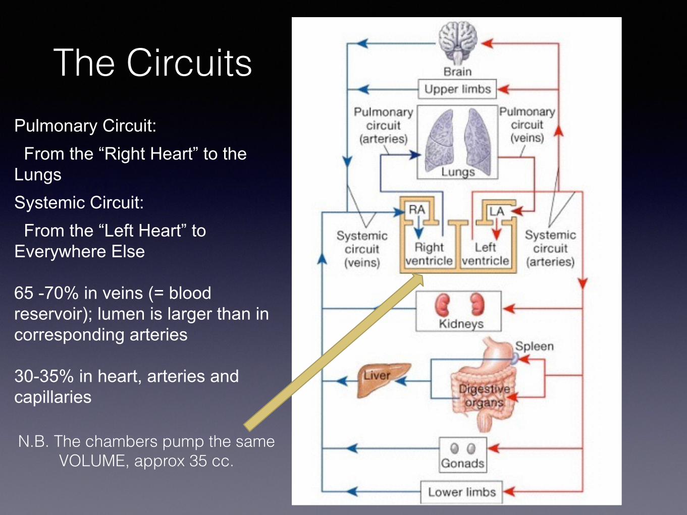
The CircuitsPulmonary Circuit From the ldquoRight Heartrdquo to the Lungs Systemic Circuit From the ldquoLeft Heartrdquo to Everywhere Else
65 -70 in veins (= blood reservoir) lumen is larger than in corresponding arteries 30-35 in heart arteries and capillaries
NB The chambers pump the same VOLUME approx 35 cc
Histology of Blood Vessels
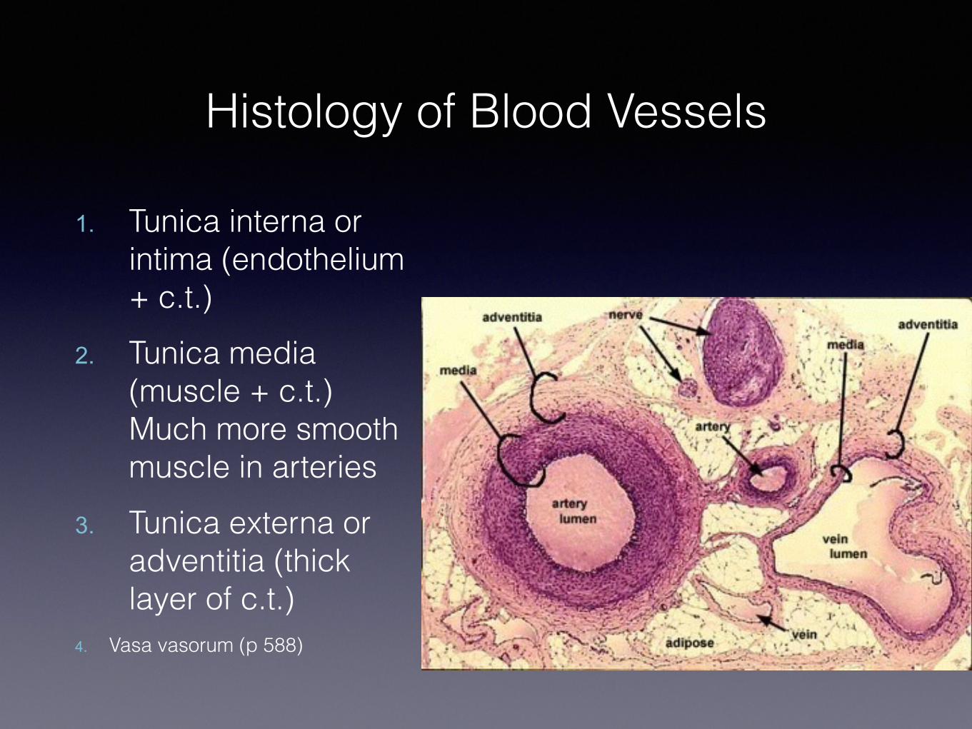
1 Tunica interna or intima (endothelium + ct)
2 Tunica media (muscle + ct) Much more smooth muscle in arteries
3 Tunica externa or adventitia (thick layer of ct)
4 Vasa vasorum (p 588)
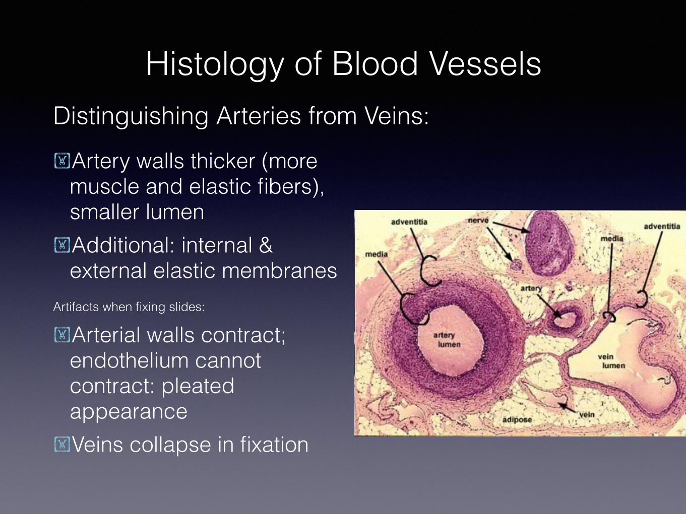
Distinguishing Arteries from Veins
Histology of Blood Vessels
Artery walls thicker (more muscle and elastic fibers) smaller lumen
Additional internal amp external elastic membranes
Artifacts when fixing slides
Arterial walls contract endothelium cannot contract pleated appearance
Veins collapse in fixation
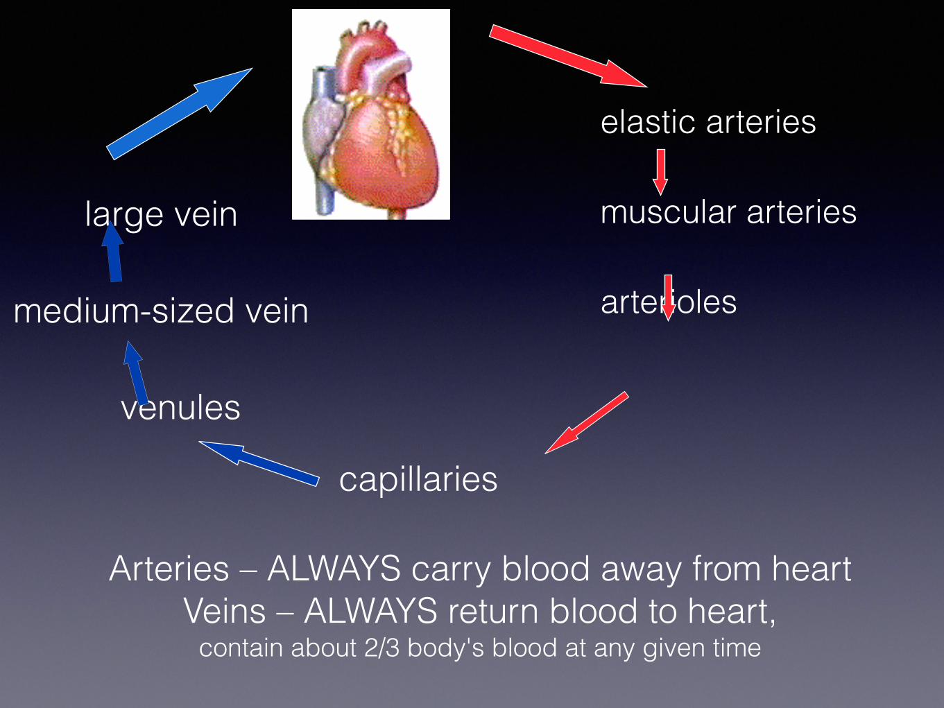
elastic arteries
muscular arteries
arterioles
capillaries
large vein
medium-sized vein
venules
Arteries ndash ALWAYS carry blood away from heart Veins ndash ALWAYS return blood to heart
contain about 23 bodys blood at any given time
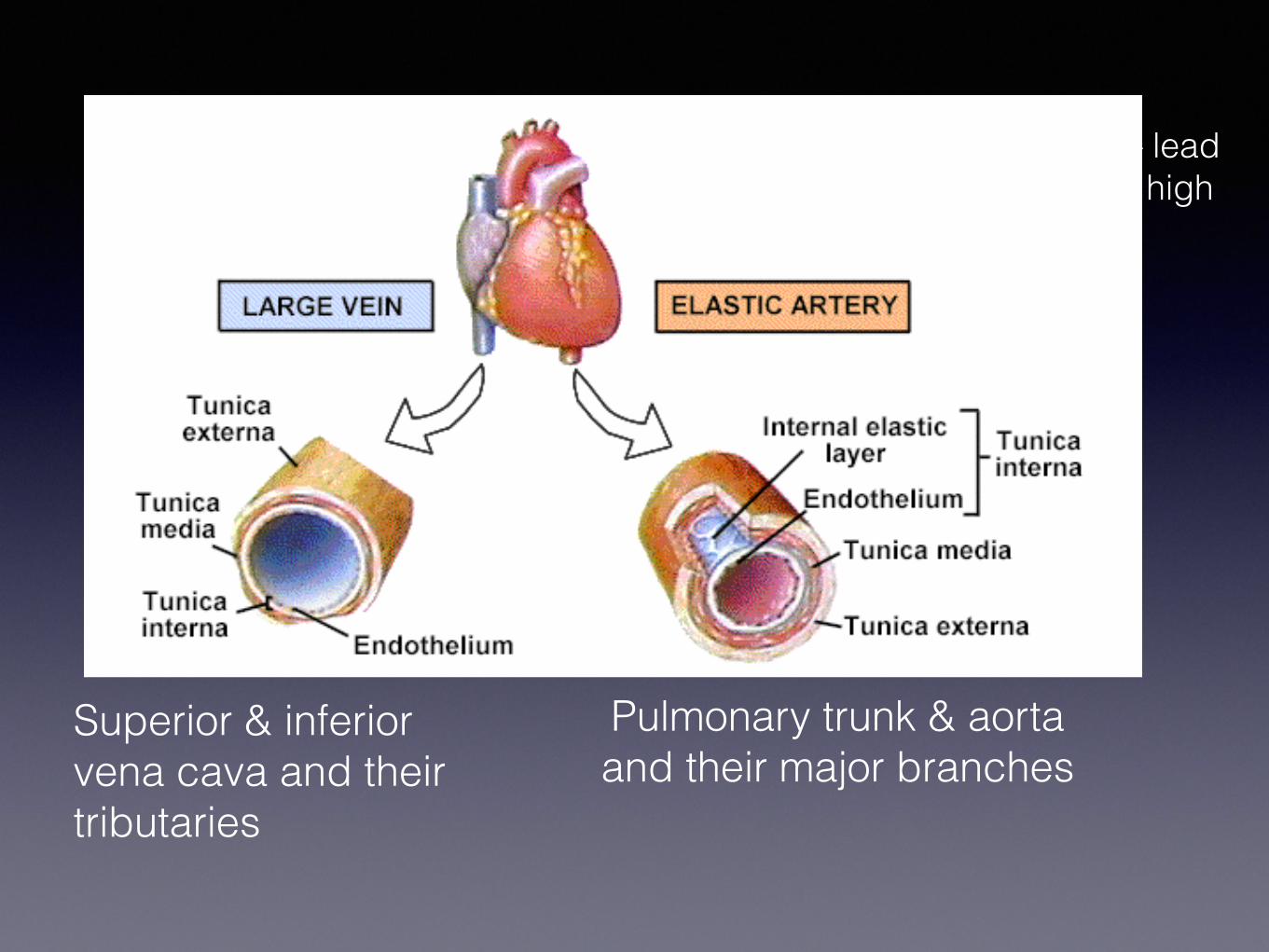
Pulmonary trunk amp aorta and their major branches
Superior amp inferior vena cava and their tributaries
Largest conducting arteries ndash lead directly from heart subject to high
pressures
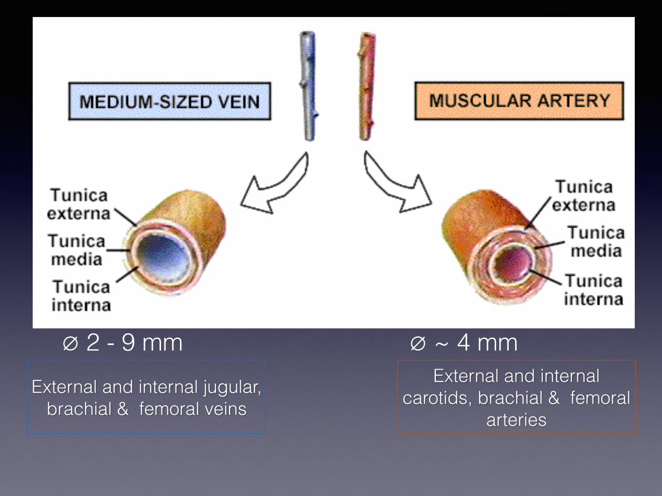
External and internal carotids brachial amp femoral
arteries
External and internal jugular brachial amp femoral veins
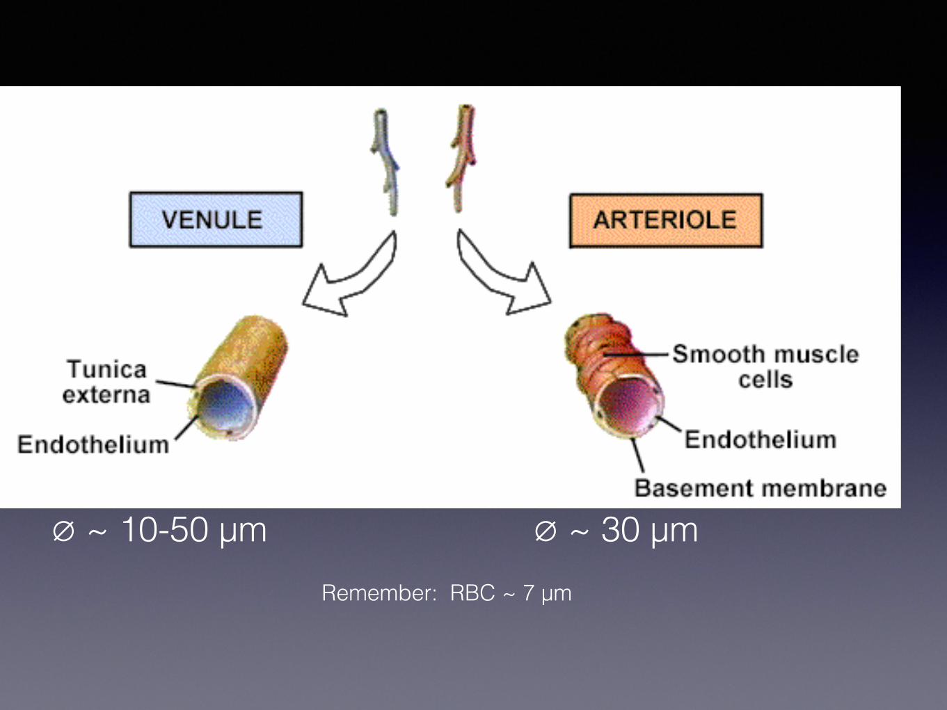
empty 2 - 9 mm empty ~ 4 mm
empty ~ 10-50 microm empty ~ 30 microm Remember RBC ~ 7 microm
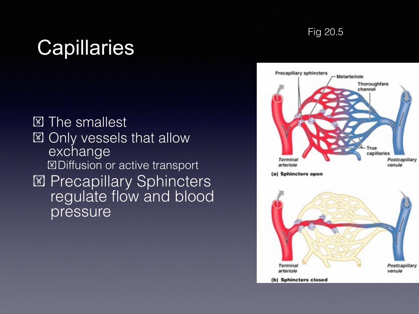
Capillaries
The smallest Only vessels that allow
exchange Diffusion or active transport
Precapillary Sphincters regulate flow and blood pressure
Fig 205
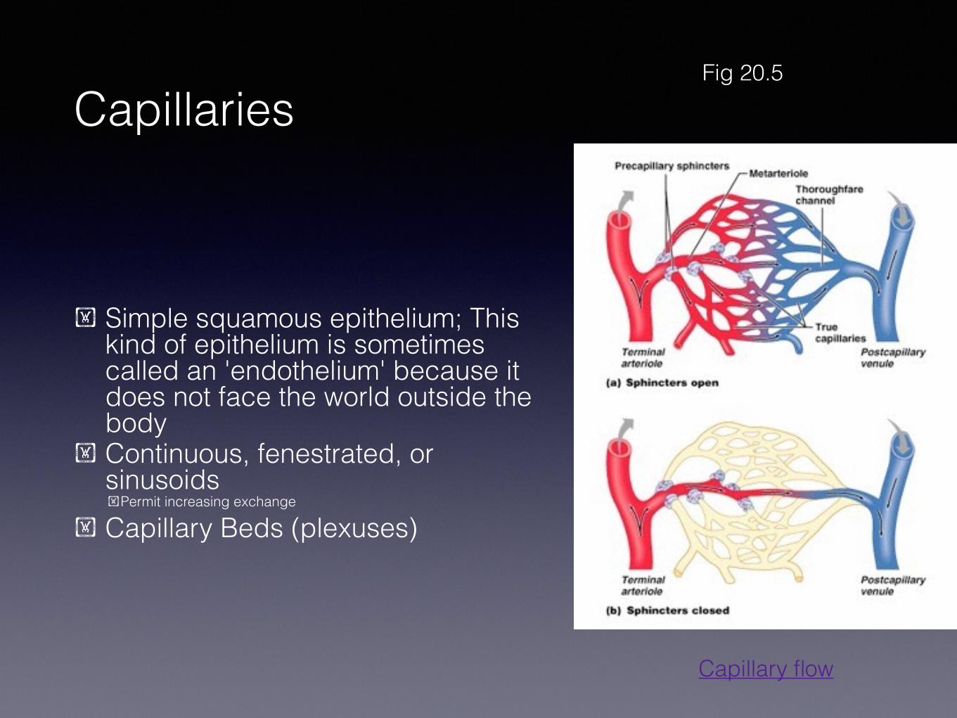
Capillaries
Simple squamous epithelium This kind of epithelium is sometimes called an endothelium because it does not face the world outside the body
Continuous fenestrated or sinusoids Permit increasing exchange
Capillary Beds (plexuses)
Fig 205
Capillary flow
Capillaries
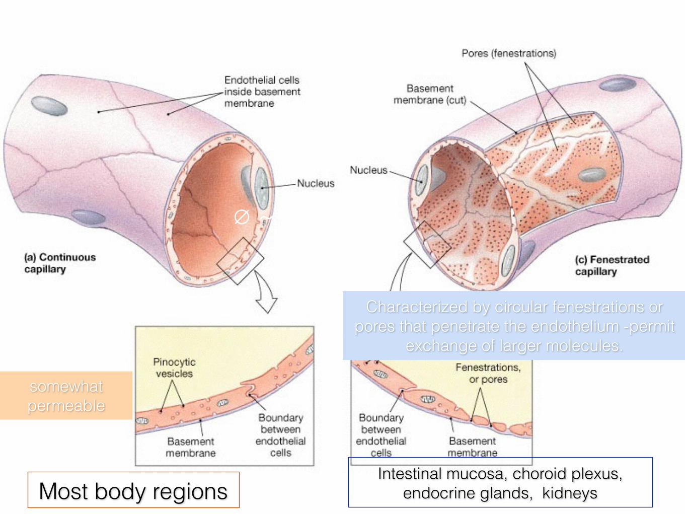
Intestinal mucosa choroid plexus endocrine glands kidneys Most body regions
Only endothelium Variably permeable
somewhat permeable
Characterized by circular fenestrations or pores that penetrate the endothelium -permit
exchange of larger molecules
empty ~ 8 microm
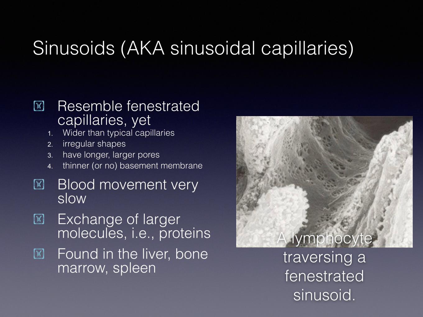
Sinusoids (AKA sinusoidal capillaries)
Resemble fenestrated capillaries yet
1 Wider than typical capillaries 2 irregular shapes 3 have longer larger pores 4 thinner (or no) basement membrane
Blood movement very slow
Exchange of larger molecules ie proteins
Found in the liver bone marrow spleen
A lymphocyte traversing a fenestrated sinusoid
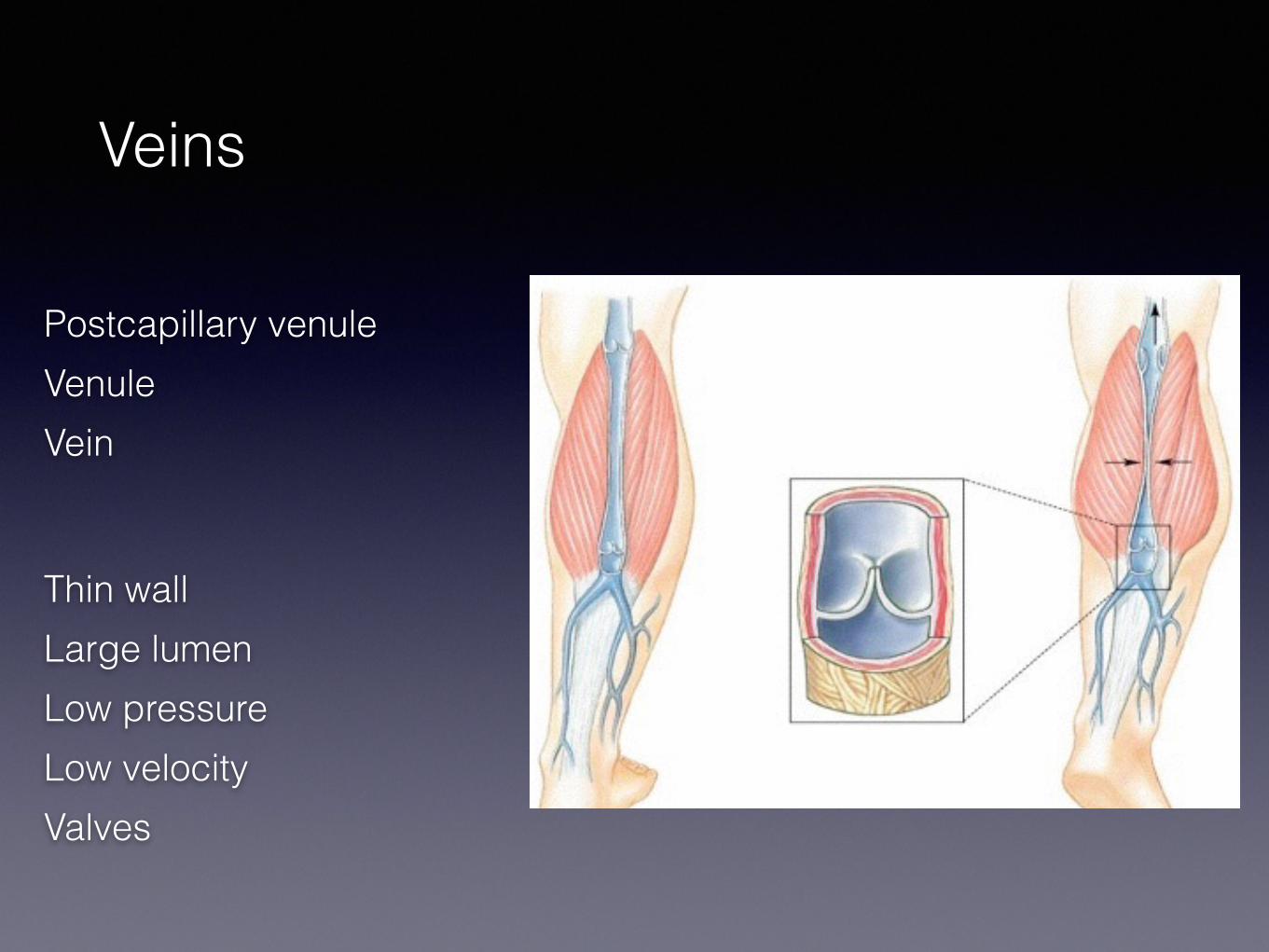
Veins
Postcapillary venule Venule Vein
Thin wall Large lumen Low pressure Low velocity Valves
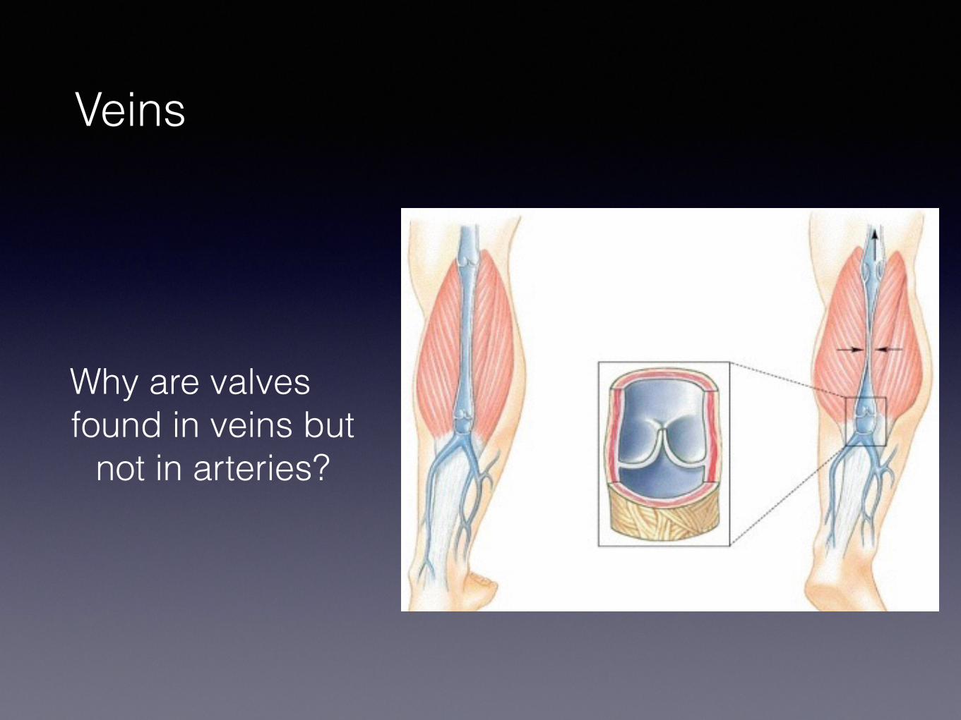
Veins
Why are valves found in veins but
not in arteries
Gross Anatomy of Circulatory System
Pulmonary amp Systemic Circulations
Right ventricle into pulmonary trunk to pulmonary arteries to lungs
Return by way of 4 pulmonary veins to left atrium
Pulmonary Circuit
Brachiocephalic trunk1
Left common carotid
Left subclavian3
2
Systemic Circulation
Right Common Carotid
Brachiocephalic Trunk
Left Common Carotid
Left Subclavian
Right Subclavian
Arteries to the Head
bull Common carotid (2) bull Vertebral Artery (2)
bull Through the transverse foramina and foramen magnum
bull and converge as the bull Basilar artery (1)
bull The carotids and basilar arteries converge on the Circle of Willis (AKA Cerebral Arterial Circle)
Circle of Willis = Cerebral Arterial Circle
bull = Ring of vessels surrounding pituitary gland - supplies cerebrum and cerebellum
bull Brain can receive blood from carotid or basilar aa
bullCollateral circulation bull(significance)
Arteriovenous malformations (AVM) are masses of abnormal blood vessels which grow in the brain
Dynamic angiogram
Arteries of the Arm
bull Difference Between Right and Left
bull Subclavian bull Axillary bull Brachial
bull Radial bull Ulnar
The Hand
Body Worlds
Ulnar Artery
Superficial Palmar Arch
Descending aorta
Abdominal aprta Common
External iliacFemoral
Thoracic aorta Abdominal aorta
Descending Aorta - Thoracic Area
Bronchial arteries - supply bronchi and lungs
Pericardial arteries - supply pericardium
Mediastinal arteries - supply mediastinal structures
Esophageal arteries - supply esophagus
Paired intercostal arteries- thoracic wall
Superior phrenic arteries - supply diaphragm
Descending Aorta Abdominal Area
Celiac trunk - 3 branches ndash to liver gallbladder esophagus stomach duodenum pancreas and spleen
Superior mesentericndash to pancreas and duodenum small intestine and colon
Paired suprarenal - to adrenal glands
Paired renal ndash to kidneys Paired gonadal ndash to testes or ovaries Inferior mesenteric ndash to terminal
colon and rectum Paired lumbar ndash to body wall
Aortic Aneurysm
An aneurysm is a bulging or ballooning of an artery
Often asymptomatic if it grows slowly
Weakness in the CT wall of the aorta
Rupture = Treatment is synthetic support
of the aorta
Circulation of the Leg
Common Iliac A and V External Iliac A amp V Femoral A amp V Popliteal A amp V
Anterior Tibial Posterior Tibial
Dorsal Pedal A Pulse checking
Great Saphenous V Used for coronary bypasses Longest vessel
Venous Circulation
Mostly parallels arterial circulation
Veins are more superficial in limbs
Major exception in the abdomen Portal Circulation
Useful Superficial Veins
Cephalic v
See Fig 2019
Median cubital v
Normal Blood Flow From artery to capillary bed to vein and back to heart
3 exceptions from artery to capillary to artery (or venule) to capillary to vein
Hypophysis Liver In kidney nephrons
Portal Systems
Portal Circulation
bull Venous return from the intestine is carried to the liver vial the Portal Vein
bull Most nutrients (not fat) are carried by the portal vein
bull Portal vein enters the liver at the hilus
Fig 2022 (a)
Portal vein
Varicose Veins
bull Page 562 bull uarr BP in legs
bull Venous dilation bull Breakdown of ct
bull Pooling bull May be painful andor
thrombogenic
Fetal circulation
Figure 2025
During fetal life all oxygen and nutrition comes not from the intestine and lungs but from the placenta The fetus has different circulatory pathways to accommodate these different needs These pathways must change AT the time of birth
Circulation Changes AT Birth
No blood coming from placenta Ductus venosus becomes ligamentum
venosus (=ligamentum teres) Foramen ovale closes amp becomes fossa
ovale Ductus arteriosus closes and becomes
ligamentum arteriosum Umbilical vein and arteries degenerate
Patent foramen ovale
The CircuitsPulmonary Circuit From the ldquoRight Heartrdquo to the Lungs Systemic Circuit From the ldquoLeft Heartrdquo to Everywhere Else
65 -70 in veins (= blood reservoir) lumen is larger than in corresponding arteries 30-35 in heart arteries and capillaries
NB The chambers pump the same VOLUME approx 35 cc
Histology of Blood Vessels
1 Tunica interna or intima (endothelium + ct)
2 Tunica media (muscle + ct) Much more smooth muscle in arteries
3 Tunica externa or adventitia (thick layer of ct)
4 Vasa vasorum (p 588)
Distinguishing Arteries from Veins
Histology of Blood Vessels
Artery walls thicker (more muscle and elastic fibers) smaller lumen
Additional internal amp external elastic membranes
Artifacts when fixing slides
Arterial walls contract endothelium cannot contract pleated appearance
Veins collapse in fixation
elastic arteries
muscular arteries
arterioles
capillaries
large vein
medium-sized vein
venules
Arteries ndash ALWAYS carry blood away from heart Veins ndash ALWAYS return blood to heart
contain about 23 bodys blood at any given time
Pulmonary trunk amp aorta and their major branches
Superior amp inferior vena cava and their tributaries
Largest conducting arteries ndash lead directly from heart subject to high
pressures
External and internal carotids brachial amp femoral
arteries
External and internal jugular brachial amp femoral veins
empty 2 - 9 mm empty ~ 4 mm
empty ~ 10-50 microm empty ~ 30 microm Remember RBC ~ 7 microm
Capillaries
The smallest Only vessels that allow
exchange Diffusion or active transport
Precapillary Sphincters regulate flow and blood pressure
Fig 205
Capillaries
Simple squamous epithelium This kind of epithelium is sometimes called an endothelium because it does not face the world outside the body
Continuous fenestrated or sinusoids Permit increasing exchange
Capillary Beds (plexuses)
Fig 205
Capillary flow
Capillaries
Intestinal mucosa choroid plexus endocrine glands kidneys Most body regions
Only endothelium Variably permeable
somewhat permeable
Characterized by circular fenestrations or pores that penetrate the endothelium -permit
exchange of larger molecules
empty ~ 8 microm
Sinusoids (AKA sinusoidal capillaries)
Resemble fenestrated capillaries yet
1 Wider than typical capillaries 2 irregular shapes 3 have longer larger pores 4 thinner (or no) basement membrane
Blood movement very slow
Exchange of larger molecules ie proteins
Found in the liver bone marrow spleen
A lymphocyte traversing a fenestrated sinusoid
Veins
Postcapillary venule Venule Vein
Thin wall Large lumen Low pressure Low velocity Valves
Veins
Why are valves found in veins but
not in arteries
Gross Anatomy of Circulatory System
Pulmonary amp Systemic Circulations
Right ventricle into pulmonary trunk to pulmonary arteries to lungs
Return by way of 4 pulmonary veins to left atrium
Pulmonary Circuit
Brachiocephalic trunk1
Left common carotid
Left subclavian3
2
Systemic Circulation
Right Common Carotid
Brachiocephalic Trunk
Left Common Carotid
Left Subclavian
Right Subclavian
Arteries to the Head
bull Common carotid (2) bull Vertebral Artery (2)
bull Through the transverse foramina and foramen magnum
bull and converge as the bull Basilar artery (1)
bull The carotids and basilar arteries converge on the Circle of Willis (AKA Cerebral Arterial Circle)
Circle of Willis = Cerebral Arterial Circle
bull = Ring of vessels surrounding pituitary gland - supplies cerebrum and cerebellum
bull Brain can receive blood from carotid or basilar aa
bullCollateral circulation bull(significance)
Arteriovenous malformations (AVM) are masses of abnormal blood vessels which grow in the brain
Dynamic angiogram
Arteries of the Arm
bull Difference Between Right and Left
bull Subclavian bull Axillary bull Brachial
bull Radial bull Ulnar
The Hand
Body Worlds
Ulnar Artery
Superficial Palmar Arch
Descending aorta
Abdominal aprta Common
External iliacFemoral
Thoracic aorta Abdominal aorta
Descending Aorta - Thoracic Area
Bronchial arteries - supply bronchi and lungs
Pericardial arteries - supply pericardium
Mediastinal arteries - supply mediastinal structures
Esophageal arteries - supply esophagus
Paired intercostal arteries- thoracic wall
Superior phrenic arteries - supply diaphragm
Descending Aorta Abdominal Area
Celiac trunk - 3 branches ndash to liver gallbladder esophagus stomach duodenum pancreas and spleen
Superior mesentericndash to pancreas and duodenum small intestine and colon
Paired suprarenal - to adrenal glands
Paired renal ndash to kidneys Paired gonadal ndash to testes or ovaries Inferior mesenteric ndash to terminal
colon and rectum Paired lumbar ndash to body wall
Aortic Aneurysm
An aneurysm is a bulging or ballooning of an artery
Often asymptomatic if it grows slowly
Weakness in the CT wall of the aorta
Rupture = Treatment is synthetic support
of the aorta
Circulation of the Leg
Common Iliac A and V External Iliac A amp V Femoral A amp V Popliteal A amp V
Anterior Tibial Posterior Tibial
Dorsal Pedal A Pulse checking
Great Saphenous V Used for coronary bypasses Longest vessel
Venous Circulation
Mostly parallels arterial circulation
Veins are more superficial in limbs
Major exception in the abdomen Portal Circulation
Useful Superficial Veins
Cephalic v
See Fig 2019
Median cubital v
Normal Blood Flow From artery to capillary bed to vein and back to heart
3 exceptions from artery to capillary to artery (or venule) to capillary to vein
Hypophysis Liver In kidney nephrons
Portal Systems
Portal Circulation
bull Venous return from the intestine is carried to the liver vial the Portal Vein
bull Most nutrients (not fat) are carried by the portal vein
bull Portal vein enters the liver at the hilus
Fig 2022 (a)
Portal vein
Varicose Veins
bull Page 562 bull uarr BP in legs
bull Venous dilation bull Breakdown of ct
bull Pooling bull May be painful andor
thrombogenic
Fetal circulation
Figure 2025
During fetal life all oxygen and nutrition comes not from the intestine and lungs but from the placenta The fetus has different circulatory pathways to accommodate these different needs These pathways must change AT the time of birth
Circulation Changes AT Birth
No blood coming from placenta Ductus venosus becomes ligamentum
venosus (=ligamentum teres) Foramen ovale closes amp becomes fossa
ovale Ductus arteriosus closes and becomes
ligamentum arteriosum Umbilical vein and arteries degenerate
Patent foramen ovale
Histology of Blood Vessels
1 Tunica interna or intima (endothelium + ct)
2 Tunica media (muscle + ct) Much more smooth muscle in arteries
3 Tunica externa or adventitia (thick layer of ct)
4 Vasa vasorum (p 588)
Distinguishing Arteries from Veins
Histology of Blood Vessels
Artery walls thicker (more muscle and elastic fibers) smaller lumen
Additional internal amp external elastic membranes
Artifacts when fixing slides
Arterial walls contract endothelium cannot contract pleated appearance
Veins collapse in fixation
elastic arteries
muscular arteries
arterioles
capillaries
large vein
medium-sized vein
venules
Arteries ndash ALWAYS carry blood away from heart Veins ndash ALWAYS return blood to heart
contain about 23 bodys blood at any given time
Pulmonary trunk amp aorta and their major branches
Superior amp inferior vena cava and their tributaries
Largest conducting arteries ndash lead directly from heart subject to high
pressures
External and internal carotids brachial amp femoral
arteries
External and internal jugular brachial amp femoral veins
empty 2 - 9 mm empty ~ 4 mm
empty ~ 10-50 microm empty ~ 30 microm Remember RBC ~ 7 microm
Capillaries
The smallest Only vessels that allow
exchange Diffusion or active transport
Precapillary Sphincters regulate flow and blood pressure
Fig 205
Capillaries
Simple squamous epithelium This kind of epithelium is sometimes called an endothelium because it does not face the world outside the body
Continuous fenestrated or sinusoids Permit increasing exchange
Capillary Beds (plexuses)
Fig 205
Capillary flow
Capillaries
Intestinal mucosa choroid plexus endocrine glands kidneys Most body regions
Only endothelium Variably permeable
somewhat permeable
Characterized by circular fenestrations or pores that penetrate the endothelium -permit
exchange of larger molecules
empty ~ 8 microm
Sinusoids (AKA sinusoidal capillaries)
Resemble fenestrated capillaries yet
1 Wider than typical capillaries 2 irregular shapes 3 have longer larger pores 4 thinner (or no) basement membrane
Blood movement very slow
Exchange of larger molecules ie proteins
Found in the liver bone marrow spleen
A lymphocyte traversing a fenestrated sinusoid
Veins
Postcapillary venule Venule Vein
Thin wall Large lumen Low pressure Low velocity Valves
Veins
Why are valves found in veins but
not in arteries
Gross Anatomy of Circulatory System
Pulmonary amp Systemic Circulations
Right ventricle into pulmonary trunk to pulmonary arteries to lungs
Return by way of 4 pulmonary veins to left atrium
Pulmonary Circuit
Brachiocephalic trunk1
Left common carotid
Left subclavian3
2
Systemic Circulation
Right Common Carotid
Brachiocephalic Trunk
Left Common Carotid
Left Subclavian
Right Subclavian
Arteries to the Head
bull Common carotid (2) bull Vertebral Artery (2)
bull Through the transverse foramina and foramen magnum
bull and converge as the bull Basilar artery (1)
bull The carotids and basilar arteries converge on the Circle of Willis (AKA Cerebral Arterial Circle)
Circle of Willis = Cerebral Arterial Circle
bull = Ring of vessels surrounding pituitary gland - supplies cerebrum and cerebellum
bull Brain can receive blood from carotid or basilar aa
bullCollateral circulation bull(significance)
Arteriovenous malformations (AVM) are masses of abnormal blood vessels which grow in the brain
Dynamic angiogram
Arteries of the Arm
bull Difference Between Right and Left
bull Subclavian bull Axillary bull Brachial
bull Radial bull Ulnar
The Hand
Body Worlds
Ulnar Artery
Superficial Palmar Arch
Descending aorta
Abdominal aprta Common
External iliacFemoral
Thoracic aorta Abdominal aorta
Descending Aorta - Thoracic Area
Bronchial arteries - supply bronchi and lungs
Pericardial arteries - supply pericardium
Mediastinal arteries - supply mediastinal structures
Esophageal arteries - supply esophagus
Paired intercostal arteries- thoracic wall
Superior phrenic arteries - supply diaphragm
Descending Aorta Abdominal Area
Celiac trunk - 3 branches ndash to liver gallbladder esophagus stomach duodenum pancreas and spleen
Superior mesentericndash to pancreas and duodenum small intestine and colon
Paired suprarenal - to adrenal glands
Paired renal ndash to kidneys Paired gonadal ndash to testes or ovaries Inferior mesenteric ndash to terminal
colon and rectum Paired lumbar ndash to body wall
Aortic Aneurysm
An aneurysm is a bulging or ballooning of an artery
Often asymptomatic if it grows slowly
Weakness in the CT wall of the aorta
Rupture = Treatment is synthetic support
of the aorta
Circulation of the Leg
Common Iliac A and V External Iliac A amp V Femoral A amp V Popliteal A amp V
Anterior Tibial Posterior Tibial
Dorsal Pedal A Pulse checking
Great Saphenous V Used for coronary bypasses Longest vessel
Venous Circulation
Mostly parallels arterial circulation
Veins are more superficial in limbs
Major exception in the abdomen Portal Circulation
Useful Superficial Veins
Cephalic v
See Fig 2019
Median cubital v
Normal Blood Flow From artery to capillary bed to vein and back to heart
3 exceptions from artery to capillary to artery (or venule) to capillary to vein
Hypophysis Liver In kidney nephrons
Portal Systems
Portal Circulation
bull Venous return from the intestine is carried to the liver vial the Portal Vein
bull Most nutrients (not fat) are carried by the portal vein
bull Portal vein enters the liver at the hilus
Fig 2022 (a)
Portal vein
Varicose Veins
bull Page 562 bull uarr BP in legs
bull Venous dilation bull Breakdown of ct
bull Pooling bull May be painful andor
thrombogenic
Fetal circulation
Figure 2025
During fetal life all oxygen and nutrition comes not from the intestine and lungs but from the placenta The fetus has different circulatory pathways to accommodate these different needs These pathways must change AT the time of birth
Circulation Changes AT Birth
No blood coming from placenta Ductus venosus becomes ligamentum
venosus (=ligamentum teres) Foramen ovale closes amp becomes fossa
ovale Ductus arteriosus closes and becomes
ligamentum arteriosum Umbilical vein and arteries degenerate
Patent foramen ovale
Capillaries
Intestinal mucosa choroid plexus endocrine glands kidneys Most body regions
Only endothelium Variably permeable
somewhat permeable
Characterized by circular fenestrations or pores that penetrate the endothelium -permit
exchange of larger molecules
empty ~ 8 microm
Sinusoids (AKA sinusoidal capillaries)
Resemble fenestrated capillaries yet
1 Wider than typical capillaries 2 irregular shapes 3 have longer larger pores 4 thinner (or no) basement membrane
Blood movement very slow
Exchange of larger molecules ie proteins
Found in the liver bone marrow spleen
A lymphocyte traversing a fenestrated sinusoid
Veins
Postcapillary venule Venule Vein
Thin wall Large lumen Low pressure Low velocity Valves
Veins
Why are valves found in veins but
not in arteries
Gross Anatomy of Circulatory System
Pulmonary amp Systemic Circulations
Right ventricle into pulmonary trunk to pulmonary arteries to lungs
Return by way of 4 pulmonary veins to left atrium
Pulmonary Circuit
Brachiocephalic trunk1
Left common carotid
Left subclavian3
2
Systemic Circulation
Right Common Carotid
Brachiocephalic Trunk
Left Common Carotid
Left Subclavian
Right Subclavian
Arteries to the Head
bull Common carotid (2) bull Vertebral Artery (2)
bull Through the transverse foramina and foramen magnum
bull and converge as the bull Basilar artery (1)
bull The carotids and basilar arteries converge on the Circle of Willis (AKA Cerebral Arterial Circle)
Circle of Willis = Cerebral Arterial Circle
bull = Ring of vessels surrounding pituitary gland - supplies cerebrum and cerebellum
bull Brain can receive blood from carotid or basilar aa
bullCollateral circulation bull(significance)
Arteriovenous malformations (AVM) are masses of abnormal blood vessels which grow in the brain
Dynamic angiogram
Arteries of the Arm
bull Difference Between Right and Left
bull Subclavian bull Axillary bull Brachial
bull Radial bull Ulnar
The Hand
Body Worlds
Ulnar Artery
Superficial Palmar Arch
Descending aorta
Abdominal aprta Common
External iliacFemoral
Thoracic aorta Abdominal aorta
Descending Aorta - Thoracic Area
Bronchial arteries - supply bronchi and lungs
Pericardial arteries - supply pericardium
Mediastinal arteries - supply mediastinal structures
Esophageal arteries - supply esophagus
Paired intercostal arteries- thoracic wall
Superior phrenic arteries - supply diaphragm
Descending Aorta Abdominal Area
Celiac trunk - 3 branches ndash to liver gallbladder esophagus stomach duodenum pancreas and spleen
Superior mesentericndash to pancreas and duodenum small intestine and colon
Paired suprarenal - to adrenal glands
Paired renal ndash to kidneys Paired gonadal ndash to testes or ovaries Inferior mesenteric ndash to terminal
colon and rectum Paired lumbar ndash to body wall
Aortic Aneurysm
An aneurysm is a bulging or ballooning of an artery
Often asymptomatic if it grows slowly
Weakness in the CT wall of the aorta
Rupture = Treatment is synthetic support
of the aorta
Circulation of the Leg
Common Iliac A and V External Iliac A amp V Femoral A amp V Popliteal A amp V
Anterior Tibial Posterior Tibial
Dorsal Pedal A Pulse checking
Great Saphenous V Used for coronary bypasses Longest vessel
Venous Circulation
Mostly parallels arterial circulation
Veins are more superficial in limbs
Major exception in the abdomen Portal Circulation
Useful Superficial Veins
Cephalic v
See Fig 2019
Median cubital v
Normal Blood Flow From artery to capillary bed to vein and back to heart
3 exceptions from artery to capillary to artery (or venule) to capillary to vein
Hypophysis Liver In kidney nephrons
Portal Systems
Portal Circulation
bull Venous return from the intestine is carried to the liver vial the Portal Vein
bull Most nutrients (not fat) are carried by the portal vein
bull Portal vein enters the liver at the hilus
Fig 2022 (a)
Portal vein
Varicose Veins
bull Page 562 bull uarr BP in legs
bull Venous dilation bull Breakdown of ct
bull Pooling bull May be painful andor
thrombogenic
Fetal circulation
Figure 2025
During fetal life all oxygen and nutrition comes not from the intestine and lungs but from the placenta The fetus has different circulatory pathways to accommodate these different needs These pathways must change AT the time of birth
Circulation Changes AT Birth
No blood coming from placenta Ductus venosus becomes ligamentum
venosus (=ligamentum teres) Foramen ovale closes amp becomes fossa
ovale Ductus arteriosus closes and becomes
ligamentum arteriosum Umbilical vein and arteries degenerate
Patent foramen ovale
Veins
Why are valves found in veins but
not in arteries
Gross Anatomy of Circulatory System
Pulmonary amp Systemic Circulations
Right ventricle into pulmonary trunk to pulmonary arteries to lungs
Return by way of 4 pulmonary veins to left atrium
Pulmonary Circuit
Brachiocephalic trunk1
Left common carotid
Left subclavian3
2
Systemic Circulation
Right Common Carotid
Brachiocephalic Trunk
Left Common Carotid
Left Subclavian
Right Subclavian
Arteries to the Head
bull Common carotid (2) bull Vertebral Artery (2)
bull Through the transverse foramina and foramen magnum
bull and converge as the bull Basilar artery (1)
bull The carotids and basilar arteries converge on the Circle of Willis (AKA Cerebral Arterial Circle)
Circle of Willis = Cerebral Arterial Circle
bull = Ring of vessels surrounding pituitary gland - supplies cerebrum and cerebellum
bull Brain can receive blood from carotid or basilar aa
bullCollateral circulation bull(significance)
Arteriovenous malformations (AVM) are masses of abnormal blood vessels which grow in the brain
Dynamic angiogram
Arteries of the Arm
bull Difference Between Right and Left
bull Subclavian bull Axillary bull Brachial
bull Radial bull Ulnar
The Hand
Body Worlds
Ulnar Artery
Superficial Palmar Arch
Descending aorta
Abdominal aprta Common
External iliacFemoral
Thoracic aorta Abdominal aorta
Descending Aorta - Thoracic Area
Bronchial arteries - supply bronchi and lungs
Pericardial arteries - supply pericardium
Mediastinal arteries - supply mediastinal structures
Esophageal arteries - supply esophagus
Paired intercostal arteries- thoracic wall
Superior phrenic arteries - supply diaphragm
Descending Aorta Abdominal Area
Celiac trunk - 3 branches ndash to liver gallbladder esophagus stomach duodenum pancreas and spleen
Superior mesentericndash to pancreas and duodenum small intestine and colon
Paired suprarenal - to adrenal glands
Paired renal ndash to kidneys Paired gonadal ndash to testes or ovaries Inferior mesenteric ndash to terminal
colon and rectum Paired lumbar ndash to body wall
Aortic Aneurysm
An aneurysm is a bulging or ballooning of an artery
Often asymptomatic if it grows slowly
Weakness in the CT wall of the aorta
Rupture = Treatment is synthetic support
of the aorta
Circulation of the Leg
Common Iliac A and V External Iliac A amp V Femoral A amp V Popliteal A amp V
Anterior Tibial Posterior Tibial
Dorsal Pedal A Pulse checking
Great Saphenous V Used for coronary bypasses Longest vessel
Venous Circulation
Mostly parallels arterial circulation
Veins are more superficial in limbs
Major exception in the abdomen Portal Circulation
Useful Superficial Veins
Cephalic v
See Fig 2019
Median cubital v
Normal Blood Flow From artery to capillary bed to vein and back to heart
3 exceptions from artery to capillary to artery (or venule) to capillary to vein
Hypophysis Liver In kidney nephrons
Portal Systems
Portal Circulation
bull Venous return from the intestine is carried to the liver vial the Portal Vein
bull Most nutrients (not fat) are carried by the portal vein
bull Portal vein enters the liver at the hilus
Fig 2022 (a)
Portal vein
Varicose Veins
bull Page 562 bull uarr BP in legs
bull Venous dilation bull Breakdown of ct
bull Pooling bull May be painful andor
thrombogenic
Fetal circulation
Figure 2025
During fetal life all oxygen and nutrition comes not from the intestine and lungs but from the placenta The fetus has different circulatory pathways to accommodate these different needs These pathways must change AT the time of birth
Circulation Changes AT Birth
No blood coming from placenta Ductus venosus becomes ligamentum
venosus (=ligamentum teres) Foramen ovale closes amp becomes fossa
ovale Ductus arteriosus closes and becomes
ligamentum arteriosum Umbilical vein and arteries degenerate
Patent foramen ovale
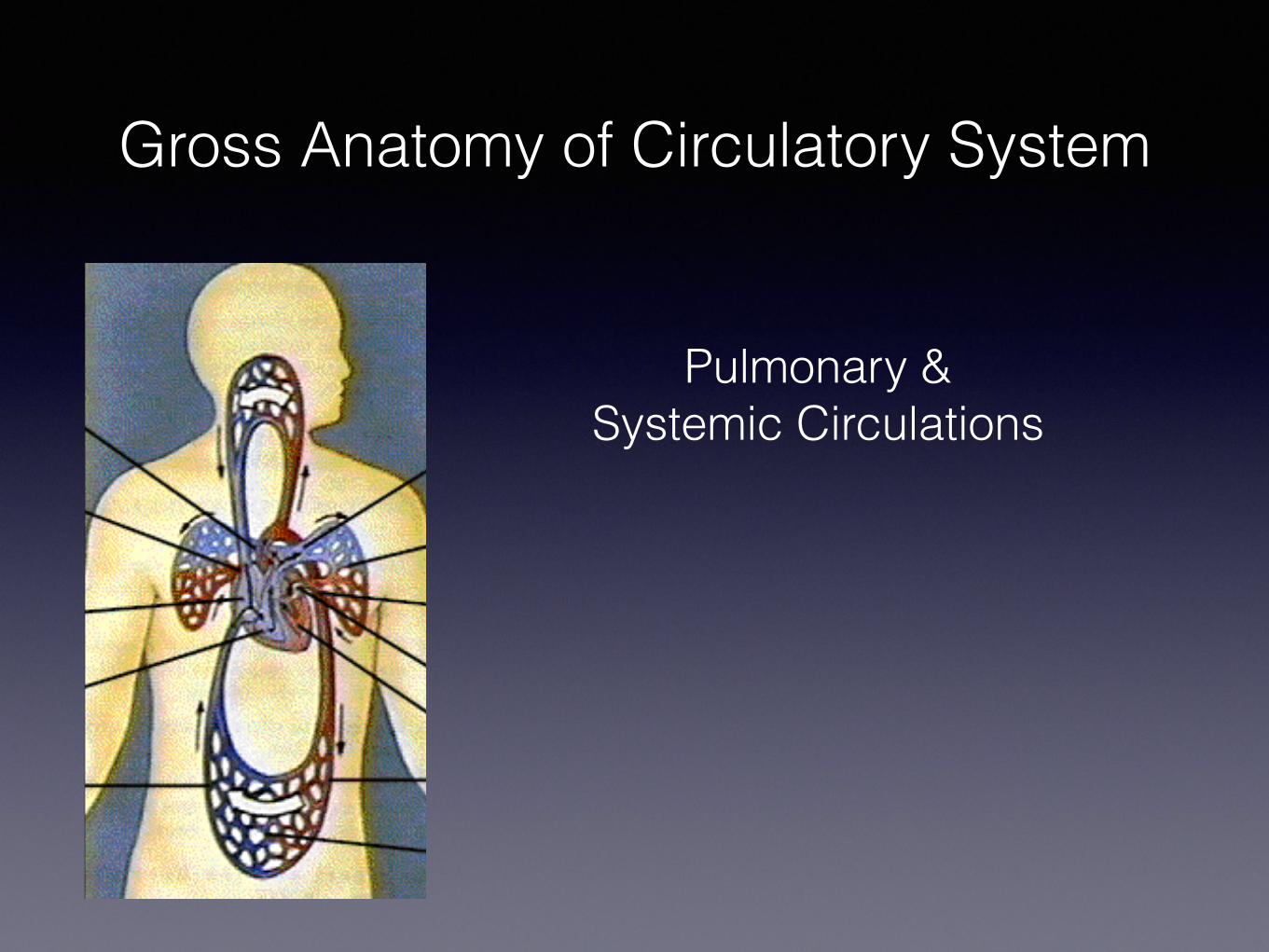
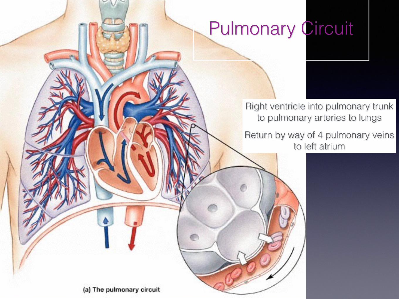
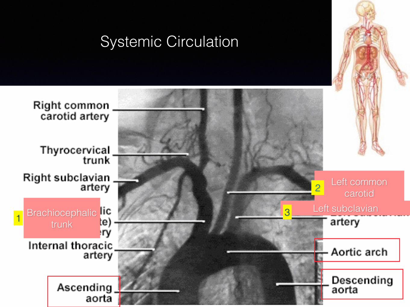
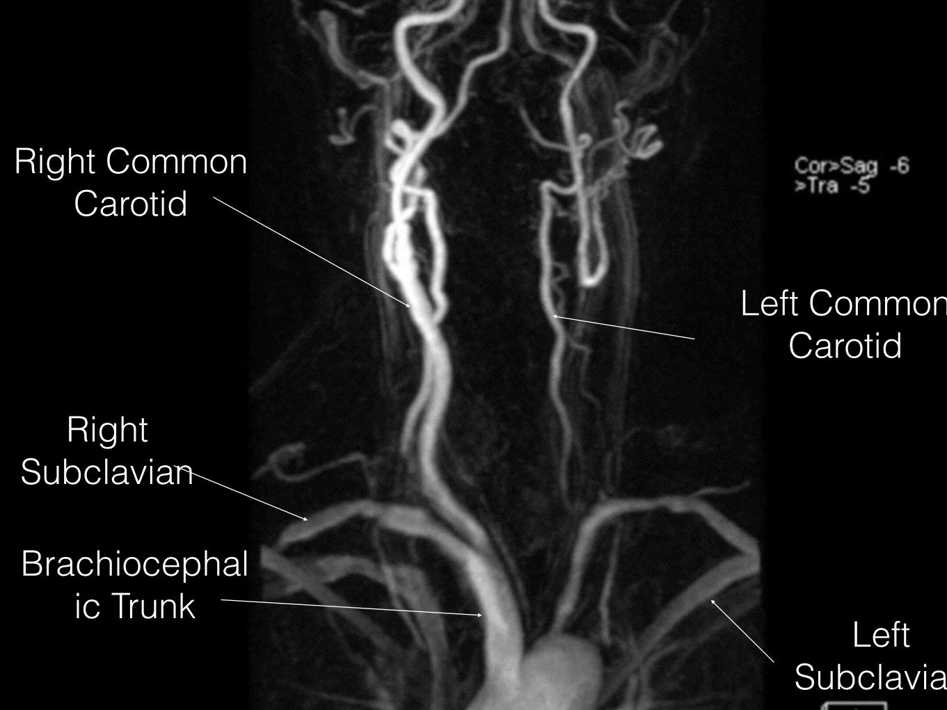
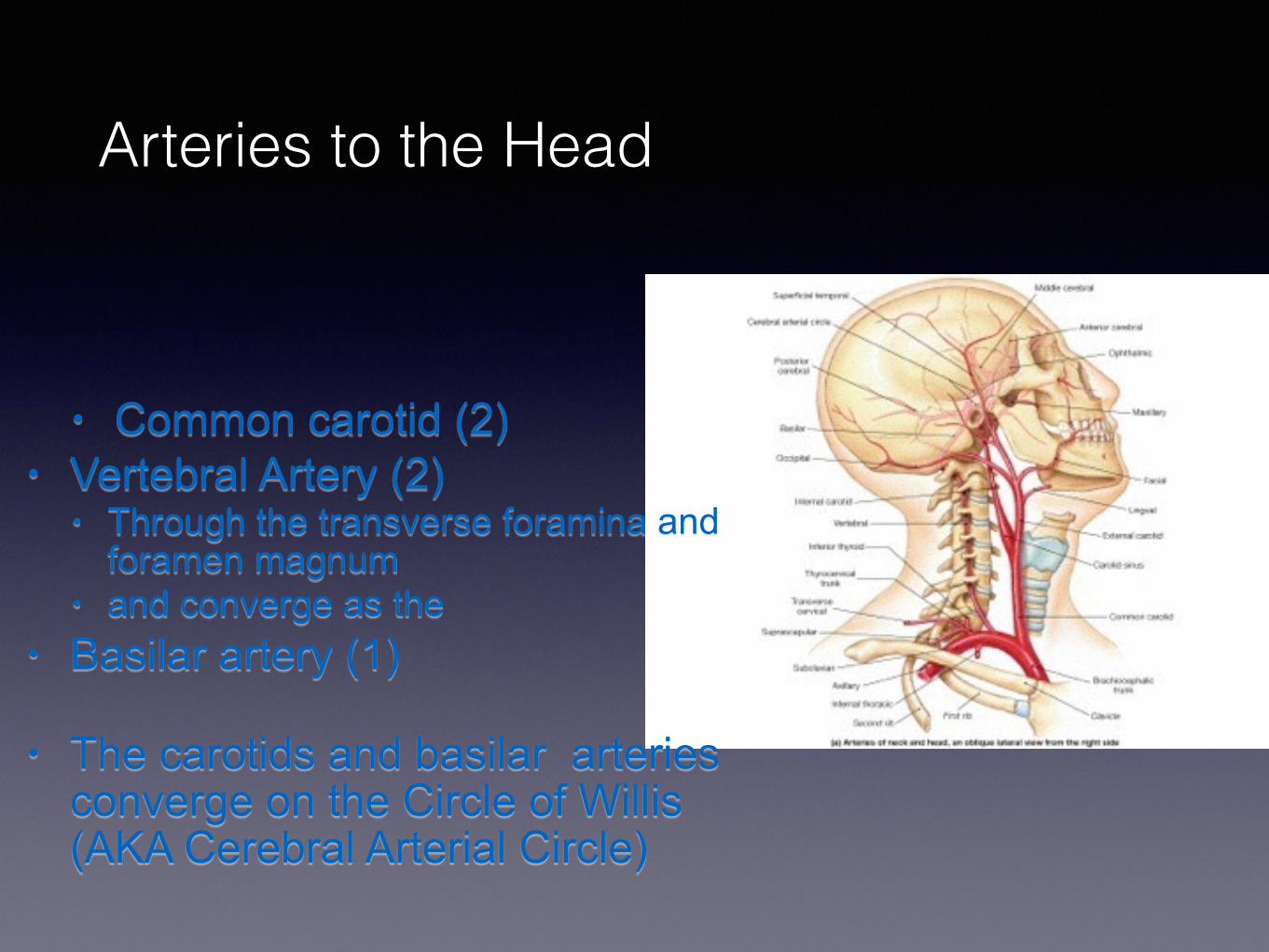
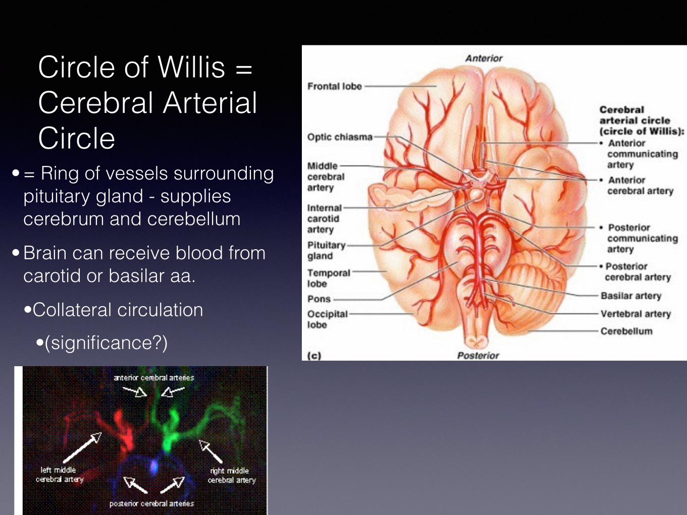
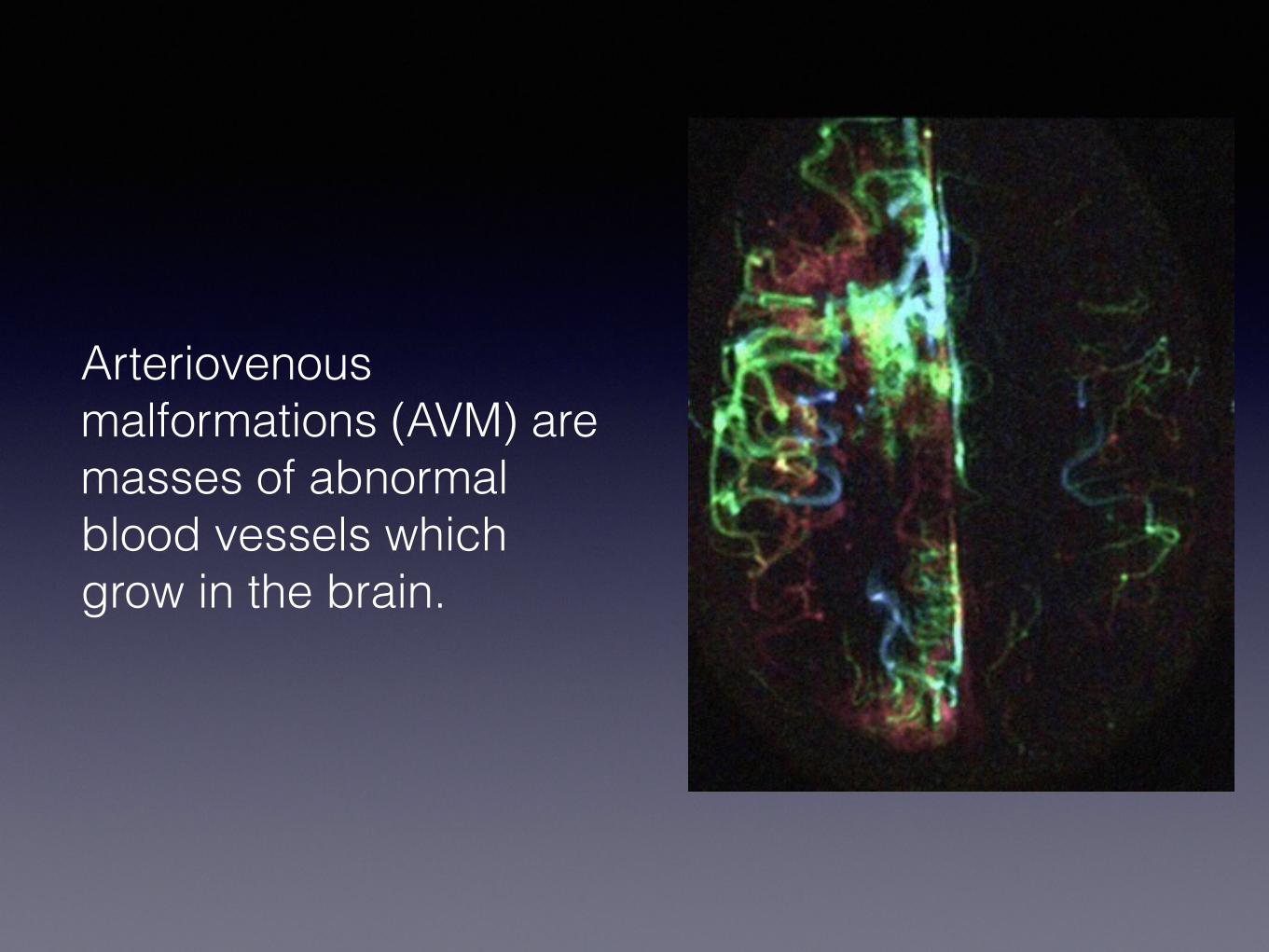
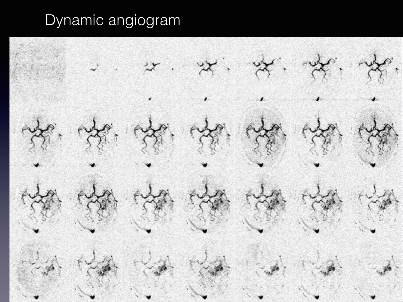
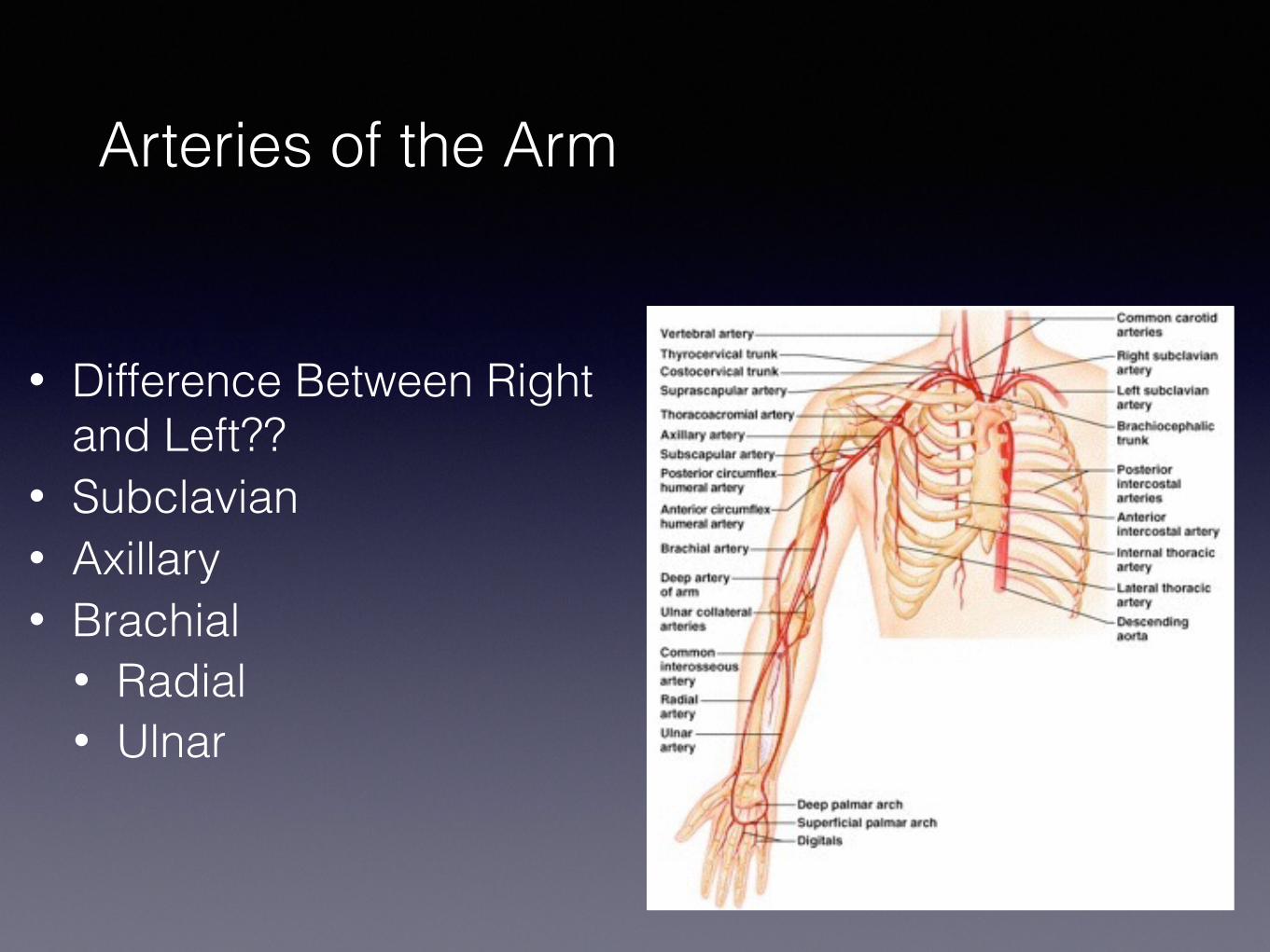
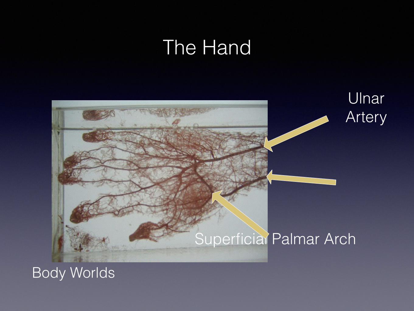
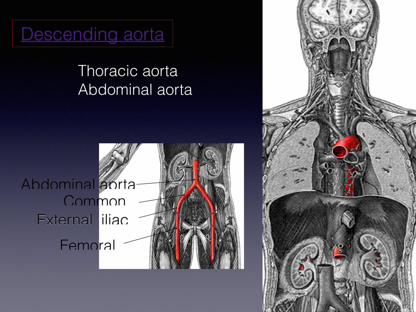
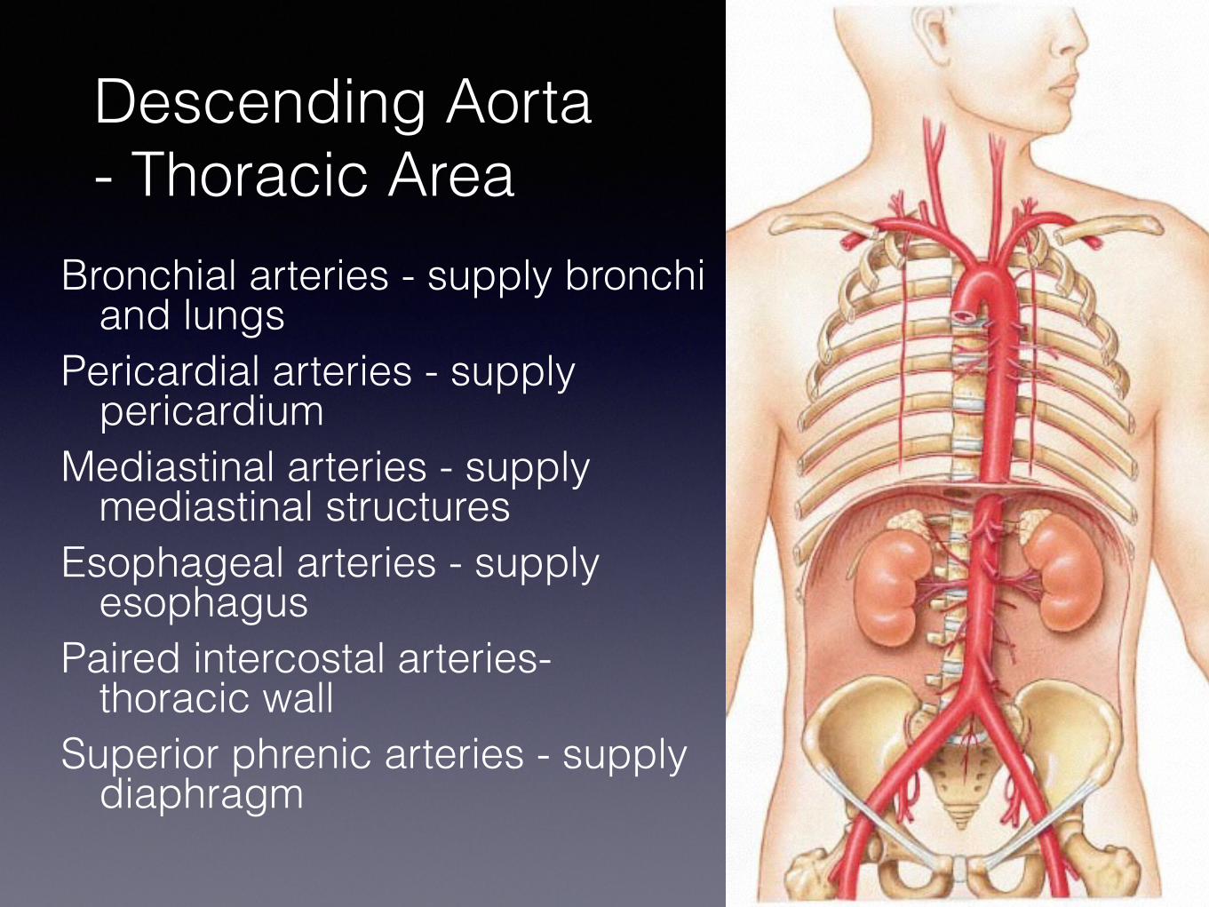
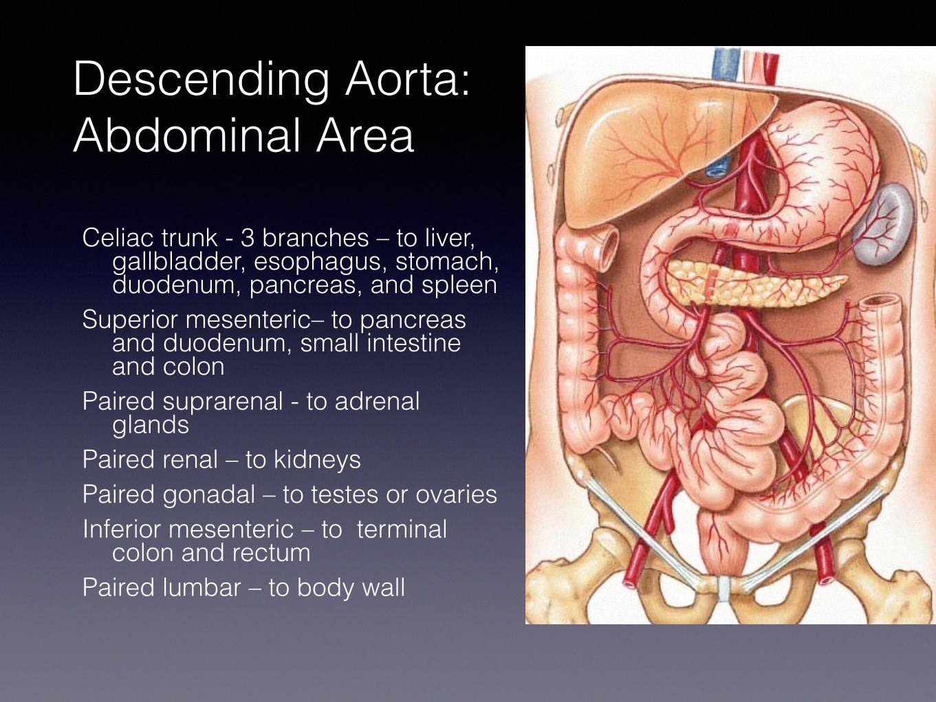
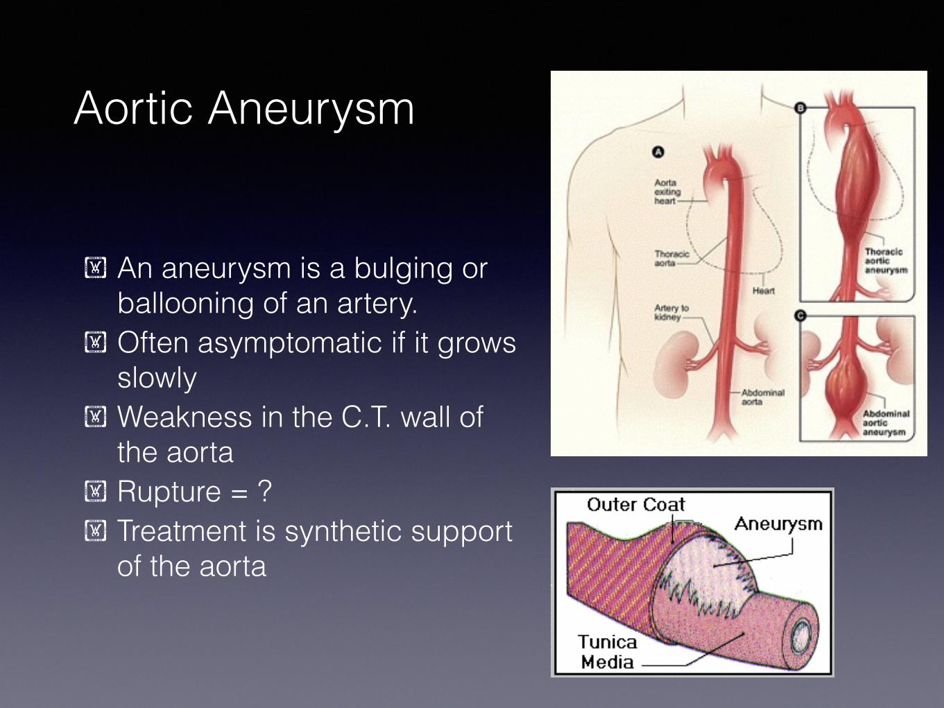
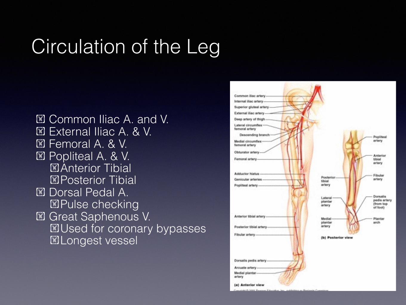
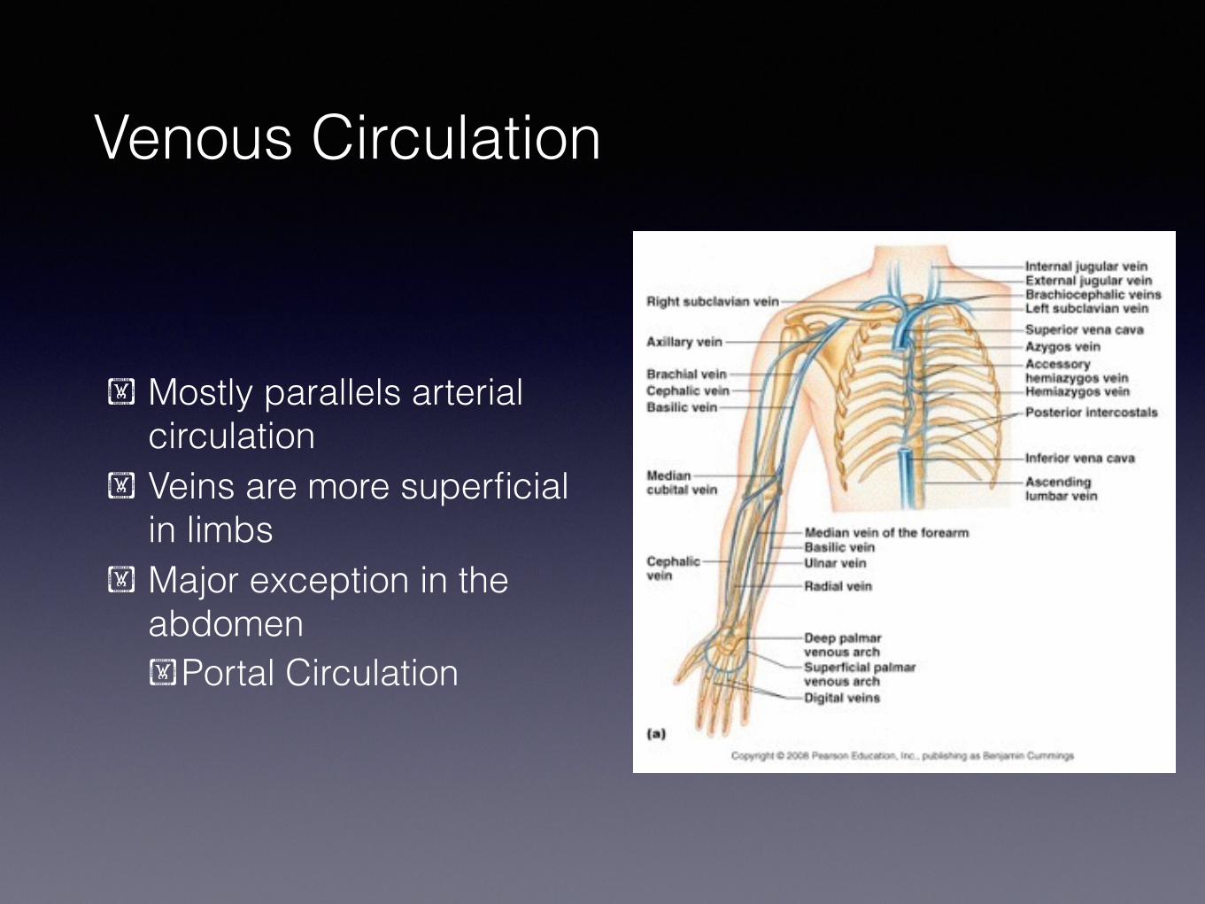
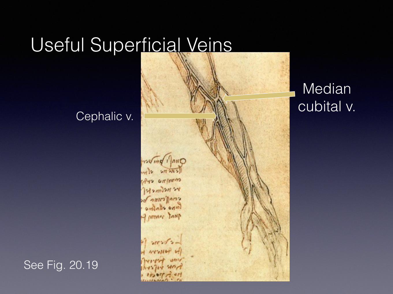
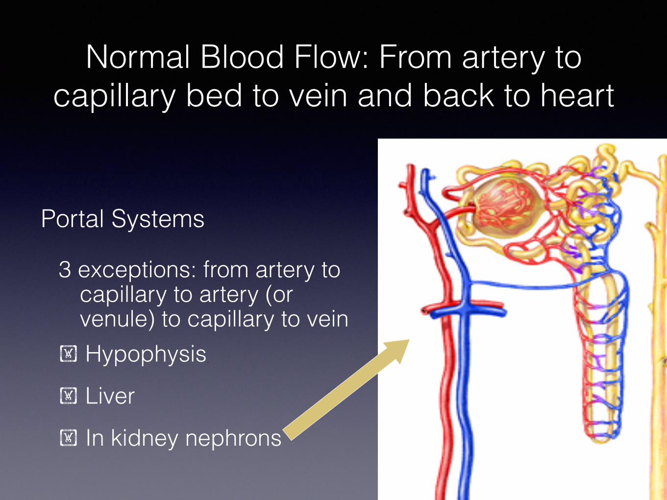
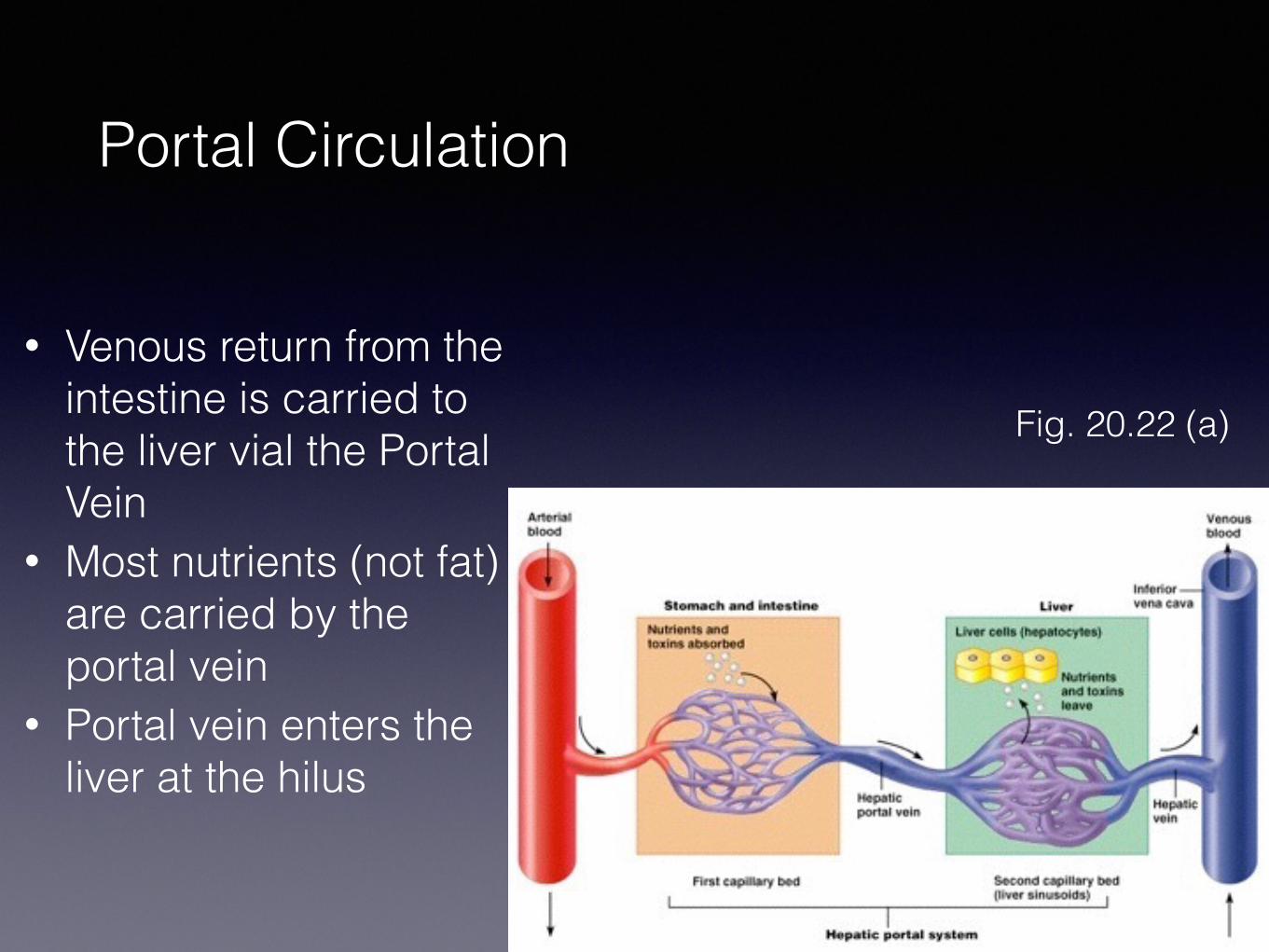
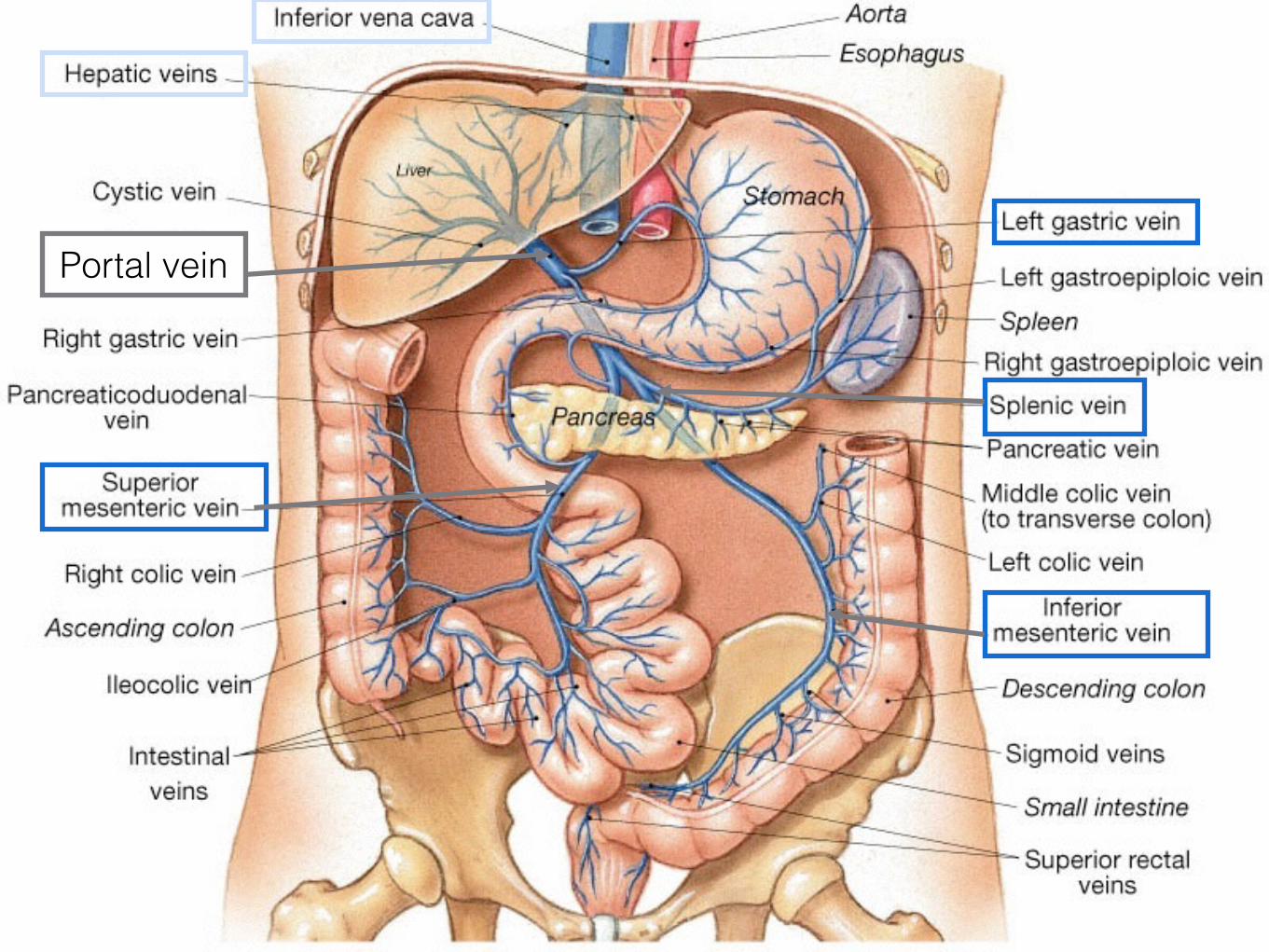
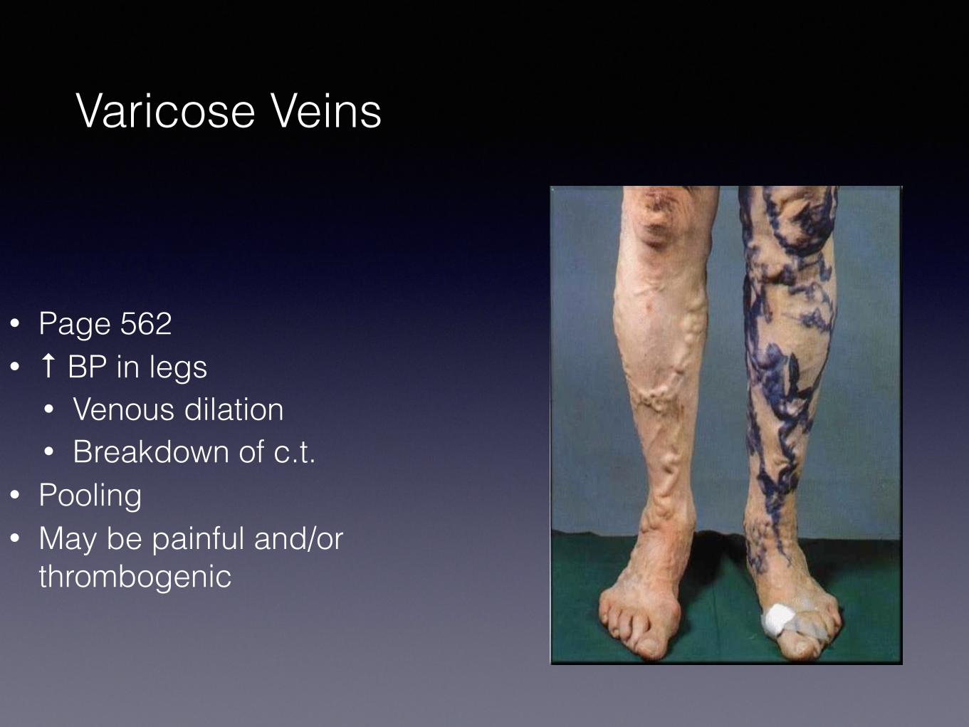
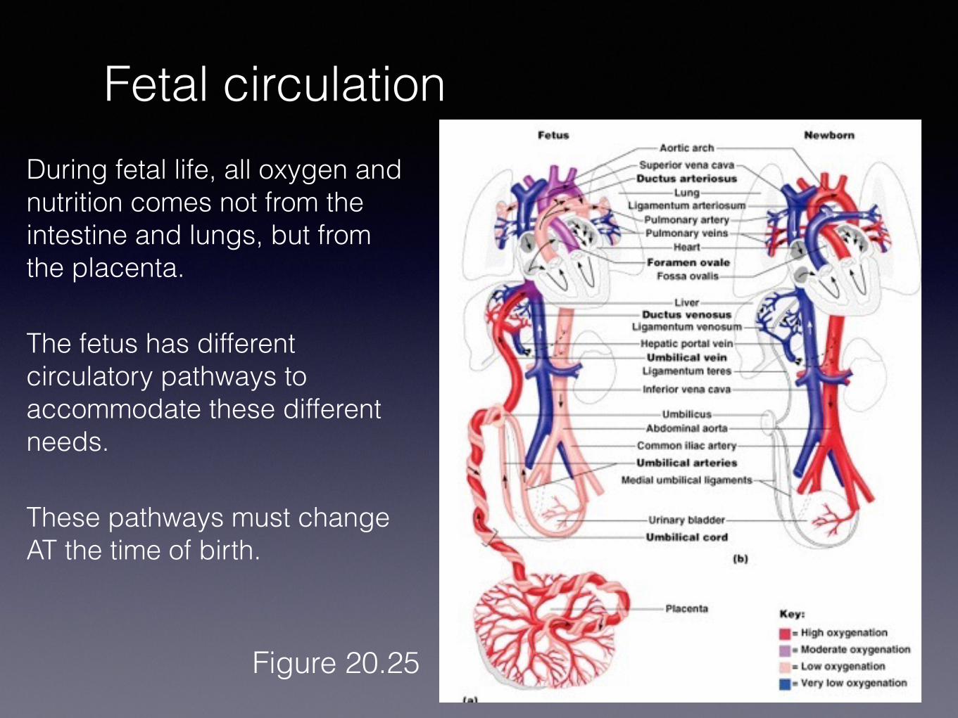
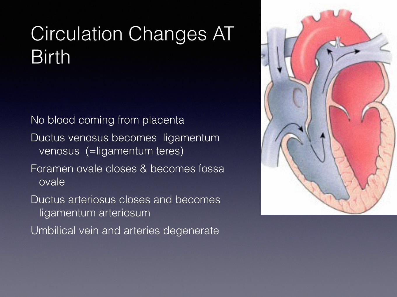
Circulation Changes AT Birth
No blood coming from placenta Ductus venosus becomes ligamentum
venosus (=ligamentum teres) Foramen ovale closes amp becomes fossa
ovale Ductus arteriosus closes and becomes
ligamentum arteriosum Umbilical vein and arteries degenerate
Patent foramen ovale
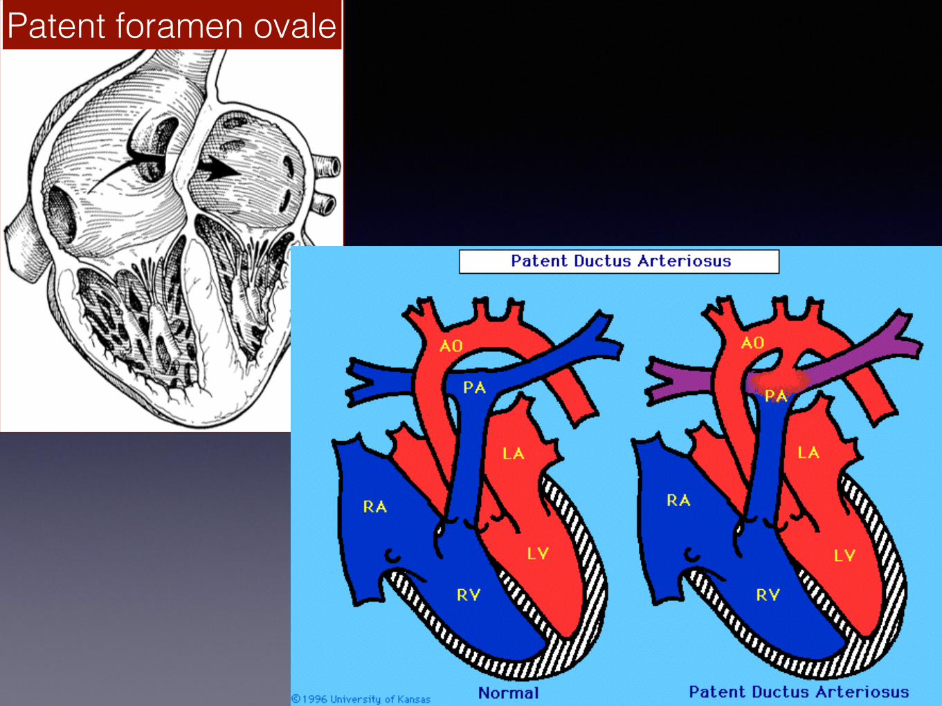
Patent foramen ovale