chain dynamic of calcified tissue
TRANSCRIPT
J O U R N A L O F M A T E R I A L S S C I E N C E : M A T E R I A L S IN M E D I C I N E 7 (1996) 1 7 5 - 1 7 9
Chain dynamic of calcified tissue
A. LAMURE, S. M E Z G H A N I , M. F. H A R M A N D * , C. LACABANNE Sofid State Physics Laboratory, URA CNRS 74, Paul Sabatier University, 118 route de Narbonne, 31 062 Toulouse Codex, France * INSERM U 306, Bordeaux II University, 146 rue Leo Saignat, 33 076 Bordeaux COdex, France
Thermally stimulated current spectroscopy has been applied to the investigation of molecular mobil i ty in human calcified tissue. A comparative study of extracts and residues at various stages of demineralization is presented. Results show that:
• the matrix (collagen) is in a glassy state at physiological temperature; • the filler (apatite) increases the static modulus; • the interfaces/interphase (non-collagenous proteins and particularly proteoglycans)
ensure cohesion and ductability for the composite.
Biomaterials for orthopaedic prostheses require the same morphology in order to phenomenologically reproduce the same dynamic behaviour.
1. In t roduct ion The development of second generation implants re- quires a thorough knowledge of the dynamic behav- iour of calcified tissue. It is well known that the matrix is constituted of collagen, and reinforced by an apa- titic mineral charge; various non-collagenous proteins (proteoglycans or glycoproteins: osteonectin, os- teocalcin, phosphoproteins) have also been detected [-1-6]. The specific mineral phase confers on the bone not only a mechanical role but also a biological role: bone contains the main ions (calcium, magnesium, sodium, phosphorus) which control certain physiolo- gical functions. In a composite, the final properties are not only dependent upon the properties of each com- ponent but also of the molecular interfaces between the various constituents. Several studies have been performed on the static modulus of bone but the chain dynamics of calcified tissues is still not understood. The aim of this work is to characterize bone dynamics and to define the nature of the organic/mineral inter- phase/interface. Extracts and residues of extraction by EDTA, at various stages of demineralization, have been studied by biochemical and biophysical methods (ELISA, radioimmunology, infrared spectroscopy, thermally stimulated current). The relaxation times of residues at various stages of demineralization have been analysed to define the characteristics of the dis- tribution functions and to establish a dynamic model of calcified tissue.
2. Mater ia ls and m e t h o d 2.1. Human bone powder sequential
extraction Non-collagenous proteins from bone, as well as sol- uble collagen, were extracted from 20 g of human bone powder (radius and cubitus), by sequential extraction
using 10 vols EDTA 0.5 M, pH 7.4, supplemented with proteases inhibitors (6-amino hexanoic acid 0.01 M, benzamidinium chloride 0.005M). As shown in TableI , ten 48 h extractions under agitation (El--+El0) were followed by two 48 h extractions (EG1, EG2) with EDTA supplemented by 4M guanidinium chloride. Finally, the two last extractions (EG3, EG4) were performed in the presence of 6 M guanidinium chloride. Supernatants were separated from the insoluble residues (R1 ~ R10, RG1 ~ RG4) by centrifugation (15 min, 2000 g, 4 °C). Proteins were precipitated from the supernatant by ammonium sul- phate 50% (v/v) in PBS, overnight at 4 °C, for further analysis. Residues were rinsed five times with ultra- pure water and freeze dried.
2.2. Thermally stimulated current (TSC) spectroscopy
In TSC experiments, a potential of 200 V was applied to the sample, placed between the plates of a conden- sor, for 2 min at 25 °C. This out-of-order configuration was quenched and the electric field cut off at liquid nitrogen temperature (LNT). Then the return to equi- librium of the sample was induced by a controlled increase of temperature (7 K min - 1). Simultaneously, the depolarization current was recorded versus tem- perature giving the "complex TSC spectrum".
For bone and bone residues, the TSC spectra were resolved into elementary spectra, i.e. well described by a single relaxation time ~ using the fractional polariza- tion method [7]. The fractional polarization experi- mental procedure was as follows: the field was applied at Tp (the polarization temperature) for 2 min allow- ing orientation of mobile units with relaxation times "C < T(Tp). Then the temperature was lowered to Td = T p - 10 °C, when the electric field was cut off
0957-4530 © 1996 Chapman & Hall 175
T A B L E I Bone demineralization sequence (48 h extraction time)
Initial products Extraction buffer Final products
Bone EDTA El, R1 R1 EDTA E2, R2 R2 EDTA E3, R3 R3 EDTA E4, R4 R,~ EDTA Es, Rs R5 EDTA E6, R6 R 6 EDTA ET, R 7 R7 EDTA E8, Rs Rs EDTA E9, R 9 R 9 EDTA Elo, Rio Rio EDTA + GuHC14M EG1, RG1 RG1 EDTA + GuHC14M EG2, RG2 RG2 EDTA + GuHC1 6M EG3, RG3 RG3 EDTA + GuHC1 6M EG4, RG~
5 x 10 -12
2 . 5 x 10 13
- 1 2 0 - 6 0
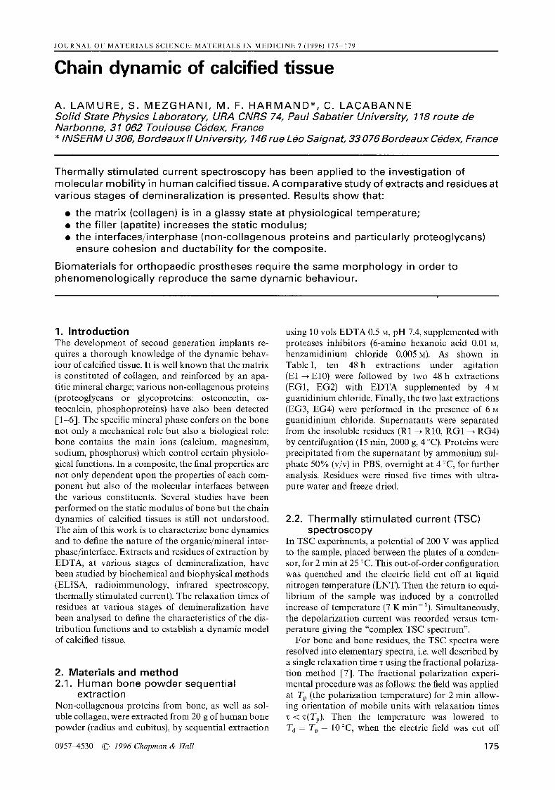
Figure 1 Complex spectrum of bone.
J 0
T ( ° C )
1 l I
- 6 0
and the temperature kept constant for t = 2 min, allowing the return to equilibrium of mobile units with relaxation times 'c < 'c(Ta). Hence, the cycle used to obtain TSC "elementary" spectra permits recording of an isolated process with relaxation times 'c such that z(Ta) < 'c < 'c(Tp). By shifting Tp along the tempera- ture axis, the whole TSC spectrum can be explored. The narrow temperature window Tp -- Td effectively selects out oriented dipoles with practically the same activation enthalpy AH and the same characteristic relaxation times to so that each elementary TSC spec- t rum may be analysed, as it arises from a single Debye relaxation process. The relaxation time depends on temperature according to the Arrhenius equation:
AH "c = "coexp k T (1)
where k is the Boltzmann constant, and
h AS 'co = ~ e x p k (2)
where h is Planck's constant and AS is the activation entropy. In apatites and collagen, some elementary processes have a particular behaviour: a linear rela- tionship exists between the Arrhenius factors log(r0) and AH. So the corresponding relaxation times are linked by a compensation law:
c = z ~ e x p ~ - - (3)
with AH
• co = "Co exp tc"To (4)
This compensation effect between the activation en- thalpy and entropy can be explained on the basis of the two sites model proposed by Hoffman et al. [8]. Molecular movements are hierarchically correlated. This dependence of activation parameters, widely ob- served in polymers [9], is characteristic of the dynam- ics of a transition.
3. Results 3.1. Biochemical analysis of extracts During the first three 48 h extractions, respectively 48%, 14% and 5% of the EDTA-soluble proteins
were extracted as assessed by the method of Lowry et al. [10]. As for the calcium determination by atomic absorption, it was shown that practically all the cal- cium disappears during the three first extractions. These results show that 3 x 48 h are not sufficient to extract all of the EDTA-soluble proteins, although the mineral phase is dissolved. The three first extracts (E 1 ~ E3) contain proteins which are probably bound to the mineral phase whereas the seven last extracts (E4 --+ Elo) are certainly constituted by proteins linked to the collagen.
Moreover, to define the origin of non-collagenic proteins, the extracts were analysed by size exclusion chromatography and the identification of the remain- ing proteins was carried out by u.v. spectroscopy at 280 nm. The presence of osteonectin was checked by ELISA [11], using polyclonal antibodies raised against bovine osteonectin (Cis Bioindustry, Gif sur Yvette/France). Finally, the presence of osteocalcin was measured by radioimmunology. Osteocalcin was found principally in E1 and E2 (57 and 29%), confirm- ing its high affinity for hydroxyapati te observed in rive [12]. In addition the last extracts with guanidinium chloride consisted principally of phosphoproteins (phosphorin) and matrix-Gla-proteins [13].
3.2. Biophysical analysis of residues Three groups of spectra can be distinguished, corre- sponding to:
• bone • residues obtained after demineralization with
EDTA • residues obtained after demineralization with
EDTA + Glu HC1.
3.2. 1. B o n e TSC spectra of bone contain two broad peaks: one main peak around room temperature and another peak around - 1 3 0 °C (Fig. 1). In order to determine their origin, the complex TSC spectrum was resolved into elementary TSC spectra using fractional polariza- tion. All TSC spectra were resolved into elementary TSC spectra using fractional polarization. All elemen- tary processes isolated between nitrogen temperature
176

- 8
e o
O
-10
-12
-14
x.,
x'-x.. T c = 130 °C " - x . "Co = 510 7 s
"x .,.
-16 I I I I I I 0.2 0.3 0.4 0.5
AH ( eV )
Figure 2 Compensation diagram of bone.
~ X
i I I
0,6 0.7
and 0 °C are characterized by relaxation times obeying a single compensation phenomenon with a Tc com- pensation temperature lying in the vicinity of 130 °C and a compensat ion time "c~ = 5 x 10 .7 s (Fig. 2). Such a compensat ion phenomenon has also been observed in synthetic apatites El4]. In stoichiometric hy- droxyapatites the value of the compensation tem- perature Tc = 211 °C corresponds to the monoclinic hexagonal transition temperature. Hence, dielectric energy losses have been associated with O H dipole reorientations inside apatite channels.
Compar ison between the bone and hydroxyapati te compensation temperatures shows that the monocli- nic-hexagonal transition is lower in calcified tissues. Complementary studies on synthetic, non-stoichio- metric hydroxyapati tes El5] have shown that hydroxyl reorientations are facilitated by the presence of foreign ions (carbonates, fluorides, chlorides) and molecules (water) inside channels. Thus the decrease of To in bone might be explained by its non-stoichiomet- ric structure. Moreover, comparison between bone and hydroxyapati te compensat ion times (7 x 10⁻⁴s in hydroxyapatite) shows that in calcified tissues the Arrhenius pre-exponential factors are shifted towards higher "Co. As the pre-exponential factor to can be linked to the activation entropy by the Eyring relation (Eqn. 2) the compensation time values indicate that the activation entropy in calcified tissues is decreased. The number of accessible sites would be lower in bone than in synthetic stoichiometric hydroxyapatite. This mobility restriction can be associated with the min- eral-organic interface.
3 . 2 . 2 . Demineral ization with EDTA TSC spectra obtained at the early stages of demineral- ization are significantly different from those of bone. It is important to note first that the magnitude of the peak located around room temperature is decreased by a factor of ten, compared with bone. Second, the residues dielectric responses below 0 °C are different from the bone response. As shown on Fig. 3, two peaks are observed below 0°C; these peaks, labelled 13 and % are located around - 1 5 0 and - 8 0 ° C , re- spectively. Note that the lower temperature peak ob- tained in bone at around - 1 3 0 °C has disappeared. Consequently, at the early stages of demineralization the complex TSC spectrum of the mineral phase van-
E
6 x 10 -13
4X10¹³ L ~3
2x10¹³ ~ J M = 1 - - - - / % 0 0
J ~ ] I - 1 2 0 -60
T(°C)
Figure 3 Complex spectrum of R1 residue.
O~
0 -60
- 8
1 0
eo -12 03 o
-14
-16
-18 ~ I i I I i I i I ~ I f
0.2 0.3 0.4 0.5 0.6 0.7 0.8 AH ( eV )
Figure 4 Compensation diagram of R1 residue: 4- 7(Tc = 100°C; • c = 1 x 10⁻⁷ s); x ~' (To = 90°C; zo = 5 x 10 6s); • 13(To - 100°C; "c o= lx10 -Ss).
ishes and the dielectric response must be associated with molecular movements of the organic matrix. Comparison of spectra of mineral and organic phases shows that movements are more complex and weaker in the organic phase.
In order to determine the origin of these peaks, they also were resolved using fractional polarizations. As shown in Fig. 4, the elementary processes isolated at temperatures lower than 0 °C follow three compensa- tion phenomena labelled ~', 13 and 3'. The compensa- tion temperatures deduced from analysis of these re- laxation processes are, respectively, 100, 90 and 100 °C. This fine structure confirms that in the early stages of demineralization, the dielectric response of the apatite has disappeared. The existence of three compensation phenomena in this residue indicates that this sample is a triphasic material. Analogous behaviour is observed in residues R2 to R10: all relax- ation processes isolated between liquid nitrogen tem- perature and 0 °C are characterized by three compen- sation phenomena with Tc temperatures around 100 °C.
3.2.3. Demineral ization with EDTA + GuHCI For the last stages of extraction, the complex TSC spectra of the residues (RG1 --. RG4) are analogous to those of the previous residues (cf. Figs 3 and 5), How- ever, as shown on Fig. 6, analysis of relaxation times
177
2 x 10 -13
A
E "" 1013 oo 1 x
b
- 1 2 0 - 6 0
C(
E
0 - 6 0
T ( ° C )
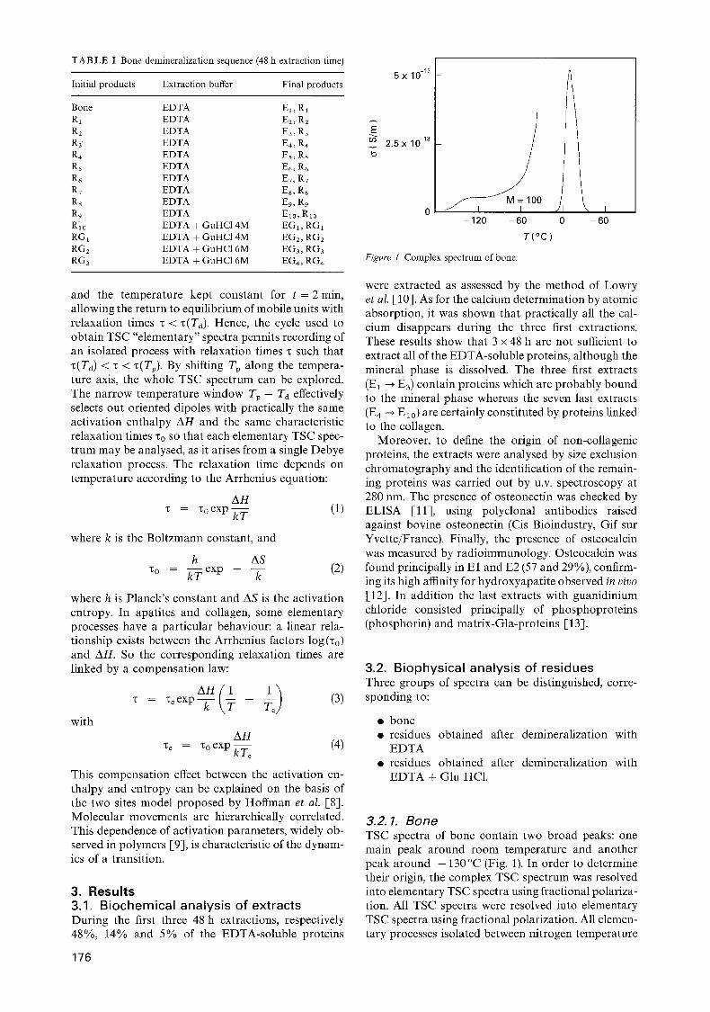
Figure 5 Complex spectrum of RG1 residue.
- 1 0
- 1 2
o
- 1 4 O
- 1 6
*.. x " x.
\ x \
x..
\
- 1 8 v I I I I I I I I 0.2 0.3 0.4 0.5 0.6 0.9
A H ( eV )
\
\ \
\
h i l l 0.7 0 .8
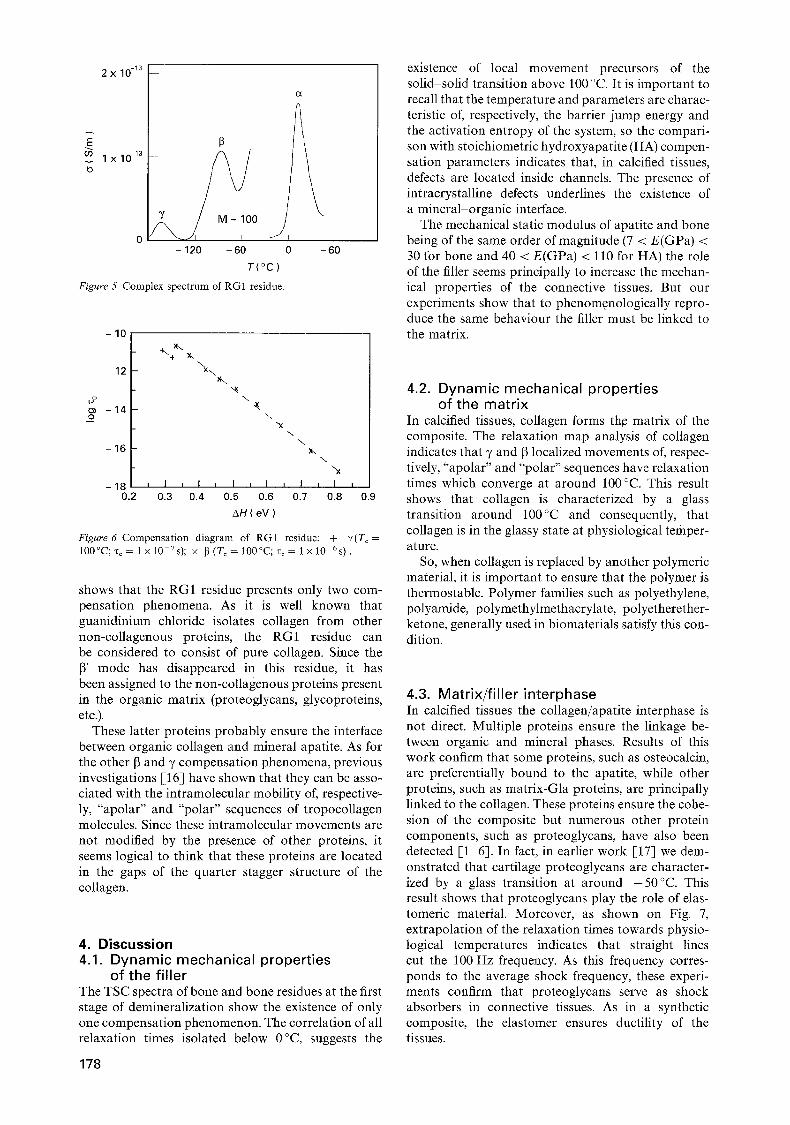
Figure 6 Compensat ion diagram of RG1 residue: + 7(To = 100°C;'c c = l x l 0 - V s ) ; x [ ~ ( T o = 1 0 0 ° C ; % = l x l 0 6s) .
shows that the RG1 residue presents only two com- pensation phenomena. As it is well known that guanidinium chloride isolates collagen from other non-collagenous proteins, the RG1 residue can be considered to consist of pure collagen. Since the 13' mode has disappeared in this residue, it has been assigned to the non-collagenous proteins present in the organic matrix (proteoglycans, glycoproteins, etc.).
These latter proteins probably ensure the interface between organic collagen and mineral apatite. As for the other 13 and 7 compensation phenomena, previous investigations [16] have shown that they can be asso- ciated with the intramolecular mobility of, respective~ ly, "apolar" and "polar" sequences of tropocollagen molecules. Since these intramolecular movements are not modified by the presence of other proteins, it seems logical to think that these proteins are located in the gaps of the quarter stagger structure of the collagen.
4. Discussion 4.1. Dynamic mechanical properties
of the filler The TSC spectra of bone and bone residues at the first stage of demineralization show the existence of only one compensation phenomenon. The correlation of all relaxation times isolated below 0°C, suggests the
178
existence of local movement precursors of the solid solid transition above 100 °C. It is important to recall that the temperature and parameters are charac- teristic of, respectively, the barrier jump energy and the activation entropy of the system, so the compari- son with stoichiometric hydroxyapatite (HA) compen- sation parameters indicates that, in calcified tissues, defects are located inside channels. The presence of intracrystalline defects underlines the existence of a mineral-organic interface.
The mechanical static modulus of apatite and bone being of the same order of magnitude (7 < E(GPa) < 30 for bone and 40 < E(GPa) < 110 for HA) the role of the filler seems principally to increase the mechan- ical properties of the connective tissues. But our experiments show that to phenomenologically repro- duce the same behaviour the filler must be linked to the matrix.
4.2. Dynamic mechanical properties of the matr ix
In calcified tissues, collagen forms the matrix of the composite. The relaxation map analysis of collagen indicates that 7 and 13 localized movements of, respec- tively, "apolar" and "polar" sequences have relaxation times which converge at around 100°C. This result shows t h a t collagen is characterized by a glass transition around 100°C and consequently, that collagen is in the glassy state at physiological temper- ature.
So, when collagen is replaced by another polymeric material, it is important to ensure that the polymer is thermostable. Polymer families such as polyethylene, polyamide, polymethylmethacrylate, polyetherether- ketone, generally used in biomaterials satisfy this con- dition.
4.3. Matrix/filler interphase In calcified tissues the collagen/apatite interphase is not direct. Multiple proteins ensure the linkage be- tween organic and mineral phases. Results of this work confirm that some proteins, such as osteocalcin, are preferentially bound to the apatite, while other proteins, such as matrix-Gla proteins, are principally linked to the collagen. These proteins ensure the cohe- sion of the composite but numerous other protein components, such as proteoglycans, have also been detected [1 6]. In fact, in earlier work [17] we dem- onstrated that cartilage proteoglycans are character- ized by a glass transition at around - 5 0 ° C . This result shows that proteoglycans play the role of elas- tomeric material. Moreover , as shown on Fig. 7, extrapolation of the relaxation times towards physio- logical temperatures indicates that straight lines cut the 100 Hz frequency. As this frequency corres- ponds to the average shock frequency, these experi- ments confirm that proteoglycans serve as shock absorbers in connective tissues. As in a synthetic composite, the elastomer ensures ductility of the tissues.

v
104
1 0 2
1
T( °C )
- 1 0 0 0
' I ' I '
. 3 8 ° C
\ "..., \ "-.
10-2 100 Hz " \
10-4
1 0 - 6 , , , I . . . . I . . . . I . . . . I . . . . 7 6 5 4 3
103 /T (K)
Figure 7 Relaxation map of proteoglycans.
5. Conclusions This study has shown the similarity between synthetic composite and calcified tissue. Bone is composed prin- cipally of a collagenic organic phase, reinforced by an apatite mineral filler. Interphase between organic and mineral phase would be ensured on one hand by multiple non-collagenous proteins linked to collagen and on the other hand by defects inside apatite chan- nels. These organic or mineral defects would explain why non-stoichiometric bone apatites have an activa- tion entropy lower than stoichiometric synthetic hy- droxyapatite.
From a physicochemical point of view, composites for bone replacement must be biphasic material, the filler reinforces the thermoplastic matrix and the cohe- sion and ductility are ensured by elastomers. The filler role seems to increase the static modulus while the elastomer could play the role of adapting dynamic strain to the stress.
References 1. W.T. BUTLER, Coll. Rel. Res. 4 (1984) 297. 2. L.W. FISHER and J. D. TERMINE, Clin. Orthop. 200 (1985)
362. 3. P .V. HAUSCHKA, J. B. LIAN and P. M. GALLOP, Proc.
Natl. Acad. Sei. USA 72 (1975) 3925. 4. P.A. PRICE and M. R. URIST, Otawara, Biochem. Biophys.
Res. Commun. 117 (1983) 765. 5. J. D. TERMINE, A. B. BELCOURT, K. M. CONN and
H. K. KLEINMANN, Y. Biol. Chem. 256 (1981) 10403. 6. A.T. ANDREWS, G. M. HERRING and P. W. KENT, Bio-
chem. J. 104 (1967) 705. 7. C. LACABANNE and D. CHATAIN, J. Polym. Sci.: Polym.
Phys. 11 (1973) 2315. 8. J. D. HOFFMAN, G. WILLIAMS and E. PASSAGLIA,
J. Polym. Sci.: Part C 14 (1966) 173. 9. C. LACABANNE, A. LAMURE, G. TESSI~YDRE, A.
BERNI~S and M. MOURGUES, J. Non-Cryst. Solids 172-174 (1994) 884.
10. O.H. LOWRY, N. J. ROSEBROUGH, A. FARR and R. J. RANDALL, J. Biol. Chem. 193 (1951) 267.
11. G.C. SAUNDERS, in "Immunoassays in the clinical laborat- ory", edited by R. N. Nakamura, W. R. Its and E. S. Tucker (Alan R. Liss, New York, 1979) p. 99.
12. F . H . WHAMS, K. KRECH and P. V. HAUSCKA, Magne- sium 2 (1983) 83.
13. M. RA IF, University Thesis number 242, Bordeaux II Univer- sity, 1993.
14. A. LAMURE, E. PLAINO, C. LACABANNE, N. HITMI, C. REY, G. BONEL and R. A. YOUNG, in "Biological and biomechanical performance of biomaterials', edited by P. Christal, A. Meunier and A. J. C. Lee (Elsevier Science, Amsterdam, 1986) p. 15.
15. A. LAMURE, F. MISKANE, A. BENNIS, N. HITM1, M. VIGNOLES and C. LACABANNE, Phosphorus, Sulfur and Silicon 77 (1993) 287.
16. A. LAMURE, N. HITMI, C. LACABANNE, M. F. HARMAND and D. HERBAGE, in Proceedings of the 5th International Symposium Electrets, Heidelberg, September 1985, edited by G. M. Sessier and R. Gerhard-Multhaupt (IEEE Service Center, Piscataway, NJ, 1985) p. 738.
17. A. LAMURE, 3rd Cycle Thesis number 2844, Toulouse III University, 1983.
Received 4 May and accepted 5 May 1995
179