changes in count and function of splenic lymphocytes from
TRANSCRIPT
in density; (2) the number of lymphocytes per gram of spleen (relative quantity) decreased significantly [(0.822 ± 0.157) × 108 vs (1.174 ± 0.254) × 108, P < 0.01]; (3) with the significant increase in the weight of the PH spleen (832.6 ± 278.2 g vs 211.7 ± 85.6 g, P < 0.01), the total quantity of lymphocytes (absolute quantity) increased significantly [(0.685 ± 0.072) × 1011 vs (0.366 ± 0.057) × 1011, P < 0.01]; and (4) the proliferative function of lymphocytes was enhanced: T lymphocytes, (0.022 ± 0.005 vs 0.015 ± 0.003, P < 0.05), and B lymphocytes (0.034 ± 0.006 vs 0.023 ± 0.001, P < 0.01).
CONCLUSION: Although lymphocyte density in the spleen decreased in patients with PH, the total quantity of lymphocytes increased because spleen weight increased greatly, along with the proliferating function. With respect to changes in lymphocytes, PH spleens may still have immune function, although it may be disordered. However, complete evaluation of the immune function of the spleen in PH requires more research.
© 2008 WJG. All rights reserved.
Key words: Portal hypertension; Spleen; Lymphocyte; Immune function
Peer reviewer: Kurt Lenz, Professor, Department of Internal Medicine, Konventhospital Barmherzige Brueder, Linz A-4020, Austria
Li ZF, Zhang S, Lv GB, Huang Y, Zhang W, Ren S, Yang J, Dang SS. Changes in count and function of splenic lymphocytes from patients with portal hypertension. World J Gastroenterol 2008; 14(15): 2377-2382 Available from: URL: http://www.wjgnet.com/1007-9327/14/2377.asp DOI: http://dx.doi.org/10.3748/wjg.14.2377
INTRODUCTIONLymphocytes reside in different organs in the human body. They circulate through the primary lymphoid organs (thymus and bone marrow), secondary lymphoid organs (spleen, lymph nodes, tonsils and Peyers patches), as well as non-lymphoid organs such as blood, lung and liver. Especially in lymphoid organs, lymphocyte subsets migrate and home to different compartments. About 15%-20% of the blood volume circulates through the spleen at any one time and about 15% of the lymphocytes reside in this organ[1,2].
CLINICAL RESEARCH
Changes in count and function of splenic lymphocytes from patients with portal hypertension
Zong-Fang Li, Shu Zhang, Gao-Bo Lv, Ying Huang, Wei Zhang, Song Ren, Jun Yang, Shuang-Suo Dang
Zong-Fang Li, Shu Zhang, Gao-Bo Lv, Wei Zhang, Song Ren, Department of General Surgery in Cadres Ward, the Second Affiliated Hospital, School of Medicine, Xi’an Jiaotong University, Xi’an 710004, Shaanxi Province, ChinaYing Huang, Jun Yang, Department of Pathology, the Second Affiliated Hospital, School of Medicine, Xi’an Jiaotong University, Xi’an 710004, Shaanxi Province, ChinaShuang-Suo Dang, Department of Infectious Diseases, the Second Affiliated Hospital, School of Medicine, Xi’an Jiaotong University, Xi’an 710004, Shaanxi Province, ChinaAuthor contributions: Li ZF designed the research; Zhang S, Lv GB, Zhang W and Ren S performed the research; Huang Y, Yang J and Dang SS contributed to reagents/materials/analytic work; Huang Y and Yang J analyzed the data; and Zhang S and Li ZF wrote the paper.Supported by The Support Project for talented man in new century from Ministry of Education of People’s Republic of China 2004, No. NCET-04-0932 and the Project of Tackle Key Problems in Science and Technology of Shaanxi Province, No. 2004K14-G1(4), 2006K14-G2(4)Correspondence to: Zong-Fang Li, Department of General Surgery in Cadres Ward, the Second Affiliated Hospital, School of Medicine, Xi’an Jiaotong University, No. 157, West 5th Road, Xi’an 710004, Shaanxi Province, China. [email protected]: +86-29-87678006 Fax: +86-29-87678634Received: January 3, 2008 Revised: February 25, 2008
AbstractAIM: To investigate changes in numbers and proliferative function of splenic lymphocytes in patients with hypersplenism due to portal hypertension (PH), to provide evidence for further study of immune status of the spleen during PH.
METHODS: Twelve sp leens f rom pat ients wi th hypersplenism due to PH served as the PH group, and four spleens from cases of traumatic spleen rupture were regarded as the control group. After weighing the spleen, lymphocytes were separated and counted using a cell counting plate to calculate the lymphocyte count per gram of spleen tissue (relative quantity) and total lymphocyte count in whole spleen (absolute quantity). The immunohistochemical SP method was used to observe the density and distribution of lymphocytes in the spleen. The MTT method was used to observe changes in lymphocyte proliferative function. RESULTS: As compared to the control group, the splenic lymphocytes in the PH group showed that: (1) There was no difference in distribution but a significant decrease
Online Submissions: wjg.wjgnet.com World J Gastroenterol 2008 April 21; 14(15): 2377-2382www.wjgnet.com World Journal of Gastroenterology ISSN [email protected] © 2008 WJG. All rights reserved.
www.wjgnet.com

Therefore, lymphocytes are the immunocytes that have the highest count in the spleen. Their functional status directly influences the immune function of the spleen[3-5].
Currently, there is still some dispute on the immune status of the spleen in patients with portal hypertension (PH)[6,7]. We have isolated splenic macrophages and demonstrated that their phagocytosis is enhanced in PH spleens[8-10], but there is little compelling experimental evidence on the distribution, count and function of lymphocytes in the PH spleen. In this study, we isolated and cultured splenic lymphocytes from PH spleen, and observed changes in their density, distribution, count, and proliferative function using the immunohistochemical SP method and 3-[4 ,5-dimethyl thiazol-2-y l ] -2 ,5-diphenyltetrazolium bromide (MTT).
MATERIALS AND METHODSPatientsTwo groups of patients were studied. Twelve patients (median age 46.8 years, range 27-62 years; eight male and four female), with hypersplenism due to PH, in our hospital from September 2005 to March 2006, were included as the PH group. All patients underwent pericardial devascularization with splenectomy. Supporting evidence for hypersplenism due to PH and cirrhosis included clinical features, abnormal laboratory tests, and postoperative pathological examination. Four patients (median age 33.5 years, range 18-38 years; three male and one female) with traumatic rupture of the spleen were enrolled into the control group. Hepatitis, cirrhosis, history of hypersplenism, and abnormalities in postoperative laboratory findings and pathological examinations were absent in the controls. All patients provided written informed consent, and the protocol was approved by the ethics committee of our hospital.
Lymphocyte count in the spleenSpleens were weighed after removal from patients. The splenic tissue samples were cut from the upper pole, lower pole, and hilum, and were transferred to the cell culture room, and kept in sealed aseptic bottles filled with 4℃ precooled PBS. Further preparations were made: Weighing 5 g tissue with an electronic balance, using a 200-mesh screen to grind the tissue sample into cell suspension, and purifying the lymphocytes with lymphocyte separating medium by gradient centrifugation. After preparation, lymphocytes were counted using a cell counting plate. The lymphocyte count per gram of spleen tissue (relative quantity) was then calculated and multiplied by the weight of the spleen to derive the total lymphocyte count in the spleen (absolute quantity).
Density and distribution of lymphocytesThe splenic tissue samples were cut from the upper pole, lower pole, and hilum, fixed in phosphate buffer (pH 7.2) containing 4% paraformaldyde, embedded in paraffin wax, and sectioned at 5 μm. CD3 and CD20 SP method staining was adopted to show T and B lymphocytes, respectively. Periarterial lymphatic sheath (PALS), splenic corpuscle (F), red pulp (RP), and marginal zone (MZ) of spleen tissue were
observed. Five fields of vision were also randomly observed in each part. Positive cells were counted respectively. For the negative control, the primary antibody was replaced by PBS.
Proliferative function of lymphocytesLymphocytes with RPMI1640 culture solution containing 10% fetal calf serum were placed in 96-well f lat-bottomed microplates in triplicate at 2 × 10⁵ cells/well, then concanavalin A (Con A) or lipopolysaccharide (LPS; both from Sigma, St. Louis, MO, USA) was added to the wells at a final concentration of 10 μg/L and 20 μg/L, respectively. The cells were then incubated in a total volume of 200 μL/well. Serum-free RPMI-1640 medium was used as a control. Cell proliferation was measured by MTT assay 44 h after culture. MTT (Sigma) solution of 20 μL (5 g/L) was added to each well. After 4 h incubation, the cells were lysed and the purple formazan crystals were solubilized. We then measured A570 of each well on an enzyme labeling instrument, and the proliferation level was calculated. Proliferation level = experimental group A (ConA or LPS) - negative control group A.
Statistical analysisP values were calculated using the independent sample t test and considered significant at P < 0.05. All the results were represented by mean ± SD.
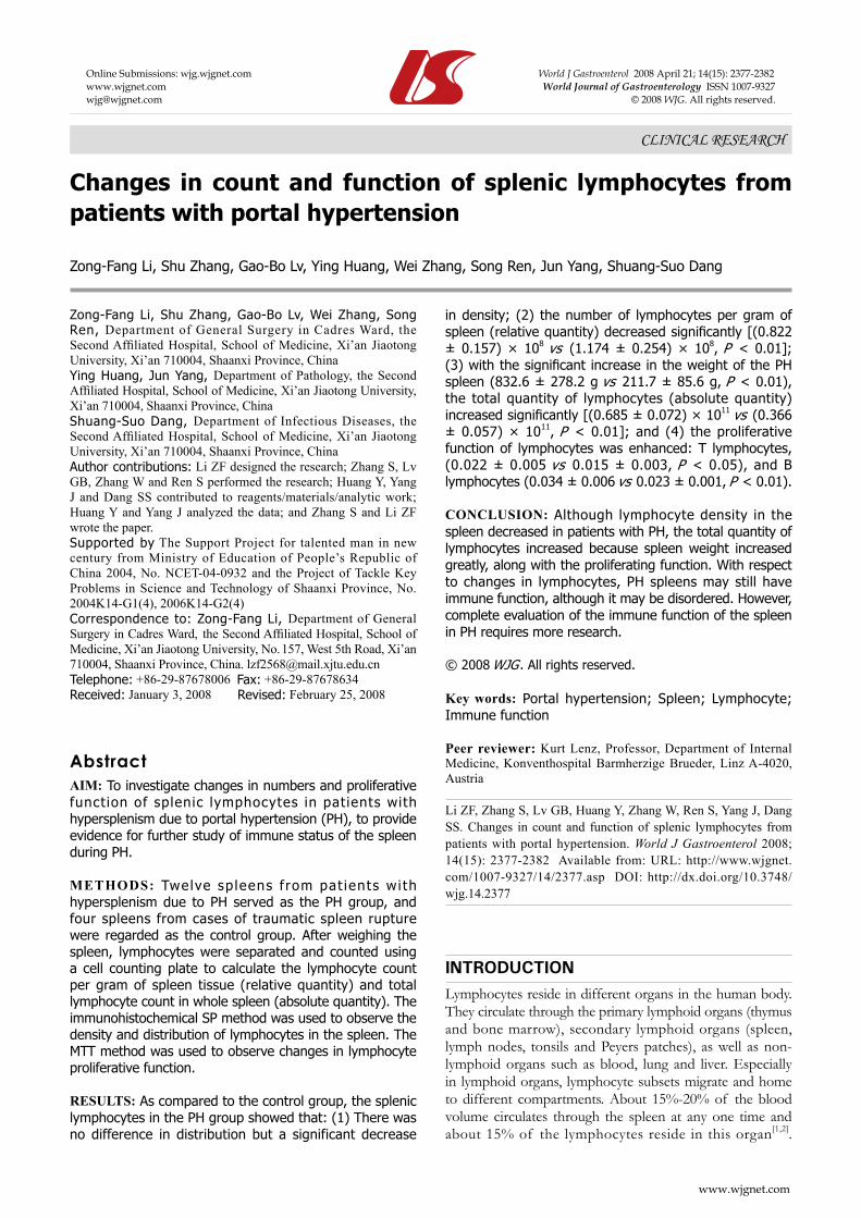
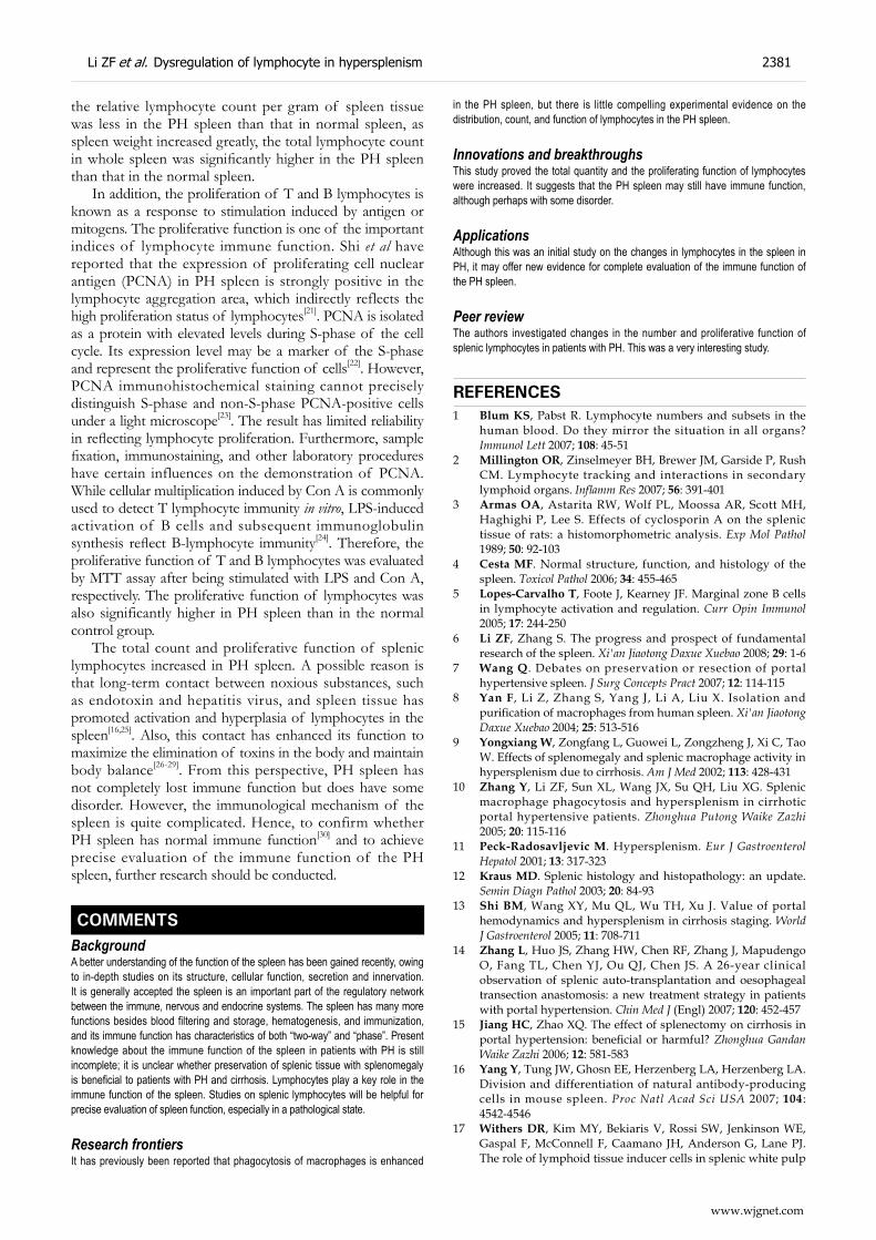
RESULTSChange in density and distribution of lymphocytesThe distribution of T and B lymphocytes was almost the same in PH spleen as in normal spleen (Figures 1 and 2). Cell counts in single fields of view were significantly less in PH spleen than in normal spleen (Table 1, Figures 3 and 4).
Change in lymphocyte countLymphocyte count was significantly less in PH spleen (relative quantity) than in normal spleen. However, with the increase in spleen weight, lymphocyte count of whole spleen (absolute quantity) was significantly greater in PH spleen than in normal spleen (Table 2).
Change in lymphocyte proliferative functionThe proliferative function of T and B lymphocytes was significantly higher in PH spleen than in normal spleen (Table 3).
DISCUSSIONAt present, the immune function of the PH spleen is still the subject of some dispute[7,11,12]. It is unclear if the preservation of splenic tissue with splenomegaly is beneficial to patients with PH and cirrhosis[13-15]. The lymphocytes are important immunocytes in the spleen. The spleen can participate in specific immunity through T-cell-mediated cellular immunity and B-cell-mediated humoral immunity[16-18]. Therefore, the evaluation of changes in lymphocyte count and functions in PH spleen is extremely important to an in-depth study of the immune status of the spleen in PH.
2378 ISSN 1007-9327 CN 14-1219/R World J Gastroenterol April 21, 2008 Volume 14 Number 15
www.wjgnet.com

Lymphocytes include T and B lymphocytes. CD3 and CD20 are important differentiation antigens on T- and B-cell membranes. CD3 and CD20 immunohistochemical staining is ideal for analyzing the distribution and count of T and B lymphocytes in tissue[19]. Wang et al have used the
method to observe lymphocytes in pathological sections of PH spleen. They believe that in PH splenomegaly, lymphocyte density in the spleen decreases, which results in a decrease in lymphocyte count[20]. We also found in our experiment that the distribution area of lymphocytes had
A1 B1
A2 B2
Figure 2 CD3 immunostaining for distribution of T lymphocytes. There was no significant difference in distribution of T lymphocytes between PH and control groups; they were all mainly located in the marginal zone and PALS. A1: Control group, marginal zone; A2: Control group, PALS; B1: PH group, marginal zone; B2: PH group, PALS (× 100).
A B
Figure 1 CD20 immunostaining for distribution of B lymphocytes. There was no significant difference in distribution of B lymphocytes between PH and control groups; they were all mainly located in the splenic corpuscle. A: Control group; B: PH group (× 100).
Table 1 Changes in density of lymphocyte in PH spleen
Group T lymphocytes B lymphocytes
F MZ PALS RP F MZ PALS RP
PH 89.5 ± 14.7 120.0 ± 14.1 122.9 ± 12.1 12.2 ± 2.9 356.5 ± 31.2 138.0 ± 19.5 113.8 ± 21.6 7.4 ± 1.7Control 126.5 ± 19.3 140.5 ± 11.6 137.0 ± 6.2 20.45 ± 4.5 418.3 ± 22.4 196.0 ± 22.0 153.8 ± 25.8 12.8 ± 4.6P value < 0.01 < 0.05 < 0.05 < 0.01 < 0.01 < 0.01 < 0.05 < 0.01
Li ZF et al. Dysregulation of lymphocyte in hypersplenism 2379
www.wjgnet.com

almost no differences between PH and normal spleens, while the lymphocyte density was significantly lower in the PH spleen. However, the lymphocyte density seen in a single microscopic field of view cannot represent the total lymphocyte count in the spleen. Therefore, in this
study, we purified and counted lymphocytes in the spleen, and then calculated lymphocyte count per gram of spleen tissue (relative quantity) and lymphocyte count in whole spleen (absolute quantity). This made the result more scientific and accurate. The results showed that although
A1 B1
A2 B2
Figure 3 CD20 immunostaining for density of B lymphocytes. Compared to the control group, the density of B lymphocytes in the PH group decreased significantly in the splenic corpuscle and RP. A1: Control group, splenic corpuscle; A2: Control group, RP; B1: PH group, splenic corpuscle; B2: PH group, RP (× 100).
A B
Figure 4 CD3 immunostaining for density of B lymphocytes. Compare to the control group, the density of T lymphocytes in the PH group decreased significantly in PALS. A: Control group; B: PH group (× 100).
Table 2 Changes in weight of spleen and lymphocyte count
Group Relative quantity (× 108)
Weight of spleen (g)
Absolute quantity (× 1011)
PH 0.822 ± 0.157 832.6 ± 278.2 0.685 ± 0.072Control 1.714 ± 0.254 211.7 ± 85.6 0.366 ± 0.057P value < 0.01 < 0.01 < 0.01
Table 3 Changes in proliferative function of lymphocyte in PH spleen
Group T lymphocyte (ConA 10 μg/L) B lymphocyte (LPS 20 μg/L)
PH 0.022 ± 0.005 0.034 ± 0.006Control 0.015 ± 0.003 0.023 ± 0.004P value < 0.05 < 0.01
2380 ISSN 1007-9327 CN 14-1219/R World J Gastroenterol April 21, 2008 Volume 14 Number 15
www.wjgnet.com
the relative lymphocyte count per gram of spleen tissue was less in the PH spleen than that in normal spleen, as spleen weight increased greatly, the total lymphocyte count in whole spleen was significantly higher in the PH spleen than that in the normal spleen.
In addition, the proliferation of T and B lymphocytes is known as a response to stimulation induced by antigen or mitogens. The proliferative function is one of the important indices of lymphocyte immune function. Shi et al have reported that the expression of proliferating cell nuclear antigen (PCNA) in PH spleen is strongly positive in the lymphocyte aggregation area, which indirectly reflects the high proliferation status of lymphocytes[21]. PCNA is isolated as a protein with elevated levels during S-phase of the cell cycle. Its expression level may be a marker of the S-phase and represent the proliferative function of cells[22]. However, PCNA immunohistochemical staining cannot precisely distinguish S-phase and non-S-phase PCNA-positive cells under a light microscope[23]. The result has limited reliability in reflecting lymphocyte proliferation. Furthermore, sample fixation, immunostaining, and other laboratory procedures have certain influences on the demonstration of PCNA. While cellular multiplication induced by Con A is commonly used to detect T lymphocyte immunity in vitro, LPS-induced activation of B cells and subsequent immunoglobulin synthesis reflect B-lymphocyte immunity[24]. Therefore, the proliferative function of T and B lymphocytes was evaluated by MTT assay after being stimulated with LPS and Con A, respectively. The proliferative function of lymphocytes was also significantly higher in PH spleen than in the normal control group.
The total count and proliferative function of splenic lymphocytes increased in PH spleen. A possible reason is that long-term contact between noxious substances, such as endotoxin and hepatitis virus, and spleen tissue has promoted activation and hyperplasia of lymphocytes in the spleen[16,25]. Also, this contact has enhanced its function to maximize the elimination of toxins in the body and maintain body balance[26-29]. From this perspective, PH spleen has not completely lost immune function but does have some disorder. However, the immunological mechanism of the spleen is quite complicated. Hence, to confirm whether PH spleen has normal immune function[30] and to achieve precise evaluation of the immune function of the PH spleen, further research should be conducted.
COMMENTSBackgroundA better understanding of the function of the spleen has been gained recently, owing to in-depth studies on its structure, cellular function, secretion and innervation. It is generally accepted the spleen is an important part of the regulatory network between the immune, nervous and endocrine systems. The spleen has many more functions besides blood filtering and storage, hematogenesis, and immunization, and its immune function has characteristics of both “two-way” and “phase”. Present knowledge about the immune function of the spleen in patients with PH is still incomplete; it is unclear whether preservation of splenic tissue with splenomegaly is beneficial to patients with PH and cirrhosis. Lymphocytes play a key role in the immune function of the spleen. Studies on splenic lymphocytes will be helpful for precise evaluation of spleen function, especially in a pathological state.
Research frontiersIt has previously been reported that phagocytosis of macrophages is enhanced
in the PH spleen, but there is little compelling experimental evidence on the distribution, count, and function of lymphocytes in the PH spleen.
Innovations and breakthroughsThis study proved the total quantity and the proliferating function of lymphocytes were increased. It suggests that the PH spleen may still have immune function, although perhaps with some disorder.
Applications Although this was an initial study on the changes in lymphocytes in the spleen in PH, it may offer new evidence for complete evaluation of the immune function of the PH spleen.
Peer reviewThe authors investigated changes in the number and proliferative function of splenic lymphocytes in patients with PH. This was a very interesting study.
REFERENCES1 Blum KS, Pabst R. Lymphocyte numbers and subsets in the
human blood. Do they mirror the situation in all organs? Immunol Lett 2007; 108: 45-51
2 Millington OR, Zinselmeyer BH, Brewer JM, Garside P, Rush CM. Lymphocyte tracking and interactions in secondary lymphoid organs. Inflamm Res 2007; 56: 391-401
3 Armas OA, Astarita RW, Wolf PL, Moossa AR, Scott MH, Haghighi P, Lee S. Effects of cyclosporin A on the splenic tissue of rats: a histomorphometric analysis. Exp Mol Pathol 1989; 50: 92-103
4 Cesta MF. Normal structure, function, and histology of the spleen. Toxicol Pathol 2006; 34: 455-465
5 Lopes-Carvalho T, Foote J, Kearney JF. Marginal zone B cells in lymphocyte activation and regulation. Curr Opin Immunol 2005; 17: 244-250
6 Li ZF, Zhang S. The progress and prospect of fundamental research of the spleen. Xi'an Jiaotong Daxue Xuebao 2008; 29: 1-6
7 Wang Q. Debates on preservation or resection of portal hypertensive spleen. J Surg Concepts Pract 2007; 12: 114-115
8 Yan F, Li Z, Zhang S, Yang J, Li A, Liu X. Isolation and purification of macrophages from human spleen. Xi'an Jiaotong Daxue Xuebao 2004; 25: 513-516
9 Yongxiang W, Zongfang L, Guowei L, Zongzheng J, Xi C, Tao W. Effects of splenomegaly and splenic macrophage activity in hypersplenism due to cirrhosis. Am J Med 2002; 113: 428-431
10 Zhang Y, Li ZF, Sun XL, Wang JX, Su QH, Liu XG. Splenic macrophage phagocytosis and hypersplenism in cirrhotic portal hypertensive patients. Zhonghua Putong Waike Zazhi 2005; 20: 115-116
11 Peck-Radosavljevic M. Hypersplenism. Eur J Gastroenterol Hepatol 2001; 13: 317-323
12 Kraus MD. Splenic histology and histopathology: an update. Semin Diagn Pathol 2003; 20: 84-93
13 Shi BM, Wang XY, Mu QL, Wu TH, Xu J. Value of portal hemodynamics and hypersplenism in cirrhosis staging. World J Gastroenterol 2005; 11: 708-711
14 Zhang L, Huo JS, Zhang HW, Chen RF, Zhang J, Mapudengo O, Fang TL, Chen YJ, Ou QJ, Chen JS. A 26-year clinical observation of splenic auto-transplantation and oesophageal transection anastomosis: a new treatment strategy in patients with portal hypertension. Chin Med J (Engl) 2007; 120: 452-457
15 Jiang HC, Zhao XQ. The effect of splenectomy on cirrhosis in portal hypertension: beneficial or harmful? Zhonghua Gandan Waike Zazhi 2006; 12: 581-583
16 Yang Y, Tung JW, Ghosn EE, Herzenberg LA, Herzenberg LA. Division and differentiation of natural antibody-producing cells in mouse spleen. Proc Natl Acad Sci USA 2007; 104: 4542-4546
17 Withers DR, Kim MY, Bekiaris V, Rossi SW, Jenkinson WE, Gaspal F, McConnell F, Caamano JH, Anderson G, Lane PJ. The role of lymphoid tissue inducer cells in splenic white pulp
Li ZF et al. Dysregulation of lymphocyte in hypersplenism 2381
www.wjgnet.com
development. Eur J Immunol 2007; 37: 3240-324518 Mueller SN, Hosiawa-Meagher KA, Konieczny BT, Sullivan
BM, Bachmann MF, Locksley RM, Ahmed R, Matloubian M. Regulation of homeostatic chemokine expression and cell trafficking during immune responses. Science 2007; 317: 670-674
19 Gong FL. Medical Immunology. Beijing: Science Press, 2000: 253-257
20 Wang Q, Zhang RD, Xiong SG, Yao M, Zhang JB, Yang YK. T, B lymphocytes and macrophages in splenomegaly of portal hypertension: an immunohistological and morphometric analysis. J Bengbu Med Coll 1992; 17: 126-130
21 Shi B, Yang Z. Vascular lesion and its mechanisms in spleen under statement of portal hypertension. Zhonghua Yixue Zazhi 2000; 80: 196-198
22 Kelman Z. PCNA: structure, functions and interactions. Oncogene 1997; 14: 629-640
23 Baserga R. Growth regulation of the PCNA gene. J Cell Sci 1991; 98 (Pt 4): 433-436
24 Zhu XL, Chen AF, Lin ZB. Ganoderma lucidum polysaccharides enhance the function of immunological effector cells in
immunosuppressed mice. J Ethnopharmacol 2007; 111: 219-22625 Kesteman N, Vansanten G, Pajak B, Goyert SM, Moser M.
Injection of lipopolysaccharide induces the migration of splenic neutrophils to the T cell area of the white pulp: role of CD14 and CXC chemokines. J Leukoc Biol 2008; 83: 640-647
26 Freitas A, Chen J. Introduction: regulation of lymphocyte homeostasis. Microbes Infect 2002; 4: 529-530
27 Li ZF, Zhang Y, Gao J, Zhang PJ, Wang JX, Liu XG. Expression and significance of Toll-like receptor 4 of splenic macrophage in patients with hypersplenism due to portal hypertension. Zhonghua Yixue Zazhi 2004; 84: 1088-1091
28 Wang Q, Xia S, Jiang H. The mechanism for splenic promoting effects on liver cirrhosis. Zhonghua Yixue Zazhi 1995; 75: 594-598, 638
29 Bertoletti A, Maini MK. Protection or damage: a dual role for the virus-specific cytotoxic T lymphocyte response in hepatitis B and C infection? Curr Opin Microbiol 2000; 3: 387-392
30 Jiang HC , Dai WJ, Hu Z. Problems related to spleen preservation in portal hypertension. J Surg Concepts Pract 2006; 11: 193-195
S- Editor Li DL L- Editor Kerr C E- Editor Ma WH
2382 ISSN 1007-9327 CN 14-1219/R World J Gastroenterol April 21, 2008 Volume 14 Number 15
www.wjgnet.com