chapter 1 introduction -...
TRANSCRIPT

Chapter 1
Introduction
This chapter has been communicated to Progress in Polymer Science as
Nanocellulose and its nanocomposites with biodegradable matrices
Introduction
Nanotechnology is the engineering of functional systems at the molecular
scale. The enormous development of science and technology that occurred in
the second half of the twentieth century has resulted in not only new, bold
ideas, but it has also created tools that enable to perceive atoms in
surrounding matter. Scientific achievements, especially those made at the
end of the twentieth century, have contributed to the significant development
of nanotechnology. It was a kind of fait accompli achieved with the
cooperation of many fields of science i.e. chemistry, biology, physics,
computer science etc, which has made nanotechnology develop very
intensively using the achievements of basic and applied sciences that
originate from chemistry, electronics, mechanics, biotechnology, medicine,
pharmacy and computer science, as well as the humanities, such as
philosophy, economics and ethics.

2 Chapter 1
1.1 Bionanotechnology
Bionanotechnology was ‘invented’ by nature. This type of technology ‘has
been chosen and perfected’ by nature for 5.5 billion years, building up, atom
by atom, the environment of life on Earth. The development of science and
technology in the twentieth century, made it possible for the scientists to
look into the structure of matter on a nanoscale (10-9 m). One of the criteria
currently used that defines nanotechnology is the size of objects
manufactured, which should have at least one dimension not greater than 100
nm. During the production of such objects, their chemical and physical
properties should be controllable, and it should be possible to build larger
objects from them [1].
Nanobiotechnology refers to the intersection of nanotechnology and biology.
Given the fact that the subject is one that has emerged only very recently,
bionanotechnology and nanobiotechnology serve as blanket terms for various
related technologies. This discipline helps to indicate the merger of
biological research with various fields of nanotechnology. Concepts that are
enhanced through nanobiology include: nanoparticles, nanodevices,
nanocomposites, and nanoscale phenomena in biological world that occurs
within the discipline of nanotechnology. This technical approach to biology
allows scientists to imagine and create systems that can be used for
biological research. Biologically inspired nanotechnology uses biological
systems as the inspirations for technologies not yet created. We can learn
from eons of evolution that have resulted in elegant systems that are
naturally created.
The most important objectives that are frequently found in nanobiology
involve applying nanotools to relevant medical/biological problems and

Introduction 3
refining these applications. Developing new tools for the medical and
biological fields is another primary objective in bionanotechnology. The
imaging of native biomolecules, biological membranes, and tissues is also a
major topic for the nanobiology researchers.
1.2 Biopolymers
Biopolymers are polymers produced by living organisms. Carbohydrates and
proteins are the major examples of biopolymers. Among the biopolymers,
the polysaccharides, which are often produced by linear polymeric
carbohydrate structures are important in the living organisms. Cellulose is
the most common organic compound and biopolymer on earth. Fig. 1.1
describes the classification of the main biopolymers.
Cellulose is the most plentiful carbohydrate in the world; 40 percent of all
organic matter is cellulose!
Starch is found in corn (maize), potatoes, wheat, tapioca (cassava), and some
other plants. Annual world production of starch is well over 70 billion
pounds, with much of it being used for non-food purposes, like making
paper, cardboard, textile sizing, and adhesives.
Collagen is the most abundant protein found in mammals. Gelatin is
denatured collagen, and is used in sausage casings, capsules for drugs and
vitamin preparations, and other miscellaneous industrial applications
including photography.
Casein, commercially produced mainly from cow's skimmed milk, is used in
adhesives, binders, protective coatings, and other products.
Soy protein and zein (from corn) are abundant plant proteins. They are used
for making adhesives and coatings for paper and cardboard.

4 Chapter 1
A number of other natural materials can be made into polymers that are
biodegradable. For example:
Polyesters are produced by bacteria, and can be made commercially on large
scales through fermentation processes. They are now being used in
biomedical applications.
Fig. 1.1 Classification of the main biopolymers [2]
Poly lactic acid (PLA): Lactic acid is now commercially produced on large
scales through the fermentation of sugar feedstocks obtained from sugar
beets or sugar cane, or from the conversion of starch from corn, potato peels,
or other starch source. It can be polymerized to produce poly (lactic acid),
which is already finding commercial applications in drug encapsulation and
biodegradable medical devices.
Natural rubber (NR) latex: Natural rubber (Hevea brasiliensis) is a high
molecular weight polymeric substance with viscoelastic properties.
Structurally it is cis 1,4-polyisoprene, Fig. 1.2 (a). Isoprene is a diene and 1,
4 addition leaves a double bond in each of the isoprene unit in the polymer.

Introduction 5
Because of this, natural rubber shows all the reactions of an unsaturated
polymer. Natural rubber is water repellent and resistant to alkalies and weak
acids. Natural rubber's elasticity, toughness, impermeability, adhesiveness,
and electrical resistance make it useful as an adhesive, a coating
composition, a fibre, a molding compound, and an electrical insulator.
Balata rubber (BR) latex: Balata latex (Manilkara bidebtata), similar to
natural rubber, has not been industrially exploited as much as natural rubber
mainly due to an enhanced level of crystallinity in the polymer which results
from the fact that the main component is trans-1, 4-polyisoprene, Fig. 1.2
(b). The trans configuration of the polymer chain leads to an increased ease
of packing and therefore increased crystallinity and concomitant brittleness
and reduced elongation at break. Balata sap is harvested in the Amazonian
shield rainforest from the mahogany tree, (Swietenia macrophylla), which is
also a sought after hardwood for fine furniture. These natural raw materials
are abundant, renewable, and biodegradable, making them attractive for
environmentally friendly products.
In this work, nanocelluloses were utilized as functional rigid fillers in natural
rubber and natural balata matrices for bionanocomposite preparation. The
morphology, thermo mechanical properties, crystallinity and structure of the
nanocomposite materials were compared.
Fig. 1.2 (a) Cis -1, 4-polyisoprene Fig. 1.2 (b) trans -1, 4-polyisoprene

6 Chapter 1
1.3 Natural fibres The term ‘natural fibres’ is used to designate numerous kinds of fibres that are
naturally produced by plants, animals and minerals [2]. To avoid any possible
misunderstanding, it is important to clarify that in this research work, ‘natural
fibres’ refer to ‘plant fibres’; also called ‘lignocellulosic fibres’, or ‘cellulosic
fibres’. More specifically, the first part of this work is focused on
‘lignocellulosic fibres’, that include banana bast fibre, jute, pineapple leaf fibre
(PALF) and coir fibre (fruit of coconut) [1].
With a desire to focus the world‘s attention on the role of natural fibres, the Food
and Agricultural Organization (FAO) declared 2009 as the International Year of
Natural Fibres [2]. According to the FAO, each year farmers harvest around 35
million tons of natural fibres from a wide range of plants. These fibres are used to
form fabrics, ropes and twines, which have played a fundamental role in the
human societies since the dawn of civilization. The most striking point regarding
the production of natural fibre is the mass quantity of the material which is
underutilised. For example, around 20 x 104 tons of sisal is produced each year in
Brazil compared to other natural fibres such as jute, ramie, curauá and rice fibres
[3]. Natural fibres play an important role in supporting the world’s population and
find variety of application with paper and packaging.
1.4 Application of biopolymers Recently, interest in composite manufacturing has shifted towards the use of
biopolymers as reinforcement because of their environmental benefits. The use of
a biodegradable matrix is worth considering since this would result in a
completely biodegradable composite. Polylactic acid is one of the main
biopolymer used in the field of biodegradable composite as the matrix material. Its
clarity makes it useful for recyclable and biodegradable packaging, such as
bottles, yogurt cups, and candy wrappers. It has also been used for food service

Introduction 7
ware, lawn and food waste bags, coatings for paper and cardboard, and fibres-for
clothing, carpets, sheets and towels, and wall coverings. In biomedical
applications, it is used for sutures, prosthetic materials for drug delivery.
� Starch-based bioplastics are important not only because starch is the
least expensive biopolymer but because it can be processed by all of the
methods used for synthetic polymers, like film extrusion and injection
moulding. utensils for catering, plates, cups and other products have
been made with starch-based plastics.
� Water soluble biopolymers such as starch, gelatin, soy protein, and
casein form flexible films when properly plasticized. Although such
films are regarded mainly as food coatings, it is recognized that they
have potential use as nonsupported stand-alone sheeting for food
packaging and other purposes.
� Starch-protein compositions have the interesting characteristic of
meeting nutritional requirements for farm animals. Hog feed, for
example, are recommended to contain 13-24% protein, complemented
with starch. If starch-protein plastics were commercialized, used food
containers and serviceware collected from fast food restaurants can be
pasteurized and turned into animal feed.
� Polyesters are now produced from natural resources-like starch and
sugars-through large-scale fermentation processes, and used to
manufacture water-resistant bottles, utensils for catering, and other
products.
� Triglycerides have recently become the basis for a new family of sturdy
composites. With glass fibre reinforcement they can be made into long-
lasting durable materials with applications in the manufacture of
8 Chapter 1
agricultural equipment, the automotive industry, construction, and other
areas.
� Natural fibres are becoming increasingly popular for use in industrial
applications, providing sustainable solutions to support technical
innovation. These versatile, natural based materials have applications in
industries ranging from textiles and consumer products to the
automotive and construction industries. Fibres like jute, hemp, flax,
wood, and even straw or hay are finding application in various industrial
and structural applications. If straw can be used instead of wood for
making composites for the construction industry, it will enable us to
conserve wood fibre which are highly precious.
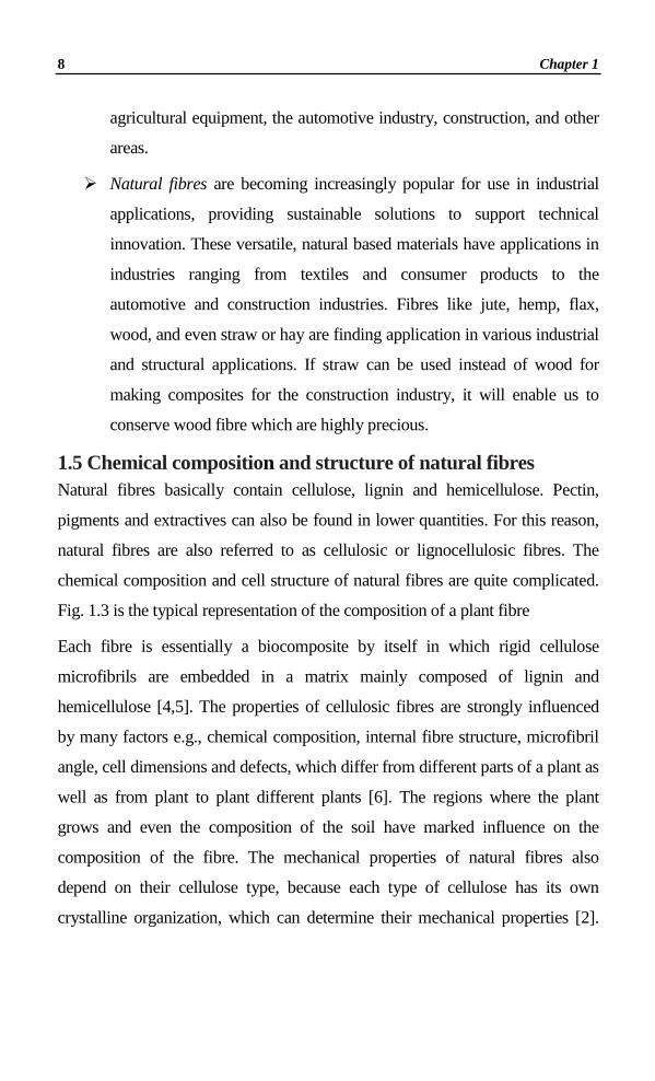
1.5 Chemical composition and structure of natural fibres Natural fibres basically contain cellulose, lignin and hemicellulose. Pectin,
pigments and extractives can also be found in lower quantities. For this reason,
natural fibres are also referred to as cellulosic or lignocellulosic fibres. The
chemical composition and cell structure of natural fibres are quite complicated.
Fig. 1.3 is the typical representation of the composition of a plant fibre
Each fibre is essentially a biocomposite by itself in which rigid cellulose
microfibrils are embedded in a matrix mainly composed of lignin and
hemicellulose [4,5]. The properties of cellulosic fibres are strongly influenced
by many factors e.g., chemical composition, internal fibre structure, microfibril
angle, cell dimensions and defects, which differ from different parts of a plant as
well as from plant to plant different plants [6]. The regions where the plant
grows and even the composition of the soil have marked influence on the
composition of the fibre. The mechanical properties of natural fibres also
depend on their cellulose type, because each type of cellulose has its own
crystalline organization, which can determine their mechanical properties [2].

Introduction 9
Table 1.1 show the chemical composition of some of the important natural
fibres which varies according to their origin.
Fig. 1.3 Scheme of the cellulose cell wall and microfibril organization [1]
Table 1.1 Chemical compositions of some lignocellulosic fibres [3]
Natural fibre Cellulose
(wt%) Hemicellulose
(wt%) Pectin (wt%)
Lignin (wt%)
Bast fibres
Flax 71 19 1 2
Hemp 75 18 1 4
Banana 72 14 >1 14
Jute 70 14 2 18
Ramie 75 15 2 1
Leaf fibres
Abaca 70 22 1 1
Sisal 73 13 1 7
PALF 85 4 <1 3
Seed hair fibres
Cotton 93 3 3 1
Wheat straw 51 26 - 7
Coir 40 <2 - 45

10 Chapter 1
The structure and chemical composition of various cellulose components
embedded within these fibres are discussed below.
1.6 Cellulose Cellulose is the most abundant renewable organic material produced in the
biosphere, having an annual production that is estimated to be over 7.5 ×
1012 tons [7]. This structural material is naturally organized as microfibrils
linked together to form cellulose fibres. Composed of long chains of glucose
molecules, cellulose fibres are arranged in an intricate web that provides both
structure and support for the cell [8]. Interestingly, within the fibres are
regions which are very well ordered: (chains are aligned parallel and are
packed close together). Crystalline is the name given to these unique fibre
regions which often measure between micrometers to nanometers in
length.1Cellulose is widely distributed in higher plants, in several marine
animals (for example, tunicates), and to a lesser degree in algae, fungi,
bacteria, invertebrates, and even amoeba (protozoa), for example,
Dictyostelium discoideum [9]. Plants contain approximately 33% cellulose
whereas wood contains around 50 % and cotton contains 90%. Paper
production is the main utilization of the cellulose raw materials. This equates
to approximately 108 tons of pulp produced annually. From this, only 4
million tons are used for further chemical processing annually [10]. It is quite
clear from these values that only a very small fraction of cellulose is used for
the production of commodity materials and chemicals. This fact was the
main motivation behind the present project to understand, design, extract and
find applications for these cellulose fibres.
1.6.1 Structure and morphology of celluloses
The cellulose microfibril is the basic structural component of cellulose,
formed during the biosynthesis. The basic chemical structure of cellulose is

Introduction 11
presented in Figure 1.4. Each monomer bears three hydroxyl groups. It is
therefore obvious that these hydroxyl groups and their ability to form
hydrogen bonds play a major role in directing the crystalline packing and
also governing the physical properties of cellulose [11]. Regardless of its
source, cellulose can be characterized as a high molecular weight
homopolymer of β-1,4-linked anhydro-D-glucose units in which every unit is
corkscrewed 180° with respect to its neighbours, and the repeat segment is
frequently taken to be a dimer of glucose, known as cellobiose (Figure 1.4).
Each cellulose chain possesses a directional chemical asymmetry with
respect to the termini of its molecular axis: one end is a chemically reducing
functionality (i.e., a hemiacetal unit) and the other has a pendant hydroxyl
group, the nominal nonreducing end. Actually, the chains of poly-β-(1→4)-
D-glucosyl residues aggregate to form a fibril, which is a long thread-like
bundle of molecules laterally stabilized by intermolecular hydrogen bonds
[12-14], as shown in Fig.1.4. Individual cellulose microfibrils have diameters
ranging from 2 to 20 nm [15-16]. Each microfibril can be considered as a
string of cellulose crystals linked along the microfibril axis by disordered
amorphous domains [15], e.g., twists and kinks [17]. Cellulose consists of a
linear homopolysaccharide composed of β-D-glucopyranose units linked
together by β-1-4-linkages [18].
The degree of polymerization (DP) of the native cellulose in wood has an
approximate value of 10,000 glucopyranose units [19] and it is around
15,000 for native cellulose in cotton. The purification procedures usually
reduce the DP, e.g., a DP of 14,000 in native cellulose can be reduced to
about 2,500 [1]. Daniel in 1985 [20] point out that the valonia fibres present
a DP of 26,500, while cotton fibres present a DP ranging from 14,000 to
20,000 depending on the part of the fibre where the analysis is performed. It

12 Chapter 1
is therefore important to keep in mind that the length of polymer chains
varies according to the source of cellulose or even to the part of the plant.
Fig. 1.4 Structure of plant fibre showing the cellobiose repeat unit
1.6.2 Polymorphs of celluloses
Infra-red spectroscopy and x-ray diffraction studies of cellulose organization
in plants have shown that the main portion of cellulose is constituted by
crystallites with interspersed amorphous regions of low degree of order [20].
There are several different crystalline arrangements of cellulose. Each one
presents a distinctive diffraction pattern. From the X-ray diffraction and
NMR analysis, six inter convertible polymorphs of cellulose, namely, I, II,
III I, III II, IV I, and IVII, have been identified. Native cellulose has been
thought to have one crystal structure, cellulose I, but evidence for the
existence of two sub allomorphs of cellulose I, termed Iα and Iβ, was
established in 1984 by cross-polarization magic angle spinning (CP-MAS)
[21]. The term regenerated cellulose, also called cellulose II, is used to refer
to cellulose precipitated out of solutions, generally alkali solutions [7,20].
These represent the two main polymorphs of cellulose. Cellulose I is
responsible for mechanical properties due to its high modulus and

Introduction 13
crystallinity. The current knowledge on the crystallography and biosynthesis
of cellulose strongly suggests that the structure of cellulose is made up of
parallel chains [22, 24], whereas the crystalline structure of cellulose II is
described as antiparallel [22, 24]. Cellulose I is not the most stable form of
cellulose. An additional hydrogen bond per glucose residue in cellulose II
makes this allomorph the most thermodynamically stable form [24].
The conversion of cellulose I to cellulose II has been widely considered
irreversible, although (partial) regeneration of cellulose I from cellulose II has
been reported [25- 26]. In 1850, Mercer discovered the transformation of
cellulose I to cellulose II when the native cellulose was treated with strong
alkali. This paved way for the interest in the subject. . The mechanism of this
transformation was a topic of intense debate that still continues. The existence
of two different crystalline forms in native cellulose, Iα and Iβ, was first
demonstrated by Attala and VanderHart [21] from nuclear magnetic resonance
(NMR) experiments with cross polarization/magic angle spinning (CP-MAS).
They proposed that most native celluloses are mixtures of cellulose Iα and Iβ,
solving a long time problem in the scientific community. The existence of
such forms was also confirmed by electron diffraction and Fourier transform
infrared spectroscopy (FTIR) analyses performed on algal cellulose during the
study of the polymorphism of native cellulose by Sugiyama et al. [23]. The
triclinic Iα allomorph is predominant in algal-bacterial celluloses, while the
monoclinic Iβ form is the allomorph present in the cellulose typical from
annual plants (ramie and cotton) [7]. Some physical properties of cellulose
fibres depend on the ratio of these two allomorphs [24].
It was discovered that the structural forms Iα and Iβ can be found not only
within the same cellulose sample [27], but also along a given microfibril
[28]. Cellulose Iα is a metastable form and can be converted into the Iβ form

14 Chapter 1
by an annealing treatment [24, 28]. In these two lattices, i.e., Iα and Iβ, the
conformation of the polysaccharide chains is similar although the hydrogen-
bonding pattern is different [29]. Nishiyama et al. [30] reported that tunicin,
the cellulose from tunicate-a sea animal-consists of nearly pure (around
90%) Iβ phase. On the contrary, freshwater alga Glaucocystis sp. consists of
nearly pure (around 90%) Iα cellulose.
Thus it is assumed that in each microfibril there are domains that conform
approximately [31] to the cellulose Iα and Iβ forms found in much more
crystalline algal and tunicate celluloses [30, 32]. Distinguishing these two
forms is difficult in wood, but it has been suggested that cellulose resembling
the Iβ form predominates in softwoods [6, 33]. Partially ordered cellulose
chains [34] and chains that differ conformationally from crystalline cellulose
are also present. [1,4, 35]. Other polysaccharides, particularly glucomannans,
may be associated closely enough with the microfibrils to be considered as a
part of their structure.
1.7 Extraction methods of CNF from various natural sources
In recent years considerable research has been done on the isolation of the
nanofibres from various natural resources. Individual cellulose nanocrystals
are produced by breaking down the cellulose fibres and isolating the
crystalline regions. Cellulose nanocrystals must be harvested from the cell
walls. Although cellulose comprises approximately 33% of most plant
cells,[36] the remainder is an assortment of lignin, hemicelluloses, lipids and
proteins that must be removed prior to crystal extraction. To achieve this,
researchers have established procedures that involve the use mechanical
grinding techniques to grind bulk cellulose followed by treatment with alkyl
hydroxides and peroxides [37].Cellulose nanocrystal production frequently
involves an additional chemical procedure. Strong acids such as Sulfuric,

Introduction 15
Nitric and Hydrochloric acid have been shown to successfully degrade
cellulose fibres. Sulfuric acid has been extensively investigated and appears
to be the most effective. The current accepted explanation depicts this
process of acid hydrolysis as a heterogeneous process that involves the
diffusion of acid into the cellulose fibres, followed by cleavage of glycosidic
bonds [37]. Acid type, acid concentration, hydrolysis time and hydrolysis
temperature are factors that have been shown to govern the products of the
hydrolysis process. It is believed that acid interacts mainly with the
amorphous regions of cellulose, as they are the most easily accessible and
have the greatest surface area. Therefore, the amorphous regions are the first
to be targeted by the strong acid, followed by regions of increased
crystallinity. A controlled hydrolysis can therefore extract regions of a
specific crystallinity from a cellulose sample (Fig. 1.5).
Fig. 1.5 Red circles demonstrate potential sites of hydrolysis. Regions of
high crystallinity possess fewer sites and therefore take longer to be broken down [36]
Strong acid hydrolysis, a process described nearly 60 years ago by Ranby et
al., has been used successfully to isolate cellulose microcrystals [38]. For the
isolated nanofibres, there are basically two families of nanosized cellulosic
particles. The first one consists of cellulose nanocrystals and the second one
is microfibrillated cellulose (MFC) [6,7,39].

16 Chapter 1
Table 1.2 The different methods adopted to extract the cellulose nanoparticles from plant fibres
Name of the material Source of the nanomaterial
Method adopted for the extraction/
preparation
Reported year and
references
Microcrystalline cellulose (MCC)
Alpha-cellulose fibres
Hydrolysis (1962) [44]
Cellulose crystallites Whatman filter paper H2SO4 hydrolysis (1996) [45]
Cellulose whiskers Cellulose fibres H2SO4 hydrolysis (1997) [46]
Cellulose microcrystal Whatman filter paper HCl hydrolysis (2001) [47]
Cellulose nanocrystals
Bacterial cellulose
H2SO4 hydrolysis
(2002) [48]
MCC (2006) [49]
Whatman filter paper (2008) [50] (2009) [51]
Cotton wool (2009) [52]
Cellulose nano whiskers
MCC LiCl:DMAc (2006) [53]
Cotton linters HCl hydrolysis (2008) [54]
Cellulose fibres
H2SO4 hydrolysis
(1997) [46]
MCC (2007) [55]
Ramie (2008) [56]
MCC (2009) [57]
Grass fibre (2009) [58]
Nanofibres Wheat straw
Mechanical treatment coupled with HCl hydrolysis
(2008) [59]
Crystalline nanocellulose MCC H2SO4 hydrolysis (2009) [60]
Microfibrillated Cellulose
Pulp Gaulin
Homogenizer (2007) [61]
Pulp Daicel (2008) [62]
Pulp Daicel (2009) [63]
Nanofbres Soybean pods
Chemical treatment coupled with high pressure defibrilator
(2007) [64]
Cellulose nanofibrils Sulfite pulp Mechanical (Sonication)
(2009) [65] (2010) [66]

Introduction 17
There are two basic approaches for creating nanostructures - bottom-up [40,
41] and top-down [42]. The bottom-up method involves construction on a
molecular scale from scratch using atoms, molecules and nanoparticles as
building blocks. This method uses chemistry- and physics derived
technologies which are based on chemical synthesis or strictly controlled
mineral growth [43]. The top-down method involves the disintegration of
macroscopic material to a nano-scale by the following methods: mechanical
(e.g. grinding), chemical (e.g., partial hydrolysis with acids or bases),
enzymatic (e.g., treatment with enzymes hydrolysing cellulose, hemicellulose,
pectin and lignin) and physical (e.g. techniques using focused ion beams or
high-power lasers) [43].
1.7.1 Cellulose nanofibres (CNF)
Cellulose nanofibres derived from plant biomass are a particularly desirable
group of nano-products. The almost unlimited availability of the raw material,
its biodegradability and biocompatibility are reasons which inspired a lot of
laboratories to conduct research on the development of nano-fibre
manufacturing technologies [67]. Both the bottom - up and top - down
processes are applicable to the production of cellulose nanofibres. In the
bottom - up method, techniques such as electrospinning can be used. [45, 68].
In the electrospinning process, nanofibre is spun from cellulose solution,
which initially may also contain solid nanoparticles [69]. The top-down
method uses physical [70, 71-72] or chemical refining [28, 70, 73], biorefining
[31] or combinations of these methods. [32] These methods consist of the
removal of plant cell constituents other than cellulose, such as pectin,
hemicellulose, lignin, and minerals. Moreover, the appropriate treatment of
cellulose fibres can increase the availability of hydroxyl groups, change the
degree of crystallinity, develop the inner surface, and break hydrogen bonds,

18 Chapter 1
which increase the reactivity of cellulose [52, 74]. Conducting the process
under appropriate conditions can lead to the separation of cellulose fibres in
macro- and micro-fibrils [32, 75, 76]. Micro-fibrils in cellulose are composed
of elongated crystalline areas separated by amorphous regions. It is assumed
that the amorphous regions may act as structural defects of the material, which
are responsible for the lateral division of micro-fibrils into nanofibres during
the hydrolysis of cellulose [77].
The oxidation of cellulose using 2,2,6,6-tetramethyl-1-piperidinyloxyl
radical (TEMPO) as a catalyst allows the formation of carboxyl groups in the
C6 position from hydroxyl groups present on the surface of the fibres. Since
the dissociated carboxyl groups are negatively charged, there are repulsive
forces between micro-fibrils, which helps generate cellulose nanofibres with
a diameter of 3-4 nm from the simple mechanical treatment of the oxidized
material. Films formed from TEMPO oxidised cellulose are characterised by
good transparency, thermal stability and low permeability to oxygen. Both
TEMPO and its analogs are soluble in water [17]. The product of cellulose
oxidation catalysed by TEMPO is, in contrast to native cellulose, resistant to
acid hydrolysis and susceptible to alkaline hydrolysis. It is also resistant to
the depolymerisation carried out using typical cellulase. However, it may
undergo biodegradation catalysed by enzymes produced by microorganisms
that occur naturally in the environment, such as bacteria of the genus
Brevundimonas [78]. For the production of cellulose nanofibres, enzymes
can also be used which, in combination with refining methods, is called
biorefining [61, 79]. For this purpose multi-enzyme preparations are usually
applied which contain enzymes degrading both cellulose and other
accompanying polymers, such as pectin, hemicelluloses and lignin. Enzymes
can be used in two ways: Firstly the treatment of biomass with enzymes such

Introduction 19
as pectinase, hemicellulase and ligninase may lead to the removal of non-
cellulose materials. Secondly, in order to obtain nanofibres, industrial
cellulases can be used, which are enzymes with various activities that can
hydrolyse cellulose into smaller structural elements, as well as into
oligosaccharides, cellobiose and ultimately – into glucose.
One of the components of the celulolytic complex is endoglucanase - an
enzyme that selectively hydrolyses amorphous areas of cellulose in a random
manner, causing the breakdown of cellulose fibres into smaller fragments
[80] and resulting in different fractions of products. This treatment allows to
obtain cellulose nanoparticles [81]. The crystalline areas of cellulose, in
contrast to the amorphous regions, show a large number of hydrogen bonds,
making them more resistant to the action of enzymes. The advantage of
enzymatic hydrolysis, in contrast to acid hydrolysis, is the fact that the fibre
surface is not esterified e.g. by sulfate groups. Nano-cellulose thus obtained
is a biocompatible material and can be used to produce biomedical and
pharmaceutical products [82]. Enzymatic processes are widely considered to
be ‘green’, i.e. environmentally friendly, unlike conventional methods of
acid hydrolysis. The inclusion of additional enzymatic hydrolysis to the
nanofibre manufacturing process, in addition to mechanical treatment,
enables the separation of fibrils in nanofibrils with diameters smaller than
those extracted using only mechanical methods. Enzymatic hydrolysis
compared to acid hydrolysis provides longer nanofibres characterised by a
greater number of connections between nanofibrils. These are desirable
characteristics for nanocellulose used as the reinforcement in composites.
Enzymatic hydrolysis applied prior to grinding or homogenisation allows the
reduction of energy consumption in these processes, thus reducing the cost of
producing nanofibres [83].

20 Chapter 1
However, different terminologies are used to describe these cellulose
nanoparticles, leading to some misunderstanding and ambiguities. These
terminologies, as well as sources of raw cellulose and extraction processes,
are summarized in Table 1.2. In most of the processes, the raw fibres are first
milled and then submitted to alkali and bleaching treatments with NaClO2.
These steps allow elimination of lignin and hemicelluloses, while leaving
cellulose moieties intact if optimal conditions are respected. The bleached
fibres are then ready to be hydrolyzed (acid hydrolysis treatment) or
disintegrated (mechanical shearing at high pressure).
1.7.2 Cellulose nanowhiskers
The extraction of crystalline cellulosic regions, in the form of nanowhiskers, is
a simple process based on acid hydrolysis. Azizi Samir et al. [15] described
cellulose whiskers as nanofibres which have been grown under controlled
conditions that lead to the formation of high-purity single crystals. As
described in Table 1.2, many different terms have been used in the literature to
designate these rod-like nanoparticles. They are mainly referred to as
‘whiskers’ or cellulose nanocrystals. The terms microfibrils, microcrystals or
microcrystallites are also used, despite their nanoscale dimensions [7].
As previously discussed, cellulose fibres and microfibrils do not display a
regular surface. This means that apart from crystalline domains, cellulose also
occurs in a non-crystalline state (amorphous). The amorphous regions are
randomly oriented in a spaghetti-like arrangement leading to a lower density
compared to nanocrystalline regions [24, 29]. The equatorial positions of the
glucopyranose residues stabilize the structure of cellulose, increasing its
rigidity and resulting in extensive intra and intermolecular hydrogen bonding
that also causes insolubility in water [84]. On the other hand, the amorphous

Introduction 21
regions are susceptible to acid attack and, under controlled conditions, they
may be removed leaving crystalline regions intact [29,84].
De Souza Lima and Borsali [29] described the principle of the disruption of
the amorphous regions of cellulose in order to produce cellulose
nanocrystals. The hydronium ions can penetrate the material in these
amorphous domains promoting the hydrolytic cleavage of the glycosidic
bonds releasing individual crystallites. Beck-Candanedo et al. [85] also
studied the properties of cellulose nanocrystals obtained by hydrolysis of
softwood and hardwood pulps. They studied the influence of hydrolysis time
and acid-to-pulp ratio in order to obtain cellulose nanocrystals. They
explained that the reaction time is one of the most important parameters to be
considered in the acid hydrolysis of wood pulp. Moreover, they reported that
too long reaction times completely digest the cellulose to yield its component
sugar molecules. On the contrary, lower reaction times will only yield large
undispersable fibres and aggregates.
Dong et al [86] were among the first researchers to study the effect of
hydrolysis conditions on the properties of resulting cellulose nanocrystals.
They proved that longer hydrolysis time leads to shorter monocrystals and
also to an increase in their surface charge. The effect of the reaction
conditions on cellulose nanocrystal surface charge and sulfur content was not
significant and was controlled by factors other than hydrolysis conditions
[87]. However, chiral nematic pitch decreases when increasing the cellulose
concentration and decreasing the nanocrystal’s length.
Dufresne and co-workers have reported recent novel techniques for the
extraction of nanocellulose [7]. They reported that the stability of nanocrystal
suspensions depends on the dimensions of their dispersed particles, their

22 Chapter 1
polydispersivity and surface charge. Araki et al. [88] compared the effects
of using Sulfuric acid or Hydrochloric acid to produce stable suspensions of
cellulosic nanocrystals. They explained that Sulfuric acid provides more
stable aqueous suspensions than hydrochloric acid. According to the same
authors, hydrochloric acid produced cellulose nanocrystals with minimum
surface charge. On the contrary, sulfuric acid-prepared nanocrystals present a
negatively charged surface [7], due to the esterification of surface hydroxyl
groups to give charged sulfate groups [85]. Later Angellier et al. [89]
evaluated similarly the influence of sulfuric and hydrochloric acids on the
hydrolysis of starch. In agreement with the previous studies, they reported
that the use of sulfuric acid not only reduces the possibility of agglomeration
of starch nanocrystals, but also limits their flocculation in aqueous medium.
Even though the process of acid hydrolysis of cellulosic material is considered
to be a well-known process, Bondeson et al. [49] considered it necessary to
optimize the process to produce a high-yield aqueous stable colloid suspension
of cellulose whiskers. They stipulated that large quantities of whiskers
suspensions are required to be used as nano reinforcement in biopolymers.
Investigating the optimization process of microcrystalline cellulose (MCC)
hydrolysis, a response surface methodology was used to evaluate the variation
of the following parameters: sulfuric acid concentration, concentration of
MCC, duration and temperature of hydrolysis, as well as duration of the
sonication step. The same authors emphasized the importance of time and
temperature of hydrolysis together with the sulfuric acid concentration as
important single factors in the process of preparation of negatively charged
isolated cellulose whiskers in water. Cellulose whiskers with a length ranging
between 200 and 400 nm and having a diameter of about 20 nm were obtained

Introduction 23
by using a 63.5 wt% sulfuric acid concentration for approximately 2 h. The
yield of the product is 30%.
To a certain extent, geometrical characteristics such as size, dimensions and
shape of cellulose nanocrystals depend on the nature of the cellulose source
as well as the hydrolysis conditions such as time, temperature, ultrasound
treatment, and purity of materials [7,15,85]. Above a critical concentration,
the rod-like shape of the charged cellulose nanocrystals leads to the
formation of an anisotropic liquid crystalline phase [85,86]. Nevertheless,
typical dimensions of whiskers range from 5 to 10 nm in diameter and from
100 to 500 nm in length.
Since the cellulose whiskers are devoid of chain folding, they contain only a
small number of defects. Their Young‘s modulus was determined by
different authors to be between 130 GPa [90] and 250 GPa [91]. This is close
to the modulus of the perfect crystal of native cellulose. The experimental
strength was assessed to be of the order of 10 GPa [15].
1.7.3 Microfibrillated cellulose
Cellulose microfibrils extracted by a mechanical disintegration process from
wood cell was first obtained by Herrick et al. [92], and Tubark et al. [93], in
1983. This new type of cellulosic material was named microfibrillated
cellulose (MFC). MFC can be viewed as a cellulosic material, composed of
expanded high-volume cellulose, moderately degraded and greatly expanded
in surface area, obtained by a homogenization process [94].
Contrary to straight cellulose whiskers, cellulose microfibrils are long and
flexible nanoparticles. MFC is composed of more or less individualized
cellulose microfibrils, presenting lateral dimensions in the order of 10 to 100
nm, and length generally in the micrometer scale [18,30], and consisting of
24 Chapter 1
alternating crystalline and amorphous domains. Another noteworthy
difference between these two kinds of nanoparticles is that MFC presents a
web like structure [16].
The leading research groups in the field of biopolymers has [89,90] reported
that the MFC as the low-cost and totally new form of cellulose. It has a large
surface area as result of heat and mechanical action. In these studies, the
authors worked with a Gaulin homogenizer, model 100-KF3-8BS, using a
pressure of 8,000 psi. Cooling was used to maintain a product temperature in
the range of 70–80 °C during the homogenization treatment. Initially, the
wood pulp was precut to reduce the fibre length to 0.6–0.7 mm. After
repeated homogenization treatments, they obtained a diluted dispersion of
MFC, having a gel-like appearance.
Swiss research work based on biopolymers with the leadership of
Zimmerman and his co-workers [91] has applied an acid hydrolysis step
before pumping the sulfite pulp through the homogenizer. In their
experiments, 5 g of oven dried pulp were hydrolyzed by 200 mL of sulfuric
acid (10 wt%) under stirring at 60°C for 16 h. After centrifugation and
washing steps, the suspension was neutralized with sodium hydroxide (0.1
M). Finally, the suspension was homogenized with a microfluidizer (M-
100Y High Pressure Pneumatic Microfluidic Processor, Newton, MA). The
sulfuric acid treatment, combined with mechanical dispersion, resulted in
finer fibril structures than MFC obtained only by a mechanical treatment.
The former produced diameters below 50 nm, but their lengths were still in
the micrometer range.
Another treatment that has been used in combination with mechanical
shearing is enzymatic hydrolysis [81]. They considered the enzymatic
Introduction 25
treatment as an environmentally friendly process since it did not involve
solvents or chemical reactants. The MFC obtained by enzymatically
pretreated pulps showed more favourable structures, with higher aspect ratio
than MFC resulting from acid hydrolysis treatment. However, they
demonstrated that a high concentration enzymatic treatment can increase the
extent of fine material and reduce the fibre length. An increasing fibre
swelling in water was observed due to the enzymatic treatment.
Tubark et al. [93] and Herrick et al. [92] suggested a wide range of potential
commercial uses for MFC in the earliest 80s. They proposed some
applications, e.g., in foods, cosmetics, paints, paper and nonwoven textiles,
oils field services, and medicine. Recently, because of its properties such as
high strength, flexibility and aspect ratio, many research groups have focused
their attention on the use of MFC as a reinforcing phase in nanocomposites.
Similar studies were carried out by the group of Ankerfors et al. [83]. First,
sulfite pulp was refined to increase the accessibility of the cell wall for
subsequent enzymatic treatment with endoglucanase (Novozym 476,
Novozymes North America Inc., Franklinton, NC). The enzymatic treatment
was done at 50°C for 2h. The concentration was 0.17 µL of monocomponent
endoglucanases per gram fibre (5 ECU/µL). After stopping the enzymatic
treatment, the material was passed through the microfluidizer (Microfluidics
M-110EH Microfluidizer Processor, Newton, MA). Additionally, the
diameter of the interaction chamber was varied by changing the interaction
chamber. They first passed the slurry through chambers of 400 and 200 µm
three times, and then five times through a chamber pair of 200 and 100 µm.
The operation pressures were 105 and 170 MPa, respectively. They
highlighted the importance of milder hydrolysis provided by enzymatic
treatment. Compared to the more aggressive acid hydrolysis treatment, the

26 Chapter 1
enzymatic treatment yielded longer and highly entangled nanoscale fibrils.
They demonstrated that the enzymatic hydrolysis step avoids blocking
problems during the homogenization treatment.
Japanese scientist [95- 96] Saito and co-workers have proposed a new
process to obtain MFC based on TEMPO reaction and strong mixing. In their
study [96], individualized MFC was obtained by TEMPO-mediated
oxidation at room temperature and stirring at 500 rpm. They determined that
at pH 10, optimal conditions were reached, giving cellulose nanofibres with
3-4 nm in width and a few microns in length.
1.7.4 Morphology and dimensions of cellulose nanofibres
Once isolated, crystals are often suspended in a solution. Evaporating the
solution on a substrate will produce a film of nanocrystals that can be imaged
and characterized using a number of techniques: 1) Optical Microscopy (OM)
which is limited to imaging objects greater than about half of the wavelength
of visible light (> 250 nm) and therefore can only image large crystal
aggregates; 2) Atomic Force Microscopy (AFM) which involves rastering a
cantilever with a very fine tip (tip radius ~ 20 nm) across the sample and
obtaining an image by measuring the cantilever deflection; and 3)
Transmission Electron Microscopy (TEM) in which electrons are accelerated
to a high voltage and detected after they pass through the sample. Both AFM
(Fig. 1.6) and TEM (Fig. 1.7) can achieve nanometer resolution and are
therefore effective for imaging cellulose nanocrystals. However, flat samples
with minimal topographical variations are required to obtain the best images
Because of their small size, they cannot be imaged using conventional
optical microscopy. It is only with the advent of higher resolution imaging
techniques such as atomic force microscopy (AFM) and transmission

Introduction 27
electron microscopy (TEM) that many nanomaterials including cellulose
nanocrystals have been successfully characterized [97]. Over the last few
decades, a number of industrial uses for microcrystalline cellulose have been
developed. Excellent moisture absorption and chemical inactivity has lead to
its widespread use in the pharmaceutical industry as a tablet excipient. On
the other hand, nanocrystalline cellulose has yet to make a significant
industrial breakthrough and its properties continue to be investigated.
Because of their large surface area-to-volume ratio and aspect ratio,
nanocrystals are predicted to have many potential applications in fields
including electronics, materials science and medicine [97].
The geometrical dimensions (length, L, and width, w) of cellulose nanofibres
are found to vary widely, depending on the source of the cellulosic material
and the conditions under which the hydrolysis is performed. Such variations
are due, in part, to the diffusion controlled nature of the acid hydrolysis. The
heterogeneity in size of cellulose nanofibres obtained from hydrolysis, for a
given source type; can be reduced by incorporating filtration [98], differential
centrifugation or ultracentrifugation (using a saccharose gradient) steps. The
precise morphological characteristics are usually studied by microscopy
(TEM, AFM, E-SEM etc.) or light scattering techniques, including small angle
neutron scattering (SANS) [99] and polarized and depolarized dynamic light
scattering (DLS, DDLS) [100]. TEM images of cellulose nanofibres typically
show aggregation of the particles, mainly due to the drying step for the
preparation of the specimens after negative staining. Besides aggregation,
additional instrumental artifacts usually lead to an overestimation of cellulose
nanofibres dimensions. To overcome these issues, the use of TEM in
cryogenic mode (cryo-TEM) will be a solution to prevent aggregation

28 Chapter 1
(Elazzouzi-Hafraoui). Transmission electron micrographs of some cellulose
nanocrystals are presented in Figure 1.7.
Fig. 1.6 Cellulose nanocrystals deposited on silicon surface [36]
Fig. 1.7 Transmission electron micrographs from diluted suspensions of hydrolyzed (a) tunicin [85], (b) ramie [56], (c) cotton [51], (d) sugar beet [106], (e) MCC [97], and (f) bacterial cellulose [48].

Introduction 29
Cellulose whiskers can be prepared from a variety of sources, e.g.,
microcrystalline cellulose [49], bacterial cellulose [48, 101], algal cellulose
(valonia) [102], hemp [103], tunicin [57, 85, 104], cotton [84], ramie [56],
sisal [105], sugar beet [106], and wood [85]. Atomic force microscopy
(AFM) has been widely used to provide valuable and rapid indication of
surface topography of cellulose nanofibres under ambient conditions at
length scales down to the angstrom level. Finally, AFM was also reported to
be a valuable technique to measure cellulose nanofibre’s mechanical
properties and interactions, such as stiffness and adhesion or pull-off forces
[107]. Typical geometrical characteristics for cellulose nanofibres originating
from different cellulose sources and obtained with a variety of techniques are
reported. Cellulose nanofibres from wood are 3-5 nm in width and 100-200
nm in length, while those for Valonia, a sea plant, are reported to be 20 nm in
width and 1000-2000 nm in length. Likewise, cotton gives cellulose
nanofibres 5-10 nm in width and 100-300 nm long, and tunicate, a sea
animal, gives ca. 10-20 nm in width and 500-2000 nm long [108]. The aspect
ratio defined as the length-to-diameter (L/D) spans a broad range and can
vary between 10 and 30 for cotton and 70 for tunicate. The morphology of
the cross section of cellulose nanofibres also depends on the origin of the
cellulose fibres. The basis of the morphological shape in the cross section
may be attributed to the action of the terminal complexes during cellulose
biosynthesis. Despite the fact that acid hydrolysis appears to erode the crystal
by preferentially peeling off angular cellulose sheets, as has been reported by
Helbert et al. [109], a number of analyses of cross sections of cellulose
nanofibres have nevertheless attempted to characterize the inherent cellulose
nanofibre geometry.

30 Chapter 1
The morphology of Cellulose nanofibres along the axis of the crystal seems
to also present different features, depending on the source of the nanocrystal.
Cellulose nanofibres from bacterial cellulose [110] and tunicate(Elazzouzi-
Hafraoui) have been reported to have ribbon-like shapes with twists having
half-helical pitches of 600-800 nm (Micrasterias denticulata) and 1.2-1.6
µm, respectively. However, these twisted features have not been clearly
evidenced in cellulose nanofibres extracted from higher plants, which are
believed to be flat with uniplanar-axial orientation.
1.8 X-ray diffraction analysis
X-ray diffraction is a non destructive method for the characterization of the
structure of crystalline material and its chemical composition. The technique
can typically be used for the lattice parameter analysis of single crystals and
it is a suitable method for the analysis of the crystalline nature of the
nanofibrils. Bragg’s equation (Eqn. 1.1) is used to investigate the lattice
parameters.
Bragg’s equation: nλ = 2 d sin θ Eqn. 1.1
Constructive interference only occurs for certain θ’s correlating to an (hkl)
plane, specifically when the path difference is equal to n wavelengths.
X-ray diffraction measurements can be performed for cellulose nanocrystals in
order to verify the alterations of cellulose crystallinity (Fig. 1.8) [111]. When a
monochromatic X-ray beam with wavelength λ is projected on to the material
at an angle θ, diffraction occurs only when the distance travelled by the rays
reflected from successive planes differs by a complete number n of
wavelengths. The molecular arrangement of these microfibrillar bundles is
sufficiently regular that cellulose exhibits a crystalline X-ray diffraction
pattern. Depending on their origin, the microfibril diameters range from ~2 to

Introduction 31
20 nm for lengths that can reach several tens of microns. These microfibrils
consist of monocrystalline cellulose domains with the microfibril axis parallel
to the cellulose chains. There is also an appreciable amount of cellulose that is
in an amorphous state within the microfibril [112]. Microfibril can be
considered a string of polymer whiskers, linked along the microfibril by
amorphous domains, and having a modulus close to that of the perfect crystal
of native cellulose (estimated to 150 GPa) and a strength that should be ~10
GPa. The crystal sizes can be calculated from full widths at half heights of the
diffraction peaks by Scherrer’s equation (Eqn. 1.2) [112].
BB
Kt
θλ
cos=
Eqn. 1.2
Where t = thickness of crystallite, K = constant dependent on crystallite
shape (0.89), λ = X-Ray wavelength, B = FWHM (full width at half max) or
integral breadth, θB = Bragg angle
Fig. 1.8 X-ray diffraction patterns of MCC and nanocellulose (isolated from different treatments) [111]
The crystallinity of the cellulose nanocrystals range from 65–90%, which is
due to the degradation of the non-crystalline components during its isolation.
The various reports suggest that cellulose nano crystals/fibres bearing high

32 Chapter 1
content of carboxyl groups on their surfaces have some contribution to their
crystallinity by ordered arrangements through intermolecular hydrogen bonds.
The crystallinity index of the material can be calculated from the crystalline
structures of cellulose nanofibres which is characterized by X-ray diffraction
by using an equation of Buschle-Diller & Zeronian (Eqn. 1.3) [113],
�� ��������
����
Eqn. 1.3
where �� � is the intensity of diffraction pattern at 2θ=18o corresponding to
the baseline as characteristic of the amorphous regions in the cellulose and
���� is the intensity the diffraction peak at 2θ = 22.6O which is associated
with the crystalline region of cellulose [114].
1.9 Chemical modifications of cellulose nanofibres
Because of a natural advantage of an abundance of hydroxyl groups at the
surface of cellulose nanofibres, different chemical modifications have been
attempted, including esterification, etherification, oxidation, silylation,
polymer grafting, etc. Noncovalent surface modification, including the use of
adsorbing surfactants and polymer coating, has also been studied. All
chemical functionalizations have been mainly conducted to (1) introduce
stable negative or positive electrostatic charges on the surface of cellulose
nanofibres to obtain better dispersion (cellulose nanofibres obtained after
sulfuric acid hydrolysis introduce labile sulfate moieties that are readily
removed under mild alkaline conditions) and (2) tune the surface energy
characteristics of cellulose nanofibres to improve compatibility, especially
when used in conjunction with nonpolar or hydrophobic matrices in
nanocomposites. The main challenge for the chemical functionalization of
cellulose nanofibres is to conduct the process in such a way that it only

Introduction 33
changes the surface of cellulose nanofibres, while preserving the original
morphology to avoid any polymorphic conversion and to maintain the
integrity of the crystal/fibre.
The modification of cellulose nanocrystals and MFC with organic
compounds via reaction of the surface hydroxyl groups has been carried out
with different grafting agents such as isocyanates [115], anhydrides [115],
chlorosilanes [116] or silanes [16,48,117]. The surface chemical
modification of cellulose whiskers prepared from Halocynthia roretzi with
different silyating agents such as isopropyldimethylchlorosilane (IPDM-
SiCl), n-butylmethylchlorosilane (BDMSiCl), n-octyldimethylchlorosilane
(ODMSiCl) and n-dodecyldimethylchlorosilane (DDMSiCl) were studied by
Goussé et al. [116-117].
The surface chemical modification of cellulose nanocrystals via the grafting-
from approach was studied by Morandi et al. [52]. Polystyrene chains were
grafted on the surface of cotton nanocrystals via surface-initiated atom
transfer radical polymerization (SI-ATRP). In this work, cotton nanocrystals
were mixed with triethylamine and DMF under nitrogen atmosphere. By
using 2-Bromoisobutyryl bromide as initiator, the reaction was carried out at
70°C for 24 h. Soxhlet extractions were used to purify the modified
nanocrystals. The mixture was extracted for 24 h using dichloromethane and
24 h with ethanol. It was possible to control the character of the
polymerization through kinetic study and following the homopolymer
number-average molecular weight versus conversion. Despite the fact that
these authors did not use the modified nanocrystals as reinforcement in a
polymer matrix, this technique is very interesting since long chain modified
nanocrystals were obtained under controlled conditions. However, it seems
that, at least under the conditions presented in this work, the cellulose

34 Chapter 1
chemical modification via grafting-from technique changes partially the
cellulose crystallinity.
In 2009, Berlioz et al. [118] developed a solvent-free method to modify the
surface of cellulose nanocrystals and MFC through esterification with
palmitoyl chloride. To achieve this modification, aerogels of tunicin
whiskers were obtained by freeze-drying aqueous suspensions (0.2% w/v),
and dehydrating suspensions of bacterial cellulose with CO2 under
supercritical conditions. The suspension was solvent exchanged from water
to ethanol and the reagent in large excess was evaporated and diffused into
the cellulose nanoparticles. The pressure of the system was kept at 100 mbar
and a nitrogen stream was provided to eliminate hydrogen chloride formed
during the ester reaction. Samples were purified through soxhlet extractions
with acetone. High efficiency was reached by this method that allowed
esterification of cellulose nanoparticles to different extents with fatty acid
chlorides. The main advantage of this process is the absence of solvent.
These modified nanoparticles can be potentially used as a reinforcing phase
in polymer nanocomposites.
1.10 Application of cellulose nanofibres
Because nanofibres can be produced from polymers which do not always fall
within the concept of classical fibre forming polymers, it is anticipated that
the scope of use of these nanoproducts may be far beyond the use of standard
fibres, micro-fibres or fibrous materials. The bottom-up method allows the
introduction of additional modifiers (at a molecular or supramolecular level
of fragmentation) into a polymer/polymer blend before the formation of
fibres, which gives them new, specific properties favourable from a practical
point of view.

Introduction 35
Some of the areas in which the use of nanofibres is expected include the
following: medicine (drug carriers, surgical materials, prostheses, dressings),
cosmetics (creams and nutritional ingredients, tampons, masks), the
environment (sensors, filters, nanofilters, adsorbers), energy (electric cells,
hydrogen storage), chemistry (catalysts with high efficiency, ultra-light
materials and composites), electronics (computers, shields for
electromagnetic radiation, electronic equipment), textiles (clothing and
functional products), defense (special-purpose clothing, face masks) [64,
119-121].
1.11 Bionanocomposites
Bionanocomposites represent an emerging group of nanostructured hybrid
materials with the combination of interdisciplinary scientific areas like
nanotechnology, biopolymers and composite technology. They are formed
by the combination of natural polymers and inorganic/green matrices and
show at least one dimension in the nanometer scale. Similar to conventional
nanocomposites, which involve synthetic polymers, these biohybrid
materials also exhibit improved structural and functional properties of great
interest for different applications. The properties inherent to the biopolymers,
that is, biocompatibility and biodegradability, open new prospects for these
hybrid materials with special incidence in regenerative medicine and in
environmentally friendly materials (green nanocomposites). Research on
bionanocomposites can be regarded as a new interdisciplinary field closely
related to significant topics such as biomineralization processes, bioinspired
materials, and biomimetic systems. The upcoming development of novel
bionanocomposites introducing multifunctionality represents a promising
research topic that takes advantage of the synergistic assembling of
biopolymers with inorganic/green matrices with nanometer-sized solids.

36 Chapter 1
1.12 Polylactic acid/cellulose composites
Polylactic acid (PLA) is a versatile biopolymer made from the renewable
agricultural raw material corn, and shows good potential for applications in
packaging, and the automotive and biomedical fields. For the packaging
industry in particular, PLA has significant potential because of the
combination of high clarity and stiffness, and excellent printability, and
because it can be manufactured using readily available production
technology. PLA is being commercialised as a food packaging polymer for
short shelf-life products in common applications such as containers, drinking
cups, and overwrap and lamination films [122]. However, the thermal
properties and water vapour and gas barrier properties of PLA are inferior to
those of conventional petroleum-based polymers [123-124]. To tailor the
properties of PLA it is desirable to prepare green composites by combining
the polymer with reinforcing elements like cellulose and its derivatives
1.12.1 Properties of polylactic acid/cellulose composites
Much research is focused on the use of natural fibres and cellulose
derivatives as reinforcements for PLA since the beginning of this decade to
produce green composites with efficient properties [125]. Purified cellulose
fibres with one nano-scale dimension have more potential as reinforcing
fillers in polymer matrices due to their good mechanical properties with very
high bending strength, and stiffness [126-127]. Cellulose in nano-scale has
been investigated extensively [15] for preparation of polymer composites and
is available from various sources, such as cotton, tunicate, bacteria, ramie
and wood. The high aspect ratio and good dispersability of cellulose
enhances barrier properties to gases and vapours, because the presence of the
impermeable crystalline fibres can increase the travel path for gas or vapour

Introduction 37
movement through the composite and lead to slower diffusion processes
[128-129]. Thus, preparation of composites with nano-scale fillers has been
considered a promising method to improve mechanical and gas barrier
properties [130].
PLA and its composite films with 2.5 and 5.0 wt% flax cellulose (FC) were
prepared and investigated by Liu et al. The morphology, structure, thermal
and mechanical properties were also studied. by [131]. The improved
mechanical properties and the nucleation effects of the reinforced FC are
their major findings. The nanocomposites based on poly (lactic acid) (PLA)
and cellulose nanowhiskers (CNW) were analysed by Norwegian scientists
in 2007 [55]. The effect of surfactant on CNW is also analysed and the
enhancement in the mechanical properties of the resultant nanocomposite is
reported. They concluded that well dispersed cellulose whiskers have a large
potential in improving the mechanical properties of biopolymers.
The fibre recycled from disposable chopsticks was chemically modified by
coupling agents and was then added to the PLA to prepare novel fibre-
reinforced green composites [132]. They reported that the glass transition
(Tg) as well as heat resistance of PLA was increased by the addition of fibre..
Mechanical properties of the composites markedly increased with the fibre
content up to about 3 times as compared to the pristine PLA. The
environmental friendliness and reuse of the waste of disposable chopsticks
are their additional achievements.

38 Chapter 1
Fig. 1.9 Effects of MFC contents (wt. %) on the temperature dependency of storage modulus [133].
Reports are there in the literature on the preparation of green composites by
reinforcing microfibrillated cellulose MFC (mostly consisting of nanofibres)
in polylactic acid (PLA) [133]. The MFC increased Young’s modulus and
tensile strength of PLA by 40% and 25%, respectively, without a reduction
of yield strain at a fibre content of 10 wt%. Furthermore, the storage modulus
(Fig 1.9) of the composite was kept constant above glass transition
temperature of matrix polymer which suggests that the cellulose fibre
network interconnected by hydrogen bonds resists the applied stress
independently irrespective of the softening of PLA. They concluded that the
MFC is a promising reinforcement of PLA composites with the advantage
over plant and pulp fibres in the reinforcement of thermoplastic polymer.
Green composites of PLA and low density polyethylene filled with cellulose
fibres were studied by Shumigin and co-workers in 2011 [134]. Tensile tests
showed that the incorporation of cellulose into PLA matrix lead to stiffer but
slightly more brittle and weaker materials, since Young’s modulus increases

Introduction 39
and tensile strength and elongation at break shows a decreasing tendency.
The composites exhibited improvement in the storage and loss moduli
compared with that of matrix polymers. The composite dynamic viscosity
increases with cellulose content in the same manner as loss and storage
moduli. The tensile properties of the cellulose-polymer composites are
presented in Table 1.3. As seen, the addition of 5 wt% of cellulose to matrix
material determines the increase of the Young’s modulus E’ that overcomes
the corresponding standard deviations. They concluded that at lower content
of cellulose no significant effect was observed. It is detected that with
increasing cellulose content the modulus E becomes up to 12 % and 30 %
higher for PLA/CELL and PE/CELL, respectively, compared to matrix
polymers indicating the reinforcing action of the filler.
Table 1.3 Tensile properties of matrix polymers (LDPE, PLA) and their composites with cellulose [134]
Polymer/Filler, wt%
Young’s modulus E, MPa (±7%)
Elongation at break εb, % (±7 %)
Tensile strength σb, MPa (±10 %)
PLA 1975 9.3 55
PLA/CELL-2 % 2017 7.1 53
PLA/CELL-5 % 2020 6.5 54
PLA/CELL-10 % 2187 6.5 54
PE 152 78 18
PE/CELL-2 % 153 69 16
PE/CELL-5% 159 63 15
PE/CELL-10 % 193 54 14

40 Chapter 1
Our study mainly focus on making sustainable novel ‘green
nanocomposites’ by reinforcing the isolated cellulose nanofibrils (CNF) in
poly-L-lactic acid (PLA) matrix. In order to find a system that would give
maximum compatibility between matrix and filler, the CNF were pretreated
with two different surfactants namely amino propyl triethoxy silane (APTES)
and a naturally extracted surfactant, saponin. The procedure to attain uniform
dispersion and maximum reinforcement of CNF in PLA matrix was assessed,
and the spectroscopic, mechanical, thermal and biodegradation properties of
the compression molded nanocomposites were also analysed.
1.13 Natural rubber/ cellulose composites Elastomers, an important class of polymers are notable for their high
flexibility, wide range of hardness, resistance to abrasion, tear, chemicals,
weather and aggressive environmental conditions. Among the elastomers,
natural rubber (NR) supplies about one third of the world demand. Natural
Rubber is an abundantly used biopolymer as a matrix material in polymer
nanocomposites. It is the second most consumed biopolymer and is the most
widely studied elastomer. NR is extensively used in fabricating green
composites and is technologically very important. The natural rubber
production has increased by 62% from 2000 to 2012 and this contributes
about 42% of the total rubber production in the world. NR is very stretchy
and flexible and extremely waterproof. A large number of applications are
possible with this material starting from pencil erasers to aerospace vehicle
parts. This amazing high molecular weight polymer is extracted from the tree
Hevea Brasiliensis in the form of latex as an aqueous emulsion or as a sap-
like dispersion, before coagulation. It is chemically represented as cis-1,4-
polyisoprene or (C5H8)n. The cis -form of the polymer tends to be amorphous
and has a glass transition temperature of about -70°C, which makes it ideally

Introduction 41
suitable for application. As elastomers, it exhibit chemical crosslinking
(vulcanization) which involves some of its C=C in saturations. The trans
form called gutta percha (GP) or balata, readily crystallizes forming rigid
materials melting at about 70°C.
Larger attention has been devoted to natural rubber composites because of its
high and reversible deformability. Owing to the presence of a double bond in
each repeat unit, natural rubber is susceptible to vulcanisation and sensitive to
ozone cracking. Rubber based systems can be classified in to several types
such as composites, blends, interpenetrating polymer networks etc. To
improve the mechanical properties and hence the practical applications of
elastomers, fillers are added. It is impossible to use most elastomers without
the reinforcing character of certain fillers, such as carbon black and high
structured silica. However, the increase of modulus and tensile properties
through higher filler loadings will generally lead to greater energy loss and
heat dissipation under dynamic conditions. Fillers like carbon black and
silicates are added to rubber matrix from the time immemorial in order to
enhance its tensile and tear strength, modulus and hardness, abrasion and
thermo oxidative resistance etc. The large size, agglomeration and the
problems in scaling up the products are considered as the usual disadvantages.
Recent years have witnessed the extensive use of nanoparticles like carbon
nanotube, graphene, graphitic fillers, nanoclay and organosesquesiloxanes to
improve the properties of natural rubber to a great extent. This has led to the
formation of a new class of composite called nanocomposites. The modulus
and tensile strength of rubber can be improved by several folds with the
addition of nanofillers such as carbon nanotubes, graphene, graphene oxide
etc. Sometimes the synergistic effect of nanofillers such as nano barite and
carbon black are also helpful in enhancing the curing characteristics, tensile

42 Chapter 1
and tear strength, abrasion resistance, wet skid resistance, thermo oxidative
resistance and corrosion resistance of natural rubber to a great extent.
Recently research has been focused on the development of other reinforcing
agents to replace carbon black in rubber compounds since it is potentially
toxic and it gives to the rubber a black color. More recently kaolin and silica
were commonly used as reinforcing agents, but their reinforcing properties
are lower than those obtained with carbon black. A variety of clays [135-
136] have been used to obtain unusual nanocomposites by exploiting the
ability of the clay silicate layers to disperse into polymeric rubber matrices.
The use of clay minerals such as montmorillonite [136] and organoclays,
[137-138] has been extended to natural rubber, and they seem to be a
potential substitute to carbon black. In the context of both biomass
valorization and nanocomposite materials development, cellulose nanofibres
used as filler for natural rubber matrix appear to be an interesting reinforcing
agent [135, 138, 155]. When compared with glass fibres, silica and carbon
black, CNF as reinforcing filler in composites has many advantages: low
cost, low density, easy processability, and little abrasion to equipment,
renewability, and biodegradability. Nanocomposites of natural rubber filled
with cellulose fibre were reported by many researchers [139-141]. The
various properties of the NR/cellulose nanocomposites are described below.
1.13.1 Mechanical properties
The primary effects of bio-fibre reinforcement on the mechanical properties
of NR composites include increased modulus, increased strength with good
bonding at high fibre concentrations, decreased elongation at failure, greatly
improved creep resistance over particulate-filled rubber, increased hardness
and a substantial improvement in cut, tear and puncture resistance.

Introduction 43
The micro reinforcement in NR by raw coir fibre has been extensively
studied by Geethamma et al [142]. Researchers have also investigated the
reinforcement effects of sisal fibre in NR matrix [143]. Attempts to
incorporate oil palm fibre in rubber matrix have also been successful. The
effect of fibre concentration on the mechanical properties of oil palm
reinforced NR composites was investigated by Ismail, et al [144]. Contrary
to other reports, they observed the general trend of reduction in tensile and
tear strength with increasing fibre concentration. An interesting report on the
reinforcement effect of grass fibre (bagasse) in NR was presented by Nassar,
et al. [145]. Pineapple [146] and kenaf fibre [147] have also found their way
as a potential reinforcement in NR.
In 2002 bamboo fibre reinforced NR composites were prepared by
incorporation of different loadings of bamboo fibre [148]. They have studied
two series of composites i.e. composites with and without the presence of a
bonding agent. Tensile modulus and hardness of the composites increase
with increasing filler loading and the presence of bonding agents. The
adhesion between the bamboo fibre and NR can be enhanced by the use of a
bonding agent. The irregular shape of bamboo fibre together with poor
adhesion (due to hydrophilic nature) to the NR matrix (hydrophobic nature)
are main factors for the deterioration of tensile strength and tear strength
with increasing filler loading. Haseena and coworkers in 2005 have also
designed a novel rubber biocomposite by using a combination of leaf and
fruit fibre in NR [149]. The incorporation of sisal and coir fibre in NR was
seen to increase the dielectric constant of the composites. These hybrid
biocomposites were found to have enormous applications as antistatic agents.
In another interesting study, the preparation of composites comprising of
waste paper in NR along with boron carbide and paraffin wax, for radiation

44 Chapter 1
shielding applications, was investigated [150]. In an innovative study, a
unique combination of sisal and oil palm fibres in NR has been utilized to
design hybrid biocomposites. It was seen that the incorporation of fibres
resulted in increased modulus [151,152]. Chemical modification of both sisal
and oil palm fibres was imperative for increased interfacial adhesion and
resulted in enhanced properties [153]. Mathew et al, used a novel fibre, isora
fibre, as the reinforcement in NR [154] with various chemical modifications
of the fibre. Isora fibre was seen to have immense potential as reinforcement
in NR. The effects of different chemical treatments, including mercerisation,
acetylation, benzoylation and treatment with toluene diisocyanate and silane
coupling agents, on isora fibre properties and mechanical properties were
analyzed by them. The results show that the chemical modifications of the
fibre leads to the total enhancement of their mechanical properties.
NR reinforced with waxy maize starch nanocrystals were prepared by
Angellier et al in 2005 [155] and in another study, waxy maize starch
nanocrystals plasticized with glycerol were used as the reinforcing phase.
The mechanical properties of both types of composites were compared [156].
The reinforcing effect of starch nanocrystals has been attributed to the
hydrogen bonding interactions between the filler and matrix. The mechanical
properties of thermoplastic waxy maize starch plasticized with 25 wt %
glycerol and reinforced with waxy maize starch nanocrystals were clearly
higher than those of the unfilled matrix, even after aging of the material.
Bras and co-workers isolated cellulose whiskers from bleached sugar cane
bagasse kraft pulp and used them as reinforcing elements in NR matrix. The
tensile properties, thermal properties, moisture sorption, water vapor
permeation, and soil biodegradation of the nanocomposites were analysed
[157]. SEM analysis revealed that the films are homogeneous. The stress–

Introduction 45
strain behavior of NR nanocomposites was significantly different from that
of neat NR and clearly shows the stiffening effect of the whiskers.
Mechanical characteristics of the nanocomposites reveal that both the
Young’s modulus and strength significantly increase upon whisker addition
to rubber. Significant improvement of Young’s modulus and tensile strength
was observed especially at high whiskers’ loading. Dynamic mechanical
thermal analysis (DMA) and differential scanning calorimetry (DSC) results
showed no change in the glass transition temperature (Tg) of the rubber
matrix upon addition of cellulose whiskers however, at the softening of
rubber, the cellulose whiskers were found to have better reinforcing effect
on the rubber (Fig.1.10). They found that the maximum increase of tensile
stress and modulus was 374 and 530%, respectively and was achieved for 10
wt% of whiskers. This high reinforcing effect is supported by the reported
mechanical percolation phenomenon of cellulose whiskers which form a stiff
continuous network of cellulosic nanoparticles linked through hydrogen
bonding by Samir et al., [158] and Dufresne, 2008 [159,160]. The formation
of this continuous network is supposed to form above the percolation
threshold of the rod-like nanoparticles. It strongly depends on the aspect ratio
of the rod like reinforcing particles and therefore on the origin of cellulose.
One of the interesting findings of their result is the increase of the elongation
at break obtained when increasing the filler content from 7.5 to 10wt% in the
nanocomposite film. They give more explanation by the homogeneity of the
filler dispersion within the polymeric matrix in addition to the plasticizing
role of the adsorbed water. It was suggested that this observation was most
probably induced by the processing technique itself and that
casting/evaporation technique results in the less homogenous films, where
the whiskers have tendency to orient randomly in to horizontal plans. Such a

46 Chapter 1
phenomenon possibly occurs for cellulose bagasse reinforced NR up to
7.5wt% filler content, inducing an increase of both stiffness and brittleness of
the material. For higher filler content, the expected increase of the viscosity
probably restricts this sedimentation phenomenon and more homogeneous
materials, displaying increased stiffness and ductility, would be obtained.
Recently, cellulose whiskers and microfibrillated cellulose (MFC) were
extracted from the rachis of date palm tree and characterized by Bendahou et
al. [139]. They applied these cellulosic nanoparticles as a reinforcing phase
to the latex of NR as matrix by the casting/evaporation method. The weight
percentage of the nanoparticle content in the latex varies from 1 to 15 %.
Under their observation, the stiffness of the NR was significantly increased
above its Tg upon nanoparticle addition. The nanoparticle content for which
the behavior changes is between 5 and 10 wt.% for nanocomposite films
reinforced with cellulose whiskers and 1 and 2.5 wt.% for MFC. It means
that the behavior of the material is mainly governed by the NR matrix over a
broader composition range for cellulose whiskers compared to MFC. The
reinforcing effect was shown to be higher for nanocomposites with MFC
compared to whiskers. They justified that this was due to the higher aspect
ratio and possibility of entanglements of the former. They concluded that the
entangled MFC provide a higher stiffness than cellulose whiskers but
induces brittleness of the material. Therefore, even if the dispersion of the
filler is expected to be lower for long entangled MFC, their higher
hydrophobic character, resulting in a higher level of adhesion with the NR
matrix, probably counter balances it.

Introduction 47
Fig.1.10 DMTA curves of rubber and rubber/cellulose whiskers (CW) nanocomposites films reinforced with 2.5 and 7.5% (wt%) bagasse cellulose whiskers [163]
The formation of a rigid three-dimensional network of chitin whiskers by the
strong interactions such as hydrogen bonds between the whiskers and its
reinforcing effect on the NR matrix has been reported by Nair and Dufresne
in 2003 [161]. Linear and nonlinear mechanical behavior of chitin whiskers
obtained from crab shell reinforced unvulcanized and vulcanized NR
nanocomposite materials were analyzed by them. The results emanating from
the successive tensile test experiments carried out by them give clear
evidence for the presence of a three-dimensional chitin network within the
evaporated samples. Cross-linking of the matrix was found to interfere with
the formation of this network.
Their DMA revealed the presence of a small percentage of crystallinity in
unvulcanized NR prepared by the evaporation method whereas no such
evidence of crystallinity was detected either in vulcanized rubber prepared
by evaporation or in unvulcanized NR prepared by hot pressing methods.
DMA also showed that the rubbery modulus of unvulcanized evaporated NR
is significantly improved by the incorporation of chitin whiskers. In addition

48 Chapter 1
to the excellent improvement in mechanical properties, for filler contents
higher than 5 wt %, an improvement in the thermal stability of the composite
is also noticed up to 220-230°C. For hot-pressed samples, the reinforcing
effect is significantly lower. Their swelling experiments support the
formation of a three-dimensional chitin whisker network, governed by a
percolation mechanism, in evaporated nanocomposites. They concluded that
the main aspect that governs the mechanical behavior of the chitin whisker
reinforced NR nanocomposites is the processing technique.
Visakh et al has reported on the effect of cellulose nanofibres isolated from
bamboo pulp residue on the vulcanized NR [162]. The nanocomposites were
prepared by two types of fillers. viz., cellulose nanofibres (CNFs) extracted
from bamboo paper and pulp waste as the reinforcing phase and NR as the
matrix phase. The NR phase was cross-linked using sulphur vulcanization.
They reported that the tensile strength and modulus at 50% elongation
increased for the nanocomposites with the addition of CNFs, accompanied
by a moderate decrease in elongation at break. The storage modulus of the
NR significantly increased above its glass-rubber transition temperature upon
nanofibre addition. The addition of CNFs also had a synergistic impact on
the thermal stability of NR. The susceptibility to organic solvents decreased
significantly for the nanocomposites compared to crosslinked NR, which
indicated restriction of polymer chain mobility in the vicinity of the
nanosized CNFs in the NR matrix.
Bras et al. in 2010 [157] and Siqueiraet al. in 2011 [140] reported significant
changes in tensile behavior with the addition of cellulose nano
reinforcements in their unvulcanised NR matrix. The matrix phase of cross-
linked NR [162] had a higher tensile strength (9 MPa) and modulus (1.7
MPa) and slightly lower elongation at break (554%) than those reported in

Introduction 49
earlier studies. Thus higher strength and modulus of the matrix material in
the Swedish groups are attributable to the vulcanization (cross-linking) of
NR as well as the processing method used. Furthermore, the tensile strength
after the addition of 6 wt% nanocellulose was 2.3 MPa, as reported by
Sequiera et al. which is lower than that of the above study (12 MPa at 5 wt%
CNF). However, the tensile modulus of NR nanocomposites with 5 wt%
bamboo waste-based nanofibres (2.3 MPa) was much lower than that
obtained using sisal nanocellulose (102 MPa). Siqueira et al. reported a
significant drop in elongation at break at 6 wt% MFC content, whereas the
elongation at break was not significantly affected by CNF in Visakh’s study.
The percent change in mechanical properties with the addition of CNFs is
not as significant as found in earlier reports on NR-based nanocelulose
composites [157,139,140]. This may be attributed to the cross-linking of
matrix, which predominates over the influence of nanofibres. They
concluded that the addition of CNFs in NR matrix had a positive impact on
its tensile strength, modulus and thermal stability, and solvent resistance.
Tan delta peak position as well as the onset degradation (To) and peak
degradation temperature (Tmax) showed a positive shift for the
nanocomposites, compared to the cross-linked matrix. Interestingly the
values of tensile strength obtained (14 MPa) in their study were superior to
some earlier reports [139,140] on NR reinforced with nanocelluloses, which
can be attributed more to the vulcanization or cross-linking of matrix phase
than the presence of nano-reinforcement.
In another work from Brazil, Pasquini and co-workers used, cassava bagasse, a
by-product of cassava starch industrialization to extract cellulose whiskers
[163] and the reinforcing capability of extracted cellulose whiskers was
investigated using NR as matrix. They have conducted the DMA of the

50 Chapter 1
nanocomposite and found that the unfilled NR displays a typical behavior of
amorphous polymer. At low temperature the nanocomposite is in the glassy
state and its storage modulus remains constant around 1GPa whereas a sharp
decrease is observed around −60oC corresponding to the glass–rubber
transition when the temperature is increased. The favorable interactions
between the NR matrix and cellulosic filler are confirmed by the relatively
high reinforcing effect provided by the latter. A significant reinforcing effect is
observed and the rubbery modulus increases upon cellulose whiskers addition.
1.13.2 Thermal degradation analysis
The thermal analysis of the nanocomposites is important since it will give a
picture about the processing techniques to be used and the use temperature of
the product. Bras et al studied the thermal stability of rubber/cellulose
whiskers nanocomposites by thermogravimetric analysis (TGA) and
compared to that of neat rubber matrix and are shown in Fig. 1.11. Thermal
decomposition of rubber in nitrogen atmosphere started at about 380oC
followed by two major losses of weight during the main volatilization and
pyrolysis, which are essentially completed by about 550oC [157].
NR/cellulose whiskers nanocomposites containing 10wt% cellulose whiskers
showed slightly lower onset degradation temperature (~265◦C) than neat NR.
The lower onset degradation in the case of rubber/cellulose whisker
nanocomposites could be due to the lower onset degradation temperature of
bagasse whiskers than rubber [164].
The degradation of the cellulose nanofibril samples involves two main
degradation stages in the NR composite which is reported by Visakh et al
[162]. The onset degradation (To) of cellulose is due to the evolution of non-
combustible gases such as carbon dioxide, carbon monoxide, formic acid,

Introduction 51
and acetic acid. In the TGA curves for pure NR and the nanocomposites,
multi-step degradation behavior was observed. The initial degradation
temperature range of neat NR matrix ranges from 242 to 315°C, but in the
case of NR/nanocellulose composites, the degradation temperature range
from 268 to 360°C. Above 400°C the volatile products of degradation are
removed. Their study on thermal degradation showed that the
nanocomposites performed synergistically compared to the used matrix and
the reinforcement. It is expected that cellulose nanofibres are encapsulated
by NR matrix and thereby have a delayed onset of decomposition. They
concluded that the increase of thermal stability for NR by the addition of
cellulose may be associated with the high amount of inter phase with
restricted chain mobility generated due to the large surface area of
nanocellulose and the entanglement of cellulose network. The restricted
chain mobility will retard the polymer chain scission as well as escape of
byproducts.
Fig. 1.11 TGA curve of rubber, bagasse cellulose whiskers (CW), and rubber/cellulose whiskers nanocomposites [157]

52 Chapter 1
1.13.3 Biodegradation analysis
Biodegradation of the nanocomposites are a very important area of research
due to strong environmental concern in the current scenario. NR degrades in
nature by specific microorganisms in a slow process and the growth of
bacteria utilizing rubber as a sole carbon source is also slow. The
biodegradation of the cellulose reinforced NR nanocomposites begins with
the degradation of the former. The degradation half-life (t1/2) of regenerated
cellulose films in soil at 10–20◦C was given to be 30–42 days, and after 2
months the films were decomposed into CO2 and water. The fast degradation
of cellulose compared to rubber is attributed to random breakdown of bonds
of cellulose macromolecules resulting from the microorganism cleavage.
Biodegradation of vulcanized rubber material is possible, although it is
difficult due to the interlinkages of the poly (cis-1,4-isoprene) chains, which
result in reduced water absorption and gas permeability of the material [165].
Bras et al [157] studied the prepared nanocomposites biodegradation by soil
burying method. They found that the presence of cellulose whiskers
significantly enhanced biodegradation of rubber in soil. The increased
biodegradation of rubber in soil as a result of presence of cellulose whiskers
could be interpreted by the mechanism suggested by Kiatkamjornwong et al
[166] who found faster disintegration of polyethylene in presence of starch.
Because cellulose biodegradation is faster than rubber, cellulose component
in the nanocomposites films is consumed by the microorganisms faster than
rubber leading to increased porosity, void formation, and the loss of the
integrity of the rubber matrix. Therefore overall faster disintegration of
nanocomposites films containing cellulose whiskers than that of neat rubber
film is observed.

Introduction 53
The biodegradation analysis of the microcrystalline cellulose from waste-
cotton fabric reinforced NR sheets were done by Chuayjuljit et al [167].
Their results are in agreement with previous findings on biodegradability of
NR reinforced with microcrystalline cellulose (MCC) where enhanced
tensile strength properties and biodegradability were noticed in case of
MCC-filled NR. Water absorption and biodegradability of the sample were
enhanced as the amount of MCC was increased. The results indicated that
MCC caused important effects in promoting the biodegradability of NR.
They concluded that the studied composites have short survival time in biotic
environment such as vermicomposting, and therefore after their use they are
suitable for disposal in landfills.
1.14 Balata rubber latex nanocomposites Balata latex (Manilkara bidebtata), similar to natural rubber, has not been
industrially exploited as much as natural rubber mainly due to an enhanced
level of crystallinity in the polymer which results from the fact that the main
component is trans-1, 4-polyisoprene. The trans configuration of the
polymer chain leads to an increased ease of packing and therefore increased
crystallinity and concomitant brittleness and reduced elongation at break.
Balata sap is harvested in the Amazonian shield rainforest from the
mahogany tree, (Swietenia macrophylla), which is also a sought after
hardwood for fine furniture. If the balata latex can be made more industrially
relevant, this would provide significant reason for avoided deforestation of
this hardwood, with related benefits to abatement of climate change. It
would also provide a sustainable livelihood for the rainforest inhabitants
such as the Makushi people of the Guyana rainforest. The prepared
nanocelluloses were utilized as functional rigid fibres in natural balata latex
matrix to prepare another class of bionanocomposite. There is no work is

54 Chapter 1
reported in the literature regarding the effective utilisation of this balata latex
and we are introducing the new class of rubber nanocomposite with its
various characterizations.
1.15 Motivation behind the work
Natural plant based cellulose nanofibres are one among the most promising for
developing engineered systems in the biomaterials arena with enhanced
biological performance. The innovative use of its characteristics enables
designing high performance solutions both for old problems (biocompatibility,
biodegradability, bioactivity of biomaterials) as well as for emerging
technologies (tissue engineering, drug delivery systems, smart/active/
adaptative biological systems). These applications require increasingly
complex and demanding architectures and properties. Nanoscience and
Nanotechnology is now emerging as the most active interdisciplinary research
field, comprising Chemistry, Physics, Biology and Engineering. In the area of
polymer composites the fibre size so far used was of the micron size and
reducing the fibre size to nano region may exhibit very interesting properties.
Such studies are just emerging in the international level. Since,
nanotechnology and nanoscience has emerged as an important area of
research, the idea of extending the work to nanocellulose fibres based on plant
fibres has been seriously thought of. Steam explosion method has currently
been identified as an efficient technique for nanocellulose preparation.
Current environmental awareness has made a paradigm shift in the usage of
various natural materials as reinforcement in different matrices. Composites
have been prepared with various natural biopolymers in the macro and micro
level. Bringing the size of these fibres down to the nano level is expected to
give several amazing properties to these composites.

Introduction 55
1.15.1 Definition of the problem
Plant fibres available abundantly in nature are a principal source of cellulose.
A variety of biological, physical and chemical methods have been assessed for
their technical and economical effectiveness at pretreating lignocellulosic
residues [168]. The best pre treatment options are those which combine
elements of both physical and chemical methods. High pressure steaming with
or without rapid decompression (explosion) has been claimed as one of the
most successful options for fractionating wood into its major components
[169]. In this research work we intend to extend the steam explosion process
for the preparation of nanocellulose from various plant fibres for the first time.
Scanning Electron Microscopy (SEM) and Atomic Force Microscopy (AFM)
were used for the morphological analysis of the prepared nanocellulose. The
surface polarities of the collected nanoparticles are also studied by Zeta
Potential measurements. Various spectroscopic methods were also used for the
characterisation of the nanocellulose. Nanocomposites based on these
cellulose fibres as filler with biocompatible matrices such as polylactic acid
(PLA), natural rubber and balata rubber were prepared and characterised. The
interfacial bond between the reinforcement and the matrix were improved by
modifying the nanocellulose. The mechanical, dynamic mechanical,
spectroscopic and biodegradation properties of the prepared composites were
characterised using various standard techniques. The prepared nanocellulose
and its composites with these different matrices are to be studied for various
biomedical applications is the future prospects for this research work [170].
The cytotoxicity and the biocompatibility of the prepared composites will
further be studied in detail to assess their biomedical applications.

56 Chapter 1
1.16 Objectives The major objectives of the research thesis are:
1. Effectively separate cellulose from natural fibres (Banana, PALF,
jute and coir fibres)
2. Isolation of nanocellulose from various plant fibres by steam
explosion process
3. Characterisation of the prepared nanocellulose using standard
techniques
4. Preparation and characterisation of prepared green
nanocomposites
a). Based on nanocellulose and polylactic acid (PLA)
b). Based on nanocellulose and natural rubber (NR) latex
c). Based on nanocellulose and balata rubber (BR) latex
5. Biodegradation analysis of the prepared nanocomposites
1.17 Conclusion The submitted research work on the Studies on Bionanocomposites suggests
a novel path for the production of nanocellulose and its applicability in
biocomposites as reinforcement. The present contribution summarizes the
works in the field of cellulose nanofibrers extracted from different sources by
steam explosion method. It has been shown that during the last 15 years an
increasing number of researches in the area of cellulose nanocomposites
have been developed. The possibility of surface chemical modification of the
cellulose via saponin has been tried for the first time and nanosized
dimensions of cellulose nanofibre have been extensively used as
reinforcement in PLA, NR and balata rubber matrices for wide variety of

Introduction 57
application. The influence of the cellulose nanofibre on the mechanical
properties of bionanocomposites has been elaborated. These different
matrices used for the preparation of biocomposites lead to different final
products, which result in nanocomposites with different mechanical, thermal,
and barrier properties. Despite the constant growing scientific production in
the field of nanocomposites, the practical transition from laboratory scale to
industrial is not so simple and requires the development of technology in the
field of chemical engineering to reduce the production costs of such
nanoparticles and the resultant nanocomposites. Moreover the
biodegradability analysis of the material is another interesting study which
suggests the complete disposal of the prepared nanocomposites. The further
extension of the study is in the biomedical application of the prepared
nanocellulose and the nanocomposites.
1.18 References
[1] Poole, C.P. Jr.; Owens, F.J. Introduction to nanotechnology, John Wiley &
Sons, 2003.
[2] Bledzki, A.K.; Gassan, J. Composites reinforced with cellulose based fibres.
Prog. Polym. Sci. 1999, 24, 221-274.
[3] Satyanarayana, K.G.; Guilmaraes, J.L.; Wypych, F. Studies on lignocellulosic
fibres of Brazil. Part I: Source, production, morphology, properties and
applications. Compos. A-Appl. Sci. Manufact. 2007, 38, 1694-1709.
[4] Jawaid, M.; Abdul Khalil, H.P.S. Cellulosic/synthetic fibre reinforced
polymer hybrid composites: A review M. Carbohydrate Polymers. 2011, 86,
1– 18.
[5] Rong, M.Z.; Zhang, M.Q.; Liu, Y.; Yang, G.C.; Zeng, H.M. The effect of
fibre treatment on the mechanical properties of unidirectional sisal-reinforced
epoxy composites. Compos. Sci. Technol. 2001, 61, 1437-1447.

58 Chapter 1
[6] Dufresne, A. Cellulose-based composites and nanocomposites. In Monomers,
Polymers and Composites from Renewable Resources, 1st ed.; Gandini, A.,
Belgacem, M.N., Eds.; Elsevier: Oxford, UK, 2008, 401-418.
[7] Dufresne, A. Polymer nanocomposites from Biological Sources. In
Encyclopedia of Nanoscience and Nanotechnology, 2nd ed.; Nalwa, H.S.,
Ed.; American Scientific Publisher: Valencia, CA, USA
[8] Fleming, K.; Gray, D.G.; Matthews, S. Cellulose Crystallites. Chem. Eur. J.
2001, 9, 1831-1835.
[9] Heux, L.; Dinand, E.; Vignon, M.R. Structural aspects in ultrathin cellulose
microfibrils followed by 13C CP-MAS NMR. Carbohydr. Polym. 1999, 40,
115-124.
[10] Hermanutz, F.; Meister, F.; Uerdingen, E. Chemical Fibres Int. 2006, 6, 342-
344.
[11] John, M.J.; Thomas, S. Biofibres and biocomposites. Carbohydr. Polym.
2008, 71, 343-364.
[12] Andresen, M.; Johansson, L.S.; Tanem, B.S.; Stenius, P. Properties and
characterization of hydrophobized microfibrillated cellulose. Cellulose. 2006,
13, 665-677.
[13] Dufresne, A.; Cavaille, J.Y.; Vignon, M.R. Mechanical behavior of sheets
prepared from sugar beet cellulose microfibrils. J. Appl. Polym. Sci. 1997, 64,
1185-1194.
[14] Stenstad, P.; Andresen, M.; Tanem, B.S.; Stenius, P. Chemical surface
modifications of microfibrillated cellulose. Cellulose. 2008, 15, 35-45.
[15] Azizi Samir, M.A.S.; Alloin, F.; Dufresne, A. Review of recent research into
cellulosic whiskers, their properties and their application in nanocomposite
field. Biomacromolecules. 2005, 6, 612-626.
[16] Lu, J.; Askeland, P.; Drzal, L.T. Surface modification of microfibrillated
cellulose for epoxy composite applications. Polymer. 2008, 49, 1285-1296.
[17] Montanari, S.; Roumani, M.; Heux, L.; Vignon, M.R. Topochemistry of
carboxylated cellulose nanocrystals resulting from TEMPO-mediated
oxidation. Macromolecules. 2005, 38, 1665-1671.

Introduction 59
[18] Brännvall, E. Aspect on Strength Delivery and Higher Utilisation of Strength
Potential of Soft Wood Kraft Pupl Fibres. Ph.D. Thesis, KTH, Royal Institute
of Technology: Stockholm, Sweden, 2007.
[19] Sjöström, E. Wood Chemistry Fundamentals and Applications; Academic Press:
New York, NY, USA, 1981.
[20] Daniel, J.R. Cellulose structure and properties. In Encyclopedia of Polymer
Science and Engineering; Kroschwitz, J.I., Ed.; Wiley-Interscience
Publication John Wiley & Sons: New York, NY, USA, 1985, 3, 86-123.
[21] Atalla, R.H.; VanderHart, L.D. Native cellulose: A composite of two distinct
crystalline forms. Science. 1984, 223, 283-285.
[22] Dinand, E.; Vignon, M.; Chanzy, H.; Heux, L. Mercerization of primary wall
cellulose and its implication for the conversion of cellulose I → cellulose II.
Cellulose. 2002, 9, 7-18.
[23] Sugiyama, J.; Persson, J.; Chanzi, H. Combined infrared and electron
diffraction study of polymorphism of native cellulose. Macromolecules.
1991, 24, 2461-2466.
[24] Saxena, I.M.; Brown, R.M.J. Cellulose Biosynthesis: Current views and
envolving Concepts. Ann. Bot. 2005, 96, 9-21.
[25] Whitmore, R. E.; Atalla, R. H. Factors influencing the regeneration of
cellulose I from phosphoric acid. Int. J. Biol. Macromol. 1985, 7, 182.
[26] Kim, N. H.; Imai, T.; Wada, M.; Sugiyama, J. Molecular directionality in
cellulose polymorphs. Biomacromolecules. 2006, 7, 274-280.
[27] Sugiyama, J.; Okano, T.; Yamamoto, H.; Horii, F. Transformation of Valonia
cellulose crystals by an alkaline hydrothermal treatment. Macromolecules.
1990, 23, 3196-3198.
[28] Sugiyama, J.; Vuong, R.; Chanzi, H. Electron diffraction study on the two
crystalline phases occurring in native cellulose from an algal cell wall.
Macromolecules 1991, 24, 4168-4175.
[29] de Souza Lima, M.M.; Borsali, R. Rodlike cellulose microcrystals: Structure,
properties and applications. Macromol. Rapid Commun. 2004, 25, 771-787.

60 Chapter 1
[30] Nishiyama, Y.; Sugiyama, J.; Chanzy, H.; Langan, P. Crystal structure and
hydrogen bonding system in cellulose 1(alpha), from synchrotron X-ray and
neutron fibre diffraction. J. Am. Chem. Soc. 2003, 125, 14300-14306.
[31] Vietor, R.J.; Newman, R.H.; Apperley, D.C.; Jarvis, M.C. Conformational
features of crystal-surface cellulose from higher plants. Plant J. 2002, 30,
721–731.
[32] Nishiyama, Y.; Johnson, G.P.; French, A.D.; Forsyth, V.T.; Langan,
P.Neutron crystallography, molecular dynamics, and quantum mechanics
studies of the nature of hydrogen bonding in cellulose I-beta.
Biomacromolecules.2008, 9, 3133–3140.
[33] Newman, R.H. Estimation of the relative proportions of cellulose I alpha and
I beta in wood by carbon-13 NMR spectroscopy. Holzforschung. 1999, 53,
335-340.
[34] Larsson, P.T.; Wickhol, K.; Iversen, T. A CP/MAS C-13 NMR investigation
of molecular ordering in celluloses. Carbohydr Res.1997, 302, 19-25.
[35] Newman, R.H; Davidson, T.C. Molecular conformations at the cellulose-
water interface. Cellulose. 2004, 11, 23–32.
[36] Oke, Isdin. Nanoscience in nature: cellulose nanocrystals. Studies by
Undergraduate Researchers at Guelph. 2010, 3, 77-80
[37] Edgar, C.D.; Grey, D.G.Smooth model cellulose Isurfaces from
nanocrystalline cellulose. Cellulose. 2003, 10, 299-306.
[38] Ranby, B. G. The colloidal properties of cellulose micelles. Discussions
Faraday Soc. 1951, 11,158-164.
[39] Lu, J.; Askeland, P.; Drzal, L.T. Surface modification of microfibrillated
cellulose for epoxy composite applications. Polymer. 2008, 49, 1285-1298.
[40] Hatzor-de, P.A.; Wissner-Gross, A.D.; Lavallee, G.; Weiss, P.S. Arrays of
Cu2+ -complexed organic clusters grown on gold nano dots. Experimental
Nanoscience 2007, 2, 3-11.
[41] Balzani, V. Nanoscience and nanotechnology. Pure Appl. Chem. 2008, 8,
1631-1650.
[42] Serkov, A.T.; Radishevskii, M.B. Fibre Chemistry. 2008, 40, 32-36.

Introduction 61
[43] Kelsall, R.W.; Hamley, I.W.; Geoghegan, M. (ed.) Nanoscale Science and
Technology. 2005, John Wiley & Sons, Ltd.
[44] Battista, O.A.; Hill, D.; Smith, P.A. Microcrystalline cellulose. Ind. Eng.
Chem. 1962, 20, 54-59.
[45] Dong, X.M.; Kimura, T.; Revol, J.F.; Gray, D.G. Effects of ionic strength on
the isotropic-chiral nematic phase transition of suspensions of cellulose
crystallites. Langmuir. 1996, 12, 2076-2082.
[46] Dufresne, A.; Cavaille, J.Y.; Helbert, W. Thermoplastic nanocomposites
filled with wheat straw cellulose whiskers. Part II. Effect of processing and
modeling. Polym. Compos. 1997, 18, 198-210.
[47] Araki, J.; Wada, M.; Kuga, S. Steric stabilization of a cellulose microcrystal
suspension by poly(ethylene glycol) grafting. Langmuir. 2001, 17, 21-27.
[48] Grunert, M.; Winter, W.T. Nanocomposites of cellulose acetate butyrate
reinforced with cellulose nanocrystals. J. Polym. Envir. 2002, 10, 27-30.
[49] Bondeson, D.; Mathew, A.; Oksman, K. Optimization of the isolation of
nanocrystals from microcrystalline cellulose by acid hydrolysis. Cellulose.
2006, 13, 171-180
[50] Paralikar, S.A.; Simonsen, J.; Lombardi, J. Poly(vinyl alcohol)/ cellulose
nanocrystals barrier membranes. J. Membr. Sci. 2008, 320, 248-258.
[51] Mangalam, A.P.; Simonsen, J.; Benight, A. Cellulose/DNA hybrid
nanomaterials. Biomacromolecules. 2009, 10, 497-504.
[52] Morandi, G.; Heath, L.; Thielemans, W. Cellulose nanocrystals grafted with
polystyrene chains through surface-initiated atom transfer radical
polymerization (SI-ATRP). Langmuir. 2009, 25, 8280-8286.
[53] Oksman, K.; Mathew, A.P.; Bondeson, D.; Kvien, I. Manufacturing process
of cellulose whiskers/polylactic acid nanocomposites. Compos. Sci. Technol.
2006, 66, 2776-2784.
[54] Braun, B.; Dorgan, J.R.; Chandler, J.P. Cellulosic nanowhiskers. Theory and
application of light scattering from polydisperse spheroids in the Rayleigh-
Gans-Debye regime. Biomacromoleucules. 2008, 9, 1255-1263.

62 Chapter 1
[55] Petersson, L.; Kvien, I.; Oksman, K. Structure and thermal properties of
poly(lactic acid)/cellulose whiskers nanocomposites materials. Compos. Sci.
Technol. 2007, 67, 2535-2544.
[56] Habibi, Y.; Goffin, A.L.; Schiltz, N.; Duquesne, E.; Dubois, P.; Dufresne, A.
Bionanocomposites based on poly(epsilon-caprolactone)- grafted cellulose
nanocrystals by ring-opening polymerization. J. Mater. Chem. 2008, 18,
5002-5010.
[57] Rojas, O.J.; Montero, G.A.; Habibi, Y. Electronspun nancomposites from
polystyrene loaded with cellulose nanowhiskers. Appl. Polym. Sci. 2009, 113,
927-935.
[58] Pandey, J.K.; Kim, S.C.; Chu, C.S.; Lee, C.S.; Jang, D.J.; Ahn, S.H.
Evaluation of morphological architecture of cellulose chains in grass during
conversion from macro to nano dimensions. e-Polymer. 2009, 102, 1-15.
[59] Alemdar, A.; Sain, M. Biocomposites from wheat straw nanofibres:
Morphology, thermal and mechanical properties. Comp. Sci. Technol. 2008,
68, 557-565.
[60] Bai, W.; Holbery, J.; Li, K. A techinique for production of nanocrystalline
cellulose with a narrow size distribution. Cellulose. 2009, 16, 455-465.
[61] Andresen, M.; Stenius, P. Water-in-oil emulsions stabilized by
hydrophobized microfibrillated cellulose. J. Disp. Sci. Technol. 2007, 28,
837-844.
[62] Lu, J.; Wang, T.; Drzal, L.T. Preparation and properties of microfibrillated
cellulose polyvinyl alcohol composite materials. Compos. A Appl. Sci. Manuf.
2008, 39, 738-746.
[63] Suryanegara, L.; Nakagaito, A.N.; Yano, H. The effect of crystallization of
PLA on the thermal and mechanical properties of microfibrillated cellulose-
reinforced PLA composites. Comp. Sci. Technol. 2009, 69, 1187-1192.
[64] Wang, B.; Mohini, S. Isolation of nanofibres from soybean source and their
reinforcing capability on synthetic polymers. Comp. Sci. Technol. 2007, 67,
2521-2527.

Introduction 63
[65] Morseburg, K.; Chinga-Carrasco, G. Assessing the combined benefits of clay
and nanofibrillated cellulose in layered TMP-based sheets. Cellulose 2009,
16, 795-806.
[66] Chinga-Carrasco, G.; Syverud, K. Computer-assisted quantification of the
multi-scale structure of films made of nanofibrillated cellulose. J. Nanopart.
Res. 2010, 12, 841-851.
[67] Chen, W.; Yu, H.;Liu, Y.; Chen, P.; Zhang, M.; Hai, Y. Individualization of
cellulose nanofibers from wood using high-intensity ultrasonication
combined with chemical pretreatments. Carbohydrate Polymers. 2011,
83,1804-1811.
[68] Hubbe, M.A.; Rojas, O.J.; Lucia, L.A.; Sain, M. Cellulosic nanocomposites: a
review. BioResources. 2008, 3, 929-980.
[69] Dabirian, F.; Hosseini, S.A. Novel Method for Nanofibre Yarn Production
Using Two Differently Charged Nozzles. Fibres & Textiles in Eastern
Europe. 2009, 17, 45-47.
[70] Syverud, K.; Chinga-Carrasco, G.; Toledo, J.; Toledo, P. A comparative
study of eucalyptus and pinus radiata pulp fibres as raw materials for
production of cellulose nanofibrils. Carbohyd. Polym. 2011, 84, 1033-1038.
[71] Seydibeyoglu, Ö.; Oksman, K. Novel nanocomposites based on polyurethane
and micro fibrillated cellulose. Comp. Scien. and Techn. 2008, 68, 908-914.
[72] Lee, S.Y.; Chun, S.J.; Kang, I.A.; Park, J.Y.; Preparation of cellulose
nanofibrils by high-pressure homogenizer and cellulose- based composite
films. J. Ind. Eng. Chem. 2009, 15, 50-55.
[73] Siró, I.; Plackett, D.; Hedenqvist, M.; Ankerfors, M.; Lindström, T. Highly
transparent films from carboxymethylated microfibrillated cellulose: the
effect of multiple homogenization steps on key properties. J. Appl. Polym.
Sci. 2011, 119, 2652-2660.
[74] Hu, Z.; Foston, M.; Arthur, J.R. Comparative studies on hydrothermal
pretreatment and enzymatic saccharification of leaves and internodes of
alamo switchgrass. Bioresource Technology. 2011, 102, 7224- 7228.

64 Chapter 1
[75] Zuluaga, R.; Putaux, J.; Cruz, J.; Vélez, J.; Mondragon, I.; Gañán, P.
Cellulose microfibrils from banana rachis: Effect of alkaline treatments on
structural and morphological features. Carbohydrate Polymers. 2009, 76, 51-
59.
[76] Bhattacharya, D.; Germinario, L.; Winter, W. Isolation, preparation and
characterization of cellulose microfibers obtained from bagasse.
Carbohydrate Polymers. 2008, 73, 371-377.
[77] Sehaqui, H.; Allais, M.; Zhou, Q.; Berglund, L. Wood cellulose
biocomposites with fibrous structures at micro- and nanoscale. Comp. Scien
and Techn. 2011, 71, 382-387.
[78] Martínez-Sanz, M.; Lopez-Rubio, A.; Lagaron, J.M. Optimization of the
nanofabrication by acid hydrolysis of bacterial cellulose nanowhiskers.
Carbohydrate Polymers. 2011, 85, 228-236.
[79] Kamm, B, Gruber P, Kamm M. Biorefineries- Industrial Processes and
Products, Ullmann’s Encyclopedia of Industrial Chemistry. 7th ed. WILEY-
VCH 2007.
[80] Ahola, S.; Turon, X.; Osterberg, M.; Laine, J.; Rojas, O.J. Enzymatic
Hydrolysis of Native Cellulose Nanofibrils and Other Cellulose Model Films:
Effect of Surface Structure. Langmuir. 2008, 24, 11592- 11599.
[81] Janardhnan, S.; Sain, M.M. Isolation of cellulose microfibrils – an enzymatic
approach. BioResources. 2006, 2, 176- 188.
[82] Satyamurthy, P.; Jain, P.; Balasubramanya, R.H.; Vigneshwaran, N.
Preparation and characterization of cellulose nanowhiskers from cotton fibres
by controlled microbial hydrolysis. Carbohydrate Polymers. 2011, 83, 122-
129.
[83] Paakko, M.; Ankerfors, M.; Kosonen, H.; Nykanen, A.; Ahola, S.; Osterberg,
M.; Ruokolainen, J.; Laine, J.; Larsson, P.T.; Ikkala, O.; Lindstrom, T.
Enzymatic hydrolysis combined with mechanical shearing and high-pressure
homogenization for nanoscale cellulose fibrils and strong gels.
Biomacromolecules. 2007, 8, 1934 -1941.

Introduction 65
[84] Thielemans, W.; Warbey, C.R.; Walsh, D.A. Permselective nanostructured
membranes based on cellulose nanowhiskers. Green Chem. 2009, 11, 531-
537.
[85] Beck-Candanedo, S.; Roman, M.; Gray, D. Effect of conditions on the
properties behavior of wood cellulose nanocrystals suspensions.
Biomacromolecules. 2005, 6, 1048-1054.
[86] Dong, X.M.; Revol, J.F.; Gray, D. Effect of microcrystallite preparation
conditions on the formation of colloid crystals of cellulose. Cellulose. 1998,
5, 19-32.
[87] Gilberto, Siqueira.; Julien, Bras.; Alain, Dufresne.; Cellulosic
Bionanocomposites: A Review of Preparation, Properties and Applications.
Polymers. 2010, 2, 728-765
[88] Araki, J.; Wada, M.; Kuga, S.; Okano, T. Low properties of microcrystalline
cellulose suspension prepared by acid treatement of native cellulose. Colloids
Surf. A. 1998, 142, 75-82.
[89] Angellier, H.; Putaux, J.L.; Molina-Boisseau, S.; Dupeyre, D.; Dufresne, A.
Starch nanocrystal fillers in a acrylic polymer matrix. Macromol. Symp. 2005,
221, 95-104.
[90] Sakurada, I.; Nukushina, Y.I.T. Experimental determination of the elastic
modulus of crystalline regions in oriented polymers. J. Polym. Sci. 1962, 57,
651-659.
[91] Zimmermann, T.; Pohler, E.; Geiger, T. Cellulose fibrils for polymer
reinforcement. Adv. Eng. Mat. 2004, 6, 754-761.
[92] Herrick, F.W.; Casebier, R.L.; Hamilton, J.K.; Sandberg, K.R.
Microfibrillated cellulose: Morphology and accessibility. J. Appl. Polym. Sci.
Appl. Polym. Symp. 1983, 37, 797-813.
[93] Turbak, A.F.; Snyder, F.W.; Sandberg, K.R. Microfibrillated cellulose, a new
cellulose product: Properties, uses, and commercial potential. J. Appl. Polym.
Sci. Appl. Polym. Symp. 1983, 37, 815-827.

66 Chapter 1
[94] Nakagaito, A.N.; Yano, H. Novel high-strength biocomposites based on
microfibrillated cellulose having nano-order-unit web-like network structure.
Appl. Phys. A-Mat. Sci. Process. 2005, 80, 155-159.
[95] Saito, T.; Nishiyama, Y.; Putaux, J.L.; Vignon, M.; Isogai, A. Homogeneous
suspensions of individualized microfibrils from TEMPO-catalyzed oxidation
of native cellulose. Biomacromolecules. 2006, 7, 1687-1691.
[96] Saito, T.; Kimura, S.; Nishiyama, Y.; Isogai, A. Cellulose nanofibres
prepared by TEMPO-mediated oxidation of native cellulose.
Biomacromolecules. 2007, 8, 2485-2491.
[97] Kvien, I.; Tamen, B.S.; Oksman, K. Characterization of cellulose whiskers
and their nanocomposites by atomic force and electron microscopy.
Biomacromolecules. 2005, 6, 3160-3165.
[98] Elazzouzi-Hafraoui, S.; Nishiyama, Y.; Putaux, J.-L.; Heux, L.; Dubreuil, F.;
Rochas, C. The shape and size distribution of crystalline nanoparticles
prepared by acid hydrolysis of native cellulose. Biomacromolecules, 2008, 9,
57-65.
[99] Terech, P.; Chazeau, L.; Cavaille, J. Y. A small-angle scattering study of
cellulose whiskers in aqueous suspensions. Macromolecules. 1999, 32, 1872-
75.
[100] De Souza Lima, M.M.; Wong, J. T.; Paillet, M.; Borsali, R.; Pecora, R.
Translational and rotational dynamics of rodlike cellulose whiskers.
Langmuir. 2003, 19, 24.
[101] Hirai, A.; Inui, O.; Horii, F.; Tsuji, M. Phase separation behavior in aqueous
suspensions of bacterial cellulose nanocrystals prepared by sulfuric acid
treatment. Langmuir. 2009, 25, 497-502.
[102] Sugiyama, J.; Chanzi, H.; Revol, J.F. On the polarity of cellulose in the cell
wall of Valonia. Planta. 1994, 193, 260-265.
[103] Cao, X.; Chen, Y.; Chang, P.R.; Stumborg, M.; Huneault, M.A. Green
Composites Reinforced with Hemp Nanocrystals in Plasticized Starch. J.
Appl. Polym. Sci. 2008, 109, 3804-3810.

Introduction 67
[104] Bonini, C.; Heux, L.; Cavaille, J.Y.; Lindner, P.; Dewhurst, C.; Terech, P.
Rodlike cellulose whiskers coated with surfactant: A small-angle neutron
scattering characterization. Langmuir. 2002, 18, 3311-3314.
[105] Garcia de Rodriguez, N.L.; Thielemans, W.; Dufresne, A. Sisal cellulose
whiskers reinforced polyvinyl acetate nanocomposites. Cellulose. 2006, 13,
261-270.
[106] Azizi Samir, M.A.S.; Alloin, F.; Paillet, M.; Dufresne, A. Tangling effect in
fibrillated cellulose reinforced nanocomposites. Macromolecules. 2004, 37,
4313-4316.
[107] Lahiji, R. R.; Reifenberger, R.; Raman, A.; Rudie, A.; Moon, R. J. NSTI
Nanotech, Nanotechnol. Conf. Trade Show, Tech. Proc. 2008, 704.
[108] Angles, M. N.; Dufresne, A. Plasticized Starch/Tunicin Whiskers
Nanocomposite Materials. 2. Mechanical Behavior. Macromolecules. 2001,
34, 2921.
[109] Helbert, W.; Nishiyama, Y.; Okano, T.; Sugiyama, J. Molecular imaging of
Halocynthia Papillosa Cellulose. J. Struct. Biol. 1998, 124, 42-50.
[110] Hanley, S. J.; Revol, J.F.; Godbout, L.; Gray, D. G. Atomic force microscopy
and transmission electron microscopy of cellulose from Micrasterias
denticulata; evidence for a chiral helical microfibril twist. Cellulose. 1997, 4,
209-220.
[111] Li, L.; Zhao, S.; Zhang, Z.; Hu, H.; Kim, J.K.TEMPO-mediated oxidation of
MCC at high temperature: Synthesis and characterization of high-hydrophilic
polymer. Fibers and Polymers. 2012, 13, 1-7.
[112] Zhou, Y. M.; Fu, S.Y.; Zheng, L.M.; Zhan, H.Y. Effect of nanocellulose
isolation techniques on the formation of reinforced poly(vinyl alcohol)
nanocomposite films. eXPRESS Polymer Letters. 2012, 6, 794–804.
[113] Buschele - Diller, G.; Zeronian, S. H. Enhancing the reactivity and strength of
cotton fibers. Journal of Applied Polymer Science, 1992, 45, 967–979.
[114] M. Östenson.; H. Järund.; G. Toriz.; P. Gatenholm. Determination of Surface
Functional Groups in Lignocellulosic Materials by Chemical Derivatization
and ESCA Analysis. Cellulose. 2006, 13, 157-170.

68 Chapter 1
[115] Siqueira, G.; Bras, J.; Dufresne, A. New process of chemical grafting of
cellulose nanoparticles with a long chain isocyanate. Langmuir. 2010, 26,
402–411.
[116] Gousse, C.; Chanzy, H.; Excoffier, G.; Soubeyrand, L.; Fleury, E. Stable
suspensions of partially silylated cellulose whiskers dispersed in organic
solvents. Polymer. 2002, 43, 2645-2651.
[117] Gousse, C.; Chanzy, H.; Cerrada, M.L.; Fleury, E. Surface silylation of
cellulose microfibrils: Preparation and rheological properties. Polymer. 2004,
45, 1569-1575.
[118] Berlioz, S.; Molina-Boisseau, S.; Nishiyama, Y.; Heux, L. Gas-phase surface
esterification of cellulose microfibrils and whiskers. Biomacromolecules.
2009, 10, 2144-2151.
[119] Vasita, R.; Katti, D.S. Nanofibers and their applications in tissue engineering.
Int J Nanomedicine. 2006, 1, 5-30.
[120] Huang, J. Syntheses and applications of conducting polymer polyaniline
nanofibres. Pure Appl. Chem. 2006, 78, 15–27.
[121] Mazalevska, O.; Struszczyk, M.H.; Chrzanowski, M.; Krucińska, I.
Application of Electrospinning for Vascular Graft Performance – Preliminary
Results. Fibres & Textiles in Eastern Europe.2011, 19, 46-52.
[122] Kale, G.; Auras, R.; Singh, S.P. Degradation of commercial biodegradable
packages under real composting and ambient exposure conditions. Journal of
Polymers and the Environment. 2006, 14, 317–334.
[123] Petersen, K.; Nielsen, P.; Olsen, M. Physical and mechanical properties of
biobased materials-starch, polylactate and polyhydroxybutyrate. Starch.
2001, 53, 356–361.
[124] Bastioli, C. Global status of the production of biobased packaging materials.
Starch. 2001, 53, 351–355.
[125] Oksman, K.; Skrifvars, M.; Selin, J.F. Natural fibres as reinforcement in
polylactic acid (PLA) composites. Composites Science and Technology.
2003, 63, 1317–1324.

Introduction 69
[126] Šturcová, A.; Davies, G.R.; Eichhorn, S.J. Elastic modulus and stress-transfer
properties of tunicate cellulose whiskers. Biomacromolecules. 2005,6, 1055–
1061.
[127] Cao, X.D.; Dong, H.; Li, C.M. New nanocomposite materials reinforced with
flax cellulose nanocrystals in waterborne polyurethane. Biomacromolecules.
2007, 8, 899–904.
[128] Lagaron, J. M.; Catalá, R.; Gavara, R. Structural characteristics defining high
barrier polymeric materials. Materials Science and Technology. 2004, 20, 1–
7.
[129] Khalil, H.P.; Shahnaz, S.A.; Sharifah, B.S.; Ratnam, M.M.; Ahmad, F.; Nik,
Fuaad, N.A. Recycled polypropylene (RPP) wood saw dust (WSD)
composites-Part1: The effect of different filler size and filler loading on
mechanical and water absorption properties. Journal of Reinforced Plastics
and Composites. 2006, 25, 1291– 1303.
[130] Mabrouk, A.B.; Ferraria, A.M.; Rego, A.M.B.; Boufi, S. Highly transparent
nancomposite films based on polybutylmethacrylate and functionalized
cellulose nanocrystals. Cellulose. 2013 DOI 10.1007/s10570-013-9916-4.
[131] Liu, D.Y.; Yuan, X.W.; Bhattacharyya, D.; Easteal, A.J. Characterisation of
solution cast cellulose nanofibre – reinforced poly(lactic acid). eXPRESS
Polymer Letters. 2010, 4, 26–31.
[132] Fong, Shih, Y.; Chung, Huang, C.; Chen, P.W. Biodegradable green
composites reinforced by the fibre recycling from disposable chopsticks
Materials Science and Engineering A. 2010, 527, 1516–1521.
[133] Iwatake, A.; Nogi, M.; Yano, H.Cellulose nanofiber-reinforced polylactic
acid. Composites Science and Technology. 2008, 68, 2103–2106.
[134] Shumigin, D.; Tarasova, E.; Krumme, A.; Meier, P. Rheological and
mechanical properties of poly(lactic) acid/cellulose and LDPE/cellulose
composites. Materials science. 2011, 17, 32-37.
[135] Kim, J.T.; Oh, T.; Lee, D.H. Curing and barrier properties of NBR/Organo-
clay nanocomposites. Polym International. 2004, 53, 406-411.

70 Chapter 1
[136] Vu, Y.T.; Mark, J.E.; Pham, L.H.; Engelhardt, M.J. Clay nanolayer
reinforcement of cis-1,4-polyisoprene and epoxidized natural rubber. J Appl
Polym Sciences. 2001, 82,1391-1403.
[137] Bala, P.; Samantaray, B.K.; Srivastava, S.K.; Nando, G.B. Organomodified
montmorillonite as filler in natural and synthetic rubber. J Appl Polym Sci.
2004, 92, 3583-3592.
[138] Angellier, H.; Molina-Boisseau, S.; Dufresne, A. Mechanical Properties of
Waxy Maize Starch Nanocrystal Reinforced Natural Rubber.
Macromolecules.2005, 38, 9161-9170.
[139] Bendahou, A.; Kaddami, H.; Dufresne, A. Investigation on the effect of
cellulosic nanoparticles’ morphology on the properties of natural rubber
based nanocomposites. European Polymer Journal. 2010, 46, 609–620.
[140] Siqueira, G.; Tapin,Lingua, S.; Bras, J.; da Silva, P.D, Dufresne A.
Mechanical properties of natural rubber nanocomposites reinforced with
cellulosic nanoparticles obtained from combined mechanical shearing, and
enzymatic and acid hydrolysis of sisal fibers. Cellulose. 2011, 18, 57–65.
[141] Visakh, P.M.; Thomas, S.; Oksman, K.; Mathew, A.P. Crosslinked natural
rubber nanocomposites reinforced with cellulose whiskers isolated from
bamboo waste: Processing and mechanical/thermal properties. Composites
Part A: Applied Science and Manufacturing. 2012, 43, 735-741.
[142] Geethamma, V.G.; Joseph, R.; Thomas, S. Short coir fiber-reinforced natural
rubber composites: Effects of fiber length, orientation, and alkali treatment. J.
Appl. Polym. Sci. 1995, 5, 583–594.
[143] Varghese, S.; Kuriakose, B.; Thomas, S.; Koshy, A.T. Mechanical and
viscoelastic properties of short fiber reinforced natural rubber composites:
Effects of interfacial adhesion, fiber loading, and orientation. J. Adhes. Sci.
Technol.1994, 8, 235–248.
[144] Ismail, H.; Rosnah, N.; Ishiaku, U.S. Oil palm fibre-reinforced rubber
composite: Effects of concentration and modification of fibre surface. Polym.
Inter. 1997, 43, 223–230.
[145] Nassar, M.M.; Ashour, E.A.; Washid, S.S. Thermal characteristics of
bagasse. J. Appl. Polym. Sci. 1996, 61, 885–890.

Introduction 71
[146] Bhattacharya, T.B.; Biswas, A.K.; Chaterjee, J.; Pramanick, D. Short
pineapple leaf fibre reinforced rubber composites. Plast., Rubbers Process.
Appl. 1986, 6, 119–125.
[147] El Sabbagh, S.H.; El Hariri, D.M.; Abd. El Ghaffar, M.A. In Proceedings
from the third international symposium on natural polymers & composites.
ISNa Pol/May 14–17, 2000, pp. 469.
[148] Ismail, H.; Edyham, M.R.; Wirjosentono, B. Bamboo fibre filled natural
rubber composites: the effects of filler loading and bonding agent. Polym.
Test. 2002, 21, 139–144.
[149] Haseena, P.; Unnikrishnan, G. In Proceedings from international conference
on advances in polymer blends and composites (ICBC) March 21–23, 2005,
pp51
[150] Madani, M.; Basta, A. H.; Abdo, A. E.S.; El-Saied, H. Utilization of waste
paper in the manufacture of natural rubber composite for radiation shielding,
Prog. Rubbers, Plast. Recycl. Technol. 2004, 20, 210–287.
[151] Jacob, M.; Varughese, K.T.; Thomas, S. Mechanical properties of sisal/oil
palm hybrid fiber reinforced natural rubber composites. Compos. Sci.
Technol. 2004 , 64, 955–965.
[152] Jacob, M.; Varughese, K.T.; Thomas, S. Natural rubber composites
reinforced with sisal/oil palm hybrid fiber: Tensile and cure characteristics. J.
Appl. Polym. Sci. 2004 , 93, 2305–2312.
[153] Jacob, M.; Francis, B.; Varughese, K.T.; Thomas, S. Dynamical mechanical
analysis of sisal/oil palm hybrid fiber-reinforced natural rubber composites.
Polym. Compos. 2006 ,27, 671–680.
[154] Mathew, L.; Joseph, K.U.; Joseph, R. Isorafibres and their composites with
natural rubber. Prog. Rubber, Plast. Recycl. Technol. 2004, 20, 337–349.
[155] Angellier, H.; Molina-Boisseau, S.; Lebrun, L.; Dufresne, A. Processing and
structural properties of waxy maize starch nanocrystals reinforced natural
rubber, Macromolecules. 2005, 38, 3783-3792.

72 Chapter 1
[156] Angellier, H.; Molina-Boisseau, S.; Dole, P.; Dufresne, A. Thermoplastic
Starch-Waxy Maize Starch Nanocrystals Nanocomposites.
Biomacromolecules. 2006, 7, 531-539.
[157] Bras, J.; Hassan, M.L.; Bruzesse, C.; Hassan, E. A.; El-Wakil, N. A.;
Dufresne, A. Mechanical, barrier, and biodegradability properties of bagasse
cellulose whiskers reinforced natural rubber nanocomposites. Ind. Crops
Prod. 2010, 32, 627–633.
[158] Samir, A.M.A.; Alloin, F.; Dufresne, A. Review of recent research into
cellulosic whiskers, their properties and their application in nanocomposite
field. Biomacromolecules. 2005, 6, 612–626.
[159] Dufresne, A.; Gandini, A.; Belgacem, M.N. Cellulose-based composites and
nanocomposites. In Monomers, polymers and composites from renewable
resources. (Eds.), Great Britain. 2008. 401–418.
[160] Dufresne, A.; Polysaccharide nanocrystals reinforced nanocomposites.
Canadian J. Chem. 2008, 86, 484–494.
[161] Nair, K.G.; Dufresne, A. Crab Shell Chitin Whisker Reinforced Natural
Rubber Nanocomposites. 2. Mechanical Behavior. Biomacromolecules. 2003,
4, 666-674.
[162] Visakh, P.M.; Thomas, S.; Oksman, K.; Mathew, A.P. Effect of cellulose
nanofibers isolated from bamboo pulp residue on vulcanized natural rubber.
BioResources. 2012, 7, 2156-2168.
[163] Pasquini, D.; Teixeira, E.D.; Curvelo, A.A. S.; Belgacem, M. N.; Dufresne,
A. Extraction of cellulose whiskers from cassava bagasse and their
applications as reinforcing agent in natural rubber. Indus. Crops Prod. 2010,
32, 486–490.
[164] Wang, N.; Ding, E.; Cheng, R. Thermal degradation behaviors of spherical
cellulose nanocrystals with sulfate groups. Polymer. 2007, 48, 3486–3493.
[165] Seal, K. J.; Morton, L. H.G. Chemical materials. Biotechnology. 1996, 8,
583–590.

Introduction 73
[166] Kiatkamjornwong, S.; Thakeow, P.; Sonsuk, M. Chemical modification of
cassava starch for degradable polyethylene sheets. Polym. Degrad. Stab.
2001, 73, 363–375
[167] Chuayjuljit, S.; Su,Uthal, S.; Tunwattanaseree, C.; Charuchinda, S.
Preparation of microcrystalline cellulose from waste-cotton fabric for
biodegradability enhancement of natural rubber sheets. J. Reinforced Plast.
Compos. 2009, 28, 1245–1254.
[168] Ramos, L.P. The chemistry involved in the steam treatment of lignocellulosic
materials. Quim. Nova. 2003, 26, 863-871.
[169] A. Dufresne.; D. Dupeyre.; M.R. Vignon. Cellulose microfibrils from potato
tuber cells: Processing and characterization of starch–cellulose microfibril
composites. J. Appl. Pol. Sci. 2000, 76, 2080.
[170] Zhou, Y.; Fuentes-Hernandez, C.; Khan, T.M.; Liu, J.C.;Hsu, J.; Shim, J.W.;
Dindar, A.; Youngblood, J.P.; Moon, R.J.; Kippelen, B. Recyclable organic
solar cells on cellulose nanocrystal substrates. Scientific reports-Nature 2013,
3, 1536.