chapter 1 various pharmaceutical disperse systems · pdf file1.2.1.2 sustained release...
TRANSCRIPT

1
BookID <BID>_ChapID <CID>_Proof# 1 - 05/10/2009
Abstract This chapter discusses various types of dispersed systems, including applications of coarse and colloidal dispersions as pharmaceutical dosage and delivery systems. Applications of colloidal dispersions as controlled drug delivery systems are also discussed in this chapter.
Introduction
Dispersed systems consist of at least two phases: the substance that is dispersed known as the dispersed (or) internal phase, and a continuous (or) external phase. Based on the particle size of the dispersed phase, dispersions are generally classified as molecular dispersions, colloidal dispersions, and coarse dispersions. Molecular dispersions have dispersed particles lower than 1.0 nm in size. Colloidal dispersions have particle sizes between 1 nm and 1 mm. Microemulsions, nanoparticles, microspheres are some of the examples of colloidal dispersions. Coarse dispersions have particle size greater than 1 mm, which includes suspensions and emulsions. The scope of this chapter is to elabo-rate coarse and colloidal dispersions as pharmaceutical dosage and delivery systems. Applications of colloidal dispersions as controlled drug delivery systems have been discussed in the final section of this chapter.
1.2 Coarse Dispersions
1.2.1 Suspensions
Suspensions are a class of dispersed system in which a finely divided solid is dispersed uniformly in a liquid dispersion medium (Nash 1988). Suspensions can be
C. Manoharan, A. Basarkar and J. Singh () Department of Pharmaceutical Sciences, College of Pharmacy, Nursing, and Allied Sciences, North Dakota State University, Fargo, ND 58105, USA e-mail: [email protected]
Chapter 1Various Pharmaceutical Disperse Systems
Chandrasekar Manoharan, Ashwin Basarkar and Jagdish Singh
A.K. Kulshreshtha et al. (eds.), Pharmaceutical Suspensions: From Formulation Development to Manufacturing, DOI 10.1007/978-1-4419-1087-5_1, © AAPS 2010
2 C. Manoharan et al.
BookID <BID>_ChapID <CID>_Proof# 1 - 05/10/2009 BookID <BID>_ChapID <CID>_Proof# 1 - 05/10/2009
classified as coarse or colloidal dispersion, depending on the size of particles. Typically, the suspensions with particle size greater than ~1 mm are classified as coarse suspension, while those below 1 mm are classified as colloidal suspension. When the particles constituting the internal phase of the suspension are therapeutically active, the suspension is known as pharmaceutical suspension. Depending on their intended route of delivery, pharmaceutical suspensions can be broadly classified as parenteral suspen-sion, topical suspensions, and oral suspensions (Martin et al. 1983).
The following can be the reasons for the formulation of a pharmaceutical suspension:
– The drug is insoluble in the delivery vehicle.– To mask the bitter taste of the drug.– To increase drug stability.– To achieve controlled/sustained drug release.
Physical characteristics of a suspension depend on their intended route of delivery. Oral suspensions generally have high viscosity and may contain high amounts of dispersed solid. A parenteral suspension on the other hand usually has low viscosity and contains less than 5% solids.
Ideally, the internal phase should be dispersed uniformly within the dispersion medium and should not sediment during storage. This, however, is practically not possible because of the thermodynamic instability of the suspension. Particles in the suspension possess a surface free energy that makes the system unstable leading to particle settling. The free energy of the system depends on the total surface area and the interfacial tension between the liquid medium and the solid particles. Thus, in order to minimize the free energy, the system tends to decrease the surface area, which is achieved by formation of agglomerates. This may lead to flocculation or aggregation, depending on the attractive and repulsive forces within the system. In a flocculated suspension, the particles are loosely connected with each other to form floccules. The particles are connected by physical adsorption of macromolecules or by long-range van der Waals forces of attraction. A flocculated suspension settles rapidly, but can be easily redispersed upon gentle agitation. This property is highly desirable in a pharmaceutical suspension to ensure uniform dosing. A deflocculated suspension on the other hand stays dispersed for a longer time, however, when the sedimentation occurs; it leads to formation of a close-packed arrangement resulting in caking. Subsequent redispersion of this type of emulsion is difficult as the energy barrier is much higher compared with a flocculated suspension.
In summary, a flocculated suspension sediments faster and is easy to redisperse, whereas a deflocculated suspension sediments slowly and is difficult to redisperse.
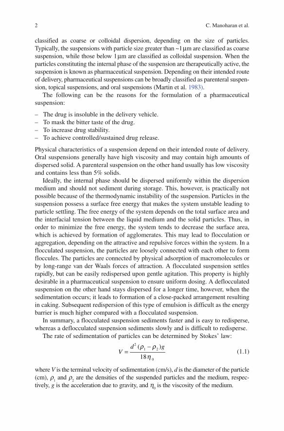
The rate of sedimentation of particles can be determined by Stokes’ law:
2
1 2
0
( )
18
d gV
r rh−
= (1.1)
where V is the terminal velocity of sedimentation (cm/s), d is the diameter of the particle (cm), r
1 and r
2 are the densities of the suspended particles and the medium, respec-
tively, g is the acceleration due to gravity, and h0 is the viscosity of the medium.

31 Various Pharmaceutical Disperse Systems
BookID <BID>_ChapID <CID>_Proof# 1 - 05/10/2009
1.2.1.1 Important Considerations in Formulation of Suspension
Formulation of a pharmaceutical suspension requires a knowledge of the properties of both the dispersed phase and the dispersion medium. The material for the formu-lation of suspensions should be carefully selected keeping in mind the route of administration, intended application, and possible adverse effects. The following are the most important factors to be considered during the formulation of pharma-ceutical suspensions:
1. Nature of suspended material: The interfacial properties of the suspended mate-rial are an important consideration during the formulation of a suspension. Particles that have low interfacial tension are easily wetted by water and there-fore can be suspended easily. Particles of materials with high interfacial tension, however, are not easily wetted. The suspension of such materials is normally achieved by using surfactants. Surfactants increase wettability of the particles by reducing their surface tension.
2. Size of suspended particles: Reduction of particle size leads to a decrease in the rate of sedimentation of the suspended particles as explained by Stoke’s law. Reduction in the size of particles can be achieved by processes such as milling, sieving, and grinding. Particle size also affects rate and extent of absorption, dis-solution, and biodistribution of the drug. However, reducing particle size beyond a certain limit may lead to formation of a compact cake upon sedimentation.
3. Viscosity of the dispersion medium: Greater viscosity of dispersion medium offers the advantage of slower sedimentation; however, it may compromise other desirable properties such as syringability for parenteral suspensions, spreadabil-ity for topical suspensions, ease of administration for oral suspensions. The property of shear thinning is highly desirable so that the suspension is highly viscous during storage when minimal shear is present so that the sedimentation is slow and has low viscosity after agitation (high shear) to facilitate ease of pourability from the bottle.
1.2.1.2 Sustained Release Suspensions
Emphasis has been placed lately on achieving sustained release of drugs by using pharmaceutical suspensions. Sustained release of drugs can be achieved after injection through intramuscular or subcutaneous route because of limited availability of dissolution medium. The particle size of suspension can be tai-lored to fall in colloidal or coarse range, depending on a variety of factors such as type of drug, formulation, site of action, and route of delivery. Depending on the aqueous solubility, the drug can be delivered in conjugation with polymers or metal salts to sustain the release (Morales et al. 2004; Gietz et al. 2000). Polymers can efficiently alter drug release kinetics through encapsulation or conjugation of the drug. A variety of micro particulate or nanoparticulate deliv-ery vectors can be prepared that can efficiently prolong the release of drug. These systems will be discussed later in the chapter. Addition of metal salts
4 C. Manoharan et al.
BookID <BID>_ChapID <CID>_Proof# 1 - 05/10/2009 BookID <BID>_ChapID <CID>_Proof# 1 - 05/10/2009
cause aggregation of drug molecules by formation of metal-hydroxy and -oxy polymers (Masuoka et al. 1993). This strategy has traditionally been used for precipitation of insulin and corticotrophin and more recently to achieve sus-tained release of recombinant hirudin (Gietz et al. 1998).
1.2.2 Emulsions
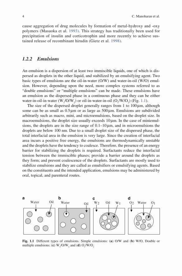
An emulsion is a dispersion of at least two immiscible liquids, one of which is dis-persed as droplets in the other liquid, and stabilized by an emulsifying agent. Two basic types of emulsions are the oil-in-water (O/W) and water-in-oil (W/O) emul-sion. However, depending upon the need, more complex systems referred to as “double emulsions” or “multiple emulsions” can be made. These emulsions have an emulsion as the dispersed phase in a continuous phase and they can be either water-in-oil-in-water (W
1/O/W
2) or oil-in-water-in-oil (O
1/W/O
2) (Fig. 1.1).
The size of the dispersed droplet generally ranges from 1 to 100 mm, although some can be as small as 0.5 mm or as large as 500 mm. Emulsions are subdivided arbitrarily such as macro, mini, and microemulsions, based on the droplet size. In macroemulsions, the droplet size usually exceeds 10 mm. In the case of miniemul-sions, the droplets are in the size range of 0.1–10 mm, and in microemulsions the droplets are below 100 nm. Due to a small droplet size of the dispersed phase, the total interfacial area in the emulsion is very large. Since the creation of interfacial area incurs a positive free energy, the emulsions are thermodynamically unstable and the droplets have the tendency to coalesce. Therefore, the presence of an energy barrier for stabilizing the droplets is required. Surfactants reduce the interfacial tension between the immiscible phases; provide a barrier around the droplets as they form; and prevent coalescence of the droplets. Surfactants are mostly used to stabilize emulsions and they are called as emulsifiers or emulsifying agents. Based on the constituents and the intended application, emulsions may be administered by oral, topical, and parenteral routes.
Fig. 1.1 Different types of emulsions. Simple emulsions: (a) O/W and (b) W/O, Double or multiple emulsions: (c) W
1/O/W
2 and (d) O
1/W/O
2
Water Oil OilWater W1 Oil O1WaterO2
a b c dW2
51 Various Pharmaceutical Disperse Systems
BookID <BID>_ChapID <CID>_Proof# 1 - 05/10/2009
1.2.2.1 Mechanism of Emulsification
When two immiscible liquids are in contact with each other, the molecules at the interface experience an imbalance of perpendicular forces. The net forces at the interface are called interfacial tension and they tend to minimize the surface area of individual liquids. In emulsions, the process of dispersion of one liquid in the other results in an increase in surface area between the dispersed droplets and dispersion medium, and surface free energy, which can be expressed as follows:
DW = g D A (1.2)
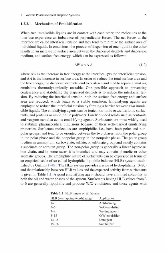
where DW is the increase in free energy at the interface, g is the interfacial tension, and D A is the increase in surface area. In order to reduce the total surface area and the free energy, the dispersed droplets tend to coalesce and tend to separate, making emulsions thermodynamically unstable. One possible approach to preventing coalescence and stabilizing the dispersed droplets is to reduce the interfacial ten-sion. By reducing the interfacial tension, both the surface free energy and surface area are reduced, which leads to a stable emulsion. Emulsifying agents are employed to reduce the interfacial tension by forming a barrier between two immis-cible liquids. The emulsifying agents can be ionic, non-ionic or zwitterionic surfac-tants, and proteins or amphiphilic polymers. Finely divided solids such as bentonite and veegum can also act as emulsifying agents. Surfactants are most widely used to stabilize pharmaceutical emulsions because of their well-marked emulsifying properties. Surfactant molecules are amphiphilic, i.e., have both polar and non-polar groups, and tend to be oriented between the two phases, with the polar group in the polar phase and the nonpolar group in the nonpolar phase. The polar group is often an ammonium, carboxylate, sulfate, or sulfonate group and mostly contains a succinate or sorbitan group. The non-polar group is generally a linear hydrocar-bon chain, and in some cases it is branched and may contain phenolic or other aromatic groups. The amphiphilc nature of surfactants can be expressed in terms of an empirical scale of so-called hydrophile–lipophile balance (HLB) system, estab-lished by Griffin (1949). The HLB system provides a scale of hydrophilicity (0–20) and the relationship between HLB values and the expected activity from surfactants is given in Table 1.1. A good emulsifying agent should have a limited solubility in both the oil and water phases of the system. Surfactants having HLB values from 3 to 6 are generally lipophilic and produce W/O emulsions, and those agents with
Table 1.1 HLB ranges of surfactants
HLB (overlapping words) range Application
1–3 Antifoaming3–6 W/O emulsifier7–9 Wetting agent8–18 O/W emulsifier13–15 Detergent15–18 Solubilizer

6 C. Manoharan et al.
BookID <BID>_ChapID <CID>_Proof# 1 - 05/10/2009 BookID <BID>_ChapID <CID>_Proof# 1 - 05/10/2009
HLB values from 8 to 18 produce O/W emulsions. Typical surfactants with their HLB values used as emulsifying agents are listed in Table 1.2.
Several theories exist that describe how emulsifying agents promote emulsifica-tion and maintain the stability of the resulting emulsion. The most prevalent theo-ries are the oriented-wedge theory, surface-tension theory, and the interfacial film theory. Indepth discussions of these theories are beyond the scope of this chapter and can be found elsewhere in literature (Becher 1977; Ansel et al. 1995). However, a general way in which emulsions are produced and stabilized has been discussed in this chapter. Emulsions do not form spontaneously when liquids are mixed and hence an input of energy is required to break up the liquids into small droplets. As the energy is applied, the interface between the oil phase and water phase is deformed resulting in the formation of droplets. Surfactant molecules get rapidly adsorbed at the interface formed between the droplets and lower the interfacial ten-sion. After the formation of emulsions, surfactants prevent coalescence of newly formed droplets by providing a strong short-ranged interfacial repulsion (Myers 1992). By lowering the interfacial tension, surfactants also reduce the energy needed to break up the large droplets into smaller ones.
1.2.2.2 Emulsion Stability
An emulsion is thermodynamically unstable, meaning that the dispersed droplets will tend to coalesce to minimize the interfacial area and break into two separate equilibrium phases with time. Three major phenomena, namely flocculation, creaming, and coalescence can take place before the emulsion separates or breaks into two phases (Fig. 1.2). The moving droplets due to Brownian motion can either adhere or repel, depending upon the Vander Waals attraction and repulsion forces that exist between the droplets. If the repulsion forces are weak, the attractive forces will pull them into contact and flocculation takes place. In flocculation, the droplets become attached to each other but are still separated by a thin film. When more droplets are involved, they aggregate and form three-dimensional clusters. At this point, the size of the droplets is not changed and the emulsifying agent is located at the surface of the individual droplets. Based on the density of the dispersed phase
Table 1.2 HLB values of typical emulsifying agents
Class Agent HLB
Anionic Triethanolamine oleate 12.0Sodium oleate 18.0Sodium dodecyl sulfate 40.0
Cationic Cetrimonium bromide 23.3Nonionic Sorbitan monolaurate (Span 20) 4.3
Sorbitan monooleate (Span 20) 8.6Polyoxyethylene sorbitan monolaurate (Tween 20) 16.7Polyoxyethylene sorbitan monooleate (Tween 80) 15.0Glyceryl monostearate 3.8

71 Various Pharmaceutical Disperse Systems
BookID <BID>_ChapID <CID>_Proof# 1 - 05/10/2009
and dispersion medium, the aggregated droplets may concentrate in one specific part of the emulsion. Creaming occurs when the aggregated droplets rise through the medium or sink to the bottom (sedimentation). Creaming depends upon the radius of the droplets, the relative difference in the densities of the two phases, and the viscosity of the continuous phase. The rate of creaming can be assessed by Stokes’ equation (1.1). It is apparent that the rate of creaming is increased by increased droplet size, a larger density difference between the two phases, and a decreased viscosity of the continuous phase. Creaming can be minimized by reduc-ing the droplet size to a fine state, keeping the difference in the densities of the two phases as small as possible and increasing the viscosity of the continuous phase. Creaming is reversible to some extent, because the dispersed droplets are still sur-rounded by the protective film and behave as a single drop. Coalescence occurs when two or more droplets fuse together to form a single larger droplet, which leads to the complete separation of the two immiscible phases. Contrary to creaming, the thin liquid film between the droplets is ruptured and therefore, coalescence process is irreversible. This phenomenon is called cracking of emulsions. Altering the vis-cosity and/or forming a strong interfacial film, using particulate solids, can stabilize coalescence. Coalescence process requires the droplets to be in close proximity. However, a different phenomenon called Ostwald ripening occurs even when the droplets are not in direct contact. Ostwald ripening is a process that involves the
1
2
3
4
5
6
Fig. 1.2 Schematic representation of different types of instability processes in an emulsion: (1) freshly prepared emulsion, (2) flocculation, (3) coalescence, (4) creaming, (5) Ostwald ripening, and (6) phase inversion
8 C. Manoharan et al.
BookID <BID>_ChapID <CID>_Proof# 1 - 05/10/2009 BookID <BID>_ChapID <CID>_Proof# 1 - 05/10/2009
growth of large particles at the expense of smaller ones because of high solubility of the smaller droplets and molecular diffusion through the continuous phase. A certain solubility of the dispersed in the continuous phase is required for Ostwald ripening to take place and is driven by the difference in Laplace pressure between droplets having different radii (Capek 2004). It is possible to stabilize emulsions against Ostwald ripening by adding components of high molecular weight that reduce the rate of diffusion of molecules within the dispersed species. Emulsions can invert from an O/W to a W/O emulsion or vice versa during homogenization or steriliza-tion procedures. This phenomenon is called as phase inversion and can be regarded as a form of instability. The temperature at which phase inversion occurs is called phase inversion temperature (PIT). The stability of an emulsion can also be affected by microbial contamination and oxidative decomposition of oils. This can be pre-vented by adding suitable preservative agents and antioxidants to the formulation.
1.2.2.3 Emulsification Methods
In an emulsion preparation, the liquid that forms the dispersed phase and the liquid that forms the continuous phase are influenced by the volume ratio of the liquids, the kind of emulsifying agent used, and its concentration in strong connection with the PIT. Emulsions may be prepared by high-pressure homogenizers, ultrasound homog-enizers, colloid mills, ball and roller mills, rotor/stator systems such as stirred vessels, and electrical and condensation devices. The principle and operation of these various equipments can be found in the literature (Walstra 1983; Becher 1977).
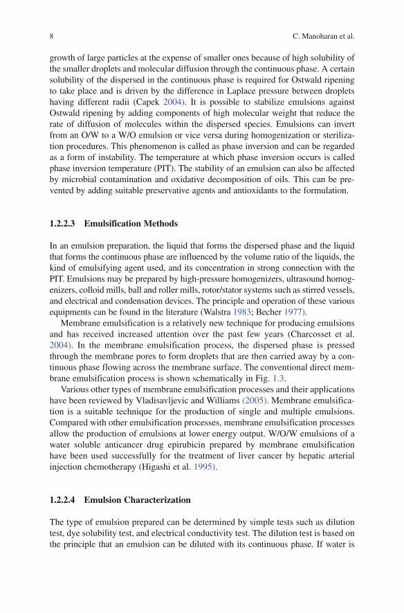
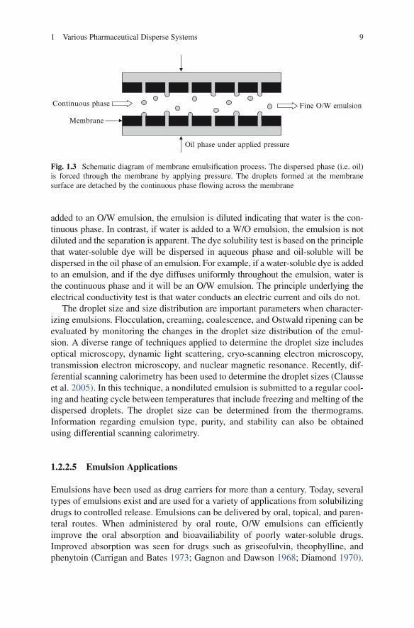
Membrane emulsification is a relatively new technique for producing emulsions and has received increased attention over the past few years (Charcosset et al. 2004). In the membrane emulsification process, the dispersed phase is pressed through the membrane pores to form droplets that are then carried away by a con-tinuous phase flowing across the membrane surface. The conventional direct mem-brane emulsification process is shown schematically in Fig. 1.3.
Various other types of membrane emulsification processes and their applications have been reviewed by Vladisavljevic and Williams (2005). Membrane emulsifica-tion is a suitable technique for the production of single and multiple emulsions. Compared with other emulsification processes, membrane emulsification processes allow the production of emulsions at lower energy output. W/O/W emulsions of a water soluble anticancer drug epirubicin prepared by membrane emulsification have been used successfully for the treatment of liver cancer by hepatic arterial injection chemotherapy (Higashi et al. 1995).
1.2.2.4 Emulsion Characterization
The type of emulsion prepared can be determined by simple tests such as dilution test, dye solubility test, and electrical conductivity test. The dilution test is based on the principle that an emulsion can be diluted with its continuous phase. If water is
91 Various Pharmaceutical Disperse Systems
BookID <BID>_ChapID <CID>_Proof# 1 - 05/10/2009
added to an O/W emulsion, the emulsion is diluted indicating that water is the con-tinuous phase. In contrast, if water is added to a W/O emulsion, the emulsion is not diluted and the separation is apparent. The dye solubility test is based on the principle that water-soluble dye will be dispersed in aqueous phase and oil-soluble will be dispersed in the oil phase of an emulsion. For example, if a water-soluble dye is added to an emulsion, and if the dye diffuses uniformly throughout the emulsion, water is the continuous phase and it will be an O/W emulsion. The principle underlying the electrical conductivity test is that water conducts an electric current and oils do not.
The droplet size and size distribution are important parameters when character-izing emulsions. Flocculation, creaming, coalescence, and Ostwald ripening can be evaluated by monitoring the changes in the droplet size distribution of the emul-sion. A diverse range of techniques applied to determine the droplet size includes optical microscopy, dynamic light scattering, cryo-scanning electron microscopy, transmission electron microscopy, and nuclear magnetic resonance. Recently, dif-ferential scanning calorimetry has been used to determine the droplet sizes (Clausse et al. 2005). In this technique, a nondiluted emulsion is submitted to a regular cool-ing and heating cycle between temperatures that include freezing and melting of the dispersed droplets. The droplet size can be determined from the thermograms. Information regarding emulsion type, purity, and stability can also be obtained using differential scanning calorimetry.
1.2.2.5 Emulsion Applications
Emulsions have been used as drug carriers for more than a century. Today, several types of emulsions exist and are used for a variety of applications from solubilizing drugs to controlled release. Emulsions can be delivered by oral, topical, and paren-teral routes. When administered by oral route, O/W emulsions can efficiently improve the oral absorption and bioavailiability of poorly water-soluble drugs. Improved absorption was seen for drugs such as griseofulvin, theophylline, and phenytoin (Carrigan and Bates 1973; Gagnon and Dawson 1968; Diamond 1970).
Fig. 1.3 Schematic diagram of membrane emulsification process. The dispersed phase (i.e. oil) is forced through the membrane by applying pressure. The droplets formed at the membrane surface are detached by the continuous phase flowing across the membrane
Oil phase under applied pressure
Membrane
Continuous phase Fine O/W emulsion

10 C. Manoharan et al.
BookID <BID>_ChapID <CID>_Proof# 1 - 05/10/2009 BookID <BID>_ChapID <CID>_Proof# 1 - 05/10/2009
Oral administration of a W/O emulsion containing ovalbumin, a model antigen has been shown to be more efficient in enhancing the immunogenic response than that of ovalbumin in saline (Masuda et al. 2003).
For parenteral drug delivery, both W/O and O/W emulsions have been investi-gated but O/W emulsions are predominantly used. W/O emulsions are easy to prepare; have good physical stability; and are easily injectable because of their low viscosity (Bjerregaard et al. 1999a). They have the potential for sustained release of hydrophilic drugs, since the surfactant layer acts as a release barrier for drugs present in the aqueous phase (Davis et al. 1985). In addition, the release properties from a W/O emulsion can be controlled within certain limits by adjusting param-eters such as droplet size, osmotic gradients, and volume fraction of the dispersed phase (Bjerregaard et al. 1999b). In vivo sustained release of aprotoin, a 58 amino acid polypeptide, from W/O emulsion has been demonstrated in mice and rabbits (Bjerregaard et al. 2001a, b). O/W or lipid emulsions are used for parenteral nutri-tion therapy, as well as for therapeutic agents. In lipid emulsions, the oil phase is typically a glyceride. Oils mostly used in lipid emulsions are soybean oil, cotton-seed oil, safflower oil, and medium-chain triglycerides. Egg phospholipids are the most commonly used emulsifying agents in lipid emulsions. Lipid emulsions offer numerous advantages as parenteral drug carriers such as solubilization of highly lipophilic drugs, stabilization of labile drugs against hydrolysis or oxidation, sus-tained release, and drug targeting. They are biocompatible, biodegradable, and reduce drug side effects by avoiding direct contact of the drug with the body fluid and tissues. Lipid emulsions have been used in parenteral nutrition for more than four decades for delivering fatty acids to patients who cannot eat or metabolize food properly. Examples of marketed formulations are Intralipid, Lipofundin, and Liposyn. Lipid emulsions have been investigated for a number of drugs to treat various disease conditions such as rhizoxin (Stella et al. 1988) and taxol (Tarr et al. 1987) for cancer, physostigmine (Rubinstein et al. 1991) for Alzheimer’s disease, and prostaglandine E
1 (Mizushima et al. 1983) for thrombosis therapy. Lipid emul-
sions of diazepam, propofol, and etomidate are commercially available.The use of lipid emulsions as ophthalmic vehicles has been explored in the last
few years. Lipid emulsions are excellent ocular delivery vehicles as already proved by Restasis®. Restasis® contains cyclosporine A indicated for increased tear produc-tion in patients with keratoconjunctivitis sicca. Drugs such as indomethacin (Klang et al. 2000), piroxicam (Klang et al. 1999), and difluprednate (Yamaguchi et al. 2005) have been investigated for ophthalmic lipid emulsions.
Double emulsions are excellent systems for the encapsulation of bioactive compounds. The presence of a reservoir phase inside droplets of another phase can be used to sustain release of active compounds, to protect sensitive mole-cules from external phase, taste masking, immobilization of enzymes, and for the enhancement of enteral and dermal absorption. The most common double emulsions used are of W/O/W type to entrap water-soluble drugs. Potential applications of double emulsions have been comprehensively reviewed by Khan et al. (2006).

111 Various Pharmaceutical Disperse Systems
BookID <BID>_ChapID <CID>_Proof# 1 - 05/10/2009
1.3 Colloidal Dispersions
1.3.1 Micelles
Micelles are self-assembling colloidal systems with particle size normally ranging from 5 to 100 nm (Kabanov et al. 1992; Torchilin 2007). They are classified as col-loidal dispersion because of their particle size. Micelles are spontaneously formed when amphiphilic molecules are placed in water at a certain concentration and tem-perature. Property of micellization is generally displayed by molecules that possess two distinct regions with opposite affinities toward a particular solvent (Mittal and Lindman 1991). At a low concentration, the molecules exist separately in a solution. However, when the concentration is increased, the molecules quickly self-assemble to form spherical micelles (Fig. 1.4). The hydrophobic portions of the molecules con-dense to form the core, whereas the hydrophilic portions constitute the shell or corona of the micelle (Lasic 1992). The concentration at which micellar association ensues is called the critical micelle concentration (CMC) and the temperature below which amphiphilic molecules exist separately is known as critical micellization temperature (CMT). The core of the micelle can solubilize lipophilic substances, whereas the hydrophilic outer portion serves as a stabilizing interface to protect the hydrophobic core from external aqueous environment. The process of micellization leads to free energy minimization of the system as the hydrophobic portions of the molecule are concealed and hydrogen bonds are established between hydrophilic portions in water.
Fig. 1.4 Schematic diagram of spontaneous micellization of amphiphilic molecules in aqueous media
Spontaneous micellization inaqueous media
Hydrophilic block
Hydrophobic block
Micelle

12 C. Manoharan et al.
BookID <BID>_ChapID <CID>_Proof# 1 - 05/10/2009 BookID <BID>_ChapID <CID>_Proof# 1 - 05/10/2009
Micelles are attractive candidates as drug carriers for delivering poorly water-soluble drugs. Micelles can solubilize a drug at concentrations much greater than its intrinsic solubility, which results in an increased bioavailability and a reduced toxic-ity. Incorporation of a drug into a micelle alters release kinetics and enhances the stability of the drug by reducing the access of water and biomolecules. Micelles generally have narrow size distribution and the size can be easily controlled by alter-ing the formulation conditions. Due to their size range, they can be conveniently sterilized by filtration through a membrane with a 0.2 mm cutoff. Specific targeting can be achieved by chemically conjugating a targeting molecule on the surface of a micelle. Passive targeting to tumors can also be achieved due to enhanced permeabil-ity and retention effect (EPR effect). Tumors have leaky vasculature and inefficient lymphatic drainage system, which results in a greater accumulation of micelles in tumors compared with normal tissues. Desirable properties of a pharmaceutical micelle include small size, narrow size distribution, low CMC value (low millimolar to micromolar), and high drug loading efficiency. Pharmaceutical micelles can be used through various routes such as parenteral, nasal, oral, otic, and ocular.
1.3.1.1 Surfactant Micelles
Micelles made from surfactants (especially nonionic surfactants) have been com-monly used to deliver drugs and biomolecules. They can efficiently entrap hydro-phobic drugs and form uniform particles. The water is distributed anisotropically within the micelle, i.e., concentration decreases from the shell to the core (Torchilin 2002). Thus, the position of a drug within the micelle depends on its polarity; more hydrophobic drug tends to stay closer to the core, whereas drugs with slight polarity are located closer to the micelle shell. A limitation of surfactant micelles is that they are not very stable and break apart rapidly upon dilution, which may cause prema-ture release and precipitation of the drug. The stability of surfactant micelles is severely compromised at concentrations lower than their CMC. Surfactant micelles typically have a rather high CMC, which may lead to concerns about surfactant-related toxicity. Micelle destabilization also compromises drug stability as the drug is exposed to blood components immediately upon systemic administration. Thus, surfactant molecules with lower CMC values and greater stability need to be devel-oped to overcome these limitations.
1.3.1.2 Polymeric Micelles
Micelles prepared from block copolymers have attracted much attention lately because of their better stability and biocompatibility (Gaucher et al. 2005). They are made from amphiphilic block copolymers with a large difference in solubility between hydrophobic and hydrophilic portions. Polymeric micelles generally have CMC values that are several orders of magnitude lower than typical CMC values for surfactants. As a result, polymeric micelles show enhanced stability and slower dissociation at lower concentrations compared with surfactant micelles (Jones and
131 Various Pharmaceutical Disperse Systems
BookID <BID>_ChapID <CID>_Proof# 1 - 05/10/2009
Leroux 1999). Surface functionalization of a polymeric micelle can be easily per-formed by chemically attaching a targeting moiety. Their low CMC values and lower rate of dissociation also allow for prolonged release of entrapped drug.
Copolymer for micellization can be synthesized by using two or more polymer blocks with contrasting solubility profiles. Poly (ethylene glycol) (PEG) is the most commonly used hydrophilic (shell forming) block (Torchilin 2002; Nishiyama and Kataoka 2003). Various molecular weights of PEG have been used for micelle preparation. PEG is highly biocompatible and forms a highly stable shell to steri-cally protect the hydrophobic core. It has also been shown to be efficient at escaping recognition by the reticuloendothelial systems (RES), thereby extending the circula-tion time of micelles in the blood (Kwon et al. 1997). Moreover, PEG copolymers usually have a low polydispersity (Mw/Mn ratio), which enables strict control of micelle size. Surface functionalization of PEG micelles can be easily performed by chemically linking a targeting moiety. Thus, PEG micelles can be used for active targeting to cells and tissues. Copolymers prepared by conjugating PEG with poly lactic acid (PLA) and poly (ethylene oxide) (PEO) have been extensively investi-gated as micellar vehicles (Yasugi et al. 1999). Other commonly used hydrophilic polymer blocks are poly (N-vinyl-2-pyrrolidone) (PVP), poly (vinyl alcohol) (PVA), and poly (vinylalcohol-co-vinyloleate). Triblock pluronic copolymers with an A-B-A structure (Ethylene oxide)x-(Propylene oxide)y-(Ethylene oxide)x have been extensively characterized (Kabanov et al. 2005). A variety of molecular weights and block lengths of Pluronic copolymers is available commercially. These copolymers have shown promise for delivering drugs and genes in vitro and in vivo.
1.3.1.3 Polymer-Lipid Micelles
A variety of hybrid micelles with lipid core and hydrophilic polymer shell has recently been investigated. Such micelles have shown good stability, longevity, and capability to accumulate into tissues with damaged vasculature (EPR effect). Micelles prepared by conjugation of PEG and phosphatidylethanolamine (PE) have been studied for delivery of anticancer drug Camptothecin (Mu et al. 2005). Such conjugation resulted in formation of very stable micelles having low toxicity and high delivery efficiency. PEG-PE conjugate form micelles with CMC in micro-molar range, which is about 100-fold lower than conventional detergent micelles. Polymer lipid micelles can be formed easily by spontaneous micellization in aque-ous media similar to surfactant and polymer micelles, and their size can be tailored by varying the molecular weight of the conjugate.
1.3.2 Microemulsions
The term “microemulsion” was first introduced by Hoar and Schulman (1943) to describe a clear solution obtained when normal O/W coarse emulsions were titrated with medium-chain length alcohols. Since then, there has been much dispute about
14 C. Manoharan et al.
BookID <BID>_ChapID <CID>_Proof# 1 - 05/10/2009 BookID <BID>_ChapID <CID>_Proof# 1 - 05/10/2009
the relationship of these systems to solubilized systems (i.e. micellar solutions, surfactant-free solutions) and to emulsions. Danielson and Lindman (1981) define microemulsion as a system of water, oil, and amphiphile which is (an) optically isotropic and thermodynamically stable liquid solution. The main difference between normal coarse emulsions and microemulsions lies in the droplet size of the dispersed phase. Microemulsions have droplets typically in the size range 10–100 nm and because of this small size range, they produce only a weak scattering of visible light and hence, they appear transparent. The features that distinguish microemulsion systems from emulsions are shown in Table 1.3.
Microemulsions are thermodynamically stable systems. The driving force for their thermodynamic stability is the ultralow interfacial tension (10−2–10−4m Nm−1). When the interfacial tension is this low, the interaction energy between droplets has been shown to be negligible and a negative free energy formation is achieved mak-ing the dispersion thermodynamically stable. The large interfacial tension between oil in water, which is typically about 50 m Nm−1, is reduced by employing surfac-tants. However, it is generally not possible to achieve the required interfacial ten-sion with the use of a single surfactant. Amphiphiles such as medium-chain length alcohols are added as cosurfactants to achieve the desired interfacial tension. Due to their amphiphilic nature, they partition between the aqueous and oil phase thereby altering the solubility properties of these phases. In addition, by interacting with surfactant monolayers at the interface, they affect their packing, which in turn can influence the curvature of the interface and interfacial free energy.
Depending upon the phase volume ratio and the nature of the surfactant used, a microemulsion can be one of the three types: O/W, bicontinous, and W/O. An O/W microemulsion is formed when the concentration of oil is low and a W/O microe-mulsion is formed when the concentration of water is low. In conditions where the volumes of oil and water are equal, a bicontinuous microemulsion is formed in which both oil and water exist as a continuous phase. A wide variety of internal structures exists within microemulsion systems. They may be spherical, spheroid, or cylindrical rod-shaped micelles and may exist in cubic, hexagonal, or lamellar phases. The relative amounts of aqueous phase, oil phase, and surfactant required to form a microemulsion can be determined with the aid of triangular/ternary phase diagrams. For example, in Fig. 1.5, each corner of the triangle represents 100% of one of the components. Moving away from that corner reduces the volume fraction of that specific component and any point on one of the axes corresponds to a mix-ture of two of those components in a defined ratio. Any point inside the triangle represents a mixture of all the three components in a defined ratio. A review written
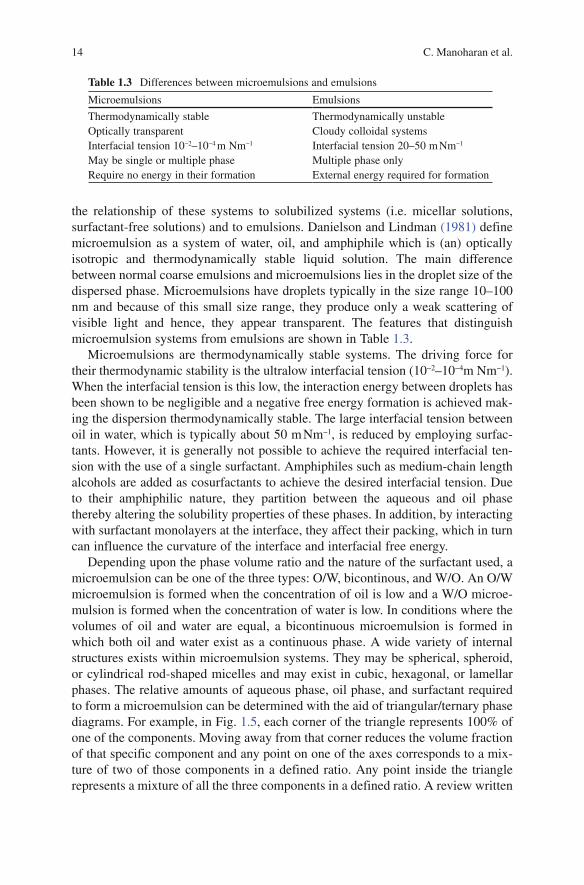
Table 1.3 Differences between microemulsions and emulsions
Microemulsions Emulsions
Thermodynamically stable Thermodynamically unstableOptically transparent Cloudy colloidal systemsInterfacial tension 10−2–10−4 m Nm−1 Interfacial tension 20–50 m Nm−1
May be single or multiple phase Multiple phase onlyRequire no energy in their formation External energy required for formation

151 Various Pharmaceutical Disperse Systems
BookID <BID>_ChapID <CID>_Proof# 1 - 05/10/2009
by Forster et al. (1995) describes in detail the physical meaning of the phase behav-ior of ternary oil/surfactant/water systems, and the concepts related to microemul-sion formation and the influence of additives on those microstructures.
1.3.2.1 Theories of Microemulsion Formation
Microemulsions form simultaneously when the interfacial tension between oil and water is reduced to close to zero. The formation and stability of a microemulsion can be affected by various factors, including the nature and molecular weight of surfactant, alcohol chain length and concentration, salinity, and temperature. Three different theories have been proposed to explain the microemulsion formation and stability (Paul and Moulik 1997). They are the interfacial mixed film theory, solu-bilization theory, and thermodynamic theory. According to the interfacial mixed film theory, the film at the interface is assumed to be a dual film and the type (W/O or O/W) of microemulsion formed depends upon the bending or curvature of the interface (Schulman et al. 1959). Solubilization theory states that oil is solubilized by normal micelles and water is solubilized by reverse micelles (Gillberg et al. 1970). Finally, according to the thermodynamic theory, the free energy of formation must be negative to form a thermodynamically stable microemulsion (Paul and Moulik 1997; Attwood 1994). Surfactants play an important role in reducing the interfacial tension in microemulsions. They can be selected based on the HLB con-cept or the critical packing parameter (CPP) concept. Surfactants with a low HLB value (3–6) are preferred for the formation of W/O microemulsion, whereas surfac-tants with high HLB value (8–18) are preferred for O/W microemulsions. The CPP describes a ratio between the hydrophobic and hydrophilic parts of a surfactant
Fig. 1.5 Schematic ternary phase diagram of an oil-surfactant-water system showing microemul-sion region and probable internal structures. An O/W microemulsion is formed when the concen-tration of oil is low, a W/O microemulsion is formed when the concentration of water is low, and a bicontinuous microemulsion is formed if the concentration of oil and water are equal. Depending upon the component concentrations and other characteristics, a variety of internal structures can exist within microemulsion systems
Bicontinuousmicroemulsion
Micelles
Lamellar pattern
Inverted micelles
Water Oil
O/W microemulsion W/O microemulsion
Surfactant (+ Co-surfactant)
O/W microemulsion
100
100500
100
5050
0
0

16 C. Manoharan et al.
BookID <BID>_ChapID <CID>_Proof# 1 - 05/10/2009 BookID <BID>_ChapID <CID>_Proof# 1 - 05/10/2009
molecule and is useful in estimating the nature of formed aggregates. The CPP can be calculated using CPP = n/a·l, where n is the partial molecular volume of the surfactant, а is the optimal head group area, and l is the length of the surfactant tail. When the CPP is between 0 and 1, O/W microemulsions are formed, and when the CPP > 1, W/O microemulsions are formed. Bicontinous microemulsions are formed when the CPP = 1. Brij, dioctyl sodium sulphosuccinate, and lecithin are some of the widely used surfactants to stabilize microemulsion formulations.
1.3.2.2 Microemulsion Characterization
Characterization of microemulsions is a difficult task because of the variety of structures and components involved, and the limitations with the available tech-niques. Therefore, it is preferable to employ a combination of techniques as far as possible. Particle size in microemulsions can be elucidated by small-angle X-ray scattering, small-angle neutron scattering, dynamic and static light scattering, freeze fracture electron microscopy, and neutron scattering techniques. NMR has been used to measure self-diffusion coefficients of the various components. Viscosity measurements can be used to determine the interaction of dispersed drop-lets and can indicate the pressure of rod-like or worm-like reverse micelles (Yu and Neuman 1995; Angelico et al. 1998). Conductivity experiments can be used to determine the type of microemulsion and to study phase inversion phenomena (Lawrence and Rees 2000).
1.3.2.3 Microemulsion Applications
Microemulsions have attracted large interest in the pharmaceutical industry as ideal delivery systems for a variety of drug molecules because of their thermodynamic stability, simplicity of preparation, and solubilization capacity. Garcia-Celma (1997) has reviewed microemulsions as drug delivery systems for a variety of drug mole-cules. Because of their unique solubilization properties, microemulsions can improve the solubility and bioavailability of poorly water-soluble drugs. Lipophilic, hydro-philic, or amphiphilic drugs can be effectively solubilized because of the existence of microdomains of different polarity within the system. Microemulsions for oral, der-mal, transdermal, parenteral, and pulmonary delivery routes have been developed. When administered via the oral route, microemulsions enhance the bioavailability of poorly soluble drugs by maintaining them in molecular dispersion in the GI tract. In addition, the presence of surfactants increases the membrane permeability of the solu-bilized drug. The oral efficacy of microemulsions is well demonstrated by the com-mercially available Cyclosporin A formulation (Neoral®). Recently, Kim et al. (2005) reported that tricaprylin microemulsions improved the oral bioavailability of low molecular weight heparin in mice and monkeys. The microemulsion was composed of tricaprylin (a surfactant mixture of Tween® 80 and Span® 20), heparin, and water. Microemulsions can serve as delivery systems for dermal applications and have been
171 Various Pharmaceutical Disperse Systems
BookID <BID>_ChapID <CID>_Proof# 1 - 05/10/2009
proved to increase the permeation rate of both lipophilic and hydrophilic drugs com-pared with conventional vehicles such as emulsions or solutions (Tenjarla 1999). Surfactants and cosurfactants play an important role in reducing the diffusional bar-rier of stratum corneum by acting as permeation enhancers and in addition, the inter-nal mobility of the drug within the vehicle also contributes to the increased permeability. Some of the commonly used surfactants for enhancing permeability are l-a-phosphatidylcholine, Azone®, and Tween® 20. Short-chain alkanols such as etha-nol and propylene glycol are used as cosurfactants in microemulsion transdermal formulations. The role of microemulsions in percutaneous penetration of drugs has been summarized in a comprehensive review by Kreilgaard (2002).
1.3.3 Nanosuspensions
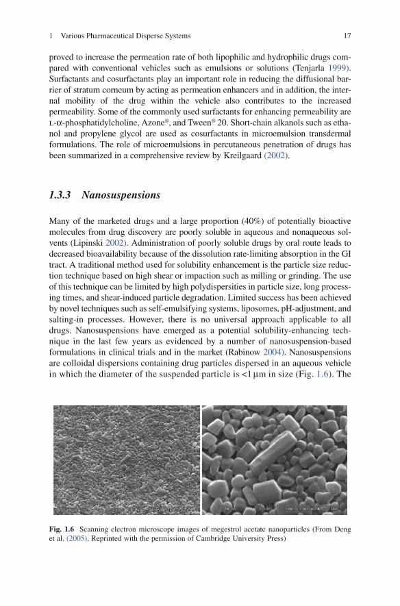
Many of the marketed drugs and a large proportion (40%) of potentially bioactive molecules from drug discovery are poorly soluble in aqueous and nonaqueous sol-vents (Lipinski 2002). Administration of poorly soluble drugs by oral route leads to decreased bioavailability because of the dissolution rate-limiting absorption in the GI tract. A traditional method used for solubility enhancement is the particle size reduc-tion technique based on high shear or impaction such as milling or grinding. The use of this technique can be limited by high polydispersities in particle size, long process-ing times, and shear-induced particle degradation. Limited success has been achieved by novel techniques such as self-emulsifying systems, liposomes, pH-adjustment, and salting-in processes. However, there is no universal approach appli cable to all drugs. Nanosuspensions have emerged as a potential solubility-enhancing tech-nique in the last few years as evidenced by a number of nanosuspension-based formulations in clinical trials and in the market (Rabinow 2004). Nanosuspen sions are colloidal dispersions containing drug particles dispersed in an aqueous vehicle in which the diameter of the suspended particle is <1 mm in size (Fig. 1.6). The
Fig. 1.6 Scanning electron microscope images of megestrol acetate nanoparticles (From Deng et al. (2005), Reprinted with the permission of Cambridge University Press)
18 C. Manoharan et al.
BookID <BID>_ChapID <CID>_Proof# 1 - 05/10/2009 BookID <BID>_ChapID <CID>_Proof# 1 - 05/10/2009
basic principle of this technique is to reduce the size of the drug particles to a submi-cron range. Reducing the particle size to a submicron range increases the surface area to be in contact with the dissolution medium and consequently the dissolution rate. Nanosuspensions have a number of potential benefits compared with conventional methods. Nanosuspensions allow to incorporate a high concentration of drug in a relatively low volume of fluid; provide a chemically and physically stable product; and can be used for controlled and targeted delivery of drugs. In addition, nanosus-pensions can be used for drugs that are water insoluble (<0.1 mg/ml) and for drugs insoluble in both water and organic solvents.
1.3.3.1 Preparation of Nanosuspensions
Nanosuspensions can be produced by bottom-up or top-down techniques. In the case of bottom-up technique, the strategy is to build particles from their constituent units, i.e., molecules, atoms. Bottom-up technique is a classical precipitation pro-cess in which the drug is dissolved in a solvent, which is subsequently added to a nonsolvent to precipitate the drug crystals. Use of solvents, the difficulty to avoid the formation of microcrystals, and the poor solubility of an increasing number of drugs in all media limit this approach. In the case of top-down technique, the coarse material is subsequently broken down until nanoscopic dimensions are reached. There are two basic techniques widely used: (1) Pearl/ball milling (Nanocrystal technology®, élan) and (2) High-pressure homogenization (Dissocubes®, Skyepharma). In pearl milling, an aqueous suspension of the drug is fed into the mill containing milling pearls made of glass, zirconium oxide, ceramic sintered aluminium oxide, or hard polystyrene with high abrasion resistance. As the pearls are rotated at a very high shear rate, the drug particles are ground into nano-sized particles between the moving milling pearls. Depending upon the hardness, the drug needs to be milled from hours to several days.
In the high-pressure homogenization technique, the drug suspension is forced under pressure through a valve that has a nanoaperture. Typical pressures applied are between 100 and 1,500 bars. As the drug suspension passes through the nano-aperture with high velocity, the static pressure is decreased, leading to the forma-tion of small gas bubbles, which implode as they exit the valve. The cavitation forces created breakdown the drug microparticles into nano-sized particles. It is not possible to obtain the desired particle size for many drugs in a single homogeniza-tion cycle. Multiple homogenization cycles are required depending on the hardness of the drug, desired mean particle size, and required homogeneity of the product. To avoid the removal of water after high-pressure homogenization in aqueous media, nanosuspensions were produced using high-pressure homogenization in nonaqueous media or in water with water-miscible liquids (Nanopure®, Pharmasol). A combinative technology (NANOEDGE TM) was introduced by Baxter Healthcare in which preci pitation step is followed by high-pressure homogenization to prevent the precipitate from crystal growth. Very recently, Moschwitzer and Muller (2006) reported a new combination method for the production of ultrafine submicron

191 Various Pharmaceutical Disperse Systems
BookID <BID>_ChapID <CID>_Proof# 1 - 05/10/2009
nanosuspensions. This method involves an evaporation step to provide a solvent-free modified starting material followed by high-pressure homogenization. Hydrocortisone acetate nanosuspensions produced using this method with reduced homogenization cycles and the nanosuspensions demonstrated excellent long-term storage stability. Application of supercritical fluid process for production of nano-suspensions has increased in recent years. The most widely used methods are rapid expansion of supercritical solution process, gas antisolvent process, and supercriti-cal antisolvent process. Cyclosporine, budesonide, and griseofulvin nanosuspen-sions have been prepared using these methods (Young et al. 2000; Steckel et al. 1997; Chattopadhyay and Gupta 2001).
1.3.3.2 Characterization of Nanosuspensions
A critical parameter that defines the quality and physicochemical behavior of nanosuspensions is the particle size distribution. The mean particle size and width of distribution can be determined using photon correlation spectroscopy (PCS), laser diffraction (LD), and coulter counter techniques. The measuring range for PCS is 3 nm to 3 mm and LD can measure particles ranging from 0.05 to 80 mm up to 2,000 mm. The coulter counter gives an absolute number of particles per unit volume for the different size classes. The shape and surface morphology of nano-suspensions can be assessed by scanning electron microscopy and atomic force microscopy. The crystalline state of nanosuspensions can be characterized by dif-ferential scanning calorimetry and X-ray diffraction. Determination of zeta potential is important to assess the physical stability of nanosuspensions. For nanosuspension formulations administered intravenously, additional parameters such as surface hydrophilicity/hydrophobicity, sterility, and pyrogenicity need to be determined.
1.3.3.3 Applications of Nanosuspensions
Nanosuspension technology is applicable to all poorly soluble drugs and an out-standing feature of this technology is its simplicity. Nanosuspensions can be applied to various administration routes such as oral, parenteral, pulmonary, otic, ophthalmic, and nasal routes. Administration of poorly water-soluble drug in the form of nanosuspensions has shown increased onset of action, increased bioavail-ability, and dissolution rate. For example, the t
max for the nanosuspension formu-
lation of naproxen, a nonsteroidal antiinflammatory drug, was 23.7 min versus 33.5 min for unmilled formulation (Liversidge and Conzentino 1995). Nanosuspension formulations of naproxen reduced the time required to achieve C
max by approximately 50% compared with marketed suspension (Naprosyn) and
tablets (Anaprox) (Merisko-Liversidge et al. 2003). The dissolution rate of naproxen tablet formulation made from naproxen particles ranging from 100 to 600 nm in size was compared with a commercially available product (Aleve) that

20 C. Manoharan et al.
BookID <BID>_ChapID <CID>_Proof# 1 - 05/10/2009 BookID <BID>_ChapID <CID>_Proof# 1 - 05/10/2009
was prepared from macro-sized naproxen (Jain et al. 2000). Dissolution rate of naproxen from the nanoparticulate formulation was found to be significantly higher compared with the drug release from the marketed product. This study showed that if properly formulated with suitable excipients, nanosuspensions can be processed into conventional dosage forms such as tablets and capsules, using standard equipment. Reducing the mean particle size of a poorly soluble investi-gational compound from 7 mm to 280 nm showed four times higher bioavailabil-ity compared with micrometric size particles of the compound (Jia et al. 2002). Nanoparticle formulation was rapidly absorbed with a t
max of 1 h, whereas the t
max
for microparticle formulation was prolonged for another 3 h. Oleanic acid nano-suspension containing particles with an average particle size of 284.9 nm was reported to have a faster dissolution rate and enhanced therapeutic effect (Chen et al. 2005). Kocbek et al. (2006) developed ibuprofen nanosuspensions prepared by a melt emulsification method traditionally used to prepare solid lipid nanopar-ticles. Ibuprofen nanosuspensions either in the form of lyophilized powder or granules showed enhanced dissolution rate compared with micronized drug. For lyophilized nanosuspension formulation, more than 65% of the drug dissolved in the first 10 min as opposed to only 15% of the micronized drug (Fig. 1.7).
Drugs that are not absorbed through the GI tract or that undergo extensive first pass metabolism can be administered intravenously as nanosuspensions. Nanosus-pen sions are ideal for intravenous route and offer many special advantages. A rapid onset of action can be achieved in case of an emergency and high concentra-tions of the drug can be administered without employing toxic cosolvents and solubility excipients. Nanosuspensions are potential targeted systems as their surface properties can be easily altered. In addition, capillary blockade is avoided, since the particle size in the nanosuspensions is less than 5 mm, which is the inner diameter of the smallest blood capillaries in the body. A chemically stable, intravenously
Fig. 1.7 In vitro dissolution profiles of ibuprofen lyophilized nanosuspension (filled circle) and micronized ibuprofen (open circle). Reproduced from Kocbek et al. (2006) with permission from Elsevier Science
100
80
60
40
20
00 10 20 30
Time (min)
Ibu
pro
fen
dis
solv
ed (
%)
40 50 60
211 Various Pharmaceutical Disperse Systems
BookID <BID>_ChapID <CID>_Proof# 1 - 05/10/2009
injectable nanosuspension formulation for omeprazole, a poorly soluble, chemi-cally labile drug with a high degradation rate in aqueous media was developed by Moschwitzer et al. (2004). Injectable nanosuspension of the poorly soluble drug tarazepide has been prepared to overcome the limited success achieved using con-ventional solubilization tecniques, such as the use of mixed micelles or cycodex-trins to improve the bioavailability of the drug (Jacobs et al. 2000). Rabinow et al. (2007) evaluated an intravenous itraconazole nanosuspension dosage form, relative to a solution formulation, in the rat. The formulation of itraconazole as a nanosus-pension enhanced the efficacy of this antifungal agent, and exhibited altered phar-macokinetics, leading to increased tolerability and high drug levels in target organs. Pulmonary administration of nanosuspensions increases rapid diffusion and disso-lution of the drug at the site of action. Budesonide, a poorly water-soluble antiin-flammatory corticosteroid, has been successfully formulated as a nanosuspension for pulmonary delivery (Jacobs and Muller 2002). The advances in nanosuspension technology are described in more detail in Chap. 10.
1.3.4 Liposomes
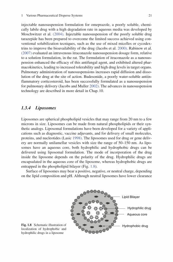
Liposomes are spherical phospholipid vesicles that may range from 20 nm to a few microns in size. Liposomes can be made from natural phospholipids or their syn-thetic analogs. Liposomal formulations have been developed for a variety of appli-cations such as diagnostic, vaccine adjuvants, and for delivery of small molecules, proteins, and nucleotides (Lasic 1998). The liposomes used for drug or gene deliv-ery are normally unilamellar vesicles with size the range of 50–150 nm. As lipo-somes have an aqueous core, both hydrophilic and hydrophobic drugs can be delivered using liposomal formulation. The mode of incorporation of the drug inside the liposome depends on the polarity of the drug. Hydrophilic drugs are encapsulated in the aqueous core of the liposome, whereas hydrophobic drugs are entrapped in the phospholipid bilayer (Fig. 1.8).
Surface of liposomes may bear a positive, negative, or neutral charge, depending on the lipid composition and pH. Although neutral liposomes have lower clearance
Fig. 1.8 Schematic illustration of localization of hydrophobic and hydrophilic drugs in a liposome
Lipid Bilayer
Hydrophobic drug
Hydrophilic drug
Aqueous core

22 C. Manoharan et al.
BookID <BID>_ChapID <CID>_Proof# 1 - 05/10/2009 BookID <BID>_ChapID <CID>_Proof# 1 - 05/10/2009
through retuculoendothelial system (RES), they have a high tendency to aggregate. Negatively charged liposomes are highly susceptible to endocytosis by mac-rophages. Positively charged liposomes, mostly used for gene delivery as poly-plexes, interact with serum protein and are subsequently removed by the RES.
Liposomes, unlike micelle, are a thermodynamically unstable system and tend to fuse together and eventually separate out of the aqueous medium on storage. To achieve long-term stability, liposomes with high charge density have been prepared. However, high charge density of liposomes can not provide long-term stabilization in vivo because of the presence of various proteins and enzymes. Another approach to stabilization of liposomes is coating the outer surface of the liposome with an inert hydrophilic polymer (Klibanov et al. 1990; Papahadjopoulos et al. 1991). Such liposomes are called sterically stabilized liposomes. They have the ability to evade the immune system and hence can achieve longer circulation time in vivo. PEG is routinely used to provide steric stability to liposomes for drug and gene delivery.
1.3.4.1 Liposomes Targeting
Liposomal delivery can lead to drastic changes in absorption, distribution, and elimination of the drug. Surface conjugation of PEG results in resistance toward aggregation and prolonged circulation in blood. This longer circulation time results in accumulation of drug at the target site overtime.
Active targeting of liposomes can also be easily achieved by conjugation of a targeting moiety on the surface of liposome. A variety of ligands and receptors has been used for active targeting of liposomes toward specific cells and tissues (Lian and Ho 2001). Antibody-coated liposomes (also referred to as Immunoliposomes) have also been commonly used to achieve specific targeting. Targeting molecule can also be conjugated to PEG through covalent linkage.
1.3.4.2 Applications of Liposomes
Drug Delivery
As the structure of liposomes resembles cell membrane, they were considered attractive delivery system for drugs especially at intracellular level. Liposomes can be formulated as gel, cream, aerosol, suspension, or dry powder and administered through a variety of routes. Several drug delivery systems using liposomes such as Doxil (Doxorubicin in stealth liposomes; Sequus Pharmaceuticals) and Daunoxome (Daunorubicin; Nexstar Pharmaceuticals) are in market. Several others are in an advanced stage of clinical trials or in preclinical studies targeted toward various cancers, autoimmunity, skin disorders etc. Fusogenic, pH-sensitive components can be added to the liposomes to cause release of drug at a certain pH in the tissue or to cause release of drug intracellularly by endosomal destabilization.
231 Various Pharmaceutical Disperse Systems
BookID <BID>_ChapID <CID>_Proof# 1 - 05/10/2009
Gene Delivery
The use of cationic liposomes to deliver polynucleotides was first reported in 1987 and since they have been commonly used in gene delivery research (Felgner et al. 1987). Cationic liposomes can efficiently condense negatively charged DNA into nanometer-sized particles. Such liposome/polynucleotide complexes are generally referred to as lipoplexes. Neutral lipid dioleoylphosphatidylethanolamine is com-monly incorporated along with cationic lipids in a lipoplex. It facilitates endosomal destabilization apart from providing structural stability to the liposome. Formation of lipoplexes also prevents DNA from degradation by extracellular and intracellular nucleases. Cellular entry of lipoplexes primarily occurs through clathrin-mediated endocytosis (Dass and Burton 2003). Liposomes have also been employed for cell-targeting, using a variety of targeting ligands (Talsma et al. 2006; Torchilin 2006). In spite of their high transfection efficiency in vitro, lipoplexes have not been very successful in vivo because of their high toxicity and immunogenicity. Lipoplex formulations have been reported to cause moderate-to-severe toxicities in animal models at both systemic and cellular levels.
Other Applications
Liposomes, depending on their lipid composition and size, can be targeted toward specific immune cells. It is well established that large liposomes are efficiently taken up by macrophages. Thus, liposome formulations provide an attractive tool for antigen delivery for stimulation of both cellular and humoral immune response. Antigens along with immunomodulatory agents can be encapsulated inside lipo-somes for efficient vaccination.
Liposomes have also been used as a diagnostic tool for transport of tracers such as fluorescent molecules or radio-isotopes for detection of tumors. They also find application in oral delivery of nutrients and in cosmetics.
1.3.5 Nanoparticles
The advent of nanotechnology has opened new avenues for drug and gene delivery in last few years. A variety of nanoscale devices such as nanoparticles, nanotubes, nano-gels, and molecular conjugates has been investigated (Ravi Kumar et al. 2004; Murakami and Nakashima 2006; Lemieux et al. 2000). Nanoparticles are the most commonly used nanometer scale delivery systems. They are typically spherical parti-cles in the size range of 1–1,000 nm (Fig. 1.9). The size of these delivery systems confers advantages such as greater and deeper tissue penetration, longer circulation time in blood, enhanced cellular uptake, ability to cross blood–brain barrier, and greater ability to target specific cell types (Kreuter et al. 1995; Davis 1997; Vogt et al. 2006). Drug or gene of interest can be incorporated in a nanoparticle by encapsulation

24 C. Manoharan et al.
BookID <BID>_ChapID <CID>_Proof# 1 - 05/10/2009 BookID <BID>_ChapID <CID>_Proof# 1 - 05/10/2009
or surface conjugation. Although majority of nanoparticles used in research have been developed from polymers, certain nonpolymeric materials have also been used.
1.3.5.1 Polymeric Nanoparticles
A variety of polymers such as poly lactide-co-glycolide (PLGA), poly lactic acid (PLA), polyethyleneimine (PEI), polymethacrylates has been investigated for for-mulation of nanoparticles. Potential applications of polymeric nanoparticles include drug and gene delivery, cell and tissue targeting, pharmacokinetic studies, tissue engineering, vaccine adjuvation, and delivery of diagnostic agents. Majority of polymers used have been biodegradable, which is highly desirable. However, some nonbiodegradable polymers have also been used, especially for gene delivery because of their high transfection efficiency. The type of the polymer used for for-mulation of nanoparticles is generally determined by the intended application of nanoparticles. For example, if the aim is to deliver the particles systemically and to evade immune recognition, hydrophilic polymers such as PEG may be incorpo-rated. Conversely, if the immune cells are to be targeted, it may be a good idea to use polymers that have high surface charge so as to cause rapid uptake by immune cells. Chemical and physical characteristics of polymers can be tailored to suit the intended application.
Drug delivery remains one of the basic and most common applications of polymeric nanoparticles. Nanoparticles can vehiculate drug of interest by either encapsulation in
Fig. 1.9 Scanning electron micrograph of polymeric nanoparticles for drug and gene delivery

251 Various Pharmaceutical Disperse Systems
BookID <BID>_ChapID <CID>_Proof# 1 - 05/10/2009
the matrix or through attachment on the surface. These polymeric nanoparticles pos-sess high drug loading efficiency and high stability to protect and securely deliver the drug in vivo. Polymeric nanoparticles are versatile systems that can be delivered through a variety of routes such as oral, nasal, parenteral, and ocular. Polymeric nano-particles have also displayed an ability to cross blood–brain barrier (Olivier 2005). Steric stabilization of nanoparticles can be achieved by coating the surface with hydrophilic polymers. It results in increased circulation time and subsequently increased bioavailability. Another important application of polymeric nanoparticles has been delivery of anticancer drugs (van Vlerken and Amiji 2006). Particles with optimum size and surface characteristics can passively travel through the leaky vas-culature in tumor tissues and can exhibit passive targeting. Active targeting of tumors can also be achieved by conjugating targeting molecule on the surface of particles.
Gene delivery has been another very extensively investigated area of nanotech-nology. Polymeric nanoparticles have been developed that can efficiently deliver genes into cells both in vitro and in vivo, with low toxicity. Nanoparticles for gene delivery can be formulated in a variety of ways. The most common mode is encap-sulation of DNA inside the polymeric matrix. Alternatively, DNA can be attached on the surface of nanoparticles by electrostatic interaction or chemical binding. Some cationic polymers such as PEI also condense DNA spontaneously in solution to form polymer/DNA complexes known as polyplexes (De Smedt et al. 2000). PEI has been by far the most commonly used polymer for gene delivery because of its very high efficiency and is widely regarded as the “gold standard” for comparing the efficiency of gene delivery vectors. Cationic charge of polymers facilitates cellular attachment and internalization. Biodegradable polymers such as PLGA and PLA have also been used to achieve sustained gene delivery.
1.3.5.2 Nonpolymeric Nanoparticles
Contrary to polymeric nanoparticles that are generally greater than 100 nm in size, nonpolymeric nanoparticles such as metallic and ceramic nanoparticles are mostly below 100 nm in size. Metal and ceramic nanoparticles have been commonly employed for various biomedical applications.
Magnetic iron oxide particles have been particularly investigated for in vivo imaging apart from other biomedical and bioengineering applications. These par-ticles can be easily synthesized with a narrow size distribution. The surface of iron oxide particles can be coated using a variety of polymeric and nonpolymeric mate-rials to modify surface characteristics (Gupta and Gupta 2005). Surface coating apart from providing stability also facilitates binding of biological ligands and receptors for tissue and cell targeting. Iron oxide nanoparticles have also shown good efficiency in delivering genes in vitro (Dobson 2006). Transfection of cells using magnetic nanoparticles is also known as magnetofection.
Gold nanoparticles have received much attention in recent years in drug delivery as an alternative to lipid and polymer-based delivery systems because of their low toxicity and easy synthesis. The particles size is generally below 50 nm with narrow

26 C. Manoharan et al.
BookID <BID>_ChapID <CID>_Proof# 1 - 05/10/2009 BookID <BID>_ChapID <CID>_Proof# 1 - 05/10/2009
distribution. Surface functionalization of gold nanoparticles, using a PEG spacer, has been shown to result in rapid cellular uptake and internalization, improved stability, and greater circulation time (Shenoy et al. 2006). Proteins can be delivered using gold nanoparticles by surface attachment using a PEG spacer. Gold nanopar-ticles surface-functionalized using quarternary ammonium chains have been used for gene delivery and have resulted in very efficient transfection of cells in vitro (Sandhu et al. 2002).
Biocompatible ceramic nanoparticles made from silica, titania, alumina, etc. have also been explored for drug delivery. High porosity of these materials makes them attractive candidates for drug/gene delivery. These nanoparticles have been used for delivery of anticancer drugs and insulin. However, the full potential of these nanosystems is yet to be explored.
1.3.6 Microspheres
Drug delivery using biodegradable polymeric microspheres has gained increased interest in the last two decades. Microspheres are solid, spherical devices containing the drug in a polymer matrix with size ranging from 1 to 1,000 mm. Microspheres are different from microcapsules. In microspheres, the drug is dispersed throughout the polymer matrix, whereas in microcapsules, the drug is the core surrounded by a poly-meric membrane. Microspheres are widely used as drug carriers for controlled release and the incorporation of drug molecules into biodegradable polymeric micro spheres has many advantages. The polymer matrix can protect drugs, such as proteins, from physiological degradation. Microspheres can control the delivery of drugs from days to months therefore reducing frequent administrations and improving patient compli-ance and comfort. Different release profiles with desired release rates can be achieved by selecting polymers with different degradation mechanisms. In addition, micro-spheres can be used to target drugs to a specific site. Microspheres have been pro-posed as delivery systems for traditional small molecular weight drugs, proteins, enzymes, vaccines, cells, and even delicate molecules such as DNA.
A wide range of natural and synthetic polymers has been used for the micro-sphere matrix. Some of the natural polymers include chitosan, alginate, albumin, and casein. Synthetic polymers include polyesters, polyanhydrides, polycaprolac-tones, polyphosphazenes, polyorthoesters, and polycarbonates. Most of the biode-gradable polymers are nontoxic; degrade within the body; are easily eliminated without the need for surgical removal. Among the aforementioned polymers, poly-esters like PLGA have received more attention because of their excellent biocom-patibility and mechanical properties for drug delivery applications. Microsphere products based on PLGA polymer commercially available are Lupron® depot, Risperdal ConstaTM, Zoladex® depot, Sandostatin LAR® depot, and Decapeptyl® depot. The encapsulated drug release from the microspheres occurs either by diffu-sion out of the polymer matrix or by degradation of the polymer or both. The release characteristics can be influenced by the molecular weight of the entrapped drug and

271 Various Pharmaceutical Disperse Systems
BookID <BID>_ChapID <CID>_Proof# 1 - 05/10/2009
polymer, size of the microspheres, excipients present in the system, and local envi-ronmental conditions such as pH.
1.3.6.1 Microsphere Preparation Techniques
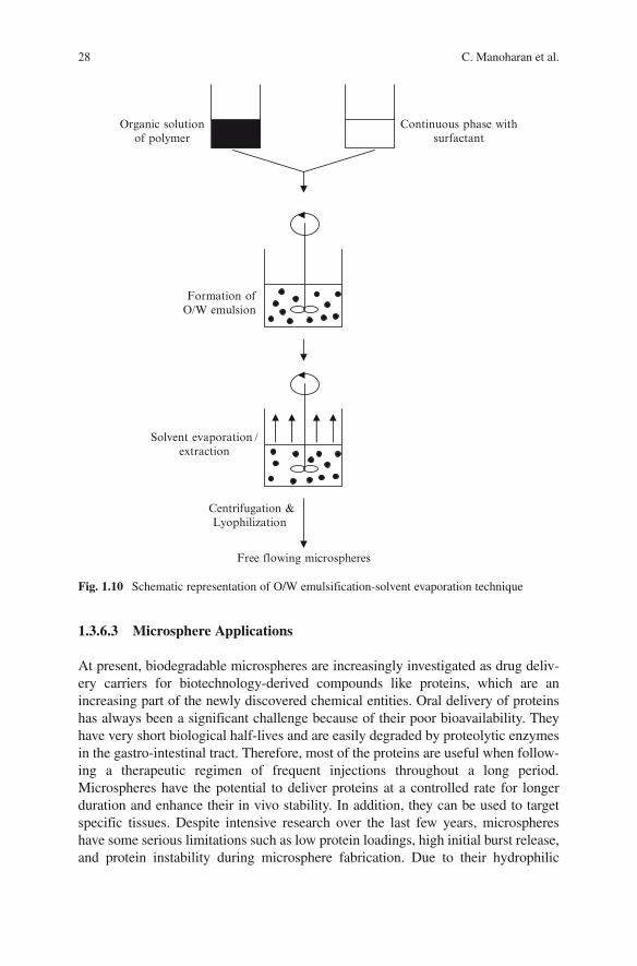
A number of techniques are currently available for preparing microspheres. A well-known and widely used technique is the emulsification-solvent evaporation tech-nique, because it allows encapsulation of the protein as an aqueous solution. In this technique, the polymer is dissolved in a volatile organic solvent such as methylene chloride or acetone. The drug is then dissolved or dispersed in the polymer solution. In the following step, the organic phase is emulsified in an aqueous phase, resulting in an O/W emulsion. The aqueous phase contains appropriate surfactant, which stabilizes the organic solvent droplets formed during the energy input. As the organic solvent is removed either by evaporation or by extraction, solid micro-spheres are formed. The microspheres can be collected by filtration or centrifuga-tion and lyophilized to obtain free-flowing microsphere product (Fig. 1.10). The O/W technique is appropriate for encapsulating hydrophobic drugs.
Hydrophilic compounds can be encapsulated by W/O/W double emulsion tech-nique. In this technique, an aqueous solution of the drug is added to an organic solution of the polymer to form a W/O emulsion. This emulsion is then added into a large-volume aqueous phase to form a W/O/W emulsion. The resulting emulsion is subjected to solvent removal either by evaporation or by extraction process. Other techniques employed for the preparation of microspheres are polymer phase separation (coacervation), spray drying, milling, and supercritical fluid techniques. For the detailed description of these methods with their merits and limitations, and for the advances in microsphere production technology, the readers are referred to an excellent chapter written by Tewes et al. (2005).
1.3.6.2 Microsphere Characterization
Particle size of microspheres can be determined by light microscopy and coulter counter. Shape and surface morphology can be examined using scanning electron microscopy and atomic force microscopy. The porosity of the microspheres can be measured by air permeability (for pores ranging from 10 to 75 mm) and mercury porosimetry (for pores <10 mm) (Burgess and Hickey 2005). The amount of drug encapsulated can be detected by dissolving the polymer in a suitable solvent and subsequent release of the drug. The release kinetics of the entrapped drug from the microspheres can be determined by in vitro and in vivo methods. In vitro methods include membrane diffusion, sample and separate, and continuous flow methods. In vivo release kinetics is usually determined indirectly from drug plasma levels in suit-able animal species. Information regarding in vivo factors that affect release rates and histopathological reactions following injection of microspheres can be obtained from animal studies (Kang and Singh 2005). Injectability and sterility testing are required in addition for microspheres administered by parenteral route.
28 C. Manoharan et al.
BookID <BID>_ChapID <CID>_Proof# 1 - 05/10/2009 BookID <BID>_ChapID <CID>_Proof# 1 - 05/10/2009
1.3.6.3 Microsphere Applications
At present, biodegradable microspheres are increasingly investigated as drug deliv-ery carriers for biotechnology-derived compounds like proteins, which are an increasing part of the newly discovered chemical entities. Oral delivery of proteins has always been a significant challenge because of their poor bioavailability. They have very short biological half-lives and are easily degraded by proteolytic enzymes in the gastro-intestinal tract. Therefore, most of the proteins are useful when follow-ing a therapeutic regimen of frequent injections throughout a long period. Microspheres have the potential to deliver proteins at a controlled rate for longer duration and enhance their in vivo stability. In addition, they can be used to target specific tissues. Despite intensive research over the last few years, microspheres have some serious limitations such as low protein loadings, high initial burst release, and protein instability during microsphere fabrication. Due to their hydrophilic
Fig. 1.10 Schematic representation of O/W emulsification-solvent evaporation technique
Organic solutionof polymer
Continuous phase withsurfactant
Formation ofO/W emulsion
Solvent evaporation /extraction
Centrifugation &Lyophilization
Free flowing microspheres

291 Various Pharmaceutical Disperse Systems
BookID <BID>_ChapID <CID>_Proof# 1 - 05/10/2009
nature, proteins tend to diffuse to the continuous external aqueous phase during the evaporation step thereby leading to low protein loadings. Burst release is the rapid release of a large part of the encapsulated drug in the first 24 h after administration. Factors affecting burst release include microsphere loading, porosity, addition of excipients, and the rapid diffusion of surface-localized drug molecules. It has also been shown that the size of the microspheres affects the burst release (Ghaderi et al. 1996). Small microspheres exhibit a greater initial burst because of the increased surface area and large microspheres exhibit a comparatively less burst release.
Protein instability during microsphere fabrication remains a major challenge for a formulation scientist, since a number of steps during fabrication imperil the three-dimensional structure of the protein which is crucial for biological activity. In the most widely used W/O/W emulsion method, the formation of water–organic sol-vent interface in the primary emulsion, use of high shear forces and freeze drying may lead to an altered protein structure. However, alternative fabrication methods such as solid-in-oil-in-water (S/O/W) emulsion, spray drying, gas antisolvent tech-nique, and new cryogenic methods have been developed to encapsulate proteins while preserving protein stability (Jiang and Schwendeman 2001; Bittner et al. 1998; Elvassore et al. 2001; Johnson et al. 1997). To stabilize proteins in micro-sphere formulations, various additives such as surfactants, sugars, salts, cyclodex-trins, and certain amino acids have also been used (Bilati et al. 2005).
Several small molecules and protein-based drugs have been encapsulated in a variety of biodegradable polymers as shown in Table 1.4. In addition, it can be seen
Table 1.4 List of some of the drugs encapsulated in biodegradable microspheres
Drug PolymerAdministration route References
Capreomycin sulfate PLGA Pulmonary Giovagnoli et al. (2007)
Naltrexone PLGA i.m. Swainston Harrison et al. (2006)
Vitamin A palmitate and acyclovir
PLGA Intraocular Martinez-Sancho et al. (2006)
Parathyroid hormone PLGA s.c. Wei et al. (2004)Levonorgestrel PLGA i.m. Wang et al. (2005)Human growth
hormonePLGA s.c. Johnson et al. (1997)
Insulin-like growth factor
PLGA s.c. Carrascosa et al. (2004)
Naproxen Polyphosphazene s.c. Veronese et al. (1998)Insulin Eudragit® Oral Morishita et al. (1993)Insulin Alginate Oral Silva et al. (2006)Insulin Chitosan Oral Wang et al. (2006)Insulin PLGA s.c. Kang and Singh (2005)Insulin Poly-e-caprolactone s.c. Shenoy et al. (2003)Insulin Poly(fumaric-co-sebacic
anhydride)s.c. Furtado et al. (2006)

30 C. Manoharan et al.
BookID <BID>_ChapID <CID>_Proof# 1 - 05/10/2009 BookID <BID>_ChapID <CID>_Proof# 1 - 05/10/2009
that PLGA has been widely used to encapsulate a variety of hormones, enzymes, and vaccines. Chitosan has been shown to be promising for the delivery of proteins by oral route because of its mucoadhesive property and permeation enhancing effect across the biological surfaces. Encapsulating insulin in chitosan microspheres improved pro-tein loading and chemical stability (Wang et al. 2006). Eudragit® microspheres have been used for targeting drugs to the colon, and calcitonin has been successfully deliv-ered (Lamprecht et al. 2004). PLGA has shown to protect the encapsulated vaccines and improve immune response when administered orally. Polymeric micros pheres for controlled delivery are discussed in the following section.
1.4 Colloidal Dispersions as Controlled Drug Delivery Systems
Controlled drug delivery systems provide several advantages compared with con-ventional dosage forms such as reduced dosing frequency, greater bioavailability, better patient compliance, and reduced toxicity. Although the concept of controlled delivery has been around since the 1960s, lack of suitable degradable delivery sys-tem is a major deterrent toward its wider acceptability. However, with the develop-ment of novel biomaterials with desirable properties such as biocompatibility, biodegradability, and ease of manufacture, a variety of controlled release system has been developed.
1.4.1 Nanosuspensions
While nanosuspension formulations of poorly soluble drugs exhibit prolonged release, nanosuspensions can also be employed for controlled drug delivery. Ibuprofen-loaded polymeric nanoparticle suspensions for intraocular delivery made from Eudragit RS100® showed a controlled release profile in vitro (Pignatello et al. 2002). Ibuprofen nanosuspensions had mean sizes around 100 nm and a positive charge making them suitable for ophthalmic applications. An inhibition of the mitotic response to the surgical trauma was achieved by the nanosuspensions com-pared with a control aqueous eye-drop formulation.
1.4.2 Nanoparticles
Various types of polymeric nanoparticles have been explored for controlled deliv-ery of therapeutic agents (Kumar 2000). The release kinetics of the drug depends on the nature of both polymer and the drug. More hydrophobic polymers generally tend to release the drug at a lower rate compared with hydrophilic polymers. The release of drug may range from a few hours to several months. These polymers

311 Various Pharmaceutical Disperse Systems
BookID <BID>_ChapID <CID>_Proof# 1 - 05/10/2009
degrade slowly in vivo and the rate of degradation is governed by polymer molecu-lar weight, temperature, pH, and polymer composition. The release rate of drug depends both on diffusion and on polymer erosion. Most commonly employed polymers for controlled delivery are PLGA, PLA, polymethacrylates, chitosan, and PEG. High initial burst release of drug is an area of concern while developing nanoparticulate formulations especially for hydrophilic drugs. High burst release may result in acute elevation of drug concentration in blood followed by little or no release. Efforts have been made to reduce burst release by changing nanoparticle size, surface characteristics, polymer composition, and surfactant type. Nanoparticles are also of value for achieving controlled gene delivery in some cases where sus-tained expression of a particular protein in small concentrations is desired (Cohen et al. 2000).
1.4.3 Liposomes
Although polymeric drug delivery devices have been the system of choice for achieving controlled delivery of therapeutics, liposomes have also aroused interest as controlled delivery systems. Several modifications have been tried to prolong the release rate of drugs from liposomes. Multivesicular liposomes (MVL) have been used to achieve sustained release of drugs up to several weeks. These liposomes are composed of several layers of phospholipids and therefore have much higher strength and stability compared with conventional unilamellar liposomes. These MLV liposomes range from 5 to 50 mm in size, which is much greater than conven-tional submicron liposomes (Jain et al. 2005). MVLs have been used for sustained delivery of a wide range of drugs from small molecules to peptides and proteins. PEGylation (PEG conjugation) of liposomes has also resulted in sustained delivery of therapeutics primarily through increasing the circulation time of liposomes in the blood (Cattel et al. 2003). Some other approaches that have been used to achieve controlled delivery from liposomes are encapsulation of liposomes inside poly-meric microspheres and entrapment of liposomes in thermosensitive gels.
1.4.4 Microspheres
Controlled drug delivery using polymeric microspheres represents one of the most rapidly advancing areas in the drug delivery field. Polymeric microspheres have been investigated for a wide range of molecules, but a few will be discussed here. Kang and Singh (2005) developed a controlled release insulin microsphere formula-tion to provide basal insulin level for a prolonged period. A mixture of PLGA micro-spheres prepared by W/O/W and S/O/W emulsion methods in a ratio of 3:1 w/w improved in vitro release of insulin from microspheres. Insulin-loaded microspheres maintained basal insulin level in diabetic rabbits for 40 days. Alginate-chitosan

32 C. Manoharan et al.
BookID <BID>_ChapID <CID>_Proof# 1 - 05/10/2009 BookID <BID>_ChapID <CID>_Proof# 1 - 05/10/2009
microspheres were developed to reduce dose/dosing frequency in the management of tuberculosis, which otherwise demands prolonged chemotherapy (Pandey and Khuller 2004). The microspheres contained three frontline antituberculous drugs, rifampicin, isoniazid, and pyrazinamide. Administration of a single oral dose of alginate–chitosan microspheres maintained drug levels in the plasma for 7 days as against 2 days for the nonencapsulated parent drugs. PLGA microspheres containing plasmid DNA both in free form and as a complex with poly (l-lysine) showed con-trolled release for 20 days (Capan et al. 1999).
References
Angelico, R., Palazzo, G., Colafemmina, G., Cirkel, P.A., Giustini, M., and Ceglie, A. 1998. Water diffusion and head group mobility in polymer-like reverse micelles: evidence of a sphere-to-rod-to-sphere transition. J Phys Chem B. 102:2883–2889.
Ansel, C.H., Popovich, N.G., and Allen, L.V., Jr. 1995. Pharmaceutical Dosage Forms and Drug Delivery Systems. Philadelphia: Williams and Wilkins.
Attwood, D. 1994. Microemulsion. In: Kreuter, J. (ed.) Colloidal Drug Delivery System. New York: Marcel Dekker, pp. 31–65.
Becher, P. 1977. Emulsions: Theory and Practice, New York: Krieger.Bilati, U., Allemann, E., and Doelker, E. 2005. Strategic approaches for overcoming peptide and
protein instability within biodegradable nano- and microparticles. Eur J Pharm Biopharm. 59:375–388.
Bittner, B., Morlock, M., Koll, H., Winter, G., and Kissel, T. 1998. Recombinant human erythro-poietin (rhEPO) loaded poly(lactide-co-glycolide) microspheres: influence of the encapsula-tion technique and polymer purity on microsphere characteristics. Eur J Pharm Biopharm. 45:295–305.
Bjerregaard, S., Wulf-Andersen, L., Stephens, R.W., Lund, L.R., Vermehren, C., Soderberg, I., and Frokjaer, S. 2001a. Sustained elevated plasma aprotinin concentration in mice following intrap-eritoneal injections of w/o emulsions incorporating aprotinin. J Control Release. 71:87–98.
Bjerregaard, S., Pedersen, H., Vedstesen, H., Vermehren, C., Soderberg, I., and Frokjaer, S. 2001b. Parenteral water/oil emulsions containing hydrophilic compounds with enhanced in vivo reten-tion: formulation, rheological characterisation and study of in vivo fate using whole body gamma-scintigraphy. Int J Pharm. 215:13–27.
Bjerregaard, S., Söderberg, I., Vermehren, C., and Frokjaer, S. 1999a. The effect of controlled osmotic stress on release and swelling properties of a water-in-oil emulsion. Int J Pharm. 183:17–20.
Bjerregaard, S., Söderberg, I., Vermehren, C., and Frokjaer, S. 1999b. Formulation and evaluation of release and swelling mechanism of a water-in-oil emulsion using factorial design. Int J Pharm. 193:1–11.
Burgess, J.D. and Hickey, J.A. 2005. Microspheres: design and manufacturing. In: Burgess, J.D. (ed.) Injectable Disperse Systems. New York: Taylor & Francis Group, pp. 305–353.
Capan, Y., Woo, B.H., Gebrekidan, S., Ahmed, S., and DeLuca, P.P. 1999. Preparation and char-acterization of poly (d,l-lactide-co-glycolide) microspheres for controlled release of poly(l-lysine) complexed plasmid DNA. Pharm Res. 16:509–513.
Capek, I. 2004. Degradation of kinetically-stable o/w emulsions. Adv Colloid Interface Sci. 107:125–155.
Carrascosa, C., Torres-Aleman, I., Lopez-Lopez, C., Carro, E., Espejo, L., Torrado, S., and Torrado, J.J. 2004. Microspheres containing insulin-like growth factor I for treatment of chronic neurodegeneration. Biomaterials. 25:707–714.
331 Various Pharmaceutical Disperse Systems
BookID <BID>_ChapID <CID>_Proof# 1 - 05/10/2009
Carrigan, P.J. and Bates, T.R. 1973. Biopharmaceutics of drugs administered in lipid-containing dosage forms. I. GI absorption of griseofulvin from an oil-in-water emulsion in the rat. J Pharm Sci. 62:1476–1479.
Cattel, L., Ceruti, M., and Dosio, F. 2003. From conventional to stealth liposomes: a new frontier in cancer chemotherapy. Tumori. 89:237–249.
Charcosset, C., Limayem, I., and Fessi, H. 2004. The membrane emulsification process – a review. J Chem Technol Biotechnol. 79:209–218.
Chattopadhyay, P. and Gupta, R.B. 2001. Production of griseofulvin nanoparticles using super-critical CO(2) antisolvent with enhanced mass transfer. Int J Pharm. 228:19–31.
Chen, Y., Liu, J., Yang, X., Zhao, X., and Xu, H. 2005. Oleanolic acid nanosuspensions: prepara-tion, in-vitro characterization and enhanced hepatoprotective effect. J Pharm Pharmacol. 57:259–264.
Clausse, D., Gomez, F., Pezron, I., Komunjer, L., and Dalmazzone, C. 2005. Morphology characteriza-tion of emulsions by differential scanning calorimetry. Adv Colloid Interface Sci. 117:59–74.
Cohen, H., Levy, R.J., Gao, J., Fishbein, I., Kousaev, V., Sosnowski, S., Slomkowski, S., and Golomb, G. 2000. Sustained delivery and expression of DNA encapsulated in polymeric nano-particles. Gene Ther. 7:1896–1905.
Danielson, I. and Lindman, B. 1981. The definition of microemulsion. Colloids Surf. 3:391–392.Dass, C.R. and Burton, M.A. 2003. Modified microplex vector enhances transfection of cells in culture
while maintaining tumour-selective gene delivery in-vivo. J Pharm Pharmacol. 55:19–25.Davis, S.S., Hadgraft, J., and Palin, J.K. 1985. Medical and pharmaceutical applications of emul-
sions. In: Becher, P. (ed.) Encyclopedia of Emulsion Technology. New York: Marcel Dekker, pp. 159–238.
Davis, S.S. 1997. Biomedical applications of nanotechnology – implications for drug targeting and gene therapy. Trends Biotechnol. 15:217–224.
De Smedt, S.C., Demeester, J., and Hennink, W.E. 2000. Cationic polymer based gene delivery systems. Pharm Res. 17:113–126.
Deng, Z., Zhao, R., Dong, L.-C., and Wong, G. 2005. Characterization of nanoparticles for drug delivery applications. Microsc Microanal. 11(Suppl 2):1934–1935.
Diamond, L. 1970. A comparison of the gastrointestinal absorption profiles of theophylline from various vehicles. Arch Int Pharmacodyn Ther. 185:246–253.
Dobson, J. 2006. Gene therapy progress and prospects: magnetic nanoparticle-based gene deliv-ery. Gene Ther. 13:283–287.
Elvassore, N., Bertucco, A., and Caliceti, P. 2001. Production of insulin-loaded poly(ethylene glycol)/poly(l-lactide) (PEG/PLA) nanoparticles by gas antisolvent techniques. J Pharm Sci. 90:1628–1636.
Felgner, P.L., Gadek, T.R., Holm, M., Roman, R., Chan, H.W., Wenz, M., Northrop, J.P., Ringold, G.M., and Danielsen, M. 1987. Lipofection: a highly efficient, lipid-mediated DNA-transfection procedure. Proc Natl Acad Sci U S A. 84:7413–7417.
Forster, T., Rybinski, W., and Wadle, A. 1995. Influence of microemulsion phases on the prepara-tion of fine disperse emulsions. Adv Colloid Interface. 58:119–149.
Furtado, S., Abramson, D., Simhkay, L., Wobbekind, D., and Mathiowitz, E. 2006. Subcutaneous delivery of insulin loaded poly(fumaric-co-sebacic anhydride) microspheres to type 1 diabetic rats. Eur J Pharm Biopharm. 63:229–236.
Gagnon, M. and Dawson, A.M. 1968. The effect of bile on vitamin A absorption in the rat. Proc Soc Exp Biol Med. 127:99–102.
Garcia-Celma, M.J. 1997. Solubilization of drugs in microemulsions. In: Solans, C. and Kunieda H. (eds.) Industrial Applications of Microemulsions. New York: Marcel Dekker, pp. 123–145.
Gaucher, G., Dufresne, M.H., Sant, V.P., Kang, N., Maysinger, D., and Leroux, J.C. 2005. Block copolymer micelles: preparation, characterization and application in drug delivery. J Control Release. 109:169–188.
Ghaderi, R., Sturesson, C., and Carlfors, J. 1996. Effect of preparative parameters on the charac-teristics of poly (d,l-lactide-co-glycolide) microspheres made by the double emulsion method. Int J Pharm. 141:205–216.
34 C. Manoharan et al.
BookID <BID>_ChapID <CID>_Proof# 1 - 05/10/2009 BookID <BID>_ChapID <CID>_Proof# 1 - 05/10/2009
Gietz, U., Arvinte, T., Haner, M., Aebi, U., and Merkle, H.P. 2000. Formulation of sustained release aqueous Zn-hirudin suspensions. Eur J Pharm Sci. 11:33–41.
Gietz, U., Arvinte, T., Mader, E., Oroszlan, P., and Merkle, H.P. 1998. Sustained release of inject-able zinc-recombinant hirudin suspensions: development and validation of in vitro release model. Eur J Pharm Biopharm. 45:259–264.
Gillberg, G., Lehtinen, H., and Friberg, S. 1970. NMR and IR investigation of the conditions determining the stability of microemulsions. J Colloid Interface Sci. 33:40–53.
Giovagnoli, S., Blasi, P., Schoubben, A., Rossi, C., and Ricci, M. 2007. Preparation of large porous biodegradable microspheres by using a simple double-emulsion method for capreomy-cin sulfate pulmonary delivery. Int J Pharm. 333:103–111.
Griffin, W.C. 1949. Classification of surface-active agents by HLB. J Soc Cosmet Chem. 1:311.Gupta, A.K. and Gupta, M. 2005. Synthesis and surface engineering of iron oxide nanoparticles
for biomedical applications. Biomaterials. 26:3995–4021.Higashi, S., Shimizu, M., Nakashima, T., Iwata, K., Uchiyama, F., Tateno, S., Tamura, S., and
Setoguchi, T. 1995. Arterial-injection chemotherapy for hepatocellular carcinoma using mono-dispersed poppy-seed oil microdroplets containing fine aqueous vesicles of epirubicin. Initial medical application of a membrane-emulsification technique. Cancer. 75:1245–1254.
Hoar, T.P. and Schulman, J.H. 1943. Transparent water-in-oil dispersions: the oleopathic hydro-micelle. Nature. 152:102–105.
Jacobs, C. and Muller, R.H. 2002. Production and characterization of a budesonide nanosuspen-sion for pulmonary administration. Pharm Res. 19:189–194.
Jacobs, C., Kayser, O., and Muller, R.H. 2000. Nanosuspensions as a new approach for the for-mulation for the poorly soluble drug tarazepide. Int J Pharm. 196:161–164.
Jain, R.A., Wei, L., and Swanson, J. 2000. Solid dose form of nanoparticulate naproxen. US Patent 6165506.
Jain, S.K., Jain, R.K., Chourasia, M.K., Jain, A.K., Chalasani, K.B., Soni, V., and Jain, A. 2005. Design and development of multivesicular liposomal depot delivery system for controlled systemic delivery of acyclovir sodium. AAPS PharmSciTech. 6:E35–E41.
Jia, L., Wong, H., Cerna, C., and Weitman, S.D. 2002. Effect of nanonization on absorption of 301029: ex vivo and in vivo pharmacokinetic correlations determined by liquid chromatogra-phy/mass spectrometry. Pharm Res. 19:1091–1096.
Jiang, W. and Schwendeman, S.P. 2001. Stabilization and controlled release of bovine serum albumin encapsulated in poly(d,l-lactide) and poly(ethylene glycol) microsphere blends. Pharm Res. 18:878–885.
Johnson, O.L., Jaworowicz, W., Cleland, J.L., Bailey, L., Charnis, M., Duenas, E., Wu, C., Shepard, D., Magil, S., Last, T., Jones, A.J., and Putney, S.D. 1997. The stabilization and encapsulation of human growth hormone into biodegradable microspheres. Pharm Res. 14:730–735.
Jones, M. and Leroux, J. 1999. Polymeric micelles – a new generation of colloidal drug carriers. Eur J Pharm Biopharm. 48:101–111.
Kabanov, A., Zhu, J., and Alakhov, V. 2005. Pluronic block copolymers for gene delivery. Adv Genet. 53:231–261.
Kabanov, A.V., Batrakova, E.V., Melik-Nubarov, N.S., Fedoseev, N.A., Dorodnich, T.Y., Alakhov, V.Y., Chekhonin, P., Nazarova, I.R., and Kabanov, V.A. 1992. A new class of drug carriers; micells poly(oxyethylene)–poly(oxypropylene) block copolymers as microcontainers for drug targeting from blood to brain. J Control. Release. 22:141–158.
Kang, F. and Singh, J. 2005. Preparation, in vitro release, in vivo absorption and biocompatibility studies of insulin-loaded microspheres in rabbits. AAPS PharmSciTech. 6:E487–E494.
Khan, A.Y., Talegaonkar, S., Iqbal, Z., Ahmed, F.J., and Khar, R.K. 2006. Multiple emulsions: an overview. Curr Drug Deliv. 3:429–443.
Kim, S.K., Lee, E.H., Vaishali, B., Lee, S., Lee, Y.K., Kim, C.Y., Moon, H.T., and Byun, Y. 2005. Tricaprylin microemulsion for oral delivery of low molecular weight heparin conjugates. J Control Release. 105:32–42.
351 Various Pharmaceutical Disperse Systems
BookID <BID>_ChapID <CID>_Proof# 1 - 05/10/2009
Klang, S.H., Siganos, C.S., and Benita, S. 1999. Evaluation of a positively charged submicron emulsion of piroxicam in the rabbit corneum healing process following alkali burn. J Control Release. 57:19–27.
Klang, S., Abdulrazik, M., and Benita, S. 2000. Influence of emulsion droplet surface charges on indomethacin ocular tissue distribution. Pharm Dev Technol. 5:521–532.
Klibanov, A.M., Maruyama, M., Torchilin, V.P., and Huang, L. 1990. Amphipathic polyethyleneg-lycols effectively prolong the circulation time of liposomes. FEBS Lett. 268:235–237.
Kocbek, P.S., Baumgartner, S., and Kristl, J. 2006. Preparation and evaluation of nanosuspensions for enhancing the dissolution of poorly soluble drugs. Int J Pharm. 312:179–186.
Kreilgaard, M. 2002. Influence of microemulsions on cutaneous drug delivery. Adv Drug Deliv Rev. 54(Suppl 1):S77–S98
Kreuter, J., Alyautdin, R.N., Kharkevich, D.A., and Ivanov, A.A. 1995. Passage of peptides through the blood–brain barrier with colloidal polymer particles (nanoparticles). Brain Res. 674:171–174.
Kumar, M. 2000. Nano and microparticles as controlled drug delivery devices. J Pharm Pharm Sci. 3:234–258.
Kwon, G.S., Naito, M., Yokoyama, M., Okano, T., Sakurai, Y., and Kataoka, K. 1997. Block copolymer micelles for drug delivery: loading and release of doxorubicin. J Control Release. 48:195–201.
Lamprecht, A., Yamamoto, H., Takeuchi, H., and Kawashima, Y. 2004. pH-sensitive microsphere delivery increases oral bioavailability of calcitonin. J Control Release. 98:1-9.
Lasic, D.D. 1992. Mixed micelles in drug delivery. Nature. 355:279–280.Lasic, D.D. 1998. Novel applications of liposomes. Trends Biotechnol. 16:307–321.Lawrence, M.J., and Rees, G.D. 2000. Microemulsion-based media as novel drug delivery sys-
tems. Adv Drug Deliv Rev. 45:89–121.Lemieux, P., Vinogradov, S.V., Gebhart, C.L., Guerin, N., Paradis, G., Nguyen, H.K., Ochietti, B.,
Suzdaltseva, Y.G., Bartakova, E.V., Bronich, T.K., St-Pierre, Y., Alakhov, V.Y., and Kabanov, A.V. 2000. Block and graft copolymers and NanoGel copolymer networks for DNA delivery into cell. J Drug Target. 8:91–105.
Lian, T. and Ho, R.J. 2001. Trends and developments in liposome drug delivery systems. J Pharm Sci. 90:667–680.
Lipinski, C. 2002. Poor aqueous solubility – an industry wide problem in drug delivery. Am Pharm Rev. 5:82–85.
Liversidge, G.G., and Conzentino, P. 1995. Drug particle reduction for decreasing gastric irritancy and enhancing absorption of naproxen in rats. Int J Pharm. 125:309–313.
Martin, A., Swarbrick, J., and Cammarata, A. (eds.) 1983. Coarse dispersions. In: Physical Pharmacy, 3rd edn. Philadelphia: Lea and Febiger, p. 544.
Martinez-Sancho, C., Herrero-Vanrell, R., and Negro, S. 2006. Vitamin A palmitate and aciclovir biodegradable microspheres for intraocular sustained release. Int J Pharm. 326:100–106.
Masuda, K., Horie, K., Suzuki, R., Yoshikawa, T., and Hirano, K. 2003. Oral-antigen delivery via a water-in-oil emulsion system modulates the balance of the Th1/Th2 type response in oral tolerance. Pharm Res. 20:130–134.
Masuoka, J., Hegenauer, J., Van Dyke, B.R., and Saltman, P. 1993. Intrinsic stoichiometric equi-librium constants for the binding of zinc(II) and copper(II) to the high affinity site of serum albumin. J Biol Chem. 268:21533–21537.
Merisko-Liversidge, E., Liversidge, G.G., and Cooper, E.R. 2003. Nanosizing: a formulation approach for poorly-water-soluble compounds. Eur J Pharm Sci. 18:113–120.
Mittal, K.L., and Lindman, B (eds.) 1991. Surfactants in solution (Vol. 3). New York: Plenum Press.Mizushima, Y., Hoshi, K., Aihara, H., and Kurachi, M. 1983. Inhibition of bronchoconstriction by
aerosol of a lipid emulsion containing prostaglandin E1. J Pharm Pharmacol. 35:397.Morales, M.E., Gallardo, V., Calpena, A.C., Doménech, J., and Ruiz, M.A. 2004. Comparative
study of morphine diffusion from sustained release polymeric suspensions. J Control Rel. 95:75–81.

36 C. Manoharan et al.
BookID <BID>_ChapID <CID>_Proof# 1 - 05/10/2009 BookID <BID>_ChapID <CID>_Proof# 1 - 05/10/2009
Morishita, I., Morishita, M., Takayama, K., Machida, Y., and Nagai, T. 1993. Enteral insulin delivery by microspheres in 3 different formulations using Eudragit L100 and S100. Int J Pharm. 91:29–37.
Moschwitzer, J., Achleitner, G., Pomper, H., and Muller, R.H. 2004. Development of an intrave-nously injectable chemically stable aqueous omeprazole formulation using nanosuspension technology. Eur J Pharm Biopharm. 58:615–619.
Moschwitzer, J. and Muller, R.H. 2006. New method for the effective production of ultrafine drug nanocrystals. J Nanosci Nanotechnol. 6: 3145–3153.
Mu, L., Elbayoumi, T.A., and Torchilin, V.P. 2005. Mixed micelles made of poly(ethylene glycol)-phosphatidylethanolamine conjugate and d-alpha-tocopheryl polyethylene glycol 1000 succi-nate as pharmaceutical nanocarriers for camptothecin. Int J Pharm. 306:142–149.
Murakami, H. and Nakashima, N. 2006. Soluble carbon nanotubes and their applications. J Nanosci Nanotechnol. 6:16–27.
Myers, D. 1992. Surfactant Science and Technology, 2nd ed. New York: VCH Publishers, Inc.Nash, R.A. 1988. Pharmaceutical suspensions. In: Liebermann, H.A., Rieger, M.M., and Banker,
G.S. (eds.) Pharmaceutical Dosage Forms: Disperse Systems (Vol. 1). New York and Basel: Mercer Dekker, Inc., pp. 151–198.
Nishiyama, N. and Kataoka, K. 2003. Polymeric micelle drug carrier systems: PEG-PAsp(Dox) and second generation of micellar drugs. Adv Exp Med Biol. 519:155–177.
Olivier, J.C. 2005. Drug transport to brain with targeted nanoparticles. NeuroRx. 2:108–119.Pandey, R. and Khuller, G.K. 2004. Chemotherapeutic potential of alginate–chitosan micro-
spheres as anti-tubercular drug carriers. J Antimicrob Chemother. 53:635–640.Papahadjopoulos, D., Allen, T.M., Gabizon, A., Mayhew, E., Matthay, K., Huang, S.K., Lee, K.D.,
Woodle, M.C., Lasic, D.D., Redemann, C., and Martin, F.J. 1991. Sterically stabilized lipo-somes: improvements in pharmacokinetics and antitumor therapeutic efficacy. Proc Natl Acad Sci U S A. 88:11460–11464.
Paul, B.K. and Moulik, S.P. 1997. Microemulsions: an overview. J Dispers Sci Technol. 18:301–67
Pignatello, R., Bucolo, C., Ferrara, P., Maltese, A., Puleo, A., and Puglisi, G. 2002. Eudragit RS100 nanosuspensions for the ophthalmic controlled delivery of ibuprofen. Eur J Pharm Sci. 16:53–61.
Rabinow, B.E. 2004. Nanosuspensions in drug delivery. Nat Rev Drug Discov. 3:785–796.Rabinow, B., Kipp, J., Papadopoulos, P., Wong, J., Glosson, J., Gass, J., Sun, C.S., Wielgos, T.,
White, R., Cook, C., Barker, K., and Wood, K. 2007. Itraconazole IV nanosuspension enhances efficacy through altered pharmacokinetics in the rat. Int J Pharm. 339:251-260
Ravi Kumar, M., Hellermann, G., Lockey, R.F., and Mohapatra, S.S. 2004. Nanoparticle-mediated gene delivery: state of the art. Expert Opin Biol Ther. 4:1213–1224.
Rubinstein, A., Pathak, Y.V., Kleinstern, J., Reches, A., and Benita, S. 1991. In vitro release and intestinal absorption of physostigmine salicylate from submicron emulsions. J Pharm Sci. 80:643–647.
Sandhu, K.K., McIntosh, C.M., Simard, J.M., Smith, S.W., and Rotello, V.M. 2002. Gold nanoparticle-mediated transfection of mammalian cells. Bioconjug Chem. 13:3–6.
Schulman, J.H., Stoeckenius, W., and Prince, L.M. 1959. Mechanism of formation and structure of microemulsions by electron microscopy. J Phys Chem. 63:1677–1680.
Shenoy, D.B., D’Souza, R.J., Tiwari, S.B., and Udupa, N. 2003. Potential applications of poly-meric microsphere suspension as subcutaneous depot for insulin. Drug Dev Ind Pharm. 29:555–563.
Shenoy, D., Fu, W., Li, J., Crasto, C., Jones, G., Dimarzio, C., Sridhar, S., and Amiji, M. 2006. Surface functionalization of gold nanoparticles using hetero-bifunctional Poly (ethylene glycol) spacer for intracellular tracking and delivery. Int J Nanomedicine. 1:51–58.
Silva, C.M., Ribeiro, A.J., Figueiredo, I.V., Goncalves, A.R., and Veiga, F. 2006. Alginate micro-spheres prepared by internal gelation: development and effect on insulin stability. Int J Pharm. 311:1–10.
371 Various Pharmaceutical Disperse Systems
BookID <BID>_ChapID <CID>_Proof# 1 - 05/10/2009
Steckel, H., Thies, J., and Muller, B.W. 1997. Micronizing of steroids for pulmonary delivery by supercritical carbon dioxide. Int J Pharm. 152:99–110.
Stella, V.J., Umprayn, K., and Waugh, W.N. 1988. Development of parenteral formulations of experimental cytotoxic agents. I. Rhizoxin (NSC-332598). Int J Pharm. 43:191–199.
Swainston Harrison, T., Plosker, G.L., and Keam, S.J. 2006. Extended-release intramuscular naltrex-one. Drugs. 66:1741–1751
Talsma, S.S., Babensee, J.E., Murthy, N., and Williams, I.R. 2006. Development and in vitro vali-dation of a targeted delivery vehicle for DNA vaccines. J Control Release. 112:271–279.
Tarr, B.D., Sambandan, T.G., and Yalkowsky, S.H. 1987. A new parenteral emulsion for the administration of taxol. Pharm Res. 4:162–165.
Tenjarla, S. 1999. Microemulsions: an overview and pharmaceutical applications. Crit Rev Ther Drug Carrier Syst. 16:461–521.
Tewes, F., Boury, F., and Benoit, J.P. 2005. Biodegradable microspheres: advances in production technology. In: Benita, S. (ed) Microencapsulation: Methods and Industrial Applications. New York: Taylor & Francis Group, pp. 1–53
Torchilin, V.P. 2002. PEG-based micelles as carriers of contrast agents for different imaging modalities. Adv Drug Deliv Rev. 54:235–252.
Torchilin, V.P. 2006. Recent approaches to intracellular delivery of drugs and DNA and organelle targeting. Annu Rev Biomed Eng. 8:343–375.
Torchilin, V.P. 2007. Micellar nanocarriers: pharmaceutical perspectives. Pharm Res. 24:1–16.Van Vlerken, L.E. and Amiji, M.M. 2006. Multi-functional polymeric nanoparticles for tumour-
targeted drug delivery. Expert Opin Drug Deliv. 3:205–216.Veronese, F.M., Marsilio, F., Caliceti, P., De Filippis, P., Giunchedi, P., and Lora, S. 1998.
Polyorganophosphazene microspheres for drug release: polymer synthesis, microsphere prepa-ration, in vitro and in vivo naproxen release. J Control Release. 52:227–237.
Vladisavljevic, G.T. and Williams, R.A. 2005. Recent developments in manufacturing emulsions and particulate products using membranes. Adv Colloid Interface Sci. 113:1–20.
Vogt, A., Combadiere, B., Hadam, S., Stieler, K.M., Lademann, J., Schaefer, H., Autran, B., Sterry, W., and Blume-Peytavi, U. 2006. 40 nm, but not 750 or 1,500 nm, nanoparticles enter epidermal CD1a + cells after transcutaneous application on human skin. J Invest Dermatol. 126:1316–1322.
Walstra, P. 1983. Formation of emulsions. In: Becher, P. (ed) Encyclopedia of Emulsion Technology. New York: Marcel Dekker, pp. 57–127.
Wang, L.Y., Gu, Y.H., Su, Z.G., and Ma, G.H. 2006. Preparation and improvement of release behavior of chitosan microspheres containing insulin. Int J Pharm. 311:187–195.
Wang, S.H., Zhang, L.C., Lin, F., Sa, X.Y., Zuo, J.B., Shao, Q.X., Chen, G.S., and Zeng, S. 2005. Controlled release of levonorgestrel from biodegradable poly (d,l-lactide-co-glycolide) micro-spheres: in vitro and in vivo studies. Int J Pharm. 301:217–225.
Wei, G., Pettway, G.J., McCauley, L.K., and Ma, P.X. 2004. The release profiles and bioactivity of parathyroid hormone from poly(lactic-co-glycolic acid) microspheres. Biomaterials. 25:345–352
Yamaguchi, M., Yasueda, S., Isowaki, A., Yamamoto, M., Kimura, M., Inada, K., and Ohtori, A. 2005. Formulation of an ophthalmic lipid emulsion containing an anti-inflammatory steroidal drug, difluprednate. Int J Pharm. 301:121–128.
Yasugi, K., Nagasaki, Y., Kato, M., and Kataoka, K. 1999. Preparation and characterization of polymer micelles from poly(ethylene glycol)-poly(d,l-lactide) block copolymers as potential drug carrier. J Control Release. 62:89–100.
Young, T.J., Mawson, S., Johnston, K.P., Henriksen, I.B., Pace, G.W., and Mishra, A.K. 2000. Rapid expansion from supercritical to aqueous solution to produce submicron suspensions of water-insoluble drugs. Biotechnol Prog. 16:402–407.
Yu, Z.J. and Neuman, R.D. 1995. Reversed micellar solution-to-bicontinuous microemulsion transition in sodium bis (2-ethylhexyl) phosphate/n-heptane /water system. Langmuir. 11:1081–1086.