chapter 10 lipids - web publishing · pdf filechapter 10 lipids . lipids: structurally diverse...
TRANSCRIPT

CHAPTER 10 Lipids

Lipids: Structurally Diverse Class
Organic molecules that are characterized by low solubility in water, that is, are relatively hydrophobic.

FIGURE 10–2a The packing of fatty acids into stable aggregates. The extent of packing depends on
the degree of saturation. (a) Two representations of the fully saturated acid stearic acid, 18:0 (stearate
at pH 7), in its usual extended conformation.
FIGURE 10–3 Glycerol and a triacylglycerol. The mixed triacylglycerol shown here has three
different fatty acids attached to the glycerol backbone. When glycerol has different fatty acids at C-1
and C-3, C-2 is a chiral center (p. 17).
FIGURE 10–9 (part 1) Glycerophospholipids. The common glycerophospholipids are
diacylglycerols linked to head-group alcohols through a phosphodiester bond. Phosphatidic acid, a
phosphomonoester, is the parent compound. Each derivative is named for the head-group alcohol (X),
with the prefix “phosphatidyl-.” In cardiolipin, two phosphatidic acids share a single glycerol (R1 and
R2 are fatty acyl groups). *Note that the phosphate esters in phosphatidylinositol 4,5-bisphosphate each
have a charge of about -1.5; one of their —OH groups is only partially ionized at pH 7.
FIGURE 10–17 Cholesterol. In this chemical structure of cholesterol, the rings are labeled A through
D to simplify reference to derivatives of the steroid nucleus; the carbon atoms are numbered in blue.
The C-3 hydroxyl group (shaded blue) is the polar head group. For storage and transport of the sterol,
this hydroxyl group condenses with a fatty acid to form a sterol ester.
FIGURE 10–22d Some other biologically active isoprenoid compounds or derivatives. Units
derived from isoprene are set off by dashed red lines. In most mammalian tissues, ubiquinone (also
called coenzyme Q) has 10 isoprene units. Dolichols of animals have 17 to 21 isoprene units (85 to 105
carbon atoms), bacterial dolichols have 11, and those of plants and fungi have 14 to 24.

Biological Functions of Lipids
• Storage of energy – Reduced compounds: lots of available energy – Hydrophobic nature: good packing
• Insulation from environment – Low thermal conductivity – High heat capacity (can “absorb” heat) – Mechanical protection (can absorb shocks)
• Water repellant – Hydrophobic nature: keeps surface of the organism dry
• Prevents excessive wetting (birds) • Prevents loss of water via evaporation
• Buoyancy control and acoustics in marine mammals – Increased density while diving deep helps sinking (just a hypothesis) – Spermaceti organ may focus sound energy: sound stun gun?

More Functions
• Membrane structure – Main structure of cell membranes
• Cofactors for enzymes – Vitamin K: blood clot formation – Coenzyme Q: ATP synthesis in mitochondria
• Signaling molecules – Paracrine hormones (act locally) – Steroid hormones (act body-wide) – Growth factors – Vitamins A and D (hormone precursors)
• Pigments – Color of tomatoes, carrots, pumpkins, some birds
• Antioxidants – Vitamin E

Lipids can provide pigment; I had to leave this one in, it is to crazy not too!

Classification of Lipids ● Based on the structure and function
• Lipids that do not contain fatty acids: cholesterol, terpenes, … • Lipids that contain fatty acids (complex lipids)
– can be further separated into:
Some common types of storage and membrane lipids.
• All the lipid types shown here have either glycerol or sphingosine as the backbone (light red screen), to which are attached one or more longchain alkyl groups (yellow) and a polar head group (blue). In triacylglycerols, glycerophospholipids, galactolipids, and sulfolipids, the alkyl groups are fatty acids in ester linkage. Sphingolipids contain a single fatty acid, in amide linkage to the sphingosine backbone. The membrane lipids of archaea are variable; that shown here has two very long, branched alkyl chains, each end in ether linkage with a glycerol moiety. In phospholipids the polar head group is joined through a phosphodiester, whereas glycolipids have a direct glycosidic linkage between the head-group sugar and the backbone glycerol.

Fatty Acids
• Carboxylic acids with hydrocarbon chains containing between 4 to 36 carbons
• Almost all natural fatty acids have an even number of carbons
• Most natural fatty acids are unbranched
• Saturated: no double bonds between carbons in the chain
• Monounsaturated: one double bond between carbons in the alkyl chain
• Polyunsaturated: more than one double bond in the alkyl chain

Fatty Acid Nomenclature
• Omega-3 fatty acids are essential nutrients – Humans need them but cannot synthesize them – Including ALA, DHA, and EPA
• Although DHA and EPA can be synthesized from ALA

Fatty Acid Nomenclature
• Two conventions for naming
fatty acids. (a) Standard
nomenclature assigns the number
1 to the carboxyl carbon (C-1), and α to the carbon next to it. Each line segment of the zigzag represents a single bond between adjacent carbons. The position of any double bond(s) is indicated by Δ followed by
a superscript number indicating
the lower-numbered carbon in the double bond. (b) For polyunsaturated fatty acids (PUFAs), an alternative convention numbers the carbons in the opposite direction, assigning the number 1 to the methyl carbon at the other end of the chain; this carbon is also designated ω
(omega; the last letter in the
Greek alphabet). The positions of
the double bonds are indicated
relative to the ω carbon.

Conformation of Fatty Acids • The saturated chain tends to adopt extended conformations
• The double bonds in natural unsaturated fatty acids are commonly in cis configuration, which kinks the chain
• The packing of fatty acids into stable aggregates. The extent of packing depends on the degree of saturation. (a) Two representations
• of the fully saturated acid stearic acid, 18:0 (stearate at pH 7), in its usual extended conformation. (b) The cis double bond (red) in oleic acid, 18:1(Δ9) (oleate), restricts
rotation and introduces a
rigid bend in the
hydrocarbon tail. All other
bonds in the chain are free
to rotate.
Melting Point and Double Bonds
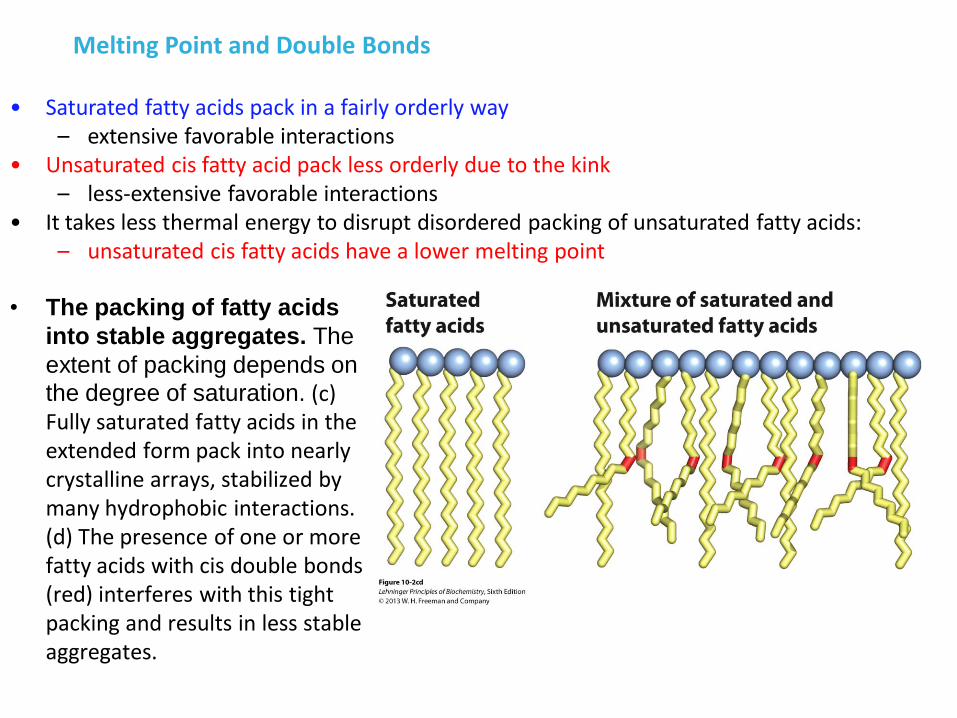
• Saturated fatty acids pack in a fairly orderly way – extensive favorable interactions
• Unsaturated cis fatty acid pack less orderly due to the kink – less-extensive favorable interactions
• It takes less thermal energy to disrupt disordered packing of unsaturated fatty acids: – unsaturated cis fatty acids have a lower melting point
• The packing of fatty acids
into stable aggregates. The
extent of packing depends on
the degree of saturation. (c) Fully saturated fatty acids in the extended form pack into nearly crystalline arrays, stabilized by many hydrophobic interactions. (d) The presence of one or more fatty acids with cis double bonds (red) interferes with this tight packing and results in less stable aggregates.

These are important food groups!
Everything taste better with bacon!!!!
• Trans fatty acids form by partial dehydrogenation of unsaturated fatty acids
– Done to increase shelf life or stability at high temperature of oils used in cooking (especially deep frying)
• A trans double bond allows a given fatty acid to adopt an extended conformation
• Trans fatty acids can pack more regularly and show higher melting points than cis forms
• Consuming trans fats increases risk of cardiovascular disease
– Avoid deep-frying partially hydrogenated vegetable oils
– Current trend: reduce trans fats in foods (Wendy’s, KFC).
GREASE FOR PEACE!

Triacylglycerols; Non-polar
• Majority of fatty acids in biological systems are found in the form of triacylglycerols
• Solid ones are called fats
• Liquid ones are called oils
• The primary storage form of lipids (body fat)
• Less soluble in water than fatty acids due to the lack of charged carboxylate group
• Less dense than water: fats and oils float
• The mixed triacylglycerol shown here has three different fatty acids attached to the glycerol backbone. When glycerol has different fatty acids at C-1 and C-3, C-2 is a chiral center

Fats Provide Efficient Fuel Storage
• The advantage of fats over polysaccharides:
– Fatty acids carry more energy per carbon
because they are more reduced
– Fatty acids carry less water per gram
because they are nonpolar
• Glucose and glycogen are for short-term
energy needs, quick delivery
• Fats are for long-term (months) energy needs,
good storage, slow delivery
• Cross section of human white adipose tissue.
Each cell contains a fat droplet (white) so
large that it squeezes the nucleus (stained
red) against the plasma membrane.

Waxes • Waxes are esters of long-chain saturated and
unsaturated fatty acids with long-chain alcohols • Insoluble and have high melting points • Variety of functions:
– Storage of metabolic fuel in plankton – Protection and pliability for hair and skin in vertebrates – Waterproofing of feathers in birds – Protection from evaporation in tropical plants and ivy – Used by people in lotions, ointments, and polishes

Structural Lipids in Membranes (Polar)
• Contain polar head groups and nonpolar tails (usually attached fatty acids)
• Diversification can come from: • modifying a different backbone • changing the fatty acids • modifying the head groups
● The properties of head groups determine the surface properties of membranes
● Different organisms have different membrane lipid head group compositions
● Different tissues have different membrane lipid head group compositions

Glycerophospholipids
• Primary constituents of cell membranes
• Two fatty acids form ester linkages with the first and second hydroxyl groups of L-glycerol-3-phosphate
• Head group is charged at physiological pH
L-Glycerol 3-phosphate, the backbone of
phospholipids. Glycerol itself is not chiral, as it has a
plane of symmetry through C-2. However, glycerol is
prochiral—it can be converted to a chiral compound by
adding a substituent such as phosphate to either of the —
CH2OH groups. One unambiguous nomenclature for
glycerol phosphate is the D, L system, in which the
isomers are named according to their stereochemical
relationships to glyceraldehyde isomers. By this system,
the stereoisomer of glycerol phosphate found in most
lipids is correctly named either L-glycerol 3-phosphate or
D-glycerol 1-phosphate. Another way to specify
stereoisomers is the sn (stereospecific numbering) system,
in which C-1 is, by definition, the group of the prochiral
compound that occupies the pro-S position. The common
form of glycerol phosphate in phospholipids is, by this
system, sn-glycerol 3-phosphate (in which C-2 has the R
configuration). In archaea, the glycerol in lipids has the
other configuration; it is D-glycerol 3-phosphate.

General Structure of Glycerophospholipids
• Unsaturated fatty acids are commonly found connected to C2
• The highly polar phosphate group may be further esterified by an alcohol; such substituent groups are called the head groups
Glycerophospholipids. The common glycerophospholipids are diacylglycerols linked to head-group alcohols through a phosphodiester bond. Phosphatidic acid, a phosphomonoester, is the parent compound. Each derivative is named for the head-group alcohol (X), with the prefix “phosphatidyl-.” In cardiolipin, two phosphatidic acids share a single glycerol (R1 and R2 are fatty acyl groups). *Note that the phosphate esters in phosphatidylinositol 4,5-bisphosphate each have a charge of about -1.5; one of their —OH groups is only partially ionized at pH 7.

Examples of Glycerophospholipids
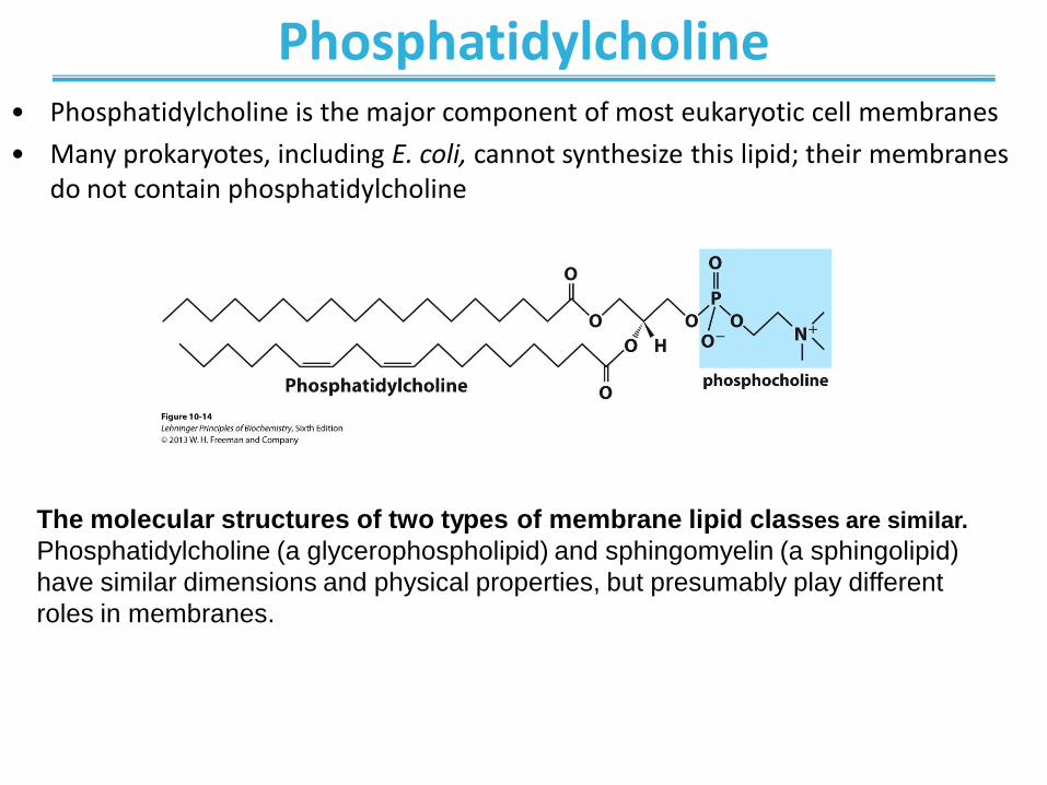
Phosphatidylcholine • Phosphatidylcholine is the major component of most eukaryotic cell membranes
• Many prokaryotes, including E. coli, cannot synthesize this lipid; their membranes do not contain phosphatidylcholine
The molecular structures of two types of membrane lipid classes are similar.
Phosphatidylcholine (a glycerophospholipid) and sphingomyelin (a sphingolipid)
have similar dimensions and physical properties, but presumably play different
roles in membranes.

Ether Lipids: Plasmalogen
Ether lipids. Plasmalogens have an ether-linked
alkenyl chain where most glycerophospholipids have
an esterlinked fatty acid. . Platelet-activating factor
has a long ether-linked alkyl chain at C-1 of glycerol,
but C-2 is ester-linked to acetic acid, which makes the
compound much more water-soluble than most
glycerophospholipids and plasmalogens. The head-
group alcohol is ethanolamine in plasmalogens and
choline in platelet-activating factor.
• Vinyl ether analog of
phosphatidylethanolamine
• Common in vertebrate heart
tissue
• Also found in some protozoa
and anaerobic bacteria
• Function is not well
understood
– Resistant to cleavage by
common lipases but
cleaved by few specific
lipases
– Increase membrane
rigidity?
– Sources of signaling
lipids?
– May be antioxidants?

Ether Lipids: Platelets-Activating Factor
• Aliphatic ether analog of phosphatidylcholine
• Acetic acid has esterified position C2
• First signaling lipid to be identified
• Stimulates aggregation of blood platelets
• Plays role in mediation of inflammation
Ether lipids Platelet-activating factor has a long ether-linked alkyl chain at C-1 of glycerol, but
C-2 is ester-linked to acetic acid, which makes the compound much more water-soluble than most
glycerophospholipids and plasmalogens. The head-group alcohol is ethanolamine in
plasmalogens and choline in platelet-activating factor.

Sphingolipids
• The backbone of sphingolipids is NOT glycerol
• The backbone of sphingolipids is a long-chain amino alcohol sphingosine
• A fatty acid is joined to sphingosine via an amide linkage rather than an ester linkage as usually seen in lipids
• A polar head group is connected to sphingosine by a glycosidic or phosphodiester linkage
• The sugar-containing glycosphingolipids are found largely in the outer face of plasma membranes

Sphingolipids
• Sphingolipids. The first three carbons at the polar end of sphingosine are analogous to the three carbons of glycerol in glycerophospholipids. The amino group at C-2 bears a fatty acid in amide linkage. The fatty acid is usually saturated or monounsaturated, with 16, 18, 22, or 24 carbon atoms. Ceramide is the parent compound for this group. Other sphingolipids differ in the polar head group (X) attached at C-1. Gangliosides have very complex oligosaccharide head groups. Standard symbols for sugars are used in this figure, as shown in Table 7–1.

Sphingomyelin • Ceramide (sphingosine + amide-linked fatty acid) +
phosphocholine attached to the alcohol
• Sphingomyelin is abundant in myelin sheath that surrounds some nerve cells in animals
The molecular structures of two types of membrane lipid classes are similar.
Phosphatidylcholine (a glycerophospholipid) and sphingomyelin (a sphingolipid)
have similar dimensions and physical properties, but presumably play different
roles in membranes.

Sphingomyelin is structurally similar to phosphatidylcholine
The molecular structures of two types of membrane lipid classes are similar. Phosphatidylcholine (a glycerophospholipid) and sphingomyelin (a sphingolipid) have similar dimensions and physical properties, but presumably play different roles in membranes.
Glycosphingolipids and Blood Groups
• The blood groups are determined in part by the type of sugars located on the head groups in glycosphingolipids.
• The structure of sugar is determined by an expression of specific glycosyltransferases
– Individuals with no active glycosyltransferase will have the O antigen
– Individuals with a glycosyltransferase that transfers an N-acetylgalactosamine group have A blood group
– Individuals with a glycosyltransferase that transfers a galactose group have B blood group

Glycosphingolipids determine blood groups
• Glycosphingolipids as
determinants of blood
groups. The human blood
groups (O, A, B) are
determined in part by the
oligosaccharide head groups
of these glycosphingolipids.
The same three
oligosaccharides are also
found attached to certain
blood proteins of individuals
of blood types O, A, and B,
respectively. Standard
symbols for sugars are used
here (see Table 7–1).
GalNAc
Gal

Defects in the turnover of membrane lipids lead to a number of diseases
1 Pathways for the breakdown of GM1, globoside, and sphingomyelin to ceramide. A defect in the enzyme hydrolyzing a particular step is indicated by ; the disease that results from accumulation of the partial breakdown product is noted.

Sterols and Cholesterol • Sterol
– Steroid nucleus: four fused rings – Hydroxyl group (polar head) in the A-ring – Various nonpolar side chains
• The steroid nucleus is almost planar
• Cholesterol. In this chemical structure of cholesterol, the rings are labeled A through D to simplify reference to derivatives of the steroid nucleus; the carbon atoms are numbered in blue. The C-3 hydroxyl group (shaded blue) is the polar head group. For storage and transport of the sterol, this hydroxyl group condenses with a fatty acid to form a sterol
ester.

Physiological Role of Sterols
• Cholesterol and related sterols are present in the membranes of most eukaryotic cells – Modulate fluidity and permeability – Thicken the plasma membrane – Most bacteria lack sterols
• Mammals obtain cholesterol from food or synthesize it de novo in the liver
• Cholesterol, bound to proteins, is transported to tissues via blood vessels – Cholesterol in low-density lipoproteins tends to deposit
and clog arteries
• Many hormones are derivatives of sterols

Steroid Hormones
• Steroids are oxidized derivatives of sterols
• Steroids have the sterol nucleus, but lack the alkyl chain found in cholesterol
• More polar than cholesterol
• Steroid hormones are synthesized from cholesterol in gonads and adrenal glands
• They are carried through the body in the bloodstream, usually attached to carrier proteins
• Many of the steroid hormones are male and female sex hormones

Steroid Hormones • Steroids derived from
cholesterol. Testosterone, the male sex hormone, is produced in the testes. Estradiol, one of the female sex hormones, is produced in the ovaries and placenta. Cortisol and aldosterone are hormones synthesized in the cortex of the adrenal gland; they regulate glucose metabolism and salt excretion, respectively. Prednisone and prednisolone are synthetic steroids used as antiinflammatory agents. Brassinolide is a growth regulator found in vascular plants.
Biologically Active Lipids
• Are present in much smaller amounts than storage or structural lipids
• Play vital roles as signaling molecules between nearby cells
• Lipid soluble vitamins (A, D, E, and K)

Arachidonic Acid Derivatives as Signaling Lipids
• Enzymatic oxidation of arachidonic acid yields
• Prostaglandins (inflammation and fever) • Thromboxanes (formation of blood clots) • Leukotrienes (smooth muscle contraction in lungs)

Vitamin D regulates calcium uptake
Vitamin D3 production and metabolism. (a) Cholecalciferol (vitamin D3) is produced in the skin by UV irradiation of 7-dehydrocholesterol, which breaks the bond shaded light red. In the liver, a hydroxyl group is added at C-25; in the kidney, a second hydroxylation at C-1 produces the active hormone, 1α,25-dihydroxyvitamin D3. This hormone regulates the metabolism of Ca2+ in kidney, intestine, and bone.

Vitamin A (Retinol)
• Involved in visual pigment
• Precursor for other hormones involved in
signaling
Vitamin A1 and its precursor and derivatives. (a) β-Carotene is the precursor of vitamin A1. Isoprene structural units are set off by dashed red lines. Cleavage of β-carotene yields two molecules of vitamin A1 (retinol) (b). Oxidation at C-15 converts retinol to the aldehyde, retinal (c), and further oxidation produces retinoic acid (d), a hormone that regulates gene expression. Retinal combines with the protein opsin to form rhodopsin (not shown), a visual pigment widespread in nature. In the dark, retinal of rhodopsin is in the 11-cis form (c). When a rhodopsin molecule is excited by visible light, the 11-cis-retinal undergoes a series of photochemical reactions that convert it to all trans-retinal (e), forcing a change in the shape of the entire rhodopsin molecule. This transformation in the rod cell of the vertebrate retina sends an electrical signal to the brain that is the basis of visual transduction.

Vitamin E, K, and other lipid quinones are antioxidants
• Some other biologically active isoprenoid compounds or derivatives. Units derived from isoprene are set off by dashed red lines. In most mammalian tissues, ubiquinone (also called coenzyme Q) has 10 isoprene units. Dolichols of animals have 17 to 21 isoprene units (85 to 105 carbon atoms), bacterial dolichols have 11, and those of plants and fungi have 14 to 24
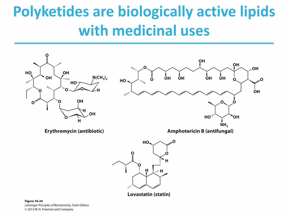
Polyketides are biologically active lipids with medicinal uses