chapter 13 perioperative pain management€¦ · clinical pharmacology of opioid analgesics opioid...
TRANSCRIPT

Perioperative Pain Management
287
Chapter 13
PERIOPERATIVE PAIN MANAGEMENT
JOHN M. STAMATOS, M.D.*; BEN H. BOEDEKER, D.V.M., M.D., PH.D.†; AND FRANK D. CROWL, M.D.‡
INTRODUCTION
PATHOPHYSIOLOGY OF PAINLocal Transmitters of PainTransmission of Pain
SECONDARY MANIFESTATIONS OF ACUTE PAINRespiratory SystemCardiovascular SystemGastrointestinal and Genitourinary SystemsEndocrine and Metabolic Systems
CLINICAL PHARMACOLOGY OF OPIOID ANALGESICSOpioid AgonistsOpioid Agonist–AntagonistsMixed AgonistsAdjunct Medications
CLINICAL PHARMACOLOGY OF NONSTEROIDAL ANTIINFLAMMATORY DRUGSMechanisms of ActionSide Effects
CLINICAL PHARMACOLOGY OF LOCAL ANESTHETICSChemical and Physical PropertiesMode of ActionFrequency-Dependent BlockadeReceptor Sites in the Sodium ChannelActions of Local Anesthetics on Nerve FibersPharmacokineticsToxicity
SELECTION OF LOCAL ANESTHETICSEstersAmides
MODE OF ADMINISTRATION OF ANALGESIC DRUGSIntramuscular and Intravenous RoutesEpidural and Intrathecal Routes
SUMMARY
*Formerly, Captain, Medical Corps, U.S. Army; Chief, Pain Management Services, Department of Anesthesiology, Walter Reed Army MedicalCenter, Washington, D. C. 20307-5001; currently, Associate Director, The New York Center for Pain Management and Rehabilitation, St.Vincent’s Hospital and Medical Center of New York, 130 West 12th Street, New York, New York 10011
†Major, Medical Corps, U.S. Army; Assistant Professor, Department of Anesthesiology, Uniformed Services University of the Health Sciences,Bethesda, Maryland 20814-4799; and Assistant Clinical Professor, Pennsylvania State University, College of Medicine, Department ofAnesthesia, Hershey Medical Center, Hershey, Pennsylvania 17033
‡Formerly, Major, Medical Corps, U.S. Army; Department of Anesthesiology, Walter Reed Army Medical Center, Washington, D. C. 20307-5001; currently, Anesthesia Department, Holy Cross Hospital, 1500 Forest Glen Road, Silver Spring, Maryland 20910

Anesthesia and Perioperative Care of the Combat Casualty
288
INTRODUCTION
Pain control has become an integral part of theanesthesiologist’s professional responsibilities. Thephysiological and humanitarian ramifications ofinadequate pain relief can be catastrophic.Postinjury pain control in past wars has not alwaysbeen optimal; in many instances, there has beennone. Every war has its own descriptions of thesuffering of untreated casualties, and it was thesound of casualties dying on the battlefield ofSolferino in 1859 that motivated Henri Dunant totake the first steps that led to the formation of theInternational Red Cross:
The stillness of the night was broken by groans, bystifled sighs of anguish and suffering. Heart-rend-ing voices kept calling for help. Who could everdescribe the agonies of that fearful night!1(p41)
More than humanitarian concerns are relevant tomanaging pain, however; postinjury or postopera-tive pain relief also blunts the trauma-induced neu-roendocrine response that leads to detrimentalmetabolic, respiratory, and cardiovascular derange-ments. Thus, effective analgesia can both make thecasualty more comfortable and decrease morbidity.
Although this chapter is oriented toward themanagement of pain in postoperative, hospitalized,combat casualties, military anesthesia providersneed to know not only the full dimensions of pro-viding analgesia to combat casualties but also whatspecifically can and should be done in the echelonsbelow the corps level. The landmark battlefieldstudy on pain in combat casualties was carried outby members of the U.S. Army Medical Corps in Italyin late 1943 and 1944.2 The impetus for this studycame from the observation that, at corps-level hos-pitals, increasing numbers of casualties were beingseen who had obvious clinical signs and symptomsof morphine overdose and even lethal poisoning.This problem was soon shown to be due to thepractice by field medical personnel of injectingmorphine—in 30-mg increments, subcutaneouslyor intramuscularly—soon after the casualty hadbeen wounded. Absorption of the morphine fromthe site of injection was sluggish because the weatherwas cold and many of the casualties were hypo-volemic or even in shock. Thus, repeated doseswere given to relieve pain. On arriving at thehospital level and with the initiation of resuscita-tion, the morphine that was deposited in peripheral
tissue was rapidly absorbed into the circulation—sometimes with fatal results.
By studying the prevalence of pain in combatcasualties, the investigators set about determininghow much morphine was really needed to obtain anacceptable degree of pain relief. On arriving at thecorps hospital, casualties were asked to describetheir pain, if any, using the following scale: none,slight, moderate, or severe. Casualties who admit-ted to pain of any degree were then asked if theywanted something to relieve it. Two hundredtwenty-five casualties, representative of the moreseverely injured, were studied. Of these, 10 weresubsequently excluded because their state of con-sciousness was altered. The remaining casualtieswere stratified into five groups according to thenature of their wounds: (1) fractures of extremitybone, (2) extensive soft-tissue injuries, (3) penetrat-ing wounds of thorax, (4) penetrating wounds ofabdomen, and (5) wounds of the head or brain orboth (Figure 13-1 and Table 13-1). Ethical consider-ations precluded a study design in which one popu-lation would serve as an unmedicated control group;thus, most casualties had received morphine beforereaching the hospital level.
Two striking findings emerged from this study.When seen at the hospital and evaluated for pain(7–14 h after their injuries, which was not a remark-ably long time by World War II standards, and atleast 5 h after last receiving morphine),
• approximately 75% of the casualties had nodesire for medication for pain relief, and
• approximately 75% of the casualties had nopain or pain that was slight to moderate inintensity.
Because the amount of morphine administeredwas not different for those with or without pain andthose who did or did not desire an analgesic, theinvestigators believed that the intensity of paincould not be explained on the basis of the amount ofmorphine administered. They concluded that
• most combat casualties do not need an anal-gesic prior to arrival at the hospital level,with the probable exception of casualtieswith abdominal wounds, in whom pain re-lief would appear to be usually indicated;and

Perioperative Pain Management
289
Degr
Fig. 13-1. When the nature of pain—assessed on admission to third-echelon hospitals—was studied in combatcasualties who were wounded in Italy during World War II, the distribution of pain intensity could be plotted as afunction of wound type. Most casualties with fractures, soft-tissue, and thoracic injuries reported little pain; mostcasualties with head wounds reported little or no pain. Only casualties with abdominal wounds demonstrated a greatneed for analgesia. Data source: Beecher HK. The control of pain in men wounded in battle. In: DeBakey ME, ed. GeneralSurgery. Vol 2. In: Coates JB Jr, ed. Surgery in World War II. Washington, DC: US Department of the Army, MedicalDepartment, Office of The Surgeon General; 1955: 4–49.
TABLE 13-1
EPIDEMIOLOGICAL ASPECTS OF A WORLD WAR II COMBAT CASUALTY PAIN STUDY
Time Since Total Dose of Time Since Last Request FurtherInjury (h) Morphine (mg) Dose (h) Pain Relief
Type of Injury (m ± sd) (m ± sd) (m ± sd) (%)
Fractured extremity bone (n = 50) 12.5 ± 1.3 27 ± 1.5 7.0 ± 0.8 22
Extensive soft-tissue extremityinjury (n = 50) 11.4 ± 1.4 27 ± 2.7 7.2 ± 0.6 18
Penetrating, of thorax (n = 50) 9.8 ± 1.0 25.0 ± 1.8 6.5 ± 0.6 20
Penetrating, of abdomen (n = 50) 7.2 ± 0.7 29.0 ± 2.2 4.8 ± 0.7 54
Head and/or brain (n = 15) 7.9 ± 1.4 19.8 ± 4.2 6.2 ± 1.5 7
Adapted from Beecher HK. The control of pain in men wounded in battle. In: DeBakey ME, ed. General Surgery. Vol 2. In: Coates JB Jr, ed.Surgery in World War II. Washington, DC: US Department of the Army, Medical Department, Office of The Surgeon General; 1955: 45.
• if morphine is required, it should be admin-istered intravenously (which, of course, maynot be practicable on the battlefield).
At the hospital level and especially followingsurgical intervention, a patient’s degree of pain is
often difficult to assess. The recovery roompersonnel’s assessment of the casualty’s pain isusually subjective. A patient’s communication skillsand cultural background will influence the abilityto express his or her level of discomfort. Severalpain scores have been developed to help eliminate
None
Slight
Moderate
Severe
Type of Injury
15
Fracture Soft Tissue Thorax Abdomen Head/Brain
10
5
0
20
25
Nu
mb
er o
f C
asu
alti
es

Anesthesia and Perioperative Care of the Combat Casualty
290
the subjectivity in the assessment of acute pain, butthese are much more useful in a research setting ora civilian hospital than on the battlefield.3
Fortunately for casualties of modern warfare,many techniques and drugs are now available thatprovide considerable analgesia. Some, such as pa-tient-controlled analgesia (PCA), will allow the ca-sualty more autonomy with respect to narcoticsadministration, which will simultaneously reducethe demand for nursing care.4,5 Administration ofnarcotics by the epidural or intrathecal routes willprovide sustained pain relief far beyond that ob-tained with the more traditional intramuscular orintravenous routes.6 By employing dilute concen-trations of local anesthetics, regional blocks can beused to provide pain relief with a minimal decre-ment of motor function.7 New nonsteroidalantiinflammatory drugs (NSAIDs), which are nearly
as effective as the narcotics used in prior wars butare not addictive, can be given intramuscularly orintravenously and act as an adjunct in the therapyof the injured soldier.8 By taking advantage of thenewer techniques, military anesthesiologists canhelp soldiers who are injured on the battlefield torecover from surgery with minimal—or at leastgreatly reduced—discomfort.
Military trauma anesthesiologists must be able tomeet the soldiers’ postoperative pain requirements,have a sound working knowledge of the drugs atour disposal, and be adept in the use of the differentmodes of delivery and administration. The battle-field is not the ideal setting in which to treat pa-tients. Therefore, we must be able to adapt to thesituation and adequately treat the casualties as theypresent, using innovative ideas and combinationsof treatment modalities to care for these soldiers.
PATHOPHYSIOLOGY OF PAIN
Visceral pain originates from organs of the ab-dominal cavity and thorax. The receptors coverlarge areas and the impulses travel through unmy-elinated sympathetic fibers. These nerves respondto stretch, crush, ischemia, and displacement. Thepain generated from these areas is dull, aching, anddifficult to localize. The normal causes of visceralpain are distention of the viscera or renal pelvissecondary to obstruction. Somatic pain, in contrast,is transmitted in unmyelinated C fibers and small,myelinated, A-delta fibers, the impulse arising inreceptors in skin, fasciae, bones, and joint spaces.These receptors innervate small discrete areas andare able to pinpoint noxious stimuli. Myelinatedsomatic nerve fibers, which are called A fibers, arethe largest in diameter and conduct impulses themost rapidly. A fibers are further divided, by theirprogressively decreasing sizes, into alpha, beta,gamma, and delta fibers. The alpha and beta fibersconvey motor and proprioception information. Thegamma fibers control muscle spindle tone. Thedelta fibers, which are the A fibers with the smallestdiameter, transmit messages concerning pain, tem-perature, and touch (Table 13-2).9
The thinly myelinated B fibers are smaller thanthe alpha fibers and have a preganglionic auto-nomic function for both sympathetic and parasym-pathetic systems.9 The unmyelinated C fibers arethe smallest diameter nerve fibers. They have thelowest rate of impulse conduction velocity and con-tain postganglionic autonomic axons as well as axonsconveying pain, temperature and touch informa-tion.10
Three major types of receptors are stimulated bynociceptive input: low-threshold mechanoreceptors,high-threshold mechanoreceptors, and polymodalnociceptors. Low-threshold mechanoreceptors re-spond to mechanical stimulation like pressure.These receptors are carried on A-delta fibers andtransmit the stimuli at about 20 m/s.11 As the inputincreases, the firing rate increases. High-thresholdmechanoreceptors respond to noxious mechanicalstimulation. As they are on low-threshold mechan-oreceptors, impulses to high-threshold mechanore-ceptors are carried on A-delta fibers. Polymodalnociceptors transmit impulses through unmyelin-ated C fibers. These receptors respond to manydifferent kinds of nociceptive input including chemi-cal, thermal, and mechanical. The receptive fieldsare larger than those of the A-delta fibers, andstimulation of these fibers lasts longer than with theother receptors.12 The generally accepted paradigmfor pain perception, modulators, and transmitters isshown in Figure 13-2.
Local Transmitters of Pain
The many different biochemical transmitters ofpain include potassium and hydrogen ions, seroto-nin, histamine, bradykinin, substance P, andleukotrienes.13 When injected locally, all these sub-stances either (a) are responsible for the transmis-sion of noxious stimulation or (b) decrease the thresh-old of the pain transmission.14
Both potassium and hydrogen ions sensitize nerveendings and are usually released from cells when

Perioperative Pain Management
291
TABLE 13-2
CLASSIFICATION OF NERVE FIBERS
Diameter ConductionFiber Type (µm) Rate (m/s) Anatomical Location Function
A
Alpha 12–20 100 Afferent to and efferent from muscles Large motor and proprioceptionand joints
Beta 5–12 30–85 Afferent to and efferent from muscles Small motor, muscle tone, touch,and joints pressure
Gamma 3–6 15–35 Efferent to muscle spindles Muscle tone
Delta 2–5 3–25 Sensory roots and afferent peripheral Sharp pain, temperature, touchnerves
B
3 3–15 Preganglionic autonomic VasomotorVisceromotorSuromotorPilomotor
C
sC 0.3–1.3 0.7–1.3 Postganglionic sympathetic VasomotorVisceromotorSuromotorPilomotor
drC 0.4–1.2 0.1–2.0 Sensory roots and afferent peripheral Dull painnerves Temperature
Touch
Data sources: (1) Raj P. Practical Management of Pain. 2nd ed. St. Louis, Mo: Mosby–Year Book; 1992: 139. (2) Barash PG, Cullen BF,Stoelting RK, eds. Clinical Anesthesia. Philadelphia, Pa: JB Lippincott; 1989: 374.
they are damaged during injury. Activity of pe-ripheral pain receptors are regulated by, amongother substances, serotonin and histamine. Seroto-nin is also a neurotransmitter for the descendinginhibitory pathways. Bradykinin, a 9–amino acidpeptide, is produced by enzymatic activity at thesite of cell breakdown. Binding sites for bradyki-nins have also been found in the dorsal columns.Substance P, an 11–amino acid peptide, is synthe-sized in the dorsal root ganglia, from where it istransferred both centrally and peripherally alongthe nerve. In the peripheral locale—a nerve end-ing—substance P activates an enzyme that cata-lyzes the formation of arachidonic acid, which, inturn, serves as the substrate of the enzymecyclooxygenase. This results in the production ofprostaglandin E2, an eicosanoid, that not only in-creases the sensitivity of nerve endings to chemicalagents and mechanical injuries but also, by itself,causes hyperalgesia.15 Substance P is also indirectlyresponsible for the vasodilation associated with
local injury.16 The leukotrienes are also derivativesof arachidonic acid, but that reaction is catalyzed bylipoxygenase. There are many different leuko-trienes, and most are associated with lowering thethreshold of the nerves associated with the trans-mission of pain.
Transmission of Pain
The events leading to the transmission of painwere first reported in 1927 and described as a tripleresponse: intense vasodilation, local edema orwheal, and secondary vasodilation spreading tothe local area.17 This response is mediated by thetransmitters of pain and stimulates the nociceptorfibers to fire. These fibers then synapse in thedorsal column of the spinal cord. There, the im-pulses are modulated and transmitted to the con-tralateral spinothalamic tract up into the cerebralcortex. The spinothalamic tract is subdivided intosubtracts, one of which, the lateral subtract (found

Anesthesia and Perioperative Care of the Combat Casualty
292
Fig. 13-2. Pain perception depends on the integrated function of three neurological pathways. First, peripheral nervesconnect pain receptors in the injured tissue with neurons in the dorsal horn of the spinal cord. Second, nerve fibers passfrom the dorsal horn neurons to the contralateral side of the cord, where they ascend to the cerebral cortex. The thirdcomponent consists of tracts that arise in the upper brain stem and descend to the dorsal horn neurons, the functionof which is thereby modulated. The ascending tract also has a component that passes to the limbic system and no doubtis important in modifying the emotional response to pain. A large number of transmitters are believed to be importantin transmitting information about pain; several of these are discussed in text. GABA: γ-aminobutyric acid; VIP:vasoactive intestinal polypeptide; CCK-8: cholecystokinin-8. Reprinted with permission from Kehlet H. Postoperativepain. In: Wilmore DW, Brennan MF, Harken AH, Holcroft JW, Meakins JL, eds. Critical Care. Vol 1. In: American Collegeof Surgeons: Care of the Surgical Patient. New York, NY: Scientific American, Inc; 1988–1993: II-12-10.
only in advanced primates), is probably responsiblefor the pinpoint perception of traumatic injury. Asthe impulses travel to the sensory tract of the cere-bral cortex, they pass through the thalamus wherethey are again modulated. Once in the sensorycortex, the painful impulse is perceived and thebody’s response is determined. Thus, the majorareas of the body in which pain is modulated are the
dorsal columns, the spinothalamic tract, the thala-mus, and the sensory cortex. The treatment of painneeds to focus on these areas.
Descending inhibitory pathways originate in thesensory cortex and terminate in the dorsal horn.These pathways are responsible for down-regula-tion of painful stimuli by releasing serotonin, nor-epinephrine, and enkephalins, which inhibit the ac-

Perioperative Pain Management
293
rally occurring endorphins and enkephalins) acti-vate both of the descending inhibitory pathwaysand bind presynaptically to the opioid receptors ofthe neurons in the dorsal horn. Opioids act toreduce the amount of substance P present.20 Localanesthetics work by blocking the axonal transmis-sion of pain.21 Antipsychotics work to increase thelevel of serotonin in the dorsal horn and thus down-regulate the pain transmissions.22 NSAIDs stop theproduction of prostaglandin E2 from prostacyclin.23
Alpha-2 adrenergic agonists inhibit substance P bystimulating presynaptic and postsynaptic spinalcord receptors, which, in turn, inhibit pain trans-mission with a mechanism of action that is differentfrom that of narcotics.24
tivity of neurons that give rise to the fibers found inthe spinothalamic tract. The release of substance Palso causes the release of vasoactive substances(serotonin, bradykinin, histamine) at the site ofinjury. These substances stimulate both a hyperemicresponse and a hyperalgesic state at the injured site.Prostaglandins that are present in the cell mem-branes are also released in response to noxiousstimuli. These substances do not themselves causepain, but they do potentiate the noxious effects ofthe other substances that are present.18
The pharmacological treatment of pain is tar-geted to work on these sites of action. Opioids (ie,any natural or synthetic drug that has morphinelikepharmacological properties,19 including the natu-
SECONDARY MANIFESTATIONS OF ACUTE PAIN
The rationale for relieving pain transcends theanesthesiologist’s professional mandate to allevi-ate suffering. Pain by itself has many deleteriouseffects, of which the inability to rest and sleep are themost obvious. Less obvious but of greater medicalimportance are such effects as pain-induced spasm ofinjured muscle, especially splinting of respiratorymuscles; reflex stimulation of the autonomic ner-vous system, which causes hypertension and therelease of catabolic hormones; and inhibition of thenormal functions of the gut and urinary tract.
Respiratory System
The respiratory system is most often affected ininjuries to the thoracic region or the upper abdo-men. Casualties who remain in pain postopera-tively splint when trying to breathe. This can causedecreased lung volumes and atelectasis, and canprogress to lung collapse and pneumonia. Therecognition of the importance of relieving pain incasualties with thoracic trauma was one of the mostimportant advances in thoracic surgery duringWorld War II:
[P]ain was an almost constant accompaniment ofany wound of the chest. The relief of pain had avital bearing on hastening recovery.… [U]ntil ithad been accomplished, the patient was unwillingto breathe deeply or cough, because of the discom-fort which followed both acts, and fluid substancestherefore accumulated in the tracheobronchial tree,which led to wet lung.25(p244)
Similar observations regarding pain in thoracic sur-gery have been made in civilian practice; pulmo-
nary function greatly improves when adequate an-algesia is provided.26
Cardiovascular System
Pain causes the amount of circulating catechola-mines to increase. The effects of catecholamines onthe cardiovascular system include increased (a) heartrate, (b) blood pressure, (c) cardiac output, and (d)systemic vascular resistance. Taken by itself, theincreased heart rate, by limiting the time availablefor diastolic flow to occur, will cause a decrease incoronary blood flow. The usual concomitant in-crease in arterial pressure and coronary vasodila-tion caused by a catechol-mediated increase incardiac metabolism negate the isolated effect ofheart rate. Thus, coronary flow increases duringtachycardia. Cardiac output may or may not change,depending on peripheral impedance. Increasedsystemic resistance will cause an increase in theworkload for the heart. Together, these factors leadto increased myocardial oxygen consumption and,therefore, to an increased risk of myocardial ischemiain patients with limited cardiac reserve.27 However,due to their age and physical condition, most in-jured soldiers should be able to tolerate this hemo-dynamic stress.
Gastrointestinal and Genitourinary Systems
Untreated pain will lead to decreased gastrointes-tinal motility with associated anorexia, nausea, andvomiting.28 This places the patient at increased riskof pulmonary aspiration, and anastomotic andincisional dehiscence, and complicates the manage-

Anesthesia and Perioperative Care of the Combat Casualty
294
TABLE 13-3
NEUROENDOCRINE AND METABOLIC RESPONSES TO TRAUMA
Response Effect Cause
Endocrine Catabolic Increased ACTH, cortisol, ADH, GH, catechol-amines, renin, angiotensin II, aldosterone,glucagon, interleukin-1
Anabolic Decreased insulin, testosterone
Metabolic
Carbohydrate Hyperglycemia, glucose intolerance, Increased hepatic glycogenolysisinsulin resistance (epinephrine, glucagon)
Increased hepatic gluconeogenesis (cortisol,glucagon, growth hormone, epinephrine,free fatty acids)
Decreased insulin secretion/action
Protein Muscle protein catabolism, increased Increased cortisol, epinephrine, glucagon,synthesis of acute-phase proteins interleukin-1
Fat Increased lipolysis and oxidation Increased catecholamines, cortisol, glucagon,growth hormone
Water and Electrolyte Flux Retention of H2O and Na+, increased Increased catecholamines, aldosterone, ADH,excretion of K+, decreased functional cortisol, angiotensin II, prostaglandins,extracellular fluid with shifts to and other factorsintracellular compartments
ACTH: adrenocorticotropic hormone; ADH: antidiuretic hormone; GH: growth hormone. Adapted with permission from CousinsM. Acute and postoperative pain. In: Wall PD, Melzac R. Textbook of Pain. New York, NY: Churchill Livingstone; 1989: Chap 18: 294.
ment of the casualty who needs to be returned to theoperating room for additional surgery. Further-more, sluggish gastrointestinal function delays theresumption of enteral nutrition. Unrelieved painhas a similar effect on the function of the urinarytract: persistent bladder retention necessitating pro-longed urinary catheterization.
Endocrine and Metabolic Systems
Increased pain causes a stress response that mani-fests as an increase in the release of endogenouscatecholamines, cortisol, and other mediators of thesympathetic response that promote harmful pro-
tein catabolism, glucose intolerance, and insulinresistance.29
An all-inclusive review of the endocrine responseto postoperative pain is not within the scope of thischapter. Suffice it to say that the endocrine re-sponse to trauma—whether surgical or otherwise—is profound. Every system in the body is affected bythese changes, most of which are secondary to cir-culating catecholamines released from the sympa-thetic nervous system (Table 13-3). Anabolism,catabolism, and substrate requirements all increase.The dual goals of postoperative pain control are thealleviation of pain and the subsequent decrease inthe levels of circulating catecholamines.
CLINICAL PHARMACOLOGY OF OPIOID ANALGESICS
The opioids, of which morphine is the prototype,are the best known and most useful narcotic analge-sics available. Morphine was first isolated fromopium—the gum obtained by drying the milky juicemade from the seedpod of the poppy plant Papaversomniferum—in 1803.19
Opioids act at two distinct anatomical sites: thesupraspinal area and the spinal cord. In thesupraspinal area, the drugs bind at receptor sites inthe periventricular and periaqueductal gray area tomodulate pain perception. In the spinal cord, theaction is in the substantia gelatinosa of the dorsal

Perioperative Pain Management
295
horn. Here, the drugs block the release of themediators of pain by binding to presynaptic recep-tors of afferent nerve terminals.20
Whether synthetic or derived from opium, thesedrugs are described by their effects at specific opioidreceptors in the central nervous system (CNS). Fourdistinct opioid receptors have been identified inhumans: mu, delta, kappa, and sigma.
The mu receptors are further subdivided into mu1 and mu 2. Mu 1 receptors are located in the spinalcord and in the supraspinal area. Stimulation ofthese receptors produces analgesia. Morphine andbeta-endorphin are agonists for this receptor. Mu 2receptors cause ventilatory depression, decreasedheart rate, euphoria, miosis, and physical depen-dence. Agonists of this group include fentanyl andmeperidine.
The delta receptors, which have no intrinsic func-tion, function as modulators of the mu receptors.Leuenkephalin is an agonist in this group.
The kappa receptors are located in the cerebralcortex and their activation causes analgesia, seda-tion, miosis, and some depression of ventilation.Agonists of this group include nalbuphine andbutorphanol.
The sigma receptors are the latest of the opioidreceptors to be described. Activation of this recep-tor stimulates dysphoria, tachycardia, and tach-ypnea. This may be the site of ketamine’s action.Naloxone is an antagonist for all the opioid re-ceptors.30
Opioid Agonists
Morphine
HO
O
N CH3
HO
23
4
5
68
9
1011
12
13
15 16
1
7
14
morphine
An opioid agonist, morphine has demonstratedmu 1 activity in the periaqueductal gray matter.These receptors modulate noxious stimuli and sendtransmissions to the medullary nuclei. Spinal mureceptors also reduce the amount of substance P inthe dorsal horn.
Morphine can be administered via the intrave-nous, intramuscular, epidural, and intrathecalroutes. When delivered intravenously, morphinehas a peak effect about 20 minutes after injection.Intramuscularly, an initial effect is seen in 15 to 30minutes and the peak effect in 45 to 90 minutes.Orally administered morphine is not reliably ab-sorbed. When given epidurally, the onset of painrelief tends to occur approximately 60 minutes later,and pain relief can last 16 to 24 hours. Withintrathecal injections, the onset of pain relief tendsto occur in approximately 10 to 15 minutes, whichcoincides with the peak level of drug in the cere-brospinal fluid.30 Regardless of the mode of admin-istration, the pain relief is relative to the concentra-tion of the drug in the cerebrospinal fluid; painrelief is not always proportional to the blood lev-els.31 The drug is eliminated from the body prima-rily by conjugation with glucuronic acid at hepaticand extrahepatic sites. A small amount of the doseis excreted unchanged in urine. Only 7% to 10% ofmorphine is eliminated by biliary excretion; the restis excreted by the kidneys.30
Respiratory Side Effects. Mu 2 receptors in thepontine and medullary centers are responsible forrespiratory depression.32 The agonists produce adose-related depression of respiration, the evidenceof which is increased PaCO2. When morphine isadministered in small doses, the respiratory rateusually decreases, which is compensated for by anincrease in the tidal volume. With increasing dosesof morphine, however, the tidal volume will de-crease. Maximal respiratory depression occurs ap-proximately 7 minutes after intravenous adminis-tration, 30 minutes after intramuscular injection, 90minutes after subcutaneous administration, and upto 16 hours after epidural or intrathecal adminis-tration.30
Cardiovascular Side Effects. Morphine adminis-tration will decrease sympathetic outflow. No ad-verse effect will probably be seen in a supine,normovolemic patient, but a casualty who is stand-ing or a supine casualty who is hypovolemic mightsuffer from hypotension. Morphine can also stimu-late histamine release, causing peripheral vasodila-tion and hypotension. The histamine release can beattenuated by limiting the rate of infusion to lessthan 5 mg/min or by administering H1 or H2 block-
Morphine, the major phenanthrene alkaloid ofopium, and codeine are the medically useful opi-oids derived from opium. Although codeine can beadministered orally, it is not as potent as morphineand therefore will not be further discussed in thischapter.

Anesthesia and Perioperative Care of the Combat Casualty
296
CH2 H2
C N N C
O
C2H5
fentenyl
ing agents.33 Morphine can also cause bradycardia,probably by stimulating the vagal nuclei in themedulla, and by exerting a depressing effect on thesinoatrial node. Although there are no known di-rect myocardial depressant effects of morphine, itcauses a decrease in myocardial oxygen consump-tion. The mechanism is probably related to periph-eral vasodilation and bradycardia. There is nodirect effect on the cerebral circulation, but thesecondary effect of the increased PaCO2 due to asso-ciated hypoventilation will cause cerebral vasodila-tion that could thereby increase intracranial pres-sure in the patient with neurotrauma.
Gastrointestinal Side Effects. Pain may causedecreased gastric emptying and motility. Mor-phine may enhance this dysmotility and lead toileus. In addition, the tone of the ileocecal and analsphincters is also increased. All these factors lead todecreased motility of and increased water absorp-tion from the gut. Opioids will also cause spasm inthe biliary smooth muscle (eg, the sphincter of Oddi).This spasm may mimic the pain of angina pectoris.
Nausea and vomiting are caused by direct stimu-lation of the chemoreceptor trigger zone of themedulla oblongata. This side effect can be treatedwith an antiemetic without reversing the analgesiceffect of the morphine.
Genitourinary Side Effects. Morphine will causeincreased peristalsis of the ureters, but the tone ofthe vesicle sphincter is also increased. The combi-nation of these effects makes it more difficult forpatients who are not catheterized to urinate. Insome patients, morphine may also inhibit the urgeto urinate.
Cutaneous Side Effects. Morphine will causevenodilation in the peripheral vessels of the ex-tremities. The histamine release can also cause anerythema.
Pruritus, which is more pronounced in epiduraland intrathecal use, can be pronounced and is sec-ondary to a CNS effect of the drug. Pruritus can bereversed by titrating small amounts of an antago-nist (eg, naloxone).30
Physical Dependence. All opioids can causephysical dependence. This complication occursmore often with agonists than with drugs that havecombined agonist–antagonist effects. Although itusually takes longer, dependence can occur after asfew as 72 hours of use. However, the possibility ofdependence should not be a factor in limiting theuse of a narcotic in the acutely injured soldier.
Dosage and Administration. For postoperativepain control, morphine should be titrated for effect.For intravenous or intramuscular injections, 0.05 to
0.1 mg/kg should provide adequate analgesia.Following this dose, the patient should be moni-tored for adequacy of pain control, changes in bloodpressure and pulse, and respiratory rate. If thepatient’s pain is not controlled, then more narcoticcan be administered; again, the patient’s vital signsshould be observed. When used intravenously orintramuscularly, morphine should be given every 3to 6 hours.34 Strict adherence to dosing intervals isneeded to maintain therapeutic drug levels.
Epidurally or intrathecally administered mor-phine can provide pain relief for 16 to 24 hours witha single dose. It is important to remember thatmorphine’s respiratory depression might not mani-fest itself for up to 20 hours after an injection. Foracute pain, the doses of epidural morphine are 0.05to 0.07 mg/kg, and can be administered every 12hours as needed.35 The anesthesiologist must bearin mind that morphine delivered via the epiduralroute requires up to 1 hour before the onset of painrelief. Morphine can be continuously infused at arate of 0.005 to 0.01 mg/kg/h. Intrathecal narcoticsalso have the benefit of extended pain relief from asingle injection. The usual dose of intrathecal mor-phine is 0.3 to 1 mg. The onset of action is almostimmediate and the duration of a single injection is12 to 24 hours.6,35–37
Fentanyl
The opioid fentanyl is chemically similar to mor-phine. However, this synthetic narcotic is 75- to125-fold more potent than morphine and, due to itshigh lipid solubility, it crosses the blood–brain bar-rier faster than morphine. The onset of action isapproximately 30 seconds. The duration of actionof the drug is also shorter than that of morphine,owing to fentanyl’s rapid uptake by inactive tissue.Because of this uptake and subsequent release, whenfentanyl has been given in repeated doses, its actioncan continue and be prolonged after the medicationhas been discontinued.38 Fentanyl is metabolizedby dealkylation, hydroxylation, and amide hydroly-sis into inactive metabolites. The drug is excreted inthe urine and feces.

Perioperative Pain Management
297
N CH3
H5C2 CO
O
meperdine
Side Effects. The side effects of fentanyl aresimilar to those of morphine. Of note, less hista-mine is released than with morphine, but there isgreater potential for physical dependence. Muscu-lar rigidity, specifically of the chest wall, has alsobeen associated with rapidly administered doses offentanyl.30
Dosage and Administration. For the postopera-tive patient with acute pain, 1 to 2 µg/kg of fentanylwill give fast relief. Fentanyl’s advantage overmorphine is its speed of onset. However, the painrelief will not last as long as that from a dose ofmorphine, and fentanyl probably is not the bestmedication if prolonged use of intermittent bolusesis anticipated. However, fentanyl can be given as anintravenous infusion to maintain pain relief. Astarting dose of 1 to 2 µg/kg/h is adequate, but canbe titrated as necessary.30 Epidural administrationof fentanyl is another useful option. A single injec-tion of 1 to 2 µg/kg in the lumbar epidural spacewill give excellent relief from pain that originates inthe abdomen or lower extremities. Long-term reliefcan be achieved with 10 µg/mL of fentanyl infusingat a rate of 1 to 2 µg/kg/h.39,40 As with epiduraladministration of morphine, the patient’s respira-tory rate must be monitored throughout the dura-tion of infusion, although the delayed respiratorydepression seen with morphine has not been dem-onstrated with fentanyl. Intrathecal fentanyl canalso be used for pain from the abdomen and lowerextremities. A dose of 0.1 to 0.2 µg/kg is adequate.The risk for respiratory depression lasts approxi-mately 4 hours—approximately as long as the painrelief from a single dose.39,40
Meperidine
Although it is structurally different from mor-phine, meperidine has many of the same properties.Meperidine is approximately one tenth the strengthof morphine when given as an intravenous or intra-muscular injection, but is much better absorbed bythe gastrointestinal system. Analgesic effects arenoted approximately 15 minutes after an oral doseand 10 minutes after an intramuscular injection.
The peak effect is usually seen within 2 hours afteroral administration and 1 hour after an intramuscu-lar injection. The effective length of analgesia is 2 to4 hours. Meperidine is metabolized in the liver tonormeperidine. The accumulation of the normeper-idine is associated with increased CNS excitation.In large or frequently repeated doses, meperidinehas caused seizures in humans. Normeperidine isexcreted renally and will accumulate in patientswith renal failure.30
Side Effects. Meperidine’s side effects, likefentanyl’s, are similar to morphine’s, but with somedifferences. Large doses exert a myocardial depres-sant effect that will decrease myocardial contractil-ity and stroke volume. This will lead to increasedleft ventricular filling pressures.41 The incidence ofnausea and vomiting associated with meperidine isequivalent to that of morphine, but patients whoexperience nausea and vomiting with morphinemight not with meperidine.
Dosage and Administration. As mentionedabove, meperidine is approximately one tenth aspotent as morphine when given by intravenous orintramuscular injection. Thus, for acute post-op-erative pain relief, a starting dose of 0.25 to 0.5 mg/kg should be adequate. Also, owing to meperidine’sbetter absorption, it is a good choice as an oraladjunct. The possibility for seizures exists withprolonged or high-dosage oral use of meperidine.30
Opioid Agonist–Antagonists
Butorphanol
Butorphanol exerts its analgesic effect by beingan agonist of both the kappa and sigma receptors.It is an antagonist of the mu receptors. A dose of 2to 3 mg is equipotent to 10 mg of morphine. Theonset, peak effect, and duration of action ofbutorphanol are similar to those of morphine. Thera-peutic dosages produce increased pulmonary ar-tery pressure, cardiac output, and systemic bloodpressure.34
Side Effects. In the low-dose, therapeutic range,the respiratory side effects of butorphanol are simi-lar to those of morphine. The cardiovascular effectsare the same as those described for meperidine. Thedrug will cause sedation, nausea, and diaphoresisin most people, and dysphoria in a small number ofpatients. Butorphanol will also antagonize mu ago-nists, thus making the choice of the proper dose ofagonists difficult in the patient who is receivingbutorphanol and will be returning to the operatingroom.
Anesthesia and Perioperative Care of the Combat Casualty
298
Dosage and Administration. Butorphanol is notavailable for enteral use. The suggested intrave-nous dose is 7 to 15 µg/kg, and the suggestedintramuscular dose is 15 to 30 µg/kg. Butorphanolshould be readministered every 3 to 4 hours.34
Nalbuphine
Nalbuphine is an agonist at the kappa and sigmareceptors and an antagonist at the mu receptors. Adose of 10 mg of nalbuphine is equipotent to 10 mgof morphine. Unlike butorphanol, nalbuphinedoes not increase cardiac output or blood pressure.One advantage of this drug is its ceiling effect onrespiratory depression42: the dose-related decreasein respiratory drive will not worsen with dosesgreater than 30 mg. Also, because nalbuphine is amu antagonist, the drug can be used to reverse therespiratory depression of morphine or fentanyl with-out decreasing analgesia. The onset, peak effect,and duration of action are similar to those of mor-phine.
For postoperative pain relief, nalbuphine shouldbe titrated for effect. For intravenous or intramus-cular injections, 0.05 to 0.1 mg/kg should provideadequate analgesia. Whether administered via theintravenous or intramuscular route, nalbuphineshould be given every 3 to 6 hours for pain control.30
Mixed Agonists
Dezocine
Dezocine is a new, mixed agonist with activity atthe mu, kappa, and delta receptors. Its affinity forthe mu receptors is 10-fold greater than for the deltareceptors, and 40-fold greater than for the kappareceptors. This drug’s advantage over other opio-ids is its ceiling effect on respiratory depression.Additionally, it is not a scheduled drug (ie, a federalnarcotics license is not required). Its physical de-pendence has not been described. The hemody-namic effects are equivalent to those of morphine.The onset, peak effect, and duration of action arealso similar to those of morphine.43 Minimal sideeffects have been found in the clinical studies so far
reported, but the number of patients reported is small.The recommended dose for dezocine is 70 to 150
µg/kg intravenously and 70 to 200 µg/kg intramus-cularly. It is suggested that dezocine be readmin-istered every 3 to 4 hours.43
Naloxone
Naloxone is a competitive inhibitor of the mu,delta, kappa, and sigma receptors. After an intrave-nous injection, the onset of action is 1 to 2 minutes.The duration of action is 1 to 4 hours but is dosedependent. Naloxone can reverse the effects of allopioid agonists. It can also cause increased heartrate and systolic blood pressure. Acute pulmonaryedema has been associated with narcotic reversalwith naloxone, although this is a relatively rarecomplication. The case reports of pulmonary edemafollowing naloxone administration have been inyoung healthy patients, and the reported doseshave been as low as 80 µg.44,45
The recommended dose is 0.5 to 1 µg/kg, withdoses being repeated until the desired effect isachieved. If the opioid being reversed has a longerhalf-life than naloxone, then it is important to starta naloxone infusion. The recommended rate ofinfusion is 5 to 10 µg/kg/h, but this rate can betitrated for effect.46,47
Adjunct Medications
Promethazine is an H1 receptor antagonist thathas been used in conjunction with narcotics to re-duce the narcotic-associated nausea. The recom-mended dose to control nausea in patients is 12.5 to25 mg every 4 to 6 hours. The dose is given as anintramuscular injection.
Droperidol is a powerful antiemetic owing to itsinhibition of the dopaminergic receptors in thechemoreceptor trigger zone of the medulla. A doseof 0.625 to 1.25 mg given to a 70-kg adult is consid-ered adequate to prevent nausea. Side effects of thedrug include drowsiness and, in a small number ofpatients, extrapyramidal reactions. Droperidol mayalso cause increased sedation in the patient emerg-ing from anesthesia.
CLINICAL PHARMACOLOGY OF NONSTEROIDAL ANTIINFLAMMATORY DRUGS
Nonsteroidal antiinflammatory drugs (NSAIDs)act by interrupting the inflammatory response.48
This interruption is thought to be due to their inhi-bition of prostaglandin biosynthesis. Aspirin (ace-
tylsalicylic acid) is the prototype of the nonopioidanalgesic, antipyretic, and antiinflammatorydrugs.49
NSAIDs are most often used

Perioperative Pain Management
299
Generic Name Trade Name Common Adult Dose for Pain
Salicylates
Aspirin 325–650 mg/4 h PO
Choline/magnesium trisalicylate Trilisate 1,000 mg, PO bid
Diflunisal Dolobid 500 mg PO q 12 h
Salsalate Disalcid 3,000 mg/d total dose divided as tid or bid
Propionic Acid Derivatives
Fenoprofen Nalfon) 200 mg qid or tid
Flurbiprofen Ansaid 200–300 mg total daily dose divided bid, tid, or qid
Ibuprofen Advil, Medipren, 300–800 mg tid to qid POMidol, Nuprin,Motrin
Ketoprofen Orudis 25–50 mg tid to qid PO
Naproxen Naprosyn 250 mg tid to qid PO
Naproxen sodium Anaprox 275 mg tid to qid
Indoles/Pyrroles
Indomethacin Indocin 25–50 mg bid to tid
Sulindac Clinoril 150 mg bid
Tolmetin Tolectin 400 mg tid
Ketorolac Toradol 30–60 mg IM loading dose, then 15–30 mg IM qid
Oxicams
Piroxicam Feldene 20 mg qid
Anthranilic Acid Derivatives
Meclofenamate sodium Meclomen 50 mg tid to qid
Mefenamic acid Ponstel 250 mg qid
Phenylacetic Acid Derivatives
Diclofenac sodium Voltaren 50 mg tid or qid or 75 mg bid
Pyrazolone Derivatives
Phenylbutazone Butazolidin 100 mg qid
TABLE 13-4
SPECIFIC NONSTEROIDAL ANTIINFLAMMATORY AGENTS BY CLASS
• as analgesics, to provide symptomatic reliefof low-intensity pain associated with head-ache and musculoskeletal disorders (eg,osteoarthritis and rheumatoid arthritis),
• as antipyretics,• to inhibit platelet aggregation in patients
vulnerable to vascular obstruction fromemboli,
• to inhibit synthesis of prostaglandins in neo-
nates to evoke closure of the ductus arterio-sus, and
• to treat excessive production of prostaglan-dins (eg, in Bartter’s syndrome).49
Important information regarding generic names,trade names, and adult dosages of selected NSAIDsis summarized in Table 13-4; pharmacokinetic dataare presented in Table 13-5.
IM: intramuscularly; PO: by mouth; bid: twice daily; tid: three times daily; qid: four times dailyData sources: (1) Physicians’ Desk Reference. 48th ed. Montvale, NJ: Medical Economics Data; 1994. (2) DiGregorio G. Handbook of PainManagement. 3rd ed. West Chester, Pa: Medical Surveillance Inc; 1991.

Anesthesia and Perioperative Care of the Combat Casualty
300
TABLE 13-5
PHARMACOKINETIC DATA OF SELECTED NONSTEROIDAL ANTIINFLAMMATORY DRUGS
Time to Peak Plasma Elimination Duration of Effective PlasmaName Concentration (h) Half-life (h) Action (h) Concentration (µg/mL)
Aspirin 0.5–1 0.25–0.35 4–6 20
Choline/magnesium 1–2 9–17 12 150–300
Ibuprofen 1–2 2 4–6 —*
Indomethacin 2 2–3 4–6 0.3–1.0
Ketorolac 0.5 4–6 4–6 —
Sulindac 2 7–18 8–12 —
Piroxicam 3–5 30–80 48–72 1.5–2
Meclofenamate sodium 0.5–1 2 2 —
Diclofenac sodium 1–4 2 2 1–2
Phenylbutazone 1–3 60–100 80 50–150
Naproxen 1–2 10–17 8–12 25–75
*[—] Indicates data not availableAdapted with permission from Digregorio GJ, Barbieri EJ, Sterling GH, Camp JF, Prout MF. Handbook of Pain Management. 3rd ed.West Chester, Pa: Medical Surveillance Inc; 1991: 129.
Mechanisms of Action
The primary actions of NSAIDs—producing an-algesia, acting as an antipyretic, and exerting anantiinflammatory effect—result from differentmechanisms. These actions and mechanisms arediscussed below.
Analgesia
NSAIDs produce analgesia by inhibiting the ac-tivity of cyclooxygenase (prostaglandin synthetase),an enzyme that leads to a decrease in the synthesisand release of prostaglandins from cells. Indi-vidual classes of NSAIDs have different methods ofinhibiting the cyclooxygenase enzyme.49 Prostag-landins enhance the potency of algesic substancessuch as bradykinin or substance P, which may stimu-late nerve endings of unmyelinated C fibers andsmall-diameter A-delta fibers to elicit noxious af-ferent input. Bradykinin stimulates formation andrelease of more prostaglandins.50 NSAIDs appearto reduce this potentiation of algesic activity ofprostaglandins by blocking their synthesis.
In contrast to that produced by opioids, whichact centrally, the analgesia produced by aspirinlikedrugs is a peripheral phenomenon. Pain is pro-
duced when prostaglandins are produced locallyaround sensitive nerve endings, as seen with in-flammation. NSAIDs are effective in relieving thispain by blocking prostaglandin synthesis. NSAIDsare not effective as analgesics when inflammation isnot present (ie, there is no increase in prostaglandinsynthesis in the area) or for the sharp, stabbing painthat is caused by direct stimulation of sensory nerves(ie, visceral pain).49
Antipyresis
NSAIDs exert an antipyretic action via a CNS mech-anism. Fever is thought to occur because endog-enous pyrogens cause a shift in the temperature-regulating system in the preoptic hypothalamus.NSAIDs are postulated to reduce the effect of en-dogenous pyrogens on the hypothalamus. In thera-peutic doses, NSAIDs do not affect either normalbody temperature or an increase in temperatureassociated with exercise, drugs, or hypothalamiclesions to which pyrogen does not contribute. Sali-cylates, acetaminophen, and ibuprofen are the onlydrugs approved in the United States as antipyretics,although indomethacin and naproxen have beenrecommended as drugs to lower fever of neoplasticdisease that is uncontrolled by other antipyretics.50

Perioperative Pain Management
301
Antiinflammatory Effect
Since prostaglandins induce symptoms of in-flammation and enhance the effects of bradykininand histamine, a reduction of prostaglandins atsites of inflammation is thought to decrease theinflammatory response. In arthritic diseases,NSAIDs reduce inflammation and swelling, therebyproviding relief.50
Side Effects
Gastrointestinal System
All NSAIDs can cause gastrointestinal pathol-ogy, including gastritis, nausea, vomiting, diarrhea,constipation, and occult blood in the stool.48,49 Gas-trointestinal problems are more common withaspirin, indomethacin, and mefenamic acid49 al-though nonacetylated salicylates may be less irri-tating.50 Alcohol ingestion increases the likelihoodthat gastrointestinal side effects will occur.49 TheUnited States Pharmacopeia recommends that allNSAIDs be taken with a full glass of water.49
NSAID-associated gastropathy has been proposedas the most frequent, serious, adverse drug effectin the United States; the risk of gastrointestinalproblems is greater in elderly and debilitated pa-tients.50
Aspirin should be avoided by patients with ahistory of ulcers and should not be used with agentsthat promote ulcer formation (eg, alcohol). Themechanisms of gastrointestinal bleeding andNSAID-induced ulceration are due in part to irrita-tion from direct contact with mucosal cells andcyclooxygenase inhibition after absorption, whichweakens the gastric-acid barrier. Chronic inhibi-tion of prostaglandin synthesis appears to decreasemucosal cytoprotection. It is likely that aspirin-induced bleeding is aggravated by its effect onplatelet aggregation.50
Renal System
Renal prostaglandins are important to ensureadequate renal perfusion and function in certaindisease states, including congestive heart failure,cirrhosis with ascites, nephrotic syndrome, anddehydration. In these patient groups, NSAIDs mayprecipitate acute renal failure. Long-term use ofhigh-dose NSAIDs may cause renal medullaryischemia and papillary necrosis. This was morecommon when phenacetin was used, especially incombination with aspirin, caffeine, and acetami-
nophen. Phenacetin has been replaced with ac-etaminophen, which may also be a risk factor forpapillary necrosis. Renal prostaglandins inhibittubular reabsorption of Na+ and water, and inhibitrenin secretion. In patients with poor renal perfu-sion states, NSAIDs may cause fluid retention, im-paired responsiveness to diuretic therapy, andhyperkalemia.50
Respiratory System
Inhibition of bronchodilator prostaglandins byNSAIDs may leave the leukotriene pathway uncon-tested and lead to bronchospasm.49 This may be aconsideration in susceptible patients such as asth-matics. Full therapeutic doses of salicylates in-crease oxygen consumption and carbon dioxideproduction in skeletal muscle. Higher doses resultin medullary stimulation, causing hyperventilationwith a respiratory alkalosis. This is compensatedfor by excretion of bicarbonate by the kidneys, whichleads to a compensated respiratory alkalosis. Toxicdoses of salicylate depress the medulla, leading toan uncompensated respiratory acidosis in a patientwith high renal bicarbonate excretion, which, inturn, leads to a coexisting metabolic acidosis.48
Hematological Effects
NSAIDs inhibit both thromboxane A2 synthesiswithin platelets and prostacyclin synthesis withinendothelial cells. Thromboxane A2, a potent vaso-constrictor, stimulates platelet aggregation, whichpromotes clotting. Prostacyclin, a potent vasodila-tor, inhibits platelet aggregation, which promotesbleeding. NSAIDs inhibit the synthesis of boththromboxane A2 and prostaglandin I2. The netbalance determines the tendency towards bleedingor clotting.51
Aspirin irreversibly inhibits platelet cyclooxy-genase which prevents the formation of thrombox-ane A2, thereby inhibiting platelet aggregation. Thebleeding time is prolonged for the lifetime of theplatelet. All other NSAIDs reversibly inhibit plate-let cyclooxygenase, so that five half-lives are re-quired to clear all the drug from the body andrestore normal platelet function.51
Reye’s Syndrome
Reye’s syndrome, a childhood illness that devel-ops after recovery from influenza or chickenpox, ischaracterized by vomiting, liver abnormalities, andencephalopathy. Nearly all victims of Reye’s syn-

Anesthesia and Perioperative Care of the Combat Casualty
302
drome had received salicylates for management ofthe viral illness. If an analgesic antipyretic is re-quired for use in children with acute viral illness,
acetaminophen should be used, as there is no evi-dence of an association between acetaminophenand Reye’s syndrome.50
CLINICAL PHARMACOLOGY OF LOCAL ANESTHETICS
Since prehistoric times, the natives of Peru havechewed the leaves of the indigenous plantErythroxylon coca, the source of cocaine, to obtain afeeling of well-being and to reduce fatigue.9,52 Chew-ing of the coca leaves caused a reversible perioralnumbness, and subsequent investigation of the cocaleaf allowed the identification of cocaine, the activeingredient. Cocaine was used as a topical ocularanesthesia and for nerve blocks during the latter18th century.49 Addictive properties and toxicitylimited the use of cocaine for local anesthesia, butthis finding stimulated research for more ideal localanesthetic agents. Einhorn introduced the estersprocaine in 1905 and tetracaine in 1932, and Lofgrenintroduced lidocaine, an amide, in 1932.9
Local anesthetics are important adjuncts to themanagement of postoperative pain; they signifi-cantly reduce the amount of narcotics necessary tomaintain a pain-free state in the postoperative pa-tient.7 A simple system governs the naming of localanesthetic drugs: those spelled with one i in thename are esters (eg, procaine, 2-chloroprocaine,tetracaine); those with two are amides (eg, lidocaine,mepivacaine, bupivacaine). Among the amides,stereoisomers of etidocaine, mepivacaine, bupiva-caine, prilocaine, and ropivacaine have been recog-nized. Although the S forms of these molecules aregenerally less toxic and have a longer duration ofaction,53 the chiral forms of local anesthetics are notdiscussed in this chapter.
Very dilute solutions of a local anesthetic com-bined with an opiate produce anesthesia compa-rable to that produced by more-concentrated solu-tions of plain local anesthetics. Use of dilute solutionof local anesthetic causes less motor blockade, whichfacilitates postoperative ambulation. The interac-tion of opiates and local anesthetics is postulated tobe due to a synergistic action of the drugs (admin-istered epidurally) acting in two or more differentsites to decrease sensory input. The local anesthet-ics act at the dorsal root ganglion while the opiatesact on the dorsal horn of the spinal cord.53
Chemical and Physical Properties
The potency, onset of action, and duration oflocal anesthetic action are directly related to thephysical properties of lipid solubility, pKa (ie, ion-
ization), and protein binding (Figure 13-3).53 Localanesthetics consist of a lipophilic, unsaturated, ben-zene-ring aromatic group; and a hydrophilic, ter-tiary amine separated by a hydrocarbon connectingchain (Figure 13-4). Clinically useful local anes-thetic agents fall into two chemically distinct groups,based on the linkage between the aromatic portionand the intermediate chain.52 This linkage can bevia an ester (–CO–) or an amide (–HNC–) bond. Theester and amide compounds differ in their chemicalstability, metabolic cycles, and allergic potentials.
In general, the greater the length of the connect-ing group and the more complicated the aromaticand amine structures, the greater the potency andthe toxicity of the local anesthetic.48 The addition ofmore complicated side chains to local anestheticscauses an increase in their tissue-protein bindingability and also makes the drug more fat soluble,thereby increasing the duration of action.49
Lipid Solubility
The aromatic group (ie, the benzene ring) that ispresent at one end of the molecule is the majordeterminant of the lipid solubility of local anes-thetic drugs. The more-lipid-soluble drugs are themore-potent local anesthetics, probably because thelipid-soluble uncharged base form can pass throughthe lipid-containing nerve membrane to reach itssite of action.53
Fig. 13-3. The general structure of a local anestheticmolecule.
N H+N H+
Intermediate chain(Ester or amide linkage)
Benzene ring(Lipophilic)
Quaternary amine(Hydrophilic)

Perioperative Pain Management
303
Cocaine
2-Chloroprocaine
Tetracaine
Lidocaine
Prilocaine
Etidocaine
Mepivacaine
Bupivacaine
Ropivacaine
Fig. 13-4. Chemical structures of commonly used local anesthetics.
Procaine H2N C
O
O C N
H
H
C2H5
C2H5
C
H
H
N
H
C C
H
H
N
OC2H5
C2H5
CH3
N
H
C C
H
CH3
N
OH
C3H7
CH3
N
H
C C
OCH3
CH3
H
C2H5
N
C2H5
C3H7
N
H
C
OCH3
CH3
N
CH3
N
H
C
OCH3
CH3
N
C4H9
Amino Amides
Amino Esters
C
H
O C
CC
C
OO
H3C
C
CH
C
CN CH3
C
O
O C N
H
H
CH3
CH3
C
H
H
NH9C4
H
C
O
O C N
H
H
C2H5
C2H5
C
H
H
NH
HCl
N
H
C
OCH3
CH3
N
C3H7
Anesthesia and Perioperative Care of the Combat Casualty
304
Ionization
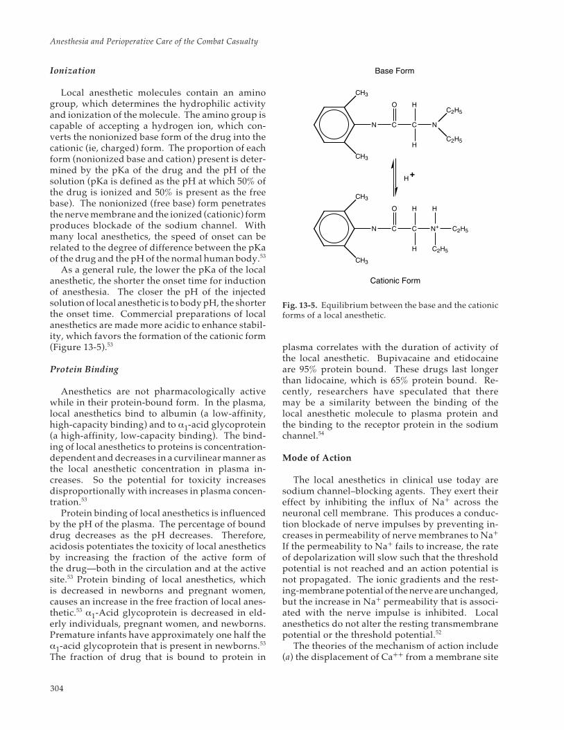
Local anesthetic molecules contain an aminogroup, which determines the hydrophilic activityand ionization of the molecule. The amino group iscapable of accepting a hydrogen ion, which con-verts the nonionized base form of the drug into thecationic (ie, charged) form. The proportion of eachform (nonionized base and cation) present is deter-mined by the pKa of the drug and the pH of thesolution (pKa is defined as the pH at which 50% ofthe drug is ionized and 50% is present as the freebase). The nonionized (free base) form penetratesthe nerve membrane and the ionized (cationic) formproduces blockade of the sodium channel. Withmany local anesthetics, the speed of onset can berelated to the degree of difference between the pKaof the drug and the pH of the normal human body.53
As a general rule, the lower the pKa of the localanesthetic, the shorter the onset time for inductionof anesthesia. The closer the pH of the injectedsolution of local anesthetic is to body pH, the shorterthe onset time. Commercial preparations of localanesthetics are made more acidic to enhance stabil-ity, which favors the formation of the cationic form(Figure 13-5).53
Protein Binding
Anesthetics are not pharmacologically activewhile in their protein-bound form. In the plasma,local anesthetics bind to albumin (a low-affinity,high-capacity binding) and to α1-acid glycoprotein(a high-affinity, low-capacity binding). The bind-ing of local anesthetics to proteins is concentration-dependent and decreases in a curvilinear manner asthe local anesthetic concentration in plasma in-creases. So the potential for toxicity increasesdisproportionally with increases in plasma concen-tration.53
Protein binding of local anesthetics is influencedby the pH of the plasma. The percentage of bounddrug decreases as the pH decreases. Therefore,acidosis potentiates the toxicity of local anestheticsby increasing the fraction of the active form ofthe drug—both in the circulation and at the activesite.53 Protein binding of local anesthetics, whichis decreased in newborns and pregnant women,causes an increase in the free fraction of local anes-thetic.53 α1-Acid glycoprotein is decreased in eld-erly individuals, pregnant women, and newborns.Premature infants have approximately one half theα1-acid glycoprotein that is present in newborns.53
The fraction of drug that is bound to protein in
Fig. 13-5. Equilibrium between the base and the cationicforms of a local anesthetic.
plasma correlates with the duration of activity ofthe local anesthetic. Bupivacaine and etidocaineare 95% protein bound. These drugs last longerthan lidocaine, which is 65% protein bound. Re-cently, researchers have speculated that theremay be a similarity between the binding of thelocal anesthetic molecule to plasma protein andthe binding to the receptor protein in the sodiumchannel.54
Mode of Action
The local anesthetics in clinical use today aresodium channel–blocking agents. They exert theireffect by inhibiting the influx of Na+ across theneuronal cell membrane. This produces a conduc-tion blockade of nerve impulses by preventing in-creases in permeability of nerve membranes to Na+
If the permeability to Na+ fails to increase, the rateof depolarization will slow such that the thresholdpotential is not reached and an action potential isnot propagated. The ionic gradients and the rest-ing-membrane potential of the nerve are unchanged,but the increase in Na+ permeability that is associ-ated with the nerve impulse is inhibited. Localanesthetics do not alter the resting transmembranepotential or the threshold potential.52
The theories of the mechanism of action include(a) the displacement of Ca++ from a membrane site
N
CH3
CH3
CH3
N
CH3
C
C
O
O
C
C
H
H
N
H
H
N+
C2H5
C2H5
H
C2H5
C2H5
Base Form
Cationic Form
H+

Perioperative Pain Management
305
that controls Na+ permeability, (b) the Meyer-Overton rule of anesthesia, (c) a change in surfacecharge, and (d) the specific-receptor theory.
Displacement of Calcium
A low calcium concentration outside the neuronenhances local anesthetic activity, while an increas-ing external calcium concentration antagonizes lo-cal anesthetic activity. However, the direct actionsof calcium and local anesthetic appear to be inde-pendent of each other.9
Meyer-Overton Rule
The Meyer-Overton rule of anesthesia postulatesthat diffusion of the relatively lipophilic anestheticmolecules into the lipid component of the neuronalmembrane expands the membrane to a critical vol-ume and interferes with sodium conductance. Lo-cal anesthetics have been shown to increase thevolume of lipid membranes and increase their de-gree of disorder. High-pressure antagonism of theanesthetic activity of certain uncharged local-anes-thetic molecules such as benzyl alcohol andbenzocaine has been seen. Pressure reversal has notbeen shown to occur in the case of charged localanesthetics. These findings indicate charged anduncharged local anesthetics may have separate sitesof action.9,55
Change in Surface Charge
A third proposal for the mechanism of action oflocal anesthetics involves the induction of alter-ations in the membrane surface charge. The cat-ionic molecule neutralizes, to a variable degree, thefixed negative charges on the inside membranesurface, altering the transmembrane potential. Ifthe molecule of local anesthetic is absorbed into theextracellular side of the axonal membrane, the extrapositive charges add to the already relatively posi-tive extracellular charge and hyperpolarize themembrane. This hyperpolarization makes it harderfor a nerve impulse to raise the transmembranepotential to the depolarization threshold. If thelocal anesthetic is absorbed into the intracellularside of the axonal membrane, the increase in posi-tive charge could prevent sufficient repolarizationof the membrane interior to allow reactivation ofthe sodium channels that were inactivated by aprevious action potential. The surface-charge theorycould account for the antagonism between divalentcations such as calcium and local anesthetics.9
Specific-Receptor Theory
The specific-receptor theory postulates that localanesthetics interact directly with specific receptorsin the neuronal membrane. This interaction affectsspecific ion channels of the neuronal membrane insuch a fashion that the ionic flux needed for initia-tion and propagation of the action potential is in-hibited. The structure of the sodium channel is thatof a lipoglycoprotein that spans the neuronal mem-brane and contains an aqueous pore that is able todiscriminate between sodium and other ions, beingselectively more permeable to sodium.9 Intrinsicelectrical properties of the macromolecules thatcompose the channel allow it to change configura-tion in response to changes in the membrane poten-tial, thus determining the conductance of sodiumions across the axolemma.
The sodium channel can exist in three states:closed (ie, resting), open, and inactivated.10 When anerve impulse occurs and the sodium channel goesthrough the open to the inactivated state, it carriesanother impulse until it repolarizes and returns tothe resting state. Immediately following an actionpotential, many of the sodium channels are in theinactivated state and cannot be reopened by a sub-sequent voltage change. So once an excitable mem-brane has been depolarized by an action potential,it cannot conduct a second impulse until it has firstrepolarized and allowed inactivated sodium chan-nels to return to their resting state. If an adequatenumber of sodium channels are not present in theresting state, sodium current sufficient for a secondaction potential cannot be generated.9,10
Frequency-Dependent Blockade
The property of frequency-dependent blockade(ie, use-dependent blockade), in which neuronalblockade by charged local anesthetic molecules in-creases with repetitive, brief, membrane depolar-izations, is one phenomenon that suggests directinteraction between sodium channel receptors andthe charged local anesthetic molecules. It is postu-lated that frequency-dependent blockade developsbecause charged, hydrophobic, anesthetic moleculesinhibit Na+ conductance through the sodium chan-nel by gaining access to a channel receptor locatedwithin the channel itself, while the pore of thesodium channel is open. For this reason, selectiveconduction blockade of nerve fibers by local anes-thetics may be related to the nerve’s characteristicfrequencies of activity (ie, selective conductionblockade occurs more readily in a rapidly firing

Anesthesia and Perioperative Care of the Combat Casualty
306
nerve).9,10 Reversal of the inhibitory effect of thelocal anesthetic also requires an open channel poreto facilitate the dissociation of the molecule of localanesthetic. Consequently, a closed channel con-taining a local anesthetic molecule would be slow toreturn to its uninhibited state.9,10
In contrast to charged anesthetics, neutral anes-thetics exhibit much less frequency-dependentblockade; this may be because they are not re-stricted to the aqueous phase and can gain accessthrough the lipid milieu of the membrane interior.Local anesthetics may also shift the sodium channelpopulation to a nonconducting state by bindingchannels that have already been inactivated, pre-venting their return to the resting depolarization-susceptible configuration. A weak tonic (ie, rest-ing) block may be induced by binding with channelsin the resting (closed) state to prevent their voltage-induced activation.
The discovery that the blocking potency of localanesthetic molecules is much greater when the in-teraction is with receptors of open and inactivatedchannels, as compared to those of closed channels,has led to the modulated-receptor hypothesis oflocal anesthetic receptor binding. According to themodulated-receptor hypothesis, local anestheticshave a higher affinity for open and inactivatedsodium channels than for closed channels. Duringstimulation, channels that are open and inactivatedbind local anesthetics more tightly. This bindingstabilizes the channels in a nonconducting state,and increasingly so with each stimulating pulse.9
The variable state of the local anesthetic receptordetermines the strength of its interaction with thelocal anesthetic molecule. An excitable membranewith a higher depolarization frequency will be moresensitive to the blocking effects of local anesthetics.This theory offers an explanation for frequency-dependent blockade.
Receptor Sites in the Sodium Channel
It is uncertain as to exactly where in the sodiumchannel the local-anesthetic receptors are located.Three sites of binding are postulated9,10:
1. near the interior opening; this area has anaffinity for charged local anesthetic molecules;
2. at the interface between the channel andmembrane lipid; this area has an affinity foruncharged molecules; and
3. at the outer aspect of the sodium channel;this area is the site of actions of toxins suchas tetrodotoxin.
Fig. 13-6. A molecule of local anesthetic moves through alipid bilayer and enters the sodium channel. On thedrawing, B represents the base form of a local anestheticand BH+ represents the cationic form. Reprinted withpermission from DiFazio CA, Woods AM. Drugs com-monly used for nerve blocking. Pharmacology of localanesthetics. In: Raj PP, ed. Practical Management of Pain.St. Louis, Mo: Mosby–Year Book; 1992: 690.
Figure 13-6 shows the nonionized base formof the local anesthetic molecule diffusing throughthe neural lipid bilayer. Inside the nerve, the equi-librium is established between the free base andcationic forms of the local anesthetic. The ionizedcationic form enters the sodium channel from theintracellular side of the nerve membrane, bind-ing to an anionic site, which blocks the sodiumchannel.53
Actions of Local Anesthetics on Nerve Fibers
Actions of local anesthetics on nerve fibers gen-erally depend on the diameter of the fiber and thelength of the fiber exposed to the agent. These tworelated characteristics are discussed below.
Minimum Length of Exposed Nerve
A minimum length of myelinated nerve fibermust be exposed to an adequate concentration oflocal anesthetic for nerve block to occur. Conduc-tion blockade is predictably present if at least threesuccessive nodes of Ranvier are exposed to ad-equate concentration of local anesthetic.49 Bothtypes of pain-conducting fibers (ie, myelinated typeA-delta and nonmyelinated type C fibers) areblocked by similar concentrations of local anesthet-ics despite their differences in diameter. Pregangli-

Perioperative Pain Management
307
onic type B fibers are more readily blocked by localanesthetics than any other fiber, even though thesefibers are larger in diameter than type C fibers (typeB fibers are myelinated; type C are not).
Differential Blockade
It has long been a clinical observation that allneuronal functions are not affected equally by localanesthetics. Blockade of the components of a pe-ripheral nerve may proceed at different rates, withloss of sympathetic function first, followed bypinprick sensation, touch and temperature, and,lastly, motor function. Or there may be relativesparing of one neuronal function over another (eg,low-dose bupivacaine labor epidural, with its rela-tively intact motor tone). There are several poten-tial explanations for differential blockade:
1. The fiber’s diameter is inversely propor-tional to the fiber’s susceptibility to localanesthetic blockade.
2. Internodal distance is proportional to theaxon diameter, so the equivalent spread oflocal anesthetic may produce conductionblockade of a thin axon but not of the adja-cent thick axon (Figure 13-7).
3. C fibers may be blocked faster, owing to therelatively unimpeded local anesthetic ac-cess to the axon.
4. The slower local anesthetic block of A fibersdepends on the pKa and the lipid partitioncoefficient of the local anesthetic molecule.
The lower the pKa (the greater the percentage oflipophilic uncharged molecules at physiological pH)and the greater the lipid partition coefficient of thelocal anesthetic molecule, the more rapid the onsetof block in A fibers. In high concentrations, even arelatively hydrophilic local anesthetic can producea rapid block of A fibers because of the greaterdiffusion gradient, causing a rapid transit acrossthe myelin sheath. Thus, the use of a less–lipid-soluble local anesthetic would most likely result indifferential block of A-delta and C fibers at theonset of the nerve block. The differential block seenwith bupivacaine is believed to be due to the rela-tively high pKa of this agent, such that fewer un-charged molecules are available to penetrate thediffusion barriers surrounding large A fibers. Dif-ferential blockade may be the manifestation of afrequency-dependent process with more–rapid-fir-ing axons being sensory and slower-firing axonsbeing somatic motor efferents.
Fig. 13-7. Differential blockade of myelinated nerve fi-bers of different diameters. Internodal distance is pro-portional to axon diameter. Conduction blockade occurswhen at least three adjacent nodes of Ranvier are blocked.This drawing shows the blockade of a thin myelinatedaxon occurring before a block will be established in athicker myelinated fiber. Adapted with permission fromFranz DN, Perry RS. Mechanisms for differential blockamong single myelinated and non-myelinated axons byprocaine. J Physiol. 1974;236:207.
Anesthetic
Thin Axon
Thick Axon
Differential block may also be a reflection of thegeographical arrangement of the nerve fibers withinthe peripheral nerve, with the outermost fibers beingblocked preferentially if the local anesthetic solutionis dilute. The local anesthetic diffuses from the outersurface (ie, the mantle) toward the center (the core)along a concentration gradient. The mantle is blockedfirst. These are the more proximal structures. The coreis the more distal structure. Skeletal muscle paralysismay precede the onset of sensory blockade if motornerve fibers are distributed peripheral to sensory fi-bers in the mixed peripheral nerve (Figure 13-8).
Pharmacokinetics
Pharmacokinetics denotes the movement of a drugthrough the body: movement into the bloodstream,movement from blood into tissues, and movementout of the body by metabolism and excretion. Whenregional anesthesia is sought, the local anesthetic isinjected in close proximity to the site of desiredeffect so that local, physical factors become moreimportant than systemic pharmacokinetic factorsfor predicting the desired pharmacodynamic ef-fects. The concentration of local anesthetic thatdevelops in neural tissue is affected by the proxim-ity of the injected anesthetic to the nerve, the flow ofanesthetic around the nerve tissue, diffusion across

Anesthesia and Perioperative Care of the Combat Casualty
308
Fig. 13-8. Somatotropic arrangement of fibers in a neu-ron. Local anesthetics diffuse along a concentration gra-dient to block the mantle fibers before the core bundle.This is the explanation for anesthesia occurring in moreproximal areas of the extremity first. Reprinted withpermission from Stoelting R, Miller R. Basics of Anesthe-sia. 2nd ed. Philadelphia, Pa; JB Lippincott; 1989: 84.
the tissue barriers and into the nerve, binding tononneural tissues, and absorption into the vascularand lymph system.
Both local carbon dioxide partial pressure andtemperature are determinants of the pharma-cokinetics of local anesthetics. Making local anes-thetics from the carbonate salt rather than the hy-drochloride salt and adding carbon dioxide to thesolution yields a better-quality neural blockade.This approach both shortens the time of onset andimproves the neural blockade. This effect may be dueto (1) elevated carbon dioxide, which causes a directneural blockade, and (2) increased carbon dioxide inthe axoplasm, which causes an increase in ion trap-ping of the local anesthetic in the axoplasm byfavoring the shift of nonionized free base form tothe impermeable ionized form of local anesthetic.53
Warming the local anesthetic solution increases thepKa of the local anesthetic so that at a constant pH,warming results in an increase in the fraction of freebase available. Warming the local anesthetic solu-tion causes a consistently faster onset of action.53
Bulk Flow
A large volume of local anesthetic solution willspread by bulk flow to a greater extent and producea greater spread of nerve block. Concentration (ortotal mass of drug) also affects the spread, probablyby influencing diffusion gradients. Separating theeffects of volume, concentration, and mass is diffi-cult. A minimum volume is probably necessary toprovide adequate spread around the nerve. A mini-mum concentration is necessary to provide an ad-equate diffusion gradient to penetrate the nerve.Once these minimum values have been attained, thetotal mass of drug becomes important.
Diffusion and Binding
The rapidity and extent of diffusion depends tolargest extent on the pKa of the local anesthetic, theconcentration injected, and the lipid solubility. Theionized form of the drug diffuses poorly, whereas,the nonionized free base is freely diffusible. Alka-linization of the injected solution will increase theproportion of nonionized drug and should facili-tate diffusion. Acidosis from local infection willretard diffusion of local anesthetic because of in-creased ionization. Local anesthetics with highlipid solubility can penetrate neural membranesmore readily but they also have more nonspecificbinding, which can impede diffusion to the specificsite of action.
Systemic Absorption
The most important factors affecting peak bloodlevel (Cmax) are (a) the total dose of local anesthetic,which is almost a linear relationship, and (b) the siteof injection (see Chapter 12, Regional Anesthesia,Figure 12-2), in the following relationship:
intercostal > caudal > epidural >brachial plexus > sciatic or femoral
When a 1 mg/kg dose of local anesthetic is givenas an epidural or caudal injection, a peak bloodlevel of approximately 1 µg/mL results. When a 1mg/kg dose of local anesthetic is given in a less-vascular area (eg, the brachial plexus) or as a subcu-taneous infiltration, a peak blood level of approxi-mately 0.5 µg/mL is seen. During an intercostalblock, a peak blood level of 1.5 µg/mL occurs. Peakblood levels may be seen 10 to 30 minutes afterinjection.53

Perioperative Pain Management
309
The peak blood levels that are seen after injectionare affected by the rate of local anesthetic biotrans-formation and elimination. For amides, this effect issmall. For example, 70% of the lidocaine presentedto the liver undergoes hepatic extraction. This islimited by hepatic blood flow and the low concen-tration of local anesthetic in the plasma after a nerveblock is performed.53 Biotransformation is the pri-mary determinant of the very low plasma levelsthat occur after a nerve block by an ester local anes-thetic. Due to their metabolism by plasma cho-linesterase, esters such as 2-chloroprocaine have aplasma half-life of approximately 45 seconds.53
Distribution, Metabolism, and Elimination
Once a local anesthetic is absorbed into the blood,it is distributed first to the lung, where local anes-thetics have a high solubility. On reaching thesystemic circulation, distribution is determined bytissue blood flow. The local anesthetics go first tovessel-rich groups and then are redistributed intotissues with lower relative perfusions.
The amide local anesthetics are extremely stableagents; esters are relatively unstable in solution.Amino esters are hydrolyzed in plasma by plasmacholinesterase. Patients with atypical plasma cho-linesterase may be at increased risk for developingexcessive plasma concentrations of ester local anes-thetics. Amide compounds undergo hepatic mi-crosomal metabolism. Hepatic disease or reduc-tions in hepatic blood blow—such as those thatoccur with congestive heart failure or during gen-eral anesthesia—can reduce the rate of local clear-ance of amide local anesthetics.
If a patient develops seizures and becomesacidotic, the local anesthetic that has accumulatedin the brain will be more ionized and unable to crossthe lipid blood–brain barrier for redistribution.Acidosis also modifies the pharmacokinetics of lo-cal anesthetics in the fetus. When a fetus is de-pressed and more acidotic, the uncharged, absorbedlocal anesthetic that crosses the placenta will be-come ionized and be trapped in the fetus.49
Expedited Onset and Prolongation of Action
Expedited Onset. Onset of local anesthesia canbe expedited by increasing the nonionized lipid-soluble form of local anesthetic. As discussed pre-viously, this is the form of local anesthetic that canpenetrate the nerve membrane. The fraction of thefree base (nonionized) form and the cationic (ion-
ized) form is determined by the pKa of the drug andthe pH of the drug solution, and the pH of the tissuein which the drug is injected.53 Adjustment of thepH by alkalinizing the injectate shifts the equilib-rium towards the nonionized free-base form of thelocal anesthetic, so that more molecules are avail-able to penetrate the nerve membrane. Increases inthe amount of free base in solution are limited bythe solubility of the free base in solution. After asaturated solution of free base is achieved, furtheralkalinization will result in precipitation of thedrug.53 Local anesthetics often fail to work in areasof local acidosis (eg, an abscess). In these areas, theequilibrium between charged and uncharged formsof the anesthetic is shifted in favor of the chargedform as a result of the abundance of hydrogen ions.This decreases the amount of uncharged local anes-thetic available to diffuse through the neural mem-brane to initiate anesthesia.49
Prolongation of Action with Epinephrine. Theaddition of epinephrine (1:200,000, 5 µg/mL) orphenylephrine (2 mg) to local anesthetic solutionsthat are to be injected produces local tissue vaso-constriction, which limits systemic absorption, andprolongs the duration of action by keeping the localanesthetic in contact with nerve fibers.52
In the presence of volatile anesthetics, systemicabsorption of epinephrine may contribute to car-diac dysrhythmias, or accentuate hypertension in se-lected patients (eg, those with preeclampsia orthyrotoxicosis). Addition of epinephrine is not rec-ommended52 in patients with
• unstable angina pectoris,• cardiac dysrhythmias,• uncontrolled hypertension,• uteroplacental insufficiency,• peripheral nerve block in areas lacking collat-
eral blood flow (eg, the digits, penis, nose), or• those receiving intravenous regional anesthesia.
Commercially prepared local anesthetic solutionswith epinephrine are pH-adjusted to a more acidicenvironment, which creates stability for the epi-nephrine (see Table 12-2 for appropriate doses ofcommonly used local anesthetics).53 Alkalinizationof these solutions improves onset time for anes-thesia. If epinephrine is added to plain local anes-thetic solution, the onset time is improved becausethe pH of the plain solution is higher (pH 6.5)than the pH of the commercially prepared, pH-ad-justed, epinephrine-containing local anesthetic solu-tion (pH 4.5).53

Anesthesia and Perioperative Care of the Combat Casualty
310
CVS depression
Respiratory arrest
Coma
ConvulsionsUnconsciousnessMuscular twitchingVisual disturbanceLightheadednessNumbness of tongue
30
25
20
15
10
5
0
Lid
oca
ine
Pla
sma
Co
nce
ntr
atio
n (
µg
/mL
)numbness and buzzing or ringing in the ears may beseen. With levels of 10 to 12 µg/kg, seizures areseen, which are due to selective blockade of inhibi-tory pathways in the brain, leaving facilitatory neu-rons unopposed. Before the onset of seizures, pa-tients have slow speech, jerky movements, tremors,and hallucinations. Cardiac toxicity is seen withlevels of 20 to 25 µg/kg.53
Bupivacaine blood levels of 4 µg/mL causes sei-zures, while with higher doses (6 µg/mL), cardiactoxicity is seen.53
Central Nervous System Toxicity
CNS toxicity manifests as seizure activity. Ifseizure activity is not rapidly stopped, acidosisprogresses due to anoxia, which decreases proteinbinding of the local anesthetic, creating more free(unbound) active drug. This is treated primarily bypreventing the detrimental effects of hypoxia and isaccomplished by the following protocol53:
• Ventilate with 100% oxygen.• Suppress the seizures by raising the seizure
threshold of the CNS with intravenousthiopental (50–100 mg if myocardial func-tion is not depressed), midazolam (1–2 mg),or diazepam (5–10 mg).
• Patients with severe toxicity should beintubated. Succinylcholine may be used tofacilitate intubation.
• Termination of the tonic clonic muscle act-ivity with muscle paralysis will preventfurther acidosis due to increased muscleactivity.
Cardiotoxicity
All local anesthetics cause a dose-dependent de-pression of contractility of cardiac muscle. Thiscardiodepressant effect on contractility parallelsthe anesthetic potency of local anesthetics in block-ing peripheral nerves (bupivacaine has 4-fold morecardiac toxicity than lidocaine).53
Progressive prolongation of ventricular conduc-tion predisposes to reentrant phenomena. Wid-ened QRS is followed by a sudden onset of ventricu-lar fibrillation. All local anesthetics produce adose-dependent depression of conduction velocityin cardiac tissue, including intraatrial, AV nodal,His-Purkinje, and intraventricular pathways.53
Bupivacaine is 4-fold more cardiotoxic thanlidocaine, perhaps because with depolarization ofthe cardiac sodium and calcium channels, lidocaine
Fig. 13-9. Toxic effects of local anesthetics produced byincreasing lidocaine plasma levels. CVS: cardiovascularsystem. Reprinted with permission from Carpenter RL,Mackey DC. Local anesthetics. In: Barash PG, Cullen BF,Stoelting RK, eds. Clinical Anesthesia. Philadelphia, Pa: JBLippincott; 1989: 389.
Toxic Effects:
Toxicity
Central nervous system (CNS) toxicity in theform of seizures occurs at a lower blood level thancardiac toxicity (Figure 13-9). Excessively highblood levels of local anesthetic can result from vari-ous mishaps, including the following:
• accidental intravascular injection of a largeamount of local anesthetic,
• premature release of a tourniquet during aBier block,
• administration of an improper dose for thesite of administration (absorption into thebloodstream depends on the site of adminis-tration), and
• administration of a local anesthetic to a pa-tient with an allergy to the drug.
Toxicity should be prevented by the means de-scribed in Exhibit 13-1. Table 13-6 compares safedoses (in mg/kg) of local anesthetics between vari-ous agents and areas of injection. Peripheral blocksare slowly absorbed, which allows administrationof a higher dose, compared with an intercostal block,which is rapidly absorbed.
Blood levels of lidocaine of 1 to 5 µg/mL aretherapeutic for treating cardiac arrhythmias and asa supplement to general anesthesia. With levels of3 to 5 µg/mL, systemic symptoms such as circumoral

Perioperative Pain Management
311
EXHIBIT 13-1
PREVENTION OF LOCAL ANESTHETIC–INDUCED TOXICITY
1. Be familiar with the local anesthetic agent being used.
2. Select a dose that should be associated with clinically safe blood levels at the site of injection.
3. Administer the drug in a manner that identifies an unintended intravascular injection:
• Fractionate the dose by injecting 3- to 5-mL increments of local anesthetic so that an intravenous orintrathecal injection can be identified before a large amount of drug has been administered.
• Aspirate frequently during injection to identify intravenous or intrathecal injections.
• Maintain verbal contact with the patient to identify early subjective sensations such as circumoral tinglingand ringing in the ears.
• Inject a small test dose of epinephrine in a 1:200,000 concentration to elicit an increase in heart rate withinadvertent intravascular injection.
• Injecting a small amount of air before injecting local anesthetic, combined with precordial Dopplermonitoring, may detect intravenous injections.
• Have resuscitation equipment available when local or regional anesthesia is performed.
4. Have a knowledge of the patient’s medical history (hepatic/renal disease or allergy to p-aminobenzoic acid).
Data source: Warfield D. Manual of Pain Management. Philadelphia, Pa: JB Lippincott; 1991.
rapidly enters and leaves the open channels (ie, thefast-in, fast-out effect). Bupivacaine, on the otherhand, rapidly enters the channels but binds theproteins in the channel causing a fast-in, slow-out
effect, which strongly blocks inactivated open car-diac channels. This prevents the channels fromrecovering during diastole, leading to increasedsusceptibility to reentrant dysrhythmias.53
TABLE 13-6
COMPARABLE SAFE DOSES OF LOCAL ANESTHETICS (MG/KG)*
Areas InjectedCentral Blocks† Intercostal Blocks§
Drugs Peripheral Blocks‡ Plain With Epi 1:200,000 with Epi 1:200,000
2-Chloroprocaine — 20 25 —
Procaine — 14 18 —
Lidocaine 20 7 9 6
Mepivacaine 20 7 9 6
Bupivacaine 5 2 2 2
Tetracaine — 2 2 —
*Estimated to produce peak plasma levels that are less than half the plasma levels at which seizures could occur†Areas of moderate vascularity (ie, caudal epidural blocks)‡Areas of low vascularity (ie, axillary blocks using local anesthetic solutions containing 1:200,000 epi)§Areas of high vascularity (ie, intercostal blocks using local anesthetic solutions containing 1:200,000 epi)Epi: epinephrineReprinted with permission from DiFazio CA, Woods AM. Drugs commonly used for nerve blocking. Pharmacology of localanesthetics. In: Raj PP, ed. Practical Management of Pain. St. Louis, Mo: Mosby–Year Book; 1992: 695.

Anesthesia and Perioperative Care of the Combat Casualty
312
Acidosis increases cardiotoxicity of local anes-thetics by decreasing protein binding, which in-creases the free (active) fraction of the drug. Acido-sis also increases the ionized form of the localanesthetic, which is the active form in blockingcardiac channels.53
Therapy for local anesthetic–induced cardiotox-icity employs the following protocol:
• Institute adequate ventilation.• Terminate the seizures.• Support blood pressure with fluids, vaso-
pressors, or cardiopulmonary resuscitation.• Institute antiarrhythmia therapy with
bretylium or magnesium sulfate.• In some cases, remove the offending agent
by means of iontophoresis.
Cardiotoxicity of the long-acting local anesthet-ics may be increased in the patient who is pregnant.Lower doses of bupivacaine are required to pro-duce cardiotoxicity in pregnant ewes compared tononpregnant controls.53 This may be due, in part, tothe fact that protein binding is decreased duringpregnancy, causing an increased fraction of freedrug.
Allergy to Local Anesthetics
p-Aminobenzoic acid is one of the metabolites ofesters and related compounds that can induce aller-gic reactions in some patients. Amino amides, onthe other hand, are not metabolized to p-aminoben-zoic acid, and reports of allergic reactions to theseagents are extremely rare.
SELECTION OF LOCAL ANESTHETICS
Local anesthetics are selected based on (a) theduration of anesthesia needed, (b) the need formotor blockade, and (c) the speed of their onset ofaction.53
Esters
Procaine
Procaine, the first synthetic local anesthetic, ischaracterized by slow onset and short duration ofaction. It penetrates tissue barriers poorly. Procaineis metabolized to p-aminobenzoic acid, which hasallergenic potential.
2-Chloroprocaine
The 2-chloro- derivative of procaine, 2-chloro-procaine has a rapid onset of action and a short(30–60 min) duration of activity. Its half-life is45 seconds in the plasma. 2-chloroprocaine hasa low potential for systemic toxicity owing toits rapid breakdown. Sensory deficits have oc-curred with inadvertent intrathecal administra-tion due to bisulfite, the preservative that wasadded. Bisulfite is an acidic solution that releas-es sulfur dioxide, forming neurotoxic sulfurousacid. Bisulfite has been removed from the prep-aration and ethyl-enediaminetetraacetic acid(EDTA) substituted. EDTA may cause spasm ofback muscles after epidural administration owingto EDTA’s binding of calcium in the paraspinalmuscles.53
Tetracaine
Tetracaine, the butyl aminobenzoic acid deriva-tive of procaine, is potent and long acting. Thisdrug is usually used for spinal analgesia in a dose of6 to 15 mg. The high degree of motor block tetracaineproduces may outlast the sensory blockade.
Cocaine
Cocaine is an ester of benzoic acid. It producestopical anesthesia and vasoconstriction. In addi-tion to its local anesthetic activity, cocaine causesCNS stimulation that manifests initially as eupho-ria and sometimes dysphoria. This is followed bypoststimulatory depression. Cocaine’s cardiovas-cular effects are caused by the drug’s blocking theuptake of catecholamines at adrenergic nerve ter-minals. This block can cause sympathetically medi-ated tachycardia, vasoconstriction leading to hy-pertension, myocardial ischemia, and dysrhythmias.Administration of epinephrine or volatile anesthet-ics may sensitize the myocardium to the effects ofcocaine. Cocaine is metabolized by plasma cho-linesterase.
Preparations of this drug consist of 4% to 10%concentrations for topical anesthesia of the nose,pharynx, and tracheobronchial tree.48 The usualtopical dose is 1 to 3 mg/kg.10
The fatal dose of cocaine has been approximatedat 1.2 g, although severe toxic effects have beenreported from doses as low as 20 mg.56 In well-premedicated, extensively monitored, anesthetized

Perioperative Pain Management
313
patients presenting for cardiac surgery, no sym-pathomimetic effect was seen due to nasal topicalcocaine (1.5 mg/kg).57 Treatment of toxicity fromcocaine consists of the following protocol:
• Cardiotoxicity may be treated with esmololto control the heart rate.
• Labetalol offers hypertension control withalpha and beta blockade.57
• Seizures from cocaine may respond to ben-zodiazepines.52
Amides
Lidocaine
Lidocaine was the first amide-derived local anes-thetic. It has excellent tissue penetration, a rapidonset, and intermediate duration (1–2 h). The addi-tion of epinephrine improves the quality of theblock and decreases absorption from the site ofinjection while also prolonging the duration of an-esthesia.
Mepivacaine
Mepivacaine is similar to lidocaine in activityand toxicity, and has an intermediate duration ofaction. This drug is poorly metabolized by the fetalliver, so therefore is infrequently used for obstetricanesthesia. It is not effective topically.
Prilocaine
Prilocaine in doses higher than 600 mg may re-sult in accumulation of the metabolite o-toluidine,
an oxidizing compound that converts hemoglobinto methemoglobin. This methemoglobin produc-tion is reversed with methylene blue 1 to 2 mg/kgadministered intravenously.
Bupivacaine
Bupivacaine was developed from mepivacaine.It has good separation of motor and sensory anes-thesia. Bupivacaine has a slow onset, with a long(3 h) duration of action. It is often used for postop-erative analgesia because of the motor–sensory sepa-ration. Hepatic enzymes for bupivacaine metabo-lism are present in the fetus, so it may be used inobstetrical anesthesia. Bupivacaine is commonlyused for subarachnoid anesthesia in hyperbaric(0.75% bupivacaine with glucose) or isobaric (0.5%bupivacaine) forms.
Etidocaine
Etidocaine is structurally similar to lidocaine.It has a rapid onset of action and a prolongedduration. Its motor block may outlast its sensoryblock.
Ropivacaine
Ropivacaine is currently undergoing clinical tri-als. It has properties similar to bupivacaine but mayhave less cardiotoxicity. Ropivacaine has a similarpKa and protein-binding characteristics as bu-pivacaine. It is less lipid soluble than bupivacaine,with similar onset and duration. It comes as 0.75%to 1% concentrations. Ropivacaine has good sepa-ration of sensory and motor block.
MODE OF ADMINISTRATION OF ANALGESIC DRUGS
Under battlefield conditions, the postoperative careof casualties should be as simple as possible. When-ever possible, casualties should take an active role intheir own care, thus limiting the amount of supportservices needed. Monitored recovery areas are smallin wartime, compared with the number of patientswho will need operations. The task of the recovery-area staff is to provide safe, adequate pain relief thatallows the casualty to be returned to an unmonitoredsetting as quickly as possible.
Intramuscular and Intravenous Routes
The mainstay of postoperative pain relief will
probably be narcotics administered via the intrave-nous and intramuscular routes. Their mechanismsof actions are well defined, they are easily adminis-tered, they are effective for acute pain, and theireffects can easily be reversed with an antagonistlike naloxone. Morphine and meperidine in par-ticular are well-known drugs and most medicalpersonnel are familiar with the dosing regimens(Tables 13-7 and 13-8).
Intravenous narcotics can also be delivered via aPCA pump. The pump can be loaded with a supplyof narcotics that will last many hours, and, withinpreset time limits (during which time the patient iseffectively locked out of the system), the patient can

Anesthesia and Perioperative Care of the Combat Casualty
314
TABLE 13-8
INTRAVENOUS MEDICATIONS
TABLE 13-7
INTRAMUSCULAR MEDICATIONS
Drug Dose (mg/kg) Peak Onset (min) Duration (h)
Narcotics
Morphine 0.05–0.10 45–90 3–6
Meperidine 0.25–0.50 60 2–4
Agonist–Antagonists
Butorphanol 0.015–0.030 45–90 3–4
Nalbuphine 0.05–0.10 45–90 3–6
Mixed Agonists
Dezocine 0.070–0.20 45–90 3–6
Nonsteroidal AntiinflammatoryDrugs
Ketorolac 30–60 mg 90 6–12
control the administration. Many authorities4,5,58,59
consider the pain relief from the pump to be supe-rior to other forms of narcotics administration. Theadded advantage of the PCA pump is that the pa-tient feels a sense of control over his or her owncare.4 This administration technique would be veryuseful in a field hospital. The drugs commonlyused in PCA pump are meperidine and morphine,both in standard concentrations. Once the patient’sPCA pump is programmed, it needs only to bechecked every 8 hours. The patient needs to beobserved every hour to check respiratory rate only.
This means that the personnel monitoring thesepatients will have more time to function in othercapacities. For an adult, the starting dose can be 1mg per dose for morphine and 5 mg per dose formeperidine. The lockout interval should be 8 to 12minutes. The dose can be increased as neededaccording to the patient’s needs. If the clinicalpicture mandates, a continuous infusion of narcot-ics can also be considered.
The usefulness of agonist–antagonists in the fieldis limited. The major disadvantage of these medica-tions is that the dose–response curves are usually
Drug Dose (mg/kg) Peak Onset (min) Duration (h)
Narcotics
Morphine 0.05–0.10 20 3–6
Fentanyl 0.001–0.002 10 1–3
Meperidine 0.25–0.50 20–30 2–4
Agonist–Antagonists
Butorphanol 0.007–0.015 20–30 3–4
Nalbuphine 0.05–0.10 20–30 3–6
Mixed Agonists
Dezocine 0.070–0.15 20–30 3–6

Perioperative Pain Management
315
shaped like an inverted U: the pain relief reaches aplateau, then decreases with higher doses. Withlower doses, the agonist effects of the drugs pre-dominate and the patient will obtain pain relief.However, if the pain is intense and the patientneeds large doses of narcotic, the antagonist effectof the drug tends to predominate and the patientwill not obtain relief.30 The next dilemma that themedical officer encounters is that the agonist–an-tagonist opioid will also inhibit the activity of othernarcotics that would be used to alleviate the patient’spain. Thus, it would be very difficult to give thecorrect amount of narcotics. Because of these prob-lems, agonist–antagonist drugs would have limitedusefulness in the field for intravenous or intramus-cular administration.
Another drug that could have a useful postop-erative role in field hospitals is ketorolac, adminis-tered intramuscularly. This drug has been usedsuccessfully for acute postoperative pain.60 Its ad-vantages are that it is not a controlled substance andit can be administered in conjunction with narcoticsfor additive pain relief without additive side ef-fects. Ketorolac would be a good adjunct to thetherapy of patients whose pain cannot be controlledwith large doses of narcotics. Once the patients aretolerating oral intake, they can be switched to anoral NSAID.
Epidural and Intrathecal Routes
Intrathecal or epidural delivery of narcoticsshould be considered whenever possible. The ad-vantage of these modes of administration is thesmall amount of observation necessary for themaintenance of good postoperative pain relief.The prolonged and profound pain relief obtainedfrom medication delivered via these modes makesthem ideal for use in field hospitals. After epiduralor intrathecal administration, patient care includesobservation for respiratory depression and side
effects common to narcotics. Depending on thetype of injury, catheters can be placed in the epi-dural space for long-term narcotic administra-tion. Not only will the patient be able to obtainnarcotics for pain relief, but if the patient needsto be returned to the operating suite, the cathetercan be used to provide anesthesia for subsequentoperations.
After the narcotics have been administered, thepatient can return to a minimally monitored settingfor continued postoperative care. The patient canreceive either intermittent boluses of a long-actingnarcotic, or a continuous infusion of a shorter-act-ing substance (Table 13-9). The side effects of nar-cotics have been described above. The most dan-gerous side effect is respiratory depression but itstrue incidence is difficult to determine. Currentstudies indicate that it ranges from 1:32 to 1:260,depending on the dose.6 The predisposing factorsto respiratory depression are advanced age, CNSdepression or concomitant systemic narcotic use,increased intrathoracic pressure, and thoracicextradural administration.6,36,37 The healthy, youngadults who comprise most of the military patientpopulation are at less potential risk for respiratorydepression. A well-tolerated treatment of narcoticside effects is a naloxone infusion: the effects ofrespiratory depression and the other side effectscan be reversed without also reversing the analge-sic effects of the narcotics.
If the need for an epidural catheter is anticipatedfor postoperative pain relief, and if the patient is toremain at relative bed rest, then epidurally admin-istered local anesthetics can be considered. Admin-istering 0.125% to 0.25% solutions of bupivacainewill not only increase the pain relief to the pre-scribed area, but will also increase the blood flow.However, for this route to be of use, injury has to belocated in a nerve distribution that can be blockedsafely by an infusion of local anesthetic through anepidural catheter.7
TABLE 13-9
POSTOPERATIVE OPIOID DOSES
Drug Intravenous Dose (mg) Epidural Dose (mg) Subarachnoid Dose (mg)
Morphine 5–10 2–5 0.20–0.50
Meperidine 50–100 30–50 10–20
Fentanyl 0.10–0.15 0.10–0.20 0.075–0.15

Anesthesia and Perioperative Care of the Combat Casualty
316
SUMMARY
Eliminating perioperative pain is justified notonly by medical ethics—the imperative to relievepain and alleviate suffering—but also because se-vere and unremitting pain can accentuate the po-tentially dangerous alterations in metabolism andthe function of various organs that are caused bysevere trauma. Foremost among the metabolic ef-fects is hypermetabolism, leading to catabolism ofmuscle protein and decreased lean body mass. Simi-larly, unrelieved pain from thoracic or upper-ab-dominal trauma may, by causing splinting of respi-ratory muscles, prevent normal pulmonary gasexchange and clearance of tracheobronchial secre-tions. Thus, morbidity may be reduced by eliminat-ing pain.
The neurological pathway by which we are madeaware of painful stimuli arising in peripheral tissuebegins in peripheral pain receptors, which connectvia peripheral nerves with ascending pathways thatarise in the dorsal horn cells of the spinal cord.From here, information travels in the anterolateralportion of the spinal cord through the brain stemand hypothalamus to the cerebral cortex. The activ-ity of the ascending tracts is modified by impulsesthat descend from higher centers via descendingtracts that synapse with neurons in the dorsal horn.Neurotransmitters are found along the neurologi-cal pathway—from substance P and histamine atthe peripheral pain receptors to serotonin and en-kephalins in the CNS.
Therapeutic interventions that are likely to beuseful in treating combat casualties with painfultrauma are of two classes: those such as local anes-thetics, which prevent transmission of impulsesalong nerves either in the periphery or in proximityto the spinal cord; and those that either interferewith the synthesis and function of neurotransmit-ters or directly inhibit neurons in the CNS, whichtransmit painful stimuli from the periphery. TheNSAIDs are examples of substances that alter neu-rotransmitter activity, while morphine and otheropiates, by acting on cells in the spinal cord, de-crease traffic passing up the ascending tracts. Opi-ates also activate the descending inhibitor tractsand act at sites along the neurological pathways forpain in the brain stem and cerebrum.
The spectrum of pain-relieving interventions that are available to military anesthesia providersranges from orally administered NSAIDs, throughintravenous morphine, through regional or epidu-ral blocks with local anesthetics, to epidural anal-gesia with opioids. Opioids have the advantageof being both potent and, by not blocking sympa-thetic and motor pathways, selective. There is,however, a risk of respiratory depression. Epidur-al block with a local anesthetic stops all afferentnerve traffic, one consequence of which is per-ipheral vasodilation in the affected body parts,which may give rise to potentially dangeroushypotension.
REFERENCES
1. Dunant H. A Memory of Solferino. Geneva, Switzerland: International Committee of the Red Cross; 1986.
2. Beecher HK. The control of pain in men wounded in battle. In: DeBakey ME, ed. General Surgery. Vol 2. In: CoatesJB Jr, ed. Surgery in World War II. Washington, DC: US Department of the Army, Medical Department, Office ofThe Surgeon General; 1955: 4–49.
3. Mitchell RWD, Smith G. The control of acute postoperative pain. Br J Anaesth. 1989;63:147–158.
4 Bullingham RES. Optimum management of postoperative pain. Drugs. 1985;29:376–386.
5. Rose PG, Piver MS, Batista E, Lau T. Patient-controlled analgesia in gynecologic oncology. J Reprod Med.1989;34:651–654.
6. Morgan M. The rational use of intrathecal and extradural opioids. Br J Anaesth. 1989;63:165–188.
7. Wildsmith JAW. Developments in local anaesthetic drugs and techniques for pain relief. Br J Anaesth. 1989;63:159–164.
8. Smith G. Management of postoperative pain. Can J Anaesth. 1989;36:S1–S4.

Perioperative Pain Management
317
9. Cousins M, Bridenbaugh P, eds. Neural Blockade in Clinical Anesthesia and Management of Pain. 2nd ed. Philadel-phia, Pa: JB Lippincott; 1988.
10. Barash, P, Cullen BF, Stoelting RR. Clinical Anesthesia. Philadelphia, Pa: JB Lippincott; 1989.
11. Adriaesen H, Gybels J, Handwerker H, Van Hees J. Response properties of thin myelinated (A-delta) fibers inhuman skin nerves. J Neurophysiol. 1969;32:1025–1031.
12. Van Hees J, Gybels JM. Pain related to single afferent C fibers from human skin. Acta Physiol Scand. 1974;92:374–380.
13. Juan H, Lembeck F. Action of peptides and other algesic agents on perivascular pain receptors of the isolatedperfused rabbit ear. Naunyn-Schmiedeberg’s Arch Pharmacol. 1974;283:151–164.
14. LaMotte RH, Campbell JN. Comparison of responses of warm and nociceptive C-fiber afferents in monkey withhuman judgement of thermal pain. J Neurophysiol. 1979;41:509–515.
15. Mariott DR, Wilkin GP, Wood JN. Substance P–induced release of prostaglandins from astrocytes: Regionalspecialization and correlation with phosphoinositol metabolism. J Neurochem. 1991;56:259–265.
16. Gamse R, Petsche U, Lemback F, Jancso G. Capsaicin applied to peripheral nerve inhibits axoplasmic transportof substance P and somatostatin. Brain Res. 1982;239:447–456.
17. Lewis T. Pain. New York, NY: Macmillan; 1942: 17.
18. Terenius L. Biochemical mediators of pain. Triangle. 1981;20:19–25.
19. Jaffe JH, Martin WR. Narcotic analgesics and antagonists. In: Goodman LS, Gilman A. The Pharmacological Basisof Therapeutics. 5th ed. New York, NY: Macmillan; 1975: 245.
20. Yaksh TL, Rudy TY. Studies on the direct spinal action of narcotics in the production of analgesia in rats. JPharmacol Exp Ther. 1977;202:411–428.
21. Richie JM, Greene NM. Local anesthetics. In: Gilman AG, Goodman LS, eds. The Pharmacological Basis ofTherapeutics. New York, NY: Macmillan; 1985: 302–321.
22. Rosenblatt RM, Reich J, Dehring D. Tricyclic antidepressants in the treatment of depression and chronic pain.Anesth Analg. 1984;63:1025–1032.
23. Flower RJ, Moncada S, Vane JR. Analgesic–antipyretics and anti-inflammatory agents: Drugs employed in thetreatment of gout. In: Gilman AG, Goodman LS, eds. The Pharmacological Basis of Therapeutics. New York, NY:Macmillan; 1985: 674–715.
24. Eisenach JC, Dewan DM, Rose JC, Angela JM. Epidural clonidine produces antinociception, but not hypotension,in sheep. Anesthesiology. 1987;66:496–501.
25. Brewer LA III. Resuscitation and preoperative preparation. In: Berry FB, ed. Thoracic Surgery. Vol 1. In: CoatesJB Jr, ed. Surgery in World War II. Washington, DC: US Department of the Army, Medical Department, Office ofThe Surgeon General; 1963.
26. Bromage PR. Epidural Analgesia. Philadelphia, Pa: WB Saunders; 1978.
27. Yeager MP, Glass DD, Neff RK, Brinck-Johnsen T. Epidural anesthesia and analgesia in high-risk surgicalpatients. Anesthesiology. 1987;66:729–736.
28. Nimmo WS. Effects of anesthesia on gastric motility and emptying. Br J Anaesth. 1984;56:29–37.

Anesthesia and Perioperative Care of the Combat Casualty
318
29. Kehlet M. Modification of responses to surgery by neural blockade: Clinical implications. In: Cousins MJ,Bridenbaugh PO, eds. Neural Blockade in Clinical Anesthesia and Management of Pain. 2nd ed. Philadelphia, Pa: JBLippincott; 1988: 145–188.
30. Stoelting RK. Opioids, agonists, and antagonists. In: Stoelting RK, ed. Pharmacology and Physiology in AnestheticPractice. New York, NY: JB Lippincott; 1987: 69–95.
31. Hug CC, Murphy MR, Rigel EP, Olson WA. Pharmacokinetics of morphine injected intravenously into theanesthetized dog. Anesthesiology. 1981;54:38–47.
32. Mueller RA, Lundberg DB, Breese GR, Medner J, Medner T, Jonason J. The neuropharmacology of respiratorycontrol. Pharmacol Rev. 1982;34:255–285.
33. Rosow CE, Moss J, Philbin DM, Savarese JJ. Histamine release during morphine and fentanyl anesthesia.Anesthesiology. 1982;56:93–96.
34. Jaffe JH, Martin WR. Opioid analgesics and antagonists. In: Gilman AG, Goodman LS, eds. The PharmacologicalBasis of Therapeutics. New York, NY: Macmillan; 1985: 473–490.
35. Cousins MJ, Mather LE. Intrathecal and epidural administration of opioids. Anesthesiology. 1984;61:276–310.
36. Gwirtz KH. Intraspinal narcotics in the management of postoperative pain. Anesthesiology Rev. 1990;17:16–28.
37. Martin R, Lamarche Y, Tetrault JP. Epidural and intrathecal narcotics. Can J Anaes. 1983;30:662–673.
38. Hug CC, Murphy MR. Tissue redistribution of fentanyl and termination of its effects in rats. Anesthesiology.1981;55:369–375.
39. Rutter DV, Skewers G, Morgan M. Extradural opioids for postoperative analgesia: A double-blinded compari-son of pethidine, fentanyl, and morphine. Br J Anaesth. 1981;53:915–929.
40. Torda TA, Pybus DA. Comparison of four narcotics for extradural analgesia. Br J Anaesth. 1982;54:291–295.
41. Freye E. Cardiovascular effects of high dosage of fentanyl, meperidine and naloxone in dogs. Anesth Analg.1974;53:40–47.
42. Gal TJ, DiFazio CA, Moscicki J. Analgesic and respiratory depressant activity of nalbuphine: A comparison withmorphine. Anesthesiology. 1982;57:367–374.
43. DiFazio CA. Pharmacology and narcotic analgesics. The Clinical Journal of Pain. 1989;5(suppl 1):S5–S7.
44. Prough DS, Roy R, Bumgarner J, Shannon G. Acute pulmonary edema in healthy teenagers following conser-vative doses of intravenous naloxone. Anesthesiology. 1984;60:485–486.
45. Partridge BL, Ward CF. Pulmonary edema following low-dose naloxone administration. Anesthesiology.1986;65:709–710.
46. Ravel N, Schott U, Dahlstrom B, et al. Influence of naloxone infusion on analgesia and respiratory depressionfollowing epidural morphine. Anesthesiology. 1986;64:194–201.
47. Thind GS, Wells CD, Wilkes RG. The effects of continuous intravenous naloxone on epidural morphineanalgesia. Anesthesia. 1986;41:582–585.
48. Jacob L. Pharmacology. 2nd ed. New York, NY: John Wiley; 1987.
49. Warfield C. Manual of Pain Management. Philadelphia, Pa: JB Lippincott; 1991.
50. Clark W. Goth’s Medical Pharmacology. St. Louis, Mo: Mosby–Year Book. 1992; 356–373.

Perioperative Pain Management
319
51. Ferrante FM. NSAIDs. In: Comprehensive Review of Pain Management. Richmond, Va: American Society ofRegional Anesthesia; 1994: 199–226.
52. Stoelting R, Miller R. Basics of Anesthesia. 3rd ed. New York, NY: Churchill Livingstone; 1994.
53. Raj P. Practical Management of Pain. 2nd ed. St. Louis, Mo: Mosby–Year Book; 1992.
54. DiFazio CA, Woods AM. Drugs commonly used for nerve blocking. Pharmacology of local anesthetics. In: RajPP, ed. Practical Management of Pain. St. Louis, Mo: Mosby–Year Book; 1992: 685–700.
55. McLaughlen S. Local anesthetics and the electrical properties of phospholipid bilayer membranes. In: Fink BR,ed. Molecular Mechanisms of Anesthesia. New York, NY: Raven Press; 1975: 193–220.
56. Physicians’ Desk Reference. 48th ed. Montville, NJ: Medical Economics Data; 1994.
57. Barash P. Cocaine: Therapeutic Uses and Problems Anesthetizing the Abuser. Richmond, Va: ASA Annual RefresherCourse Lectures. October 19, 1991.
58. Hecker BR, Albert L. Patient-controlled analgesia: A randomized, prospective comparison between twocommercially available PCA pumps and conventional analgesia therapy for postoperative pain. Pain. 1988;35:115–120.
59. Zacharias M, Pfeifer MV, Herbison P. Comparison of two methods of intravenous administration of morphinefor postoperative pain relief. Anaesth Intensive Care. 1990;18:205–209.
60. Brown CR, Moodie JE, Yee JP. Efficacy of intramuscular ketorolac and meperidine in pain following major oralsurgery. Clin Pharmacol Ther. 1988;43:161–165.