chapter 16 dna & rna. heredity (genetic inheritance) heredity is the passing on of features from...
TRANSCRIPT

Chapter 16 DNA & RNAChapter 16 DNA & RNA
Heredity (Genetic Inheritance) Heredity is the passing on of features from parents by means of genes.
Humans inherit features such as number of fingers.
Plants inherit features such as number of petals.
GenesA gene is a section of DNA that causes the production of a protein.Genes control a cells activities.
Genes are the units of heredity.

Gene Expression
Gene expression is the way in which the genetic information in a gene is decoded in the cell and used to make a protein (Gene expression refers to the way genes work).
Characteristics are traits or features that are inherited genetically, characteristics arise from the interaction of heredity and your environment. A child may inherit the gene for tallness but if the child’s diet lacks the correct nutrients the genes may not be able to cause tallness i.e. the genes may not be expressed.Chromosomes
Chromosomes are composed of 60% protein and 40% DNA

• DNA – Deoxyribonucleic Acid
• The protein in DNA is responsible for holding the DNA in a tightly packed configuration so it can fit into the nucleus.
• DNA is heavily folded and coiled to fit into the nucleus.
• A typical human chromosome had a DNA strand 6cm long

Non-coding DNANon-coding DNA (junk DNA) is DNA that does not carry the code
for the formation of a protein.
Recent research shows that large amounts of this non-coding DNA may act as a genetic control panel switching genes on and off.
Two types of non-coding DNA
a) Some of it occurs between genes
b) Some of it is found within genes

Structure of DNA4 Chemicals called bases are used in DNA
adenine (A) thymine (T)guanine (G)cytosine (C)
Each of the 4 bases can only join or bond with one other base.A joins with TG joins with C
The pairs A/T and G/C are called complementary base pairs
Remember:
At The Giant’s Causeway

The DNA Molecule is made of two attach strands similar to the two sides of a ladder.
The strands are held together by complementary bases.
If one strand of DNA has the sequence TAGCAT then the sequence on the partner strand must be ATCGTA
DNA is arranged to form a double helix shape

Genetic Code
The genetic code is the sequence of bases in DNA that provide the instruction for a cell (Using RNA) to form a protein.
Human chromosome Number 1 has about 300 million base pairs. The precise sequence of bases is called the genetic code.
Each group of three bases (triplet or Codon is the code for an amino acid.
A triplet (codon) is a sequence of 3 bases in DNA (or RNA) that codes for an amino acid
A sequence of bases that produce a protein is called a gene.

DNA (or a gene) makes protein as follows:• the DNA strands separate• the bases on DNA link up with complementary bases to
form mRNA (the code is transcribed)

The detailed structure of DNA consists of:
• nucleotides, which contain phosphate, deoxyribose sugar and a base. The four bases are:
the purines adenine (A) and guanine (G) the pyridimines thymine (T) and cytosine (C)
• nucleotide or base pairs, i.e. A=T or G≡C, join together due to hydrogen bonding •The base pairs are held together by hydrogen bonds.
• a double helix, where the phosphates and sugars form the sides of the molecule and the base pairs are like rungs inside the double helix

DNA Replication
At the end of mitosis each new cell has a single stranded chromosome. Before the cell can divide again the DNA strand must produce a new copy of its self. This process is called DNA Replication . This takes place in the nucleus during interphase.
Mechanism of DNA Replication1.The double helix unwinds.2.An enzyme breaks the bonds between the base
pairs.3.DNA bases present in the cytoplasm enter the
nucleus. The incoming bases attach to the exposed
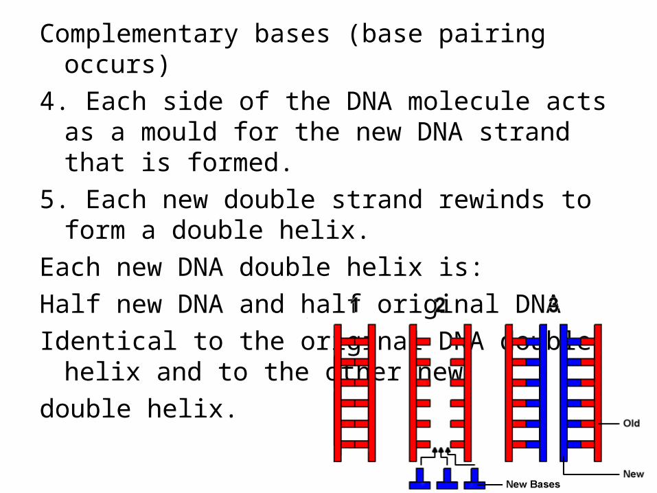
Complementary bases (base pairing occurs)
4. Each side of the DNA molecule acts as a mould for the new DNA strand that is formed.
5. Each new double strand rewinds to form a double helix.
Each new DNA double helix is:
Half new DNA and half original DNA
Identical to the original DNA double helix and to the other new
double helix.

DNA Replication

Significance of DNA ReplicationEach new strand of DNA will have exactly the same sequence of
bases as the original DNA strand.
This allows the same DNA to be passed on to each new generation of cells.
DNA Profiling
A DNA Profile (DNA or genetic
fingerprint) is a method of making
a unique patter of bands from the
DNA of a person, which can then
be used to compare with the DNA
profile of another person.

Method of Preparing a DNA profilePreparing a DNA Profile Involves 4 steps:
1. Releasing DNA from cells2.Cutting the DNA into fragments using restriction enzymes3. Separating the fragments according to their size4. Comparing the patterns of the bands

DNA profiling in more detail
1. Cells are broken down to release DNA
If the amount of DNA is too small to work with it can be increased (amplified) using the polymerase chain reaction (PCR).
2. DNA is cut into fragments
Isolated DNA is cut into fragments using special enzymes called restriction enzymes. Different restriction enzymes cut the DNA at specific base sequences. e.g. one restriction enzyme always cuts the DNA at the sequence CAATTC
3. The fragments are separated

The section of DNA that have been cut are separated on the basis of their size using a process called gel electrophoresis. An electric current is applied along a gel in a small glass tank. The current draws the negatively charged DNA to one end of the gel. Small DNA fragments move faster along the gel than larger DNA fragments.
When the electrophoresis is finished a permanent record is obtained by adding radioactive material which combines with the DNA fragments. A photographic copy of the bands is then obtained.
4. Patterns are compared
It is highly unlikely that two people will have the same DNA Profile (unless they are identical twins).
Application of DNA Profiles
DNA profiles can be used to:
• Forensic Science. To establish whether biological tissue e.g blood, hair, saliva or semen at a crime scene matches or does not match a suspect. (Forensic Medicine is the way in which medical knowledge is used in legal situations to convict criminals)
• Medical. To determine whether a person is or is not the parent of a child

Genetic or DNA screening means that a person’s DNA can be tested to show the presence of normal or altered genes (which may cause disease).
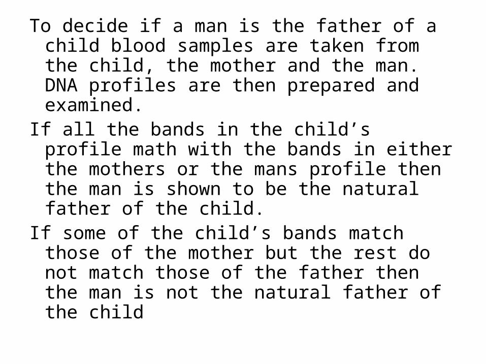
To decide if a man is the father of a child blood samples are taken from the child, the mother and the man. DNA profiles are then prepared and examined.
If all the bands in the child’s profile math with the bands in either the mothers or the mans profile then the man is shown to be the natural father of the child.
If some of the child’s bands match those of the mother but the rest do not match those of the father then the man is not the natural father of the child


Genetic ScreeningGenetic screening means testing DNA for the presence or
absence of a particular gene or altered gene.Genetic Disorders caused by defective genes include:a) Albinism (pigment melanin cannot be made in skin)b) Cystic fibrosis (build up of mucus in the lungs and
intestines)c) Haemochromatosis (in which too much iron
accumulates in the body and has to be removed by regular bleeding)
d) Some cancers
Genetic screening can be carried out in two main ways1. Adult screening2. Foetal screening

1. Adult ScreeningSometimes carried out on adults who although they do not suffer from a genetic disorder themselves may carry a defective gene in their cells (carriers). It is now possible to identify “carriers” of many disorders e.g. cystic fibrosis, sickle cell anaemia etc.
People who are carriers are may be given information about the chances of them having a child with the disorder which allows them to prepare for a medical condition that might affect their family.

2. Embryonic or Foetal ScreeningCells are removed from the embryo, placenta or the fluid around the foetus. These cells can be tested to detect if the embryo or foetus has one of a number of genetic disorders.
Ethics of Genetic screening
Ethics relates to whether behaviour is proper or improper.
Genetic screening may cause ethical problems
If the results are released to the public people involved may feel embarrassed, be treated unfairly, be isolated, lose their jobs etc.
If genetic screening is carried out on a foetus it may encourage termination of the pregnancy?

RNA (ribonucleic Acid)Also a nucleic acid
Consists of 4 bases: adenine, guanine, cytosine and uracil

A and U are complementary bases and G and C are complementary bases
RNA is single stranded
The sequence of bases in RNA depends on the sequence of bases on the DNA strand.
e.g. If DNA strand has bases GGAATC then RNA will have the complementary sequence CCUUAG
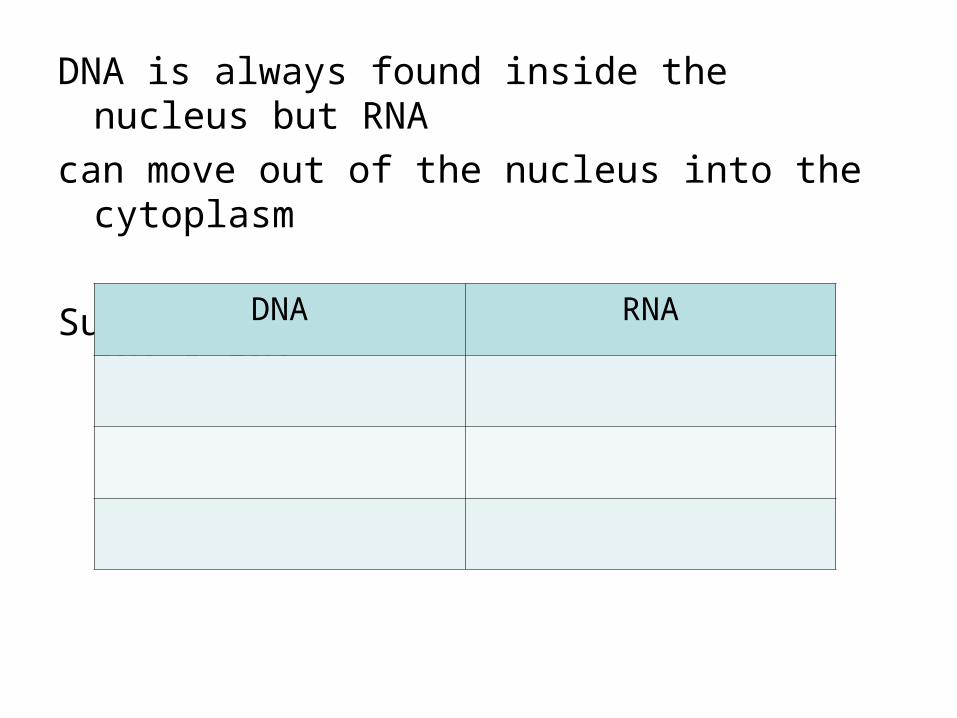
DNA is always found inside the nucleus but RNA
can move out of the nucleus into the cytoplasm
Summary table: Differences between RNA & DNA
DNA RNA
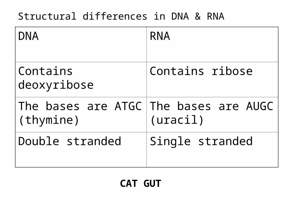
Structural differences in DNA & RNA
DNA RNA
Contains deoxyribose Contains ribose
The bases are ATGC (thymine)
The bases are AUGC (uracil)
Double stranded Single stranded
CAT GUT
New Vocabulary
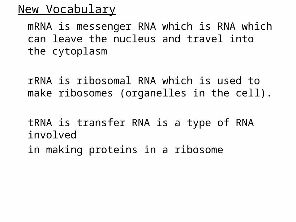
mRNA is messenger RNA which is RNA which can leave the nucleus and travel into the cytoplasm
rRNA is ribosomal RNA which is used to make ribosomes (organelles in the cell).
tRNA is transfer RNA is a type of RNA involved
in making proteins in a ribosome

New Vocabulary
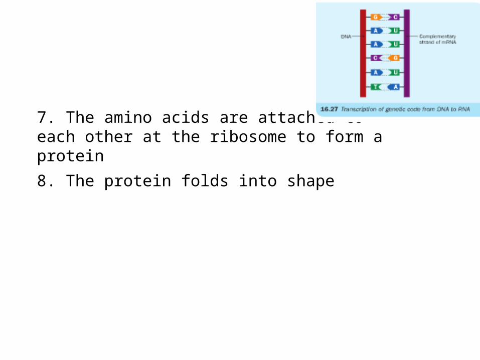
Transcription is the copying of a sequence of genetic bases from DNA onto messenger RNA (mRNA)
Translation is the conversion of a sequence of genetic bases on messenger RNA into a sequence of amino acids.

Steps in protein synthesis (ordinary level):
1. Enzymes open up the DNA at the site of a gene
2. The DNA code is transcribed (copied)onto a complementary mRNA strand
3. mRNA enters a ribosome in the cytoplasm
4. Every tRNA has a complementary triplet to the triplets on the mRNA
5. tRNA molecules enter the ribosome
6. every tRNA has a specific amino acid
7. The amino acids are attached to each other at the ribosome to form a protein
8. The protein folds into shape


Detailed structure of DNAThe structure of DNA was worked out by James
Watson and Francis Crick.
They shared the Nobel Prize in 1962
DNA is made up of units called nucleotides. These are arranged in very long chains called polynucleotides.
Nucleotides
Nucleotides consist of 3 parts:
A phosphate group, a sugar and a nitrogen containing base.

The sugar deoxyribose is a 5 carbon sugar similar to ribose
The phosphate group PO4 is normally written a P
There are four distinct bases i.e. A,T,G,C
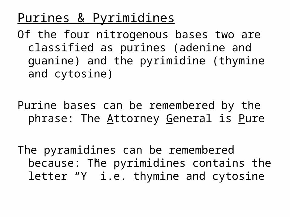
Purines & PyrimidinesOf the four nitrogenous bases two are classified as
purines (adenine and guanine) and the pyrimidine (thymine and cytosine)
Purine bases can be remembered by the phrase: The Attorney General is Pure
The pyramidines can be remembered because: The pyrimidines contains the letter “Y” i.e. thymine and cytosine

Double Helix
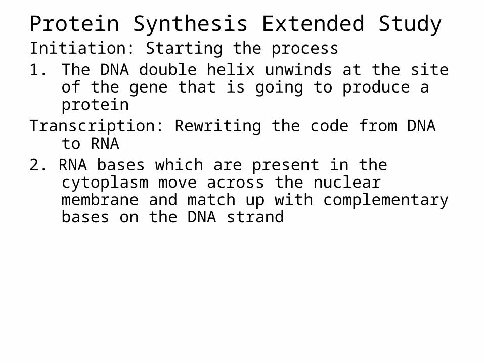
Protein Synthesis Extended StudyInitiation: Starting the process1. The DNA double helix unwinds at the site of the gene
that is going to produce a proteinTranscription: Rewriting the code from DNA to RNA2. RNA bases which are present in the cytoplasm move
across the nuclear membrane and match up with complementary bases on the DNA strand

3. RNA polymerase (enzyme) causes the sequence of RNA bases to join together to form mRNA. A sequence of three bases on DNA or RNA is called a codon.
Codons may cause three possible outcomesa) A start codon indicates the beginning of a geneb) Most codons in a gene specify for a particular amino
acidc) A stop codon indicates the end of a gene

4. Every gene has one start codon, many codons specifying amino acids and one stop codon.
Translation: the production of a protein according to the RNA code
5. mRNA moves from the nucleus into the cytoplasm
6. Ribosomes are made up of rRNA (ribosomal RNA) and proteins.
7. The mRNA strand forms weak bonds with the rRNA in a ribosome. This will be the site of protein synthesis
8. The cytoplasm contains a supply for tRNA (transfer RNA). Each tRNA carries:
a) A specific triplet or anticodon
b) A particular amino acid which is specific to the anticodon.
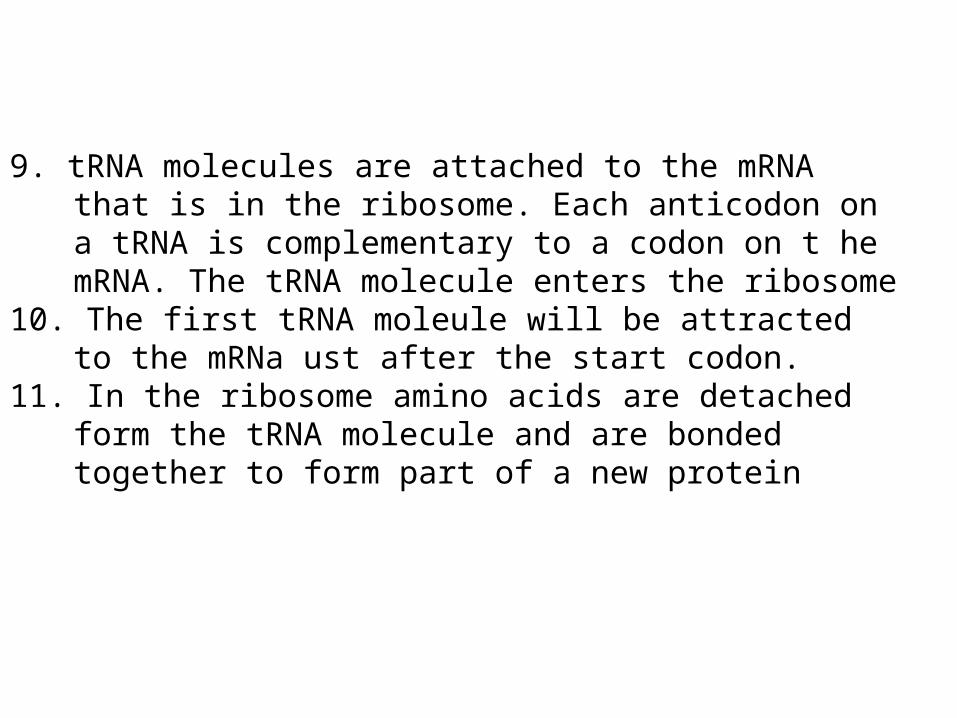
9. tRNA molecules are attached to the mRNA that is in the ribosome. Each anticodon on a tRNA is complementary to a codon on t he mRNA. The tRNA molecule enters the ribosome
10. The first tRNA moleule will be attracted to the mRNa ust after the start codon.
11. In the ribosome amino acids are detached form the tRNA molecule and are bonded together to form part of a new protein

12. tRNA moleules leave the ribosome without any attached amino acids. As they leave they pull the mRNA strand through the ribosome.
13. tRNA molecules continue to bind with mRNA until a stop codon is reached.
At this point:
a) The mRNA code sequence is complete
b) The new protein is produced.
Type of RNA Functions
mRNA (messenger RNA)
tRNA (transfer RNA)
rRNA (ribosomal RNA)
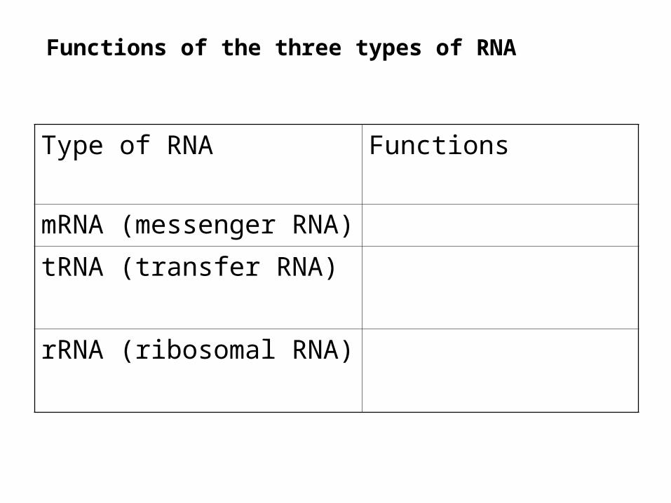
Functions of the three types of RNA

Type of RNA Functions
mRNA (messenger RNA) Complementary strand to DNA
Carries instruction for the production of a protein from DNA to a ribosome
tRNA (transfer RNA) Has a complementary anticodon to mRNA codon
Carries an amino acid to the ribosome
rRNA (ribosomal RNA) Forms part of the structure of a ribosome
Forms a weak bond with mRNA in the ribosome