chapter 187 juvenile angiofibroma
TRANSCRIPT
187Juvenile angiofibroma
MICHAEL GLEESON
Introduction 2437
Pathogenesis 2437
Presentation 2438
Assessment 2438
Treatment 2439
Preoperative embolization 2440
Preoperative chemotherapy 2441
Surgical resection – techniques and approaches 2441
Complications 2442
Key points 2443
Best clinical practice 2443
Deficiencies in current knowledge and areas for future
research 2443
References 2443
SEARCH STRATEGY
The data in this chapter are supported by a PubMed search of publications focussing on the genetics, pathology andmanagement of juvenile angiofibroma.
INTRODUCTION
Recognized since ancient times by Hippocrates, juvenileangiofibroma is an uncommon, benign and extremelyvascular tumour that arises in the tissues within thesphenopalatine foramen. Rarely, it is found at other sitesin the nasal cavity and paranasal sinuses.1 Juvenileangiofibroma accounts for less than 0.5 percent of allhead and neck tumours. It develops almost exclusively inadolescent males, though there are reports of this tumourbeing found in children, the elderly, young and evenpregnant women. As it grows, the tumour extends intothe nasopharynx, paranasal sinuses, pterygopalatine andinfratemporal fossa. Larger tumours can involve the orbitand cavernous sinus. Juvenile angiofibromas are locallyinvasive, though a few have been reported to behave in amore malignant fashion. The vascular nature of juvenileangiofibroma has posed a significant problem for thosecharged with its management. In recent years, with theadvent of endoscopic techniques, the surgical approach tothis tumour has changed. This chapter aims to summarizecurrent knowledge and management.
PATHOGENESIS
Juvenile angiofibromas present as well-defined, lobulatedtumours that are covered by nasopharyngeal mucosa. Thetumour consists of proliferating, irregular vascular chan-nels within a fibrous stroma. Tumour blood vesselstypically lack smooth muscle and elastic fibres, this featurecontributing to its reputation for sustained bleeding. Thestromal compartment is made up of plump cells that canbe spindle or stellate in shape and give rise to varyingamounts of collagen. It is this that makes some tumoursvery hard or firm, while others may be relatively soft.
As this tumour is almost exclusively found in adolescentboys, there has always been much speculation and indirectevidence that sex-hormone receptors play some part in itsdevelopment. Recent immunocytochemical techniqueshave been used to show that androgen receptors arepresent in at least 75 percent of tumours, these receptorsbeing present in both the vascular and stromal elements. Amuch smaller proportion of tumours also have someprogesterone receptors. In contast, oestrogen receptorshave not been demonstrated.2 Other factors also play their

part in the development of this tumour. The angiogenicgrowth factor (vascular endothelial growth factor (VEGF))has been found localized on both endothelial and stromalcells, perhaps indicating that both cell types play a rolein tumour development.3 Vessel density and both theexpression and localization of VEGF correlate with theproliferative marker Ki67.4 However, neither the prolif-erative index nor VEGF expression seem to bear anyrelation whatsoever to the stage of the tumour at the timeof presentation; in other words, its degree of aggressiveness.Overexpression of insulin-like growth factor II (IGFII) hasalso been found in a large number of juvenile angio-fibromas. The IGFII gene is situated on the short arm ofchromosome 11 and at that site is the target of genomicimprinting, expressing the paternal allele only. It is thoughtthat overexpression of IGFII might be associated with atendency to recurrence and poorer prognosis.5
Juvenile angiofibromas have also been reported todevelop 25 times more frequently in patients with familialadenomatous polyposis, a condition that is associatedwith mutations of the adenomatous polyposis coli (APC)gene. As a result, it has been suggested that germlinemutations in the APC gene on chromosome 5q might alsobe involved in the pathogenesis of sporadic juvenileangiofibromas. This gene regulates the beta-cateninpathway which influences cell to cell adhesion. Mutationsof beta-catenin have been found in sporadic and recurrentjuvenile angiofibromas.6 Localization of beta-catenin onlyto the nuclei of stromal cells has been interpreted by someto suggest that the stromal cells have a critical role in thedevelopment of these neoplasms.
PRESENTATION
Recurrent severe epistaxes accompanied by progressivenasal obstruction are the classical symptoms of juvenileangiofibromas at the time of presentation. These tumoursdo not grow fast and so many months or even years maypass before it occurs to the patient or their parents thatthere is anything seriously amiss. In most, there is a delayof at least six or seven months between the onset ofsymptoms and presentation. By that time, it is usual forthe youth to have other signs and symptoms of tumourgrowth and extension. These may include swelling of thecheek, trismus, hearing loss secondary to Eustachian tubeobstruction, anosmia and a nasal intonation or plummyquality to the voice. More extensive tumour growth withinvasion of the orbit and cavernous sinus may causeproptosis, diplopia, visual loss, facial pain and headache.
Anterior rhinoscopy is likely to confirm the presenceof abundant mucopurulent secretions in the nasal cavitythat usually obscure the tumour from vision, though afew patients have tumour prolapsing from the anteriornares. The soft palate is often displaced inferiorly by thebulk of the tumour which can be seen clearly as a pink orreddish mass that fills the nasopharynx.
ASSESSMENT
In the past, the exact nature of these tumours wassuggested by the plain lateral skull radiographic appear-ance that would show anterior bowing of the posteriorwall of the maxillary sinus (Figure 187.1). Nowadays, thediagnosis is based on the CT and MR appearances that aresometimes confirmed by angiography. A trans-nasalbiopsy is not necessary and can provoke brisk haemor-rhage. The exact extent or stage of the tumour can only bedetermined by a combination of CT and MR imaging andthis is vital when planning the surgical resection (Figures187.2 and 187.3). Several staging systems have beenproposed but that of Fisch is the most robust andpractical (Table 187.1).7 It defines clearly which tumourscan be resected by endonasal techniques and those thatwould be better tackled by more open or infratemporalfossa/neurosurgical approaches. The Radkowski stagingsystem appeals to those involved with the management ofsmaller tumours as there are more subdivisions but, inreality, this adds little to its utility (Table 187.2).8
Diagnostic angiography is undertaken to evaluate the
Figure 187.1 Typical plain radiographic appearance of an
angiofibroma. The posterior wall of the maxillary sinus has been
bowed anteriorly (arrowed).
2438 ] PART 17 HEAD AND NECK TUMOURS

source of blood supply and as a prelude to selectiveembolization (Figure 187.4). [*]
TREATMENT
Hippocrates (470–410 BC) is said to have removed what hecalled a ‘hard nasal polyp’ through a midline, nose-splitting incision and it is suggested that this was ajuvenile angiofibroma. The famous surgeon, Liston, atUniversity College London performed the first successful
resection of an angiofibroma on a 21-year-old man fromGibraltar on 6th September 1841.9 The tumour had beenpresent for at least three and a half years and filled thepharynx to the extent that it caused significant airwayobstruction. It extended into his cheek and had erodedthe alveolar process. The patient had experienced a
Figure 187.2 (a) Axial and (b) coronal CT images of a type
3a, right-sided, juvenile angiofibroma. There is destruction of
the pterygoid plates and extension of tumour through the skull
base (arrowed).
Figure 187.3 MR appearances of a type 3a, right-sided
juvenile angiofibroma. It has invaded the infratemporal fossa.
The operative specimen gives a more objective assessment of
the tumour size.
Table 187.1 Fisch staging system of juvenile angiofibromas.
Type Details
1 Tumour limited to the nasopharyngeal cavity; bone destruction negligible or limited to the sphenopalatine foramen
2 Tumour invading the pterygopalatine fossa or the maxillary, ethmoid or sphenoid sinus with bone destruction
3 Tumour invading the infratemporal fossa or orbital region:
(a) without intracranial involvement
(b) with intracranial extradural (parasellar) involvement
4 Intracranial intradural tumour:
(a) without infiltration of the cavernous sinus, pituitary fossa or optic chiasm
(b) with infiltration of the cavernous sinus, pituitary fossa or optic chiasm
Chapter 187 Juvenile angiofibroma ] 2439

number of severe epistaxes, losing two to three pints ofblood on each occasion. Liston removed the tumour byperforming a total maxillectomy through a Weber–Fergusson incision without anaesthesia! The patient borethe procedure ‘with remarkable fortitude’. He wasdischarged 24 days later able to eat a normal diet!Histopathological examination of the operative specimenshowed the tumour had a ‘fibro-vascular nature.’
In the years that followed it became apparent that alarge number of these tumours could be removedrelatively safely by open approaches but not withoutsignificant morbidity. A number of patients also died as adirect result of blood loss. Attempts were made to reducethe blood supply of the tumour preoperatively and toshrink them by chemical means. Very extensive tumourswere treated by radiotherapy as were recurrences.Strategies for the surgical management of these tumourshave evolved over the intervening years and never morequickly than in the last decade with the advent ofendonasal endoscopic techniques.10
PREOPERATIVE EMBOLIZATION
The role of preoperative embolization in the surgicalmanagement of this tumour is controversial. While somesurgeons regard it as essential, others have less strict viewsor frankly disagree. There is little doubt that for smalltumours the blood supply is predictable, usually theterminal branches of the internal maxillary artery, andthese can be controlled easily at the time of surgery.Embolization in such cases would seem to be unnecessary.More extensive tumours acquire a blood supply fromother vessels, branches of both the external and internalcarotid circulations. Surgical resection of these tumourscan be formidable and preoperative selective emboliza-tion, some days before surgery, is prudent at the veryleast. It is the medium-sized tumours where the benefitsof preoperative embolization are doubtful. Intraoperativeblood loss after embolization is certainly less.11 Themaxillary or external carotid artery can be controlled orligated relatively easily at an early stage in any openprocedure, regardless of whether embolization has beenundertaken or not. [**]
It might be thought that any measure undertaken toimprove operative conditions might influence the ade-quacy of resection and reduce the rate of recurrence. Onthe contrary, recurrence rates seem to be increased bypreoperative embolization.12 Perhaps shrinkage of thetumour makes it more difficult to define its entirety at thebottom of a deep and bloody operative field.
Table 187.2 Radkowski classification of juvenile angiofibromas.
Stage Details
Ia Limited to the nose and nasopharyngeal area
Ib Extension into one or more sinuses
IIa Minimal extension into pterygopalatine fossa
IIb Occupation of the pterygopalatine fossa without orbital erosion
IIc Infratemporal fossa extension without cheek or pterygoid plate involvement
IIIa Erosion of the skull base (middle cranial fossa or pterygoids)
IIIb Erosion of the skull base with intracranial extension with or without cavernous sinus involvement
Figure 187.4 External carotid angiogram of a juvenile
angiofibroma demonstrating its blood supply from the internal
maxillary artery which is much larger than normal.
2440 ] PART 17 HEAD AND NECK TUMOURS
PREOPERATIVE CHEMOTHERAPY
Other means of reducing tumour size before surgery havealso been evaluated. Oestrogens have been reported toinduce shrinkage in some but their effect is variable andnot without complications. At the very least, oestrogentherapy delays surgery and the secondary feminizingeffects are certainly unwanted by an adolescent boy. In asmall series of patients given the nonsteroidal androgenreceptor blocker, flutamide, tumour shrinkage of up to 44percent was reported by Gates et al.13 Flutamide isregularly used in the management of prostatic cancer.While not without side effects, nausea, breast tendernessand gynaecomastia, these effects were only temporary anddisappeared completely at the end of therapy. It seemedthat this drug might have a role in the preoperativepreparation of patients with very advanced tumours,certainly those with intracranial extension. Unfortunately,in a pilot study of seven patients with stage IV diseaseundertaken by Labra et al.14 the mean shrinkage achievedwas 7.5 percent (median 6.1 percent, range 4.8–11.1percent) and this was considered to be insignificant. [**/*]
SURGICAL RESECTION – TECHNIQUES ANDAPPROACHES
Until relatively recently, most small tumours wereresected either through a transpalatal approach, lateralrhinotomy or mid-facial degloving approach. Openapproaches can be used for tumours of all stages andcertainly were the only option before the application ofendonasal endoscopic techniques became more wide-spread. Nowadays, stage Fisch 1, 2 and some type 3tumours are suitable for endoscopic resection using oneor two surgeon techniques.15, 16, 17, 18, 19
There is much to be gained by endonasal endoscopictechniques, for example, reduced intraoperative bloodloss, fewer postoperative complications and a reducedlength of hospital stay. It is difficult to know how realthese claims are as no prospective trials have beenundertaken and those reported are either small case seriesor rely on historical controls. Furthermore, the tumoursresected by endoscopic techniques tend to be relativelysmall. Fisch type 3 tumours suitable for endoscopicresection have limited medial invasion of the infratem-poral fossa. Larger tumours and those extending across orthrough the skull base present a very different surgicalchallenge which the enthusiastic endoscopist shouldconsider carefully before embarking on a potentiallylife-threatening procedure.
Endoscopic endonasal techniques
Preoperative embolization is usually undertaken, not-withstanding its doubtful benefit. After the induction of
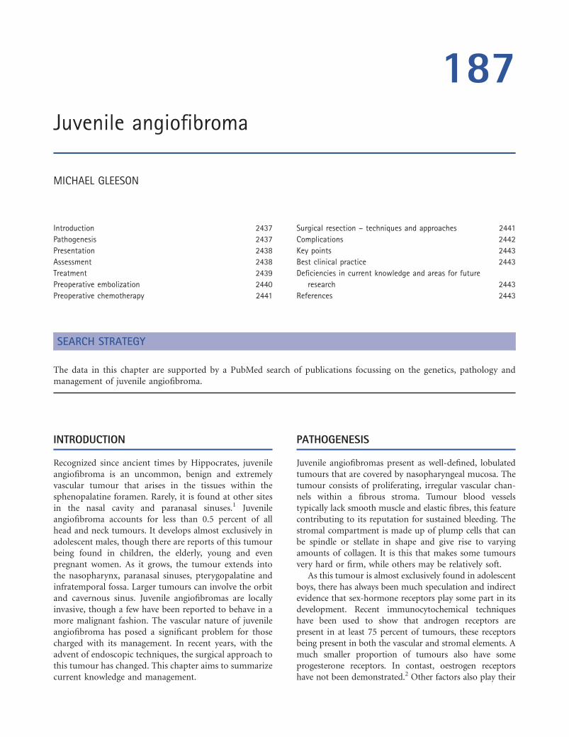
anaesthesia the nose is prepared with a vasoconstrictorsolution, 4 percent cocaine or epinephrine 1:10,000. Theanterior end of the middle turbinate is resected at theoutset of the procedure (Figure 187.5).
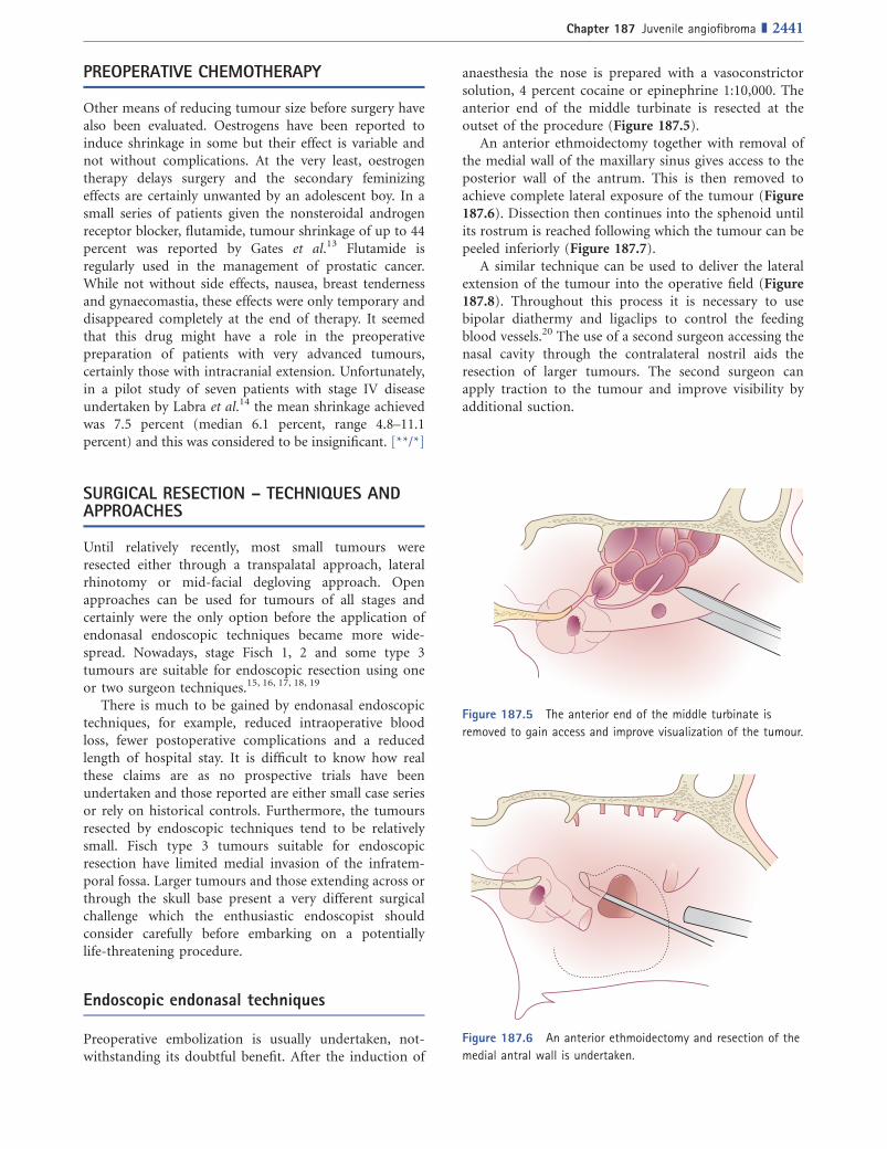
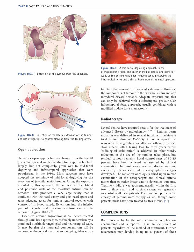
An anterior ethmoidectomy together with removal ofthe medial wall of the maxillary sinus gives access to theposterior wall of the antrum. This is then removed toachieve complete lateral exposure of the tumour (Figure187.6). Dissection then continues into the sphenoid untilits rostrum is reached following which the tumour can bepeeled inferiorly (Figure 187.7).
A similar technique can be used to deliver the lateralextension of the tumour into the operative field (Figure187.8). Throughout this process it is necessary to usebipolar diathermy and ligaclips to control the feedingblood vessels.20 The use of a second surgeon accessing thenasal cavity through the contralateral nostril aids theresection of larger tumours. The second surgeon canapply traction to the tumour and improve visibility byadditional suction.
Figure 187.5 The anterior end of the middle turbinate is
removed to gain access and improve visualization of the tumour.
Figure 187.6 An anterior ethmoidectomy and resection of the
medial antral wall is undertaken.
Chapter 187 Juvenile angiofibroma ] 2441
Open approaches
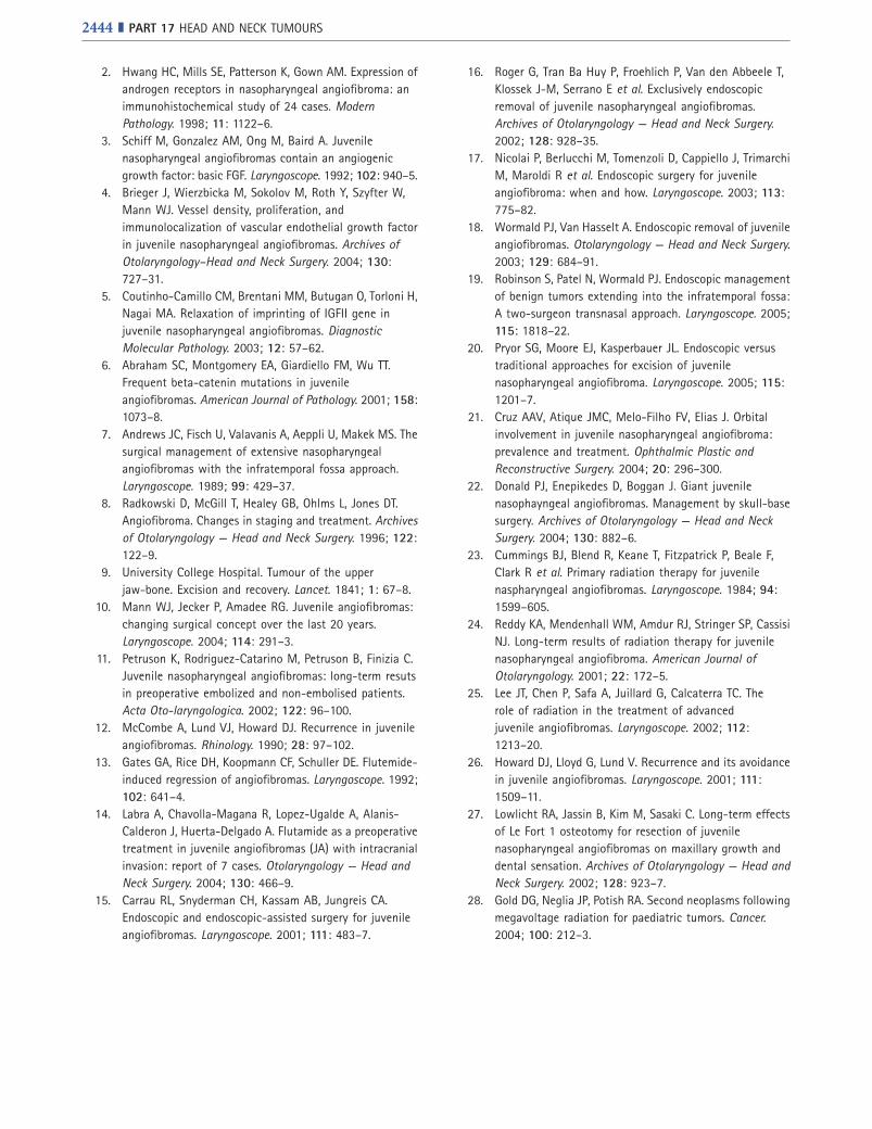
Access for open approaches has changed over the last 20years. Transpalatal and lateral rhinotomy approaches havelargely, but not completely, given way to mid-facialdegloving and infratemporal approaches that werepopularized in the 1980s. Most surgeons now haveadopted the technique of mid-facial degloving for theresection of juvenile angiofibromas. Using the exposureafforded by this approach, the anterior, medial, lateraland posterior walls of the maxillary antrum can beremoved. This produces a very large cavity that isconfluent with the nasal cavity and post-nasal space andgives adequate access for tumour removal together withcontrol of its blood supply. Extensions into the inferiorpart of the orbit and infratemporal fossa can also beremoved (Figure 187.9).21
Extensive juvenile angiofibromas are better resectedthrough skull base approaches, preferably undertaken by acombined otorhinolaryngological and neurosurgical team.It may be that the intranasal component can still beremoved endoscopically or that endoscopic guidance may
facilitate the removal of paranasal extensions. However,the components of tumour in the cavernous sinus and anyintradural disease demands adequate exposure and thiscan only be achieved with a subtemporal pre-auricularinfratemporal fossa approach, usually combined with amodified middle fossa craniectomy.22
Radiotherapy
Several centres have reported results for the treatment ofadvanced disease by radiotherapy.23, 24, 25 External beamradiation was delivered in several fractions to achieve atotal tumour dose of 30–55Gy. All series report thatregression of angiofibromas after radiotherapy is veryslow indeed, often taking two to three years before‘radiological stabilization’ is achieved. In other words,reduction in the size of the tumour takes place, butresidual tumour remains. Local control rates of 80–85percent have been achieved as assessed by clinicalexamination. In most series, residual disease was notassessed by interval scans unless new symptoms or signsdeveloped. The radiation oncologists relied upon mirrorexamination of the nasopharynx and clinical criteriarather than objective image data to determine outcome.Treatment failure was apparent, usually within the firsttwo to three years, and surgical salvage was generallysuccessful in all these patients. There are no reports on theefficacy of gamma-knife therapy as yet, though somepatients must have been treated by this means. [**]
COMPLICATIONS
Recurrence is by far the most common complicationencountered and is reported in up to 25 percent ofpatients regardless of the method of treatment. Furtherrecurrences may develop in up to 40 percent of these
Figure 187.7 Extraction of the tumour from the sphenoid.
Figure 187.8 Resection of the lateral extension of the tumour
and use of ligaclips to control bleeding from the feeding artery.
Figure 187.9 A mid-facial degloving approach to the
pterygopalatine fossa. The anterior, medial, lateral and posterior
walls of the antrum have been removed while preserving the
infra-orbital nerve and a rim of bone around the nasal aperture.
2442 ] PART 17 HEAD AND NECK TUMOURS

patients. Not surprisingly, recurrence is more likely inpatients with advanced disease and in those treated byinexperienced surgeons. As stated previously, preoperativeembolization is not associated with a reduced rate ofrecurrence as might be expected. The one single factorthat seems to correlate with recurrence is the age of thepatient at the time of presentation. The younger thepatient the more likely that future recurrence will develop.[**/*]
From a surgical standpoint, most recurrences developas a consequence of invasion of the basisphenoid. A moremeticulous exploration of this area at the time of primarysurgery has been reported to have a dramatic effect on therate of recurrent disease.26 Rather than rely on macro-scopic clearance of disease, drilling out the basisphenoidensures that no residual tumour remains in the pterygoidcanal or cancellous bone of the sphenoid.
In view of the very high incidence of recurrent disease,prolonged clinical and radiological monitoring is neces-sary for all these patients. Disease-free status five yearsafter primary surgery probably represents cure. The samecannot be said for those who have experienced recurrentdisease.
Interestingly, few surgeons have quoted complicationsother than recurrence. It is unlikely that this is the onlypossible complication, but rather that all others pale intoinsignificance in comparison. Surgically induced infra-orbital nerve sensory deficits are recognized as a potentialcomplication of mid-facial degloving, as is nasal vestib-ular stenosis.27 Prolonged nasal crusting is also commonand this may well develop into ozaena. Regular nasaldouching with saline and the use of glucose in glycerinedrops can do much to alleviate this unpleasant complica-tion. With more extensive resections, ocular problemsmay be experienced. Displacement of the globe caused byloss of bony support, ophthalmoplegia and visual lossmust have been experienced by some, but perhaps notconsidered a complication so much as the unavoidableconsequence of a complete craniofacial resection.
Late complications also develop following radiother-apy and are relatively common. Up to 33 percent ofpatients in the reported series have been affected. Growthretardation, panhypopituitarism, temporal lobe necrosis,cataracts, radiation keratopathy, together with skin,thyroid and nasopharyngeal malignancies were the mostcommon problems encountered in the first 10–15 yearsafter treatment. Some second neoplasms have developedin the radiation field at an even later date. Whatever thesuccess rate of radiotherapy to control disease, it has to beremembered that even 30 years after radiotherapy thepatient will still be very young28 and none of thesereported complications is negligible. Furthermore, it mustbe recognized that all patients with residual disease are atrisk of future regrowth or recurrence. For these patients,lifetime monitoring and assessment is necessary. Serialinterval MR imaging should become a standard of carefor these patients.
KEY POINTS
� Angiofibromas should be suspected whenevera young male patient complains of unilateralnasal obstruction, particularly if they havebeen experiencing epistaxes.
� The diagnosis should be made on the basis ofimaging and not biopsy.
� Complete surgical resection is the treatmentof choice with radiotherapy being reservedfor those in whom this is not possible orwho develop recurrence.
� There is a high rate of recurrence.� Recurrence rates can be reduced by
meticulous dissection of the sphenopalatineforamen.
� Recurrence usually becomes evident withintwo to three years of the initial resection.
� Response to radiotherapy is very slow, theeffect may not be fully apparent for one totwo years.
Best clinical practice
[ Both CT and MR images should be acquired and
clinical staging made on the basis of these data.
[ Angiography and embolization is advised for those
undergoing endoscopic endonasal resection and for
those with intracranial extension.
[ Patients should have interval imaging for at least five
years before being declared disease free and cured.
Deficiencies in current knowledge andareas for future research
$ The precise molecular mechanisms involved in the
development of this tumour.
$ Long-term outcome data for patients treated by
endoscopic endonasal techniques.
$ Outcome data for patients treated by gamma-knife or
stereotactic radiotherapy.
REFERENCES
1. Windfuhr JP, Remmert S. Extranasopharyngeal
angiofibromas: etiology, incidence and management. ActaOto-laryngologica. 2004; 124: 880–9.
Chapter 187 Juvenile angiofibroma ] 2443
2. Hwang HC, Mills SE, Patterson K, Gown AM. Expression of
androgen receptors in nasopharyngeal angiofibroma: an
immunohistochemical study of 24 cases. ModernPathology. 1998; 11: 1122–6.
3. Schiff M, Gonzalez AM, Ong M, Baird A. Juvenile
nasopharyngeal angiofibromas contain an angiogenic
growth factor: basic FGF. Laryngoscope. 1992; 102: 940–5.4. Brieger J, Wierzbicka M, Sokolov M, Roth Y, Szyfter W,
Mann WJ. Vessel density, proliferation, and
immunolocalization of vascular endothelial growth factor
in juvenile nasopharyngeal angiofibromas. Archives ofOtolaryngology–Head and Neck Surgery. 2004; 130:727–31.
5. Coutinho-Camillo CM, Brentani MM, Butugan O, Torloni H,
Nagai MA. Relaxation of imprinting of IGFII gene in
juvenile nasopharyngeal angiofibromas. DiagnosticMolecular Pathology. 2003; 12: 57–62.
6. Abraham SC, Montgomery EA, Giardiello FM, Wu TT.
Frequent beta-catenin mutations in juvenile
angiofibromas. American Journal of Pathology. 2001; 158:1073–8.
7. Andrews JC, Fisch U, Valavanis A, Aeppli U, Makek MS. The
surgical management of extensive nasopharyngeal
angiofibromas with the infratemporal fossa approach.
Laryngoscope. 1989; 99: 429–37.8. Radkowski D, McGill T, Healey GB, Ohlms L, Jones DT.
Angiofibroma. Changes in staging and treatment. Archivesof Otolaryngology — Head and Neck Surgery. 1996; 122:122–9.
9. University College Hospital. Tumour of the upper
jaw-bone. Excision and recovery. Lancet. 1841; 1: 67–8.10. Mann WJ, Jecker P, Amadee RG. Juvenile angiofibromas:
changing surgical concept over the last 20 years.
Laryngoscope. 2004; 114: 291–3.11. Petruson K, Rodriguez-Catarino M, Petruson B, Finizia C.
Juvenile nasopharyngeal angiofibromas: long-term resuts
in preoperative embolized and non-embolised patients.
Acta Oto-laryngologica. 2002; 122: 96–100.12. McCombe A, Lund VJ, Howard DJ. Recurrence in juvenile
angiofibromas. Rhinology. 1990; 28: 97–102.13. Gates GA, Rice DH, Koopmann CF, Schuller DE. Flutemide-
induced regression of angiofibromas. Laryngoscope. 1992;102: 641–4.
14. Labra A, Chavolla-Magana R, Lopez-Ugalde A, Alanis-
Calderon J, Huerta-Delgado A. Flutamide as a preoperative
treatment in juvenile angiofibromas (JA) with intracranial
invasion: report of 7 cases. Otolaryngology — Head andNeck Surgery. 2004; 130: 466–9.
15. Carrau RL, Snyderman CH, Kassam AB, Jungreis CA.
Endoscopic and endoscopic-assisted surgery for juvenile
angiofibromas. Laryngoscope. 2001; 111: 483–7.
16. Roger G, Tran Ba Huy P, Froehlich P, Van den Abbeele T,
Klossek J-M, Serrano E et al. Exclusively endoscopic
removal of juvenile nasopharyngeal angiofibromas.
Archives of Otolaryngology — Head and Neck Surgery.2002; 128: 928–35.
17. Nicolai P, Berlucchi M, Tomenzoli D, Cappiello J, Trimarchi
M, Maroldi R et al. Endoscopic surgery for juvenile
angiofibroma: when and how. Laryngoscope. 2003; 113:775–82.
� 18. Wormald PJ, Van Hasselt A. Endoscopic removal of juvenile
angiofibromas. Otolaryngology — Head and Neck Surgery.2003; 129: 684–91.
19. Robinson S, Patel N, Wormald PJ. Endoscopic management
of benign tumors extending into the infratemporal fossa:
A two-surgeon transnasal approach. Laryngoscope. 2005;115: 1818–22.
20. Pryor SG, Moore EJ, Kasperbauer JL. Endoscopic versus
traditional approaches for excision of juvenile
nasopharyngeal angiofibroma. Laryngoscope. 2005; 115:1201–7.
21. Cruz AAV, Atique JMC, Melo-Filho FV, Elias J. Orbital
involvement in juvenile nasopharyngeal angiofibroma:
prevalence and treatment. Ophthalmic Plastic andReconstructive Surgery. 2004; 20: 296–300.
22. Donald PJ, Enepikedes D, Boggan J. Giant juvenile
nasophayngeal angiofibromas. Management by skull-base
surgery. Archives of Otolaryngology — Head and NeckSurgery. 2004; 130: 882–6.
� 23. Cummings BJ, Blend R, Keane T, Fitzpatrick P, Beale F,
Clark R et al. Primary radiation therapy for juvenile
naspharyngeal angiofibromas. Laryngoscope. 1984; 94:1599–605.
24. Reddy KA, Mendenhall WM, Amdur RJ, Stringer SP, Cassisi
NJ. Long-term results of radiation therapy for juvenile
nasopharyngeal angiofibroma. American Journal ofOtolaryngology. 2001; 22: 172–5.
25. Lee JT, Chen P, Safa A, Juillard G, Calcaterra TC. The
role of radiation in the treatment of advanced
juvenile angiofibromas. Laryngoscope. 2002; 112:1213–20.
� 26. Howard DJ, Lloyd G, Lund V. Recurrence and its avoidance
in juvenile angiofibromas. Laryngoscope. 2001; 111:1509–11.
27. Lowlicht RA, Jassin B, Kim M, Sasaki C. Long-term effects
of Le Fort 1 osteotomy for resection of juvenile
nasopharyngeal angiofibromas on maxillary growth and
dental sensation. Archives of Otolaryngology — Head andNeck Surgery. 2002; 128: 923–7.
28. Gold DG, Neglia JP, Potish RA. Second neoplasms following
megavoltage radiation for paediatric tumors. Cancer.2004; 100: 212–3.
2444 ] PART 17 HEAD AND NECK TUMOURS