chapter 19 viruses. copyright © 2008 pearson education inc., publishing as pearson benjamin...
TRANSCRIPT

Chapter 19Viruses

Copyright © 2008 Pearson Education Inc., publishing as Pearson Benjamin Cummings
Overview: A Borrowed Life
Viruses called bacteriophages can infect and set in motion a genetic takeover of bacteria, such as Escherichia coli.
Viruses lead “a kind of borrowed life” between life-forms and chemicals.
The origins of molecular biology lie in early studies of viruses that infect bacteria.

0.5 µm
Figure 19.1 Are the tiny viruses infecting this E. coli cell alive?

Concept 19.1: A virus consists of a nucleic acid surrounded by a protein
coat.
Viruses were detected indirectly long before they were actually seen.
Copyright © 2008 Pearson Education Inc., publishing as Pearson Benjamin Cummings
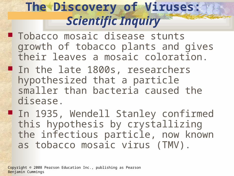
The Discovery of Viruses: Scientific Inquiry
Tobacco mosaic disease stunts growth of tobacco plants and gives their leaves a mosaic coloration.
In the late 1800s, researchers hypothesized that a particle smaller than bacteria caused the disease.
In 1935, Wendell Stanley confirmed this hypothesis by crystallizing the infectious particle, now known as tobacco mosaic virus (TMV).

Copyright © 2008 Pearson Education Inc., publishing as Pearson Benjamin Cummings
Fig. 19-2
RESULTS
1 2 3Extracted sapfrom tobaccoplant withtobaccomosaic disease
Passed sapthrough aporcelain filter knownto trapbacteria
Rubbed filteredsap on healthytobacco plants
4 Healthy plantsbecame infected

Copyright © 2008 Pearson Education Inc., publishing as Pearson Benjamin Cummings
Structure of Viruses
Viruses are not cells. Viruses are very small infectious
particles consisting of nucleic acid enclosed in a protein coat and, in some cases, a membranous envelope.

Copyright © 2008 Pearson Education Inc., publishing as Pearson Benjamin Cummings
Viral Genomes
Viral genomes may consist of either Double- or single-stranded DNA, or Double- or single-stranded RNA
• Depending on its type of nucleic acid, a virus is called a DNA virus or an RNA virus.

Copyright © 2008 Pearson Education Inc., publishing as Pearson Benjamin Cummings
Capsids and Envelopes
A capsid is the protein shell that encloses the viral genome.
Capsids are built from protein subunits called capsomeres.
A capsid can have various structures.
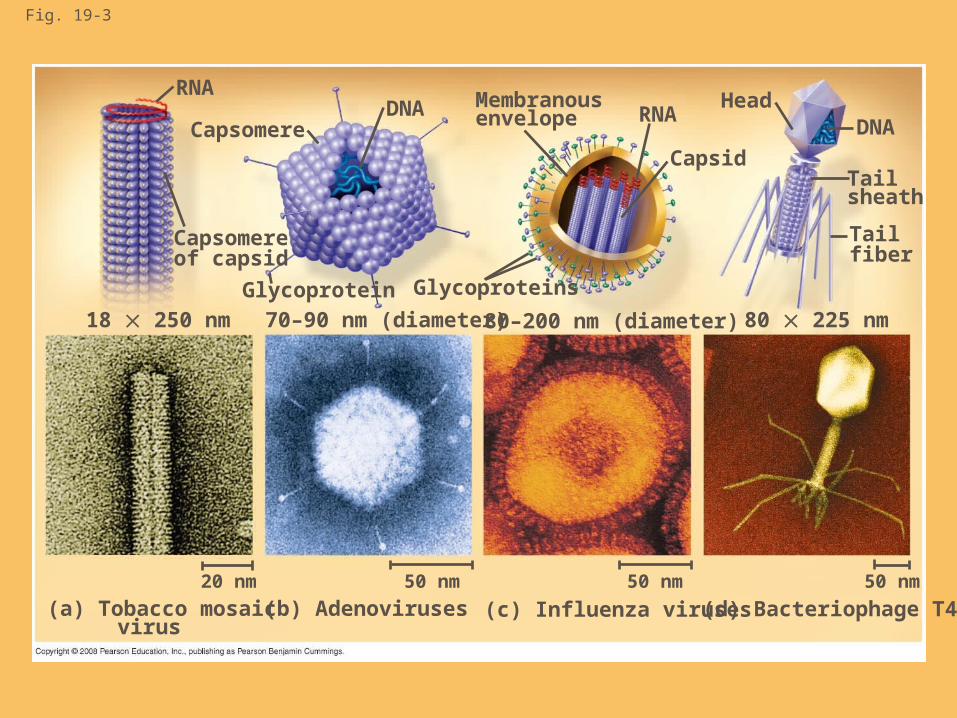
Fig. 19-3
RNA
Capsomere
Capsomereof capsid
DNA
Glycoprotein
18 250 nm 70–90 nm (diameter)
Glycoproteins
80–200 nm (diameter) 80 225 nm
Membranousenvelope RNA
Capsid
HeadDNA
Tailsheath
Tailfiber
50 nm50 nm50 nm20 nm
(a) Tobacco mosaic virus
(b) Adenoviruses (c) Influenza viruses (d) Bacteriophage T4
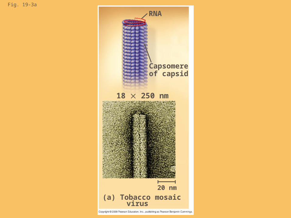
Fig. 19-3a
(a) Tobacco mosaic virus
20 nm
18 250 nm
Capsomereof capsid
RNA
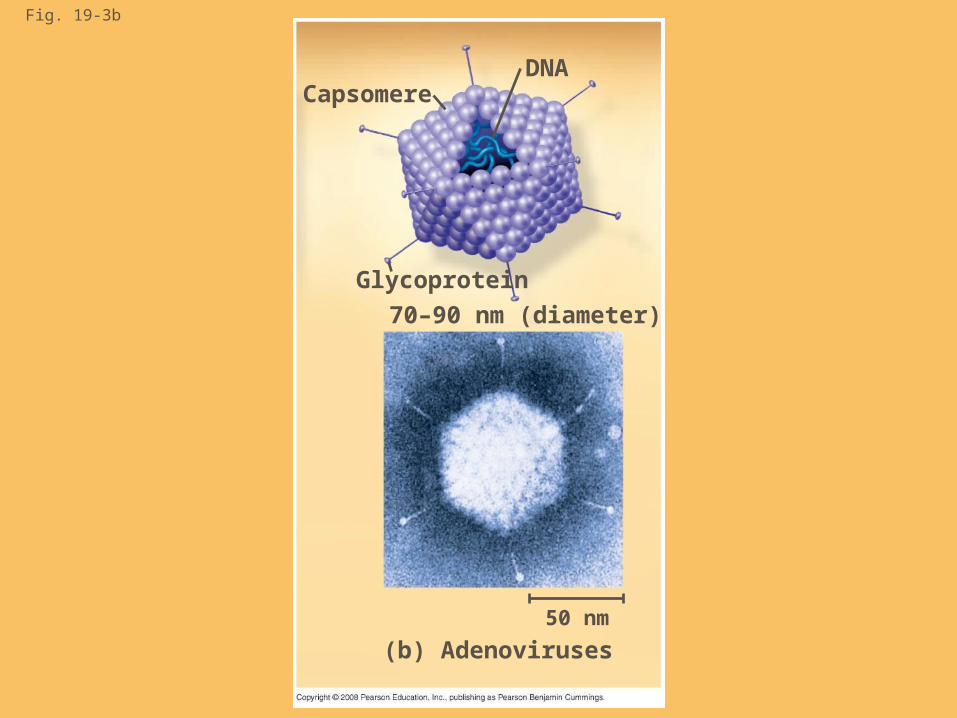
Fig. 19-3b
DNACapsomere
Glycoprotein
70–90 nm (diameter)
50 nm
(b) Adenoviruses

Fig. 19-3c
Membranousenvelope RNA
Capsid
Glycoproteins
80–200 nm (diameter)
50 nm
(c) Influenza viruses

Fig. 19-3d
HeadDNA
Tailsheath
Tailfiber
80 225 nm
50 nm
(d) Bacteriophage T4

Copyright © 2008 Pearson Education Inc., publishing as Pearson Benjamin Cummings
Some viruses have membranous envelopes that help them infect hosts.
These viral envelopes surround the capsids of influenza viruses and many other viruses found in animals.
Viral envelopes, which are derived from the host cell’s membrane, contain a combination of viral and host cell molecules.

Copyright © 2008 Pearson Education Inc., publishing as Pearson Benjamin Cummings
Bacteriophages, also called phages, are viruses that infect bacteria.
They have the most complex capsids found among viruses.
Phages have an elongated capsid head that encloses their DNA.
A protein tail piece attaches the phage to the host and injects the phage DNA inside.
Copyright © 2008 Pearson Education Inc., publishing as Pearson Benjamin Cummings
Concept 19.2: Viruses reproduce only in host cells
Viruses are obligate intracellular parasites, which means they can reproduce only within a host cell.
Each virus has a host range, a limited number of host cells that it can infect.

Copyright © 2008 Pearson Education Inc., publishing as Pearson Benjamin Cummings
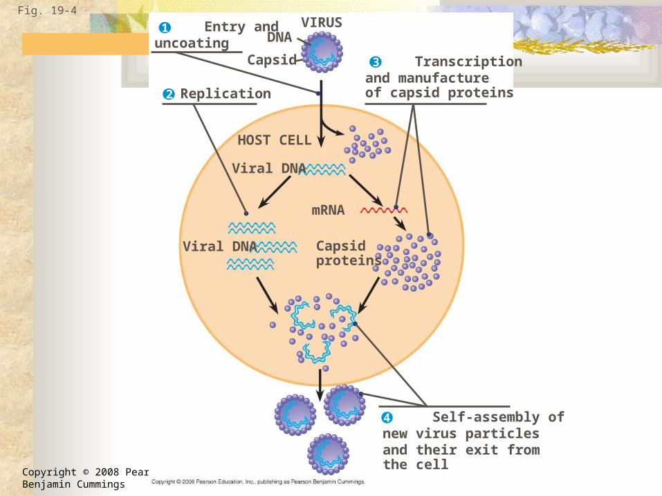
General Features of Viral Reproductive Cycles
Once a viral genome has entered a cell, the cell begins to manufacture viral proteins.
The virus makes use of host enzymes, ribosomes, tRNAs, amino acids, ATP, and other molecules.
Viral nucleic acid molecules and capsomeres spontaneously self-assemble into new viruses.
Animation: Simplified Viral Reproductive CycleAnimation: Simplified Viral Reproductive Cycle
Copyright © 2008 Pearson Education Inc., publishing as Pearson Benjamin Cummings
Transcriptionand manufactureof capsid proteins
Self-assembly of new virus particles and their exit from the cell
Entry anduncoating
Fig. 19-4VIRUS1
2
3
DNA
Capsid
4
Replication
HOST CELL
Viral DNA
mRNA
Capsidproteins
Viral DNA
Copyright © 2008 Pearson Education Inc., publishing as Pearson Benjamin Cummings
Reproductive Cycles of Phages
Phages are the best understood of all viruses Phages have two reproductive mechanisms:
the lytic cycle and the lysogenic cycle.

Copyright © 2008 Pearson Education Inc., publishing as Pearson Benjamin Cummings
The Lytic Cycle
The lytic cycle is a phage reproductive cycle that culminates in the death of the host cell.
The lytic cycle produces new phages and digests the host’s cell wall, releasing the progeny viruses.
A phage that reproduces only by the lytic cycle is called a virulent phage.
Bacteria have defenses against phages, including restriction enzymes that recognize and cut up certain phage DNA.
Animation: Phage T4 Lytic CycleAnimation: Phage T4 Lytic Cycle

Copyright © 2008 Pearson Education Inc., publishing as Pearson Benjamin Cummings
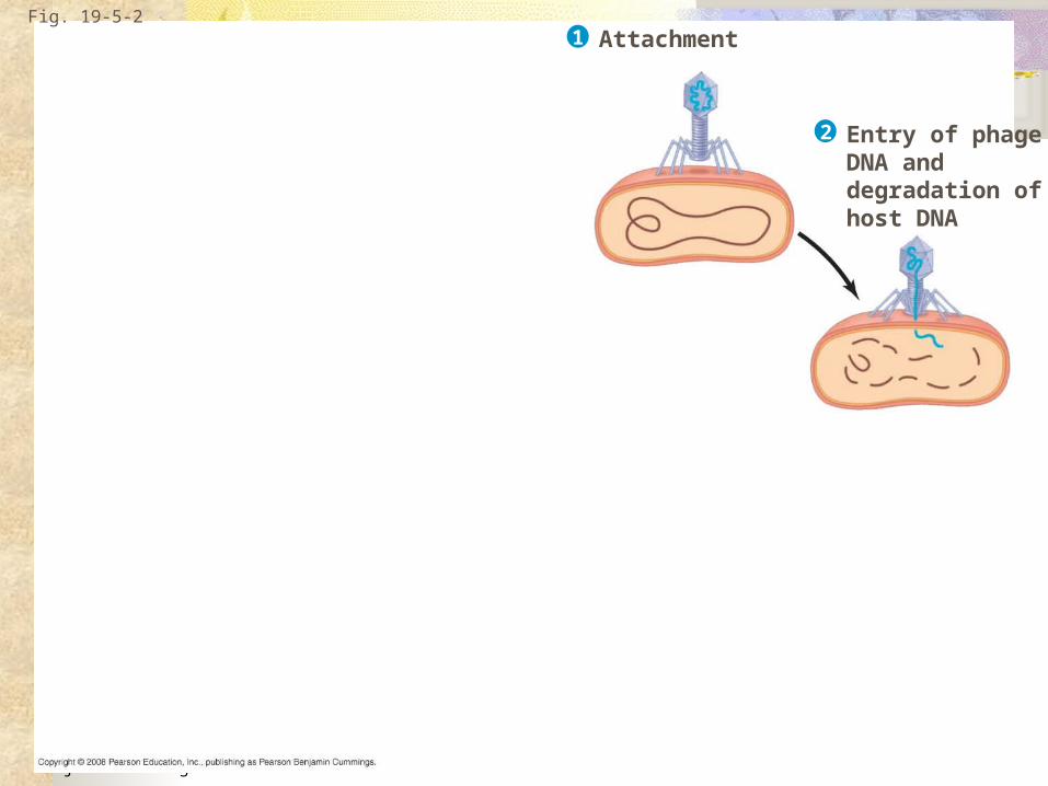
Fig. 19-5-1
Attachment1
Copyright © 2008 Pearson Education Inc., publishing as Pearson Benjamin Cummings
Fig. 19-5-2
Entry of phageDNA anddegradation ofhost DNA
Attachment1
2

Copyright © 2008 Pearson Education Inc., publishing as Pearson Benjamin Cummings
Fig. 19-5-3
Synthesis of viralgenomes andproteins
Entry of phageDNA anddegradation ofhost DNA
Attachment1
2
3

Copyright © 2008 Pearson Education Inc., publishing as Pearson Benjamin Cummings
Fig. 19-5-4
Phage assembly
Assembly
Synthesis of viralgenomes andproteins
Entry of phageDNA anddegradation ofhost DNA
Attachment1
2
4
Head Tail Tail fibers
3
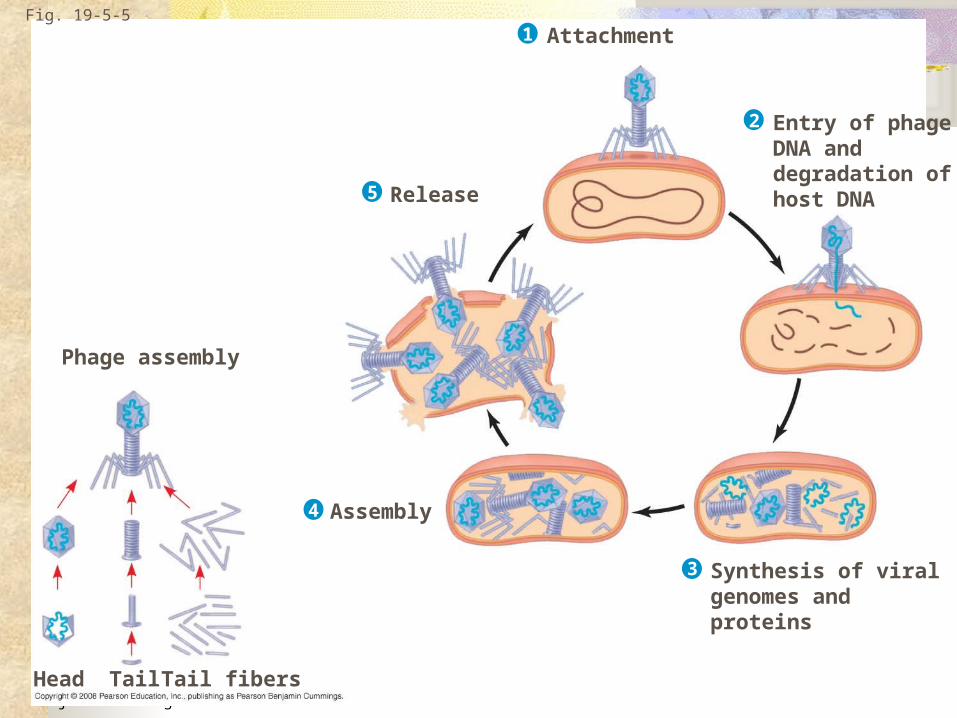
Copyright © 2008 Pearson Education Inc., publishing as Pearson Benjamin Cummings
Fig. 19-5-5
Phage assembly
Head Tail Tail fibers
Assembly
Release
Synthesis of viralgenomes andproteins
Entry of phageDNA anddegradation ofhost DNA
Attachment1
2
4
5
3
Copyright © 2008 Pearson Education Inc., publishing as Pearson Benjamin Cummings
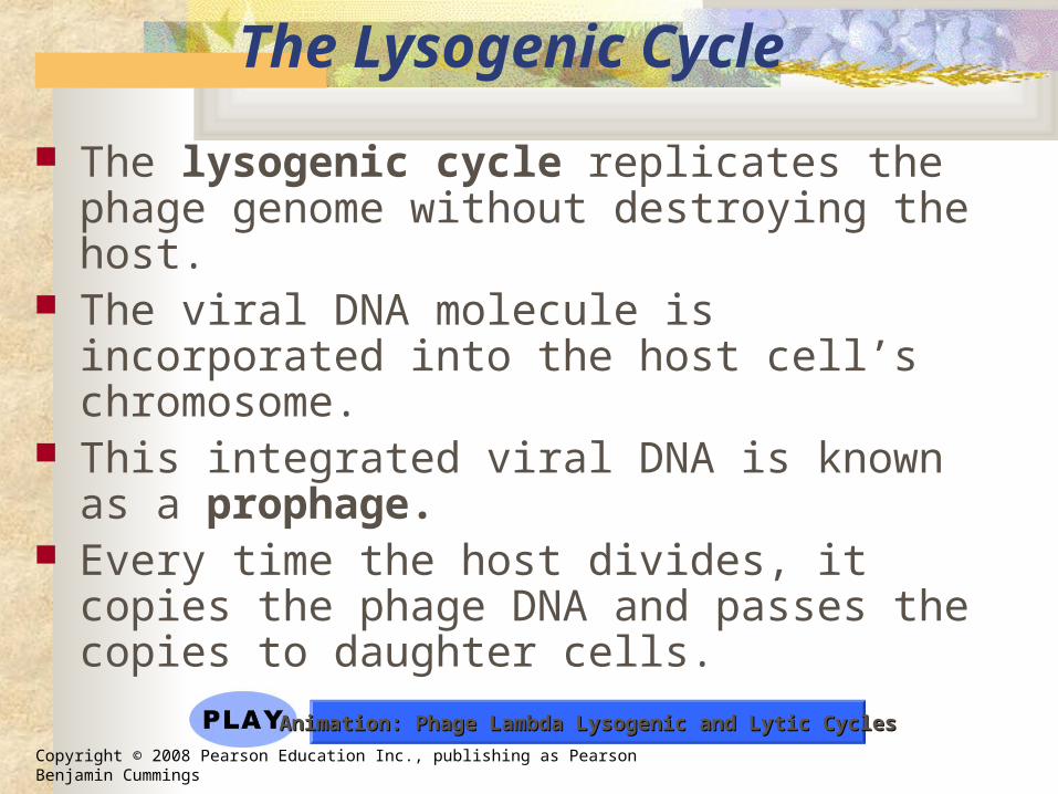
The Lysogenic Cycle
The lysogenic cycle replicates the phage genome without destroying the host.
The viral DNA molecule is incorporated into the host cell’s chromosome.
This integrated viral DNA is known as a prophage.
Every time the host divides, it copies the phage DNA and passes the copies to daughter cells.
Animation: Phage Lambda Lysogenic and Lytic CyclesAnimation: Phage Lambda Lysogenic and Lytic Cycles

Copyright © 2008 Pearson Education Inc., publishing as Pearson Benjamin Cummings
An environmental signal can trigger the virus genome to exit the bacterial chromosome and switch to the lytic mode.
Phages that use both the lytic and lysogenic cycles are called temperate phages.

Copyright © 2008 Pearson Education Inc., publishing as Pearson Benjamin Cummings
Fig. 19-6
PhageDNA
Phage
The phage injects its DNA.
Bacterialchromosome
Phage DNAcircularizes.
Daughter cellwith prophage
Occasionally, a prophageexits the bacterialchromosome,initiating a lytic cycle.
Cell divisionsproducepopulation ofbacteria infectedwith the prophage.
The cell lyses, releasing phages.
Lytic cycle
Lytic cycleis induced or Lysogenic cycle
is entered
Lysogenic cycle
Prophage
The bacterium reproduces,copying the prophage andtransmitting it to daughter cells.
Phage DNA integrates intothe bacterial chromosome,becoming a prophage.
New phage DNA and proteinsare synthesized andassembled into phages.
Copyright © 2008 Pearson Education Inc., publishing as Pearson Benjamin Cummings
Reproductive Cycles of Animal Viruses
• There are two key variables used to classify viruses that infect animals: DNA or RNA? Single-stranded or double-stranded?
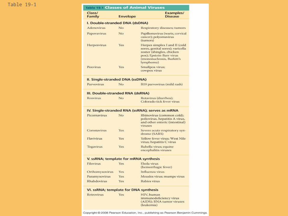
Table 19-1

Table 19-1a
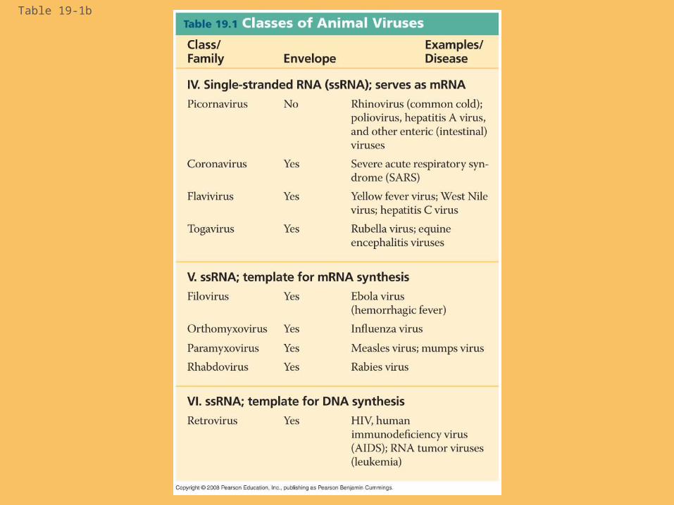
Table 19-1b

Copyright © 2008 Pearson Education Inc., publishing as Pearson Benjamin Cummings
Viral Envelopes
Many viruses that infect animals have a membranous envelope.
Viral glycoproteins on the envelope bind to specific receptor molecules on the surface of a host cell.
Some viral envelopes are formed from the host cell’s plasma membrane as the viral capsids exit.

Copyright © 2008 Pearson Education Inc., publishing as Pearson Benjamin Cummings
Other viral membranes form from the host’s nuclear envelope and are then replaced by an envelope made from Golgi apparatus membrane.

Copyright © 2008 Pearson Education Inc., publishing as Pearson Benjamin Cummings
Fig. 19-7
Capsid
RNA
Envelope (withglycoproteins)
Capsid and viral genomeenter the cell
HOST CELL
Viral genome (RNA)
Template
mRNA
ER
Glyco-proteins
Capsidproteins Copy of
genome (RNA)
New virus

Copyright © 2008 Pearson Education Inc., publishing as Pearson Benjamin Cummings
RNA as Viral Genetic Material
The broadest variety of RNA genomes is found in viruses that infect animals.
Retroviruses use reverse transcriptase to copy their RNA genome into DNA.
HIV (human immunodeficiency virus) is the retrovirus that causes AIDS (acquired immunodeficiency syndrome).

Fig. 19-8Glycoprotein Viral envelope
Capsid
RNA (twoidenticalstrands)Reverse
transcriptase HIV
HIVMembrane ofwhite blood cell
HIV entering a cell
0.25 µm
Viral RNA
RNA-DNAhybrid
HOST CELL
Reversetranscriptase
DNA
NUCLEUS
Provirus
ChromosomalDNA
RNA genomefor the next viralgeneration
mRNA
New virusNew HIV leaving a cell

Fig. 19-8aGlycoprotein
Reversetranscriptase HIV
RNA (twoidenticalstrands)
Capsid
Viral envelope
HOST CELL
Reversetranscriptase
Viral RNA
RNA-DNAhybrid
DNA
NUCLEUS
Provirus
ChromosomalDNA
RNA genomefor thenext viralgeneration
mRNA
New virus

Fig. 19-8b
HIVMembrane ofwhite blood cell
HIV entering a cell
0.25 µm
New HIV leaving a cell

Copyright © 2008 Pearson Education Inc., publishing as Pearson Benjamin Cummings
The viral DNA that is integrated into the host genome is called a provirus.
Unlike a prophage, a provirus remains a permanent resident of the host cell.
The host’s RNA polymerase transcribes the proviral DNA into RNA molecules.
The RNA molecules function both as mRNA for synthesis of viral proteins and as genomes for new virus particles released from the cell.
Animation: HIV Reproductive CycleAnimation: HIV Reproductive Cycle
Copyright © 2008 Pearson Education Inc., publishing as Pearson Benjamin Cummings
Evolution of Viruses
Viruses do not fit our definition of living organisms.
Since viruses can reproduce only within cells, they probably evolved as bits of cellular nucleic acid.
Candidates for the source of viral genomes are plasmids, circular DNA in bacteria and yeasts, and transposons, small mobile DNA segments.
Plasmids, transposons, and viruses are all mobile genetic elements.
Copyright © 2008 Pearson Education Inc., publishing as Pearson Benjamin Cummings
Mimivirus, a double-stranded DNA virus, is the largest virus yet discovered.
There is controversy about whether this virus evolved before or after cells.
Copyright © 2008 Pearson Education Inc., publishing as Pearson Benjamin Cummings
Concept 19.3: Viruses, viroids, and prions are formidable pathogens in animals and
plants
Diseases caused by viral infections affect humans, agricultural crops, and livestock worldwide.
Smaller, less complex entities called viroids and prions also cause disease in plants and animals, respectively.
Copyright © 2008 Pearson Education Inc., publishing as Pearson Benjamin Cummings
Viral Diseases in Animals
Viruses may damage or kill cells by causing the release of hydrolytic enzymes from lysosomes.
Some viruses cause infected cells to produce toxins that lead to disease symptoms.
Others have envelope proteins that are toxic.

Copyright © 2008 Pearson Education Inc., publishing as Pearson Benjamin Cummings
Vaccines are harmless derivatives of pathogenic microbes that stimulate the immune system to mount defenses against the actual pathogen.
Vaccines can prevent certain viral illnesses. Viral infections cannot be treated by
antibiotics. Antiviral drugs can help to treat, though not
cure, viral infections.
Copyright © 2008 Pearson Education Inc., publishing as Pearson Benjamin Cummings
Emerging Viruses
Emerging viruses are those that appear suddenly or suddenly come to the attention of scientists.
Severe acute respiratory syndrome (SARS) recently appeared in China.
Outbreaks of “new” viral diseases in humans are usually caused by existing viruses that expand their host territory.
Copyright © 2008 Pearson Education Inc., publishing as Pearson Benjamin Cummings
Flu epidemics are caused by new strains of influenza virus to which people have little immunity.
Viral diseases in a small isolated population can emerge and become global.
New viral diseases can emerge when viruses spread from animals to humans.
Viral strains that jump species can exchange genetic information with other viruses to which humans have no immunity.

Copyright © 2008 Pearson Education Inc., publishing as Pearson Benjamin Cummings
These strains can cause pandemics, global epidemics.
The “avian flu” is a virus that recently appeared in humans and originated in wild birds.

Fig. 19-9
(a) The 1918 flu pandemic
(b) Influenza A H5N1 virus
(c) Vaccinating ducks
0.5 µm

Fig. 19-9a
(a) The 1918 flu pandemic
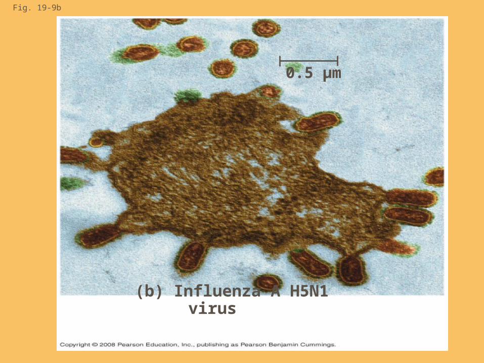
Fig. 19-9b
(b) Influenza A H5N1 virus
0.5 µm
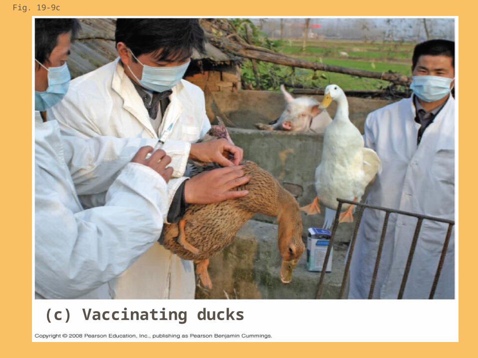
Fig. 19-9c
(c) Vaccinating ducks

Viral Diseases in Plants
More than 2,000 types of viral diseases of plants are known and cause spots on leaves and fruits, stunted growth, and damaged flowers or roots.
Most plant viruses have an RNA genome.
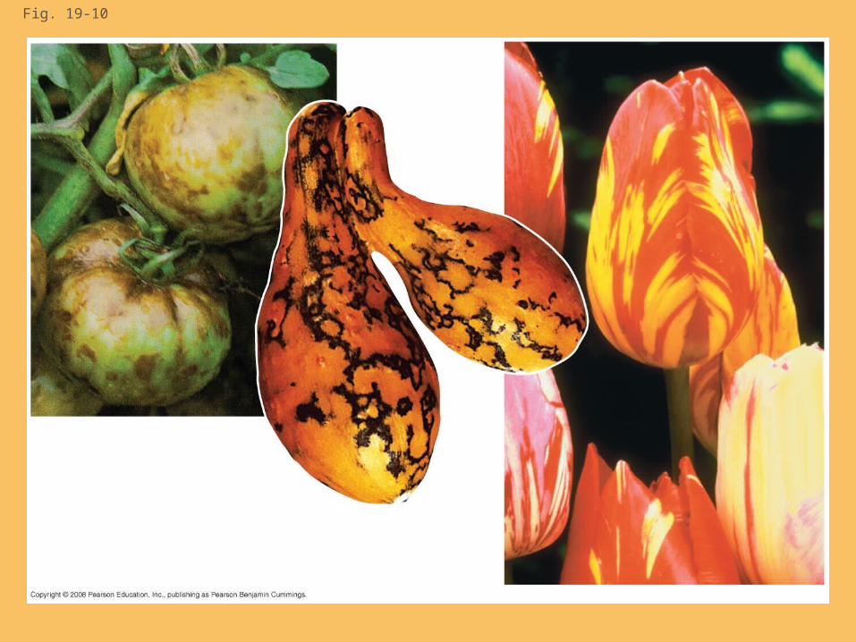
Fig. 19-10
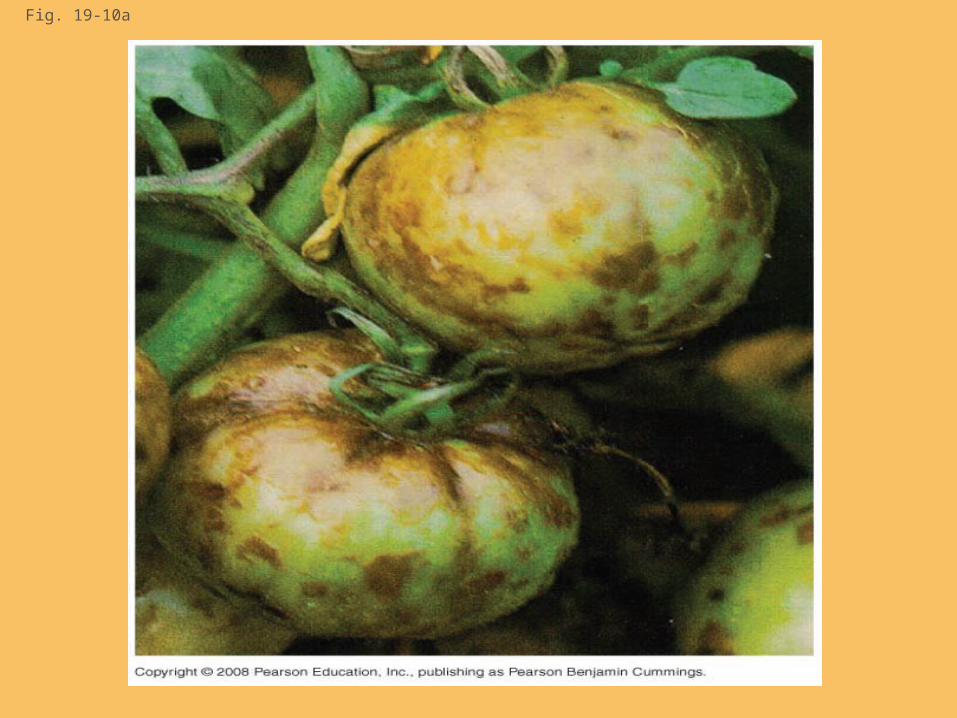
Fig. 19-10a

Fig. 19-10b

Fig. 19-10c

Copyright © 2008 Pearson Education Inc., publishing as Pearson Benjamin Cummings
Plant viruses spread disease in two major modes: Horizontal transmission, entering
through damaged cell walls. Vertical transmission, inheriting
the virus from a parent.
Copyright © 2008 Pearson Education Inc., publishing as Pearson Benjamin Cummings
Viroids and Prions: The Simplest Infectious Agents
Viroids are circular RNA molecules that infect plants and disrupt their growth.
Prions are slow-acting, virtually indestructible infectious proteins that cause brain diseases in mammals.
Prions propagate by converting normal proteins into the prion version
Scrapie in sheep, mad cow disease, and Creutzfeldt-Jakob disease in humans are all caused by prions.

Copyright © 2008 Pearson Education Inc., publishing as Pearson Benjamin Cummings
Fig. 19-11
Prion
Normalprotein
Originalprion
Newprion
Aggregatesof prions

Copyright © 2008 Pearson Education Inc., publishing as Pearson Benjamin Cummings
You should now be able to:
1. Explain how capsids and envelopes are formed.
2. Distinguish between the lytic and lysogenic reproductive cycles.
3. Explain why viruses are obligate intracellular parasites.
4. Describe the reproductive cycle of an HIV retrovirus
5. Describe three processes that lead to the emergence of new diseases.
6. Describe viroids and prions.

Copyright © 2008 Pearson Education Inc., publishing as Pearson Benjamin Cummings
Fig. 19-UN1
PhageDNA
Bacterialchromosome
The phage attaches to ahost cell and injects its DNA
Prophage
Lysogenic cycle• Temperate phage only• Genome integrates into bacterial chromosome as prophage, which (1) is replicated and passed on to daughter cells and (2) can be induced to leave the chromosome and initiate a lytic cycle
Lytic cycle• Virulent or temperate phage• Destruction of host DNA• Production of new phages• Lysis of host cell causes release of progeny phages
Copyright © 2008 Pearson Education Inc., publishing as Pearson Benjamin Cummings
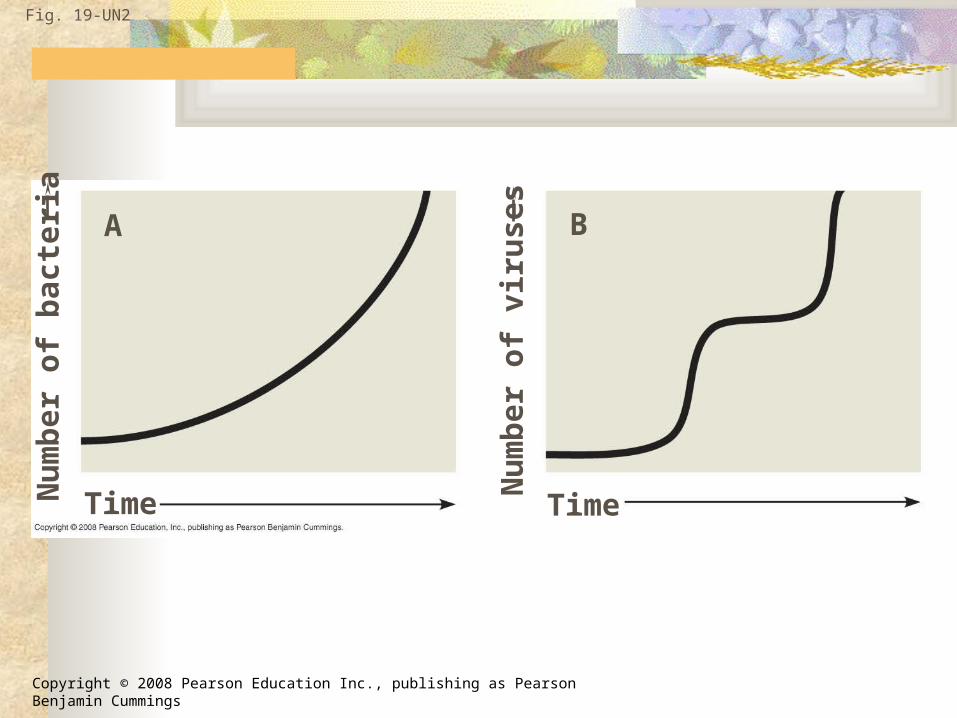
Fig. 19-UN2
Time Time
A B
Nu
mb
er
of
ba
cte
ria
Nu
mb
er
of
vir
use
s

Copyright © 2008 Pearson Education Inc., publishing as Pearson Benjamin Cummings
Fig. 19-UN3