chapter 2: basic radiobiology - international atomic … · · 2016-02-192.1 basic radiobiology ....
TRANSCRIPT

IAEA International Atomic Energy Agency
Slide set of 60 slides based on the chapter authored by R.G. Dale and J. Wondergem of the IAEA publication (ISBN 92-0-107304-6): Nuclear Medicine Physics: A Handbook for Teachers and Students
Objective: To familiarize with the possible effects induced by ionizing
radiation on living matter.
Chapter 2: Basic Radiobiology
Slide set prepared in 2015 by M. Cremonesi (IEO European Institute of Oncology, Milano, Italy)

IAEA Nuclear Medicine Physics: A Handbook for Teachers and Students – Chapter 2 – Slide 2/60 Nuclear Medicine Physics: A Handbook for Teachers and Students – Chapter 2 – Slide 2/60
2.1. Introduction
2.2. Radiation effects and timescales
2.3. Biological properties of ionizing radiation
2.4. Molecular effects of radiation and their modifiers
2.5. DNA damage and repair
2.6. Cellular effects of radiation
2.7. Gross radiation effects on tumours and tissues/organs
2.8. Special radiobiological consideration in targeted radionuclide therapy
CHAPTER 2 TABLE OF CONTENTS

IAEA Nuclear Medicine Physics: A Handbook for Teachers and Students – Chapter 2 – Slide 3/60 Nuclear Medicine Physics: A Handbook for Teachers and Students – Chapter 2 – Slide 3/60
Radiobiology is the qualitative and quantitative study of the effects of ionizing radiation on living matter.
Radiation may induce cells to become malignant, alter their functionality, or directly induce cell death.
Consideration of the associated radiobiology is: - important in diagnostic applications of radiation - essential in therapy applications of radiation.
2.1 BASIC RADIOBIOLOGY 2.1 Introduction

IAEA Nuclear Medicine Physics: A Handbook for Teachers and Students – Chapter 2 – Slide 4/60 Nuclear Medicine Physics: A Handbook for Teachers and Students – Chapter 2 – Slide 4/60
2.2 BASIC RADIOBIOLOGY
At the microscopic level, incident rays or particles may interact with orbital electrons within the cellular atoms and molecules to cause:
The irradiation of cellular material with such radiation gives rise to the production of a flux of energetic secondary particles (electrons).
- Excitation: raising a bound electron to a higher energy state; the electron does not have sufficient energy to leave the host atom. - Ionization, the electron receives sufficient energy to be ejected from its orbit and to leave the host atom. Ionizing radiation is able to induce electron ejection process.
Energetic and unbound, they are capable of migrating away from the site of production, interact with other atoms and molecules, and give up their energy to the surrounding medium.
2.2 Radiation effects and timescales
electron
e-
atom atom
(ionized)
e-
e- +

IAEA Nuclear Medicine Physics: A Handbook for Teachers and Students – Chapter 2 – Slide 5/60 Nuclear Medicine Physics: A Handbook for Teachers and Students – Chapter 2 – Slide 5/60
2.2 BASIC RADIOBIOLOGY
This energy absorption process gives rise to radicals and other chemical species → chemical interactions → cause of radiation damage.
Irrespective of the nature of the primary radiation (particles and/or electro-magnetic waves), energy is transferred to matter always via the secondary electrons which are produced.
Chemical changes operate over a short timescale (~10–5 s), but this period is a factor of ~1018 longer than the time taken for the original particle to traverse the cell nucleus. Thus, on the microscopic scale, there is a relatively long period during which chemical damage is inflicted.
Initial ionization events in the biological material (near-instantaneous at the microscopic level) are the precursors to a chain of subsequent events which may lead to the clinical (macroscopic) manifestation of radiation damage.
2.2 Radiation effects and timescales

IAEA Nuclear Medicine Physics: A Handbook for Teachers and Students – Chapter 2 – Slide 6/60 Nuclear Medicine Physics: A Handbook for Teachers and Students – Chapter 2 – Slide 6/60
Expression of cell death in individual lethally damaged cells occurs later, usually at the point when the cell next attempts to enter mitosis.
Gross (macroscopic and clinically observable) radiation effects are a result of the wholesale functional impairment that follows from lethal damage being inflicted to large numbers of cells or critical substructures.
In clinical studies, deleterious health effects may be seen long after the diagnostic test or treatment
The whole process may need months/ years
2.2 BASIC RADIOBIOLOGY 2.2 Radiation effects and timescales

IAEA Nuclear Medicine Physics: A Handbook for Teachers and Students – Chapter 2 – Slide 7/60 Nuclear Medicine Physics: A Handbook for Teachers and Students – Chapter 2 – Slide 7/60
2.3. BIOLOGICAL PROPERTIES OF IONIZING RADIATION
In nuclear medicine, four types of radiation which play a relevant role in tumour and normal tissue effects:
gamma radiation (γ)
beta radiation (β)
alpha particles (α)
Auger electrons (e-Auger)
2.3.1. Types of ionizing radiation
paper aluminum,
plastic lead
α
β
γ
Fermi level
Auger electron
Core Hole
Auger process
L2,3
L1
K

IAEA Nuclear Medicine Physics: A Handbook for Teachers and Students – Chapter 2 – Slide 8/60 Nuclear Medicine Physics: A Handbook for Teachers and Students – Chapter 2 – Slide 8/60
2.3. BIOLOGICAL PROPERTIES OF IONIZING RADIATION
electromagnetic (EM) radiation of high energy (usually >25 keV)
produced by subatomic particle interactions
as a stream of wave-like particle bundles (photons) moving at the speed of light
interaction properties governed mainly by their associated wavelength
Ionization behaviour of large numbers of photons can be accurately predicted; individual photon interactions occur at random and, while passing through matter, a photon may interact one or more times, or never
Gamma radiation
γ: very short wavelenght EM radiation
2.3.1.1. Gamma radiation
wavelenght frequency
long
short
low
high

IAEA Nuclear Medicine Physics: A Handbook for Teachers and Students – Chapter 2 – Slide 9/60 Nuclear Medicine Physics: A Handbook for Teachers and Students – Chapter 2 – Slide 9/60
undergo many ionizing events relatively close to the site of their creation → contribute mostly to the locally absorbed dose
In each interaction (normally involving a photoelectric, Compton, or pair production event), secondary particles are produced, usually electrons (directly ionizing) or another photon of reduced energy, which can undergo further interactions
secondary electrons
carry energy further away from the initial interaction site → following subsequent electron-producing interactions → responsible for the dose deposition at more distant sites from the original interaction
secondary photons
2.3. BIOLOGICAL PROPERTIES OF IONIZING RADIATION 2.3.1.1. Gamma radiation
IAEA Nuclear Medicine Physics: A Handbook for Teachers and Students – Chapter 2 – Slide 10/60 Nuclear Medicine Physics: A Handbook for Teachers and Students – Chapter 2 – Slide 10/60
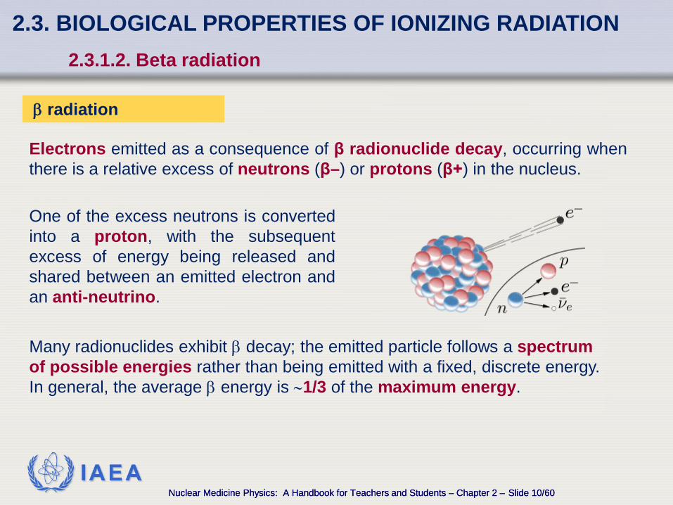
Electrons emitted as a consequence of β radionuclide decay, occurring when there is a relative excess of neutrons (β–) or protons (β+) in the nucleus.
β radiation
Many radionuclides exhibit β decay; the emitted particle follows a spectrum of possible energies rather than being emitted with a fixed, discrete energy. In general, the average β energy is ∼1/3 of the maximum energy.
One of the excess neutrons is converted into a proton, with the subsequent excess of energy being released and shared between an emitted electron and an anti-neutrino.
2.3. BIOLOGICAL PROPERTIES OF IONIZING RADIATION 2.3.1.2. Beta radiation

IAEA Nuclear Medicine Physics: A Handbook for Teachers and Students – Chapter 2 – Slide 11/60 Nuclear Medicine Physics: A Handbook for Teachers and Students – Chapter 2 – Slide 11/60
Most β emitting radionuclides also emit γ photons as a consequence of the initial β decay, leaving the daughter nucleus in an excited, metastable state. Since β particles are electrons, once ejected from the host atom, they behave exactly as the electrons created following the passage of a γ ray, giving up their energy (usually of the order of several hundred keV) to other atoms and molecules through a series of collisions. For radionuclides which emit both β particles and γ photons, it is usually the particulate radiation which delivers the greatest fraction of the radiation dose to the organ which has taken up the activity. E.g.:
- about 90% of the dose delivered to the thyroid by 131I is from the β component. - emissions contribute more significantly to the overall whole body dose
2.3. BIOLOGICAL PROPERTIES OF IONIZING RADIATION 2.3.1.2. Beta radiation

IAEA Nuclear Medicine Physics: A Handbook for Teachers and Students – Chapter 2 – Slide 12/60 Nuclear Medicine Physics: A Handbook for Teachers and Students – Chapter 2 – Slide 12/60
- emitted when heavy, unstable nuclides undergo decay. - helium nucleus (2 n + 2 p) emitted in a nuclear decay - mα ≅ 7000 mβ - twice the electronic charge of β - give up their energy over a very
short range (<100 µm).
α radiation
α particles usually possess energies in the MeV range and lose this energy in a short range making them biologically very efficacious, i.e. they possess a high LET (linear energy transfer; see Section 2.6.3) and are associated with high RBE (relative biological effectiveness; see Section 2.6.4).
2.3. BIOLOGICAL PROPERTIES OF IONIZING RADIATION 2.3.1.3. Alpha particles

IAEA Nuclear Medicine Physics: A Handbook for Teachers and Students – Chapter 2 – Slide 13/60 Nuclear Medicine Physics: A Handbook for Teachers and Students – Chapter 2 – Slide 13/60
Radionuclides which decay by electron capture or internal conversion leave the atom in a highly excited state with a vacancy in one of the inner shell electron orbitals. This vacancy is rapidly filled by either a fluorescent transition (characteristic X ray) or a non-radiative (Auger) transition: the energy gained by the electron transition to the deeper orbital is used to eject another electron from the same atom.
Auger electrons
Auger electrons are very short range, low energy particles often emitted in cascades, a consequence of the inner shell atomic vacancy that traverses up through the atom to the outermost orbital, ejecting additional electrons at each step. This cluster of very low energy electrons can produce ionization densities comparable to those produced by an α particle track. → Radionuclides which decay by electron capture and/or internal conversion can exhibit high-LET-like behavior close (within 2 nm) to the site of the decay.
2.3. BIOLOGICAL PROPERTIES OF IONIZING RADIATION 2.3.1.4. Auger electrons

IAEA Nuclear Medicine Physics: A Handbook for Teachers and Students – Chapter 2 – Slide 14/60 Nuclear Medicine Physics: A Handbook for Teachers and Students – Chapter 2 – Slide 14/60
2.4. MOLECULAR EFFECTS OF RADIATION AND THEIR MODIFIERS
Radiation effects on atoms or molecules which are not parts of the biological target. Cells exist in a rich aqueous environment → the majority of indirect actions involve the ionization or excitation of water molecules. The free radicals created may then migrate and damage the adjacent biological targets. Indirect action is the main cause of radiation damage and, in normally normoxic cells, accounts for ∼2/3 of the damage.
Ionization or excitation (via Coulomb interactions) of the atoms in the biological target → chain of events eventually leading to the observable (macroscopic) damage. In normally oxygenated mammalian cells, direct effects account for ∼1/3 of the damage for low LET radiations such as electrons and photons.
Radiation induced damage to biological targets may result from:
Direct action
Indirect action
predominant with high LET radiation, e.g. α, neutrons
predominant with low LET radiation, e.g. X, γ rays

IAEA Nuclear Medicine Physics: A Handbook for Teachers and Students – Chapter 2 – Slide 15/60 Nuclear Medicine Physics: A Handbook for Teachers and Students – Chapter 2 – Slide 15/60
2.4. MOLECULAR EFFECTS OF RADIATION AND THEIR MODIFIERS
Difference between direct and indirect damage to cellular DNA

IAEA Nuclear Medicine Physics: A Handbook for Teachers and Students – Chapter 2 – Slide 16/60 Nuclear Medicine Physics: A Handbook for Teachers and Students – Chapter 2 – Slide 16/60
2.4. MOLECULAR EFFECTS OF RADIATION AND THEIR MODIFIERS 2.4.1. Role of oxygen
Radiation effects may be influenced especially by the presence/absence of oxygen.
The free radicals (R) produced as a result of direct or indirect effects are very reactive and seek to interact with other molecules which can share/donate electrons.
Molecular oxygen (O2) has 2 unpaired electrons and readily reacts with free radicals, causing an increased likelihood that deoxyribonucleic acid (DNA) will be damaged by indirect process.
oxygen enhancement ratio (OER) to achieve
equivalent biological effect
Important reactions via which oxygen can increase biological damage are:
~ 3 for low LET radiation (as γ rays) D hypoxia
D in air OER =
~ 1 for high LET radiation (as α particles)

IAEA Nuclear Medicine Physics: A Handbook for Teachers and Students – Chapter 2 – Slide 17/60 Nuclear Medicine Physics: A Handbook for Teachers and Students – Chapter 2 – Slide 17/60
2.4. MOLECULAR EFFECTS OF RADIATION AND THEIR MODIFIERS 2.4.2. Bystander effects
Occur when a cell which has not been traversed by a charged particle is damaged as a result of radiation interactions occurring in neighbouring cells
Its discovery poses a challenge to the traditional view that all radiation damage stems from direct interactions of charged particles with critical cellular targets
It still remains controversial in radiobiology
A possible explanation is that irradiated cells may send out a stress signal to nearby cells → a response, e.g. the initiation of apoptosis, in those cells
It is probably most significant in radiation protection involving low doses since it amplifies the overall radiation effect in situations where not all of the cells in a tissue are subjected to particle transversal, i.e. the overall radiation risk to that tissue is higher than would be expected
Bystander effects

IAEA Nuclear Medicine Physics: A Handbook for Teachers and Students – Chapter 2 – Slide 18/60 Nuclear Medicine Physics: A Handbook for Teachers and Students – Chapter 2 – Slide 18/60
2.5. DNA DAMAGE AND REPAIR 2.5.1. DNA damage
DNA damage is the primary cause of cell death caused by radiation. Radiation exposure produces a wide range of lesions in the DNA:
DNA damage
The number of DNA lesions generated by irradiation is large, but there are a number of mechanisms for DNA repair → the percentage of lesions causing cell death is very small
single strand breaks (SSBs) double strand breaks (DSBs) base damage protein–DNA cross-links protein–protein cross-links
In the DNA of a cell, a dose of 1–2 Gy leads to base damages >1000 SSBs ~1000 DSBs ~40
IAEA Nuclear Medicine Physics: A Handbook for Teachers and Students – Chapter 2 – Slide 19/60 Nuclear Medicine Physics: A Handbook for Teachers and Students – Chapter 2 – Slide 19/60
2.5. DNA DAMAGE AND REPAIR 2.5.1. DNA damage
DSBs play a critical role in cell killing, carcinogenesis, hereditary effects
Experimental evidence:
the initially produced DSBs correlate with radiosensitivity and survival at lower dose
unrepaired or mis-repaired DSBs also correlate with survival after higher doses
there is causal link between the generation of DSBs and the induction of chromosomal translocations with carcinogenic potential

IAEA Nuclear Medicine Physics: A Handbook for Teachers and Students – Chapter 2 – Slide 20/60 Nuclear Medicine Physics: A Handbook for Teachers and Students – Chapter 2 – Slide 20/60
2.5. DNA DAMAGE AND REPAIR 2.5.2. DNA repair
Important for the recovery of cells from radiation and other damaging agents.
There are multiple enzymatic mechanisms for detecting and repairing radiation induced DNA damage.
DNA repair mechanisms
cleavage of the damaged DNA strand by enzymes that cleave the polynucleotide chain on either side of the damage, and enzymes which cleave the end of a polynucleotide chain allowing removal of a short segment containing the damaged region. DNA polymerase can then fill in the resulting gap using the opposite undamaged strand as a template.
base excision repair, mismatch repair, nucleotide excision repair
DNA repair mechanisms:
base oxidation, alkylation, strand intercalation
respond to damages as:
Excision repair

IAEA Nuclear Medicine Physics: A Handbook for Teachers and Students – Chapter 2 – Slide 21/60 Nuclear Medicine Physics: A Handbook for Teachers and Students – Chapter 2 – Slide 21/60
2.5. DNA DAMAGE AND REPAIR 2.5.2. DNA repair
For DSB, there are two primary repair pathways:
Repair operates on blunt ended DNA fragments. This process involves the repair proteins recognizing lesion termini, cleaning up the broken ends of the DNA molecule, and the final ligation of the broken ends. NHEJ operates throughout the cell cycle but dominates in G1/S-phases. The process is error-prone because it does not rely on sequence homology.
Non-homologous end joining
(NHEJ)
DSB repair utilizes sequence homology with an undamaged copy of the broken region and, hence, can only operate in late S- or G2-phases of the cell cycle. Undamaged DNA from both strands is used as templates to repair the damage. In contrast to NHEJ, homologous recombination is error-free.
Homologous recombination
unrepaired or misrepaired damage to DNA → mutations and/or chromosome damage in the cell.
cell death cancer or hereditary effects

IAEA Nuclear Medicine Physics: A Handbook for Teachers and Students – Chapter 2 – Slide 22/60 Nuclear Medicine Physics: A Handbook for Teachers and Students – Chapter 2 – Slide 22/60
2.6. CELLULAR EFFECTS OF RADIATION 2.6.1. Concept of cell death
Radiation doses of the order of several Gy may lead to cell loss. Cells are regarded as having been ‘killed’ by radiation if they have lost reproductive integrity, which can occur by apoptosis, necrosis, mitotic catastrophe or by induced senescence and may take a significant time.
cells attempt to divide without proper repair of DNA damage leading to a reproductive cell death which can occur in the first few cell divisions after irradiation, and with increasing frequency after increasing doses.
Apoptosis or programmed cell death can occur naturally or result from insult to the cell environment. It occurs in particular cell types after low doses of irradiation, e.g. lymphocytes, serous salivary gland cells, and certain cells in the stem cell zone in testis and intestinal crypts.
is a form of cell death associated with loss of cellular membrane activity. Cellular necrosis generally occurs after high radiation doses.
Necrosis
Senescent cells are metabolically active but have lost the ability to divide.
Senescence
Mitotic catastrophe

IAEA Nuclear Medicine Physics: A Handbook for Teachers and Students – Chapter 2 – Slide 23/60 Nuclear Medicine Physics: A Handbook for Teachers and Students – Chapter 2 – Slide 23/60
2.6. CELLULAR EFFECTS OF RADIATION 2.6.2. Cell survival curves
For quantitative understanding of biological responses to radiation: behaviour of the cell survival (dose response) characteristics structure and meaning of such curves role played by the various factors which influence radiation response
Typical shape of a cell survival curve for mammalian tissue: fractional survival of cells resulting from delivery of single acute doses of the specified radiation (in this case γ). Acute dose to mean a dose delivered at high dose rate, i.e. near instantaneously.
Radiation cell survival curve: Fraction of plated cells retaining colony forming ability vs. radiation absorbed dose.
Main characteristics: a finite initial slope (at zero dose) and a gradually increasing slope as dose increases

IAEA Nuclear Medicine Physics: A Handbook for Teachers and Students – Chapter 2 – Slide 24/60 Nuclear Medicine Physics: A Handbook for Teachers and Students – Chapter 2 – Slide 24/60
2.6. CELLULAR EFFECTS OF RADIATION 2.6.3. Dose deposition characteristics: linear energy transfer
The energy transfer to the absorbing medium is via secondary electrons created by the passage of the primary ionizing particle or ray. LET is a measure of the linear rate at which radiation is absorbed in the absorbing medium by the secondary particles and is defined by ICRU:
where dE is the average energy locally imparted to the medium by a charged particle of specified energy in traversing a distance dℓ.
dE dℓ
LET =
[LET ] = keV/ µm

IAEA Nuclear Medicine Physics: A Handbook for Teachers and Students – Chapter 2 – Slide 25/60 Nuclear Medicine Physics: A Handbook for Teachers and Students – Chapter 2 – Slide 25/60
2.6. CELLULAR EFFECTS OF RADIATION 2.6.3. Dose deposition characteristics: linear energy transfer
For radiobiological studies, the concept of LET is problematic since it relates to an average linear rate of energy deposition but, at the microscopic level (i.e. at dimensions comparable with the critical cellular targets), the energy deposited per unit length along different parts of a single track may vary dramatically.
The directly measured RBE is of much greater usefulness as an indicator of the differing biological efficacies of various radiation types.
As charged particles lose energy in their passage through a medium via the result of collision and ionizing processes, the LET rises steeply to its highest value towards the very end of their range. The change in LET value along the track length is one reason why average LET values correlate poorly with observed (i.e. macroscopic) biological effects.

IAEA Nuclear Medicine Physics: A Handbook for Teachers and Students – Chapter 2 – Slide 26/60 Nuclear Medicine Physics: A Handbook for Teachers and Students – Chapter 2 – Slide 26/60
2.6. CELLULAR EFFECTS OF RADIATION 2.6.4. Determination of relative biological effectiveness
For a given biological end point, the RBE of the high LET radiation is defined as:
In particular, the RBE of a radiation is defined as the ratio of the dose required to produce the same reduction in cell survival as a reference low LET radiation.
d low LET
d high LET
RBE =
d low LET or dL, d high LET or dH: isoeffective doses for the reference (low LET, 60Co γ rays or high energy (250 kVp) X rays) and high LET radiation.
dL
dH
=

IAEA Nuclear Medicine Physics: A Handbook for Teachers and Students – Chapter 2 – Slide 27/60 Nuclear Medicine Physics: A Handbook for Teachers and Students – Chapter 2 – Slide 27/60
2.6. CELLULAR EFFECTS OF RADIATION 2.6.4. Determination of relative biological effectiveness
If the cell survival curves are described in terms of the linear–quadratic (LQ) model, the surviving fraction S as a function of acute doses at low- (L) high- (H) LET is:
RBEs determined at any particular end-point (cell surviving fraction) vary with changing dose for a given radiation fraction size for a low LET radiation.
(αH and αL are the high and low LET linear radio-sensitivity constants)
The maximum RBE (RBEmax) occurs at zero dose and, in terms of microdosimetric theory, corresponds to

IAEA Nuclear Medicine Physics: A Handbook for Teachers and Students – Chapter 2 – Slide 28/60 Nuclear Medicine Physics: A Handbook for Teachers and Students – Chapter 2 – Slide 28/60
2.6. CELLULAR EFFECTS OF RADIATION 2.6.4. Determination of relative biological effectiveness
Relative biological effectiveness (RBE) as a function of the radiation dose per fraction, derived using: RBEmax = 5, RBEmin = 1 (α/β)L = 3 Gy, The general trend of a steadily falling RBE with increasing dose per fraction is independent of the chosen values.

IAEA Nuclear Medicine Physics: A Handbook for Teachers and Students – Chapter 2 – Slide 29/60 Nuclear Medicine Physics: A Handbook for Teachers and Students – Chapter 2 – Slide 29/60
2.6. CELLULAR EFFECTS OF RADIATION 2.6.4. Determination of relative biological effectiveness
If the quadratic radiosensitivity coefficients (βH and βL) are unchanged with changing LET (i.e. βH = βL), at high doses, the RBE tends to unity. However, this constancy of β has been challenged and, if β does change with LET, then RBE will tend asymptotically to an alternative minimum value (RBEmin) given by:
and the ‘working’ RBE at any given dose per fraction is given as
in terms of the low-LET dose per fraction dL
in terms of the high-LET dose per fraction dH

IAEA Nuclear Medicine Physics: A Handbook for Teachers and Students – Chapter 2 – Slide 30/60 Nuclear Medicine Physics: A Handbook for Teachers and Students – Chapter 2 – Slide 30/60
2.6. CELLULAR EFFECTS OF RADIATION 2.6.4. Determination of relative biological effectiveness
The rate of change of RBE with dose/fraction is influenced by the existence of a non-unity RBEmin parameter.
Even for a fixed value of RBEmax, the potential uncertainty in RBE at the fraction sizes might be very large if RBEmin is erroneously assumed to be unity.
These uncertainties would be compounded if there were an additional linkage between RBEmax and the tissue α/β value.
The assumption of a fixed value of RBE, if applied to all fraction sizes, could lead to gross clinical errors. The previous equations point out that determination of RBEs in a clinical setting is potentially complex and will depend on accurate knowledge of RBEmax and RBEmin (if it is not unity).

IAEA Nuclear Medicine Physics: A Handbook for Teachers and Students – Chapter 2 – Slide 31/60 Nuclear Medicine Physics: A Handbook for Teachers and Students – Chapter 2 – Slide 31/60
2.6. CELLULAR EFFECTS OF RADIATION 2.6.4. Determination of relative biological effectiveness
The RBE value at any dose fraction size will also be governed by the low-LET α/β ratio (a tissue dependent parameter which provides a measure of how tissues respond to changes in dose fractionation) and the dose fraction size (a purely physical parameter) at the point under consideration. The RBEmax value may itself be tissue dependent, likely being higher for the dose-limiting normal tissues than for tumours (as seen in clinical experience with neutron therapy, a variety of ion species as well as by theoretical microdosimetric studies).
This potentially deleterious effect may be offset by the fact that, in carbon- helium- and argon-ion beams, LET (and, hence, RBE) will vary along the track in such a way that it is low at the entry point (adjacent to normal tissues) and highest at the Bragg peak located in the tumour. Although this might be beneficial, it means that the local RBE is more spatially variable than is indicated by the low-LET dose per fraction dLequation.

IAEA Nuclear Medicine Physics: A Handbook for Teachers and Students – Chapter 2 – Slide 32/60 Nuclear Medicine Physics: A Handbook for Teachers and Students – Chapter 2 – Slide 32/60
2.6. CELLULAR EFFECTS OF RADIATION 2.6.4. Determination of relative biological effectiveness
Difficulties in setting reference doses for clinical inter-comparisons
determined at 2 Gy fractions on a biological system
representative end-point, (e.g. the overall late tolerance of
normal tissues)
Wambersie: distinction between the ‘reference’ RBE and the ‘clinical’ RBE.
as more clinical experience becomes available, a more
practical ‘clinical’ RBE evolves, this being the reference RBE
empirically weighted by collective clinical experience and by volume
effects related to the beam characteristics, geometry or
technical conditions
IAEA Nuclear Medicine Physics: A Handbook for Teachers and Students – Chapter 2 – Slide 33/60 Nuclear Medicine Physics: A Handbook for Teachers and Students – Chapter 2 – Slide 33/60
2.6. CELLULAR EFFECTS OF RADIATION 2.6.5. The dose rate effect and the concept of repeat treatments
The critical nuclear target (DNA) is subjected to a large deposition of energy which physically breaks both strands of the double helix structure and disrupts the code sufficiently to disallow any opportunity of repair. This process can be thought of as a single-hit process and the total amount of DNA damage is directly proportional (∝) to the dose delivered.
When mammalian cells are irradiated, it is helpful to visualize their subsequent death as resulting from either of two possible processes:
An ionizing event occurs and releases only sufficient energy to disrupt the coding carried by one strand of the DNA. If the irradiation continues, two outcomes are possible: either the broken strand will restore itself to its original state (no lethality) or, prior to full repair taking place, a second, independent radiation event may occur in the same location and damage the opposite strand of the DNA, a complementary action between the two damaged strands then leading to cell lethality in what is called a two-hit process.
1
2

IAEA Nuclear Medicine Physics: A Handbook for Teachers and Students – Chapter 2 – Slide 34/60 Nuclear Medicine Physics: A Handbook for Teachers and Students – Chapter 2 – Slide 34/60
2.6. CELLULAR EFFECTS OF RADIATION
2.6.5. The dose rate effect and the concept of repeat treatments
Once created, the radiation damage due to these two routes is indistinguishable (i.e. both processes are lethal). The observed radiation response characterized in the cell survival curve consists of two components: one linear (∝ dose) and the other quadratic (∝ dose2).
The amount of damage created in the second process is dependent on the ability of the second break to be induced before the first break has repaired itself, and, thus, is dependent on the dose rate.
This phenomenological description qualitatively explains the shape of a radiation survival curve, with a finite initial slope at low dose followed by an increasingly downward curvature as dose increases.
This route depends on two independent events, each having a probability ∝ to dose → the number of damaged DNA targets is ∝ to dose×dose: dose2

IAEA Nuclear Medicine Physics: A Handbook for Teachers and Students – Chapter 2 – Slide 35/60 Nuclear Medicine Physics: A Handbook for Teachers and Students – Chapter 2 – Slide 35/60
2.6. CELLULAR EFFECTS OF RADIATION
2.6.5. The dose rate effect and the concept of repeat treatments
different dose rates
Reducing the dose rate causes the shape of the response curve to become less ‘curvy’ than in the acute case, but the initial slope remains unchanged. When the doses are all delivered at a very low dose rate, as for most radionuclide therapies (e.g. 0.1–0.5 Gy/h), the response is essentially a straight line, when the curves are plotted on a log–linear scale. This means that the low dose response is purely exponential.
Range of response curves with doses delivered at different dose rates

IAEA Nuclear Medicine Physics: A Handbook for Teachers and Students – Chapter 2 – Slide 36/60 Nuclear Medicine Physics: A Handbook for Teachers and Students – Chapter 2 – Slide 36/60
2.6. CELLULAR EFFECTS OF RADIATION 2.6.6. The basic linear–quadratic model
The basic equation of the LQ model describes the shape of the cell survival curves and has a biophysical origin. Cell survival after delivery of an acute dose d is given is:
with α (Gy-1) and β (Gy-2) being the linear and quadratic sensitivity coefficients
If the treatment is repeated in N spaced fractions, the net survival is SN:

IAEA Nuclear Medicine Physics: A Handbook for Teachers and Students – Chapter 2 – Slide 37/60 Nuclear Medicine Physics: A Handbook for Teachers and Students – Chapter 2 – Slide 37/60
2.6. CELLULAR EFFECTS OF RADIATION 2.6.7. Modification to the linear–quadratic model for radionuclide therapies
Targeted radionuclide therapy normally involves irradiation
of the tumour/normal tissues at a dose rate which is not
constant but which reduces as treatment proceeds, as a
consequence of the combination of radionuclide decay and
biological clearance of the radiopharmaceutical.
A more extensive formulation of the LQ model is required.

IAEA Nuclear Medicine Physics: A Handbook for Teachers and Students – Chapter 2 – Slide 38/60 Nuclear Medicine Physics: A Handbook for Teachers and Students – Chapter 2 – Slide 38/60
2.6. CELLULAR EFFECTS OF RADIATION
2.6.8. Quantitative intercomparison of different treatment types
In the LQ modelling, the term called the ‘biological effective dose’ (BED) is used to assess and inter-compare different treatment types. BED is defined as:
The parameters α and β are rarely known in detail for individual tumours or tissues, but values of the ratio α/β (Gy) are becoming increasingly known from clinical and experimental data.
In general, α/β is systematically higher for tumours (5–20 Gy) than for critical, late-responding normal tissues (2–5 Gy).
This difference makes the BED concept useful in practice.

IAEA Nuclear Medicine Physics: A Handbook for Teachers and Students – Chapter 2 – Slide 39/60 Nuclear Medicine Physics: A Handbook for Teachers and Students – Chapter 2 – Slide 39/60
2.6. CELLULAR EFFECTS OF RADIATION 2.6.8. Quantitative intercomparison of different treatment types
For non-acute treatments (dose delivery is protracted over a long time period on account of a lower dose rate), the BED is re-written as:
where g(t) is a function of the time t taken for delivery
µ is the time constant relating to the repair of sublethal damage with tissue repair half-time T1/2
µ = 0.693/ T1/2

IAEA Nuclear Medicine Physics: A Handbook for Teachers and Students – Chapter 2 – Slide 40/60 Nuclear Medicine Physics: A Handbook for Teachers and Students – Chapter 2 – Slide 40/60
2.6. CELLULAR EFFECTS OF RADIATION
2.6.8. Quantitative intercomparison of different treatment types
For a treatment delivery at constant dose rate R, the delivered dose d is related to treatment time t via d = R × t, thus:
When t > 12 h the equation simplifies to:

IAEA Nuclear Medicine Physics: A Handbook for Teachers and Students – Chapter 2 – Slide 41/60 Nuclear Medicine Physics: A Handbook for Teachers and Students – Chapter 2 – Slide 41/60
2.6. CELLULAR EFFECTS OF RADIATION
2.6.9. Cellular recovery processes
At lower doses and dose rates (multiple exposures), cellular recovery may play an important role in the fixation of the radiation damage.
There are three broad types of cellular radiation damage:
in which partially damaged DNA is left with sufficient capacity to restore itself over a period of a few hours, provided there is no further damage during the repair period
in which the cellular DNA is irreversibly damaged to such an extent that the cell dies or loses its proliferative capacity
Lethal damage
Sublethal damage
Potentially lethal
damage
in which repair of what would normally be a lethal event is made possible by manipulation of the post-irradiation cellular environment.

IAEA Nuclear Medicine Physics: A Handbook for Teachers and Students – Chapter 2 – Slide 42/60 Nuclear Medicine Physics: A Handbook for Teachers and Students – Chapter 2 – Slide 42/60
2.6. CELLULAR EFFECTS OF RADIATION
2.6.10. Consequence of radionuclide heterogeneity
The one possible exception would be if a radiopharmaceutical would selectively localize at sensitive target cells, within an organ, that are key for organ regeneration or function, e.g. crypt cells in the colon.
The effectiveness per unit dose of a radiopharmaceutical depends on the heterogeneity of the radionuclide distribution
The cellular response also depends on microdosimetry, especially if the radiopharmaceutical selectively localizes on the cell surface or internalizes within a certain cohort of cells within a tumour/normal organ. These radiolabels may exhibit geometric enhancement factors that modulate response (see ICRU Report 67)
Global non-uniformity of a source distribution, which results in pockets of cells (tumour or normal tissue) receiving less than the average dose, almost always leads to a greater fraction of cell survivors, than if all cells receive a uniform dose

IAEA Nuclear Medicine Physics: A Handbook for Teachers and Students – Chapter 2 – Slide 43/60 Nuclear Medicine Physics: A Handbook for Teachers and Students – Chapter 2 – Slide 43/60
2.7. GROSS RADIATION EFFECTS ON TUMOURS AND TISSUES/ORGANS
2.7.1. Classification of radiation damage (early versus late)
Clinically observed radiation effects in whole tissues or organs reflect the damage inflicted to large numbers of constituent cells and, thus, appear on a timescale which is governed largely by the underlying proliferation rates of those cells. Such observable effects are classified depending on the speed at which they manifest themselves following irradiation as:
appear within days, weeks or months of irradiation and are associated with fast-proliferating epithelial tissues, e.g. bone marrow, mucosa, intestinal tract, etc.
Cells lethally affected by radiation may continue to function, only dying when attempting to undergo subsequent cell division (mitosis).
Late effects appear months or years after irradiation and appear in structures which proliferate very slowly, e.g. kidney.
Early (or acute) effects

IAEA Nuclear Medicine Physics: A Handbook for Teachers and Students – Chapter 2 – Slide 44/60 Nuclear Medicine Physics: A Handbook for Teachers and Students – Chapter 2 – Slide 44/60
2.7. GROSS RADIATION EFFECTS ON TUMOURS AND TISSUES/ORGANS
2.7.1. Classification of radiation damage (early versus late)
In most types of radiotherapy, late effects are considered to be most critical and generally limit the total dose which may be delivered to the tumour.
If the tolerance of the late-responding tissues is exceeded, the subsequent reactions may seriously affect mobility, quality of life, even be life threatening. Such problems arise long after the treatment and are impossible to correct.
Acute reactions in external beam radiotherapy (EBRT) are usually transient and easier to control by adjustment of the treatment dose delivery pattern and/or simple medication. In radionuclide therapies, acute radiation toxicities are generally possible to circumvent once they begin to occur (e.g. by accelerating clearance of the radiopharmaceutical). Chronic toxicities (e.g. to the kidney) usually occur at times which are long relative to the lifetime of the radionuclide. Safe activities of therapeutic radionuclides should be administered, taking into account dose limiting constraints.

IAEA Nuclear Medicine Physics: A Handbook for Teachers and Students – Chapter 2 – Slide 45/60 Nuclear Medicine Physics: A Handbook for Teachers and Students – Chapter 2 – Slide 45/60
2.7. GROSS RADIATION EFFECTS ON TUMOURS AND TISSUES/ORGANS
2.7.2. Determinants of tumour response
The potential advantage of radionuclide therapy over other forms of radiation therapy is its ability to deliver dose to the local disease and to occult tumour deposits.
In nuclear medicine, the primary determinants of treatment effectiveness are:
tumour specificity of the radionuclide carrier. homogeneity of uptake of the carrier within the targeted tumour(s). intrinsic RBE of the radiation used for the therapy, determined primarily by the nature of the radionuclide emissions (e.g. α, β, γ, Auger e-…) range of the particles, as determined by their energies. total dose delivered. responsiveness of the targeted tumour cells to radiation.
radiobiological properties such as the cellular radiosensitivity, variations of sensitivity within the cell cycle, oxygen status of the cells (fully oxic, partially oxic or hypoxic), ability of the cells to recover from sublethal damage, degree to which tumour growth (re-population) may occur during therapy.

IAEA Nuclear Medicine Physics: A Handbook for Teachers and Students – Chapter 2 – Slide 46/60 Nuclear Medicine Physics: A Handbook for Teachers and Students – Chapter 2 – Slide 46/60
2.7. GROSS RADIATION EFFECTS ON TUMOURS AND TISSUES/ORGANS
2.7.2. Determinants of tumour response
These factors are complementary and interactive, and should not be considered in isolation from each other. Thus, for example, significant non-uniformity of tumour uptake may result in activity/dose ‘cold spots’, but the detrimental potential of these might be offset by the selection of a radionuclide which emits particles of sufficient range to produce a cross-fire effect within the cold spots from those adjacent cells which are properly targeted. The significance of cold spot and cross-fire effects is further dependent on the size of the tumour deposit under consideration.

IAEA Nuclear Medicine Physics: A Handbook for Teachers and Students – Chapter 2 – Slide 47/60 Nuclear Medicine Physics: A Handbook for Teachers and Students – Chapter 2 – Slide 47/60
2.7. GROSS RADIATION EFFECTS ON TUMOURS AND TISSUES/ORGANS
2.7.3. The concept of therapeutic index in radiation therapy and radionuclide therapy
The therapeutic index (or ‘therapeutic ratio’)* of a particular radiation treatment is a measure of the damage to the tumour vs. the damage to critical normal structures.
(*) The therapeutic index can be considered as a qualitative concept, quantitative definitions are not necessary. Any new treatment improving tumour control and/or reducing morbidity is said to be associated with an improved therapeutic index.
high therapeutic index → good tumour control, low normal tissue morbidity low therapeutic index → low tumour control, high morbidity.
normal tissues at risk are those immediately adjacent to the tumour. Doses to the normal tissues (and risk of toxicity) may be reduced by a good combination of physical and radiobiological factors.
tumour may be single/discrete or may consist of distributed masses/ metastases within the body. Normal tissues at risk may be widely distributed and may be a reflection of the particular uptake pattern of the targeting compound used for the therapy.
EBRT
Targeted radionuclide
therapy

IAEA Nuclear Medicine Physics: A Handbook for Teachers and Students – Chapter 2 – Slide 48/60 Nuclear Medicine Physics: A Handbook for Teachers and Students – Chapter 2 – Slide 48/60
2.7. GROSS RADIATION EFFECTS ON TUMOURS AND TISSUES/ORGANS
2.7.4. Long term concerns: stochastic and deterministic effects
Stochastic effects (e.g. hereditary damage, cancer induction) are those for which the likelihood of them occurring is dose related, but the severity of the resultant condition is not related to the dose received.
Deterministic effects (e.g. cataract induction, general radiation syndromes, bone marrow ablation, etc.) manifest with a severity which is dose related.
From diagnostic uses of radionuclides predominantly stochastic effects need to be considered as potential side effects, although deterministic damage may result if the embryo or fetus is irradiated.
For radionuclide therapy applications, the concerns relate to both stochastic and deterministic effects.
The radiation detriment from radiation exposure may be classified as stochastic or deterministic in nature.

IAEA Nuclear Medicine Physics: A Handbook for Teachers and Students – Chapter 2 – Slide 49/60 Nuclear Medicine Physics: A Handbook for Teachers and Students – Chapter 2 – Slide 49/60
2.8. SPECIAL RADIOBIOLOGICAL CONSIDERATIONS IN TARGETED RADIONUCLIDE THERAPY
2.8.1. Radionuclide targeting
very promising approach for the treatment of wide-spread metastasis and disseminated tumour cells to deliver therapeutic irradiation doses to the tumour while sparing normal tissues by targeting a structure that is abundant in tumour cells, but rare in normal tissues
Tumour targeted radiotherapy
antibodies labelled with a therapeutic radionuclide acting against a specific tumour target. (e.g. 131I-tositumomab (Bexxar®) and 90Y-ibritumomab tiuxetan (Zevalin® in the treatment of non-Hodgkin’s lymphoma)
epidermal growth factor (EGF) labelled with 125I which binds EGF receptors. EGF receptors are overexpressed on tumour cells in many malignancies such as highly malignant gliomas
At present, several other radiolabelled antibodies/molecules are being used in experi-mental models and in clinical trials to study their feasibility in other types of cancer.

IAEA Nuclear Medicine Physics: A Handbook for Teachers and Students – Chapter 2 – Slide 50/60 Nuclear Medicine Physics: A Handbook for Teachers and Students – Chapter 2 – Slide 50/60
2.8. SPECIAL RADIOBIOLOGICAL CONSIDERATIONS IN TARGETED RADIONUCLIDE THERAPY
2.8.2. Whole body irradiation
involves controlled irradiation of a carefully delineated target volume. Normal structures adjacent to the tumour will likely receive a dose, in some cases moderately high, but the volumes involved are relatively small. The rest of the body receives only a minimal dose, mostly arising from radiation scattered within the patient from the target volume and from a small amount of leakage radiation emanating from the treatment machine outside the body.
commonly administered intravenously, give rise to substantial whole body doses and doses to the radiation sensitive bone marrow. Once the untargeted activity is removed from the blood, it may give rise to substantial doses in normal structures, especially the kidneys. Furthermore, the activity taken up by the kidneys and targeted tumour deposits may (if γ ray emissions are involved) continue to irradiate the rest of the body.
Conventional EBRT
Targeted radionuclide
therapies

IAEA Nuclear Medicine Physics: A Handbook for Teachers and Students – Chapter 2 – Slide 51/60 Nuclear Medicine Physics: A Handbook for Teachers and Students – Chapter 2 – Slide 51/60
2.8. SPECIAL RADIOBIOLOGICAL CONSIDERATIONS IN TARGETED RADIONUCLIDE THERAPY
2.8.3. Critical normal tissues for radiation and radionuclide therapies
Radiation doses used in radionuclide therapies are much higher than for diagnosis; systemic therapies → retention of the pharmaceuticals within the blood; increased accumulation of radionuclides in non-tumour cells
Very sensitive towards ionizing radiation. Exposure with high doses → rapid depression of white blood cells followed by platelet depression a few weeks later, and in a later stage (∼1 month after exposure) also by depression of the red blood cells. Patients could suffer from infections, bleeding and anaemia.
possible unwanted toxicities main critical organs: bone marrow, kidney, liver, intestinal tract, lungs
Bone marrow

IAEA Nuclear Medicine Physics: A Handbook for Teachers and Students – Chapter 2 – Slide 52/60 Nuclear Medicine Physics: A Handbook for Teachers and Students – Chapter 2 – Slide 52/60
radiation induced damage several months after exposure. A reduction of proximal tubule cells is observed. These pathological changes finally lead to nephropathy
Hepatocytes are the radiosensitive targets. The life-span of the cells is about a year → deterioration of liver function apparent 3 -9 months after exposure
GI tract
characterized by de-population of the intestinal mucosa (usually 3-10 days after) leading to prolonged diarrhoea, dehydration, loss of weight, etc.
Kidneys
radiation induced damage several months after exposure. Pulmonary damage is observed as acute pneumonitis and later fibrosis
Liver
Lungs
2.8. SPECIAL RADIOBIOLOGICAL CONSIDERATIONS IN TARGETED RADIONUCLIDE THERAPY
2.8.3. Critical normal tissues for radiation and radionuclide therapies

IAEA Nuclear Medicine Physics: A Handbook for Teachers and Students – Chapter 2 – Slide 53/60 Nuclear Medicine Physics: A Handbook for Teachers and Students – Chapter 2 – Slide 53/60
2.8. SPECIAL RADIOBIOLOGICAL CONSIDERATIONS IN TARGETED RADIONUCLIDE THERAPY
2.8.3. Critical normal tissues for radiation and radionuclide therapies
Radionuclide therapy: diversity of radiopharmaceuticals, with different pharmacokinetics and biodistribution → different responses / tolerances.
If radionuclides are tightly conjugated to a targeting molecule, then the biodistribution and clearance is determined by that molecule. For high molecular weight targeting agents, such as an antibody injected intravenously, the slow plasma clearance results in marrow toxicity. For smaller radiolabelled peptides, renal toxicity becomes of concern.
Determinants of normal tissue response:
radionuclide employed E.g. isotopes of iodine localize in the thyroid, salivary glands, stomach, bladder. Strontium, yttrium, samarium, fluorine, radium concentrate in bone. Several radiometals, such as bismuth, can accumulate in the kidney
radiolabelled molecules

IAEA Nuclear Medicine Physics: A Handbook for Teachers and Students – Chapter 2 – Slide 54/60 Nuclear Medicine Physics: A Handbook for Teachers and Students – Chapter 2 – Slide 54/60
2.8. SPECIAL RADIOBIOLOGICAL CONSIDERATIONS IN TARGETED RADIONUCLIDE THERAPY
2.8.3. Critical normal tissues for radiation and radionuclide therapies
When studying a new radiopharmaceutical or molecular imaging agent it is always important to study in detail the biodistribution at trace doses, to ensure the absence of radionuclide sequestration within potentially sensitive tissue, such as the retina of the eye or the germ cells of the testes. Meredith et al. (2008) offers a review of normal tissue toxicities resulting from radionuclide therapies.

IAEA Nuclear Medicine Physics: A Handbook for Teachers and Students – Chapter 2 – Slide 55/60 Nuclear Medicine Physics: A Handbook for Teachers and Students – Chapter 2 – Slide 55/60
2.8. SPECIAL RADIOBIOLOGICAL CONSIDERATIONS IN TARGETED RADIONUCLIDE THERAPY
2.8.4. Imaging the radiobiology of tumours
A tracer that is becoming more widely available for PET imaging is fluorothymidine (FLT). It is selectively entrapped within cells that are progressing through S-phase (DNA replication) of the cell cycle → signal ∝ to cell proliferation, minimizing the signal from cells in G0 or in cell cycle arrest.
Ability to identify only replicating cells separate from all tumour cells within the tumour volume identified by CT → more accurate measures of the initial viable tumour burden and tumour response.
The development of molecular imaging using PET has given rise to new radiotracers which have the potential to assess radiobiological features of relevance for therapy planning.
Replicating cells, tumour response

IAEA Nuclear Medicine Physics: A Handbook for Teachers and Students – Chapter 2 – Slide 56/60 Nuclear Medicine Physics: A Handbook for Teachers and Students – Chapter 2 – Slide 56/60
2.8. SPECIAL RADIOBIOLOGICAL CONSIDERATIONS IN TARGETED RADIONUCLIDE THERAPY
2.8.4. Imaging the radiobiology of tumours
Complementary to measuring tumour response is the measurement of therapeutic efficacy through radiotracers that selectively target cell death. Radiotracers such as radiolabelled annexin V are under development to selectively bind to receptors expressed on cells undergoing programmed cell death.
Cells within a tumour microenvironmental region of low partial oxygen pressure (hypoxia), exhibit a great radio-resistance to radiation and chemotherapy relative to those under normoxic conditions. PET radiotracers under evaluation for imaging tumour hypoxia are, e.g. fluoromisonidazole (18F-FMISO), fluoroazomycin arabinoside (18F-FAZA) and copper-diacetyl-bis(N4-methylthiosemicarbazone) (64Cu-ATSM).
The ability to measure the radiobiological attributes of a tumour prior to therapy may provide invaluable information about relative resistance/ aggressiveness of tumours → improved management of patients.
Selectively targeting cell death
Hypoxia, resistance

IAEA Nuclear Medicine Physics: A Handbook for Teachers and Students – Chapter 2 – Slide 57/60 Nuclear Medicine Physics: A Handbook for Teachers and Students – Chapter 2 – Slide 57/60
2.8. SPECIAL RADIOBIOLOGICAL CONSIDERATIONS IN TARGETED RADIONUCLIDE THERAPY
2.8.5. Choice of radionuclide to maximize therapeutic index
The choice of the optimum radionuclide to maximize the therapeutic index in clinical therapeutic applications depends on several factors:
It should depend on the type of tumour treated. For leukaemia or micro-metastatic deposits (individual/small clusters of tumour cells) → distinct advantage of radionuclides which emit very short range particles; α particle ranges <100 μm in tissue → advantage of α particle emitters, if the targeting molecule were able to reach all tumour cells.
However: α-emitting radionuclides are not widely available and extremely expensive, and the short α range can be a disadvantage for bulk tumours.
range of the emitted particles from the radionuclide 1

IAEA Nuclear Medicine Physics: A Handbook for Teachers and Students – Chapter 2 – Slide 58/60 Nuclear Medicine Physics: A Handbook for Teachers and Students – Chapter 2 – Slide 58/60
For these reasons almost all therapeutic radionuclides consist of medium (131I) or long range. (90Y, 186Re) β emitters: advantageous for treating solid tumours for which target receptor (antigen) expression may be heterogeneous, or with non-uniform delivery, due to greater cross-fire range of their β emissions (up to a 1 cm range in unit density tissue)
2.8. SPECIAL RADIOBIOLOGICAL CONSIDERATIONS IN TARGETED RADIONUCLIDE THERAPY
2.8.5. Choice of radionuclide to maximize therapeutic index
It is necessary to consider ease and stability of the radiolabelled end product radiochemistry.
radiochemistry 2

IAEA Nuclear Medicine Physics: A Handbook for Teachers and Students – Chapter 2 – Slide 59/60 Nuclear Medicine Physics: A Handbook for Teachers and Students – Chapter 2 – Slide 59/60
2.8. SPECIAL RADIOBIOLOGICAL CONSIDERATIONS IN TARGETED RADIONUCLIDE THERAPY
2.8.5. Choice of radionuclide to maximize therapeutic index
If the T1/2 is too short, then the radiolabelled agent may have insufficient time to reach the tumor target → minimal therapeutic index.
Increasing T1/2 increases the therapeutic index, but renders the patient radioactive for a longer period of time → prolonged confinement, greater expense and radiation risks to staff and family. Pure β emitting radionuclides (e.g.90Y, 32P) have advantages in that they minimize the exposure to personnel assisting the patient.
The T1/2 of the radionuclide should ideally match the biological uptake and retention kinetics of the tumour-targeting carrier. For large protein (e.g. antibodies), radionuclides with T1/2 of several days are required to optimize the therapeutic index. For smaller molecular targeting agents (e.g. peptides), radionuclides with short T1/2 may be better suited to minimize radioactive waste.
choice of radionuclide half-life (T1/2) 3

IAEA Nuclear Medicine Physics: A Handbook for Teachers and Students – Chapter 2 – Slide 60/60 Nuclear Medicine Physics: A Handbook for Teachers and Students – Chapter 2 – Slide 60/60
BIBLIOGRAPHY
Dale RG, Jones B. (Eds) Radiobiological Modelling in Radiation Oncology, The British Institute of Radiology, London (2007).
Hall EJ, Giacca AJ. Radiobiology for the Radiologist, 6th edn, Lippincott,
Williams and Wilkins, Philadelphia, PA (2006).
ICRU - INTERNATIONAL COMMISSION ON RADIATION UNITS, Absorbed-dose Specification in Nuclear Medicine, Rep. 67, Nuclear Technology Publishing, Ashford, United Kingdom (2002).
Meredith R, Wessels B, Knox S. Risks to normal tissue from radionuclide therapy, Semin. Nucl. Med. 38 (2008) 347–357.