chapter 2 neurons and glia. introduction “neurophilosophy” –brain is the origin of mental...
Post on 22-Dec-2015
227 views
TRANSCRIPT
Chapter 2 Neurons and Glia
Introduction
• “Neurophilosophy”– Brain is the origin of mental abilities
• Glia and Neurons– Glia: Insulates, supports, and nourishes neurons– Neurons
• Process information• Sense environmental changes • Communicate changes to other neurons• Command body response
The Neuron Doctrine
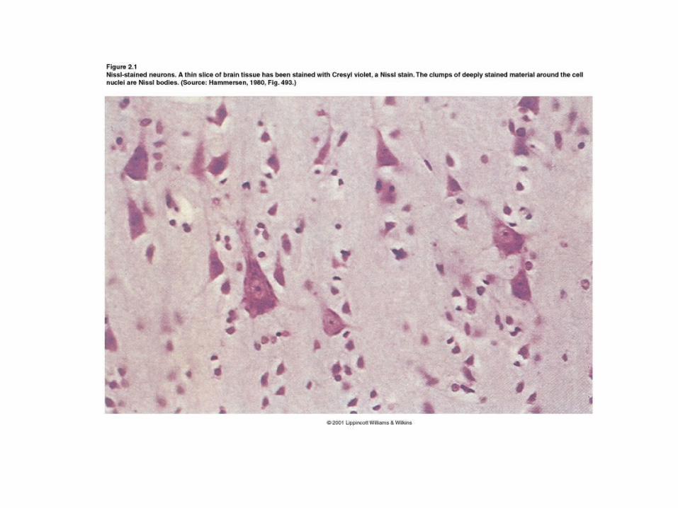
• Histology– Study of tissue structure– The Nissl Stain
• Facilitates the study of cytoarchitecture in the CNS
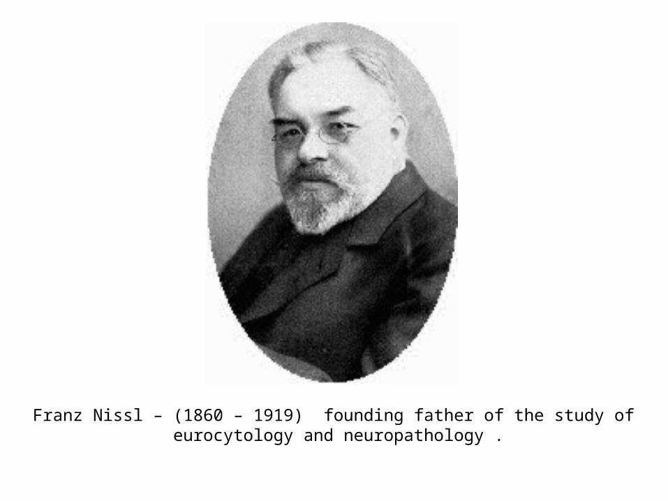
Franz Nissl – (1860 – 1919) founding father of the study of eurocytology and neuropathology .
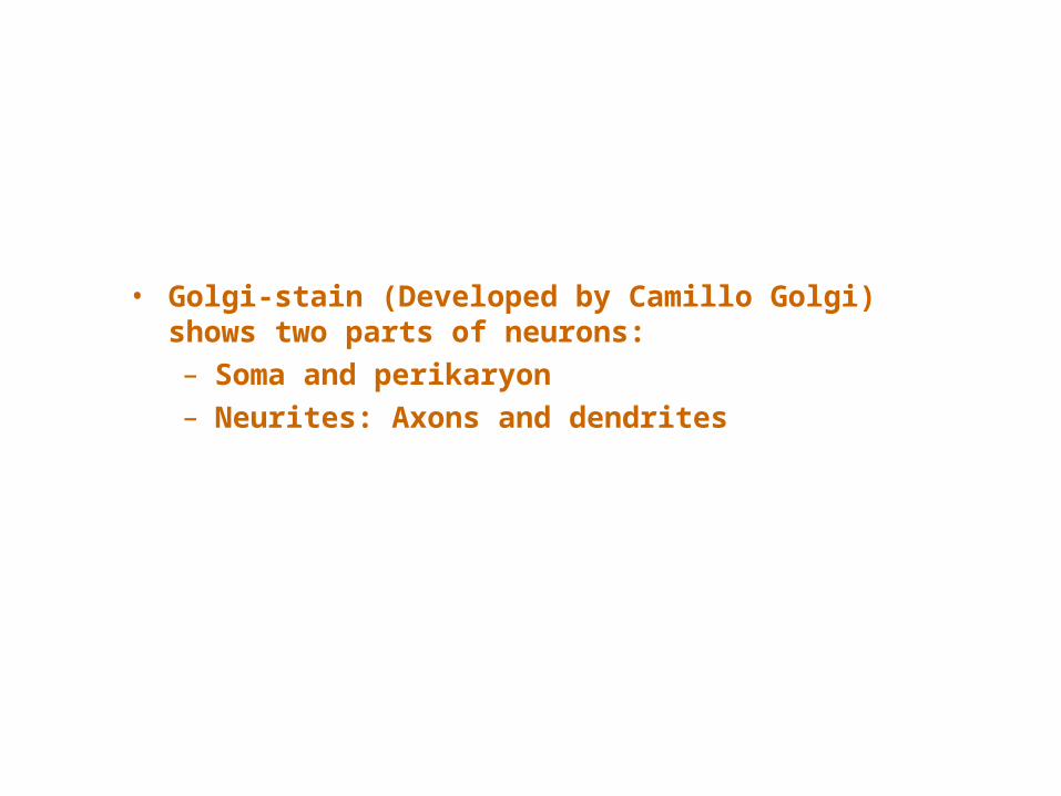
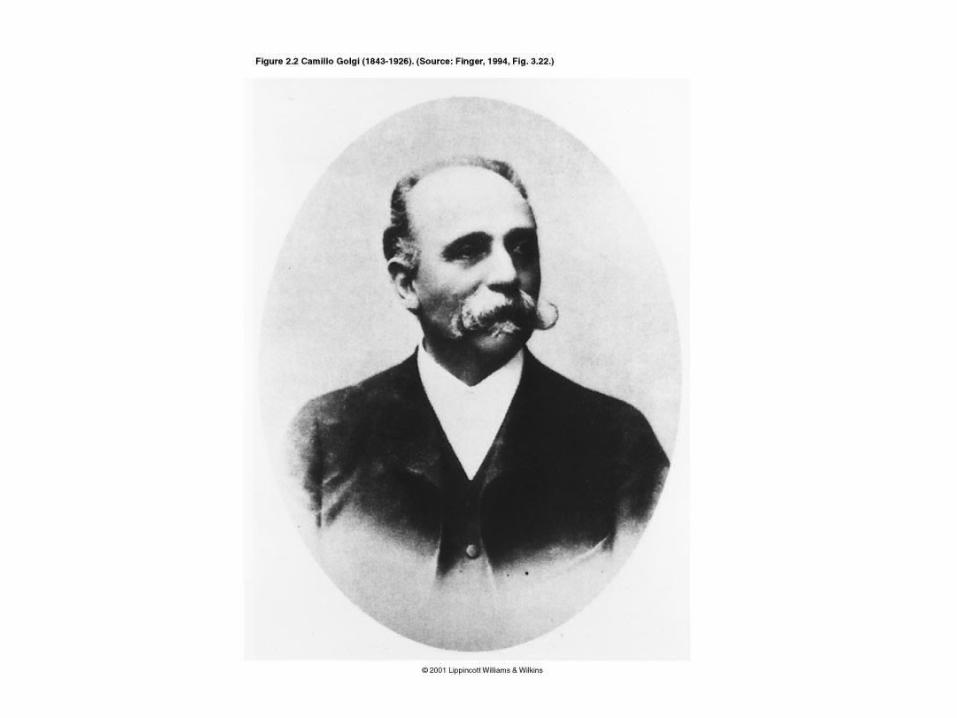
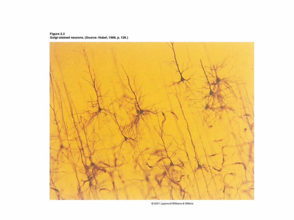
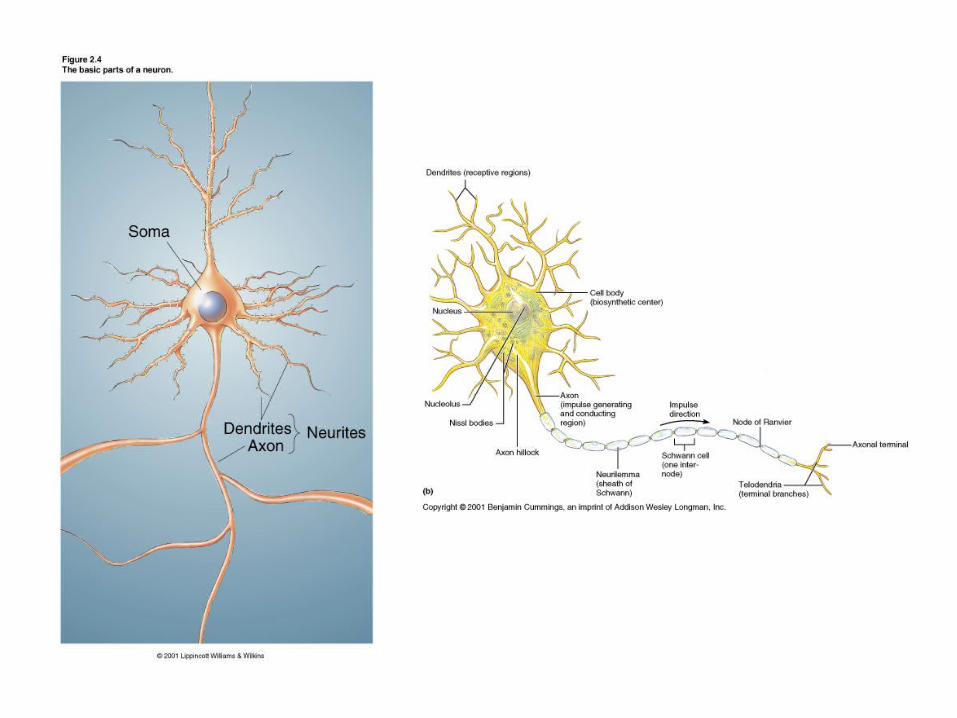
• Golgi-stain (Developed by Camillo Golgi) shows two parts of neurons:– Soma and perikaryon– Neurites: Axons and dendrites
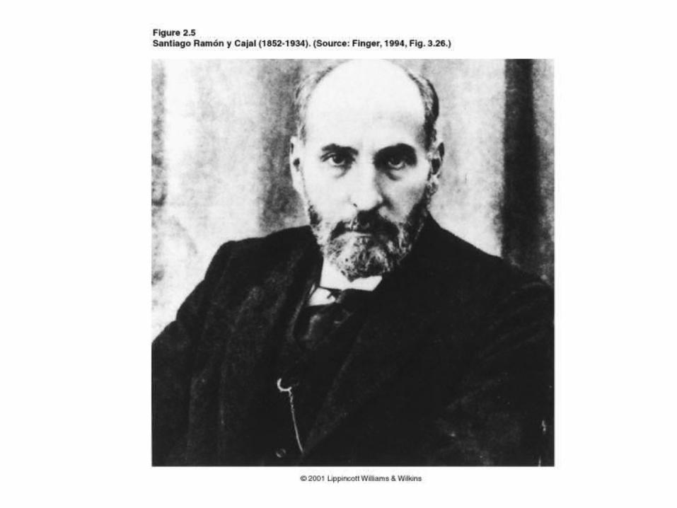
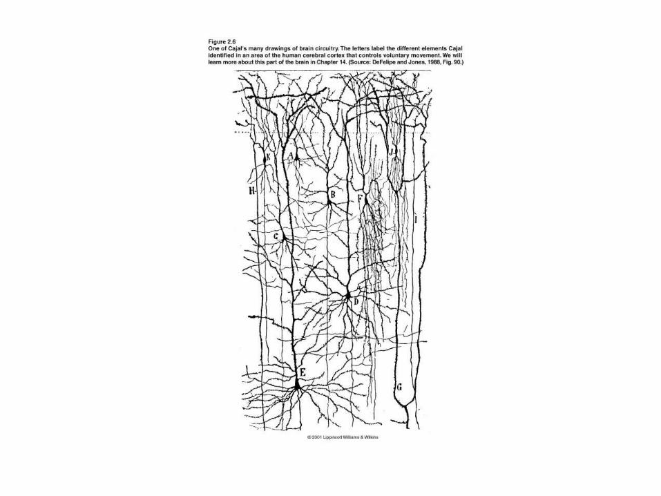
• Cajal’s Contributions– Neural circuitry– Neurons communicate by
contact, not continuity• Neuron doctrine
• Neurons adhere to cell theory
• Based in Golgi stain

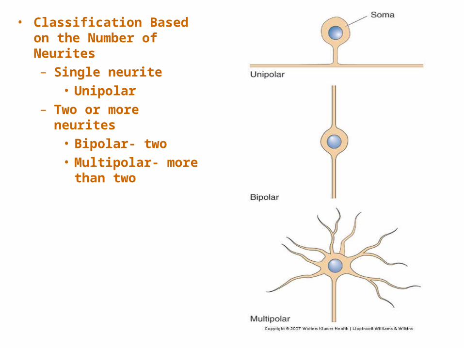
• Classification Based on the Number of Neurites – Single neurite
• Unipolar– Two or more
neurites • Bipolar- two• Multipolar- more
than two
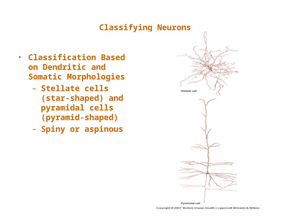
Classifying Neurons
• Classification Based on Dendritic and Somatic Morphologies – Stellate cells
(star-shaped) and pyramidal cells (pyramid-shaped)
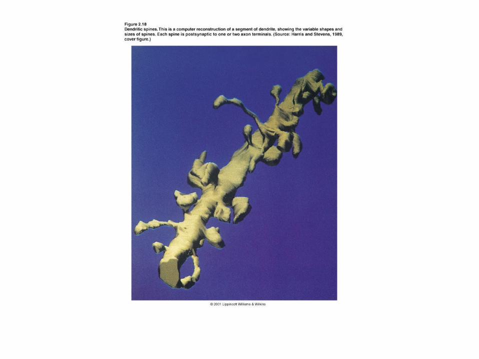
– Spiny or aspinous
• Further Classification– By connections within the CNS
• Primary sensory neurons, motor neurons, interneurons
– Based on axonal length• Golgi Type I• Golgi Type II
– Based on neurotransmitter type• e.g., – Cholinergic = Acetycholine at synapses
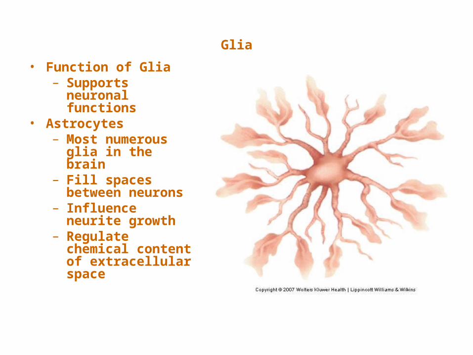
Glia
• Function of Glia – Supports
neuronal functions
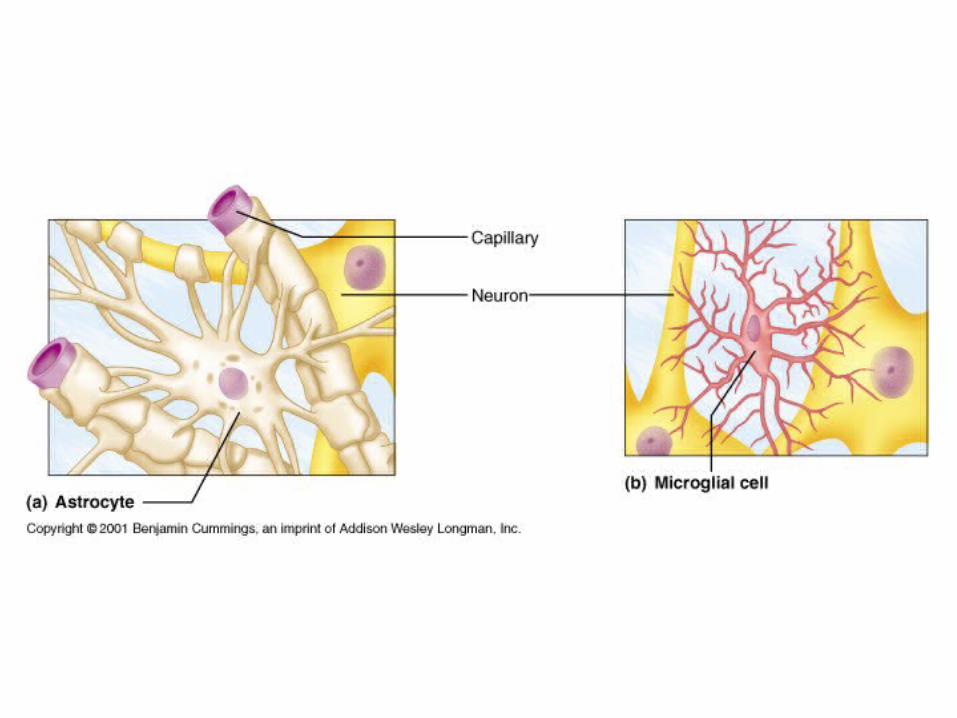
• Astrocytes– Most numerous
glia in the brain– Fill spaces
between neurons– Influence neurite
growth – Regulate
chemical content of extracellular space

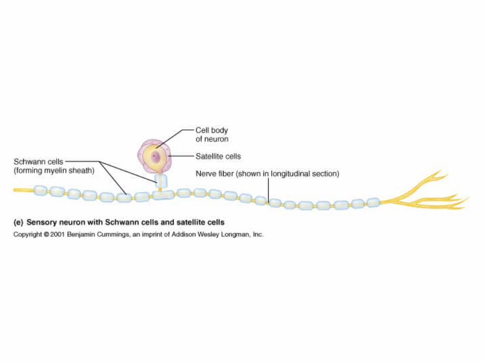
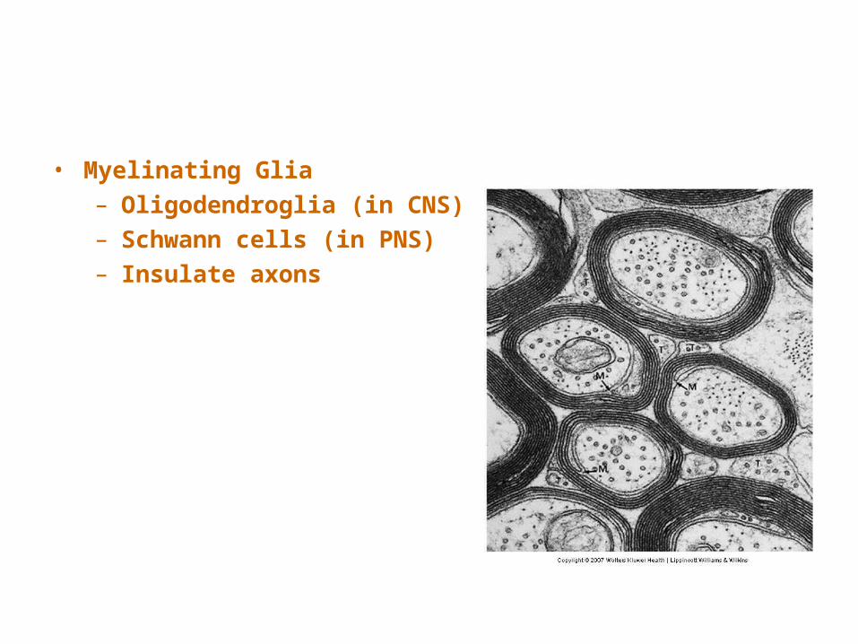
• Myelinating Glia– Oligodendroglia (in CNS)– Schwann cells (in PNS)– Insulate axons
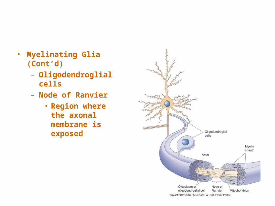
• Myelinating Glia (Cont’d)– Oligodendroglial
cells– Node of Ranvier
• Region where the axonal membrane is exposed
• Other Non-Neuronal Cells– Microglia as phagocytes (immune)
• The Soma• Cytosol: Watery fluid inside the cell• Organelles: Membrane-enclosed structures
within the soma• Cytoplasm: Contents within a cell membrane
(e.g., organelles, excluding the nucleus)
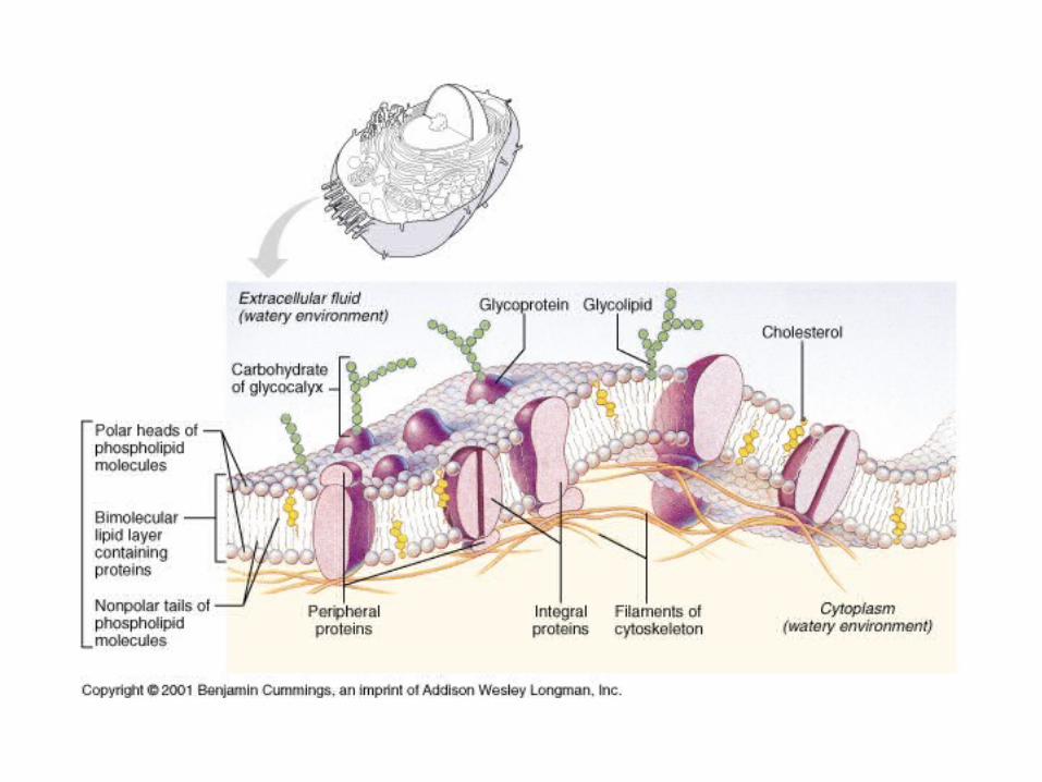
• The Membrane– Barrier that encloses cytoplasm– ~5 nm thick– Protein concentration in membrane varies– Structure of discrete membrane regions
influences neuronal function
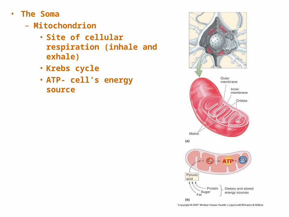
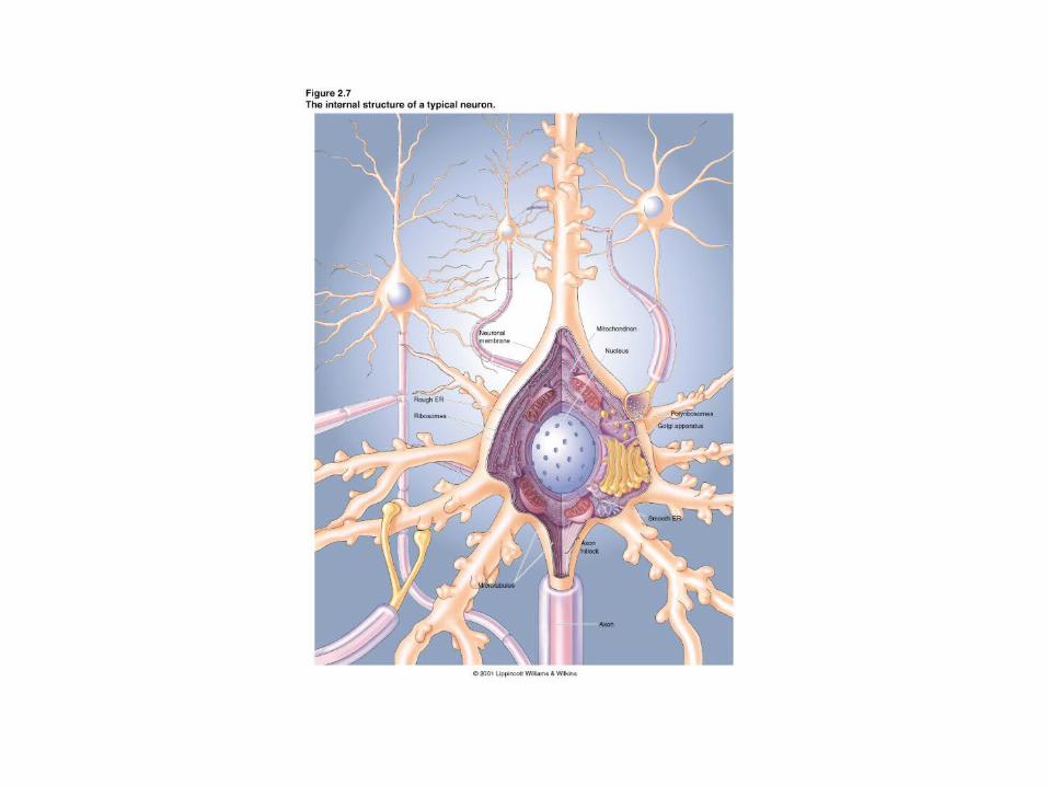
• The Soma– Mitochondrion
• Site of cellular respiration (inhale and exhale)
• Krebs cycle• ATP- cell’s energy source
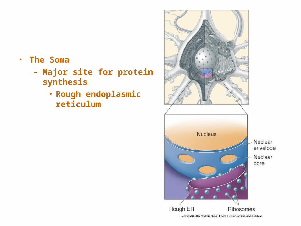
• The Soma– Major site for protein
synthesis• Rough endoplasmic
reticulum
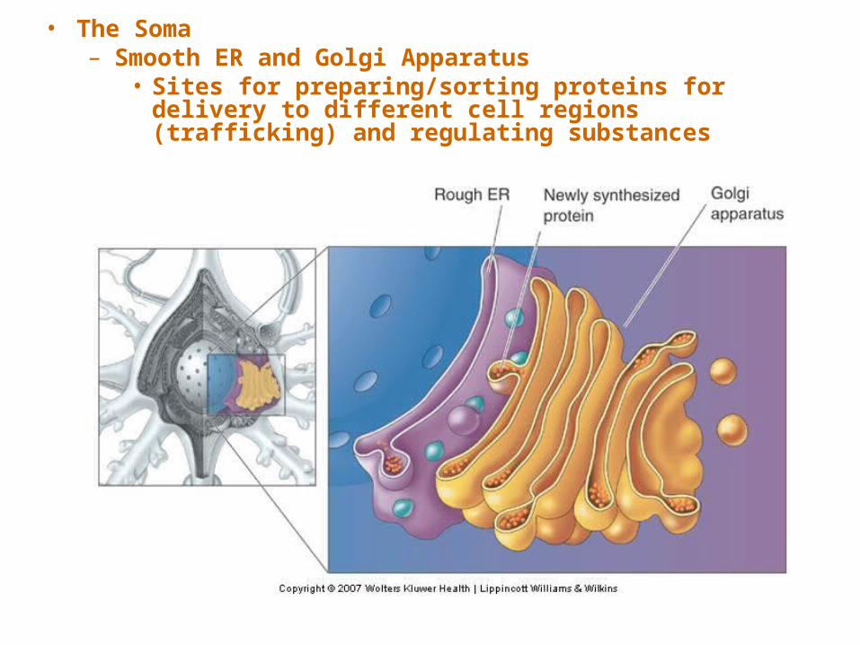
• The Soma– Smooth ER and Golgi Apparatus
• Sites for preparing/sorting proteins for delivery to different cell regions (trafficking) and regulating substances
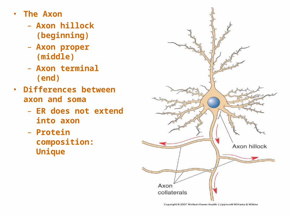
• The Axon– Axon hillock
(beginning)– Axon proper
(middle)– Axon terminal
(end)• Differences between
axon and soma– ER does not extend
into axon– Protein
composition: Unique
• The Axon– The Axon Terminal
• Differences between the cytoplasm of axon terminal and axon – No microtubules in terminal– Presence of synaptic vesicles– Abundance of membrane proteins– Large number of mitochondria
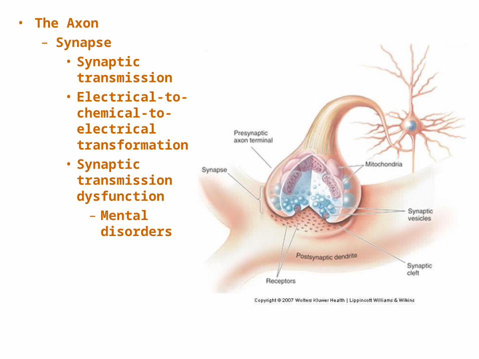
• The Axon– Synapse
• Synaptic transmission
• Electrical-to-chemical-to-electrical transformation
• Synaptic transmission dysfunction – Mental
disorders
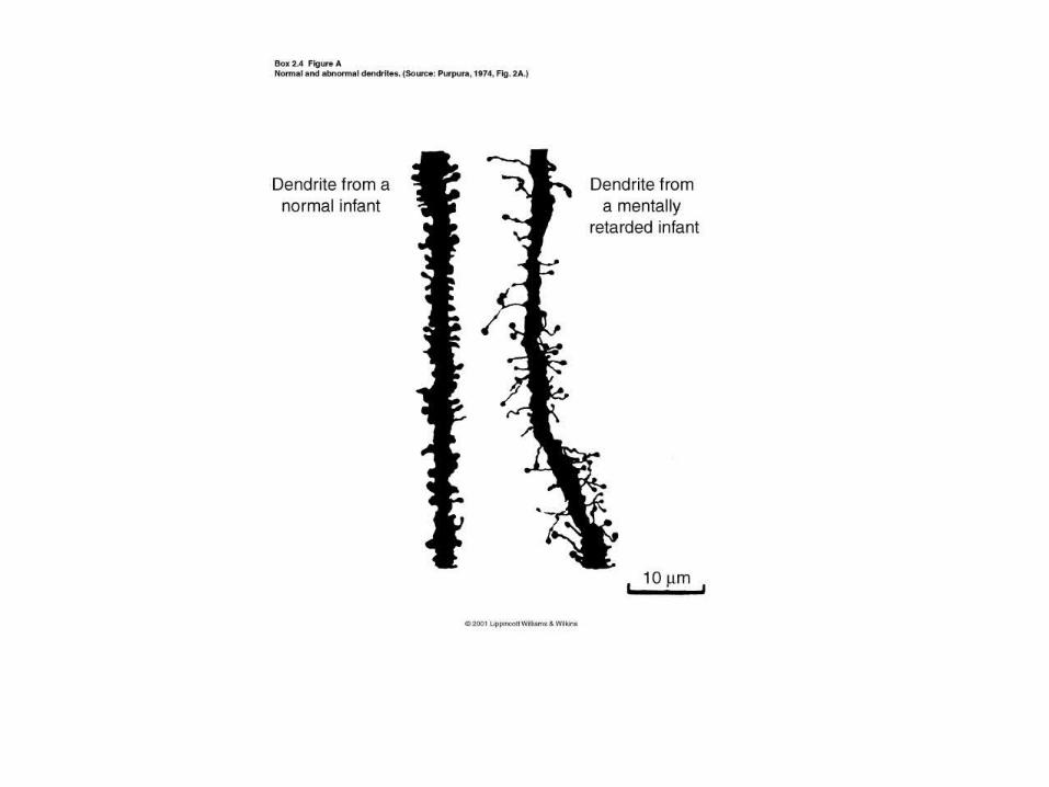
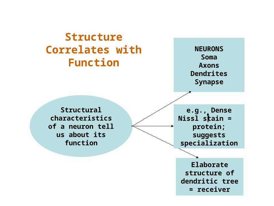
Structural characteristics of a neuron tell us about
its function
NEURONSSomaAxons
DendritesSynapse
Elaborate structure of
dendritic tree = receiver
e.g., Dense Nissl stain = protein;
suggests specialization
Structure Correlates with
Function