chapter 2 structure and functions of cells of the...
TRANSCRIPT
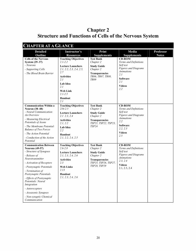
Chapter 2 Structure and Functions of Cells of the Nervous System
CHAPTER AT A GLANCE Detailed Outline
Instructor’s Resources
PrintSupplements
MediaSupplements
Professor Notes
Cells of the Nervous System (29–37) - Neurons - Supporting Cells - The Blood Brain Barrier
Teaching Objectives2.1-2.2 Lecture Launchers 2.1, 2.2, 2.3, 2.4, 2.5, 2.7Activities 2.1 Lab Idea 2.1Web Link 2.1-2.5 Handout2.1
Test Bank Chapter 2 Study Guide Chapter 2 Transparencies TR06, TR07, TR08, TR09
CD-ROM Terms and Definitions Self testFigures and DiagramsAnimations2.1 Software 2.1Videos2.2
Communication Within a Neuron (38–48) - Neural Communication: An Overview - Measuring Electrical Potentials of Axons - The Membrane Potential: Balance of Two Forces - The Action Potential - Conduction of the Action Potential
Teaching Objectives 234-2.5Lecture Launchers 2.1 2.3, 2.4 Activities 2.1, 2.2 Lab Idea 2.2Handout2.1, 2.2, 2.4, 2.5
Test Bank Chapter 2 Study Guide Chapter 2 Transparencies TSP21, TSP22, TSP23, TSP24
CD-ROM Terms and Definitions Self testFigures and DiagramsAnimations2.2Software 2.2, 2.3 Videos2.5
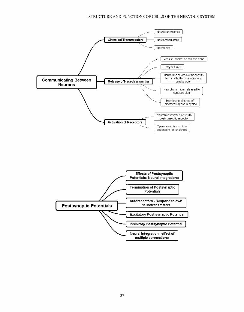
Communication Between Neurons (49-57) - Structure of Synapses - Release of Neurotransmitter - Activation of Receptors - Postsynaptic Potentials - Termination of Postsynaptic Potentials - Effects of Postsynaptic Potentials: Neural Integration- Autoreceptors - Axoaxonic Synapses - Non-synaptic Chemical Communication
Teaching Objectives 2.6-2.8 Lecture Launchers 2.1, 2.3, 2.4, 2.6 Activities 2.3Web Links 2.13Handout2.1, 2.3, 2.4, 2.6
Test Bank Chapter 2 Study Guide Chapter 2 Transparencies TSP25, TSP26, TSP27, TSP28, TSP29
CD-ROM Terms and Definitions Self testFigures and DiagramsAnimations2.3, 2.4 Videos2.1, 2.3, 2.4
20

LECTURE OUTLINE1: Cells of the Nervous System
1.1: Neurons 1.1.1: Classification of neurons
1.1.1.1: Sensory neurons 1.1.1.2: Motor neurons 1.1.1.3: Interneurons
1.1.2: Subdivisions of the nervous system 1.1.2.1: Central nervous system 1.1.2.2: Peripheral nervous system
1.1.3: Basic structure 1.1.3.1: Soma 1.1.3.2: Dendrites 1.1.3.3: Axon 1.1.3.4: Terminal buttons
1.1.4: Types of neurons 1.1.4.1: Multipolar - 1 axon, many dendritic trees 1.1.4.2: Bipolar - 1 axon and 1 dendritic tree (usually sensory) 1.1.4.3: Unipolar - one connection to soma (sensory)
1.1.5: Internal structure 1.1.5.1: Membrane 1.1.5.2: Cytoplasm 1.1.5.3: Mitochondria
1.1.5.3.1: Extract energy from nutrients 1.1.5.3.2: Adenosine triphosphate (ATP)
1.1.5.4: Nucleus 1.1.5.4.1: Chromosomes
1.1.5.4.1.1: Made of deoxyribonucleic acid (DNA) 1.1.5.4.1.2: Contain genes
1.1.5.4.1.2.1: Genes code proteins 1.1.5.4.1.2.1.1: Cytoskeleton
1.1.5.4.1.2.1.2: Enzymes 1.1.5.5: Microtubules
1.1.5.5.1: Axoplasmic transport 1.1.5.5.1.1: Anterograde - soma to terminal buttons 1.1.5.5.1.2: Retrograde - terminal buttons to soma
1.2: Supporting cells 1.2.1: Types of glial cells
1.2.1.1: Astrocyte 1.2.1.1.1: Morphology:
1.2.1.1.1.1: Star-shaped 1.2.1.1.1.2: Wrap around neurons and blood vessels
1.2.1.1.2: Functions 1.2.1.1.2.1: Physical support 1.2.1.1.2.2: Scavenge debris via phagocytosis 1.2.1.1.2.3: Regulate fluid around neurons 1.2.1.1.2.4: Provide nourishment to neurons 1.2.1.1.2.5: Isolate synapses
1.2.1.2: Oligodendrocyte 1.2.1.2.1: Found in central nervous system 1.2.1.2.2: Functions
1.2.1.2.2.1: Support 1.2.1.2.2.2: Produce myelin sheath
1.2.1.2.2.2.1: 80% lipid, 10% protein
STRUCTURE AND FUNCTIONS OF CELLS OF THE NERVOUS SYSTEM
21

1.2.1.2.2.2.2: Makes several separate segments of myelin 1.2.1.2.2.2.3: Node of Ranvier – gap between myelin segments
1.2.1.3: Microglia 1.2.1.3.1: Morphology
1.2.1.3.1.1: Small 1.2.1.3.2: Function
1.2.1.3.2.1: Break down dead and dying neurons via phagocytosis 1.2.1.3.2.2: Protect brain from invading organisms 1.2.1.3.2.3: Part of brain’s inflammation response to damage
1.2.1.4: Schwann cells 1.2.1.4.1: Found in peripheral nervous system 1.2.1.4.2: Function
1.2.1.4.2.1: Produce myelin 1.2.1.4.2.1.1: One cell makes one segment of myelin
1.3: Blood brain barrier 1.3.1: Selectively permeable
1.3.1.1: Some molecules pass freely 1.2.1.2: Others molecules (e.g. glucose) require protein transporters to help
1.3.2: In brain, the capillary walls lack gaps between cells 1.3.3: Functions to maintain chemical balance in brain 1.3.4: More permeable in some areas
1.3.4.1: Area postrema 1.3.4.1.1: Detects toxins 1.3.4.1.2: Causes emesis (vomiting)
2: Communication Within a Neuron 2.1: Neural communication: an overview
2.1.1: Control of the withdrawal reflex as an example of neural communication 2.1.1.1: Excitation 2.1.1.2: Inhibition
2.2: Measuring the electrical potentials of axons 2.2.1: Electrodes
2.2.1.1: Microelectrode 2.2.1.2: Oscilloscope - measures voltages across time
2.2.2: Membrane potential 2.2.2.1: Potential is amount of stored energy
2.2.3: Resting potential 2.2.4: Depolarization 2.2.5: Hyperpolarization 2.2.6: Action potential
2.2.6.1: Threshold of excitation 2.3: The Membrane potential: balance of two forces
2.3.1: The force of diffusion 2.3.1.1: Diffusion – process in which molecules distribute themselves evenly throughout a medium 2.3.1.2:Molecules diffuse from regions of high to low concentrations
2.3.2: The force of electrostatic pressure 2.3.2.1:Electrolytes
2.3.2.1.2: Ions 2.3.2.1.2.1: Cations 3.3.2.1.2.2: Anions
2.3.2.2: Electrostatic pressure - Like charges repel, opposite charges attract 2.3.3: Ions in the extracellular and intracellular fluid
2.3.3.1: Ions and their distribution at resting potential 2.3.3.1.1: Organic anions (A-) – only in intracellular fluid 2.3.3.1.2: Chloride ions (Cl-) – more in extracellular fluid 2.3.3.1.3: Sodium ions (Na+) – more in extracelluar fluid 2.3.3.1.4: Potassium ions (K+) – more in intracellular fluid
CHAPTER 2
22

2.3.3.2: Forces of diffusion and electrostatic pressure on ions 2.3.3.2.1: A- unable to cross axon membrane 2.3.3.2.2: K+ diffusion- out, electrostatic pressure – in; no net movement 2.3.3.2.3: Cl- diffusion – in, electrostatic pressure – out; no net movement 2.3.3.2.4: Na+ diffusion and electrostatic pressure – in; membrane largely impermeable to Na+ so
no net movement 2.3.3.3: Sodium-potassium pump (transporters)
2.3.3.3.1: Exchange 3 NA+ for 2 K+ 2.3.3.3.2: Requires up to 40% of neurons energy
2.4: The Action potential 2.4.1: Ionic movements during action potential
2.4.1.1: Voltage dependent ion channels - sodium channels open and NA+ rushes in 2.4.1.2: Voltage dependent potassium channels less sensitive open later 2.4.1.3: Peak action potential – Na+ channels refractory 2.4.1.4: Potassium channels open to allow K+ ions to reach a normal potential 2.4.1.5: Potassium & sodium channels closed - Potential to resting level 2.4.1.6: Membrane potential briefly overshoots resting potential
2.5: Conduction of the action potential 2.5.1: Laws
2.5.1.1: All or none 2.5..1.2: Rate law
2.5.2: Saltatory conduction 2.5.2.1: Occurs only in myelinated axons 2.5.2.2: Action potential “jumps” from node to node 2.5.2.3: Advantages
2.5.2.3.1: Need less energy for active sodium transport 2.5.2.3.2: Increases speed of transmission
3: Communication Between Neurons 3.1: The Concept of synaptic transmission
3.1.1: Synaptic transmission 3.1.2: Neurotransmitters attach to binding sites
3.1.2.1: Ligand – chemical that attach to binding sites 3.1.3: Results in postsynaptic potentials
3.2: Structure of synapses 3.2.1: Types of synapses
3.2.1.1: Axodendritic 3.2.1.2: Axosomatic 3.2.1.3: Axoaxonic
3.2.2: Synaptic structures 3.2.2.1: Presynaptic membrane
3.2.2.1.1: Synaptic vesicles 3.2.2.1.2: Release zone
3.2.2.2: Postsynaptic membrane 3.2.2.3: Synaptic cleft
3.3: Release of neurotransmitter 3.3.1: Vesicles fuse with presynaptic membrane
3.4: Activation of receptors 3.4.1: Neurotransmitter binds with postsynaptic receptor 3.4.2.: Ionotropic receptor
3.4.2.1: Ions flow through the receptor 3.4.2.2: Metabotropic receptor
3.4.2.2.1: Second messengers 3.4.2.2.1.1: G protein 3.4.2.2.1.2: Cyclic AMP
3.5: Postsynaptic potentials: 3.5.1: Receptor, not the neurotransmitter, determines the nature of the postsynaptic response
STRUCTURE AND FUNCTIONS OF CELLS OF THE NERVOUS SYSTEM
23
3.5.2: Excitatory postsynaptic potential (EPSP): Depolarizes postsynaptic membrane 3.5.3: Inhibitory postsynaptic potential (IPSP): Hyperpolarizes postsynaptic membrane
3.6: Termination of postsynaptic potentials 3.6.1: Reuptake of neurotransmitter by transporter molecules 3.6.2: Enzymatic deactivation
3.6.2.1: Acetylcholine/acetylcholinesterase 3.7: Effects of postsynaptic potentials: neural integration
3.7.1: Neural integration involves the effects of EPSP’s and IPSP’s on the likelihood a neuron will fire an action potential
3.8: Autoreceptors 3.8.1: Presynaptic receptors -respond neurotransmitters released by that neuron 3.8.2: metabotropic – regulate internal processes like neurotransmitter synthesis
3.9: Axoaxonic synapses 3.9.1:Alter amount of neurotransmitter released
3.9.1.1: Presynaptic inhibition 3.9.1.2: Presynaptic facilitation
3.10: Nonsynaptic communication: neuromodulators and hormones 3.10.1: Neurotransmitters 3.10.2: Neuromodulators
3.10.2.1: Usually peptides 3.10.2.2: Travel over relatively long distance to reach target3.10.3: Hormones 3.10.3.1: Released endocrine glands and act on target cells
TEACHING OBJECTIVESThe student should be able to:
2-1 Name and describe the parts of a neuron and explain their functions. 2-2 Describe the supporting cells of the central and peripheral nervous systems and describe and explain
the importance of the blood-brain barrier. 2-3 Briefly describe the neural circuitry responsible for a withdrawal reflex and its inhibition by neurons in
the brain. 2-4 Describe the measurement of the action potential and explain the how the balance between the forces
of diffusion and electrostatic pressure is responsible for the membrane potential. 2-5 Describe the role of ion channels in action potentials and explain the all-or-none law and the rate law. 2-6 Describe the structure of synapses, the release of the neurotransmitter, and the activation of
postsynaptic receptors. 2-7 Describe postsynaptic potentials: the ionic movements that cause them, the processes that terminate
them, and their integration. 2-8 Describe the role of autoreceptors and axoaxonic synapses in synaptic communication and describe the
role of neuromodulators and hormones in nonsynaptic communication.
CHAPTER SUMMARYChapter 2 covers the structure and function of the cells that comprise the nervous system. The basic structure and function of neurons, glia and the blood-brain barrier are described in the first section. The second section focuses on the electrical potentials used by neurons for intracellular communication. The forces underlying the establishment of the membrane potential and the mechanism by which the action potential is generated are discussed. In the final section, the mechanism by which communication between neurons occurs is described.
CHAPTER 2
24
KEY TERMSsensory neuron (28) motor neuron (28) interneuron (29) central nervous system (CNS) (29) peripheral nervous system (PNS) (29) soma (31) dendrite (29) synapse (29) axon (29) multipolar neuron (30) bipolar neuron (30) unipolar neuron (30) terminal button (31) neurotransmitter (31) membrane (31) cytoplasm (31) mitochondria (31) adenosine triphosphate (ATP) (31) nucleus (31) chromosome (31) deoxyribonucleic acid (DNA) (33) gene (33) cytoskeleton (33) enzyme (33) axoplasmic transport (33) microtubule (33) glia (33) astrocyte (33)
phagocytosis (34) oligodendrocyte (34) myelin sheath (34) node of Ranvier (35) microglia (35) Schwann cell (36) blood-brain barrier (36) area postrema (37) electrode (40) microelectrode (40) membrane potential (40) oscilloscope (40) resting potential (40) depolarization (41) hyperpolarization (41) action potential (41) threshold of excitation (42) diffusion (42) electrolyte (42) ion (42) electrostatic pressure (42) intracellular fluid (42) extracellular fluid (42) sodium-potassium transporter (44) ion channel (45) voltage-dependent ion channel (46) all-or-none law (47) rate law (47) saltatory conduction (47) postsynaptic potential (49)
binding site (49) ligand (49) dendritic spine (49) presynaptic membrane (49) postsynaptic membrane (49) synaptic cleft (49) synaptic vesicle (50) postsynaptic receptor (51) neurotransmitter-dependent ion channel (51) ionotropic receptor (51) metabotropic receptor (52) G protein (52) second messenger (52) excitatory postsynaptic potential (EPSP) (52) inhibitory postsynaptic potential (IPSP) (53) reuptake (53) enzymatic deactivation (54) acetylcholine (54) acetylcholinesterase (54) neural integration (54) autoreceptor (55) presynaptic facilitation (56) presynaptic inhibition (56) neuromodulator (56) peptides (56) hormones (56) endocrine gland (56) target cell (56)
CHAPTER GUIDECells of the Nervous System
Neurons Supporting Cells The Blood- Brain Barrier
Animation 2.1 Neurons and Supporting Cells Transparency 06 The Major External Features of a Neuron Transparency 07 The Major Internal Features of a Neuron Transparency 08 Types of Neurons Transparency 09 The Two Types of Neurons Lecture 2.1 Metaphors Lecture 2.2 Photography Lecture 2.3 Animations Lecture 2.4 Neuron Skits Lecture 2.5 New Thoughts About the Functions of Glial Cells Lecture 2.7 Drug Effects and the Blood-Brain Barrier Online 1.1 Neuron Pets Activity 2.1 Simulate a Neuron and Measure Potentials Lab Idea: 2.1 Histology, Neurons, and Glia
STRUCTURE AND FUNCTIONS OF CELLS OF THE NERVOUS SYSTEM
25
Software 2.1 Brainiac Video 2.2: The Brain Teaching Modules, Module 1 Web Link 2.1 The Virtual CellWeb Link 2.2 The Story of a Membrane Web Link 2.3 Neuroanatomy and Neuropathology on the Internet. Web Link 2.4 Glia: The Forgotten Brain Cell Web Link 2.5 Molecular Biology: A Gallery of Animations Web Link 2.6 Millions and Billions of Cells: Types of Neurons Handout 2.1 Concept Maps
Communicating Within a Neuron Neural Communication: An Overview Measuring Electrical Potential of Axons The Membrane Potential: Balance of Two Forces The Action Potential Conduction of the Action Potential
Animation 2.2 The Action Potential Lecture 2.1 Metaphors Lecture 2.3 Animations Lecture 2.4 Neuron Skits Lecture 2.6 The Behavioral Consequences of Neural Inhibition Activity 2.1 Simulate a Neuron and Measure Potentials Activity 2.2 Axonal Transmission Speed Measurement Lab Idea 2.2 Crawdad – A CD ROM Lab Manual for Neurophysiology Video 2.5 Nerve Impulse Conduction Handout 2.1 Concept Maps Handout 2.2 Neuron Skit: Firing of a Neuron Handout 2.4 Name Tags for Skits Handout 2.5 Things That You Need to Know About Neurons
Communication Between Neurons The Concept of Chemical Transmission The Structure of Synapses Release of Neurotransmitter Activation of Receptors Postsynaptic Potentials Termination of Postsynaptic Potentials Effects of Postsynaptic Potentials: Neural Integration Autoreceptors Axoaxonic Synapses Nonsynaptic Chemical Communication
Animation 2.3 Synapses Animation 2.4 Postsynaptic Potentials Laboratory 2.1 Crawdad: A CD-ROM Lab Manual for Neurophysiology Lecture 2.1 Metaphors Lecture 2.2 Photography Lecture 2.3 Animations Lecture 2.4 Neuron Skits Activity 2.3 How to Murder a Neuron Software 2.2 The Action Potential Simulator Software 2.3 Electrophysiology of the Neuron Video 2.1 Biological Psychology: A Video Supplement, 2000 (Module 4) Video 2.3 Neurons and Chemicals Video 2.4 The Neuron and Neural Transmission Web Link 2.7 Synapse (Tutorial) Web Link 2.8 Synapse Web Web Link 2.12 Synaptic Transmission: A Four Step Process
CHAPTER 2
26
Handout 2.1 Concept Maps Handout 2.3 Neuron Skit: Synapse Handout 2.4 Name Tags for Skits Handout 2.6 How to Murder a Neuron
LECTURE LAUNCHERSLecture Launcher 2.1: Metaphors
Some times it is helpful to take concepts that students are unfamiliar with and place them in a more familiar context. Remind the students that these are models and may not work the same as the real thing, but you can get past some cognitive barriers by making connections to the student’s current experience.
A simplistic (and probably not entirely accurate) explanationIf you are having trouble understanding Excitatory (EPSP) and Inhibitory (IPSP) postsynaptic potentials, you might find these explanations and metaphors helpful. Please remember that, like our model neuron, the following description is not how things really work, but it may help you to get a picture of the events that will then allow you to explore the information in more detail and revise and correct your understanding.
Concentrations of various chemicals in and around the cell. The postsynaptic membrane has protein receptors in the membrane made of phospholipids (fat). Each receptor has a shape that fits at least one neurotransmitter molecule. Imagine a molecule of neurotransmitter floating through the extra cellular space in the synapse until it reaches one of these receptors. When the neurotransmitter gets close, it fits into the protein molecule like a key in a lock. This changes the shape of the protein molecule and sets off a change in the electrical potential of the cell. If the neurotransmitter is excitatory at that receptor, it will depolarize the cell membrane (make it more likely to transmit information) around the receptor site. You might think of this as dropping a stone into a still lake. The ripples move away from the receptor, getting weaker and weaker. At some point, a ripple will cross the cell body and move down the axonal hillock. If the receptor is close to the axonal hillock, the ripple will still be strong when it gets there.
Axonal HillockThe axonal hillock is a small “hill” at the beginning of the axon. It is here that the decision is made to "fire.” The cell. The neuron “gun” is fired at the axonal hillock trigger. A small squeeze on the trigger will not fire the neuron. There will be a point when the trigger moves far enough to fire the neuron, and like a gun, once fired, it has to be reloaded.
Postsynaptic Receptors Cells can be seen as a mini version of the world. Just as the cell seems to make decisions based on multiple inputs, in society we often make decisions based on information from a number of people.
Imagine the axonal hillock as a meeting of 100 people - - (100 postsynaptic potentials).
1. The meeting is to decide whether to send a message encouraging another group of people to move to a different building (The goal is not important in this example). To make a decision, the meeting must have a Quorum of at least 50 people. Out of the people at the meeting, at least two-thirds must vote in favor of the action (be positive).
2a. The meeting room has just a few people wandering around. (The resting potential) More people show up until there are 57 people in the room. The meeting begins. There is a vote on sending the message. Forty-five people vote for sending the message (EPSPs) and 12 vote against sending the message. Since the vote is more than 2/3 in favor, the message is sent.
2b. The meeting room has just a few people wandering around. (The resting potential) More people show up until there are 45 people in the room. The meeting begins. There is a vote on sending the message. Forty people vote for sending the message (EPSPs) and five vote against sending the message. The vote is more than 2/3 in favor but there was not a quorum (not enough EPSPs) so the message is not sent.
2c. The meeting room has just a few people wandering around. (The resting potential) More people show up until there are 57 people in the room. The meeting begins. There is a vote on sending the message. Twenty
STRUCTURE AND FUNCTIONS OF CELLS OF THE NERVOUS SYSTEM
27

people vote for sending the message (EPSPs) and 37 vote against sending the message. Since the vote is not more than 2/3 in favor, the message is not sent.
In these three situations, the number of excitatory and inhibitory potentials that reach the axonal hillock at the same time will be combined to determine whether or not the cell fires.
Let’s look at several examples of meeting outcomes.
Excitatory versus Inhibitory Postsynaptic Potential
Excitatory influences in the nervous system make things more likely to happen
Inhibitory influences in the nervous system make things less likely to happen
Pre-synaptic versus Post-Synaptic Terminal button of the axon Dendrite or cell body side of the synapse
First, let’s look at the terms that discriminate an EPSP from an IPSP, Excitatory and Inhibitory. Post Synaptic Potentials
� Excitatory influences in the nervous system make things more likely to happen.
� Inhibitory influences in the nervous system make things less likely to happen.
How does the axonal hillock know how far is far enough to fire the neuron? Here is another metaphor. It does a little basic math. Addition and subtraction.
If the ripple of potential is excitatory, when it reaches the axonal hillock it will be added to other excitatory potentials that arrive at about the same time. If the sum of the potentials is great enough, the axonal hillock will send an action potential down the axon.
If the ripple is inhibitory, it changes the cell potentials by making the cell less likely to fire or by subtracting from the potentials arriving at about the same time.
In general, the farther away from the axonal hillock the stimulated receptor is – the less of a depolarization will occur because the postsynaptic potential fades as it moves away from the receptor.
What about situations where not all the postsynaptic potentials reach the axonal hillock at exactly the same time?
Which is going to be the most disruptive? (Have the most influence on communication.) 1. One person talking during a class or 10 people talking during a class at the same time?
2. Ten people distributed over a large classroom and talking during a class or the same ten people sitting together and talking during a class.
3. Ten people each talking for one minute at different times in a one-hour class, or the same ten people talking for one minute at the same time?
Each of these represents a different situation at the cell membrane.
1. If one receptor is stimulated by an excitatory transmitter, it is not likely to create a large enough change in the neuron potential to cause the cell to fire. Multiple stimulations, even if at different locations, are more likely to be successful in depolarizing the membrane and firing the cell.
More is better.
1. Even if multiple receptors are stimulated; if they are closer together, they have a greater effect as the depolarization from one enhances that of the others. This is referred to as spatial summation.
CHAPTER 2
28

Together is better.
Neurotransmitters take time to float across the synapse. Not all will reach the receptors at the same time. If it takes too long, the effect of the early neurotransmitters will be almost gone before the other arrives
Lecture Launcher 2.2 Photography Photographs are always useful to make concepts concrete and to keep students interested. Microphotographs of cells can assist the students in imagining the cell structure and relating this to its function. While drawings and models are always essential in learning about physiology, it helps to periodically tie these back to the real structures that are being modeled. Microphotography in the form of still and moving images can be a great starting point for a lecture or a review after discussing the model. Can the student locate the real structures after working with a diagram?
Web Links: Web Link 2.8: Synapse Web
Lecture Launcher 2.3 Animations Since the process of transmission takes place over time, students may be confused while looking at still diagrams. The Internet is a great place to find animation that focuses on the level of detail that you wish to emphasize. Projecting these animations during a lecture and making them available for further examination online can help students to catch on the way that a potential travels along the neuron and the changes that take place at a single point over time.
AnimationsAnimation 2.1 Neurons and Supporting Cells Animation 2.2 The Action Potential
Web links Web Link 2.5 Molecular Biology: A Gallery of Animations
Lecture Launcher 2.4: Neuron Skits Scientific courses often miss out on activities that students find both entertaining and useful learning activities. One of these is “Role Playing.” Choose students who are outgoing and willing to volunteer to stand in front of the class. Give them the instruction sheets the class before they will be doing the activity so that they can prepare their character. (Handout 2.2 or 2.3)
Before the next class, have the students come a few minutes early to discuss their interaction. You may wish to project an image of a synapse at the front of the room or suggest that class members turn to an appropriate illustration in their text. During the class, have the students go through the activity. Ask the class to identify the “actors” and pin appropriate signs on them. Allow the class to act as directors, making revisions to the action. Encourage discussion of the difference between the model as portrayed by the actors and the interactions within the nervous system.
Have class members assist in figuring out what each element should do with the actors following class instructions. Have the actors try to literally interpret what they are being told to do. (If the class suggests that the neurotransmitter should go through the cell membrane before the vesicle attaches to it, the vesicle membrane should keep tightly closed - they have not been told to let go or to merge with the presynaptic membrane - and the presynaptic membrane should not let the neurotransmitter through.)
When the correct steps have been figured out, have the actors go through the process one or two times correctly before joining the class.
Handouts: Handout 2.2 Neuron Skits: Firing of a Neuron Handout 2.3 Neuron Skits: The Synapse Handout 2.4 Name Tags for Skits
STRUCTURE AND FUNCTIONS OF CELLS OF THE NERVOUS SYSTEM
29

Lecture Launcher 2.5: New Thinking about the Functions of Glial Cells A recent review article published in Scientific American may be helpful in expanding student’s appreciation for the complexity of neural functioning. This paper details new research on the potential for glial cells to act as a signaling system in the brain, a function previously believed exclusive to neurons. This research is not only poignant because of its content but also because it shows the dynamic nature of our understanding of neural functioning. This paper can be used as a springboard to discuss a variety of topics such as: the importance of technology in our understanding of brain function, the implications for our understanding of neural functions if in fact glial cells do act as a signaling system in the brain, and the possible role of glial communication in consciousness, learning, memory, and any other neural activity.
Fields, R.D. (2004) The other half of the brain. Scientific American. April; 2004,.55-61.
Lecture Launcher 2.6: The Behavioral Consequences of Neural Inhibition When discussing synaptic summation and inhibitory postsynaptic potentials, students will often confuse synaptic inhibition with behavioral inhibition. I tell students that many of our neurons serve to inhibit our behavior and that synaptic inhibition of inhibitory neurons can result in behavioral excitation. Several examples can help clarify this point:
We have all heard the expression “running around like a chicken with its head cut off” which is literally true when a chicken is decapitated. It will run around aimlessly for a short time because neurons in the brain normally act to inhibit much of their movement and when decapitated the inhibitory control of movement is removed.
During sexual intercourse a female Praying Mantis will often bite the head off of her male partner. Again, this decapitation removes the inhibitory control of movement by the brain and increases their likelihood of reproductive success. This behavior has also been observed in several species of spiders, but I tell students not to try this at home!
When people consume alcohol, one of its effects is that a person may engage in behavior that they might normally not perform (unplanned unprotected sexual behavior, violent and criminal behavior). One of the many neurochemical effects of alcohol is to bind to receptors for the inhibitory neurotransmitter GABA and thus inhibiting neural activity. Low dose inhibition of neural activity by alcohol is believed to increase the likelihood of behaviors a person would not normally engage in while sober. Alcohol inhibits the inhibitory mechanisms that normally prevent people from engaging in these behaviors. It is always important to point out however, that at higher doses alcohol’s inhibitory effects on the brain are global and can inhibit brainstem mechanisms that control our vital life functions and can lead to death.
Lecture Launcher 2.7: Drug Effects and the Blood-Brain Barrier The use of L-dopa to treat Parkinson’s disease and the difference in the effectiveness between heroin and morphine are excellent examples of the importance of the blood brain barrier on drug effects. After providing students with these and other examples of the influence of the blood brain barrier on drug action you might ask them to search for there own examples on the internet or pharmacology texts. You might also ask students to research ways in which Pharmaceutical companies are trying to get drugs to cross the blood brain barrier which normally would not. Alternately, you can ask students to think of their own ways of “tricking” the blood brain barrier into allowing certain drugs to pass. Finally, you might visit the issue of what might be the consequences of loosing the blood brain barrier or having on severely compromised as is the case for human infants.
CLASSROOM ACTIVITIES , LAB IDEAS, ASSIGNMENTS, AND HANDOUTSActivities
Activity 2.1 Simulate a neuron and measure potentials Neurosim II This PC software package provides a set of neural simulations including the cable properties of a neuron,
CHAPTER 2
30

calculations of the resting membrane potential using the Goldman equation, the Hodgkin-Huxley model of neuron properties, and a simulation of the postsynaptic response to various transmitters including ACh, GABA, and 5-HT. Neurosim II is available from BIOSOFT (01223 368622; Email: [email protected]).
The Axon Potential SimulatorA membrane potential simulation program by Jeffrey L. Ram, Department of Physiology, Wayne State University. This site provides a powerful simulation program of the ionic and electrical events that occur during an action potential. The simulator allows the instructor to demonstrate EPSPs, IPSPs, and the effects of toxins such as TTX and TEA on the membrane potential.
Electrophysiology of the Neuron This software program (PC or MAC formats) provides a simulation of the effects of manipulating electrophysiological parameters on resting potentials, action potentials, and postsynaptic potentials. The program is available from Oxford University Press (001 212 7266069). A shareware version is available.
SoftwareSoftware 2.2 The Axon Potential Simulator Software 2.3 Electrophysiology of the Neuron
Activity 2.2 Axonal Transmission Speed Measurement Equipment
Stopwatch or watch with second hand Tape Measure (Optional) Calculator
It would be very difficult to measure the speed of transmission through an axon in a classroom if a single axon were used, but an estimate of the speed of transmission can be easily calculated in a class activity, and the larger the class, the better. Scientists often use multiple measurements of rapidly occurring phenomena and then divide by the number of measurements.
Begin by having the class estimate the distance that an impulse must travel to go from a person’s shoulder to the hand on the same side. (Having a tape measure available is useful, but not necessary.) Multiply this by the number of students in the class. This is the distance the signal must travel.
Have students stand. (Moving to the outside of the room works best but may not be possible for a large lecture class.) Each student places his or her right hand on the right shoulder of the next person. The instructor begins the action by squeezing the shoulder of the first student in line while keeping track of the time. The students each squeeze the shoulder of the next person as soon as he or she feels a squeeze. The last person needs to indicate that he or she has felt the squeeze so that the instructor can stop timing.
You may need to run through the action a few times to get the estimate to stabilize.
Divide the number of seconds from start to finish by the distance and the number of students to get an estimate of transmission time.
Activity 2.3 How to Murder a Neuron An understanding of the fragile nature of a single neuron can be represented by having the students explore the manner in which a neuron can be damaged. This Handout gives some suggestions in an informal manner. Have the students pair up to decide what could happen that would result in the different types of neural damage.
Handouts Handout 2.6 How to Murder a Neuron
Lab Ideas Laboratory 2.1 Lab exercise in histology, neurons, and glia:
Examining neural tissue under a microscope can help students to better visualize neurons and glia as well as appreciate the complexity of neural organization. For this lab you can introduce students to histology and discuss what can be learned from examining the microscopic organization of the brain. The importance of staining techniques can be addressed and students can be introduced to the stereotaxic atlas. Students can then
STRUCTURE AND FUNCTIONS OF CELLS OF THE NERVOUS SYSTEM
31

be asked to examine a number of microscope slides of neural tissue and ask them to draw and identify cells and structures discussed in class. Students generally do not have the background to do this, so I point out what they should be looking for in pictures on an overhead projector. Pictures can be readily obtained from histology or neuroscience texts. A variety of histology slides are commercially available (Carolina Science and Math) for this exercise. I have used slides of mammal peripheral nerve (cross section), mammal peripheral nerve (teased), human cerebellum, human cerebral cortex, and coronal sections of the whole rat brain.
Laboratory 2.2 Crawdad: A CD-ROM Lab Manual for Neurophysiology This CD-ROM is an electronic manual designed to accompany an undergraduate course in neurophysiology. There are 10 different exercises covered in the manual that range from an electronic circuit for membrane potential to the neuromuscular junction. The manual includes a number of diagrams that will be useful for lecture material and includes several video clips that illustrate electrophysiological recording in the crawdad. The CD ROM is available from Sinauer Associates (PO Box 47, Sunderland, MA, 10375: www.sinauer.com).
HandoutsHandout 2.1 Concept Maps
These maps may assist students in organizing the material in this chapter. You can make these maps available to the students or encourage them to construct their own maps.
Handout 2.2 Neuron Skits: Firing of a Neuron Script for Student Role Playing Activity (Lecture Launcher 2.4)
Handout 2.3 Neuron Skits: The Synapse Script for Student Role Playing Activity (Lecture Launcher 2.4)
Handout 2.4 Name Tags for Skits Name tags to be used with scripts for Student Role Playing Activity (Lecture Launcher 2.4)
Handout 2.5 Things That You Need to Know About Neurons A few basic facts about neurons.
Handout 2.6 How to Murder a Neuron To be used with Activity 2.4
SOFTWARE AND VIDEO RESOURCESSoftware Software 2.1 Brainiac
http://www.webcom.com/~medmult/brainiac.htmlOver 50 stained sections from the spinal cord through the thalamus; coronal and horizontal modules; review and test Medical Multimedia Systems
Software 2.2 The Axon Potential Simulator http://www.phypc.med.wayne.edu/jeffram/axon3.htmA membrane potential simulation program by Jeffrey L. Ram, Department of Physiology, Wayne State University. This site provides a powerful simulation program of the ionic and electrical events that occur during an action potential. The simulator allows the instructor to demonstrate EPSPs, IPSPs, and the effects of toxins such as TTX and TEA on the membrane potential.
Software 2.3 Electrophysiology of the Neuron: This software program (PC or MAC formats) provides a simulation of the effects of manipulating electrophysiological parameters on resting potentials, action potentials, and postsynaptic potentials. The program is available from Oxford University Press (001 212 7266069). A shareware version is available.
CHAPTER 2
32
VideoVideo 2.1 Biological Psychology: A Video Supplement, 2000
Allyn and Bacon Module 4: Neurons and Synaptic Transmission, (7 min: 49 sec)
Video 2.2 The Brain Teaching Modules (2e, 1997) Module 1: Organization and Evaluation of Brain Function
Video 2.3 Neurons and Chemicals (1998) Part of The Brain: An Owners Manual Series (90 minutes)
Video 2.4 The Neuron and Neural transmission (2001) Insight Media This video covers neural structure and transmission of nerve impulses (30 min)
Video 2.5 Nerve Impulse Conduction (1997) Insight Media This video addresses the electrochemical nature of nerve impulses and uses simulations to illustrate the effects of chemicals on membrane potential (29 min)
ON-LINE COURSE MATERIALSDiscussion Questions
Online 1.1 Neuron Pets What would a single neuron be like if you had one for a pet? How would you take care of it? What behavior would you expect? Would an afferent neuron make a better pet than an efferent neuron?
WEB LINKSStructure and Functions of Cells of the Nervous System
Web Link 2.1 The Virtual Cell web page http://personal.tmlp.com/Jimr57/textbook/chapter3/chapter3.htmThere is great online information about animal cells. You can find diagrams, photographs, and even movies of living cells.
Web Link 2.2 The Story of a Membrane http://www.concord.org/~barbara/workbench_web/unitIII_mini/cf_membranes/about_pores.htmlStructure of the Plasma Membrane Proteins, lipids and sugars at work in a single pore
Web Link 2.3 Neuroanatomy and Neuropathology on the Internet. http://www.neuropat.dote.hu/histol.htmThis site contains a collection of light and electron micrographs of various tissues including nerve and muscle.
NeuronStructure
Web Link 2.4 Glia: The Forgotten Brain Cell http://faculty.washington.edu/chudler/glia.html
Web Link 2.5 Molecular Biology: A Gallery of Animations http://www.neuroguide.com/cajal_gallery.htmlThis site contains links that will lead students to one of four different animations
STRUCTURE AND FUNCTIONS OF CELLS OF THE NERVOUS SYSTEM
33

including neurotransmitter release, acetylcholinesterase in action, blockade of dopamine reuptake by cocaine, and action of nicotine at the cholinergic receptor.
Web Link 2.6 Millions and Billions of Cells: Types of Neurons http://faculty.washington.edu/chudler/cells.html
SynapsesWeb Link 2.7 The Synapse
http://faculty.washington.edu/chudler/synapse.htmlNeuroscience for Kids
Web Link 2.8 Synapse Web http://synapses.bu.edu / This site is devoted to the anatomy of synapses and includes images of synaptic connections as well as links to other sites relating to synapses.
Transmission Web Link 2.9 How Nerve Cells Work
http://www.epub.org.br/cm/n09/fundamentos/transmissao/voo_i.htmWeb Link 2.10 Building Blocks of the Nervous System
http://www.wwnorton.com/gleitman/ch2/tutorials/2tut1.htmTransmission of Neural Impulses Interactive Tutorial
Web Link 2.11 Tutorial on Basic Neural Functioning http://psych.hanover.edu/Krantz/neurotut.htmlStudents can complete a tutorial on nerve activity at this site.
Web Link 2.12 Synaptic Transmission: A Four Step Process http://www.williams.edu/imput/synapse/pages/about.htmlRequires QuickTime for Animations
Web Link 2.13 Transmission of Signals through a Neuron and Across a Synapse http://www.msu.edu/user/lounsbu1/w2thneurons.htmlA nice summary table
CHAPTER 2
34

Handout 2.1 Concept Maps
STRUCTURE AND FUNCTIONS OF CELLS OF THE NERVOUS SYSTEM
35

CHAPTER 2
36
STRUCTURE AND FUNCTIONS OF CELLS OF THE NERVOUS SYSTEM
37

Han
dout
2.2
Neu
ron
Skit:
Fir
ing
of a
Neu
ron
TH
E C
HA
RA
CT
ER
S T
he p
resy
napt
ic m
embr
ane
on th
e te
rmin
al b
utto
n (tw
o to
four
peo
ple,
arm
s out
stre
tche
d, h
oldi
ng h
ands
.)
The
ves
icle
(thr
ee p
eopl
e ho
ldin
g ha
nds o
n th
e in
side
of t
he p
resy
napt
ic m
embr
ane)
A m
olec
ule
of n
euro
tran
smitt
er (i
nsid
e th
e ci
rcle
mad
e by
the
vesi
cle
arm
s)
The
den
drite
(Tw
o pe
ople
, one
han
d on
eac
h sh
ould
er o
f “th
e re
cept
or”)
The
rec
epto
r (S
tand
s bet
wee
n th
e de
ndrit
e m
embr
anes
– a
rms o
ut)
The
act
ion
pote
ntia
ls fo
r th
e pr
esyn
aptic
and
pos
t syn
aptic
neu
rons
. (O
ne a
t the
bac
k of
the
clas
sroo
m n
ear
one
aisl
e. T
he se
cond
act
ion
pote
ntia
l is s
tand
ing
behi
nd th
e ba
rrie
r for
med
by
the
rece
ptor
and
two
dend
rite
mem
bran
e se
ctio
ns.)
TH
E S
ET
TIN
G
The
syna
ptic
gap
bet
wee
n tw
o ne
uron
s
CHAPTER 2
38

STRUCTURE AND FUNCTIONS OF CELLS OF THE NERVOUS SYSTEM
TH
E A
CT
ION
Th
e ac
tion
pote
ntia
l Th
e ac
tion
pote
ntia
l run
s dow
n th
e ai
sle
(Axo
n) y
ellin
g “F
ire! F
ire!”
Whe
n it
reac
hes t
he v
esic
le, i
t hel
ps th
e ve
sicl
e ov
er to
the
mem
bran
e.
The
vesi
cle
The
vesi
cle
open
s at o
ne g
rasp
ed h
and,
as d
oes t
he p
resy
napt
ic m
embr
ane.
The
ves
icle
join
s han
ds w
ith th
e m
embr
ane
to b
ecom
e pa
rt of
the
pres
ynap
tic m
embr
ane.
The
neur
otra
nsm
itter
Th
e ne
urot
rans
mitt
er w
ande
rs o
ut a
nd m
eand
ers a
roun
d, e
vent
ually
find
ing
the
rece
ptor
.
The
rece
ptor
Th
e re
cept
or a
nd n
euro
trans
mitt
er g
rasp
han
ds fo
r a m
omen
t.
The
rece
ptor
turn
s tel
ls a
nd th
e ot
her p
arts
of t
he m
embr
ane
that
som
ethi
ng e
xciti
ng h
as h
appe
ned.
The
seco
nd a
ctio
n po
tent
ial
The
seco
nd a
ctio
n po
tent
ial m
oves
aw
ay fr
om th
e re
cept
or u
p th
e op
posi
te a
isle
(axo
n)
The
neur
otra
nsm
itter
Th
ene
urot
rans
mitt
erle
tsgo
and
wan
ders
arou
ndfo
rafe
wm
ore
mom
ents
befo
rere
turn
ing
toth
epr
esyn
aptic
The
mem
bran
e
The
mem
bran
e op
ens a
nd th
e ne
urot
rans
mitt
er m
oves
insi
de
39

Han
dout
2.3
Neu
ron
Skit:
Syn
apse
T
HE
CH
AR
AC
TE
RS:
Tr
anel
la (a
neu
rotra
nsm
itter
)
Agi
e A
goni
st (A
n ag
onis
t)
Aun
tie G
onis
t (A
n an
tago
nist
)
Reg
gie
Rec
epto
r (on
e of
the
post
syna
ptic
rece
ptor
s in
the
syna
pse)
Som
a (T
he c
ell b
ody
– Pl
ayed
by
rem
aini
ng c
lass
mem
bers
)
TH
E S
ET
TIN
G:
You
are
in th
e sy
naps
e of
a se
nsor
y ne
uron
.
TH
E A
CT
ION
R
eggi
e R
ecep
tora
nd S
oma
(Reg
gie
Rec
epto
r is h
angi
ng o
ut o
n th
e po
stsy
napt
ic m
embr
ane.
He
is b
ored
, wai
ting
for s
omet
hing
stim
ulat
ing
to h
appe
n.)
Tran
ella
, Agi
e, a
nd A
untie
Gon
ist:
(Tra
nella
, Agi
e, a
nd A
untie
Gon
ist a
re w
ande
ring
arou
nd th
e sy
napt
ic g
ap lo
okin
g fo
r Reg
gie.
)
Aun
tie G
onis
t and
Reg
gie
Rec
epto
r and
Som
a(A
untie
Gon
ist f
inds
Reg
gie
first
. The
y gr
asp
hand
s. Sh
e be
gins
telli
ng h
im th
at th
ere
is n
othi
ng o
f any
im
porta
nce
goin
g on
and
that
he
does
n’t n
eed
to d
o an
ythi
ng.)
Aun
tie w
ande
rs o
ff, t
o be
repl
aced
by
Agi
e.
Agi
e an
d R
eggi
e R
ecep
tor a
nd S
oma
(Agi
e Te
lls R
eggi
e ab
out w
hat i
s goi
ng o
n ar
ound
him
, but
she
wai
ts fo
r a m
omen
t or t
wo
befo
re sh
e co
mm
ents
on
wha
t she
sees
mos
t of t
he ti
me.
He
pass
es o
n th
e in
form
atio
n to
the
wai
ting
cell
body
-- T
he c
lass
).
Agi
e le
aves
, and
Tra
nella
mov
es c
lose
r to
Reg
gie
Tran
ella
and
Reg
gie
Rec
epto
r and
Som
a (T
rane
lla te
lls h
im a
bout
thin
gs th
at sh
e is
aw
are
of a
nd h
e pa
sses
the
info
rmat
ion
on to
Som
a.)
CHAPTER 2
40
Han
dou
t 2
.4 N
ame
Tags
for
Ski
ts
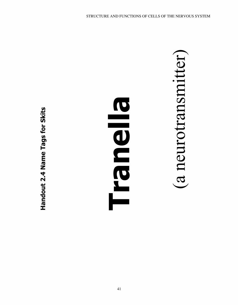
Tran
ella
(a n
euro
trans
mitt
er)
STRUCTURE AND FUNCTIONS OF CELLS OF THE NERVOUS SYSTEM
41

Agi
e A
goni
st
(An
agon
ist)
CHAPTER 2
42
Aun
tie G
onis
t
(An
anta
goni
st)
STRUCTURE AND FUNCTIONS OF CELLS OF THE NERVOUS SYSTEM
43

Reg
gie
Rec
epto
r
(a p
ost s
ynap
tic
rece
ptor
)
CHAPTER 2
44

Pres
ynap
ticM
embr
ane
on th
ete
rmin
al b
utto
n
STRUCTURE AND FUNCTIONS OF CELLS OF THE NERVOUS SYSTEM
45
Ves
icle
mem
bran
e
CHAPTER 2
46

A m
olec
ule
of
neur
otra
nsm
itter
STRUCTURE AND FUNCTIONS OF CELLS OF THE NERVOUS SYSTEM
47

Den
drite
Mem
bran
e
CHAPTER 2
48

The
Rec
epto
r
STRUCTURE AND FUNCTIONS OF CELLS OF THE NERVOUS SYSTEM
49

The
Act
ion
Pote
ntia
l
CHAPTER 2
50
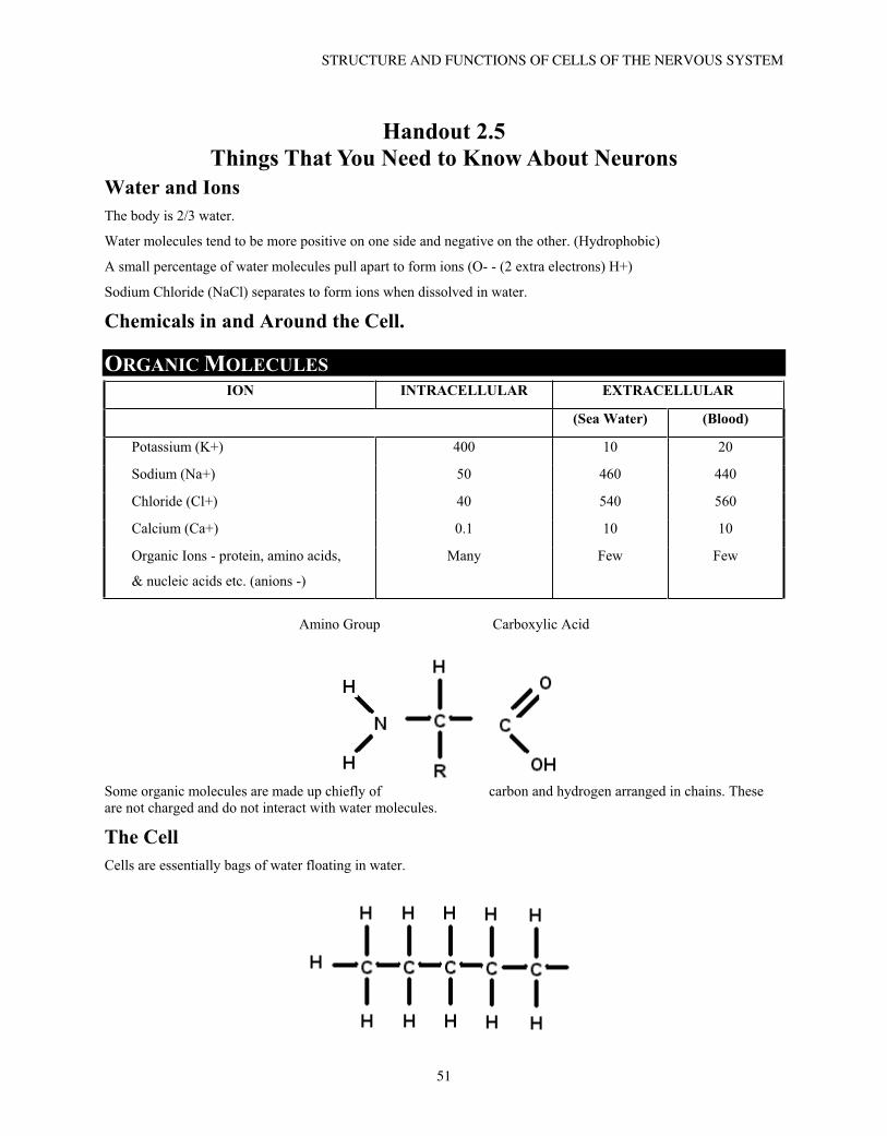
Handout 2.5 Things That You Need to Know About Neurons
Water and Ions The body is 2/3 water.
Water molecules tend to be more positive on one side and negative on the other. (Hydrophobic)
A small percentage of water molecules pull apart to form ions (O- - (2 extra electrons) H+)
Sodium Chloride (NaCl) separates to form ions when dissolved in water.
Chemicals in and Around the Cell.
ORGANIC MOLECULESION INTRACELLULAR EXTRACELLULAR
(Sea Water) (Blood)
Potassium (K+) 400 10 20
Sodium (Na+) 50 460 440
Chloride (Cl+) 40 540 560
Calcium (Ca+) 0.1 10 10
Organic Ions - protein, amino acids,
& nucleic acids etc. (anions -)
Many Few Few
Amino Group Carboxylic Acid
Some organic molecules are made up chiefly of carbon and hydrogen arranged in chains. These are not charged and do not interact with water molecules.
The Cell Cells are essentially bags of water floating in water.
STRUCTURE AND FUNCTIONS OF CELLS OF THE NERVOUS SYSTEM
51

Phospholipids have a charged phosphorous containing group at one end and can interact with water (hydrophilic) and the other end cannot (hydrophobic) making the molecule (amphipathic – likes both).
When exposed to water the molecules line up. (Form a membrane.)
Membranes are 25 to 60% protein.
Proteins are built using RNA as a pattern to link amino acids.
Protein molecules span the thickness of the membrane. They form hydrophilic channels and pumps. Some are anchored, others float.
Neurons are very small The membrane of a neuron is so thin that it cannot be seen under a light microscope. It is thinner than a wavelength of light. Seven nanometers or 10-9 meters.
Each neuron uses a the same neurotransmitter/s at all it’s synapses (In most cases)
Transmission speed is dependent on several factors: 1. Size/Diameter of the cell
2. Myelinization
The myelin cells are made up of a large percentage of lipid (fat).
The information flows in only one direction, unless forced.
Transmission changes the electrical difference between the interior and exterior of the cell.
Transmission is “all-or-nothing”
Vesicles use calcium to merge with the membrane and release intracellular material to the outside. (This is also related to how the cell grows.)
The membrane can also pinch in to bring extra cellular material inside. (Cell retraction)
CHAPTER 2
52
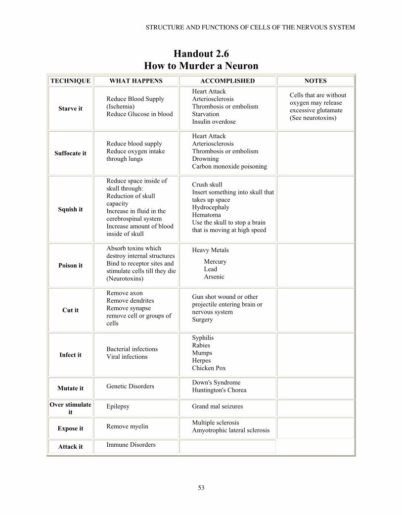
Handout 2.6 How to Murder a Neuron
TECHNIQUE WHAT HAPPENS ACCOMPLISHED NOTES
Starve itReduce Blood Supply (Ischemia) Reduce Glucose in blood
Heart Attack Arteriosclerosis Thrombosis or embolism Starvation Insulin overdose
Cells that are without oxygen may release excessive glutamate (See neurotoxins)
Suffocate itReduce blood supply Reduce oxygen intake through lungs
Heart Attack Arteriosclerosis Thrombosis or embolism Drowning Carbon monoxide poisoning
Squish it
Reduce space inside of skull through: Reduction of skull capacityIncrease in fluid in the cerebrospinal system Increase amount of blood inside of skull
Crush skull Insert something into skull that takes up space Hydrocephaly Hematoma Use the skull to stop a brain that is moving at high speed
Poison it
Absorb toxins which destroy internal structures Bind to receptor sites and stimulate cells till they die (Neurotoxins)
Heavy Metals
Mercury LeadArsenic
Cut it
Remove axon Remove dendrites Remove synapse remove cell or groups of cells
Gun shot wound or other projectile entering brain or nervous system Surgery
Infect itBacterial infections Viral infections
Syphilis Rabies Mumps Herpes Chicken Pox
Mutate it Genetic Disorders Down's Syndrome Huntington's Chorea
Over stimulate it
Epilepsy Grand mal seizures
Expose it Remove myelin Multiple sclerosis Amyotrophic lateral sclerosis
Attack it Immune Disorders
STRUCTURE AND FUNCTIONS OF CELLS OF THE NERVOUS SYSTEM
53