chapter 26: the urinary system bio 211 lecture instructor: dr. gollwitzer 1
TRANSCRIPT
Chapter 26: The Urinary System
BIO 211 LectureInstructor: Dr. Gollwitzer
1
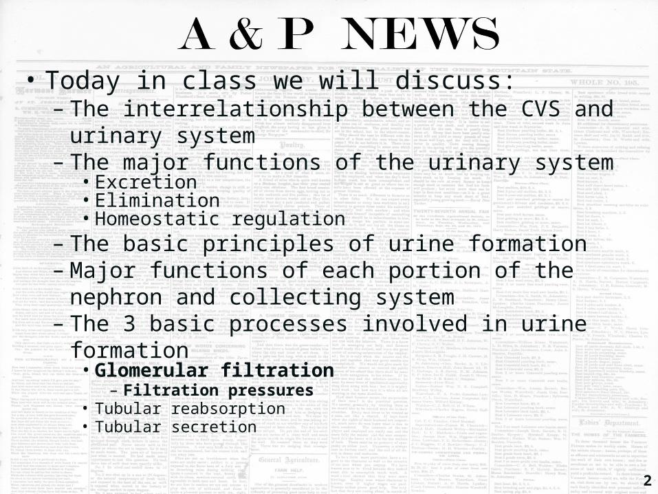
• Today in class we will discuss:– The interrelationship between the CVS and urinary
system– The major functions of the urinary system• Excretion• Elimination• Homeostatic regulation
– The basic principles of urine formation– Major functions of each portion of the nephron and
collecting system– The 3 basic processes involved in urine formation• Glomerular filtration
– Filtration pressures• Tubular reabsorption• Tubular secretion
2
CVS and Urinary System
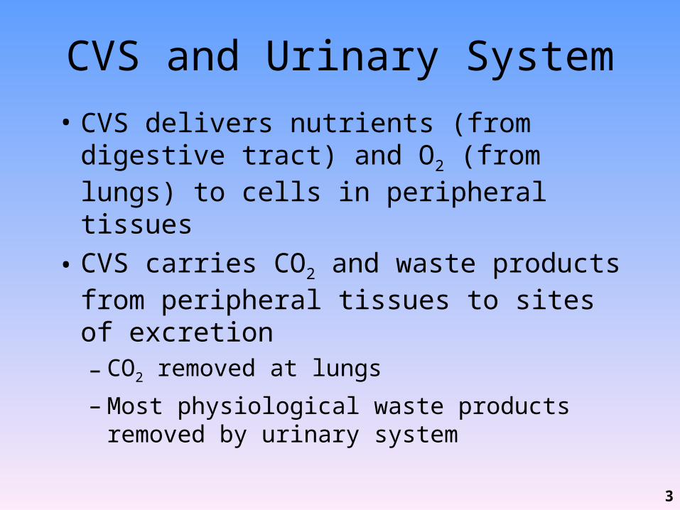
• CVS delivers nutrients (from digestive tract) and O2 (from lungs) to cells in peripheral tissues
• CVS carries CO2 and waste products from peripheral tissues to sites of excretion– CO2 removed at lungs
– Most physiological waste products removed by urinary system
3
Major Functions of Urinary System
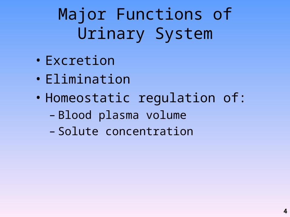
• Excretion• Elimination• Homeostatic regulation of:– Blood plasma volume– Solute concentration
4

Major Functions of Urinary System
• Excretion– Removal of organic wastes (e.g., urea, uric acid,
creatinine) from body fluids (= urine formation)– Performed by kidneys which act as filtering units
• Elimination– Discharge of waste products into environment
(urination)– Occurs when urinary bladder contracts and forces
urine through urethra and out of body
5
Major Functions of Urinary System: Homeostatic Regulation
• Regulation of blood volume (water balance) and BP– Adjusts volume of water lost in urine– Releases• Renin
– Involved in production of angiotensin II that affects BP, thirst, and other hormones (ADH, aldosterone) that affect water retention by kidneys
• Erythropoietin– Stimulates erythropoiesis in bone marrow, maintains RBC
volume
6

Major Functions of Urinary System: Homeostatic Regulation
• Regulation of plasma ion concentrations (electrolyte balance)– Controls amounts lost in urine (e.g., Na+, K+, Cl-)– Controls Ca2+ levels by synthesis of calcitriol
• Reabsorption (conservation) of valuable nutrients– Recycles valuable nutrients• e.g., amino acids, glucose
– Prevents excretion in urine
7
Major Functions of Urinary System: Homeostatic Regulation
• Stabilization of blood pH (acid-base balance)– Controls loss of H+ and HCO3
- in urine
• Detoxification– Of poisons, e.g., drugs
• Deamination– Removes NH2 (amino group) so amino acids can be
metabolized
8
Basic Principles of Urine Formation
• Urine = fluid containing:– Water– Ions– Soluble compounds
• Goal of urine production– To maintain homeostasis– By regulating volume and composition of blood
9
Basic Principles of Urine Formation
• Involves excretion of solutes (i.e., metabolic/organic waste products)– Urea• Most abundant• Produced by breakdown of amino acids
– Creatinine• Generated in skeletal muscle by breakdown of
creatine phosphate (CP, high energy compound that plays a role as energy source in muscle contraction)
– Uric acid• Formed by recycling nitrogenous bases from RNA
10
Basic Principles of Urine Formation
• Waste products dissolved in bloodstream can only be eliminated when dissolved in urine– Thus removal accompanied by unavoidable
water loss• To avoid dehydration, kidneys concentrate
filtrate (i.e., reabsorb water) produced by glomerular filtration
11
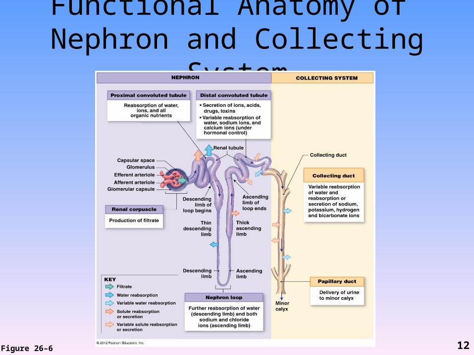
Functional Anatomy of Nephron and Collecting System
Figure 26–6 12

3 Processes Involved in Urine Formation
• Glomerular filtration– Forces water and solutes out of blood in
glomerulus into capsular space– filtrate
• Tubular reabsorption– Recovers useful materials from filtrate
• Tubular secretion– Ejects waste products, toxins, and other
undesirable solutes into tubules13
Glomerular Filtration
• Occurs in renal corpuscle• Hydrostatic pressure forces water and solutes:– Out of blood in glomerulus– Into capsular space filtrate
• Occurs solely on basis of size– Small solute molecules carried with filtrate
14
Glomerular Filtration• Involves passage across filtration membrane
which is composed of 3 cellular units– Glomerular capillary endothelium– Lamina densa– Filtration slits
15
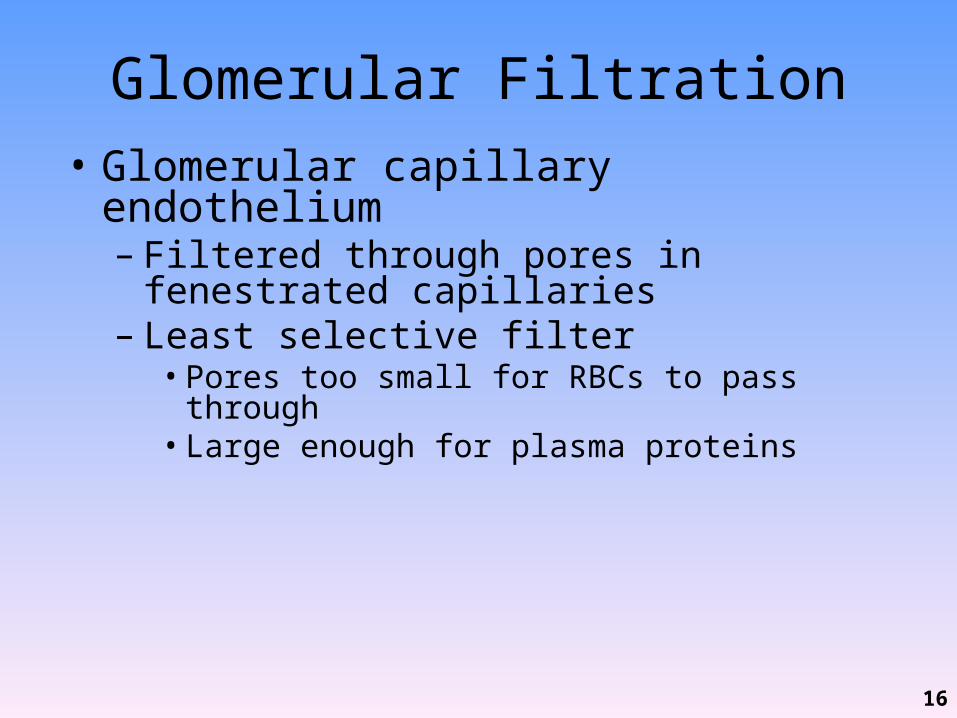
Glomerular Filtration• Glomerular capillary endothelium– Filtered through pores in fenestrated capillaries– Least selective filter• Pores too small for RBCs to pass through• Large enough for plasma proteins
16
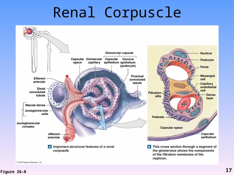
Renal Corpuscle
Figure 26–8 17
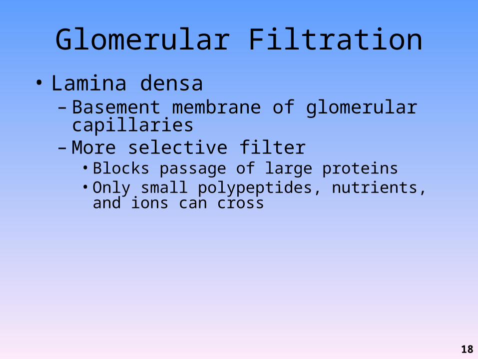
Glomerular Filtration• Lamina densa– Basement membrane of glomerular capillaries– More selective filter• Blocks passage of large proteins• Only small polypeptides, nutrients, and ions can cross
18
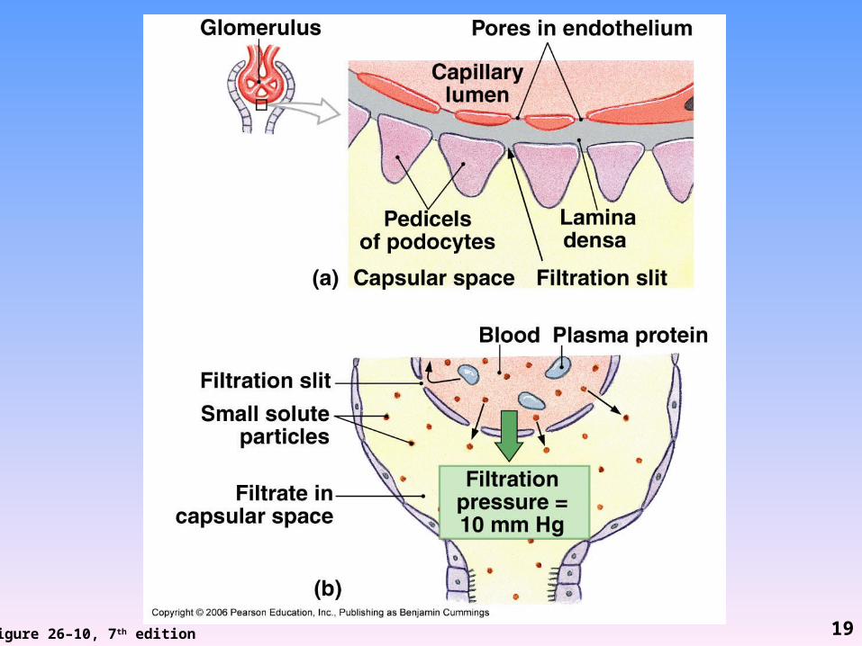
Figure 26–10, 7th edition 19
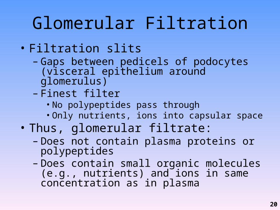
Glomerular Filtration• Filtration slits– Gaps between pedicels of podocytes (visceral
epithelium around glomerulus)– Finest filter• No polypeptides pass through• Only nutrients, ions into capsular space
• Thus, glomerular filtrate:– Does not contain plasma proteins or polypeptides– Does contain small organic molecules (e.g.,
nutrients) and ions in same concentration as in plasma
20

Filtration Pressures
• Filtration pressure = balance between:– Hydrostatic (fluid) pressures• Glomerular hydrostatic pressure (GHP) in capillaries (50
mmg Hg)• Capsular hydrostatic pressure (CHP) (15 mm Hg)
– Blood osmotic pressure (BOP) (25 mm Hg)
21
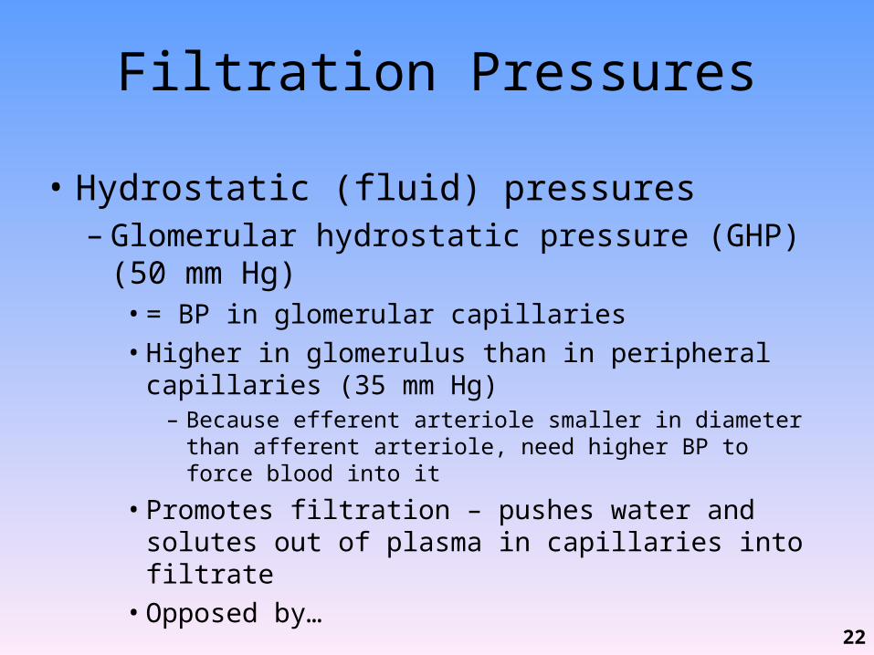
Filtration Pressures
• Hydrostatic (fluid) pressures– Glomerular hydrostatic pressure (GHP) (50 mm Hg)• = BP in glomerular capillaries• Higher in glomerulus than in peripheral capillaries (35
mm Hg)– Because efferent arteriole smaller in diameter than afferent
arteriole, need higher BP to force blood into it
• Promotes filtration – pushes water and solutes out of plasma in capillaries into filtrate• Opposed by…
22
Filtration Pressures
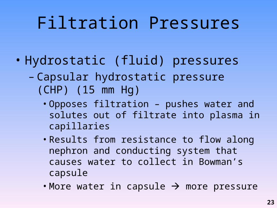
• Hydrostatic (fluid) pressures– Capsular hydrostatic pressure (CHP) (15 mm Hg)• Opposes filtration – pushes water and solutes out of
filtrate into plasma in capillaries• Results from resistance to flow along nephron and
conducting system that causes water to collect in Bowman’s capsule• More water in capsule more pressure
23
Filtration Pressures
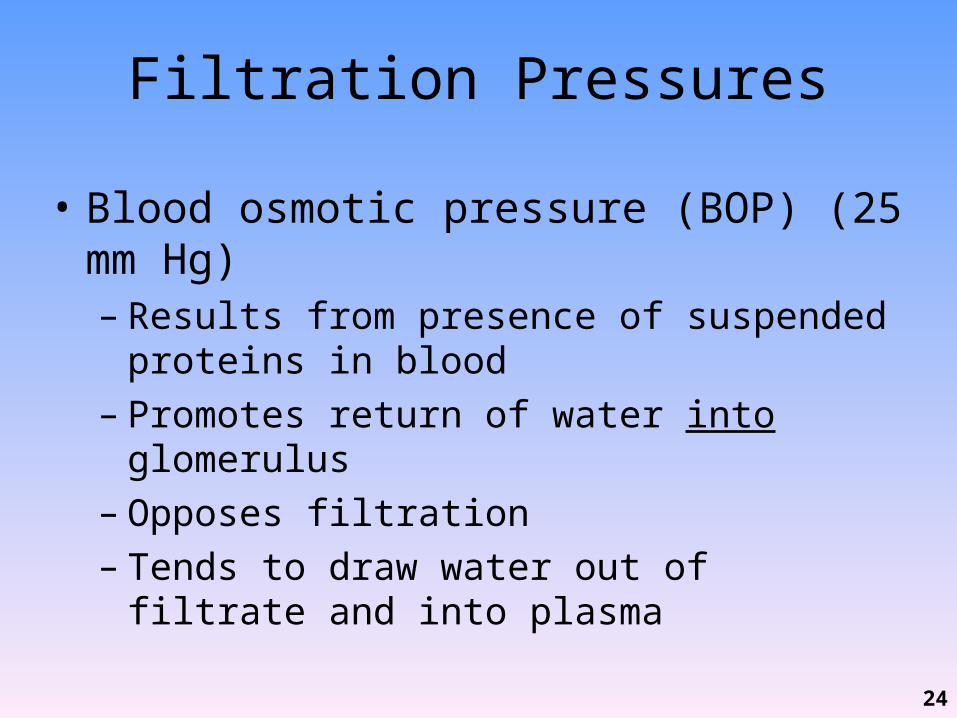
• Blood osmotic pressure (BOP) (25 mm Hg)– Results from presence of suspended proteins in
blood– Promotes return of water into glomerulus– Opposes filtration– Tends to draw water out of filtrate and into plasma
24
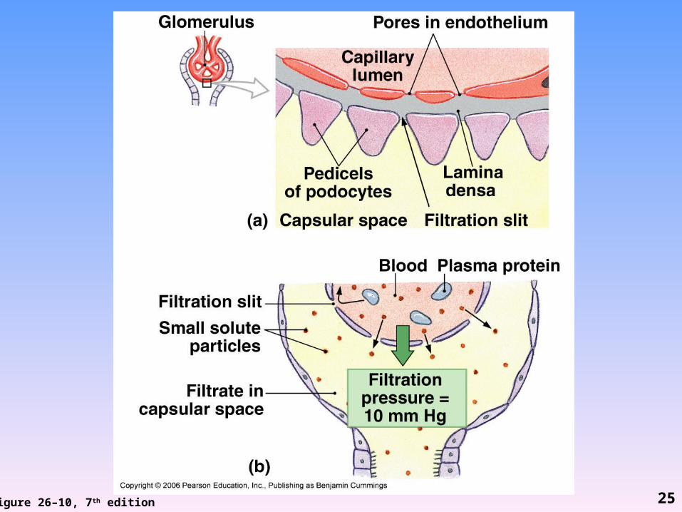
Figure 26–10, 7th edition 25
Summary of Filtration Pressures
• Hydrostatic pressures– GHP (pushing out of glomerulus) = 50 mm Hg– CHP (pushing into glomerulus) = 15 mm Hg– Net = 35 mm Hg (pushing out of glomerulus)
• Osmotic pressure– BOP (draws into glomerulus) = 25 mm Hg
• Filtration pressure = 10 mm Hg– Difference between net hydrostatic pressure and
blood osmotic pressure
26
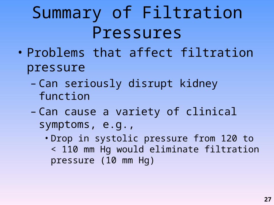
Summary of Filtration Pressures
• Problems that affect filtration pressure– Can seriously disrupt kidney function– Can cause a variety of clinical symptoms, e.g.,• Drop in systolic pressure from 120 to < 110 mm Hg
would eliminate filtration pressure (10 mm Hg)
27
• Today in class we will discuss:– The 3 basic processes involved in urine formation• Glomerular filtration
– Glomerular Filtration Rate– Renal Failure
• Tubular reabsorption– PCT, Loop of Henle & Countercurrent Exchange,DCT– Collecting System
• Tubular secretion– PCT, DCT and Collecting system
– Urine• Compare/contrast to plasma• General characteristics• Hormone influence of volume and concentration
– Voluntary & involuntary regulation of urination and the micturition reflex
28
Glomerular Filtration Rate (GFR)• Gomerular filtration– Vital first step essential to all other kidney functions– Must occur so:
• Waste products excreted• pH controlled• Blood volume maintained
• GFR = amount of filtrate kidneys produce per minute• Avg GFR = 125 mL/min or 50 gal/day (out of 480 gallons
of blood flow/day)– 10% of fluid delivered by renal arteries enters capsular spaces– 99% of this reabsorbed so urinate only 0.5 gallons/day
29
Glomerular Filtration Rate (GFR)• Measured using creatinine clearance test (CCT)– Breakdown of CP in muscle creatinine– Creatinine enters filtrate at glomerulus and is not
reabsorbed so is excreted in urine– Can compare amount of creatinine in blood vs. in
urine during 24 hour and estimate GFR– If glomerulus damaged, GFR will be altered (have
more or less creatinine in urine than normal)
30
Glomerular Filtration Rate (GFR)• GFR depends on:– Adequate blood flow to glomerulus– Maintenance of normal filtration pressures
• Affected by anything that reduces renal blood flow or BP, e.g.,– Hypotension, hemorrhage, shock, dehydration
• Decreased renal blood volume and/or BP decreased filtration pressure decreased GFR
31
Control of GFR• GFR increased by:– EPO (relatively minor) – Renin-angiotensin system– Natriuretic peptides (ANP and BNP)
32
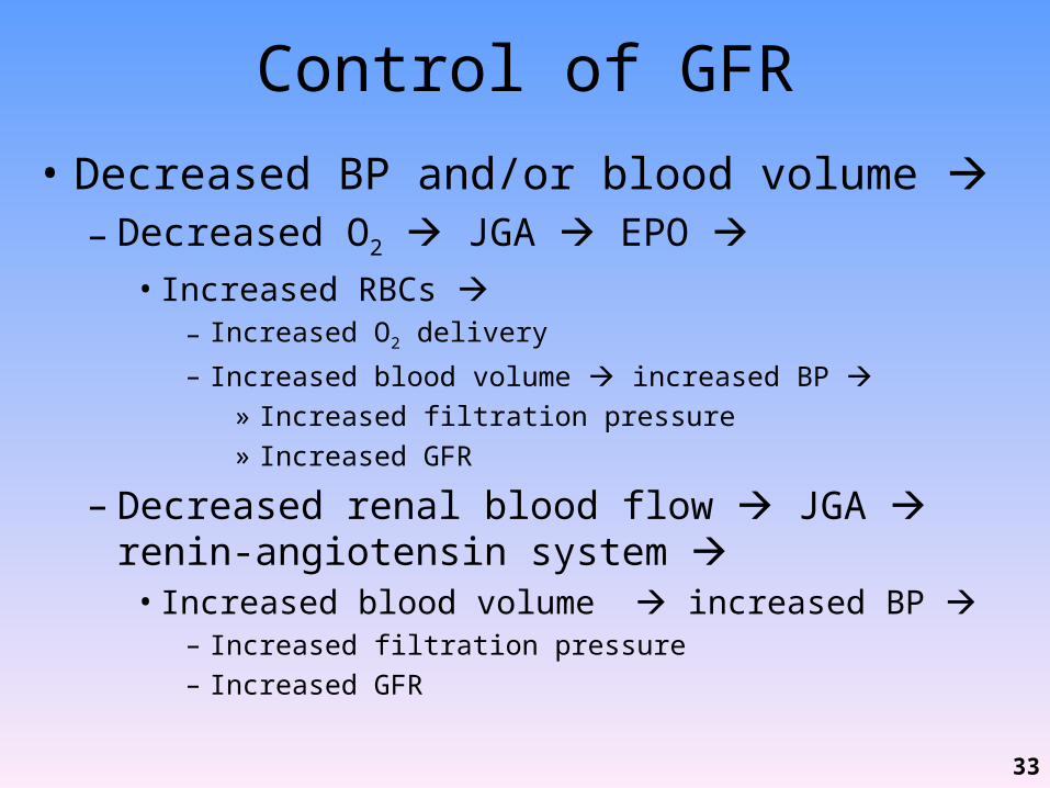
Control of GFR
• Decreased BP and/or blood volume – Decreased O2 JGA EPO • Increased RBCs
– Increased O2 delivery
– Increased blood volume increased BP » Increased filtration pressure» Increased GFR
– Decreased renal blood flow JGA renin-angiotensin system • Increased blood volume increased BP
– Increased filtration pressure– Increased GFR
33
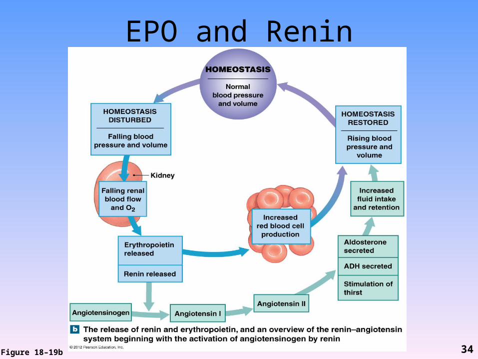
EPO and Renin
Figure 18–19b 34
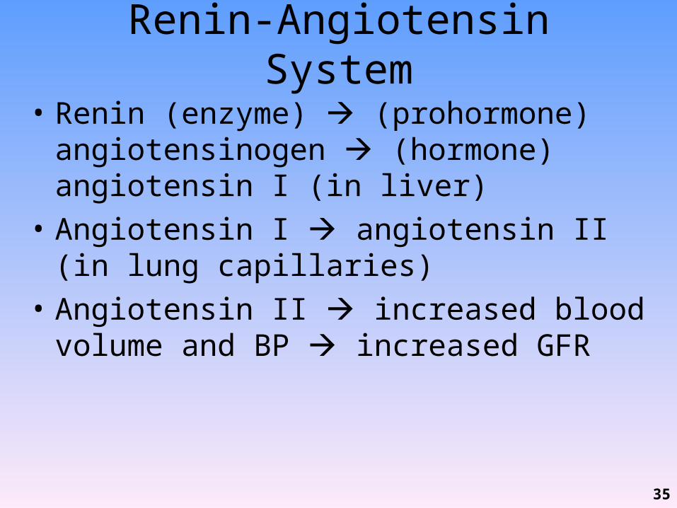
Renin-Angiotensin System
• Renin (enzyme) (prohormone) angiotensinogen (hormone) angiotensin I (in liver)
• Angiotensin I angiotensin II (in lung capillaries)
• Angiotensin II increased blood volume and BP increased GFR
35

Primary Effects of Angiotensin II• Stimulates constriction of efferent arterioles
increased glomerular pressure• Directly stimulates reabsorption of Na+ and H2O in DCT
increased blood volume and BP• Stimulates adrenal cortex aldosterone
reabsorption of Na+ (and H2O) increased blood volume and BP
• Stimulates posterior pituitary ADH reabsorption of H2O increased blood volume and BP
• Stimulates thirst increased blood volume and BP• Stimulates vasoconstriction of arterioles
36
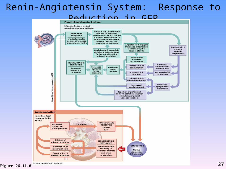
Renin-Angiotensin System: Response to Reduction in GFR
Figure 26–11-0 37
Control of GFR• Increased blood volume or BP stretched
cardiac muscle cells natriuretic peptides – ANP = atrial NP– BNP = brain NP (produced by ventricles)
• Natriuretic peptides– Increase GFR– Decrease blood volume and BP– Via 2 mechanisms
38

Natriuretic Peptides Increase GFR
• Act opposite to angiotensin II– Increase Na+ and H2O loss• Inhibit renin release• Inhibit secretion of aldosterone and ADH
– Suppress thirst– Prevent increased BP by angiotensin II and NE
• Increase glomerular pressures– Dilate afferent arterioles– Constrict efferent arterioles
• Also increase tubular reabsorption of Na+
– Decreases blood volume and BP39
Renal Failure• When filtration (GFR) slows, urine production
decreases• Symptoms appear because water, ions, and metabolic
wastes retained rather than excreted• Almost all systems affected: fluid balance, pH,
muscular contraction, neural function, digestive function, metabolism
• Leads to:– Hypertension (due to blood “backing up”)– Anemia due to lack of erythropoietin production– CNS problems (sleepiness, seizures, delirium, coma,
death)40

Renal Failure
• Acute renal failure– From exposure to toxic drugs, renal ischemia,
urinary obstruction, trauma– Develops quickly, but usually temporary– With supportive treatment can survive
• Chronic renal failure– Condition deteriorates gradually– Cannot be reversed– Dialysis or kidney transplant may prolong life
41
Reabsorption and Secretion
• Occur in all segments of renal tubules• Relative importance changes from segment to
segment
42
Tubular Reabsorption
• Molecules move from filtrate across tubular epithelium into peritubular interstitial fluid and blood– Water, valuable solutes (e.g., nutrients, proteins,
amino acids, glucose)
• Occurs through diffusion, osmosis (H2O), active transport by carrier proteins
• Occurs primarily along PCT (also along renal tubule and collecting system)
43
Tubular Secretion
• Molecules move from peritubular fluid into tubular fluid
• Lowers plasma concentration of undesirable materials
• Necessary because filtration does not force all solutes out of plasma
• Primary method of excretion for many drugs• Occurs primarily at PCT and DCT
44

Reabsorption and Secretion: PCT
• Primarily reabsorption– 60-70% of filtrate– Includes:• Organic nutrients (99-100%), e.g., glucose, amino acids,
proteins, lipids, vitamins• Water (60-70%)• Ions (60-70%), e.g., Na+, Cl-; also K+, Ca2+, HCO3
- – Reabsorbed materials enter peritubular fluid and
capillaries• Secretion– H+, NH4
+, creatinine, drugs, toxins45

Reabsorption: Loop of Henle
• Reabsorption– Na+, Cl-
– Water
• Accomplished by countercurrent exchange– Refers to exchange by tubular fluids moving in
opposite directions– Fluid in descending limb flows toward renal pelvis– Fluid in ascending limb flows toward cortex
46

Countercurrent Exchange
• Occurs because of different permeabilities of segments of LOH
• Descending limb (thin)– Permeable to water– Relatively impermeable to solutes
• Ascending limb (thick)– Relatively impermeable to water and solutes– Has active transport mechanisms• Pump Na+ and Cl- from tubular fluid into peritubular fluid
47

Countercurrent Exchange
• Na+ and Cl- pumped out of thick ascending limb into peritubular fluid
• Increases osmotic concentration in peritubular fluid around thin descending limb
• Results in osmotic flow of H2O out of thin descending limb into peritubular fluid increased solute concentration in thin descending limb
• Arrival of concentrated solution in thick ascending limb increases transport of Na+ and Cl- into peritubular fluid
48
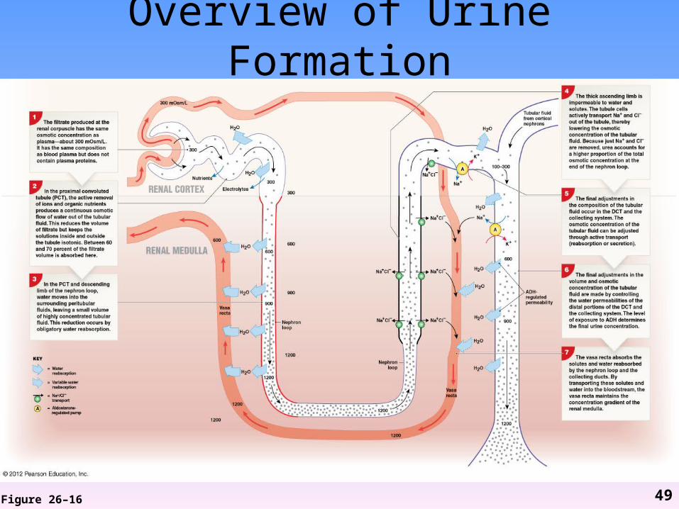
Overview of Urine Formation
Figure 26–16 49
Reabsorption and Secretion: DCT
• Reabsorption (by vasa recta)– Na+ (under influence of aldosterone), Cl-
– Ca2+(under influence of PTH and calcitriol)– H2O (under influence of ADH)
• Secretion– K+ (in exchange for Na+), H+ – NH4
+ (from deamination; produces lactic acid, ketone bodies acidosis)
– Creatinine, drugs, toxins
50
Reabsorption and Secretion: Collecting System
• Makes final adjustments to ion concentration and urine volume
• Reabsorption – Na+ (under influence of aldosterone)– H2O (under influence of ADH)
– HCO3-
– Urea (distal portion)
• Secretion– K+, H+
51
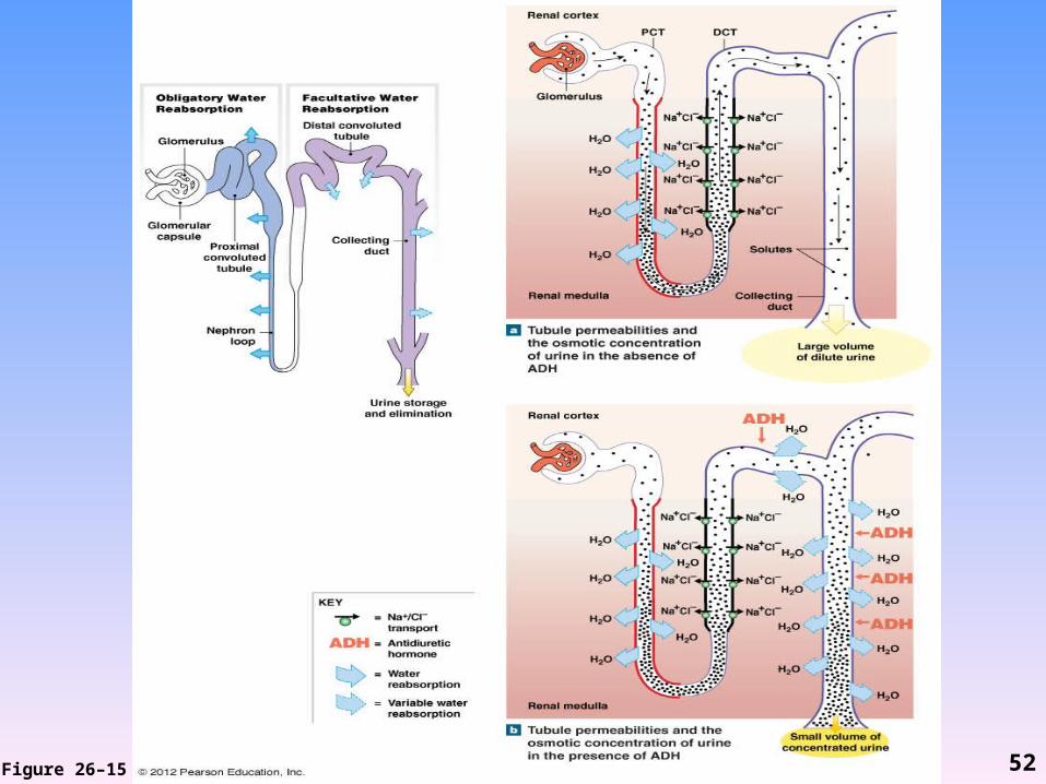
Figure 26–15 52

Summary: Urine Formation
• Involves all parts of nephron and collecting system
• Processes occur primarily in certain areas– Glomerular filtration at the renal corpuscle– Nutrient reabsorption in the PCT– Water and salt conservation in loop of Henle– Tubular secretion in the DCT
• Regulation of final volume and solute concentration occurs in loops of Henle and collecting system
53
Normal Kidney Function
• Continues as long as filtration, reabsorption, and secretion function within narrow limits
• Disruption of kidney function has immediate effects on composition of circulating blood
• If both kidneys affected, death occurs within few days
54
Normal Kidney Function
• Glomeruli produce approx 48 gallons (180 L) of filtrate/day– 70X plasma volume!
• Almost all fluid volume must be reabsorbed to avoid fatal dehydration
55
Urine
• Clear, sterile solution• Yellow (“straw”) color due to pigment
(urobilin)• Urinalysis = analysis of urine sample• Results from filtration, absorption,
secretion activities of nephron
56
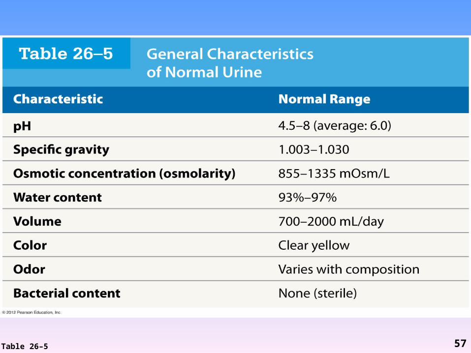
Table 26–5 57
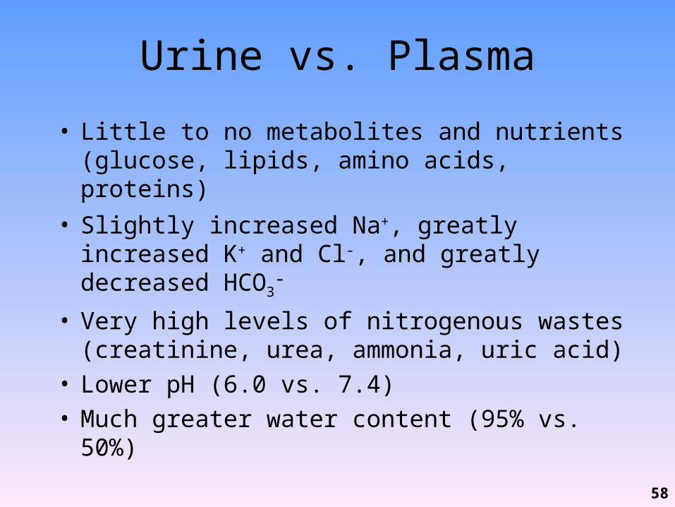
Urine vs. Plasma
• Little to no metabolites and nutrients (glucose, lipids, amino acids, proteins)
• Slightly increased Na+, greatly increased K+ and Cl-, and greatly decreased HCO3
-
• Very high levels of nitrogenous wastes (creatinine, urea, ammonia, uric acid)
• Lower pH (6.0 vs. 7.4)• Much greater water content (95% vs. 50%)
58
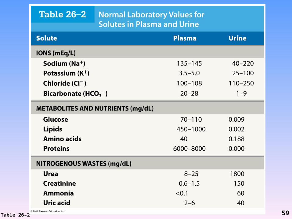
Table 26–2 59
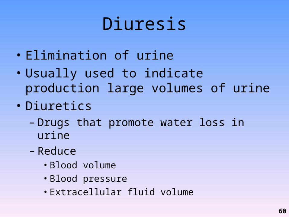
Diuresis
• Elimination of urine• Usually used to indicate production large
volumes of urine• Diuretics– Drugs that promote water loss in urine– Reduce• Blood volume• Blood pressure• Extracellular fluid volume
60
Micturition Reflex
• Coordinates the process of urination• Begins when stretch receptors in bladder
stimulate parasympathetic neurons– Results in contraction of detrusor muscle
contraction
• Voluntary relaxation of external urethral sphincter causes relaxation of internal urethral sphincter
61
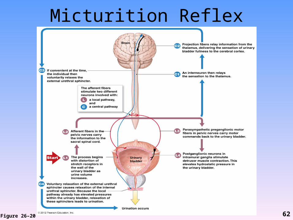
Micturition Reflex
Figure 26–20 62
Voluntary Control
• Infants– Lack voluntary control over urination–Corticospinal connections are not
established
• Incontinence =– Inability to voluntarily control urination–May be caused by trauma to internal or
external urethral sphincter
63
Age-Related Changes in Urinary System
• Decline in number of functional nephrons• Reduction in GFR • Reduced sensitivity to ADH
64
Age-Related Changes in Urinary System
• Problems with micturition reflex– Sphincter muscles lose tone incontinence– Lose control due to:• Stroke• Alzheimer’s disease• CNS problems
– In males, enlarged prostate compresses urethra, restricts urine flow urinary retention
65
The Excretory System
• Includes all systems with excretory functions that affect body fluids composition– Urinary system– Integumentary system– Respiratory system– Digestive system
66