chapter 29 development and inheritance lecture outline files/bio132lecture... · chapter 29...
TRANSCRIPT
Chapter 29 Development and Inheritance Lecture Outline
Fertilization 23 Chromosome/Haploid ovum + 23 Chromosome/Haploid sperm = 46 Chromosome/Diploid zygote 1. Sperm transport & capacitation 2. Acrosomal reaction & sperm penetration Ampulla-isthmus junction A. Hyaluronidase: penetrate corona radiata B. Bind zona pellucida Acrosomal reaction Acrosin C. Bind sperm receptor on membrane Membrane fusion Oocyte activation 3. Oocyte activation Fusion → Na+ channels → depolarization → Ca2+ release from SER A. Cortical reaction Inactivate sperm receptors Harden zona pellucida Prevent polyspermy B. Complete Meiosis II Polar body C. ↑ metabolism 4. Nuclear fusion Pronuclei fuse: Amphimixis Zygote Mitosis Prenatal development 1. Pre-embryonic development A. Cleavage & balstocyst formation (d 1-6) Zygote → preembryo Blastomeres Morula Blastocyst Trophoblast Inner cell mass Blastocoele Endometrial gland secretions B. Implantation (d 6-14) Trophoblast erodes endometrium Cellular trophoblast Syncytial trophoblast Lacunae Human Chorionic Gonadotropin (hCG) Maintain corpus luteum Progesterone Maintain uterus Gestational trophoblastic neoplasia 2. Embryonic development A. Gastrulation (w 2-3) Inner cell mass → embryonic disc Epiblast Hypoblast
Embryonic membranes 1. Amnion Amniotic fluid 2. Yolk sac Blood cell production 3. Allantois Umbilical cord & bladder 4. Chorion Chorionic villi Gastrulation = Embryonic disc → Primary germ layers Primary germ layers 1. Ectoderm 2. Endoderm 3. Mesoderm Primitive streak B. Placentation (w 2-12) Chorionic villi Body stalk Decidua capsularis Decidua basalis Placenta = -chorionic villi -syncytial trophoblast -decidua basalis Endocrine function: 1. Human Chorionic Gonadotropin 2. Human Placental Lactogen 3. Placental prolactin 4. Relaxin 5. Progesterone 6. Estrogen C. Organogenesis Notochord: mesoderm Umbilical cord: yolk sac, vessels 1. Ectoderm A. Neuralization Neural tube B. Epidermis & epidermal structures Hair, nails, glands Mouth, anus Special sense organs 2. Endoderm -Epithelia of digestive & respiratory -Thyroid, parathyroid, thymus -Liver, pancreas -Urethra, urinary bladder 3. Mesoderm -Muscle, bone, bone marrow -Blood, vessels -Connective tissue, serosa 3. Fetal development Summary A. First trimester (m 0-3)
Amy Warenda Czura, Ph.D. 1 SCCC BIO132 Chapter 29 Handout
Zygote Embryogenesis B. Second trimester (m 4-6) Amniochorionic membrane C. Third trimester (m 7-9) Adipose Pregnancy & maternal systems 1. Physical -vascularization of reproductive organs -uterus expands -diaphragm invades throax -lordosis -↑ mammary gland & milk 2. Functional -GI: nausea, heartburn, constipation -Urinary: ↑ GFR, incontinence -Respiratory: ↑ rate, ↑ TV, ↓ residual volume -Cardiovascular: ↑ volume, ↑BP, ↑ CO -Metabolism: hunger Parturition ↑ estrogen = -↑ oxytocin receptors in myometrium -block progesterone -Braxton Hicks contractions Fetus initiates: Oxytocin Protaglandins Contractions Maternal oxytocin Positive feedback Stages of labor 1. Dilation stage Contractions 10-30 min Cervix dilates Amniochorionic membrane ruptures 2. Expulsion stage Contractions 1-3 min 3. Placental stage Afterbirth Complications 1. Teratogens 2. Spontaneous abortion 3. Premature labor 4. Difficult deliveries A. Face up B. Dystocia Cesarean section C. Breech Multiple births Dizygotic twins: fraternal Monozygotic twins: identical Conjoined twins Lactation ↑ estrogen + lactogen → ↑ PRH → ↑ prolactin Colostrum Milk: water, proteins, amino acids, lipids, sugars,
ions, vitamins, antimycrobials (lysozyme & complement) Milk let down reflex Tactile → Oxytocin release Myoepithelial cells Post natal development 1. Neonatal -high respiratory rate -ductus arteriosus & formaen ovale close -high heart rate -meconium cleared -high water loss at kidney -body temp fluctuates -high metabolic rate 2. Infancy 1 month to 2 years 3. Childhood 2 years to adolescence 4. Adolescence Puberty to maturity 5. Senescence Maturity to death Human Genetics Gene Genotype Phenotype Patterns of inheritance Somatic cells 23 pair homologus chromosomes 1 pair sex chromosomes: XX, XY 22 pair autosomal Locus Alleles Autosomal chromosome alleles Homozygous Heterozygous 1. Dominant 2. Recessive 3. Incomplete dominance 4. Codominance Simple inheritance Punnett square Sex-linked inheritance X linked XX = female XY = male Polygenic inheritance 1. Suppression 2. Complementary gene action Penetrance Expressivity Genomic imprinting Individual variation 1. Genetic recombination Translocation (crossing over) Defects:
Amy Warenda Czura, Ph.D. 2 SCCC BIO132 Chapter 29 Handout

A. Translocation defect B. Extra/missing chromosomes 1. Trisomy e.g. Down syndrome 2. Monosomy e.g Turner syndrome 2. Mutation Spontaneous mutation: 1/109 Fetal testing 1. Amniocentesis 2. Chorionic villi sampling 3. In vitro fertilization 8 cell morula
Amy Warenda Czura, Ph.D. 3 SCCC BIO132 Chapter 29 Handout

Amy Warenda Czura, Ph.D. 4 SCCC BIO132 Chapter 29 Handout
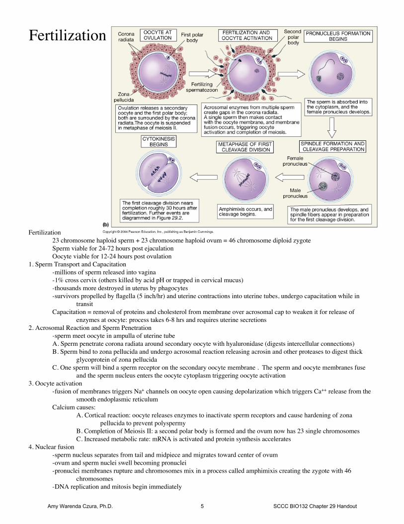
Fertilization23 chromosome haploid sperm + 23 chromosome haploid ovum = 46 chromosome diploid zygoteSperm viable for 24-72 hours post ejaculationOocyte viable for 12-24 hours post ovulation
1. Sperm Transport and Capacitation-millions of sperm released into vagina-1% cross cervix (others killed by acid pH or trapped in cervical mucus)-thousands more destroyed in uterus by phagocytes-survivors propelled by flagella (5 inch/hr) and uterine contractions into uterine tubes, undergo capacitation while in
transitCapacitation = removal of proteins and cholesterol from membrane over acrosomal cap to weaken it for release of
enzymes at oocyte: process takes 6-8 hrs and requires uterine secretions2. Acrosomal Reaction and Sperm Penetration
-sperm meet oocyte in ampulla of uterine tubeA. Sperm penetrate corona radiata around secondary oocyte with hyaluronidase (digests intercellular connections)B. Sperm bind to zona pellucida and undergo acrosomal reaction releasing acrosin and other proteases to digest thick
glycoprotein of zona pellucidaC. One sperm will bind a sperm receptor on the secondary oocyte membrane . The sperm and oocyte membranes fuse
and the sperm nucleus enters the oocyte cytoplasm triggering oocyte activation3. Oocyte activation
-fusion of membranes triggers Na+ channels on oocyte open causing depolarization which triggers Ca++ release from thesmooth endoplasmic reticulum
Calcium causes:A. Cortical reaction: oocyte releases enzymes to inactivate sperm receptors and cause hardening of zona
pellucida to prevent polyspermyB. Completion of Meiosis II: a second polar body is formed and the ovum now has 23 single chromosomesC. Increased metabolic rate: mRNA is activated and protein synthesis accelerates
4. Nuclear fusion-sperm nucleus separates from tail and midpiece and migrates toward center of ovum-ovum and sperm nuclei swell becoming pronuclei-pronuclei membranes rupture and chromosomes mix in a process called amphimixis creating the zygote with 46
chromosomes-DNA replication and mitosis begin immediately
Fertilization
Amy Warenda Czura, Ph.D. 5 SCCC BIO132 Chapter 29 Handout

Prenatal Development
1. Pre-embryonic Development (fertilization to implantation) A. Cleavage and Blastocyst Formation (~day1-6)
Cleavage = rapid mitotic divisions of zygote with little growth-@36hr post fertilization, first division is complete: zygote → pre-embryo
-pre-embryo consists of 2 identical blastomeres-blastomeres continue division to create a cell cluster called a morula-@ day 4 morula differentiates into a blastocyst of 100+ cells:
-outer layer of cells called the trophoblast (will become part of placenta)-inner cluster of cells called the inner cell mass (will become the embryonic disc)-central cavity called the blastocoele which is filled with fluid
-trophoblast cells digest zona pellucida and blastocyst receives nourishment from uterine secretionsfrom endometrial glands
B. Implantation (~day 6-14)-blastocyst contacts uterine lining on inner cell mass side-trophoblast cells secrete digestive enzymes and growth factors triggering thickening of endometrial
lining at point of contact-blastocyst erodes a path into endometrium-trophoblast proliferates and forms two layers:
1. inner cellular trophoblast: remains as wall of blastocyst2. outer syncytial trophoblast: multinuclear cytoplasmic mass in contact with endometrium
-syncytial trophoblast digests endometrial cells and blood vessels creating channels called lacunaethat fill with maternal blood bringing nutrients to blastocyst
-trophoblast cells produce hCG/ human chorionic gonadotropin to maintain the corpus luteum,progesterone from the corpus luteum prevents mensus
Amy Warenda Czura, Ph.D. 6 SCCC BIO132 Chapter 29 Handout

2. Embryonic Development (week 1-8) A. Gastrulation (week 2-3)
-inner cells mass divides into epiblast (superficial) and hypoblast (deep) forming a two layerembryonic disc
-embryonic membranes form:1. amnion
-develops superior to epiblast-forms a transparent membrane sac that fills with amniotic fluid to support and protect
fetus during development2. yolk sac
-develops inferior to hypoblast-serves as site of early blood cell production-later forms part of the gut
3. allantois-forms as out-pocketing at caudal end of yolk sac-serves as structural basis for umbilical cord-later develops into part of urinary bladder
4. chorion-forms from cellular trophoblast-develops chorionic villi that are later vascularized to become fetal half of the placenta
-two-layer embryonic disc differentiates into three primary germ layers (gastrulation)1. ectoderm - faces the amnion2. endoderm - faces the yolk sac3. mesoderm - layer of cells that migrates between endoderm and ectoderm
-three-layer embryonic disc further differentiates to form the embryo-embryo is centered on a raised groove called the primitive streak that appears on dorsal surface of
embryonic disc
Amy Warenda Czura, Ph.D. 7 SCCC BIO132 Chapter 29 Handout

B. Placentation (week 2-12)-chorionic villi enlarge and become vascularized-arteries and veins connect to developing embryo at body stalk which forms from allantois-as embryo enlarges, it bulges out of endometrium in amniotic sac-chorion surrounding bulge thins and is covered by decidua capsularis (endometrium)-chorion facing uterine wall retains large vascularized chorionic villi that extend into blood filled
lacunae in thickened endometrium (fetal half of placenta)-blood filled endometrium called decidua basalis forms maternal half of placentaPlacenta = disc shaped tissue consisting of chorionic villi + syncytical trophoblst + decidua basalis,
functions to connect fetal blood supply to large surface area for nutrient, gas, and wasteexchange with maternal blood supply (complete at 12 weeks)
Amy Warenda Czura, Ph.D. 8 SCCC BIO132 Chapter 29 Handout

C. Organogenesis (week 3-8)-notochord develops in mesoderm under
primitive streak of embryonic disc and defineslong axis on body
-as three germ layers differentiate, they foldaround toward yolk sac creating a cylindricalshape with ectoderm on the outside andendoderm on the inside
-fetal vessels and yolk sac protrude throughcylinder and will form the umbilical cord
1. Specialization of the ectoderma. Neuralization
-ectoderm overlying notochord differentiates and folds inward forming neural tube-neural tube pinches off into mesoderm: anterior end will form brain, remainder will form
spinal cordb. Epidermis
-most of ectoderm differentiates into epidermis and epidermal structures (hair, nails, skinglands, lining of mouth and anus, special sense organs)
2. Specialization of endoderm-differentiates to form epithelial linings of digestive and respiratory tracts-forms all associated glandular tissues (thyroid, parathyroid, thymus, liver, pancreas)-forms urethra and most of urinary bladder (some from allantois)
3. Specialization of mesoderm-differentiates to form all tissues and structures between epidermis and mucosal linings: muscle,
bone, bone marrow, blood, blood vessels, lymphatic vessels, all connective tissue, serosa,reproductive organs, kidneys
At the end of embryonic period (week 8), all body systems are present, ossification has begun, andcardiovascular system is fully functional. Embryo (week 0-8) now called a fetus (week 8-birth).
3. Fetal Development (week 9-38)-body structures and organ systems continue development to form all specific cell types and tissues of
human body
Amy Warenda Czura, Ph.D. 9 SCCC BIO132 Chapter 29 Handout
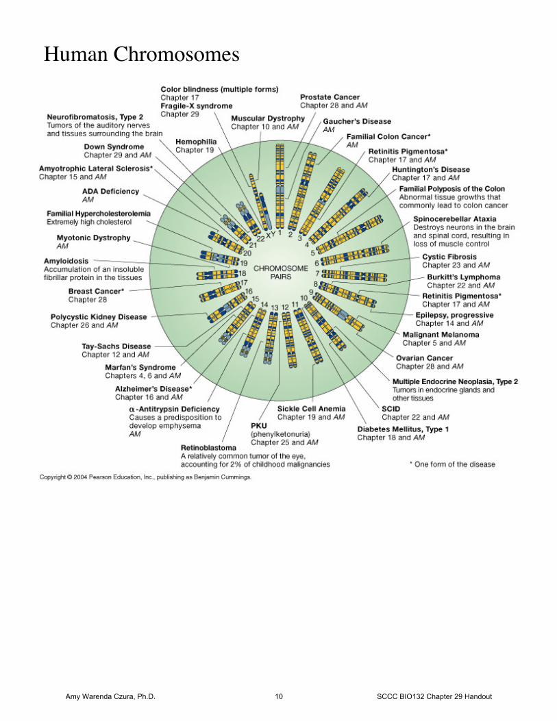
Human Chromosomes
Amy Warenda Czura, Ph.D. 10 SCCC BIO132 Chapter 29 Handout