chapter 3 · 2016-09-06 · chapter 3 lecture by edward j. zalisko ... 3. proteins, 4. nucleic...
TRANSCRIPT

PowerPoint Lectures
Campbell Biology: Concepts & Connections, Eighth EditionREECE • TAYLOR • SIMON • DICKEY • HOGAN
Chapter 3
Lecture by Edward J. Zalisko
The Molecules of Cells
© 2015 Pearson Education, Inc.
Introduction
© 2015 Pearson Education, Inc.
© 2015 Pearson Education, Inc.
Figure 3.0-2
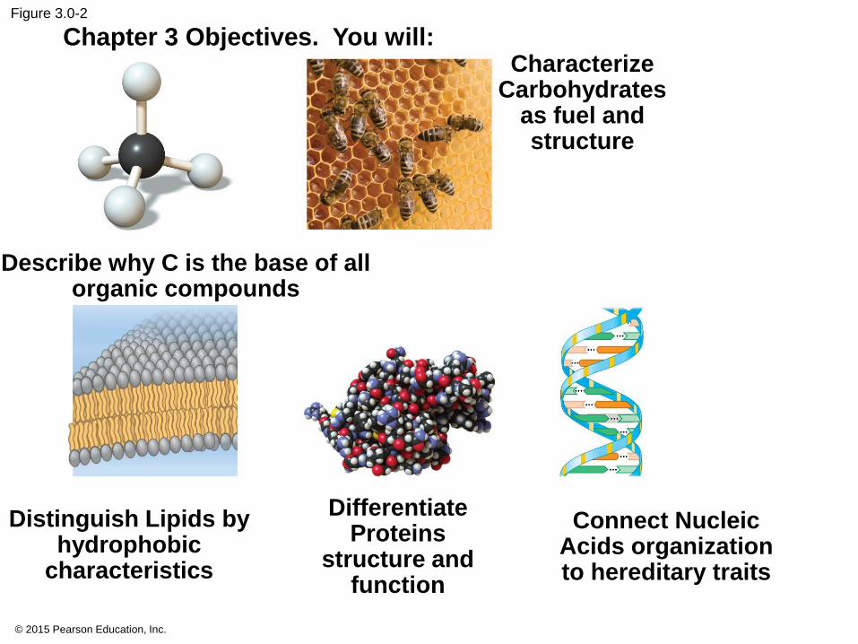
Chapter 3 Objectives. You will:
Describe why C is the base of all organic compounds
Characterize Carbohydrates
as fuel and structure
Distinguish Lipids by hydrophobic
characteristics
Differentiate Proteins
structure and function
Connect Nucleic Acids organization to hereditary traits
INTRODUCTION TO ORGANIC
COMPOUNDS
© 2015 Pearson Education, Inc.
3.1 Life’s molecular diversity is based on the properties of carbon
• carbon bonded to
• other carbons and
• atoms of other elements.
• organic compounds.
© 2015 Pearson Education, Inc.
Draw a carbon atom with valence shell
© 2015 Pearson Education, Inc.
© 2015 Pearson Education, Inc.
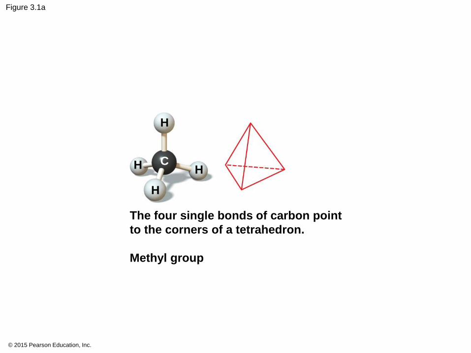
Figure 3.1a
The four single bonds of carbon point
to the corners of a tetrahedron.
Methyl group
H
H
H
HC
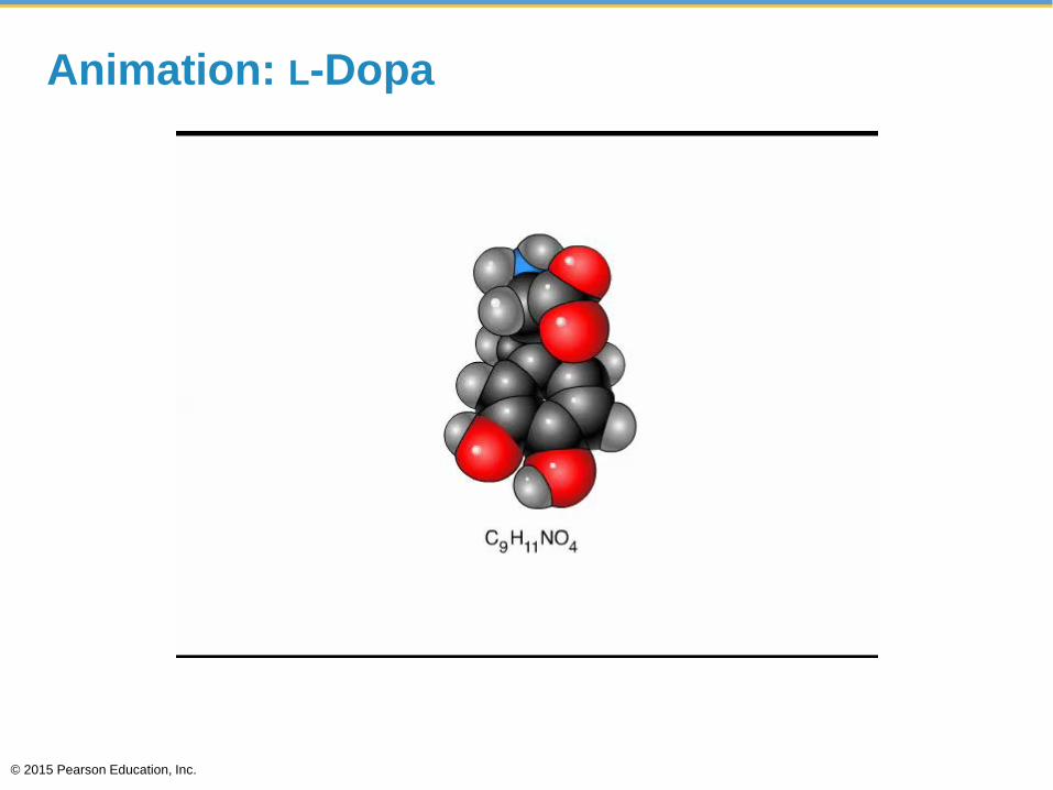
Animation: L-Dopa
© 2015 Pearson Education, Inc.
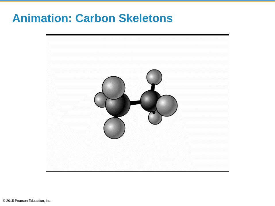
Animation: Carbon Skeletons
© 2015 Pearson Education, Inc.
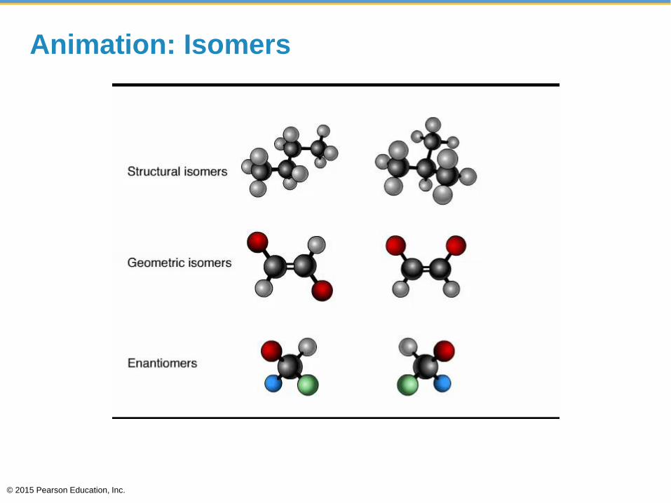
Animation: Isomers
© 2015 Pearson Education, Inc.
© 2015 Pearson Education, Inc.
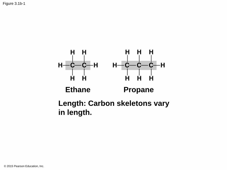
Figure 3.1b-1
Length: Carbon skeletons vary
in length.
PropaneEthane
© 2015 Pearson Education, Inc.
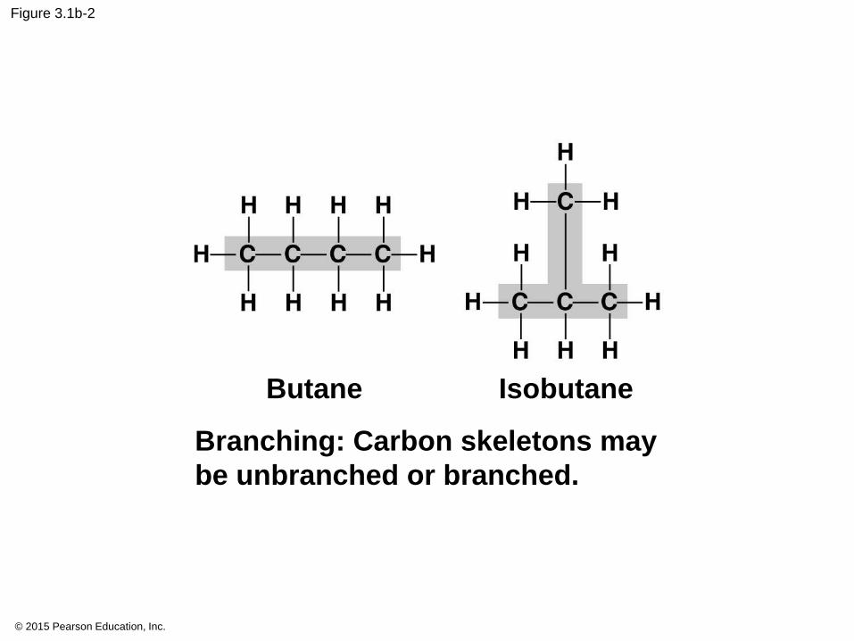
Figure 3.1b-2
Butane Isobutane
Branching: Carbon skeletons may
be unbranched or branched.
© 2015 Pearson Education, Inc.
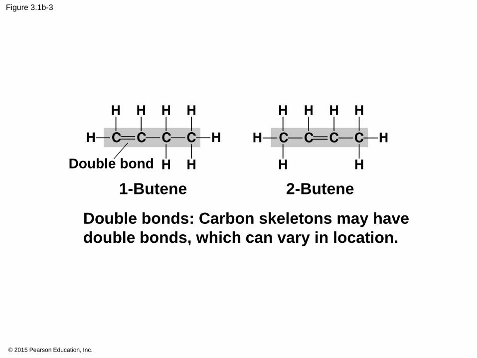
Figure 3.1b-3
1-Butene 2-Butene
Double bonds: Carbon skeletons may have
double bonds, which can vary in location.
Double bond
© 2015 Pearson Education, Inc.
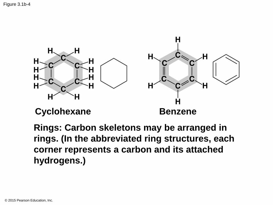
Figure 3.1b-4
Cyclohexane Benzene
Rings: Carbon skeletons may be arranged in
rings. (In the abbreviated ring structures, each
corner represents a carbon and its attached
hydrogens.)

© 2015 Pearson Education, Inc.
Figure 3.1b-0
Butane
Length: Carbon skeletons vary
in length.
Propane 1-Butene 2-Butene
Double bonds: Carbon skeletons may have
double bonds, which can vary in location.
Double bond
Isobutane Cyclohexane Benzene
Branching: Carbon skeletons may
be unbranched or branched.
Rings: Carbon skeletons may be arranged in
rings. (In the abbreviated ring structures, each
corner represents a carbon and its attached
hydrogens.)
Ethane
© 2015 Pearson Education, Inc.
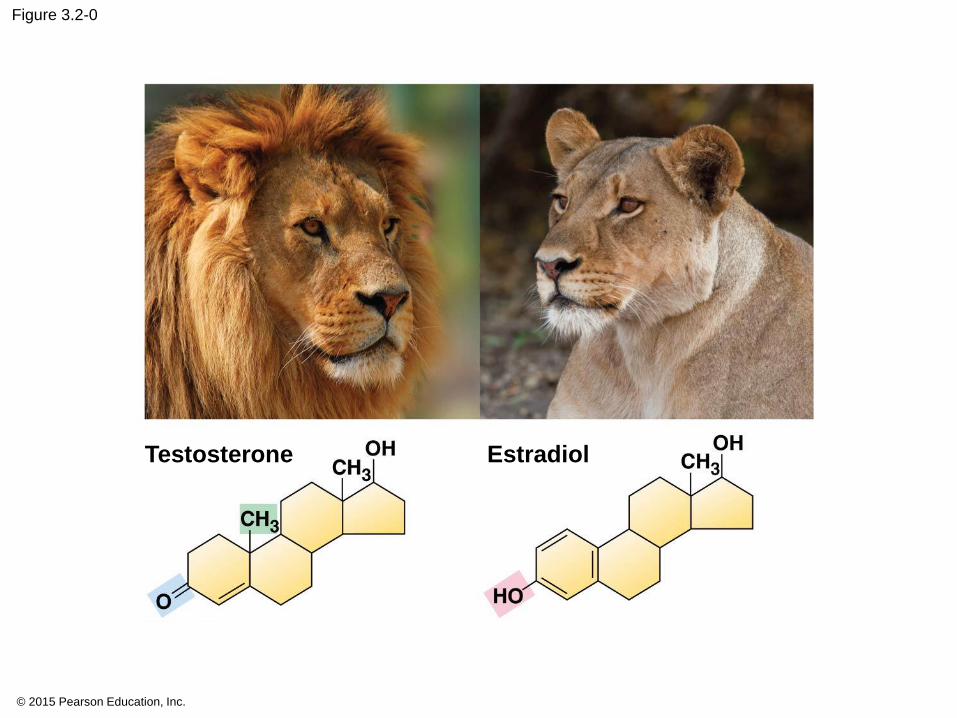
Figure 3.2-0
Testosterone Estradiol
© 2015 Pearson Education, Inc.
© 2015 Pearson Education, Inc.
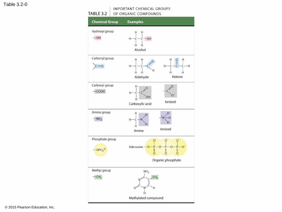
Table 3.2-0
3.3
1. carbohydrates,
2. lipids,
3. proteins,
4. nucleic acids.
© 2015 Pearson Education, Inc.
3.3
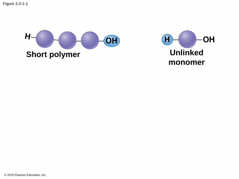
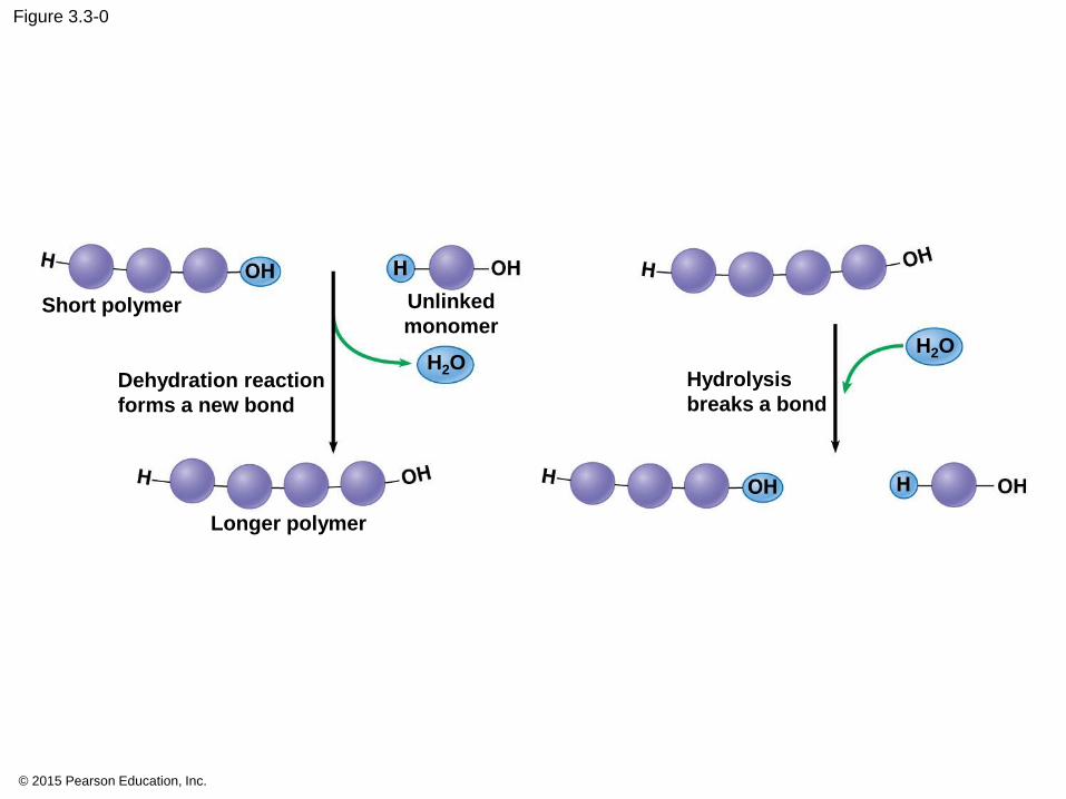
• Monomers make polymers
• Polymers make macromolecules
© 2015 Pearson Education, Inc.
3.3
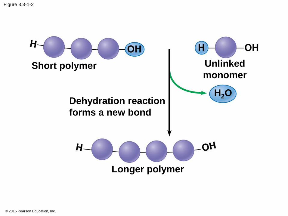
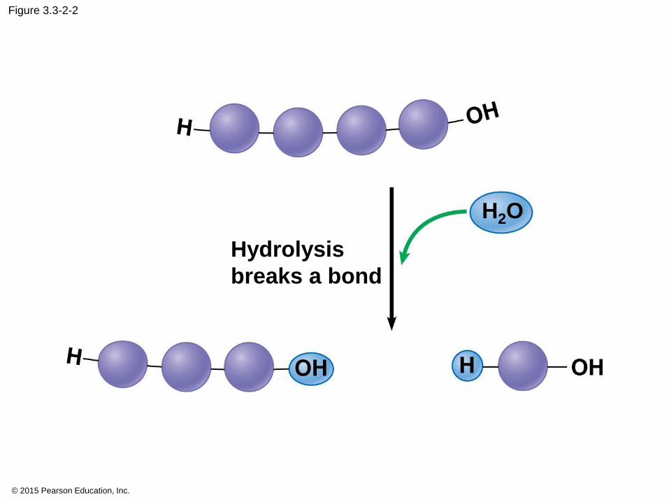
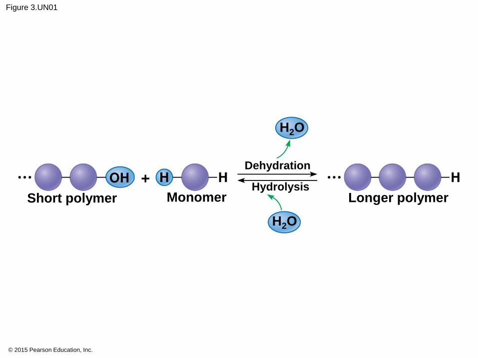
• Monomers linked through dehydration reactions.
• Polymers broken apart by hydrolysis.
• Mediated by enzymes
© 2015 Pearson Education, Inc.
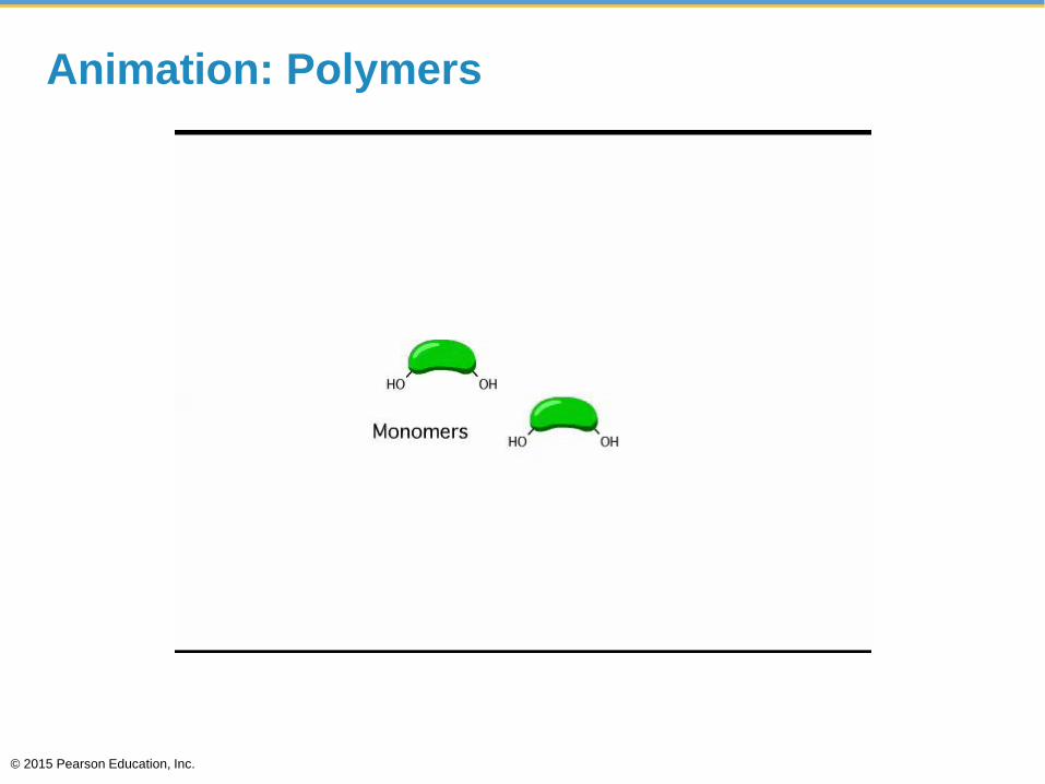
Animation: Polymers
© 2015 Pearson Education, Inc.
© 2015 Pearson Education, Inc.
Figure 3.3-1-1
Short polymer Unlinked
monomer
© 2015 Pearson Education, Inc.
Figure 3.3-1-2
Short polymer Unlinked
monomer
Dehydration reaction
forms a new bond
H2O
Longer polymer
© 2015 Pearson Education, Inc.
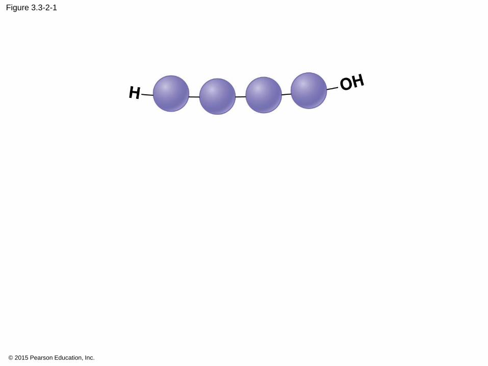
Figure 3.3-2-1
© 2015 Pearson Education, Inc.
Figure 3.3-2-2
Hydrolysis
breaks a bond
H2O
© 2015 Pearson Education, Inc.
Figure 3.3-0
Short polymer Unlinked
monomer
Dehydration reaction
forms a new bond
H2O
Longer polymer
Hydrolysis
breaks a bond
H2O
© 2015 Pearson Education, Inc.
Figure 3.UN01
Dehydration
Hydrolysis
H2O
H2O
Short polymer Monomer Longer polymer
You should now be able to
1. Describe the importance of carbon to life’s
molecular diversity.
2. Describe the chemical groups that are important
to life.
3. Explain how a cell can make a variety of large
molecules from a small set of molecules.
© 2015 Pearson Education, Inc.
CARBOHYDRATES
© 2015 Pearson Education, Inc.

Figure 3.0-2
Chapter 3 Objectives. You will:
Describe why C is the base of all organic compounds
Characterize Carbohydrates
as fuel and structure
Distinguish Lipids by hydrophobic
characteristics
Differentiate Proteins
structure and function
Connect Nucleic Acids organization to hereditary traits
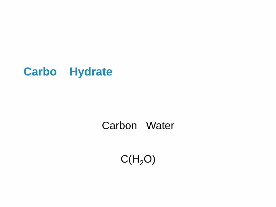
Carbo Hydrate
Carbon Water
C(H2O)
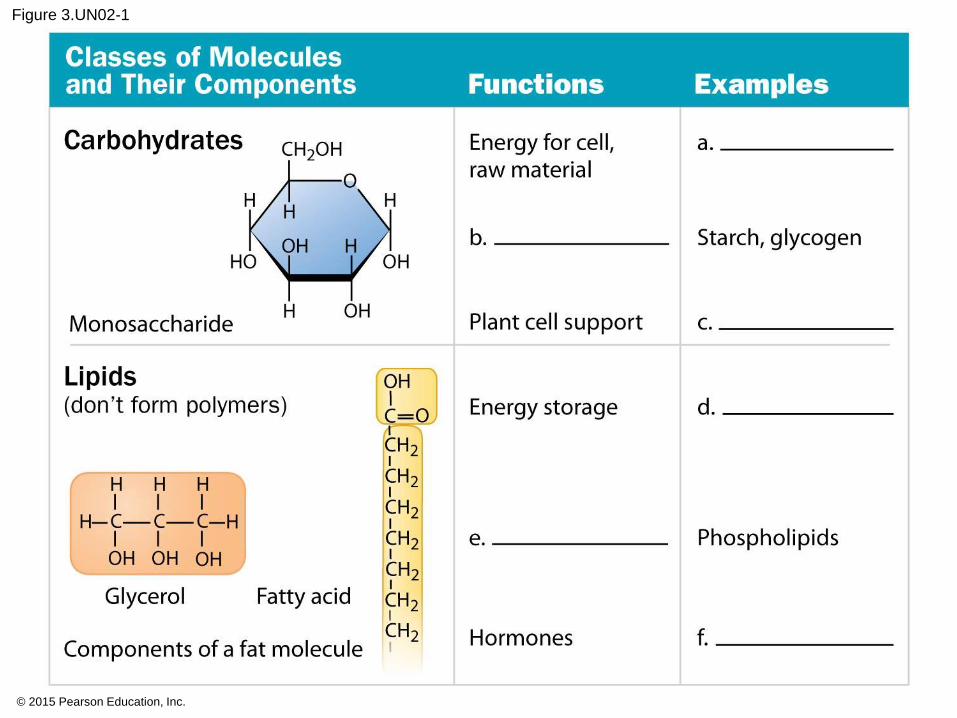
3.4 Monosaccharides are the simplest carbohydrates
• fructose,
• glucose, and
• honey.
© 2015 Pearson Education, Inc.

Cellular Respiration
• Breakdown glucose to release energy from bonds
© 2015 Pearson Education, Inc.
© 2015 Pearson Education, Inc.
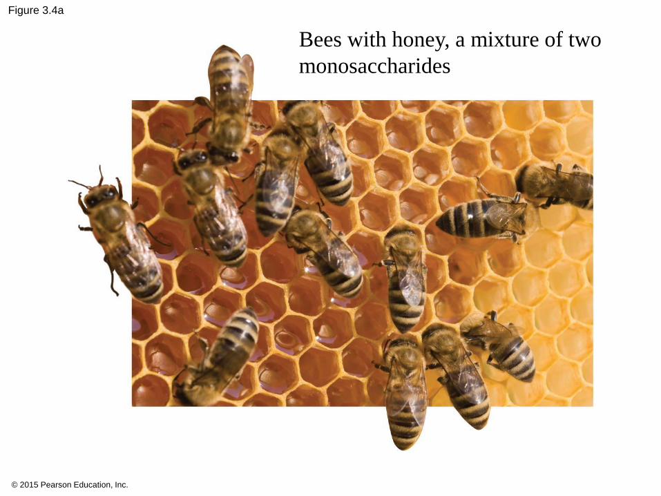
Figure 3.4a
Bees with honey, a mixture of two
monosaccharides
© 2015 Pearson Education, Inc.
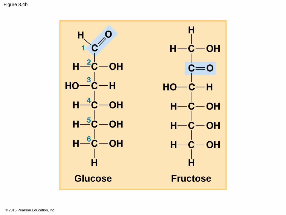
Figure 3.4b
Glucose Fructose
© 2015 Pearson Education, Inc.
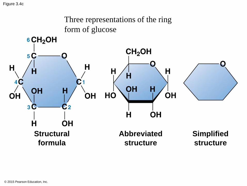
Figure 3.4c
Structural
formula
Abbreviated
structure
Simplified
structure
Three representations of the ring
form of glucose
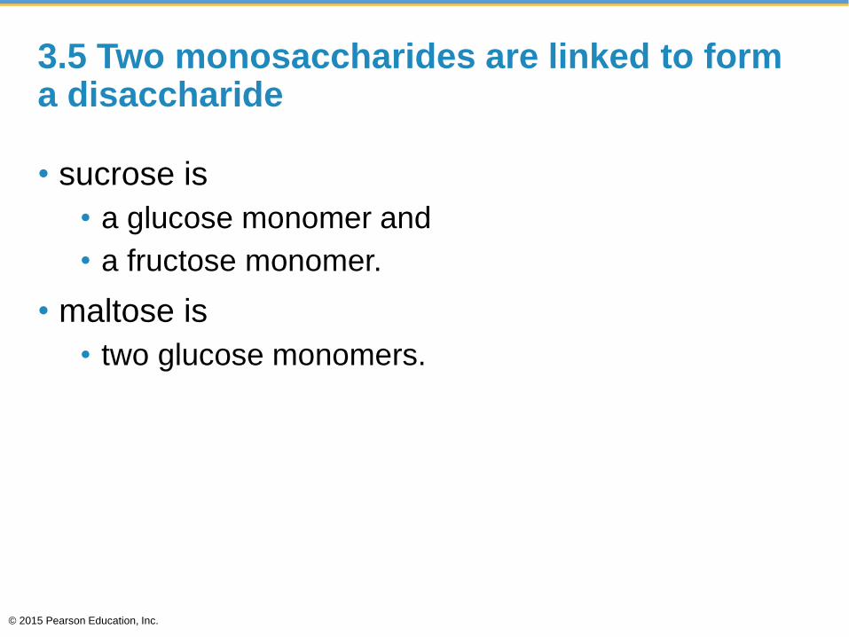
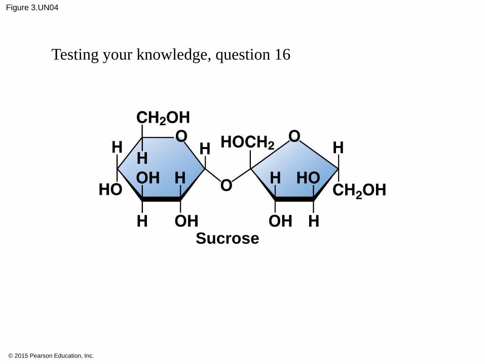
3.5 Two monosaccharides are linked to form a disaccharide
• sucrose is
• a glucose monomer and
• a fructose monomer.
• maltose is
• two glucose monomers.
© 2015 Pearson Education, Inc.

Animation: Disaccharides
© 2015 Pearson Education, Inc.
© 2015 Pearson Education, Inc.
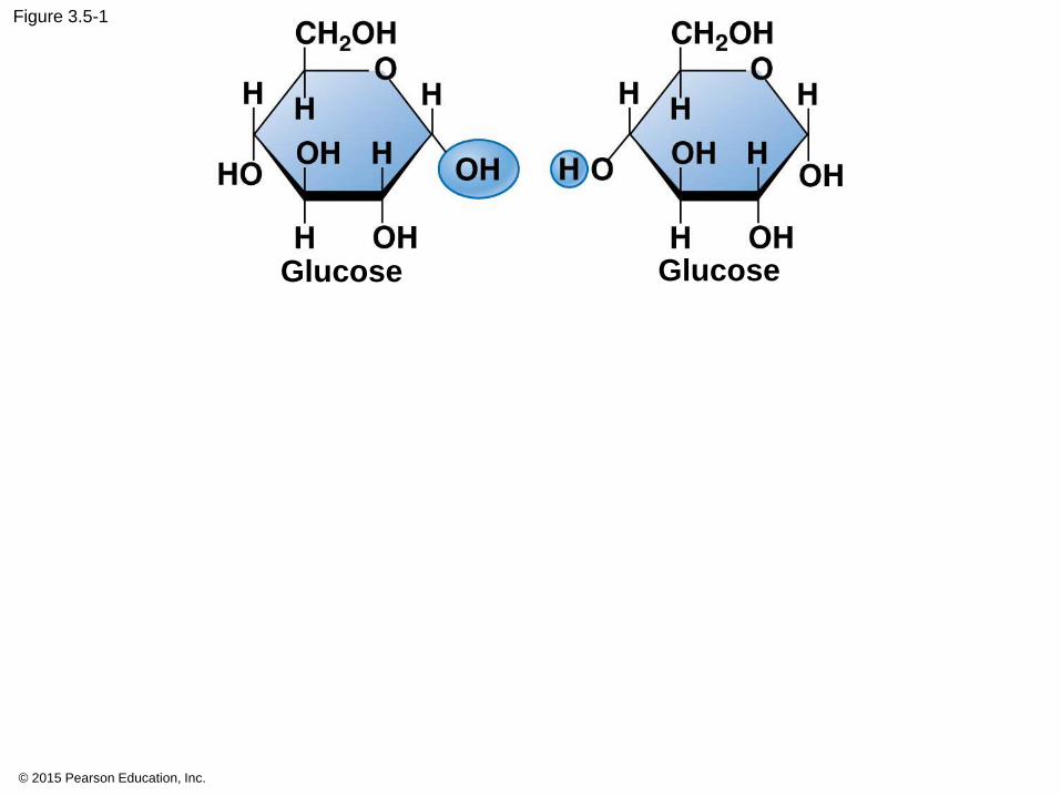
Figure 3.5-1
Glucose Glucose
© 2015 Pearson Education, Inc.
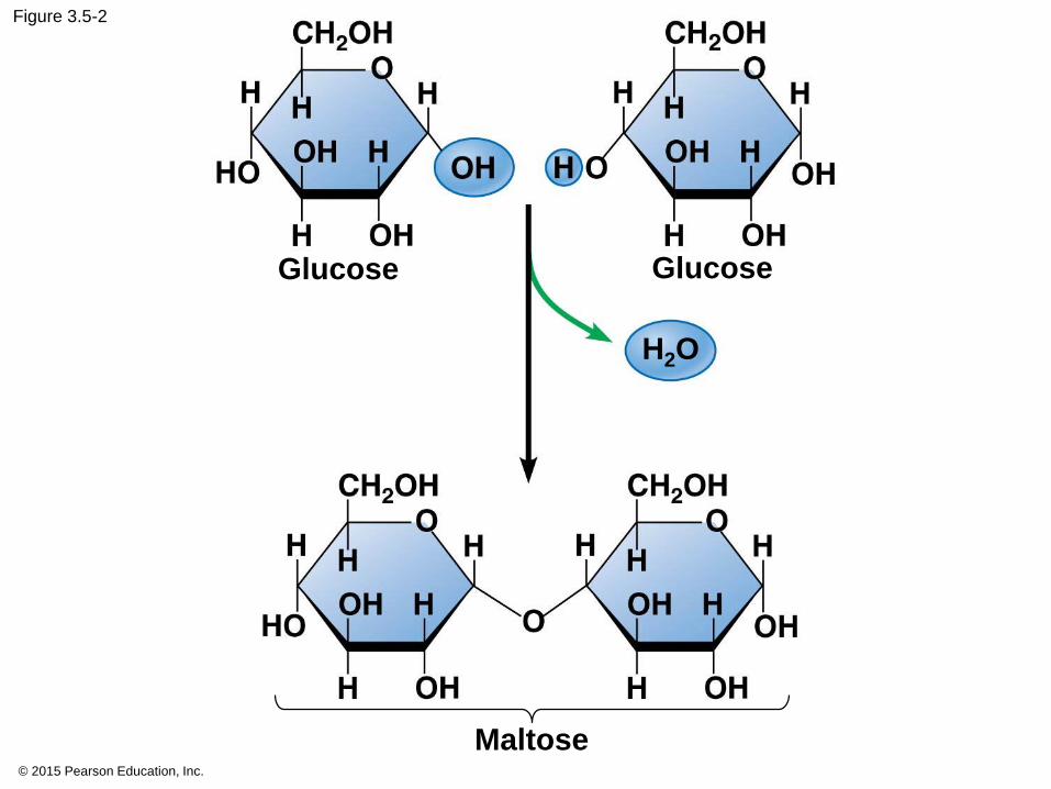
Figure 3.5-2
Glucose Glucose
Maltose
H2O

3.6 CONNECTION: What is high-fructose corn syrup, and is it to blame for obesity?Page 38
• Summarize:
© 2015 Pearson Education, Inc.
3.7 Polysaccharides are long chains of monosaccharides
• thousands of monosaccharides.
• storage
• structural compounds.
© 2015 Pearson Education, Inc.
3.7
• Starch is
• glucose monomers,
• plants energy storage.
• Glycogen is
• glucose monomers,
• animals for energy storage.
© 2015 Pearson Education, Inc.
3.7
• Cellulose
• glucose monomers
• plant cell walls
• Chitin is
• glucose monomers
• insects and crustaceans exoskeleton
• fungus cell walls.
© 2015 Pearson Education, Inc.
© 2015 Pearson Education, Inc.
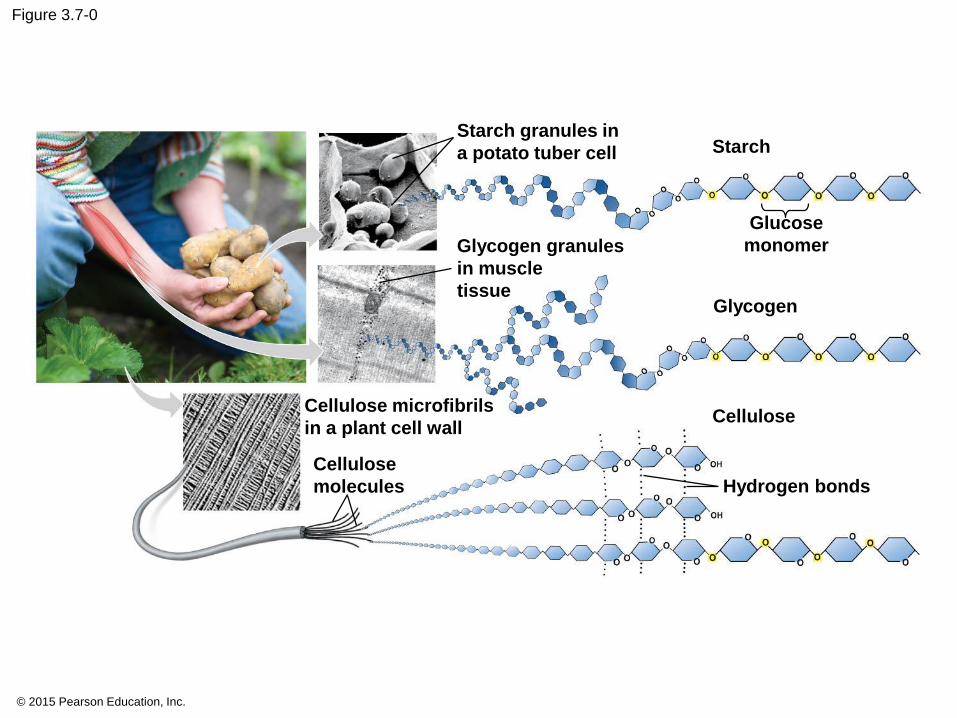
Figure 3.7-0
Starch granules in
a potato tuber cell Starch
Glycogen granules
in muscle
tissue
Cellulose microfibrils
in a plant cell wall
Cellulose
molecules Hydrogen bonds
Cellulose
Glycogen
Glucose
monomer
3.7
• hydrophilic (water-loving).
• Bath towel made of cotton, which is mostly
cellulose, and therefore water absorbent.
© 2015 Pearson Education, Inc.
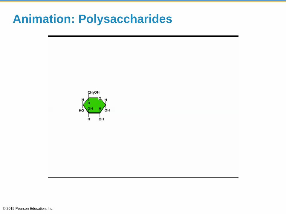
Animation: Polysaccharides
© 2015 Pearson Education, Inc.

You should now be able to
1. Define monosaccharides, disaccharides, and
polysaccharides and explain their functions.
© 2015 Pearson Education, Inc.
LIPIDS
© 2015 Pearson Education, Inc.
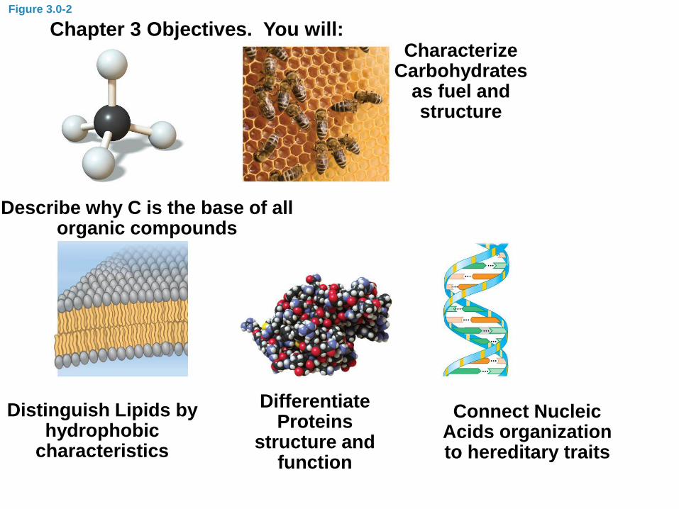
Figure 3.0-2
Chapter 3 Objectives. You will:
Describe why C is the base of all organic compounds
Characterize Carbohydrates
as fuel and structure
Distinguish Lipids by hydrophobic
characteristics
Differentiate Proteins
structure and function
Connect Nucleic Acids organization to hereditary traits
3.8 Fats are lipids that are mostly energy-storage molecules
• Lipids
• hydrophobic, or water-fearing, compounds,
• long-term energy storage,
• contain twice as much energy as a polysaccharide,
and
• consist mainly of carbon and hydrogen atoms
linked by nonpolar covalent bonds.
• No oxygen
© 2015 Pearson Education, Inc.
3.8
• not huge molecules and
• not built from monomers.
• Lipids vary a great deal in structure and function.
© 2015 Pearson Education, Inc.
3.8
• three types of lipids:
1. fats,
2. phospholipids, and
3. steroids.
© 2015 Pearson Education, Inc.
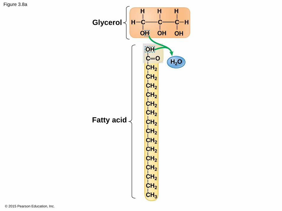
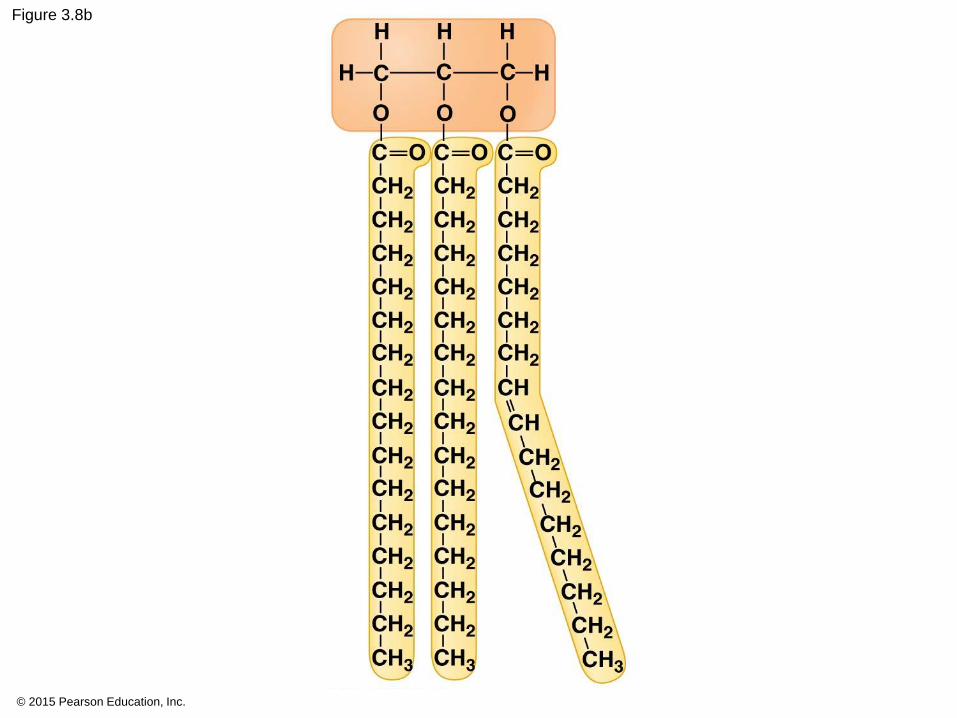
3.8 Fats
• one glycerol linked to three fatty acids.
• Triglycerides
• Energy storage and insulation
© 2015 Pearson Education, Inc.
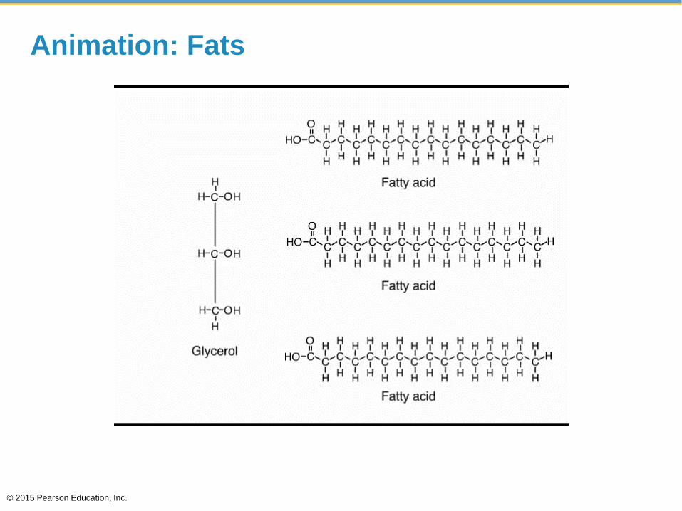
Animation: Fats
© 2015 Pearson Education, Inc.
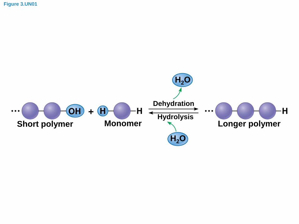
Figure 3.UN01
Dehydration
Hydrolysis
H2O
H2O
Short polymer Monomer Longer polymer
© 2015 Pearson Education, Inc.
Figure 3.8a
Glycerol
Fatty acid
H2O

© 2015 Pearson Education, Inc.
Figure 3.8b
© 2015 Pearson Education, Inc.
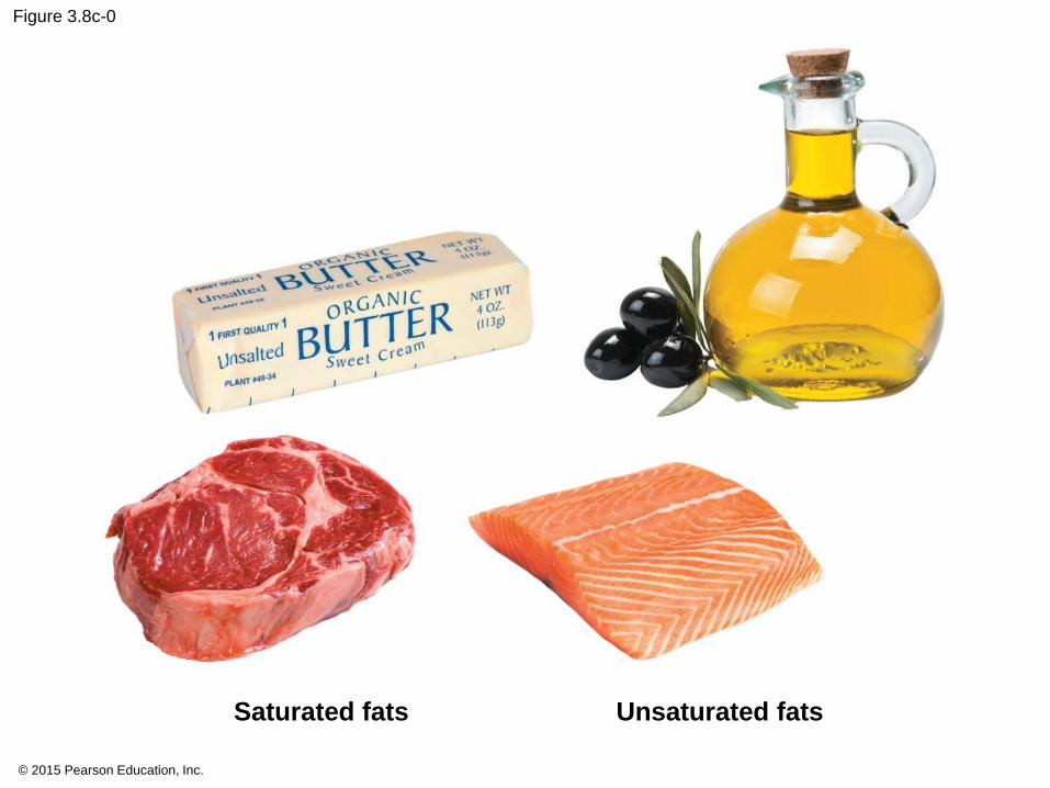
Figure 3.8c-0
Saturated fats Unsaturated fats
3.8
• Fats with the maximum number of hydrogens are
called saturated fatty acids.
• Stack together making a solid at room temperature
• Insulation
• Long term storage
• Draw a saturated fatty acid:
© 2015 Pearson Education, Inc.
© 2015 Pearson Education, Inc.
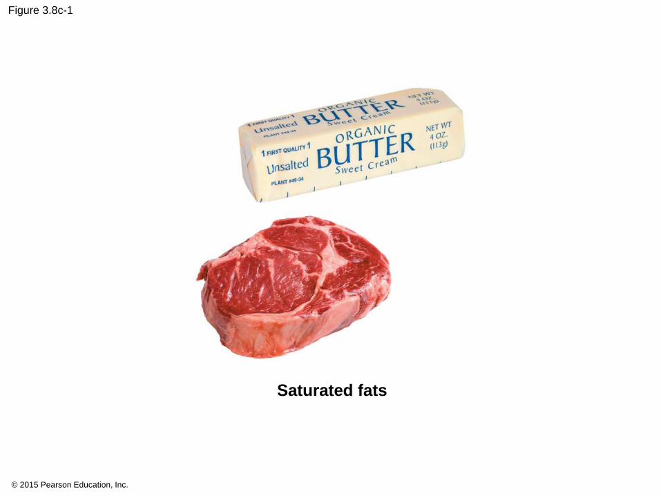
Figure 3.8c-1
Saturated fats

© 2015 Pearson Education, Inc.

© 2015 Pearson Education, Inc.
Blubber Glove Demonstration
3.8
• Fatty acids with one or more double bonds form
unsaturated fatty acids.
• kinks or bends prevent them from packing together
tightly and are liquid at room temperature.
• Easier access
Draw and unsaturated fatty acid.
© 2015 Pearson Education, Inc.
© 2015 Pearson Education, Inc.
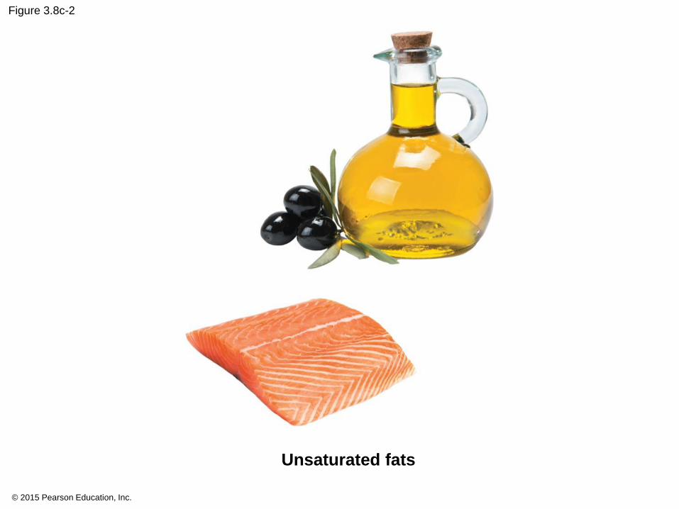
Figure 3.8c-2
Unsaturated fats
© 2015 Pearson Education, Inc.
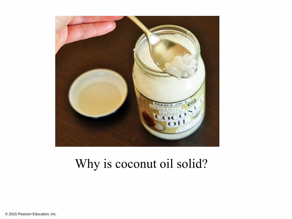
Why is coconut oil solid?
3.8
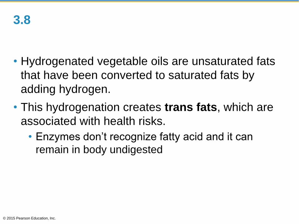
• Hydrogenated vegetable oils are unsaturated fats
that have been converted to saturated fats by
adding hydrogen.
• This hydrogenation creates trans fats, which are
associated with health risks.
• Enzymes don’t recognize fatty acid and it can
remain in body undigested
© 2015 Pearson Education, Inc.
© 2015 Pearson Education, Inc.
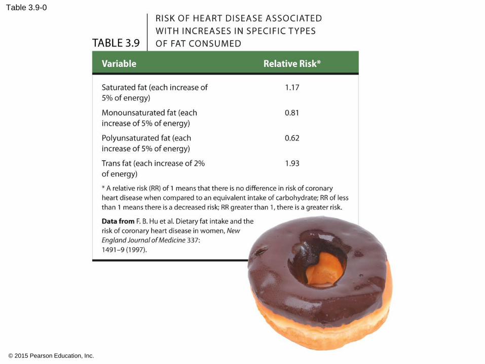
Table 3.9-0
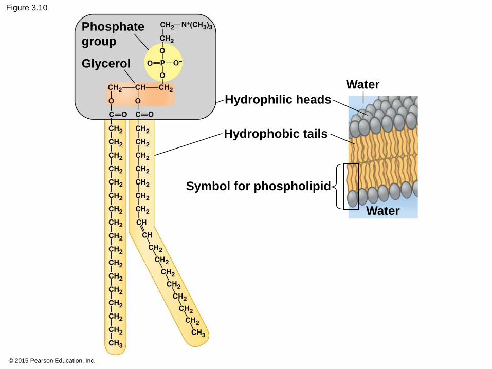
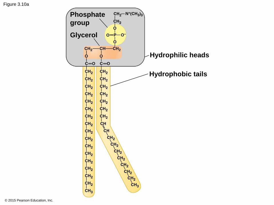
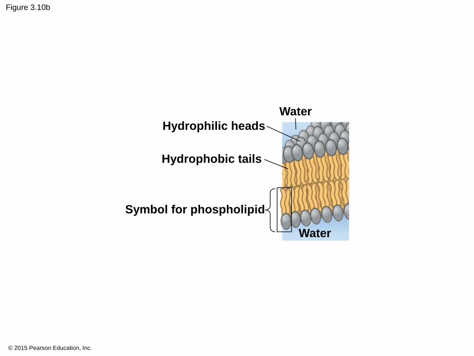
3.10 Phospholipids
• Cell membranes.
• two fatty acids attached to glycerol.
© 2015 Pearson Education, Inc.
© 2015 Pearson Education, Inc.
Figure 3.10
Phosphate
group
Glycerol
Hydrophilic heads
Hydrophobic tails
Symbol for phospholipid
Water
Water
© 2015 Pearson Education, Inc.
Figure 3.8b
© 2015 Pearson Education, Inc.
Figure 3.10a
Phosphate
group
Glycerol
Hydrophilic heads
Hydrophobic tails
3.10
• hydrophilic heads
• exterior watery environment and
• internal watery part of the cell.
• The hydrophobic tails cluster together away from
water
© 2015 Pearson Education, Inc.
© 2015 Pearson Education, Inc.
Figure 3.10b
Hydrophilic heads
Hydrophobic tails
Symbol for phospholipid
Water
Water
3.10 Steroids
• Steroids contain four fused rings.
• Cholesterol
• animal cell membranes and
• starting material
• Steroids
• Hormones
© 2015 Pearson Education, Inc.

© 2015 Pearson Education, Inc.
Figure 3.10c
Cholesterol, a steroid
3.10 CONNECTION: Anabolic steroids pose health risks p41
• Anabolic steroids
• synthetic testosterone
• violent mood swings,
• depression,
• liver damage,
• cancer,
• high cholesterol, and
• high blood pressure.
© 2015 Pearson Education, Inc.
You should now be able to
1. Define lipids, phospholipids, and steroids and
explain their functions.
2. Explain how trans fats are formed in food.
Describe the evidence that suggests that eating
trans fats is more unhealthy than consuming
saturated fats.
© 2015 Pearson Education, Inc.
PROTEINS
© 2015 Pearson Education, Inc.

Figure 3.0-2
Chapter 3 Objectives. You will:
Describe why C is the base of all organic compounds
Characterize Carbohydrates
as fuel and structure
Distinguish Lipids by hydrophobic
characteristics
Differentiate Proteins
structure and function
Connect Nucleic Acids organization to hereditary traits
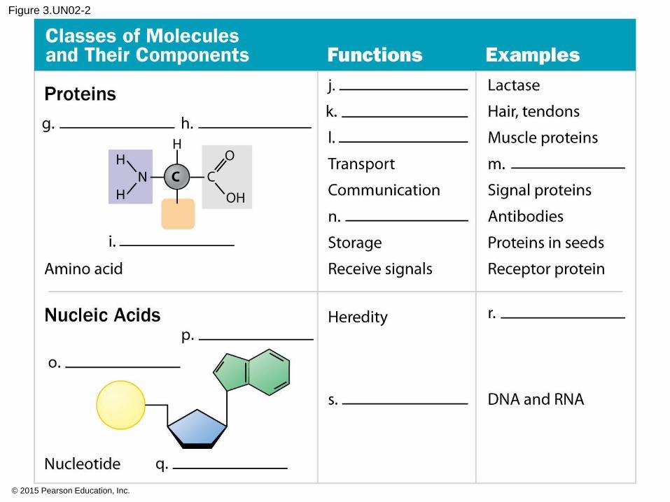
3.12 Proteins
• involved in nearly every dynamic function
• very diverse,
• tens of thousands of different proteins,
Composed of differing arrangements of 20 amino
acid monomers.
© 2015 Pearson Education, Inc.
3.12
• enzymes
• catalysts
• regulate chemical reactions within cells
• transport proteins embedded in cell membranes
• antibodies of the immune system,
• many hormones and chemical messengers.
© 2015 Pearson Education, Inc.
3.12
• receptor proteins on cell membranes,
• contractile proteins in muscle cells,
• structural proteins, collagen
• storage proteins, eggs and seeds.
© 2015 Pearson Education, Inc.
3.12
• The function depend on shape.
• hundreds or thousands of amino acids
• sequence determines particular shape.
© 2015 Pearson Education, Inc.
© 2015 Pearson Education, Inc.
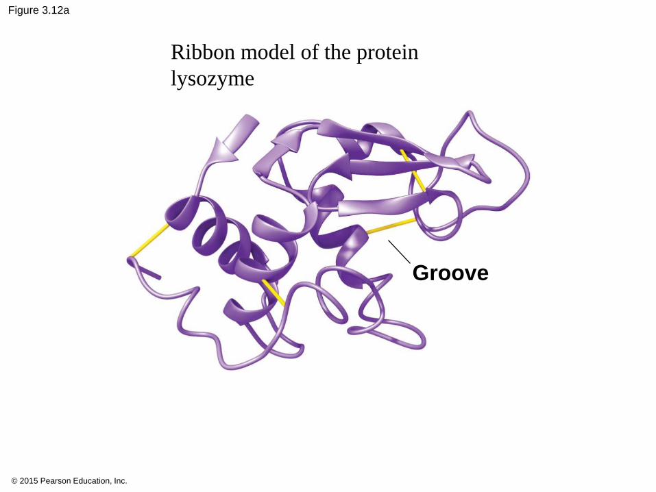
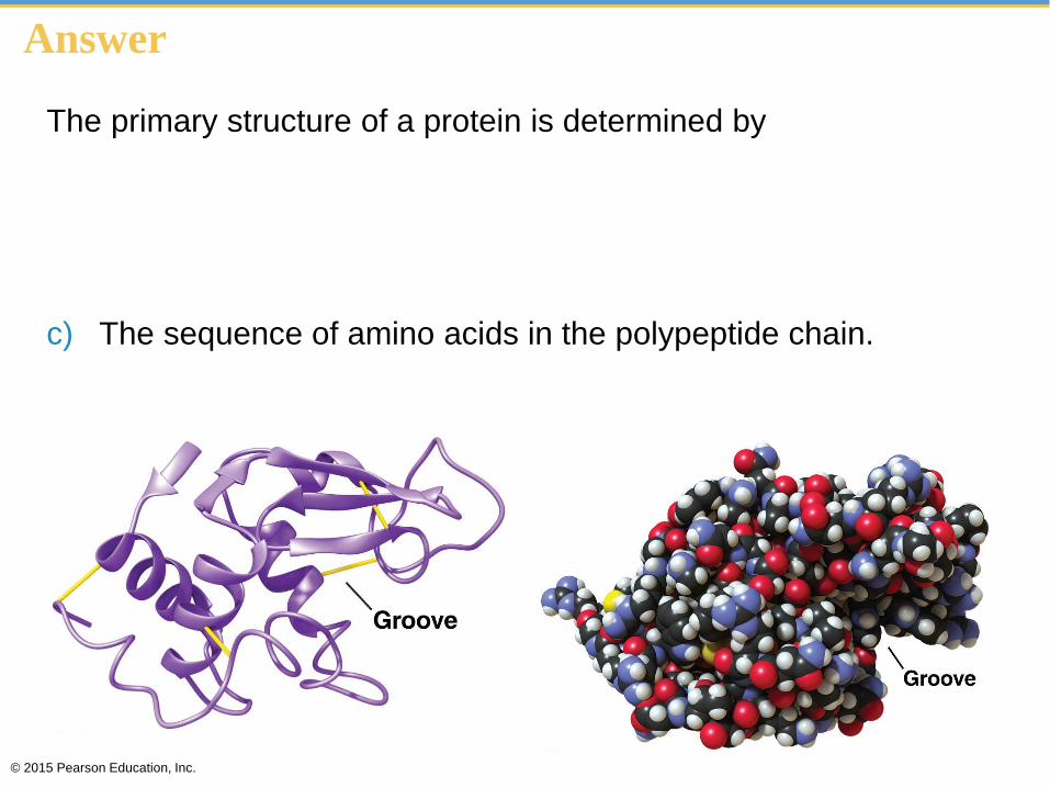
Figure 3.12a
Groove
Ribbon model of the protein
lysozyme
© 2015 Pearson Education, Inc.
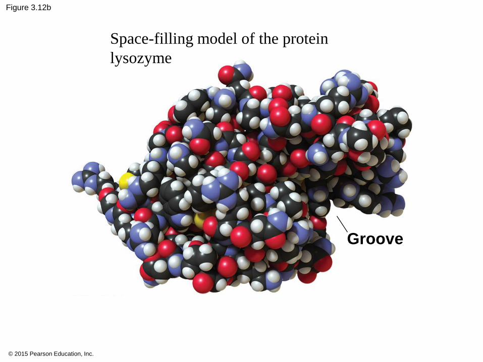
Figure 3.12b
Groove
Space-filling model of the protein
lysozyme
© 2015 Pearson Education, Inc.
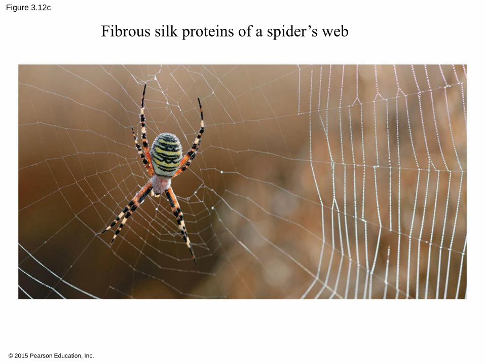
Figure 3.12c
Fibrous silk proteins of a spider’s web
3.12
• denature,
• unravels,
• loses its specific shape, and
• loses its function.
• changes in salt concentration, pH, or high heat.
© 2015 Pearson Education, Inc.
© 2015 Pearson Education, Inc.
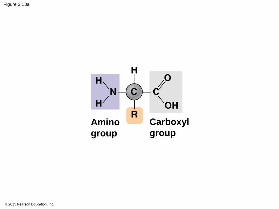
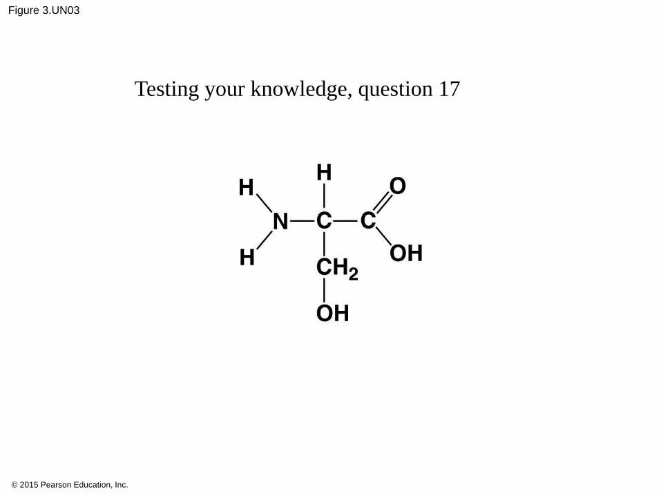
Figure 3.13a
Amino
group
Carboxyl
group
© 2015 Pearson Education, Inc.
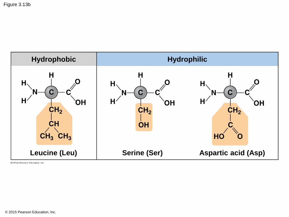
Figure 3.13b
Hydrophobic Hydrophilic
Leucine (Leu) Serine (Ser) Aspartic acid (Asp)

© 2015 Pearson Education, Inc.
Figure 3.UN01
Dehydration
Hydrolysis
H2O
H2O
Short polymer Monomer Longer polymer
© 2015 Pearson Education, Inc.
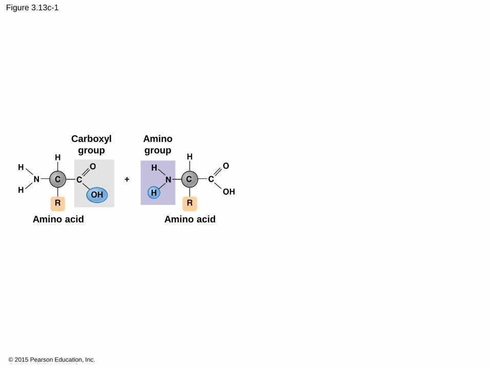
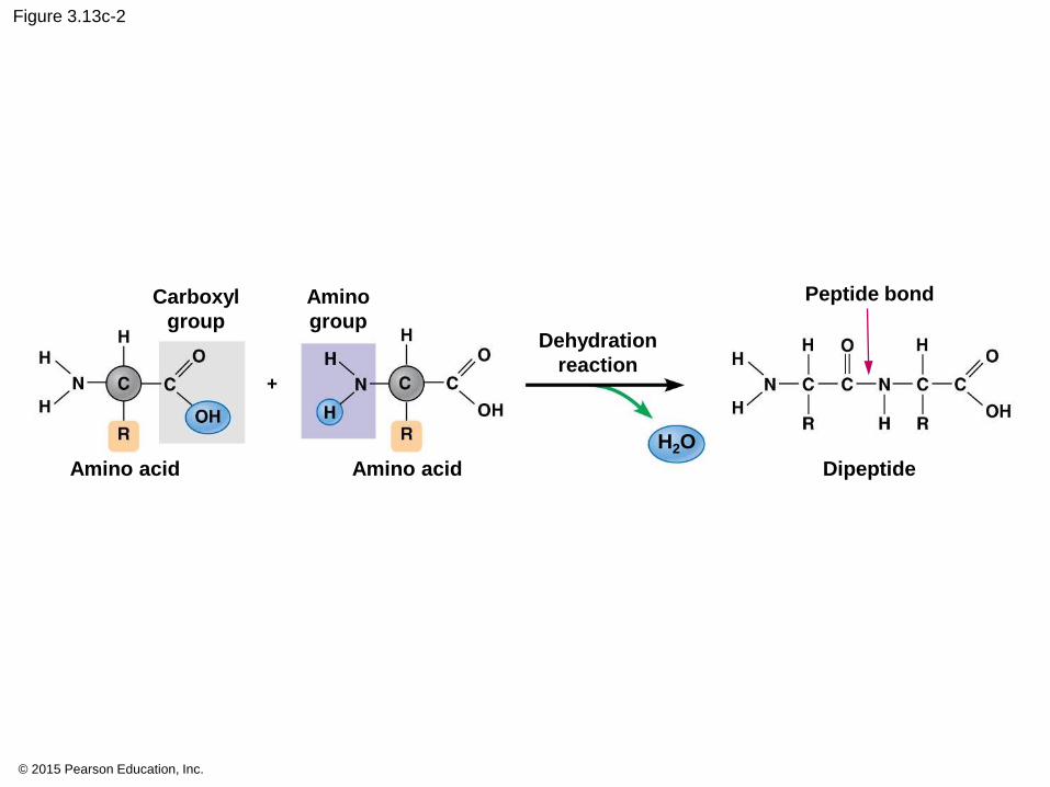
Figure 3.13c-1
Carboxyl
group
Amino
group
Amino acid Amino acid
© 2015 Pearson Education, Inc.
Figure 3.13c-2
Carboxyl
group
Amino
group
Amino acid
Dehydration
reaction
Peptide bond
Dipeptide
H2O
Amino acid
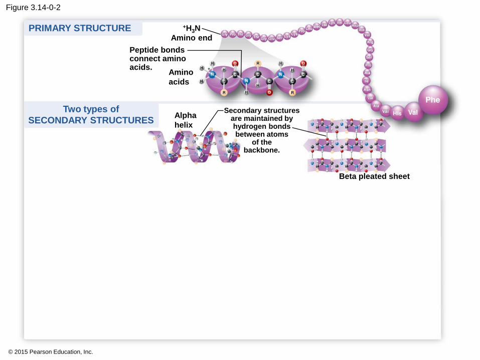
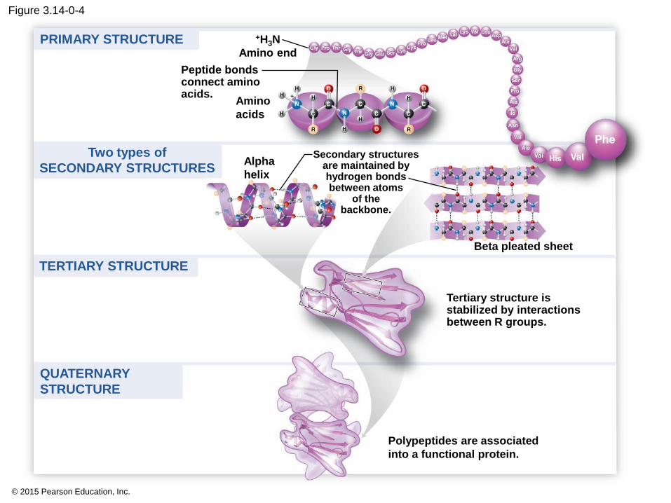
four levels of structure
1. primary structure,
2. secondary structure,
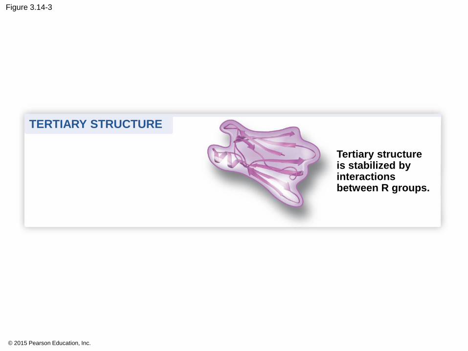
3. tertiary structure, and
4. quaternary structure.
© 2015 Pearson Education, Inc.
Animation: Protein Structure Introduction
© 2015 Pearson Education, Inc.
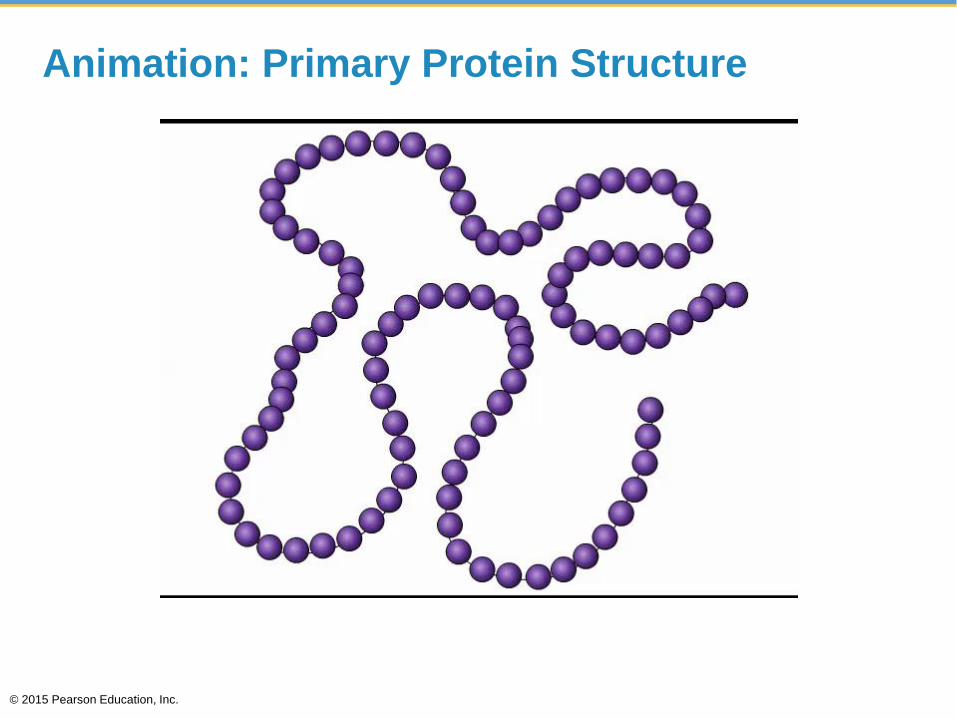
Animation: Primary Protein Structure
© 2015 Pearson Education, Inc.
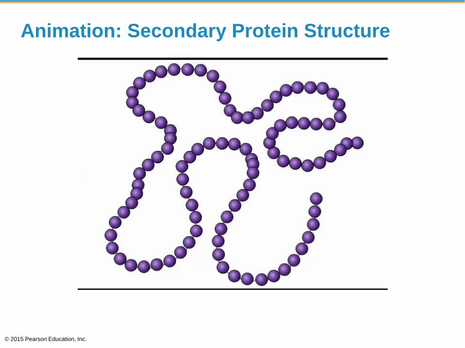
Animation: Secondary Protein Structure
© 2015 Pearson Education, Inc.

Animation: Tertiary Protein Structure
© 2015 Pearson Education, Inc.
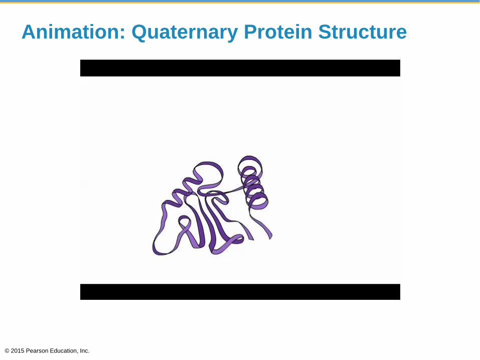
Animation: Quaternary Protein Structure
© 2015 Pearson Education, Inc.

© 2015 Pearson Education, Inc.
Figure 3.14-0
Amino
acids
+H3N
Amino end
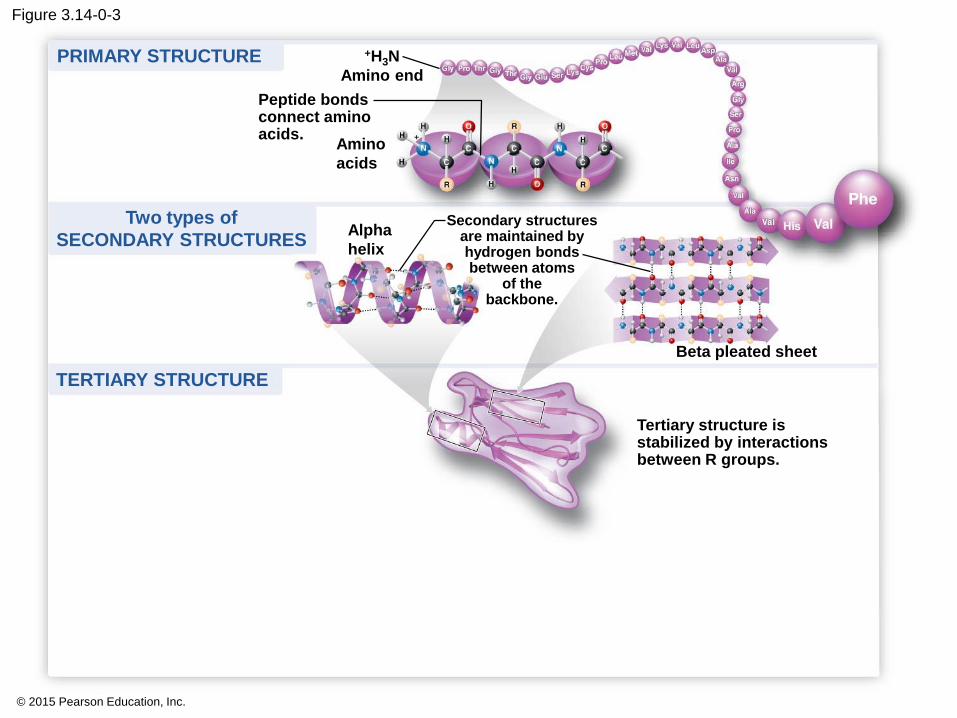
Peptide bondsconnect aminoacids.
Alpha
helix
Secondary structuresare maintained byhydrogen bondsbetween atoms
of thebackbone.
Beta pleated sheet
Tertiary structure isstabilized by interactionsbetween R groups.
TERTIARY STRUCTURE
PRIMARY STRUCTURE
Two types of
SECONDARY STRUCTURES
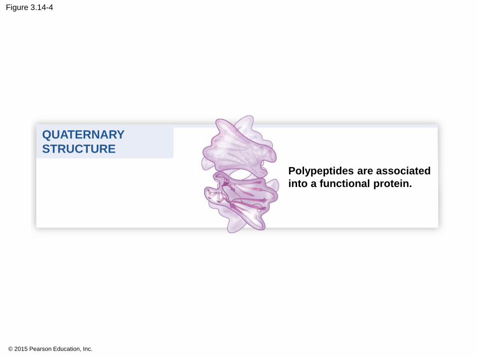
Polypeptides are associated
into a functional protein.
QUATERNARY
STRUCTURE
© 2015 Pearson Education, Inc.
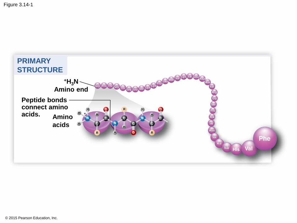
Figure 3.14-1
Amino
acids
+H3N
Amino end
Peptide bondsconnect aminoacids.
PRIMARY
STRUCTURE
© 2015 Pearson Education, Inc.
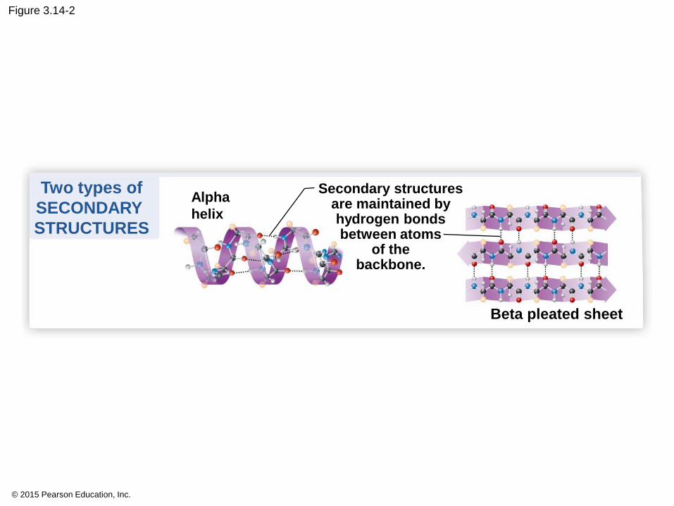
Figure 3.14-2
Alpha
helix
Secondary structuresare maintained byhydrogen bondsbetween atoms
of thebackbone.
Beta pleated sheet
Two types of
SECONDARY
STRUCTURES
© 2015 Pearson Education, Inc.
Figure 3.14-0-2
Amino
acids
+H3N
Amino end
Peptide bondsconnect aminoacids.
Alpha
helix
Secondary structuresare maintained byhydrogen bondsbetween atoms
of thebackbone.
Beta pleated sheet
PRIMARY STRUCTURE
Two types of
SECONDARY STRUCTURES
© 2015 Pearson Education, Inc.
Figure 3.14-3
Tertiary structure is stabilized by interactionsbetween R groups.
TERTIARY STRUCTURE
© 2015 Pearson Education, Inc.
Figure 3.14-0-3
Amino
acids
+H3N
Amino end
Peptide bondsconnect aminoacids.
Alpha
helix
Secondary structuresare maintained byhydrogen bondsbetween atoms
of thebackbone.
Beta pleated sheet
Tertiary structure isstabilized by interactionsbetween R groups.
TERTIARY STRUCTURE
PRIMARY STRUCTURE
Two types of
SECONDARY STRUCTURES
© 2015 Pearson Education, Inc.
Figure 3.14-4
Polypeptides are associated
into a functional protein.
QUATERNARY
STRUCTURE
© 2015 Pearson Education, Inc.
Figure 3.14-0-4
Amino
acids
+H3N
Amino end
Peptide bondsconnect aminoacids.
Alpha
helix
Secondary structuresare maintained byhydrogen bondsbetween atoms
of thebackbone.
Beta pleated sheet
Tertiary structure isstabilized by interactionsbetween R groups.
Polypeptides are associated
into a functional protein.
TERTIARY STRUCTURE
PRIMARY STRUCTURE
QUATERNARY
STRUCTURE
Two types of
SECONDARY STRUCTURES
Cook egg with acid demonstration

© 2015 Pearson Education, Inc.
Sickle Cell
You should now be able to
6. Describe the chemical structure of proteins and
the importance of proteins to cells.
© 2015 Pearson Education, Inc.
NUCLEIC ACIDS
© 2015 Pearson Education, Inc.

Figure 3.0-2
Chapter 3 Objectives. You will:
Describe why C is the base of all organic compounds
Characterize Carbohydrates
as fuel and structure
Distinguish Lipids by hydrophobic
characteristics
Differentiate Proteins
structure and function
Connect Nucleic Acids organization to hereditary traits

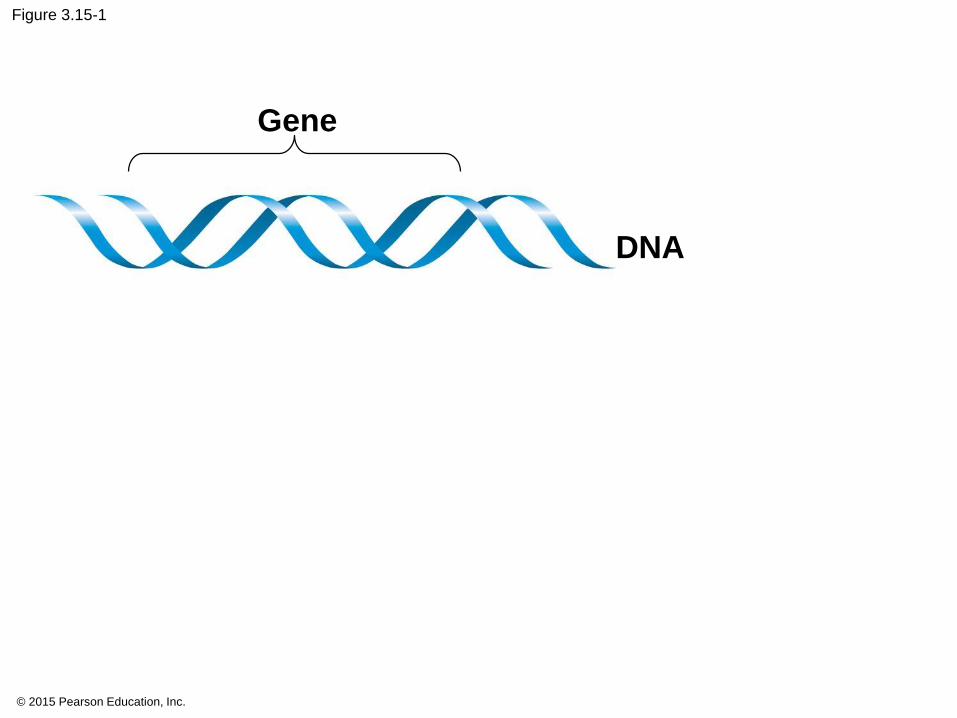
3.15 DNA
• Genes consist of DNA (deoxyribonucleic acid), a
type of nucleic acid.
• inherited from parents.
• directions for its own replication.
• directs the synthesis of proteins.
© 2015 Pearson Education, Inc.
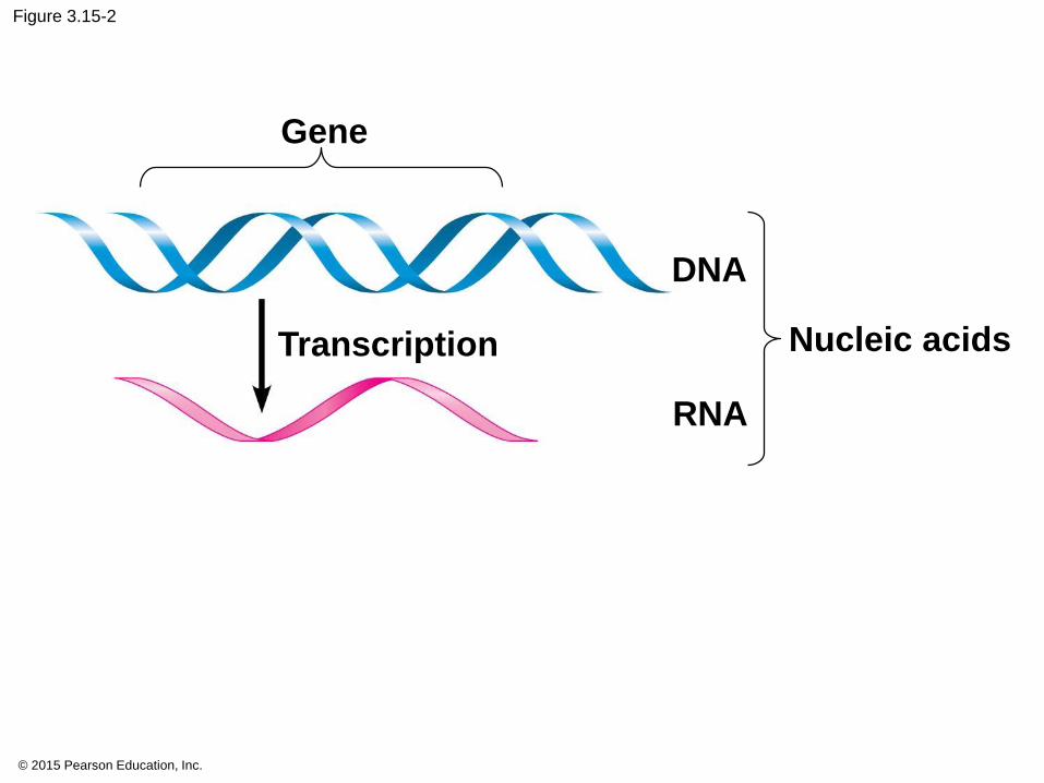
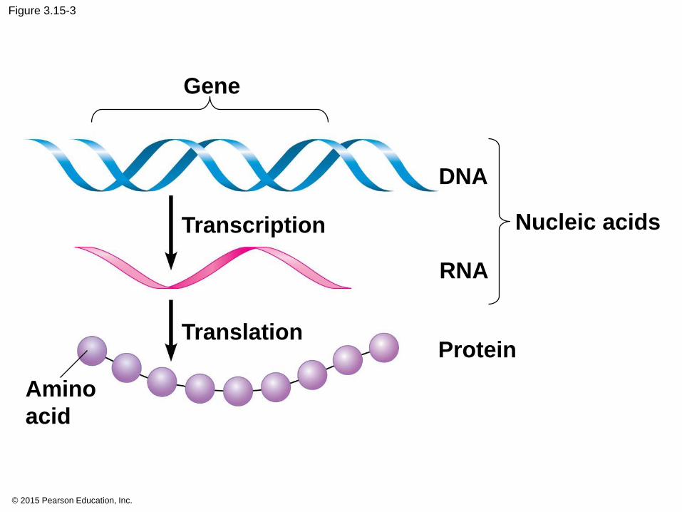
3.15 RNA
• RNA (ribonucleic acid).
• DNA is transcribed into RNA
• RNA is translated into proteins
•Central Dogma
© 2015 Pearson Education, Inc.
© 2015 Pearson Education, Inc.
Figure 3.15-1
Gene
DNA
© 2015 Pearson Education, Inc.
Figure 3.15-2
Gene
Transcription
DNA
RNA
Nucleic acids
© 2015 Pearson Education, Inc.
Figure 3.15-3
Gene
Transcription
Translation
Amino
acid
DNA
RNA
Protein
Nucleic acids
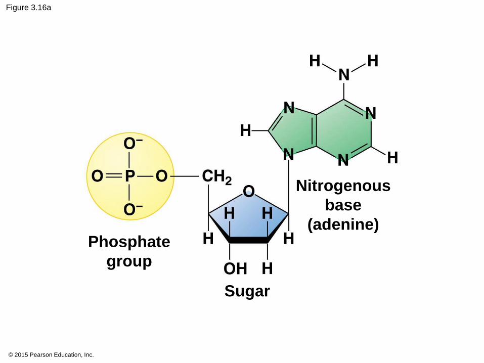
3.16
• monomers called nucleotides.
1. a five-carbon sugar
2. a phosphate group, and
3. a nitrogenous base.
© 2015 Pearson Education, Inc.

Figure 3.UN01
Dehydration
Hydrolysis
H2O
H2O
Short polymer Monomer Longer polymer
© 2015 Pearson Education, Inc.
Figure 3.16a
Sugar
Phosphate
group
Nitrogenous
base
(adenine)
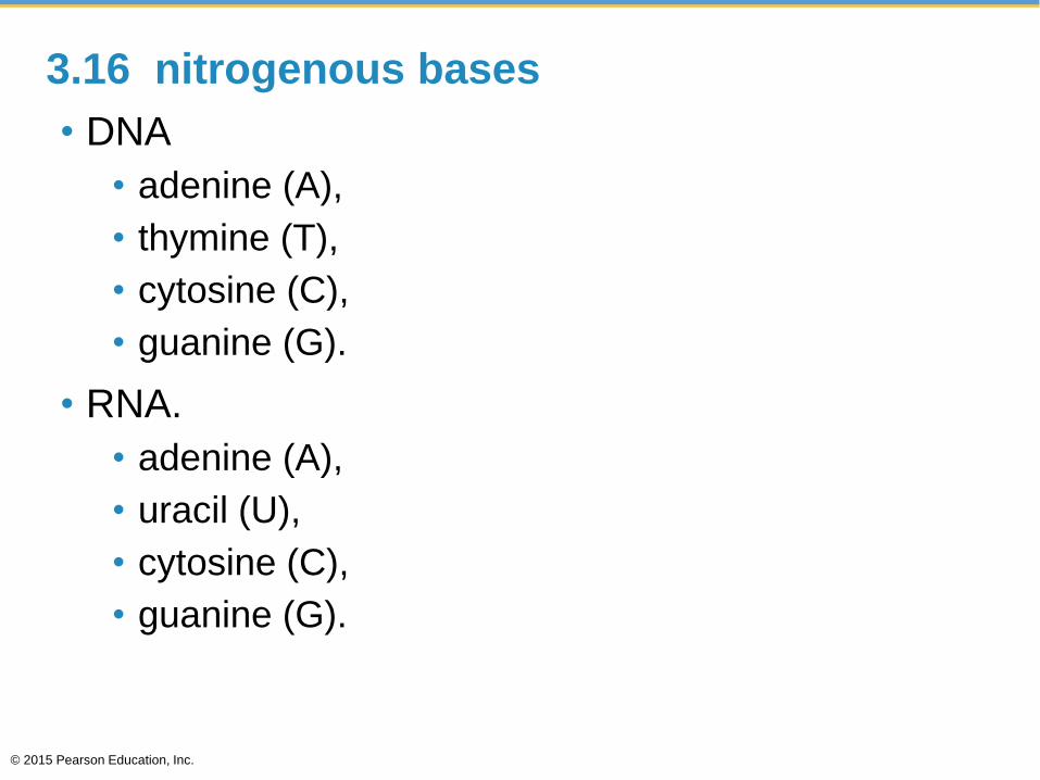
3.16 nitrogenous bases
• DNA
• adenine (A),
• thymine (T),
• cytosine (C),
• guanine (G).
• RNA.
• adenine (A),
• uracil (U),
• cytosine (C),
• guanine (G).
© 2015 Pearson Education, Inc.
© 2015 Pearson Education, Inc.
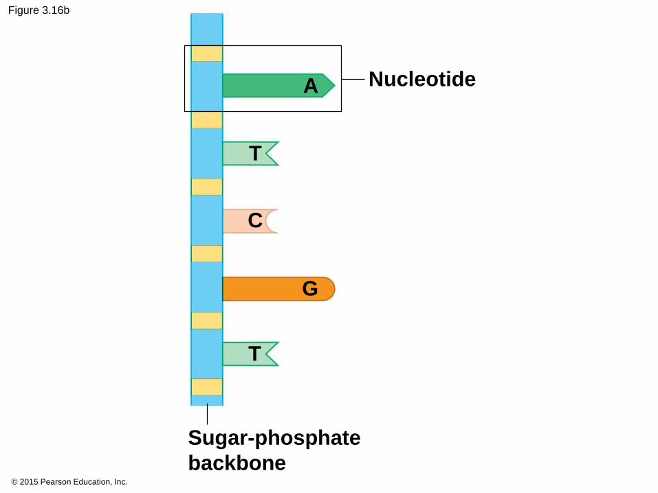
Figure 3.16b
Nucleotide
Sugar-phosphate
backbone
A
T
C
G
T

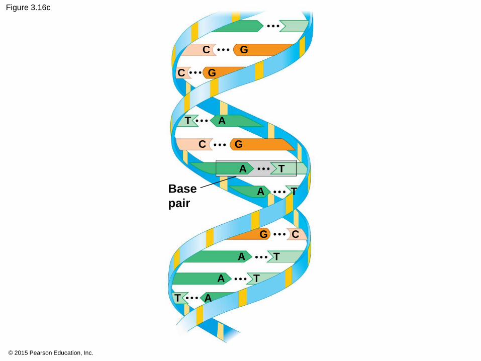
3.16 Nucleic acids are polymers of nucleotides
• RNA is a single strand.
• DNA is a double helix,
© 2015 Pearson Education, Inc.
© 2015 Pearson Education, Inc.
Figure 3.16c
Base
pair
C G
C G
C G
CG
T A
T A
TA
TA
TA
TA

© 2015 Pearson Education, Inc.

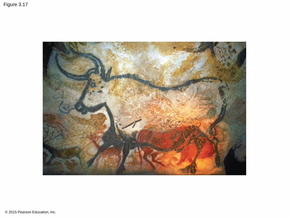
3.16 EVOLUTION CONNECTION: Lactose tolerance is a recent event in human evolution page 47
• Summarize:
© 2015 Pearson Education, Inc.
© 2015 Pearson Education, Inc.
Figure 3.17
You should now be able to
6. Describe the chemical structure of nucleic acids
and explain how they relate to inheritance.
7. Explain how lactose tolerance has evolved in
humans.
© 2015 Pearson Education, Inc.
© 2015 Pearson Education, Inc.
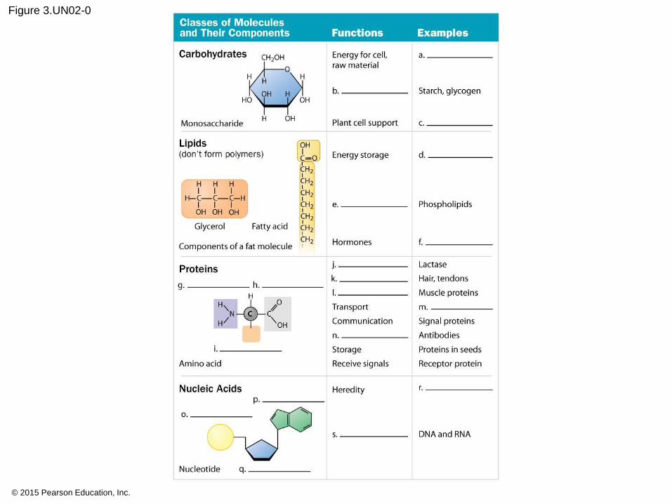
Figure 3.UN02-0
© 2015 Pearson Education, Inc.
Figure 3.UN02-1
© 2015 Pearson Education, Inc.
Figure 3.UN02-2
© 2015 Pearson Education, Inc.
Figure 3.UN03
Testing your knowledge, question 17
© 2015 Pearson Education, Inc.
Figure 3.UN04
Sucrose
Testing your knowledge, question 16

© 2015 Pearson Education, Inc.
Figure 3.UN06
Enzyme A Enzyme B
Temperature (C)
Rate
of
reacti
on
0 20 40 60 80 100
Testing your knowledge, question 18
Clicker Questions for
Campbell Biology: Concepts & Connections, Eighth EditionREECE • TAYLOR • SIMON • DICKEY • HOGAN
Chapter 3
Updated by Shannon Datwyler
The Molecules of Cells
© 2015 Pearson Education, Inc.
Concept Check
The formation of starch from simple sugars such as glucose
involves a series of _______________ reactions.
a) hydrolysis
b) dehydration
c) hydrophobic
d) denaturation
© 2015 Pearson Education, Inc.
Answer
The formation of starch from simple sugars such as glucose
involves a series of _______________ reactions.
a) hydrolysis
b) dehydration
c) hydrophobic
d) denaturation
© 2015 Pearson Education, Inc.
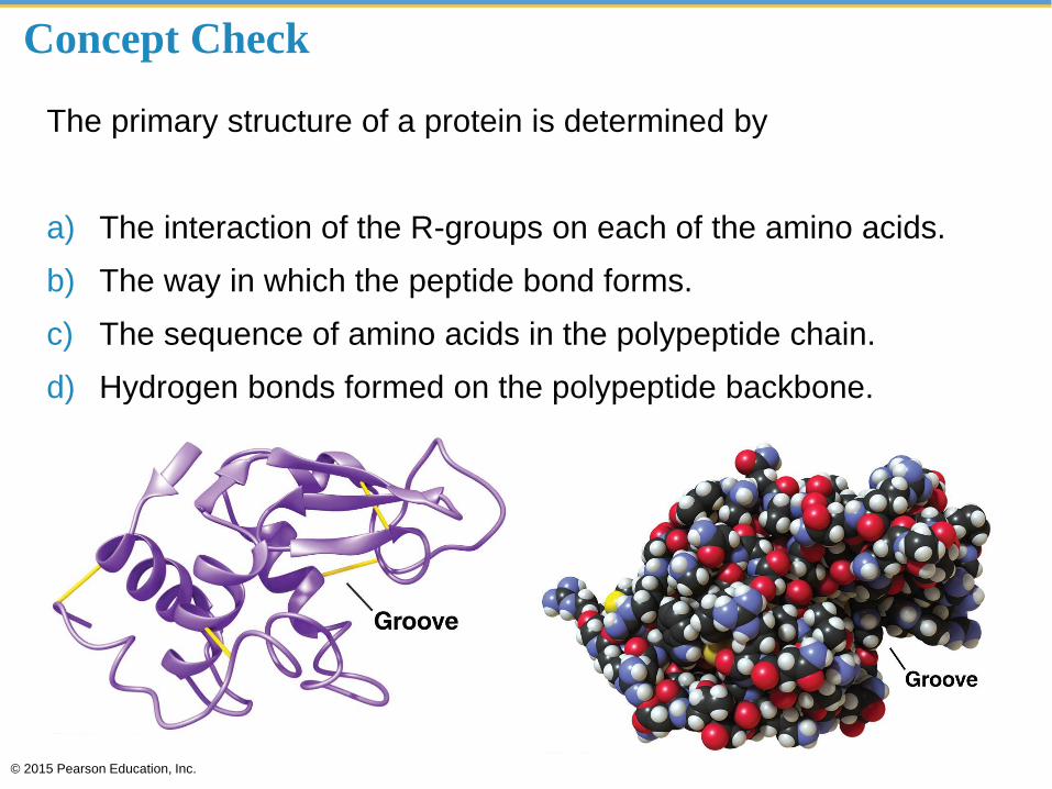
Concept Check
The primary structure of a protein is determined by
a) The interaction of the R-groups on each of the amino acids.
b) The way in which the peptide bond forms.
c) The sequence of amino acids in the polypeptide chain.
d) Hydrogen bonds formed on the polypeptide backbone.
© 2015 Pearson Education, Inc.
Answer
The primary structure of a protein is determined by
a) The interaction of the R-groups on each of the amino acids.
b) The way in which the peptide bond forms.
c) The sequence of amino acids in the polypeptide chain.
d) Hydrogen bonds formed on the polypeptide backbone.
© 2015 Pearson Education, Inc.
Concept Check
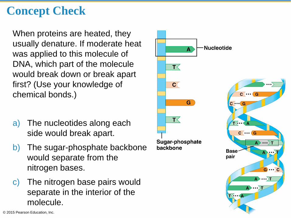
When proteins are heated, they
usually denature. If moderate heat
was applied to this molecule of
DNA, which part of the molecule
would break down or break apart
first? (Use your knowledge of
chemical bonds.)
a) The nucleotides along each
side would break apart.
b) The sugar-phosphate backbone
would separate from the
nitrogen bases.
c) The nitrogen base pairs would
separate in the interior of the
molecule.© 2015 Pearson Education, Inc.
Answer
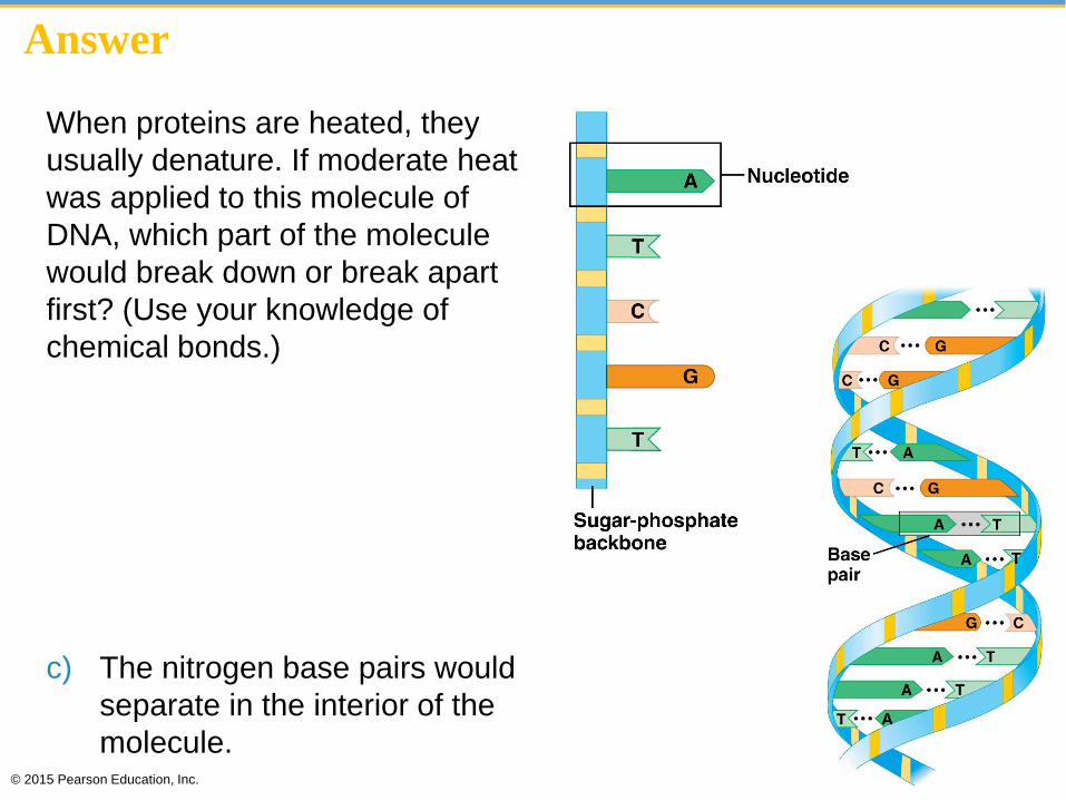
When proteins are heated, they
usually denature. If moderate heat
was applied to this molecule of
DNA, which part of the molecule
would break down or break apart
first? (Use your knowledge of
chemical bonds.)
a) The nucleotides along each
side would break apart.
b) The sugar-phosphate backbone
would separate from the
nitrogen bases.
c) The nitrogen base pairs would
separate in the interior of the
molecule.© 2015 Pearson Education, Inc.
Concept Check
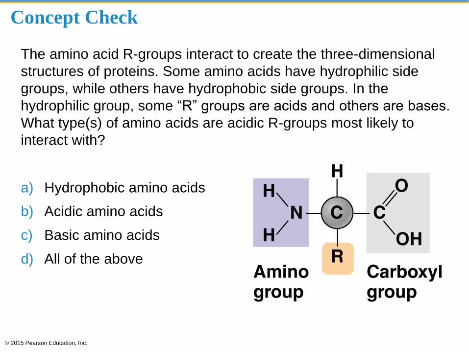
The amino acid R-groups interact to create the three-dimensional
structures of proteins. Some amino acids have hydrophilic side
groups, while others have hydrophobic side groups. In the
hydrophilic group, some “R” groups are acids and others are bases.
What type(s) of amino acids are acidic R-groups most likely to
interact with?
a) Hydrophobic amino acids
b) Acidic amino acids
c) Basic amino acids
d) All of the above
© 2015 Pearson Education, Inc.

Answer
The amino acid R-groups interact to create the three-dimensional
structures of proteins. Some amino acids have hydrophilic side
groups, while others have hydrophobic side groups. In the
hydrophilic group, some “R” groups are acids and others are bases.
What type(s) of amino acids are acidic R-groups most likely to
interact with?
a) Hydrophobic amino acids
b) Acidic amino acids
c) Basic amino acids
d) All of the above
© 2015 Pearson Education, Inc.
Concept Check
The key to a protein’s function is its shape. The shape can be
altered (or denatured) under unfavorable conditions. By heating a
protein such as that found in egg whites, the protein’s shape
changes. What best describes why this happens?
a) Interactions between R-groups change, resulting in a change in
the secondary structure of the protein.
b) Peptide bonds undergo a series of dehydration reactions.
c) Peptide bonds undergo a series of hydrolysis reactions.
© 2015 Pearson Education, Inc.
Answer
The key to a protein’s function is its shape. The shape can be
altered (or denatured) under unfavorable conditions. By heating a
protein such as that found in egg whites, the protein’s shape
changes. What best describes why this happens?
a) Interactions between R-groups change, resulting in a change in
the secondary structure of the protein.
b) Peptide bonds undergo a series of dehydration reactions.
c) Peptide bonds undergo a series of hydrolysis reactions.
© 2015 Pearson Education, Inc.
Science and Society
Seeking to gain an edge over
the competition, some athletes
have turned to anabolic steroids
to enhance their performance.
Due in part to the negative
health effects of steroid use,
most sports organizations ban
the use of steroids.
Do you believe that sports
organizations should ban the
use of performance-
enhancing drugs?
© 2015 Pearson Education, Inc.
Disagree Agree
Strongly A B C D E Strongly
Science and Society
Olympic track and field records
appear to have leveled off and
perhaps even declined from a
high point in the early to mid-
1990s. Some have suggested
that many of the records set
during this time period were due
to drug-based enhancement.
Should these records be
thrown out?
© 2015 Pearson Education, Inc.
Disagree Agree
Strongly A B C D E Strongly