chapter 3 muscle physiology - yolasalahmartin.yolasite.com/resources/hsc_205_cell_physiology... ·...
TRANSCRIPT

1
Lecture 9a
Muscle structure
Topics
• Smooth, skeletal, and cardiac muscle
tissues
• Structure and function of skeletal muscle
cells.
• Sarcomeres structure and contraction
• Actin-myosin interaction and sliding
filament theory
Muscle Similarities
• Muscle types: skeletal, cardiac, smooth
• Skeletal and smooth muscle cells are elongated
and are called muscle fibers
• Muscle contraction depends on two kinds of
myofilaments – actin and myosin
• Muscle terminology is similar
– Sarcolemma – muscle plasma membrane
– Sarcoplasm – cytoplasm of a muscle cell
– Prefixes – myo, mys, and sarco all refer to muscle
Classification of Muscle Cells
• Striated (muscle cells with a banded
appearance) or nonstriated (not banded)
• Muscle cells can have a single nucleus or
be multinucleate
• Muscle cells can be controlled voluntarily
(consciously) or involuntarily (automatically)
Skeletal Muscle
• Striated, “voluntary”, and multinucleated
• Cells can be very long
• Contracts rapidly but tires easily
• Is extremely adaptable and can exert forces
ranging from a fraction of an ounce to over 70
pounds
• Satellite cells: Like a muscle “stem cell,” can
divide to become new skeletal muscle cells
(adult skeletal muscle cells do not divide).
Cardiac Muscle Cells
• Occurs only in the heart
• Is striated, not voluntary, uni- or bi- nucleate
• Contracts at a fairly steady rate set by the heart’s pacemaker cells
• Cells are called cardiac myocytes
• Form branching networks connected at intercalated disks
• Neural controls allow the heart to respond to changes in bodily needs
Limited capacity for repair

2
Smooth Muscle Cells
• Nonstriated, involuntary, and have a single nucleus
• Smooth muscle cells are small and tapered
• can divide and regenerate
• Found in walls of hollow organs and blood vessels
• Contract alone or under nervous system control
• Smooth muscle helps maintain blood pressure, and squeezes or propels substances (i.e., food, feces) through organs
Functional Characteristics of
Muscle Tissue• Excitability, or irritability – the ability to
receive and respond to stimuli
• Contractility – the ability to shorten forcibly
• Extensibility – the ability to be stretched or
extended
• Elasticity – the ability to recoil and resume
the original resting length
Characteristics of Skeletal,
Cardiac, and Smooth Muscle
Table 10–4
The Muscular System
• Includes only skeletal muscles
– attached to the skeletal system
– allow us to move
• Muscle tissue (muscle cells or fibers)
• Connective tissues
• Nerves
• Blood vessels
Functions of Skeletal Muscles
1. Produce skeletal movement
2. Maintain body posture
3. Support soft tissues
4. Stabilize joints
5. Guard body openings
6. Generate heat
Skeletal Muscle
Figure 9.2a

3
Organization of Connective
Tissues• Muscles have 3 layers of connective tissues:
–Epimysium – an overcoat of dense regular and
irregular connective tissue that surrounds the entire
muscle; Separates muscle from surrounding tissues
–Perimysium – fibrous connective tissue that
surrounds groups of muscle fibers called fascicles;
Contains blood vessel and nerve supply to fascicles
–Endomysium – fine sheath of connective tissue
composed of collagen and reticular fibers surrounding
each muscle cell/fiber; Contains capillaries and nerve
fibers contacting muscle cells; Contains satellite cells
(stem cells) that repair damage
Levels of organization
Figure 10–6 (1 of 5)
Level 1: Skeletal Muscle Level 2: Muscle Fascicle
Figure 10–6 (2 of 5)
Level 3: Muscle Cell (Fiber)
Figure 10–6 (3 of 5)
Level 4: Myofibril
Figure 10–6 (4 of 5)

4
Level 5: Sarcomere
Figure 10–6 (5 of 5)
Summary – muscle orgnaization
• Epimysium surrounds muscle (which are bundles of fascicles)
• Perimysium surrounds fascicles (which are bundles are fibers/cells)
• Endomysium surrounds muscle fibers (which are filled with myofibrils)
• Myofibrils are long cylinders of sarcomeres
• Sarcomeres contract to shorten muscles. (Made up of myofilaments)
Muscle Attachments
• Direct – epimysium of the muscle is fused
to the periosteum of a bone
• Indirectly – connective tissue wrappings
(endomysium, perimysium, and
epimysium) come together at ends of
muscles and extend beyond it as a tendon
(bundle) or aponeurosis (sheet)
Innervation and Vascularization
• Nerves
– Skeletal muscles are voluntary muscles,
controlled by nerves of the somatic nervous
system
• Muscles have extensive vascular systems:
– supply large amounts of oxygen and nutrients
– carry away wastes
Formation of Skeletal Muscle Fibers• Skeletal muscle cells are called fibers
• Myoblasts join to form muscle fibers
Figure 10–2

5
Skeletal Muscle Fibers
• Are very long cylindrical cell with hundreds
of nuclei just beneath the sarcolemma
• Each cell is a syncytium produced by
fusion of embryonic mesodermal cells
(myoblasts)
• Fibers are 10 to 100 µm in diameter, and
up to hundreds of centimeters long
Organization of
Skeletal Muscle Fibers
Figure 10–3
Myofibrils
• Myofibrils are densely packed, rodlike
contractile elements
• Make up most of the muscle cell volume
• Made up sarcomeres, which are
themselves bundles of protein filaments
(myofilaments)
Sarcomeres
• The smallest contractile unit of a muscle
• The region of a myofibril between two
successive Z discs
• Composed of myofilaments made up of
contractile proteins
• The repeating pattern of myofibrils notice the
presence of a repeating portion known as a
sarcomere
Myofilaments
• Myofibrils and sarcomeres consist of thick and thin myofilaments
• These filaments are responsible for the striations of muscle, which are alternating dark and lightbands
• Myofilaments are responsible for muscle contraction – Thin filaments:
• made of the protein actin
– Thick filaments: • made of the protein myosin
Sarcomeres
Figure 10–4

6
Sarcomeres
• The contractile units of muscle
• Structural units of myofibrils (that is,
myofibrils are made up of many
sarcomeres postioned end to end)
• Form visible striated patterns within
myofibrils:
– alternating dark, thick filaments (A bands) and
light, thin filaments (I bands)
Sarcomere
M Lines and Z Lines
• M line:
– the center of the A band
– at midline of sarcomere
• Z lines/discs:
– the centers of the I bands
– at 2 ends of sarcomere (like z is at the end of the
alphabet)
– coin-shaped sheet of proteins (connectins) that
anchors the thin filaments and connects myofibrils to
one another
Zone of Overlap
• The densest, darkest area on a light
micrograph
• Where thick and thin filaments overlap
The H Zone
• The area around the M line
• Has only thick filaments but no thin
filaments
Titin
• Strands of protein that reach from tips of
thick filaments to the Z line
• Stabilize the filaments

7
Sarcomere Sarcomere Structure
Figure 10–5
Special names for skeletal
muscle cell structures
• Sarcolemma: plasma membrane
• Sarcoplasm: cytoplasm
• Sarcoplasmic reticulum (like smooth ER)
New to skeletal muscle cells:
• Transverse tubules (T tubules) are extensions of the sarcolemma that join with the SR at specialized regions
The Sarcolemma
• The cell membrane of a muscle cell
• Surrounds the sarcoplasm (cytoplasm of
muscle fiber)
• Muscle contractions are started by a
change in transmembrane potential
(electrical charge on either side of the
membrane)
Action potential
• A rapid, transitory reversal of the
transmembrane potential that propagates
quickly along the length of an electrically
excitable cell.
• Huh? Basically, a portion of a cell goes
from negative to positive charge very
quickly and this spreads from one part of
the cell to the next to the next and so on.
Transverse Tubules (T tubules)
• T tubules are continuous with the
sarcolemma and have the same properties
• They conduct action potentials to the
deepest regions of the muscle
• These impulses signal for the release of
Ca2+ from adjacent terminal cisternae
• Allow entire muscle fiber to contract
simultaneously

8
Zone of overlap and T tubules
• Transverse tubules encircle the sarcomere
near zones of overlap (why?)
• Ca2+ released by SR causes thin and thick
filaments to interact
Sarcoplasmic Reticulum
• An elaborate membranous structure that runs longitudinally, surrounding each myofibril
• Similar in structure to smooth endoplasmic reticulum
• Helps transmit action potential to myofibril
• Forms chambers (terminal cisternae) attached to T tubules that release calcium during muscle contraction
Terminal Cisternae
• Concentrate Ca2+ inside (via ion pumps)
• When stimulated by an action potential,
they release Ca2+ into sarcomeres to begin
muscle contraction
A Triad
• Structure formed by 1 T tubule and 2
terminal cisternae (thickenings of the SR)
• T tubules and SR provide tightly linked
signals for muscle contraction
• T tubule proteins act as voltage sensors
• SR has receptors that regulate Ca2+
release from the terminal cisternae
Organization of
Skeletal Muscle Fibers
Figure 10–3
Muscle Contraction
• Is caused by interactions of thick and thin
filaments
• Structures of protein molecules detemine
interactions

9
Thin
Thick
Thin and thick filamentsMyofilaments: Thick Filaments
• Composed of the protein
myosin (approximately 500)
• Each myosin molecule has
a rod-like tail and two
globular heads
– Tails – two interwoven, heavy
polypeptide chains, bound
together, pointing towards the
M line
– Heads – two smaller, light
polypeptide chains that reach
out and grab onto actin
The Myosin Molecule
Figure 10–7d
Myofilaments: Thin Filaments• Thin filaments are chiefly composed of the
protein actin held together by nebulin
• The subunits contain the active sites to which myosin heads attach during contraction
• Tropomyosin strands block active sites
• Troponin holds tropomyosin and actintogether (at rest)
Arrangement of the Filaments in
a Sarcomere• Longitudinal section within one sarcomere
Figure 9.4d
• Troponin binds tropomyosin to actin
– consists of three subunits
• TnI: binds to actin
• TnT: bonds to tropomyosin
• TnC: binds calcium
– controlled by Ca2+, kind of like the “lock” and
Ca2+ is the “key”
Troponin and Tropomyosin

10
Initiating Contraction
• Ca2+ binds to receptor on troponin
molecule
• Troponin–tropomyosin complex changes
shape, moves troponin out of the way
• Exposes the active site of each actin
molecule (bead)
Myosin Action
• During contraction, myosin heads:
– interact with actin filaments, forming cross-
bridges
– pivot, producing motion
It is the pivoting of myosin heads that causes
muscle contraction and therefore all
movements
Sliding Filaments
Figure 10–8
Notice that during contraction,
the sarcomere shortens.
Sliding Filament Model of
Contraction• Thin filaments slide past the thick ones so that the actin
and myosin filaments overlap to a greater degree
• In the relaxed state, thin and thick filaments overlap only slightly
• Upon stimulation, myosin heads bind to actin and sliding begins
• Myosin heads pull the actin thin filaments closer together, sliding them in between the thick filaments
• As this event occurs throughout the sarcomeres, the muscle shortens– Z lines move closer together
– width of A band stays the same
– width of the I band and the H zone both shrink
Movie
• contraction

11
Summary
• Smooth. Skeletal, and cardiac muscle
tissues
• Structure and function of skeletal muscle
cells.
• Sarcomere structure and contraction
• Actin-myosin interaction and sliding
filament theory
Lecture 9b.
Muscle Contraction
Topics
• The excitation part of excitation –
contraction coupling: events at the
neuromuscular junction
• The contraction part of excitation –
contraction coupling: the contraction cycle
and ATP
• Tension and motor units
• Muscle metabolism and fiber types
What needs to happen for a
contraction to occur?• In order to contract, a skeletal muscle must:
– Be stimulated by a nerve ending
– Propagate an electrical current, or action potential,along its sarcolemma, thorought the muscle cell via T tubules
– Have a rise in intracellular Ca2+ levels, the final trigger for contraction
– Thick and thin filaments need to interact
– ATP is required
• Linking the electrical signal to the contraction is excitation-contraction coupling
Contraction begins at the
neuromuscular junctionThe Neuromuscular Junction
• Is the location of neural stimulation
• Action potential (electrical signal):
– Travels along nerve axon
– Axons of motor neurons branch profusely as they enter muscles
– Each brance ends at synaptic terminalforming a neuromuscular junction with a single muscle fiber
– Message is passed on through synapse to muscle and this causes contraction
12
Neuromuscular Junction
• The neuromuscular junction is formed from:– Axonal endings, which have small membranous sacs
(synaptic vesicles) that contain the neurotransmitter acetylcholine (ACh)
– The motor end plate of a muscle, which is a specific part of the sarcolemma that contains ACh receptors and helps form the neuromuscular junction
• Though exceedingly close, axonal ends and muscle fibers are always separated by a space called the synaptic cleft (a gap between synaptic terminal and motor end plate)
The muscle
membrane sends the
message to the SR
through the T-
tubules, leading to
calcium release
Excitation – contraction coupling
Excitation–Contraction Coupling
• An action potential induced in the muscle by a nerve propagates from the site of nerve contact to a triad (through the T-tubules)
• SR releases Ca2+,which triggers the interaction of thick and thin filaments
• ATP is required to “cock” the myosin in the ready position, then Ca2+ allows contraction to begin producing tension
Figure 9.10
ADP
Pi
Net entry of Na+ Initiatesan action potential which
is propagated along the
sarcolemma and downthe T tubules.
T tubuleSarcolemma
SR tubules (cut)
SynapticcleftSynaptic
vesicle
Axon terminal
ACh ACh ACh
Neurotransmitter released diffuses
across the synaptic cleft and attaches
to ACh receptors on the sarcolemma.
Action potential in
T tubule activates
voltage-sensitive receptors,which in turn trigger Ca2+
release from terminal
cisternae of SRinto cytosol.
Calcium ions bind to troponin;troponin changes shape, removing
the blocking action of tropomyosin;
actin active sites exposed.
Contraction; myosin heads alternately attach toactin and detach, pulling the actin filaments toward
the center of the sarcomere; release of energy by
ATP hydrolysis powers the cycling process.
Removal of Ca2+ by active transportinto the SR after the action
potential ends.
SR
Tropomyosin blockage restored,blocking myosin binding sites on
actin; contraction ends and
muscle fiber relaxes.
Ca2+
Ca2+
Ca2+
Ca2+
Ca2+Ca2+
Ca2+
Ca2+
Ca2+
Ca2+
1
2
3
4
5
6
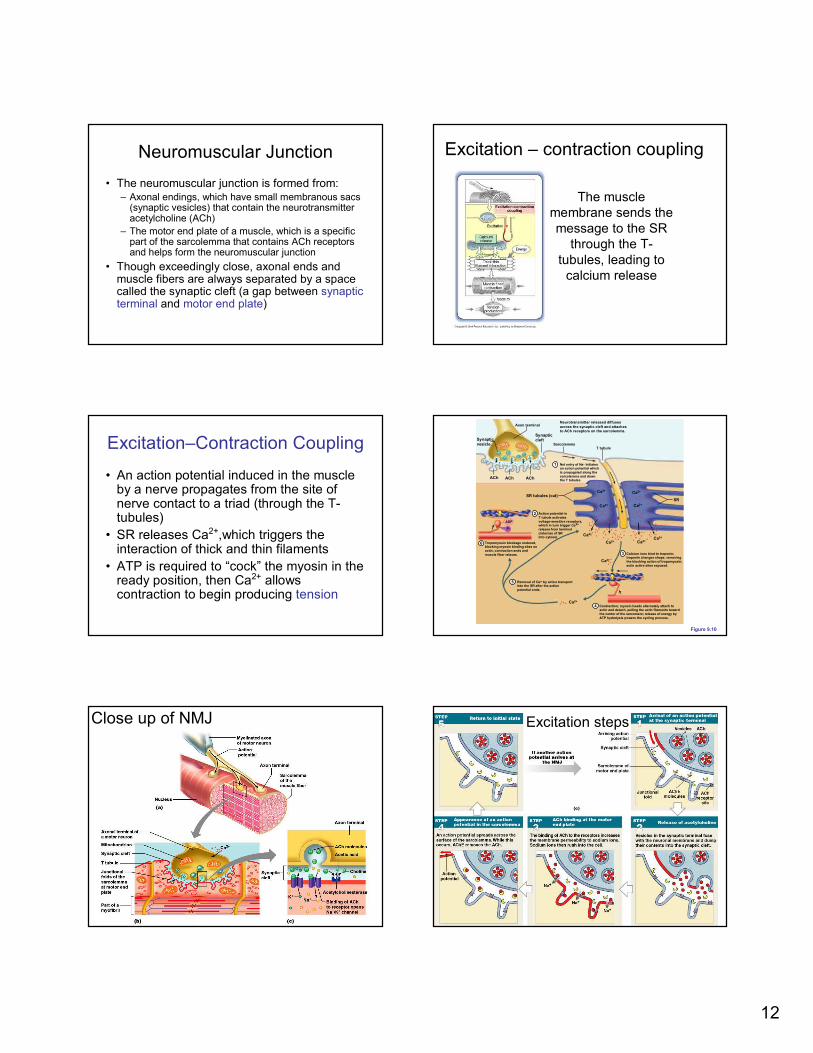
Close up of NMJ
Figure 10–10c
Excitation steps

13
Excitation: Nerve Synaptic
Terminal
• Arrival of action potential into terminal
• Voltage-regulated calcium channels open
and allow Ca2+ to enter the axon
• Ca2+ inside the axon terminal causes
axonal vesicles to fuse with the axonal
membrane release vesicles full of
neurotransmitter (acetylcholine or ACh)
into the synaptic cleft
Excitation: The Neurotransmitter
• Acetylcholine or ACh:
– travels across the synaptic cleft
– binds to membrane receptors on sarcolemma
(motor end plate)
– causes sodium ions to rush into sarcoplasm
through ACh receptors (that are Na+
channels)
– is quickly broken down by enzyme
(acetylcholinesterase or AChE)
Excitation: Action Potential
• Generated by increase in sodium ions in the sarcolemma (coming in through AChreceptors)
• Travels along the T tubules
– Note: T-tubules are just like the sacrolemmaand are filled with extracellular fluid (hi Na+)
• Depolarization causes Calcium to be released from the SR terminal cisternae
• Next: contraction
Excitation: AChE stops the signal
• Acetylcholinesterase (AChE) present in
the synaptic cleft breaks down the ACh
and thus it is no longer present to bind to
AChE receptors and cause an action
potential in the muscle
• Signal ends, NO more calcium is released,
contractions ceases
Contraction: Calcium ions are
released….now what happens?Contraction
• Myosin cross bridges alternately attach
and detach
• Thin filaments move toward the center of
the sarcomere
• Hydrolysis of ATP powers this cycling
process
• Ca2+ is removed into the SR, tropomyosin
blockage is restored, and the muscle fiber
relaxes

14
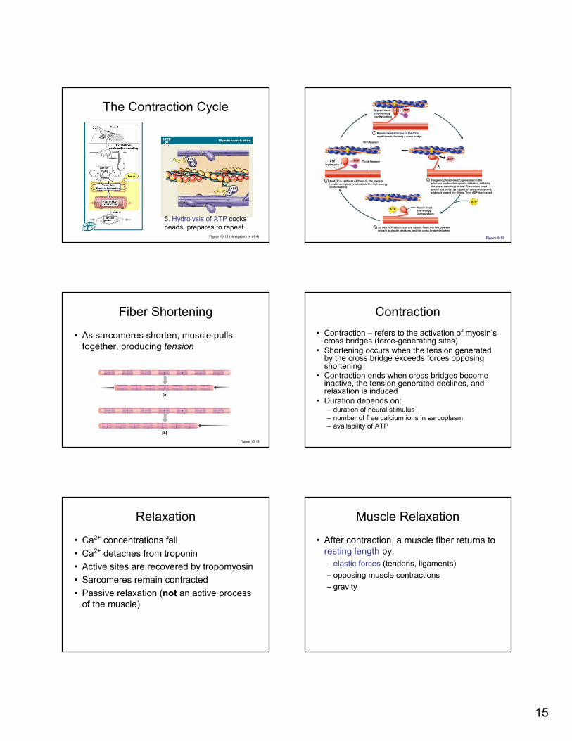
5 Steps of the Contraction Cycle
1. Exposure of active sites
2. Formation of cross-bridges
3. Pivoting of myosin heads
4. Detachment of cross-bridges
5. Reactivation of myosin
Movie
• Muscle contraction
The Contraction Cycle
Figure 10–12 (1 of 4)
At Rest
Myosin heads are
cocked and ready
(because ATP was
already hydrolyzed to
ADP) But myosin can’t bind
to actin yet because
tropomyosin blocks the
active site
The Contraction Cycle
Figure 10–12 (2 of 4)
1. Heads are “cocked” & point
away from M line
2. When active sites
exposed, cocked heads
can bind
Calcium exposes the Active Site
Figure 10–11
Calcium from
SR cisternae
binds to
troponin,
causes
tropomyosin
to move and
allowing
myosin to
bind actin
The Contraction Cycle
Figure 10–12 (3 of 4)
3. Loss of ADP causes
heads to pivot4. Binding of ATP causes
heads to dissociate
15
The Contraction Cycle
Figure 10–12 (Navigator) (4 of 4)
5. Hydrolysis of ATP cocks
heads, prepares to repeat
Figure 9.12
ATP
ADP
ADPATP
hydrolysis
ADP
ATP
Pi
Pi
Myosin head
(high-energy
configuration)
Myosin head attaches to the actin
myofilament, forming a cross bridge.
Thin filament
As ATP is split into ADP and Pi, the myosin
head is energized (cocked into the high-energy
conformation).
Inorganic phosphate (Pi) generated in the
previous contraction cycle is released, initiating
the power (working) stroke. The myosin head
pivots and bends as it pulls on the actin filament,
sliding it toward the M line. Then ADP is released.
Myosin head
(low-energy
configuration)
As new ATP attaches to the myosin head, the link between
myosin and actin weakens, and the cross bridge detaches.
Thick filament
1
4 2
3
Fiber Shortening
• As sarcomeres shorten, muscle pulls
together, producing tension
Figure 10–13
Contraction
• Contraction – refers to the activation of myosin’s cross bridges (force-generating sites)
• Shortening occurs when the tension generated by the cross bridge exceeds forces opposing shortening
• Contraction ends when cross bridges become inactive, the tension generated declines, and relaxation is induced
• Duration depends on:– duration of neural stimulus
– number of free calcium ions in sarcoplasm
– availability of ATP
Relaxation
• Ca2+ concentrations fall
• Ca2+ detaches from troponin
• Active sites are recovered by tropomyosin
• Sarcomeres remain contracted
• Passive relaxation (not an active process
of the muscle)
Muscle Relaxation
• After contraction, a muscle fiber returns to
resting length by:
– elastic forces (tendons, ligaments)
– opposing muscle contractions
– gravity

16
Rigor Mortis
• A fixed muscular contraction after death
• Caused when:
– ion pumps cease to function
– calcium builds up in the sarcoplasm
– no ATP to remove myosin heads from actin
– only ends during autolysis ~36 hrs
A Review of Muscle Contraction
Table 10–1 (1 of 2)
A Review of Muscle Contraction
Table 10–1 (2 of 2)
What happens when you get
botulism?
Muscles and Tension
We will look at the following
• What two factors affect the tension
produced by a muscle fiber?
• What is a twitch?
• What is a motor unit?
• What is the difference between isotonic
and isometric contractions?

17
Tension Production
• Tension = pulling force
• The all–or–none principal: as a whole, a muscle fiber is either contracted or relaxed
• Amount of tension in a muscle is determined by the following: 1.Resting length of the sarcomere at the time of
stimulation
2.Frequency of stimulation….which impacts the concentration of calcium ions
1. Resting length of Sarcomere
determines the amount of
tension
2. Frequency of Stimulation
• A single neural stimulation produces:
– a single contraction or twitch
– which lasts about 7–100 msec
– Does NOT generate any action on its own
• Sustained muscular contractions:
– require many repeated stimuli
Twitch Contraction
Muscle Response to Varying Stimuli
• A single stimulus results in a single contractile response – a muscle twitch
• Frequently delivered stimuli (muscle does not have time to completely relax) increases contractile force – wave summation
Figure 9.15
Motor Unit

18
Motor unit
• Consists of a motor neuron and all the muscle fibers it supplies
• All fibers activated at the same time
• Size varies greatly, from four to perhaps thousands of muscle fibers per motor unit
• Muscle fibers from a motor unit are spread throughout the muscle; therefore, contraction of a single motor unit causes weak contraction of the entire muscle
Motor Units in a skeletal muscle
Size of motor unit
• Determines how finely you can control a muscle
• Fine control requires small motor units
– Small: 1 nerve controls few fibers (2-6), e.g. fingers, eye
• Large weight-bearing muscles (thighs, hips) have large motor units
– Large: 1 nerve controls many fibers (2000), e.g. thigh
Contractions vary based on the
pattern of tension produced• Isotonic
– Tension rises and the skeletal muscle length
changes
• Isometric
– Tension rises but the muscle does not change
length because the tension never exceeds the
resistance
Isometric and Isotonic ContractionsRecruitment
(Motor Unit Summation)• In a whole muscle or group of muscles,
smooth motion and increasing tension is
produced by slowly increasing number of
motor units stimulated
• Often, the smaller fibers are recruited first,
then larger fibers

19
Rotation
• Sustained Tension = less than maximum
tension
• Allows motor units to rest in rotation: units
cycle through periods of activity and
inactivity
Rotation
KEY CONCEPT
• Force is increased by increasing the
number of stimulated motor units
(recruitment)
• Force is maintained by cycling the activity
of motor units within a muscle (rotation)
Muscle Tone• Is the constant, slightly contracted state of all
muscles, which does not produce active movements
• Keeps the muscles firm, healthy, and ready to respond to stimulus
• Motor units actively maintain body position, without motion
• Spinal reflexes account for muscle tone by:– Activating one motor unit and then another
– Responding to activation of stretch receptors in muscles and tendons
• Increasing muscle tone increases metabolic energy used, even at rest!
Muscle Metabolism
Contractions require lots of ATP
• If one muscle fiber contains 15 billion thick
filaments, and you need 2500 ATP/thick
filament, then how much ATP do you
need?
� Lots
• Muscle fibers must continuously
manufacture ATP as needed

20
Three ways to make ATP
• Creatine phosphate is stored energy that
can quickly be made into ATP
• Aerobic respiration with oxygen (generates
36 ATP/glucose)
• Anaerobic glycolysis (generates 2
ATP/glucose)
– Stores of glucose or fatty acids are needed to
do the latter two
Muscle Metabolism: Energy for
Contraction
Figure 9.20
Energy Storage in Muscle Fiber
Table 10–2
Creatine
• Made by muscle cells from amino acids
• It is phosphorylated by the enzyme
creatine phosphokinase (CPK) and
becomes creatine phosphate
• Damaged muscle cells leak CPK and it is
detected in the bloodstream
• Can quickly regenerate ATP from ADP by
transferring a phosphate group to it
Aerobic Metabolism
• Is the primary energy source of most
resting muscles
• Requires oxygen and a source of energy
glucose (stored glycogen, fatty acids)
• Produces 30-36 ATP molecules per
glucose molecule
But there’s a problem:
• During peak muscle activity oxygen
demands are too great
• When muscle contractile activity reaches
70% of maximum:
– Bulging muscles compress blood vessels
– Oxygen delivery is impaired
• Oxygen cannot enter the cell fast enough,
metabolism switches to anaerobic,
generating lactic acid

21
Anaerobic Glycolysis
• Is often the primary energy source for
peak muscular activity
• Breaks down glucose from glycogen
stored in skeletal muscles
• Produces 2 ATP molecules per molecule
of glucose (inefficient) without requiring
oxygen
Energy Use and Muscle Activity
• At rest
– Fatty acids from fats are the primary energy
source
– Because oxygen is abundant, aerobic
respiration is used
– Glucose taken in is stored as glycogen for late
use
– Creatine phosphate reserves are built up
Energy Use and Muscle Activity
• At during light or normal exertion:
– Creatine phosphate reserves are used first
but are quickly exhausted
– Muscles use aerobic respiration of fatty acids
and glucose released from glycogen stores to
make more ATP
Energy Use and Muscle Activity
• At peak exertion:
– muscles lack oxygen to support mitochondria
– muscles rely on glycolysis for ATP
– pyruvic acid builds up, is converted to lactic
acid
The Cori Cycle
• The removal and recycling of lactic acid by
the liver
• Liver converts lactic acid to pyruvic acid
• Glucose is released to recharge muscle
glycogen reserves
Muscle Fatigue
• When muscles can no longer perform a
required activity, they are fatigued
• Caused by:
– ATP production fails to keep pace with ATP
use
– There is a relative deficit of ATP, causing
contractures
– Lactic acid accumulates in the muscle
– Ionic imbalances are present

22
Oxygen Debt
• After exercise:
– the body needs more oxygen than usual to
normalize metabolic activities
– results in heavy breathing
– Oxygen reserves must be replenished
– Lactic acid must be converted to pyruvic acid
– Glycogen stores must be replaced
– ATP and CP reserves must be resynthesized
The Recovery Period
• The time required after exertion for
muscles to return to normal
• Oxygen becomes available
• Mitochondrial activity resumes
Heat Production and Loss
• Active muscles produce heat
– The vast majority of the heat generated by
your body comes from muscle activity. What
reaction occurs repeatedly in muscles?
• Up to 70% of muscle energy can be lost
as heat, raising body temperature
KEY CONCEPT
• Skeletal muscles at rest metabolize fatty acids and store glycogen
• During light activity, muscles generate ATP through aerobic breakdown of carbohydrates, lipids or amino acids
• At peak activity, energy is provided by anaerobic reactions that generate lactic acid as a byproduct
Are all muscle fibers the same?
• No
• Why can you stand all day on your feet yet
get tired during the 100 meter dash?
Muscle fibers vary in the body
• Fast fibers (white, fast twitch, fast
glycolytic)
• Slow fibers (red, slow twitch, slow
oxidative)
• Intermediate fibers (fast twitch, fast
oxidative)

23
Fast Fibers
• Contract very quickly
• Have large diameter, large glycogen
reserves, few mitochondria
• Have strong contractions, fatigue quickly
• Mostly anerobic (glycolytic)
Slow Fibers
• Are slow to contract, but also slow to
fatigue
• Have small diameter, more mitochondria
• Have high oxygen supply
• Contain myoglobin (red pigment) to bind
and store extra oxygen
• Mostly aerobic
Intermediate Fibers
• Also: fast oxidative fibers
• Are mid-sized
• Have low myoglobin
• Have more capillaries than fast fibers,
slower to fatigue
• Both aerobic and anerobic
Slow fibers vs. Fast fibers
Comparing Skeletal
Muscle Fibers
Table 10–3
Muscles and Fiber Types
• White muscle:– mostly fast fibers
– pale (e.g., chicken breast, ”white meat”)
• Red muscle:– mostly slow fibers
– dark (e.g., chicken legs, “dark meat”)
• Most human muscles are a mix of fiber types and therefore appear pink

24
Muscle Hypertrophy
• Muscle growth from weight training:
– increases diameter of muscle fibers
– increases number of myofibrils (but not
number of muscle fibers)
– increases numbers of mitochondria, glycogen
reserves
Anaerobic activity
• Anaerobic activities (e.g., 100-meter dash, competitive weightlifting):
– use fast fibers
– fatigue quickly with strenuous activity
• Performance improved by:
– resistance exercise
– frequent, brief, intensive workouts
– hypertrophy
– increased glycogen stores
Aerobic activity
• Aerobic activities (prolonged activity):
– supported by mitochondria
– require oxygen and nutrients
• Improved by:
– repetitive training (muscle memory)
– cardiovascular training leading to improved
delivery of oxygen and nutrients, increase
mitochondria and myoglobin synthesis
Fiber type “switching”
• Most fibers in human muscles are fast,
and the relative amount of fast versus slow
is genetic
• However, endurance training can cause
some fast fibers to take on the appearance
and properties of intermediate fibers,
improving aerobic performance
Summary
• Excitation: events at the neuromuscular
junction (5 steps)
• Contraction: the contraction cycle and
ATP (5 steps)
• Tension and motor units
• Muscle metabolism and fiber types