chapter 3 procaryotic cell structure and function an overview of
TRANSCRIPT
1
Copyright © The McGraw-Hill Companies, Inc. Permission required for reproduction or display.
1
Chapter 3
Procaryotic Cell Structure and Function
Copyright © The McGraw-Hill Companies, Inc. Permission required for reproduction or display.
2
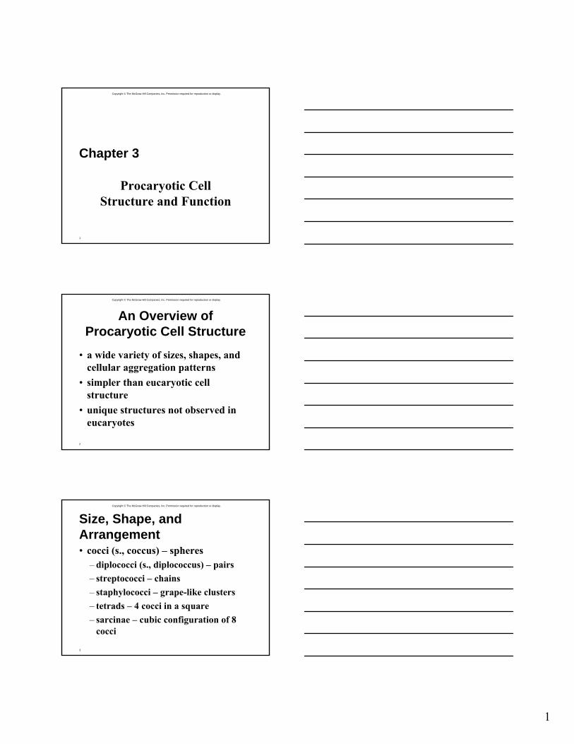
An Overview of Procaryotic Cell Structure
• a wide variety of sizes, shapes, and cellular aggregation patterns
• simpler than eucaryotic cell structure
• unique structures not observed in eucaryotes
Copyright © The McGraw-Hill Companies, Inc. Permission required for reproduction or display.
3
Size, Shape, and Arrangement• cocci (s., coccus) – spheres
– diplococci (s., diplococcus) – pairs– streptococci – chains– staphylococci – grape-like clusters– tetrads – 4 cocci in a square– sarcinae – cubic configuration of 8
cocci
2
Copyright © The McGraw-Hill Companies, Inc. Permission required for reproduction or display.
4
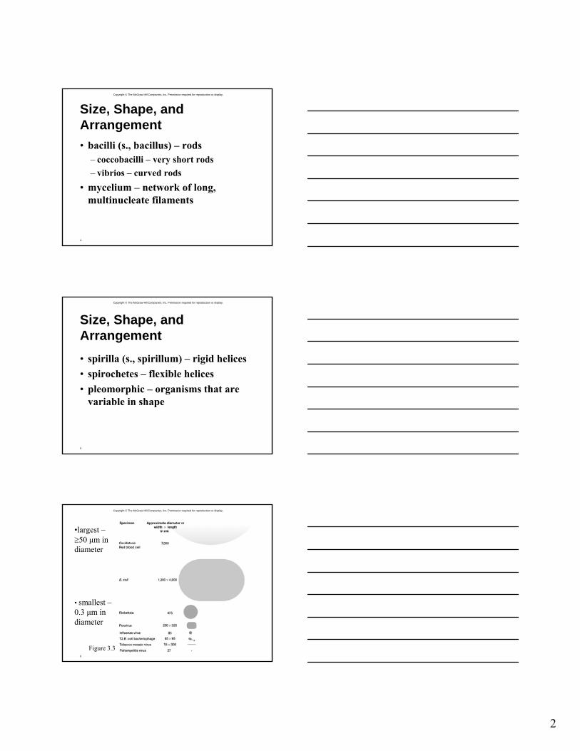
Size, Shape, and Arrangement• bacilli (s., bacillus) – rods
– coccobacilli – very short rods– vibrios – curved rods
• mycelium – network of long, multinucleate filaments
Copyright © The McGraw-Hill Companies, Inc. Permission required for reproduction or display.
5
Size, Shape, and Arrangement• spirilla (s., spirillum) – rigid helices• spirochetes – flexible helices• pleomorphic – organisms that are
variable in shape
Copyright © The McGraw-Hill Companies, Inc. Permission required for reproduction or display.
6
•largest –≥50 μm indiameter
• smallest –0.3 μm in diameter
Figure 3.3

3
Copyright © The McGraw-Hill Companies, Inc. Permission required for reproduction or display.
7
Procaryotic Cell Organization
Copyright © The McGraw-Hill Companies, Inc. Permission required for reproduction or display.
8Figure 3.4
Copyright © The McGraw-Hill Companies, Inc. Permission required for reproduction or display.
9
4
Copyright © The McGraw-Hill Companies, Inc. Permission required for reproduction or display.
10
Procaryotic Cell Membranes
• membranes are an absolute requirement for all living organisms
• plasma membrane encompasses the cytoplasm
• some procaryotes also have internal membrane systems
Copyright © The McGraw-Hill Companies, Inc. Permission required for reproduction or display.
11
The Plasma Membrane
• contains lipids and proteins– lipids usually form a bilayer– proteins are embedded in or associated
with lipids• highly organized, asymmetric,
flexible, and dynamic
Copyright © The McGraw-Hill Companies, Inc. Permission required for reproduction or display.
12
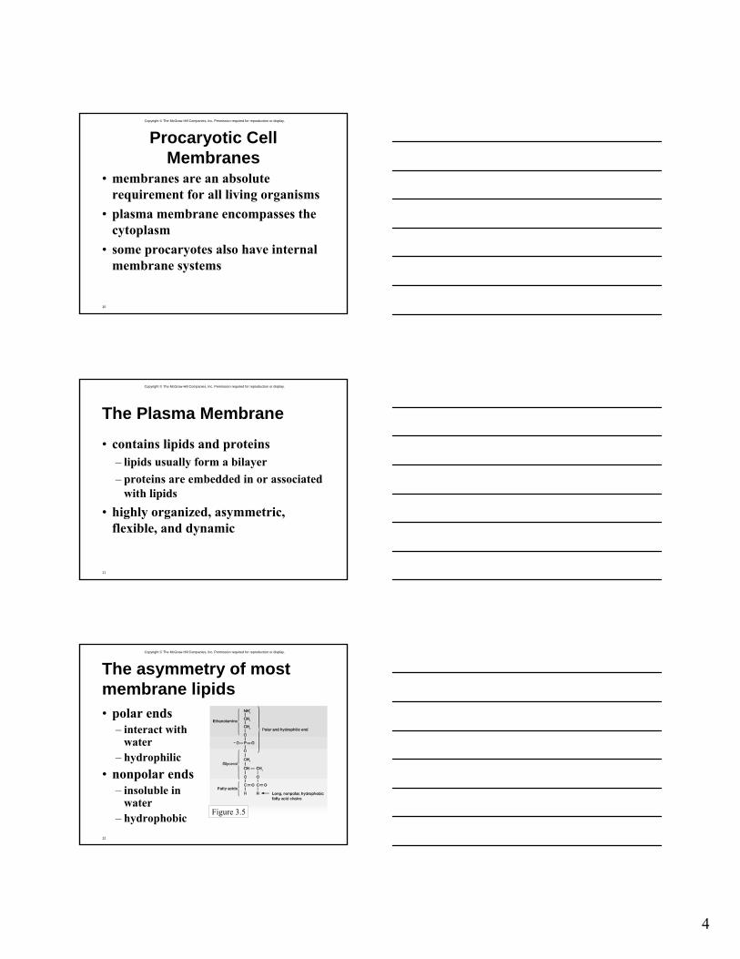
The asymmetry of most membrane lipids• polar ends
– interact with water
– hydrophilic• nonpolar ends
– insoluble in water
– hydrophobic Figure 3.5
5
Copyright © The McGraw-Hill Companies, Inc. Permission required for reproduction or display.
13
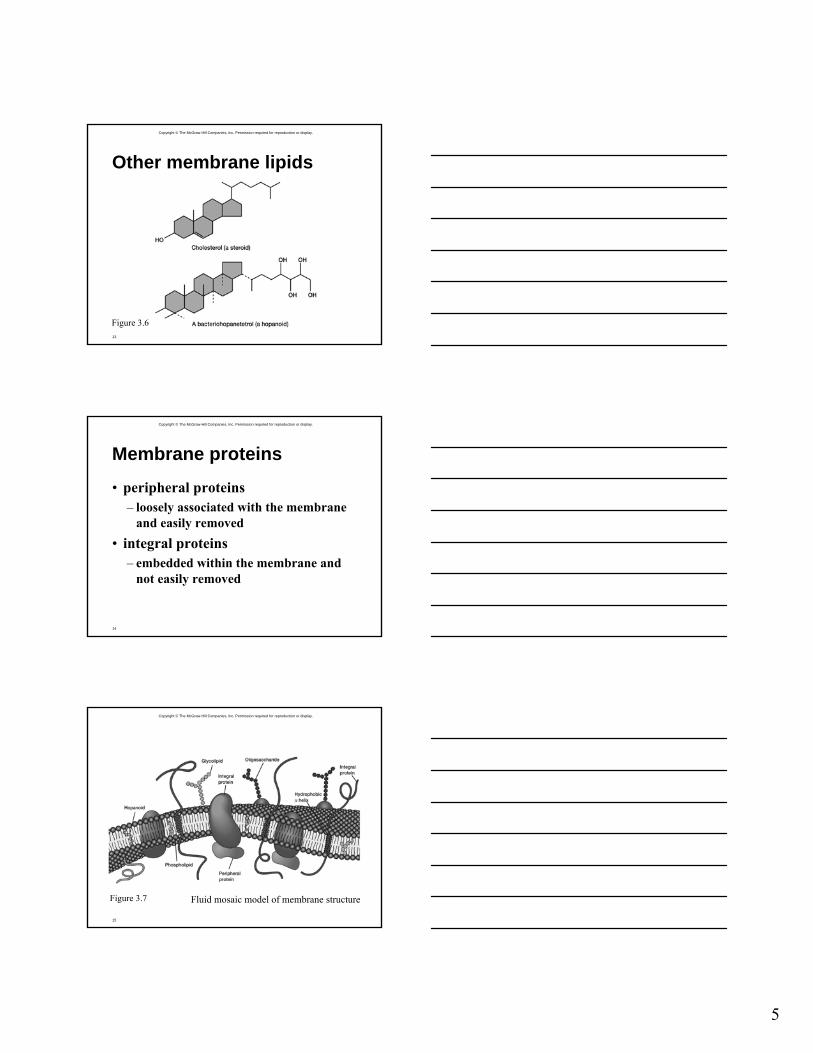
Other membrane lipids
Figure 3.6
Copyright © The McGraw-Hill Companies, Inc. Permission required for reproduction or display.
14
Membrane proteins
• peripheral proteins– loosely associated with the membrane
and easily removed• integral proteins
– embedded within the membrane and not easily removed
Copyright © The McGraw-Hill Companies, Inc. Permission required for reproduction or display.
15
Figure 3.7 Fluid mosaic model of membrane structure
6
Copyright © The McGraw-Hill Companies, Inc. Permission required for reproduction or display.
16
Archaeal membranes
• composed of unique lipids• some have a monolayer structure
instead of a bilayer structure
Copyright © The McGraw-Hill Companies, Inc. Permission required for reproduction or display.
17
Functions of the plasma membrane• separation of cell from its
environment• selectively permeable barrier
– some molecules are allowed to pass into or out of the cell
– transport systems aid in movement of molecules
Copyright © The McGraw-Hill Companies, Inc. Permission required for reproduction or display.
18
More functions…
• location of crucial metabolic processes
• detection of and response to chemicals in surroundings with the aid of special receptor molecules in the membrane
7
Copyright © The McGraw-Hill Companies, Inc. Permission required for reproduction or display.
19
Internal Membrane Systems• mesosomes
– may be invaginations of the plasma membrane• possible roles
– cell wall formation during cell division– chromosome replication and distribution– secretory processes
– may be artifacts of chemical fixation process
Copyright © The McGraw-Hill Companies, Inc. Permission required for reproduction or display.
20
Other internal membrane systems• complex in-foldings of the plasma
membrane– observed in many photosynthetic
bacteria and in procaryotes with high respiratory activity
– may be aggregates of spherical vesicles, flattened vesicles, or tubular membranes
Copyright © The McGraw-Hill Companies, Inc. Permission required for reproduction or display.
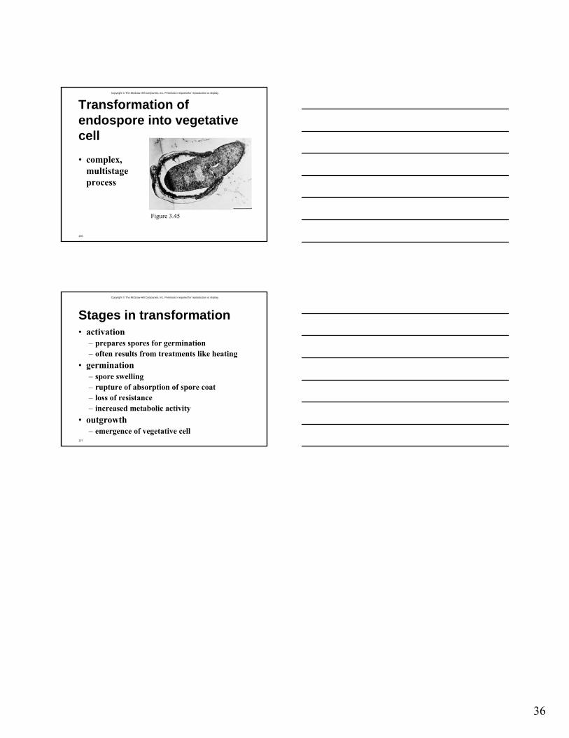
21
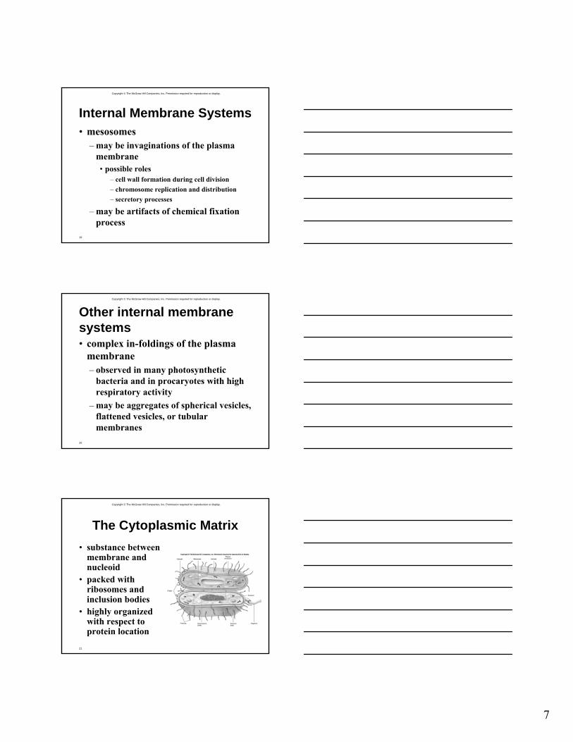
The Cytoplasmic Matrix• substance between
membrane and nucleoid
• packed with ribosomes and inclusion bodies
• highly organized with respect to protein location
8
Copyright © The McGraw-Hill Companies, Inc. Permission required for reproduction or display.
22
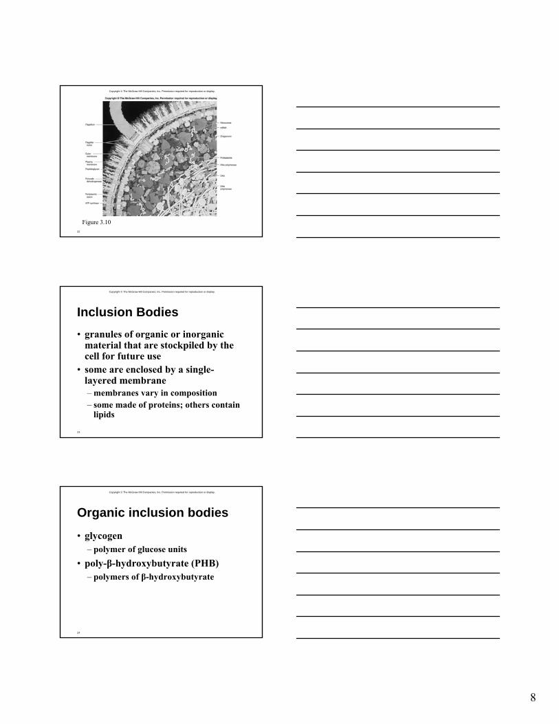
Figure 3.10
Copyright © The McGraw-Hill Companies, Inc. Permission required for reproduction or display.
23
Inclusion Bodies• granules of organic or inorganic
material that are stockpiled by the cell for future use
• some are enclosed by a single-layered membrane– membranes vary in composition– some made of proteins; others contain
lipids
Copyright © The McGraw-Hill Companies, Inc. Permission required for reproduction or display.
24
Organic inclusion bodies
• glycogen– polymer of glucose units
• poly-β-hydroxybutyrate (PHB)– polymers of β-hydroxybutyrate
9
Copyright © The McGraw-Hill Companies, Inc. Permission required for reproduction or display.
25
Organic inclusion bodies
• cyanophycin granules– large polypeptides containing about
equal quantities of arginine and aspartic acid
• carboxysomes– contain the enzyme ribulose-1,5,-
bisphosphate carboxylase
Copyright © The McGraw-Hill Companies, Inc. Permission required for reproduction or display.
26
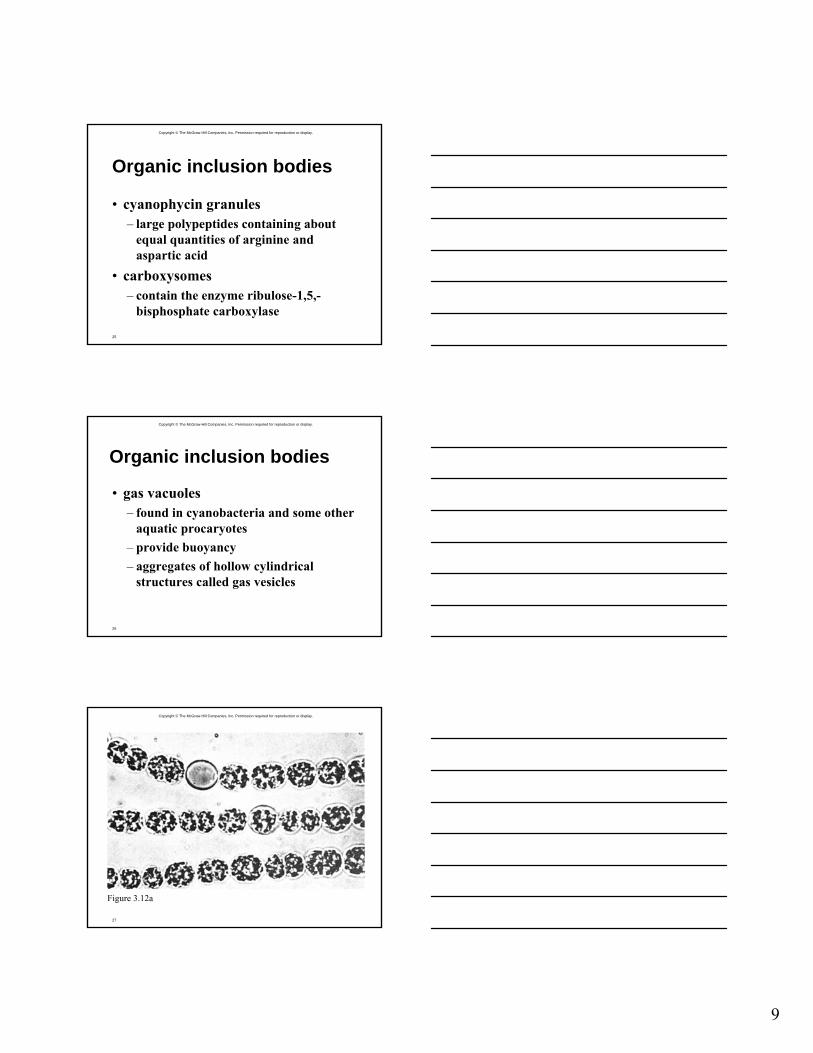
Organic inclusion bodies
• gas vacuoles– found in cyanobacteria and some other
aquatic procaryotes– provide buoyancy– aggregates of hollow cylindrical
structures called gas vesicles
Copyright © The McGraw-Hill Companies, Inc. Permission required for reproduction or display.
27
Figure 3.12a
10
Copyright © The McGraw-Hill Companies, Inc. Permission required for reproduction or display.
28
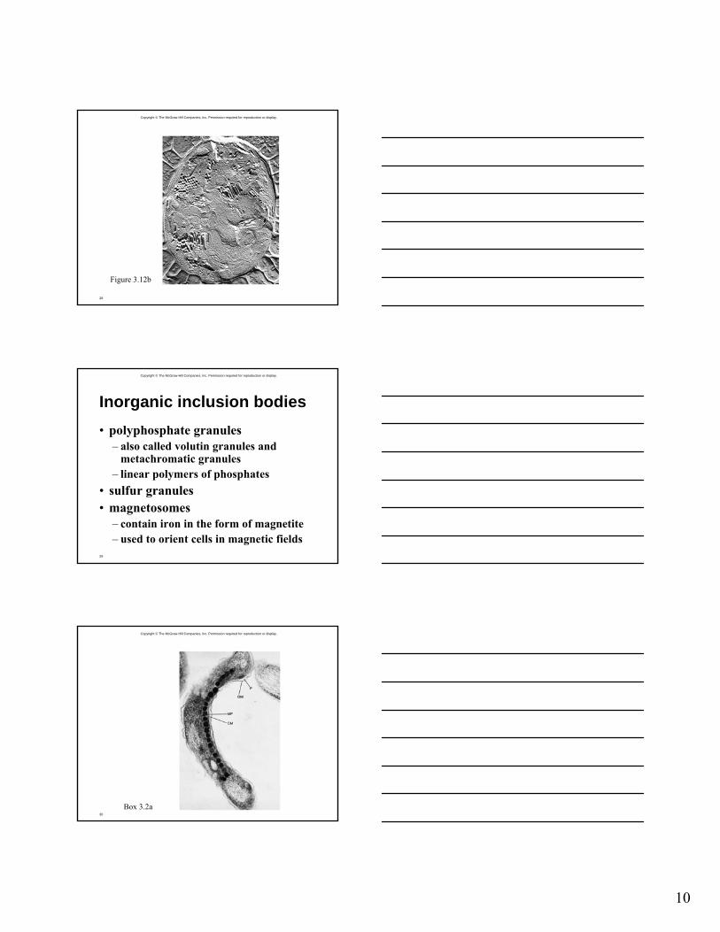
Figure 3.12b
Copyright © The McGraw-Hill Companies, Inc. Permission required for reproduction or display.
29
Inorganic inclusion bodies• polyphosphate granules
– also called volutin granules and metachromatic granules
– linear polymers of phosphates• sulfur granules• magnetosomes
– contain iron in the form of magnetite– used to orient cells in magnetic fields
Copyright © The McGraw-Hill Companies, Inc. Permission required for reproduction or display.
30
Box 3.2a
11
Copyright © The McGraw-Hill Companies, Inc. Permission required for reproduction or display.
31
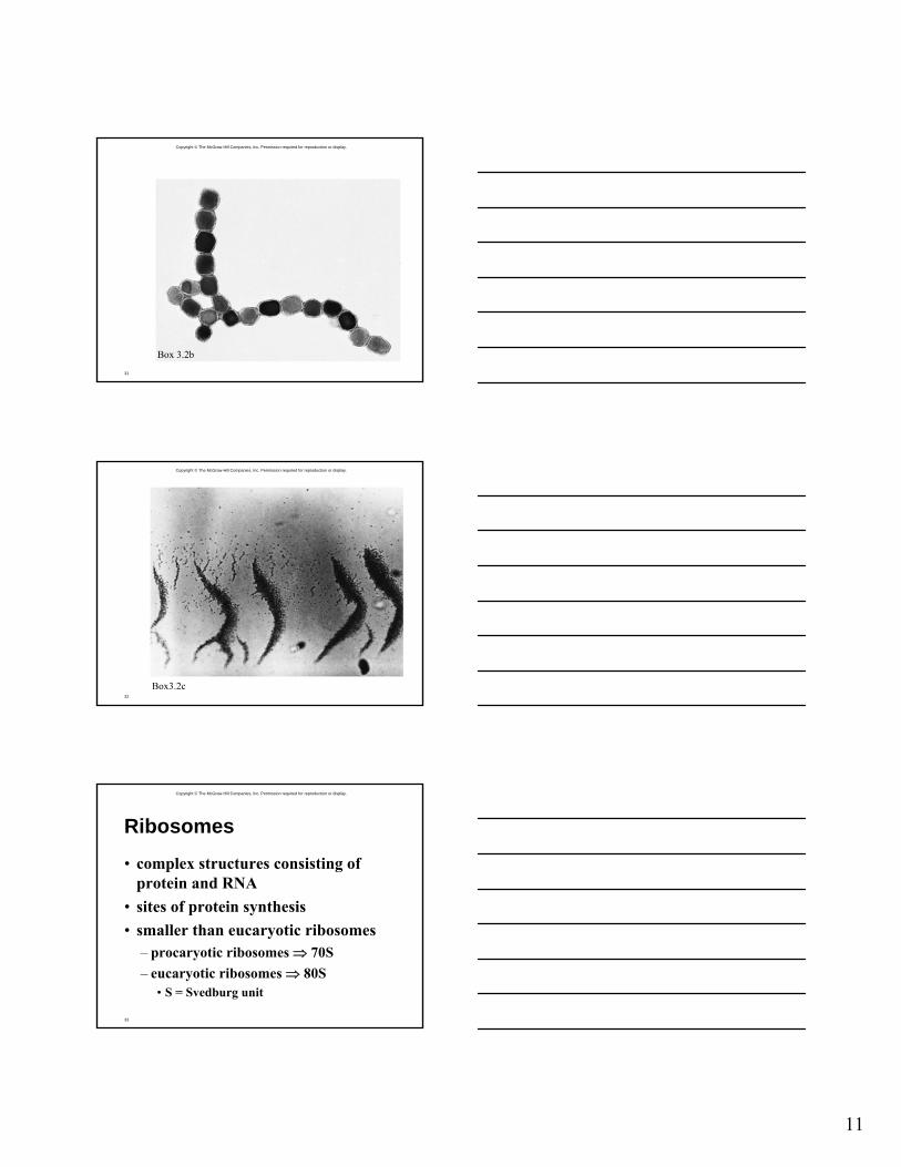
Box 3.2b
Copyright © The McGraw-Hill Companies, Inc. Permission required for reproduction or display.
32
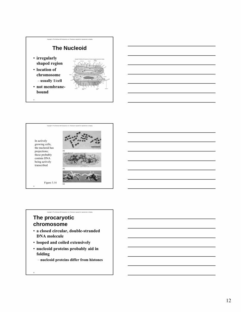
Box3.2c
Copyright © The McGraw-Hill Companies, Inc. Permission required for reproduction or display.
33
Ribosomes
• complex structures consisting of protein and RNA
• sites of protein synthesis• smaller than eucaryotic ribosomes
– procaryotic ribosomes ⇒ 70S– eucaryotic ribosomes ⇒ 80S
• S = Svedburg unit
12
Copyright © The McGraw-Hill Companies, Inc. Permission required for reproduction or display.
34
The Nucleoid
• irregularly shaped region
• location of chromosome– usually 1/cell
• not membrane-bound
Copyright © The McGraw-Hill Companies, Inc. Permission required for reproduction or display.
35
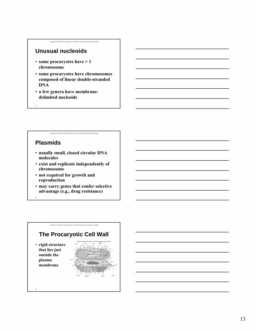
Figure 3.14
In actively growing cells, the nucleoid has projections; these probably contain DNA being actively transcribed
Copyright © The McGraw-Hill Companies, Inc. Permission required for reproduction or display.
36
The procaryotic chromosome• a closed circular, double-stranded
DNA molecule• looped and coiled extensively• nucleoid proteins probably aid in
folding– nucleoid proteins differ from histones
13
Copyright © The McGraw-Hill Companies, Inc. Permission required for reproduction or display.
37
Unusual nucleoids
• some procaryotes have > 1 chromosome
• some procaryotes have chromosomes composed of linear double-stranded DNA
• a few genera have membrane-delimited nucleoids
Copyright © The McGraw-Hill Companies, Inc. Permission required for reproduction or display.
38
Plasmids• usually small, closed circular DNA
molecules• exist and replicate independently of
chromosome• not required for growth and
reproduction• may carry genes that confer selective
advantage (e.g., drug resistance)
Copyright © The McGraw-Hill Companies, Inc. Permission required for reproduction or display.
39
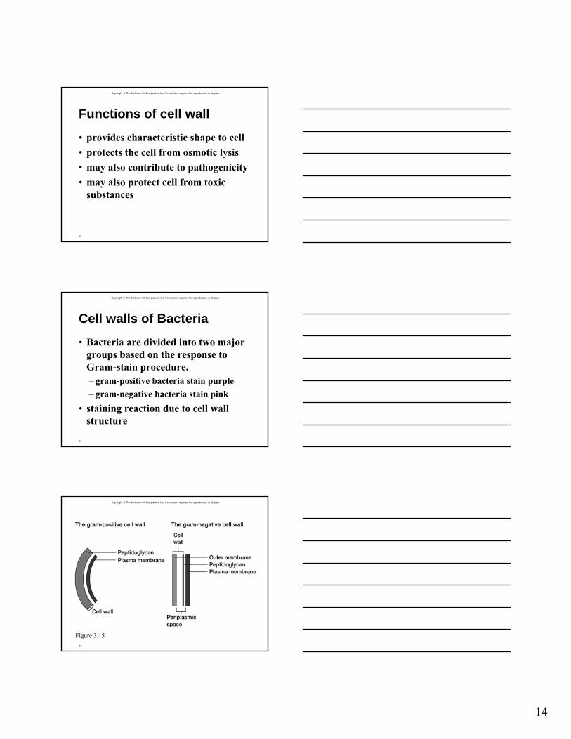
The Procaryotic Cell Wall• rigid structure
that lies just outside the plasma membrane
14
Copyright © The McGraw-Hill Companies, Inc. Permission required for reproduction or display.
40
Functions of cell wall
• provides characteristic shape to cell• protects the cell from osmotic lysis• may also contribute to pathogenicity• may also protect cell from toxic
substances
Copyright © The McGraw-Hill Companies, Inc. Permission required for reproduction or display.
41
Cell walls of Bacteria
• Bacteria are divided into two major groups based on the response to Gram-stain procedure.– gram-positive bacteria stain purple– gram-negative bacteria stain pink
• staining reaction due to cell wall structure
Copyright © The McGraw-Hill Companies, Inc. Permission required for reproduction or display.
42
Figure 3.15
15
Copyright © The McGraw-Hill Companies, Inc. Permission required for reproduction or display.
43
Periplasmic space• gap between plasma membrane and
cell wall (gram-positive bacteria) or between plasma membrane and outer membrane (gram-negative bacteria)
• periplasm– substance that occupies periplasmic
space
Copyright © The McGraw-Hill Companies, Inc. Permission required for reproduction or display.
44
Periplasmic enzymes
• found in periplasm of gram-negative bacteria
• some of their functions– nutrient acquisition– electron transport– peptidoglycan synthesis– modification of toxic compounds
Copyright © The McGraw-Hill Companies, Inc. Permission required for reproduction or display.
45
Exoenzymes
• secreted by gram-positive bacteria• perform many of the same functions
that periplasmic enzymes do for gram-negative bacteria
16
Copyright © The McGraw-Hill Companies, Inc. Permission required for reproduction or display.
46
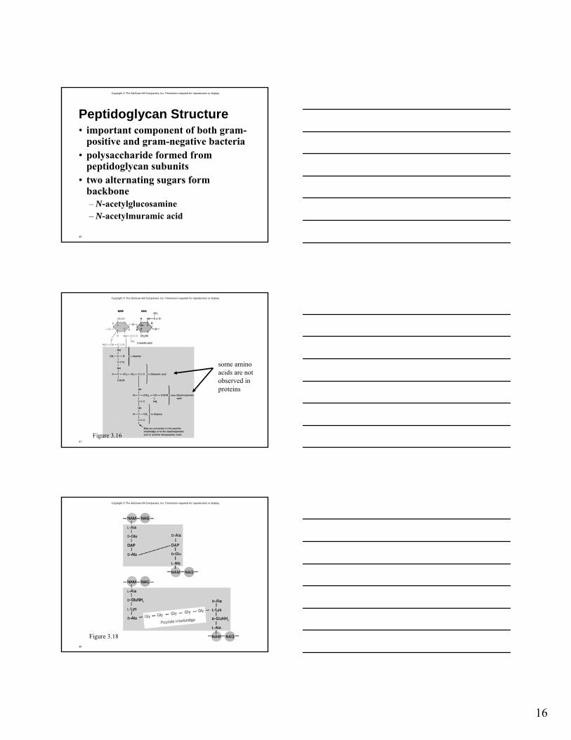
Peptidoglycan Structure• important component of both gram-
positive and gram-negative bacteria• polysaccharide formed from
peptidoglycan subunits• two alternating sugars form
backbone– N-acetylglucosamine– N-acetylmuramic acid
Copyright © The McGraw-Hill Companies, Inc. Permission required for reproduction or display.
47
some amino acids are not observed in proteins
Figure 3.16
Copyright © The McGraw-Hill Companies, Inc. Permission required for reproduction or display.
48
Figure 3.18
17
Copyright © The McGraw-Hill Companies, Inc. Permission required for reproduction or display.
49
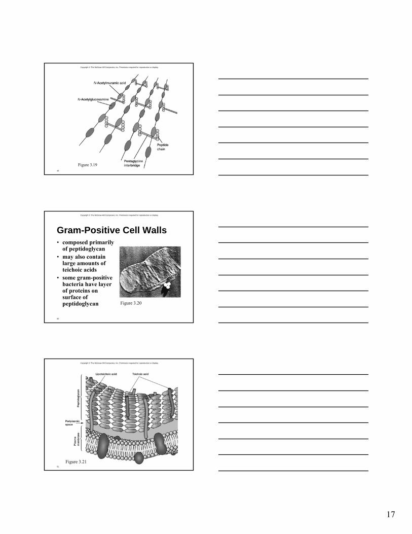
Figure 3.19
Copyright © The McGraw-Hill Companies, Inc. Permission required for reproduction or display.
50
Gram-Positive Cell Walls• composed primarily
of peptidoglycan• may also contain
large amounts of teichoic acids
• some gram-positive bacteria have layer of proteins on surface of peptidoglycan Figure 3.20
Copyright © The McGraw-Hill Companies, Inc. Permission required for reproduction or display.
51
Figure 3.21
18
Copyright © The McGraw-Hill Companies, Inc. Permission required for reproduction or display.
52
Figure 3.22
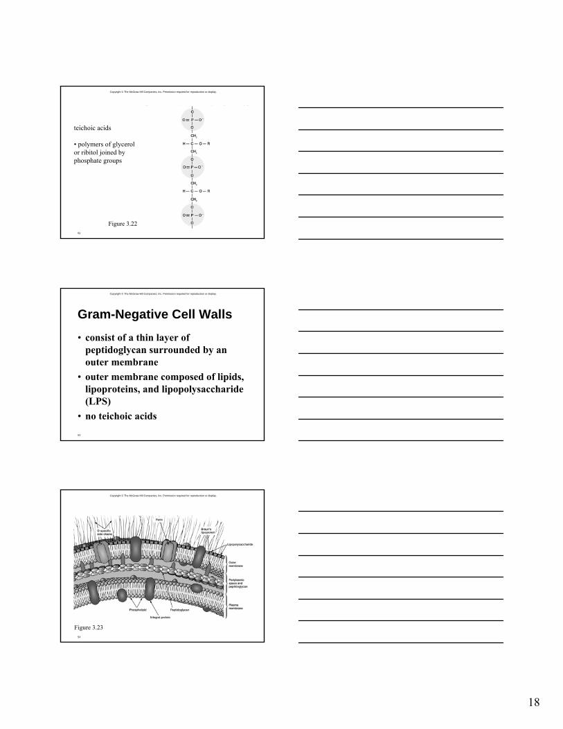
teichoic acids
• polymers of glycerolor ribitol joined byphosphate groups
Copyright © The McGraw-Hill Companies, Inc. Permission required for reproduction or display.
53
Gram-Negative Cell Walls
• consist of a thin layer of peptidoglycan surrounded by an outer membrane
• outer membrane composed of lipids, lipoproteins, and lipopolysaccharide(LPS)
• no teichoic acids
Copyright © The McGraw-Hill Companies, Inc. Permission required for reproduction or display.
54
Figure 3.23
19
Copyright © The McGraw-Hill Companies, Inc. Permission required for reproduction or display.
55
Important connections• Braun’s lipoproteins connect outer
membrane to peptidoglycan• Adhesion sites
– sites of direct contact (possibly true membrane fusions) between plasma membrane and outer membrane
– substances may move directly into cell through adhesion sites
Copyright © The McGraw-Hill Companies, Inc. Permission required for reproduction or display.
56
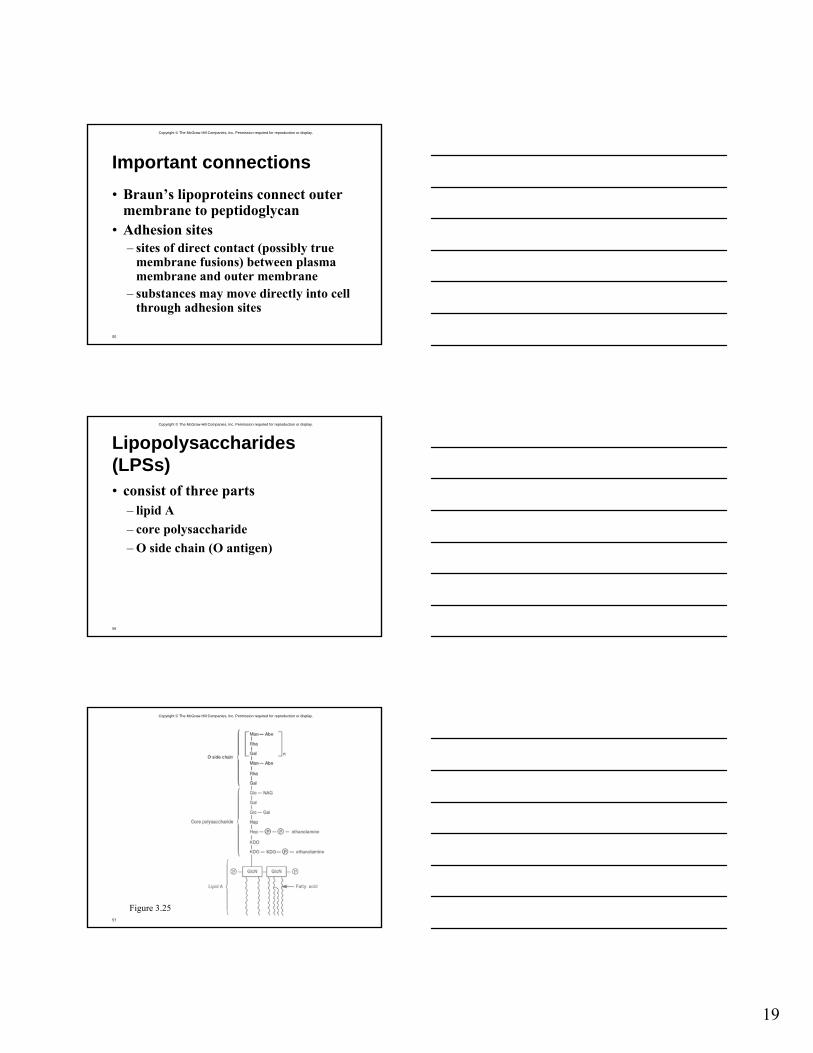
Lipopolysaccharides (LPSs)• consist of three parts
– lipid A– core polysaccharide– O side chain (O antigen)
Copyright © The McGraw-Hill Companies, Inc. Permission required for reproduction or display.
57
Figure 3.25

20
Copyright © The McGraw-Hill Companies, Inc. Permission required for reproduction or display.
58
Importance of LPS
• protection from host defenses (O antigen)
• contributes to negative charge on cell surface (core polysaccharide)
• helps stabilize outer membrane structure (lipid A)
• can act as an exotoxin (lipid A)
Copyright © The McGraw-Hill Companies, Inc. Permission required for reproduction or display.
59
Other characteristics of outer membrane• more permeable than plasma
membrane due to presence of porin proteins and transporter proteins– porin proteins form channels through
which small molecules (600-700 daltons) can pass
Copyright © The McGraw-Hill Companies, Inc. Permission required for reproduction or display.
60
The Mechanism of Gram Staining• thought to involve constriction of the
thick peptidoglycan layer of gram-positive cells– constriction prevents loss of crystal
violet during decolorization step• thinner peptidoglycan layer of gram-
negative bacteria does not prevent loss of crystal violet
21
Copyright © The McGraw-Hill Companies, Inc. Permission required for reproduction or display.
61
The Cell Wall and Osmotic Protection• osmosis
– movement of water across selectively permeable membrane from dilute solutions to more concentrated solutions
• cells are often in hypotonic solutions[solute]outside cell < [solute]inside cell
Copyright © The McGraw-Hill Companies, Inc. Permission required for reproduction or display.
62
The Cell Wall and Osmotic Protection• osmotic lysis
– can occur when cells are in hypotonic solutions
– movement of water into cell causes swelling and lysis due to osmotic pressure
• cell wall protects against osmotic lysis
Copyright © The McGraw-Hill Companies, Inc. Permission required for reproduction or display.
63
Cell walls do not protect against plasmolysis
• plasmolysis– occurs when cells are in hypertonic
solutions[solute]outside cell > [solute]inside cell
– water moves out of cell causing cytoplasm to shrivel and pull away from cell wall
22
Copyright © The McGraw-Hill Companies, Inc. Permission required for reproduction or display.
64
Practical importance of plasmolysis and osmotic lysis• plasmolysis
– useful in food preservation– e.g., dried foods and jellies
• osmotic lysis– basis of lysozyme and penicillin action
Copyright © The McGraw-Hill Companies, Inc. Permission required for reproduction or display.
65
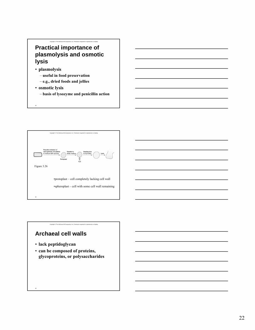
Figure 3.26
•protoplast – cell completely lacking cell wall
•spheroplast – cell with some cell wall remaining
Copyright © The McGraw-Hill Companies, Inc. Permission required for reproduction or display.
66
Archaeal cell walls
• lack peptidoglycan• can be composed of proteins,
glycoproteins, or polysaccharides
23
Copyright © The McGraw-Hill Companies, Inc. Permission required for reproduction or display.
67
Protein Secretion in Procaryotes
• numerous protein secretion pathways have been identified
• four major pathways are:– Sec-dependent pathway– type II pathway– type I (ABC) protein secretion
pathway– type III protein secretion pathway
Copyright © The McGraw-Hill Companies, Inc. Permission required for reproduction or display.
68
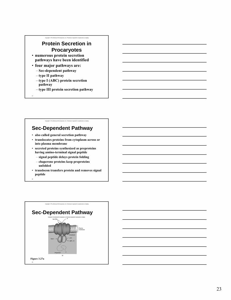
Sec-Dependent Pathway• also called general secretion pathway• translocates proteins from cytoplasm across or
into plasma membrane• secreted proteins synthesized as preproteins
having amino-terminal signal peptide– signal peptide delays protein folding– chaperone proteins keep preproteins
unfolded• translocon transfers protein and removes signal
peptide
Copyright © The McGraw-Hill Companies, Inc. Permission required for reproduction or display.
69
Sec-Dependent Pathway
Figure 3.27a
24
Copyright © The McGraw-Hill Companies, Inc. Permission required for reproduction or display.
70
Type II Protein Secretion Pathway• transports proteins from periplasmic
across outer membrane• observed in some gram-negative
bacteria, including some pathogens• complex systems consisting of up to
12-14 proteins– most are integral membrane proteins
Copyright © The McGraw-Hill Companies, Inc. Permission required for reproduction or display.
71
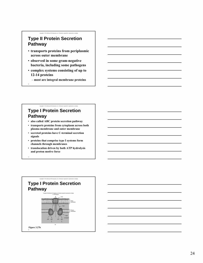
Type I Protein Secretion Pathway• also called ABC protein secretion pathway• transports proteins from cytoplasm across both
plasma membrane and outer membrane• secreted proteins have C-terminal secretion
signals• proteins that comprise type I systems form
channels through membranes• translocation driven by both ATP hydrolysis
and proton motive force
Copyright © The McGraw-Hill Companies, Inc. Permission required for reproduction or display.
72
Type I Protein Secretion Pathway
Figure 3.27b
25
Copyright © The McGraw-Hill Companies, Inc. Permission required for reproduction or display.
73
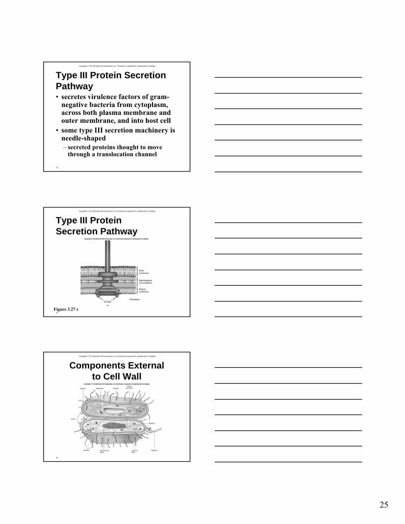
Type III Protein Secretion Pathway• secretes virulence factors of gram-
negative bacteria from cytoplasm, across both plasma membrane and outer membrane, and into host cell
• some type III secretion machinery is needle-shaped– secreted proteins thought to move
through a translocation channel
Copyright © The McGraw-Hill Companies, Inc. Permission required for reproduction or display.
74
Type III Protein Secretion Pathway
Figure 3.27 c
Copyright © The McGraw-Hill Companies, Inc. Permission required for reproduction or display.
75
Components External to Cell Wall

26
Copyright © The McGraw-Hill Companies, Inc. Permission required for reproduction or display.
76
Capsules, Slime Layers, and S-Layers• layers of material lying outside the cell
wall– capsules
• usually composed of polysaccharides• well organized and not easily removed from cell
– slime layers• similar to capsules except diffuse, unorganized
and easily removed
Copyright © The McGraw-Hill Companies, Inc. Permission required for reproduction or display.
77
Capsules, Slime Layers, and S-Layers• glycocalyx
– network of polysaccharides extending from the surface of the cell
– a capsule or slime layer composed of polysaccharides can also be referred to as a glycocalyx
Copyright © The McGraw-Hill Companies, Inc. Permission required for reproduction or display.
78
Capsules, Slime Layers, and S-Layers• S-layers
– regularly structured layers of protein or glycoprotein
– common among Archaea, where they may be the only structure outside the plasma membrane
27
Copyright © The McGraw-Hill Companies, Inc. Permission required for reproduction or display.
79
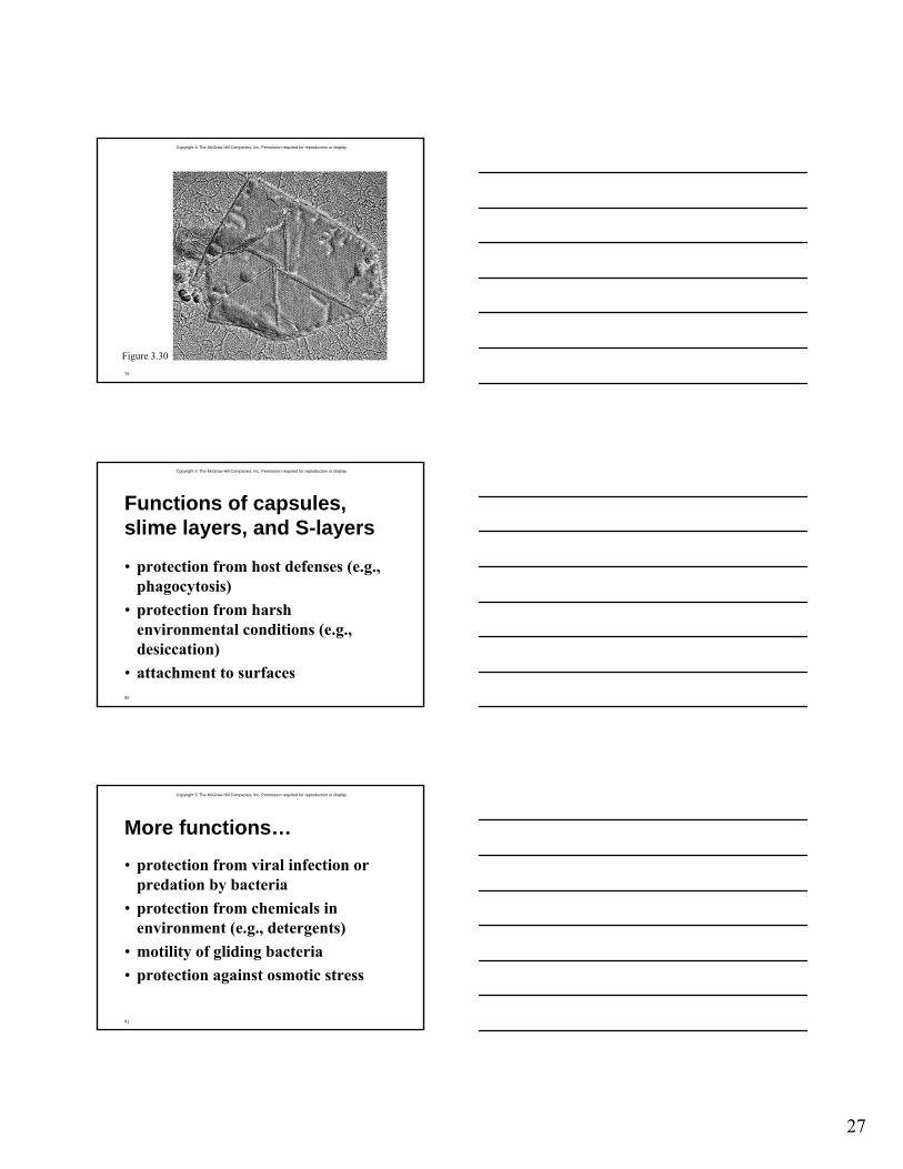
Figure 3.30
Copyright © The McGraw-Hill Companies, Inc. Permission required for reproduction or display.
80
Functions of capsules, slime layers, and S-layers
• protection from host defenses (e.g., phagocytosis)
• protection from harsh environmental conditions (e.g., desiccation)
• attachment to surfaces
Copyright © The McGraw-Hill Companies, Inc. Permission required for reproduction or display.
81
More functions…
• protection from viral infection or predation by bacteria
• protection from chemicals in environment (e.g., detergents)
• motility of gliding bacteria• protection against osmotic stress
28
Copyright © The McGraw-Hill Companies, Inc. Permission required for reproduction or display.
82
Pili and Fimbriae• fimbriae (s., fimbria)
– short, thin, hairlike, proteinaceous appendages
• up to 1,000/cell– mediate attachment to surfaces– some (type IV fimbriae) required for
twitching motility or gliding motility that occurs in some bacteria
• sex pili (s., pilus)– similar to fimbriae except longer, thicker,
and less numerous (1-10/cell)– required for mating
Copyright © The McGraw-Hill Companies, Inc. Permission required for reproduction or display.
83
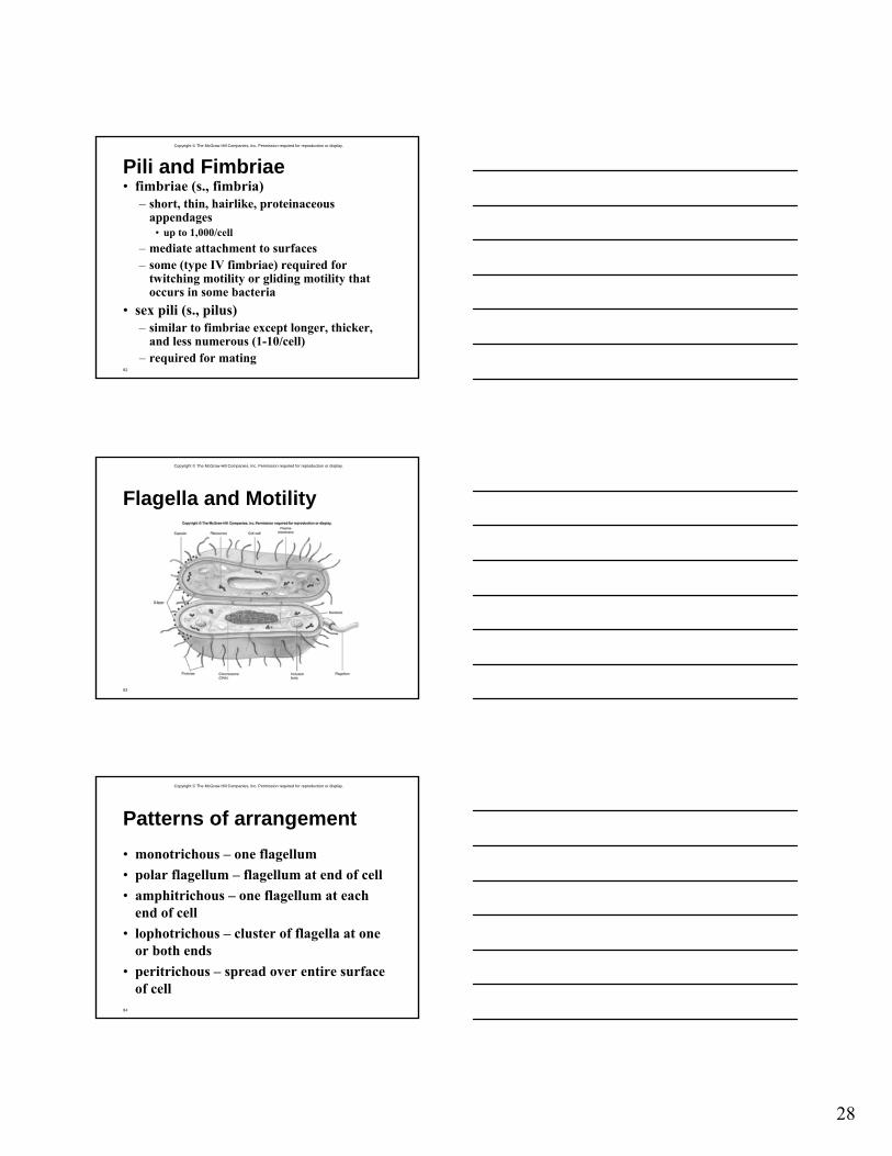
Flagella and Motility
Copyright © The McGraw-Hill Companies, Inc. Permission required for reproduction or display.
84
Patterns of arrangement• monotrichous – one flagellum• polar flagellum – flagellum at end of cell• amphitrichous – one flagellum at each
end of cell• lophotrichous – cluster of flagella at one
or both ends• peritrichous – spread over entire surface
of cell
29
Copyright © The McGraw-Hill Companies, Inc. Permission required for reproduction or display.
85
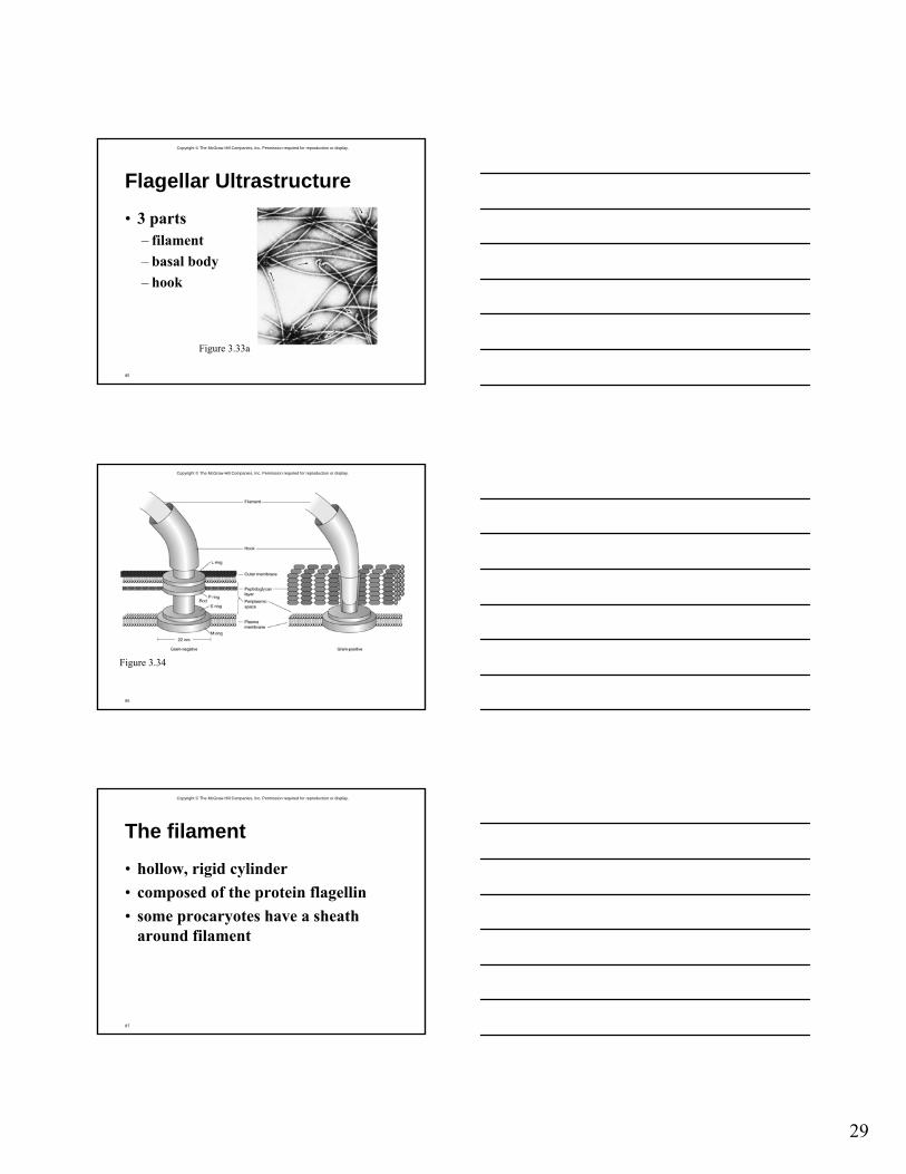
Flagellar Ultrastructure
• 3 parts– filament– basal body– hook
Figure 3.33a
Copyright © The McGraw-Hill Companies, Inc. Permission required for reproduction or display.
86
Figure 3.34
Copyright © The McGraw-Hill Companies, Inc. Permission required for reproduction or display.
87
The filament
• hollow, rigid cylinder• composed of the protein flagellin• some procaryotes have a sheath
around filament
30
Copyright © The McGraw-Hill Companies, Inc. Permission required for reproduction or display.
88
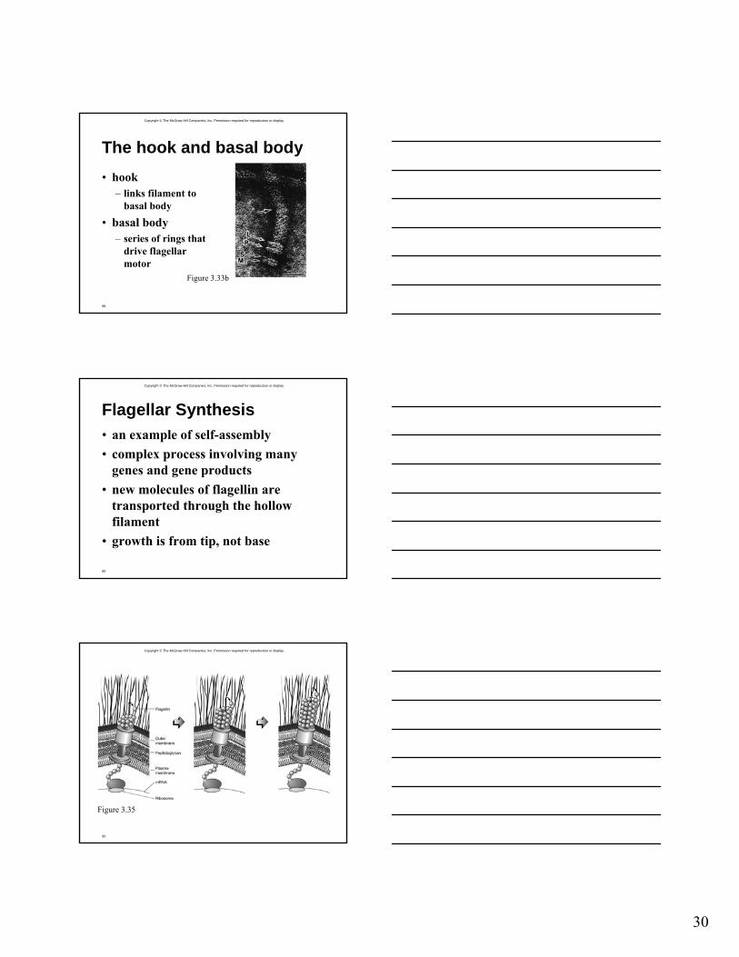
The hook and basal body• hook
– links filament to basal body
• basal body– series of rings that
drive flagellar motor
Figure 3.33b
Copyright © The McGraw-Hill Companies, Inc. Permission required for reproduction or display.
89
Flagellar Synthesis• an example of self-assembly• complex process involving many
genes and gene products• new molecules of flagellin are
transported through the hollow filament
• growth is from tip, not base
Copyright © The McGraw-Hill Companies, Inc. Permission required for reproduction or display.
90
Figure 3.35
31
Copyright © The McGraw-Hill Companies, Inc. Permission required for reproduction or display.
91
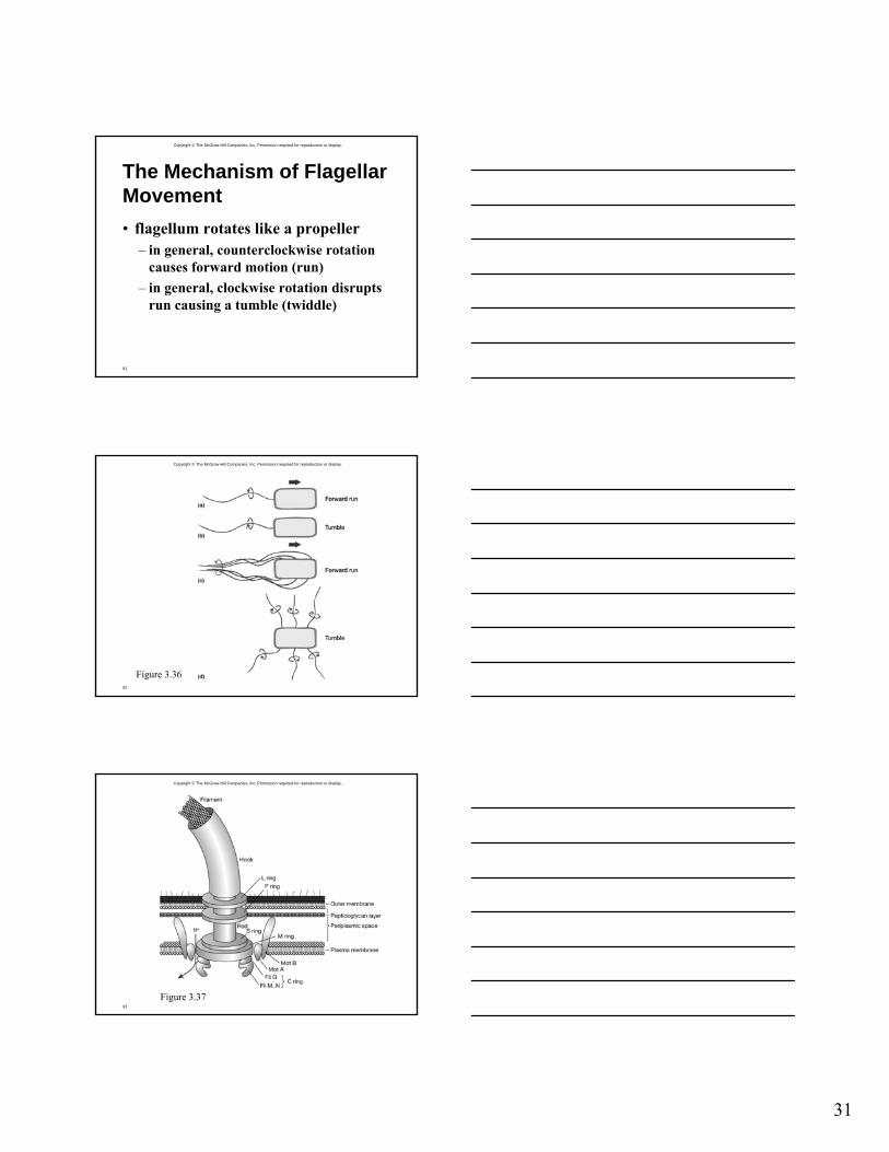
The Mechanism of Flagellar Movement• flagellum rotates like a propeller
– in general, counterclockwise rotation causes forward motion (run)
– in general, clockwise rotation disrupts run causing a tumble (twiddle)
Copyright © The McGraw-Hill Companies, Inc. Permission required for reproduction or display.
92
Figure 3.36
Copyright © The McGraw-Hill Companies, Inc. Permission required for reproduction or display.
93
Figure 3.37
32
Copyright © The McGraw-Hill Companies, Inc. Permission required for reproduction or display.
94
Other types of motility
• spirochetes– axial filaments cause flexing and
spinning movement• gliding motility
– cells coast along solid surfaces– no visible motility structure has been
identified
Copyright © The McGraw-Hill Companies, Inc. Permission required for reproduction or display.
95
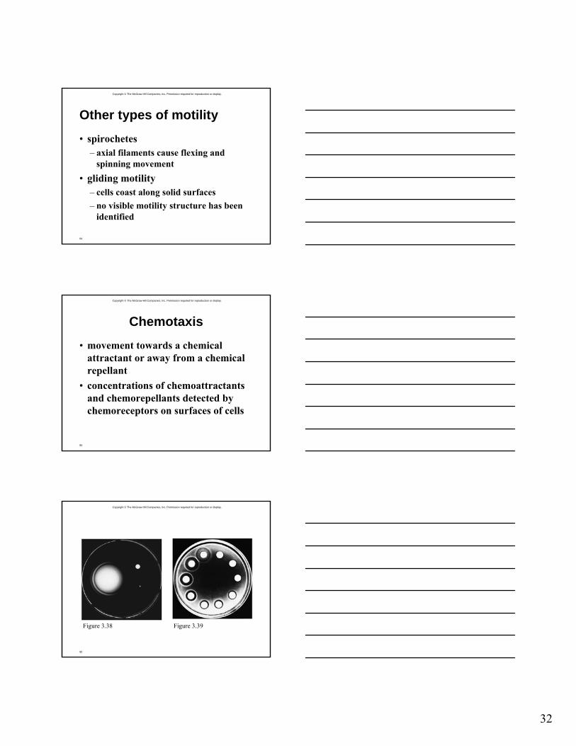
Chemotaxis
• movement towards a chemical attractant or away from a chemical repellant
• concentrations of chemoattractants and chemorepellants detected by chemoreceptors on surfaces of cells
Copyright © The McGraw-Hill Companies, Inc. Permission required for reproduction or display.
96
Figure 3.38 Figure 3.39
33
Copyright © The McGraw-Hill Companies, Inc. Permission required for reproduction or display.
97
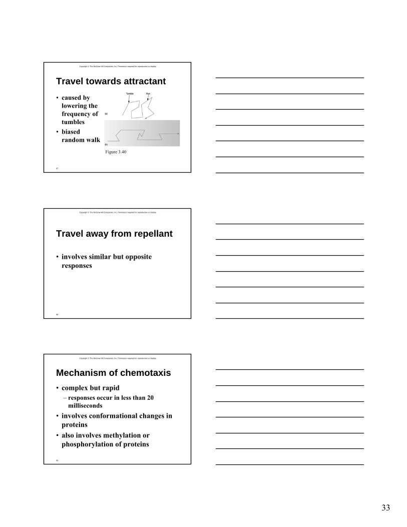
Travel towards attractant• caused by
lowering the frequency of tumbles
• biased random walk
Figure 3.40
Copyright © The McGraw-Hill Companies, Inc. Permission required for reproduction or display.
98
Travel away from repellant
• involves similar but opposite responses
Copyright © The McGraw-Hill Companies, Inc. Permission required for reproduction or display.
99
Mechanism of chemotaxis• complex but rapid
– responses occur in less than 20 milliseconds
• involves conformational changes in proteins
• also involves methylation or phosphorylation of proteins
34
Copyright © The McGraw-Hill Companies, Inc. Permission required for reproduction or display.
100
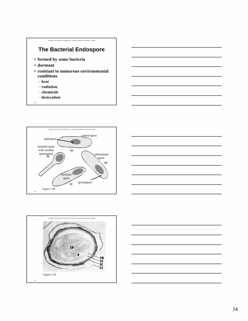
The Bacterial Endospore• formed by some bacteria• dormant• resistant to numerous environmental
conditions– heat– radiation– chemicals– desiccation
Copyright © The McGraw-Hill Companies, Inc. Permission required for reproduction or display.
101
Figure 3.40
endospore
sporangium
central spore
subterminalspore
terminalspore
terminal sporewith swollensporangium
Copyright © The McGraw-Hill Companies, Inc. Permission required for reproduction or display.
102
Figure 3.42
35
Copyright © The McGraw-Hill Companies, Inc. Permission required for reproduction or display.
103
What makes an endospore so resistant?• calcium (complexed with dipicolinic
acid)• acid-soluble, DNA-binding proteins• dehydrated core• spore coat• DNA repair enzymes
Copyright © The McGraw-Hill Companies, Inc. Permission required for reproduction or display.
104
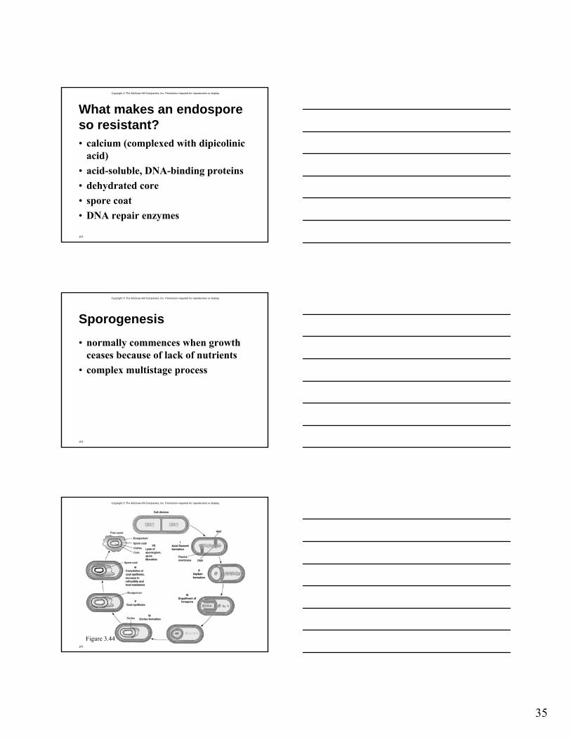
Sporogenesis
• normally commences when growth ceases because of lack of nutrients
• complex multistage process
Copyright © The McGraw-Hill Companies, Inc. Permission required for reproduction or display.
105
Figure 3.44
36
Copyright © The McGraw-Hill Companies, Inc. Permission required for reproduction or display.
106
Transformation of endospore into vegetative cell
• complex, multistage process
Figure 3.45
Copyright © The McGraw-Hill Companies, Inc. Permission required for reproduction or display.
107
Stages in transformation• activation
– prepares spores for germination– often results from treatments like heating
• germination– spore swelling– rupture of absorption of spore coat– loss of resistance– increased metabolic activity
• outgrowth– emergence of vegetative cell