chapter 4 & 5
TRANSCRIPT

Membrane proteins

Extrinsic (Peripheral)
• Bind weakly and reversibly
• Electrostatic interactions• Sediment out with
membrane and removed with change in pH
• Can be important in signaling, endocytosis, lipid rafts, interaction with cytoskeleton
Cytochrome C

Role of ankyrin in attaching the cytoskeleton

Phospholipase A2 binding to a lipid bilayer

Structure of annexins

Some peripheral proteins are Lipid-anchored proteins

0.5% of all eukaryotic proteins are linked with GPI

Different modes of binding amphoteric proteins to membranes
1) Electrostatic switch2) Lipid anchor3) Protein has a
binding pocket for a particular head group
4) Amphipathic helix in the interfacial region of the bilayer

Different effects of peripheral protein binding on lipid organization

Classification of Intrinsic (Integral) membrane proteins

A.A. in intrinsic proteins
1) Nonpolar amino acids side chains typically point towards the hydrophobic interior
2) Acidic and basic residues remain uncharged, form ion pairs that neutralize their charge or play a special role
3) Hydrogen bond often link side chain to carbonyl group to cap the end helices or stabilize between helices
4) Glycine and proline often “break” the helix
5) Aromatic groups often interface the hyrodrophilic and nonpolar domains
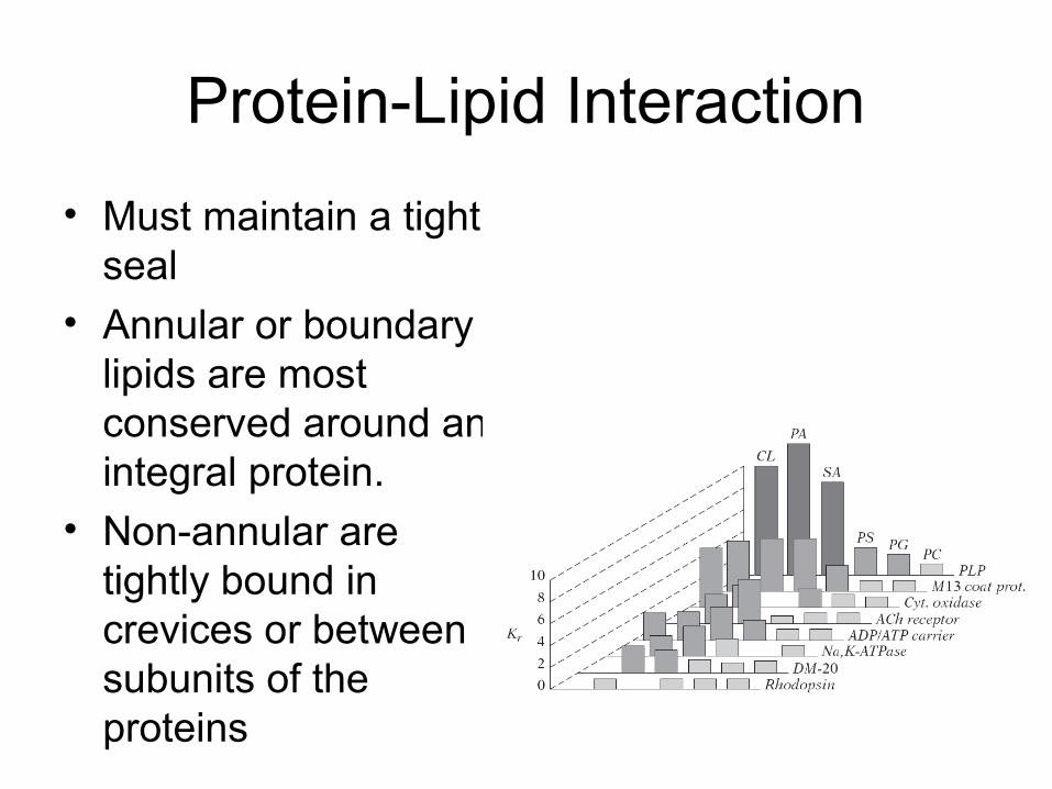
Protein-Lipid Interaction
• Must maintain a tight seal
• Annular or boundary lipids are most conserved around an integral protein.
• Non-annular are tightly bound in crevices or between subunits of the proteins

EPR detection of two populations of lipids: bulk and annular

Distortion of lipid bilayers due to hydrophobic mismatch
• Proteins with small number of helices are likely to tilt to accommodate thickness
• Larger proteins likely induce changes in thickness
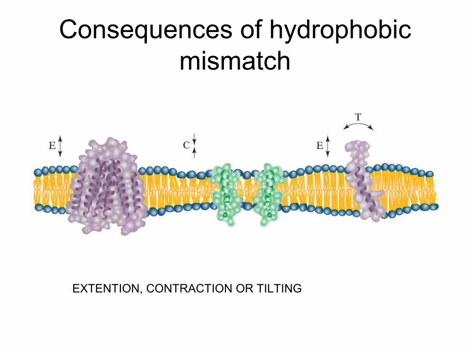
Consequences of hydrophobic mismatch
EXTENTION, CONTRACTION OR TILTING

Bundles and barrels
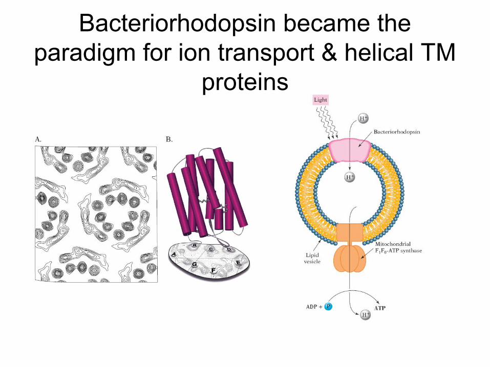
Bacteriorhodopsin became the paradigm for ion transport & helical TM
proteins

High resolution structure of BR

The structure of the photosynthetic reaction center of
B. viridis
- Membrane spanning surface is very hydrophobic
- TM consists of 5 helices from M, 5 from L and 1 from H
- Has 30-35 annular lipids

Organization of the light harvesting complexes

First x-ray structure of porin from Rhodobacter capsulatus (1991) confirmed
β-barrel structure.

Porins are found in the outer membrane of Gram negative bacteria (and mitochondria and chloroplasts).

An antiparallel β-sheet is formed with adjacent β-strands running in opposite directions. Every other side chain extends above or below the sheet.
H-bonds are quite perpendicular to the chains.

Beta strands
• Extended peptide backbone needs only seven a.a. residues to cross the nonpolar domain
• Typically have 9 – 11 residues tilted at 45 degrees
• Must partner with other strand
• Interstrand H bonds make structure rigid and stable
• Even numbers strands vary from 8 – 24
• amphipathic