chapter 40: head, face, and neck trauma
TRANSCRIPT

9/11/2012
1
1
Chapter 40
Head, Face, and Neck Trauma
2
Learning Objectives
• Describe the mechanisms of injury, assessment, and management of maxillofacial injuries.
• Describe the mechanisms of injury, assessment, and management of ear, eye, and dental injuries.
• Describe the mechanisms of injury, assessment, and management of anterior neck trauma.
3
Copyright © 2013 by Jones & Bartlett Learning, LLC, an Ascend Learning Company

9/11/2012
2
Learning Objectives
• Describe the mechanisms of injury, assessment, and management of injuries to the scalp, cranial vault, or cranial nerves.
• Distinguish between types of traumatic brain injury based on an understanding of pathophysiology and assessment findings.
4
Learning Objectives
• Outline the prehospital management of the patient with cerebral injury.
• Calculate a Glasgow Coma Scale, trauma score, Revised Trauma Score, and pediatric trauma score when given appropriate patient information.
5
Maxillofacial Injury
• In descending order of frequency, major causes of maxillofacial trauma are– Motor vehicle crashes
– Home injuries
– Athletic injuries
– Animal bites
– Intentional violent acts
– Industrial injuries
• Maxillofacial trauma may include soft tissue injuries and facial fractures
6
Copyright © 2013 by Jones & Bartlett Learning, LLC, an Ascend Learning Company
9/11/2012
3
Soft Tissue Injuries
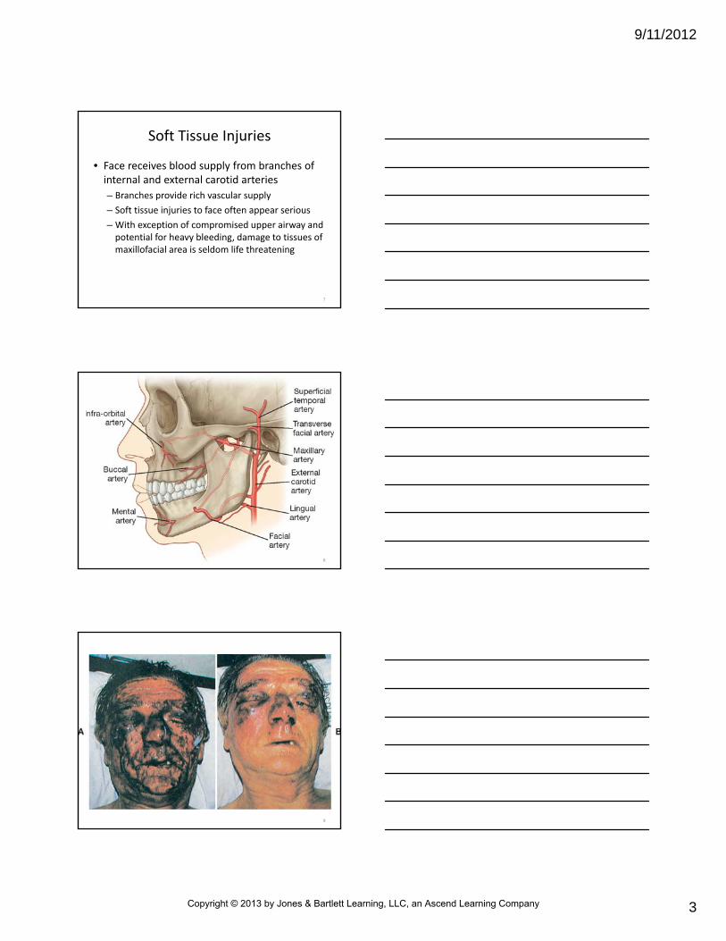
• Face receives blood supply from branches of internal and external carotid arteries
– Branches provide rich vascular supply
– Soft tissue injuries to face often appear serious
– With exception of compromised upper airway and potential for heavy bleeding, damage to tissues of maxillofacial area is seldom life threatening
7
8
9
Copyright © 2013 by Jones & Bartlett Learning, LLC, an Ascend Learning Company
9/11/2012
4
Soft Tissue Injuries
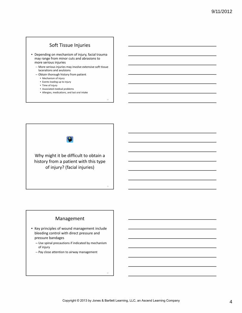
• Depending on mechanism of injury, facial trauma may range from minor cuts and abrasions to more serious injuries– More serious injuries may involve extensive soft tissue lacerations and avulsions
– Obtain thorough history from patient• Mechanism of injury
• Events leading up to injury
• Time of injury
• Associated medical problems
• Allergies, medications, and last oral intake
10
Why might it be difficult to obtain a history from a patient with this type
of injury? (facial injuries)
11
Management
• Key principles of wound management include bleeding control with direct pressure and pressure bandages
– Use spinal precautions if indicated by mechanism of injury
– Pay close attention to airway management
12
Copyright © 2013 by Jones & Bartlett Learning, LLC, an Ascend Learning Company

9/11/2012
5
Management
• Soft tissue injuries to nose and mouth are common with facial injuries
– Assess airway for obstruction caused by
• Blood
• Vomitus
• Bone fragments
• Broken teeth
• Dentures
• Damage to anterior neck
13
Management
• Soft tissue injuries to nose and mouth are common with facial injuries
– Suction may be needed to clear airway
– Oral or nasal adjuncts
– Tracheal intubation
– Cricothyrotomy
14
Facial Fractures
• Facial bones can withstand tremendous forces from impact of energy– Facial fractures are common after blunt trauma
• Anatomical structure of facial bones allows stepwise fracture to absorb impact of blunt trauma
– Blunt trauma injuries may be classified anatomically as fractures to
• Mandible• Midface• Zygoma• Orbit• Nose
15
Copyright © 2013 by Jones & Bartlett Learning, LLC, an Ascend Learning Company

9/11/2012
6
Facial Fractures
• Signs and symptoms of facial fractures
– Asymmetry of cheek bone prominences
– Crepitus
– Dental malocclusion
– Discontinuity of orbital rim
– Displacement of nasal septum
– Ecchymosis
– Lacerations and bleeding
16
Facial Fractures
• Signs and symptoms of facial fractures
– Limitation of forward movement of the mandible
– Limited ocular movements
– Numbness
– Pain
– Swelling
– Visual disturbances
17
Fractures of the Mandible
• Mandible
– Single facial bone in lower third of face
– Fractures rank second in frequency after nasal fractures
– Hemicircle of bone
• May break in multiple locations, often distant from point of impact
18
Copyright © 2013 by Jones & Bartlett Learning, LLC, an Ascend Learning Company
9/11/2012
7
Fractures of the Mandible
• Signs and symptoms– Dental malocclusion
• Patients may complain their teeth do not “feel right” when their mouths are closed
– Numbness in chin
– Inability to open mouth
– Difficulty swallowing
– Excessive salivation
• Most patients with mandibular fractures require hospitalization
19
Fractures of the Mandible
• Anterior dislocation of mandible in absence of fracture also may occur as a result of– Blunt trauma to face (rare)
– Abnormally wide yawn
– Dental treatment requiring that jaws remain open for long periods
• In these cases, condylar head advances forward beyond articular surface of temporal bone
• Jaw‐closing muscles spasm
• Mouth becomes locked in wide‐open position
20
Fractures of the Mandible
• Anterior dislocation of mandible
– Patient usually feels severe pain from spasm
– Experiences anxiety and discomfort that perpetuate spasm
– Reduced manually in emergency department with aid of muscle relaxant or sedative or in operating room with general anesthetic
21
Copyright © 2013 by Jones & Bartlett Learning, LLC, an Ascend Learning Company

9/11/2012
8
What will your patient care priority be with these patients? (fracture of
the mandible)
22
Fractures of the Midface
• Middle third of face includes– Maxilla
– Zygoma
– Floor of orbit
– Nose
• Fractures result from direct or transmitted force– Injuries often associated with CNS injury and spinal trauma
23
24
Copyright © 2013 by Jones & Bartlett Learning, LLC, an Ascend Learning Company
9/11/2012
9
Fractures of the Midface
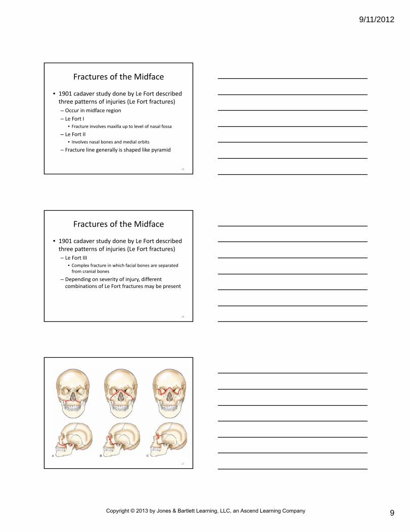
• 1901 cadaver study done by Le Fort described three patterns of injuries (Le Fort fractures)
– Occur in midface region
– Le Fort I
• Fracture involves maxilla up to level of nasal fossa
– Le Fort II
• Involves nasal bones and medial orbits
– Fracture line generally is shaped like pyramid
25
Fractures of the Midface
• 1901 cadaver study done by Le Fort described three patterns of injuries (Le Fort fractures)
– Le Fort III
• Complex fracture in which facial bones are separated from cranial bones
– Depending on severity of injury, different combinations of Le Fort fractures may be present
26
27
Copyright © 2013 by Jones & Bartlett Learning, LLC, an Ascend Learning Company

9/11/2012
10
Fractures of the Midface
• Signs and symptoms specific to midface fractures
– Midfacial edema
– Unstable maxilla
– Lengthening of face (donkey face)
– Epistaxis
– Numb upper teeth
– Nasal flattening
– Cerebrospinal fluid rhinorrhea (cerebrospinal fluid leakage caused by ethmoid cribriform plate fracture)
28
Fractures of the Midface
• Patients with midface fractures require hospitalization
– Risk of having serious airway problems related to swelling and bleeding
– Because of extent of fractures, risk exists of placing nasogastric or even nasotracheal tubes into brain tissue
29
Fractures of the Zygoma
• Zygoma (malar eminence) articulates with frontal, maxillary, and temporal bones
– Commonly called the cheek bone
– Rarely gets fractured because of its sturdy construction
– When fractures occur, usually result of physical assaults and vehicle crashes
– Zygomatic fractures often associated with orbital fractures and manifest similar clinical signs distinguished by x‐ray exam
30
Copyright © 2013 by Jones & Bartlett Learning, LLC, an Ascend Learning Company

9/11/2012
11
Fractures of the Zygoma
• Signs and symptoms specific to zygomaticfractures
– Flatness of usually rounded cheek area
– Numbness of cheek, nose, and upper lip (particularly if orbital fracture involved)
– Epistaxis
– Altered vision
31
32
Fractures of the Orbit
• Orbital contents are protected by bony ring
– Ring resembles pyramid, with apex pointed toward back of head
– Bones of walls, floor, roof of orbit are thin and fractured easily by direct blows and transmitted forces
– Many orbital fractures associated with other facial injuries, such as Le Fort II and III fractures
33
Copyright © 2013 by Jones & Bartlett Learning, LLC, an Ascend Learning Company

9/11/2012
12
Fractures of the Orbit
• Blowout fracture to orbit can occur when object of greater diameter than that of bony orbital rim strikes globe of eye and surrounding soft tissue
– Impact pushes globe into orbit and in turn compresses orbital contents
– Sudden increase in intraocular pressure is transmitted to orbital floor
– Orbital floor is weakest part of orbital structure
34
35
Fractures of the Orbit
• If orbital floor fractures, orbital contents may be forced into maxillary sinus
– Soft tissue and extraocular muscles may be trapped in defect
36
Copyright © 2013 by Jones & Bartlett Learning, LLC, an Ascend Learning Company

9/11/2012
13
Fractures of the Orbit
• Signs and symptoms of blowout fractures– Periorbital edema
– Subconjunctival ecchymosis
– Diplopia (double vision)
– Enophthalmos (recessed globe)
– Epistaxis
– Anesthesia in region of infraorbital nerve (anterior cheek)
– Impaired extraocular movements
37
How do you assess a patient’s eye movement?
38
Fractures of the Orbit
• Orbital fractures often associated with other fractures
– Le Fort II and III injuries
– Those of zygomatic complex
– Injury to orbital contents is common
• Suspect such injury with any facial fracture
39
Copyright © 2013 by Jones & Bartlett Learning, LLC, an Ascend Learning Company

9/11/2012
14
Fractures of the Nose
• Of all facial bones, nasal bones have least structural strength
– Fractured most frequently
– External portion of nose, formed mostly of hyaline cartilage, supported mainly by nasal bones and frontal processes of maxillary bones
40
Fractures of the Nose
• Injuries to nose may– Depress dorsum of nose
– Displace it to one side
– Result only in epistaxis and swelling without apparent skeletal deformity
• Fractures to orbit also may be present
• In children, minimal displacement of nasal bones can result in growth changes and ultimate deformity
41
Facial Fracture Management
• When caring for patient with facial fractures
– Assume spine has been injured
– Use spinal precautions
• Facial fractures associated with high percentage of related cervical spine fractures
42
Copyright © 2013 by Jones & Bartlett Learning, LLC, an Ascend Learning Company

9/11/2012
15
Facial Fracture Management
• Treatment
– Assess patient’s airway for obstruction caused by
• Blood
• Vomitus
• Bone fragments
• Broken teeth
• Dentures
• Damage to anterior neck
43
Facial Fracture Management
• Treatment
– Suction may be needed to clear airway of debris and fluid
– May need to maintain airway with
• Nasal (in absence of suspected midface or basal skull fracture) or oral adjunct
• Tracheal intubation
• Cricothyrotomy
44
Facial Fracture Management
• Bleeding usually can be controlled by direct pressure and pressure bandages
– Epistaxis may be severe and should be controlled by applying external pressure to anterior nares
– Mild epistaxis
• To prevent blood from draining down throat, instruct patient to sit upright or to lean forward (in absence of spinal injury) while compressing nares
• Unconscious patient should be positioned on side (if not contraindicated by injury)
• If bleeding is severe, evaluate patient for hemorrhagic shock
45
Copyright © 2013 by Jones & Bartlett Learning, LLC, an Ascend Learning Company

9/11/2012
16
Why would you not want the blood to drain posteriorly?
46
Ear, Eye, and Dental Trauma
• Ears, eyes, or teeth may be injured separately or along with other forms of head and facial trauma
– Injury to these regions may be minor
– May result in permanent sensory function loss and disfigurement
– Regardless of severity, evaluate ear, eye, and dental trauma and treat only after identifying and managing life‐threatening problems
47
Ear Trauma
• Trauma to ear may include
– Lacerations and contusions
– Thermal injuries
– Chemical injuries
– Traumatic perforations
– Barotitis
48
Copyright © 2013 by Jones & Bartlett Learning, LLC, an Ascend Learning Company

9/11/2012
17
Lacerations and Contusions
• Usually result from blunt trauma– Common in victims of domestic violence
– Treated by direct pressure to control bleeding
– Application of ice or cold compresses decreases soft tissue swelling
– If portion of outer ear (pinna) has been avulsed, retrieve avulsed tissue if possible
• Wrap in moist gauze
• Seal in plastic
• Place on ice
• Transport with patient for surgical repair
– Cartilage tears often heal poorly and are easily infected
49
50
Thermal Injuries
• May occur from
– Prolonged exposure to extreme cold
– Exposure of lesser duration to extreme heat
– Contact with hot liquids or electrical currents
• Prehospital treatment usually limited to
– Dressings to prevent contamination
– Transportation for evaluation by physician
51
Copyright © 2013 by Jones & Bartlett Learning, LLC, an Ascend Learning Company

9/11/2012
18
Chemical Injuries
• Strong acids or alkalis produce burns on contact– Emergency care consists of copious irrigation
– After irrigation, bathe ear and ear canal with saline or sterile water
• Allow irrigation liquid to remain in ear canal for 2 to 3 minutes
• Repeat 3 to 4 times
• Dry and cover ear to prevent contamination
• Transport
52
Traumatic Perforations
• Traumatic perforation can occur by objects such as a cotton‐tipped applicators and by changes in pressure
– Pressure injuries may result from explosions (blast injuries) or scuba diving (barotrauma)
– Usually heal spontaneously without treatment
– Physician evaluation advised
53
Traumatic Perforations
• If injury is caused by a penetrating object, stabilize object in place and cover ear to prevent further contamination– Inner or middle ear canal may have been contaminated
• Antibiotic therapy usually is prescribed
– Serious complications• Facial nerve palsy
• Temporal bone fractures
• Hearing loss
• Vertigo
54
Copyright © 2013 by Jones & Bartlett Learning, LLC, an Ascend Learning Company

9/11/2012
19
Barotitis
• Occurs when person is exposed to changes in barometric pressure great enough to produce inflammation and injury to middle ear
– Flying at high altitudes
– Scuba diving
55
Barotitis
• Gas pressure in air‐filled spaces of middle ear normally equals that of environment
– Boyle’s law
• At constant temperature, volume of gas is inversely proportional to pressure
• On ascent, gas expands
• On descent, gas contracts
• When gases become trapped or partially trapped, expand in direct proportion to decrease in pressure
56
Barotitis
• When trapped gas cannot reach equilibrium with environmental pressure, pain and sensation of blocked ear may develop– To equalize pressure in middle ear, patient can be directed to
• Bear down (Valsalva’s maneuver)
• Yawn
• Swallow
• Move lower jaw
– These methods may cause Eustachian tube to open• Will equalize pressure in middle ear cavity
57
Copyright © 2013 by Jones & Bartlett Learning, LLC, an Ascend Learning Company

9/11/2012
20
Eye Trauma
• 2,000 or more eye and orbital injuries are estimated to occur each day in United States
– Common causes are blunt and penetrating trauma from
• Motor vehicle crashes
• Sport and recreational activities
• Violent altercations
• Chemical exposure from household and industrial accidents
• Foreign bodies
• Animal bites and scratches
58
Evaluation
• Acute eye injuries may be difficult to identify
– Patient with normal vision may have serious underlying injury
59
Evaluation
• Symptoms requiring high degree of suspicion– Obvious trauma with eye injury
– Visual loss or blurred vision that does not improve with blinking
• Indicates possible damage to globe, ocular contents, or optic nerve
– Loss of portion of visual field• Possible detachment of retina
• Hemorrhage into eye
• Optic nerve injury
60
Copyright © 2013 by Jones & Bartlett Learning, LLC, an Ascend Learning Company

9/11/2012
21
Evaluation
• Evaluation of eye injury
– Thorough history
– Measurement of visual acuity, pupillary reaction, and extraocular movements
– Assessing patient’s vision will be rough estimation at best
• Will be reevaluated in emergency department under controlled circumstances
61
Aside from trauma, what are some other causes of visual disturbances?
62
History
• Thorough history should include
– Exact mode of injury
– Previous ocular, medical, and drug history
• Cataracts
• Glaucoma
• Hepatitis
• HIV
63
Copyright © 2013 by Jones & Bartlett Learning, LLC, an Ascend Learning Company

9/11/2012
22
History
• Thorough history should include
– Use of eye medications
– Use of corrective glasses or contact lenses
– Presence of ocular prostheses
– Duration of symptoms and treatment interventions attempted before EMS arrival
64
Visual Acuity
• Measurement of visual acuity is usually first step in any examination of patient’s eyes
– Exception is chemical burn to eye
• Irrigation should come before measurement of visual acuity
– Visual acuity can be measured with handheld visual acuity chart (e.g., Snellen chart), or any printed material with small, medium, and large point sizes
– Record distance that printed item was held from patient’s face
65
Visual Acuity
• Vision of each eye should be assessed separately while covering other eye
– No pressure should be applied
– Test injured eye first for acuity comparison to uninjured eye
– If patient wears corrective lenses, measure with lenses first and then without lenses
66
Copyright © 2013 by Jones & Bartlett Learning, LLC, an Ascend Learning Company

9/11/2012
23
Visual Acuity
• Vision of each eye should be assessed separately while covering other eye
– Illiterate or non‐English‐speaking patients require alternative method of evaluation
• Finger counting
• Hand motion
• Presence/absence of light perception
– Abnormal responses to any of these methods indicate significant loss of vision
67
The assessment of visual acuity may be difficult on some calls. What factors in the prehospital setting
may make it difficult?
68
Pupillary Reaction
• Pupils should be black, round, and equal in size– Should react to light in same way and at same time
– Both eyes should constrict in response to light and dilate in response to dark
• Direct response to light refers to constriction of illuminated pupil
• Consensual response to light refers to constriction of opposite pupil
69
Copyright © 2013 by Jones & Bartlett Learning, LLC, an Ascend Learning Company

9/11/2012
24
Pupillary Reaction
• Abnormal pupillary responses after blunt trauma to eye are common and may be caused by
– Tearing
– Direct trauma to pupillary sphincter muscle
• May suggest more serious injury involving optic nerve or globe
70
Pupillary Reaction
• Causes of pupil abnormalities in absence of recent injury
– Drug use
– Cataracts
– Previous surgical procedures
– Ocular prosthesis
– Anisocoria (normal or congenital unequal pupil size)
– CNS disease
– Strokes
– Previous injury
• Document all pupil abnormalities
71
Extraocular Movements
• Extraocular muscles are responsible for movements of globe, or eyeball– Voluntary muscles are innervated by cranial nerves III, IV, and VI
• Attached to outside of eyeball and bones of orbit and move globe in any desired direction
– Involuntary eye muscles are innervated by sympathetic nerves
• Located within eye iris and ciliary muscle
• Muscles dilate and constrict pupil and change shape of lens, respectively
72
Copyright © 2013 by Jones & Bartlett Learning, LLC, an Ascend Learning Company
9/11/2012
25
Extraocular Movements
• To evaluate extraocular movement of eyes
– Instruct patient to visually track movement of object
• Ask to track object up, down, right, left
– Abnormalities in movement may indicate
• Orbital content edema
• Cranial nerve injury
• Contusions or lacerations of extraocular muscles
• Muscle entrapment in fracture
73
Extraocular Movements
• To evaluate extraocular movement of eyes
– Patients with limited or abnormal extraocular movements often complain of double vision in one or more directions of gaze
– Document all findings
74
• Few eye injuries are truly urgent– All victims of ocular trauma should be evaluated by physician
– Some patients need specialized care by an ophthalmologist
– If paramedic suspects serious injury that may call for specialized care, medical direction should be advised as soon as possible
• Services will be ready when patient arrives in emergency department
Eye Injury Evaluation and Management
75
Copyright © 2013 by Jones & Bartlett Learning, LLC, an Ascend Learning Company
9/11/2012
26
76
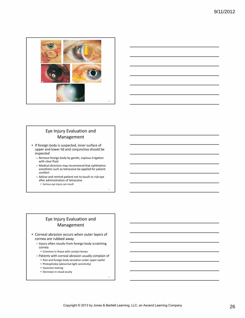
• If foreign body is suspected, inner surface of upper and lower lid and conjunctiva should be inspected– Remove foreign body by gentle, copious irrigation with clear fluid
– Medical direction may recommend that ophthalmic anesthetic such as tetracaine be applied for patient comfort
– Advise and remind patient not to touch or rub eye after administration of tetracaine
• Serious eye injury can result
Eye Injury Evaluation and Management
77
• Corneal abrasion occurs when outer layers of cornea are rubbed away– Injury often results from foreign body scratching cornea
• Common in those with contact lenses
– Patients with corneal abrasion usually complain of• Pain and foreign body sensation under upper eyelid
• Photophobia (abnormal light sensitivity)
• Excessive tearing
• Decrease in visual acuity
Eye Injury Evaluation and Management
78
Copyright © 2013 by Jones & Bartlett Learning, LLC, an Ascend Learning Company

9/11/2012
27
• Often signs and symptoms are delayed
• Prehospital management of corneal abrasion
– Gentle irrigation with clear fluid
– Application of double patch to eyes to prevent injured eye from moving when uninjured eye moves, causing aggravation
– Generally heal within 24 to 48 hours
Eye Injury Evaluation and Management
79
80
81
Copyright © 2013 by Jones & Bartlett Learning, LLC, an Ascend Learning Company

9/11/2012
28
82
Will the patient with a suspected corneal abrasion need to be evaluated by a physician?
83
• Blunt trauma to eye or its adjacent structures may result in
– Contusion injury
– Traumatic hyphema (bleeding into anterior chamber)
– Globe or scleral rupture
Eye Injury Evaluation and Management
84
Copyright © 2013 by Jones & Bartlett Learning, LLC, an Ascend Learning Company

9/11/2012
29
• Blunt injury to eye may be associated with other serious injuries
– Orbital fracture
– Vitreous hemorrhage
– Dislocation of lens
• Prehospital care
– Limited to control of any bleeding with gentle, direct pressure
– Protection of eye with metal shield or cardboard cup
– Rapid transport
Eye Injury Evaluation and Management
85
• If paramedic suspects traumatic hyphema or globe or scleral rupture
– Place patient on spine board and elevate head of spine board 40 degrees to decrease intraocular pressure
– Instruct patient to avoid any activity that might increase intraocular pressure
– Analgesics and antiemetics may be indicated for pain relief and nausea
• Can reduce movement, straining, coughing, retching that may increase intraocular pressure
Eye Injury Evaluation and Management
86
Pupillary Reaction
• Abnormal pupillary responses after blunt trauma to eye are common and may be caused by
– Tearing
– Direct trauma to pupillary sphincter muscle
– May suggest more serious injury involving optic nerve or globe
87
Copyright © 2013 by Jones & Bartlett Learning, LLC, an Ascend Learning Company

9/11/2012
30
• Treatment
– Control any bleeding by gentle, direct pressure
– Protect globe from dehydration or contamination from foreign material
• Cover orbital area with plastic or damp, sterile dressings and eye shield
Eye Injury Evaluation and Management
88
• Stabilize foreign bodies protruding from eye
– Cover these with cardboard cup and secure cup with tape
– Unaffected eye should also be covered to prevent consensual movement
– Do not attempt to remove object
– If needed, penetrating object may be shortened for transport (after consulting with medical direction)
– Oxygen and IV fluids also may be recommended in these cases
Eye Injury Evaluation and Management
89
• Chemical injury to eye associated with– Loss of corneal epithelial tissue
– Globe perforation
– Scarring and deformation of eyelids and conjunctiva
– True emergencies• Require immediate intervention
• Mandate extensive, continuous irrigation of both eyes with neutral fluid for 20 minutes before and during transport
Eye Injury Evaluation and Management
90
Copyright © 2013 by Jones & Bartlett Learning, LLC, an Ascend Learning Company

9/11/2012
31
Should you wait until medical direction is contacted before you
begin irrigation of the eye?
91
Contact Lenses
• Contact lenses are of three general types: hard, soft hydrophilic, rigid gas‐permeable– Hard lenses
• Microlenses sometimes prescribed for astigmatism
• Rarely used today
– Soft (hydrophilic) lenses • Usually large in diameter (extending onto conjunctiva)
• May be designed for daily or extended wear
– Rigid gas‐permeable lenses• Similar in size to microlenses
• Have low water content and high oxygen permeability
92
Contact Lenses
• Do not attempt to remove contact lenses in patients with eye injuries
– May cause more damage and may aggravate injury
– If complicated by presence of contact lenses (e.g., chemical burns to the eyes), medical direction may recommend that lenses be removed
• If patient is unable to remove lenses, paramedic may be instructed to do so
93
Copyright © 2013 by Jones & Bartlett Learning, LLC, an Ascend Learning Company

9/11/2012
32
Dental Trauma
• Adult normally has 32 teeth– Each tooth consists of two sections
• Crown: projects above gingiva (portion of oral mucosa surrounding tooth),
• Root: fits into bony socket (alveolus) of maxilla or mandible
– Three layers make up hard tissues of teeth • Enamel• Dentin (ivory)• Cementum
– Soft tissues of teeth• Pulp• Periodontal membrane
94
95
Dental Trauma
• Teeth and associated alveolar process may be injured alone or along with fractures of jaw or facial bones
– Two most common types of dental trauma involve fractures and avulsions of the anterior teeth
96
Copyright © 2013 by Jones & Bartlett Learning, LLC, an Ascend Learning Company

9/11/2012
33
Dental Trauma
• If tooth is fractured, examine oral cavity carefully for tooth fragments– Removal of fragments reduces risk of aspiration and obstruction of airway
– Lacerations and avulsions to tongue and surrounding mucous membranes often occur with dental trauma
• Painful
• May bleed profusely
• May compromise patient’s airway
97
Dental Trauma
• Tooth avulsions are common
– Many teeth can be saved with proper emergency treatment
– Permanent teeth that have been avulsed have good survival rate if reimplanted and stabilized within 1 hour
98
Dental Trauma
• If avulsed tooth has been out of patient’s mouth less than 15 minutes, medical direction may recommend reimplanting tooth into original socket
– Take care not to reimplant tooth backward
– Be alert for possible aspiration
99
Copyright © 2013 by Jones & Bartlett Learning, LLC, an Ascend Learning Company
9/11/2012
34
Dental Trauma
• If reimplantation is impossible, follow guidelines established by American Dental Association and the American Association of Endodontists
– Never place avulsed tooth in anything that can dry or crush outside of tooth
– Do not handle tooth roughly
• Do not rinse it off or rub, scrape, or disinfect outside of tooth in any way
• Any adherent membrane or fibrous tissue should be left in place to avoid stripping off periodontal membrane and ligament, which are critical to survival of reimplanted tooth
100
Dental Trauma
• Guidelines (cont.)– Place tooth in a nurturing, break‐resistant storage device (e.g., Emergency Tooth Preserving System)
• Device should have a tightly fitted top and soft inner walls
– Store tooth in a pH‐balanced, isotonic, glucose‐, calcium‐, and magnesium‐enriched cell‐preserving fluid (e.g., Hank’s solution)
• Use refrigerated fresh whole milk as the best alternative storage medium (powdered milk is not suitable)
• For short periods (1 hour or less), use sterile saline
• Do not use tap water because it damages the periodontal ligament
101
Dental Trauma
• Guidelines (cont.)
– Advise medical direction of avulsed teeth so that appropriate services will be available when the patient arrives in the emergency department
102
Copyright © 2013 by Jones & Bartlett Learning, LLC, an Ascend Learning Company

9/11/2012
35
Anterior Neck Trauma
• Anterior neck injuries are caused by blunt and penetrating trauma
– May result in damage to
• Skeletal structures
• Vascular structures
• Nerves
• Muscles
• Glands of neck
103
104
Anterior Neck Trauma
• Common mechanisms of injury to anterior neck– Strangulation injuries from clothing, jewelry, or personal equipment getting caught in machinery
– All‐terrain vehicles and other small motor vehicles• Clothesline injuries to neck from running into wires, ropes, or fences
– Blows to neck
– Contact sports
– Hangings
– Horseback riding
– Hyperextension and hyperflexion injuries
105
Copyright © 2013 by Jones & Bartlett Learning, LLC, an Ascend Learning Company

9/11/2012
36
Anterior Neck Trauma
• Common mechanisms of injury to anterior neck
– Industrial injuries
– Missile injury from firearms
– Motor vehicle crashes
– Neck striking dashboard or steering column
– Snow skiing
– Sport and recreational activities
– Stab wounds
– Violent altercations
– Water sports
106
Anterior Neck Trauma
• With blunt and penetrating neck injuries, assume patient has cervical spine injury also
– Assume such injury until ruled out by clinical examination and x‐ray films (radiography) of cervical region of neck
– X‐ray examination alone does not rule out cervical spine injury
107
Evaluation
• Neck can be divided into three zones defined by horizontal planes
– Zone I represents base of neck
• Extends from sternal notch to top of clavicles or cricoid cartilage
• Injuries to this zone have highest mortality rate because of risk of injury to major vascular and thoracic structures
108
Copyright © 2013 by Jones & Bartlett Learning, LLC, an Ascend Learning Company

9/11/2012
37
109
Evaluation
• Neck zones– Zone II extends from the clavicles or cricoid cartilage cephalad to angle of mandible with vital structures
• Carotid artery
• Jugular vein
• Trachea
• Larynx
• Esophagus
• Cervical spine
– Injuries to this zone are most common
– Lower mortality rate than zone I injuries
110
Evaluation
• Neck zones
– Zone III is part of neck above angle of mandible, with risk of injury to
• Distal carotid artery
• Salivary glands
• Pharynx
111
Copyright © 2013 by Jones & Bartlett Learning, LLC, an Ascend Learning Company

9/11/2012
38
Soft Tissue Injuries
• Soft tissue injuries to neck from blunt trauma often produce
– Hematomata
– Associated edema
– Direct laryngeal or tracheal injury
– Both can result in airway compromise
– May produce lacerations and puncture wounds with resultant vascular, laryngotracheal, or esophageal injury
– Blunt trauma may cause vascular injuries as well(uncommon)
112
Soft Tissue Injuries
• Treatment
– Begin with rapid assessment
– Control of airway
– Spinal injury consideration
113
Is prehospital airway control always possible in patients with anterior
neck injuries?
114
Copyright © 2013 by Jones & Bartlett Learning, LLC, an Ascend Learning Company
9/11/2012
39
Hematomata and Edema
• Edema may produce enough pressure in neck tissues to obstruct airway completely, especially in
– Pharynx
– Larynx
– Trachea
– Epiglottis
– Vocal cords
115
Hematomata and Edema
• If airway is compromised, consider oral or nasal intubation with spinal precautions
– Stabilizes damaged areas of neck
– Protects airway
– Provides means for ventilatory support
• Slightly smaller endotracheal tube may be needed to ensure passage through airway
116
Hematomata and Edema
• When direct intubation is impossible, cricothyrotomy or translaryngeal canulaventilation may be indicated
– Cool, humidified oxygen helps treat edematous airways
– Slight elevation of patient’s head (if not contraindicated by injury)
117
Copyright © 2013 by Jones & Bartlett Learning, LLC, an Ascend Learning Company

9/11/2012
40
Lacerations and Puncture Wounds
• Lacerations and puncture wounds may be superficial or deep– Superficial
• Usually can be managed by covering wound
• Covering helps to prevent further contamination
– Deep • Associated with more serious injuries to underlying structures
• May require aggressive airway therapy and ventilatorysupport, suction, hemorrhage control by direct pressure, and fluid replacement
118
Lacerations and Puncture Wounds
• Signs and symptoms of significant penetrating neck trauma– Active bleeding
– Dysphagia
– Dyspnea
– Hematemesis
– Hemoptysis
– Hoarseness
– Large or expanding hematoma
– Mobility and crepitus
– Neurological deficit (stroke, brachial plexus injury, spinal cord injury)
– Pulse deficit
– Shock
– Stridor
– Subcutaneous emphysema
– Tenderness to palpation
119
Why is rapid transport crucial when caring for a patient who has anterior
neck injuries?
120
Copyright © 2013 by Jones & Bartlett Learning, LLC, an Ascend Learning Company

9/11/2012
41
Vascular Injury
• Blood vessels are most commonly injured structures in neck– May be injured by blunt or penetrating trauma
– Vessels at risk of injury• Carotid
• Vertebral
• Subclavian
• Innominate
• Internal mammary arteries
• Jugular and subclavian veins
121
Vascular Injury
• First priority
– Secure airway (with spinal precautions)
– Provide adequate ventilatory support
122
Vascular Injury
• Second priority
– Control hemorrhage
• Use constant, direct pressure
• Apply pressure only to affected vessels
• Blood flow to the brain will not be obstructed completely
• If bleeding cannot be controlled in this manner, medical direction may advise applying direct pressure with gloved finger to vessel
123
Copyright © 2013 by Jones & Bartlett Learning, LLC, an Ascend Learning Company

9/11/2012
42
Vascular Injury
• If paramedic suspects venous injury, patient should be kept supine or in slight Trendelenburgposition– Will help to prevent air embolism (rare but lethal complication)
• If paramedic suspects air embolism, turn immobilized patient on left side– Head should be lower than feet in attempt to trap air embolus in right ventricle
– Venous neck wounds should be dressed with occlusive dressing
124
Vascular Injury
• Fluid replacement for hypovolemia should be guided by medical direction– May include using large‐bore catheters and isotonic crystalloid (lactated Ringer’s solution or normal saline)
– If penetrating injury to base of neck (zone I) has occurred, upper extremity venous drainage may be compromised by laceration
• Placement of at least one IV line in lower extremity should be considered
• Medical direction may advise that second IV line be placed in upper extremity on side opposite injury
125
Why might application of a pneumatic antishock garment be harmful with this type of injury?
126
Copyright © 2013 by Jones & Bartlett Learning, LLC, an Ascend Learning Company

9/11/2012
43
Laryngeal or Tracheal Injury
• Injury from blunt or penetrating trauma to anterior neck may cause
– Fracture or dislocation of laryngeal and tracheal cartilages
– Hemorrhage
– Swelling of air passages
127
Laryngeal or Tracheal Injury
• All these injuries can compromise airway and cause respiratory distress– Airway injury can lead to death in head and neck trauma patients
– Rapid and judicious control of airway and prevention of aspiration are crucial
– High degree of suspicion for associated vascular disruption and esophageal, chest, and intra‐abdominal injury is critical aspect of preventing death
128
Laryngeal or Tracheal Injury
• Injuries that may be associated with laryngeal and tracheal injuries
– Fracture of hyoid bone
• Resulting in laceration and distortion of epiglottis
– Separation of hyoid and thyroid cartilages
• Resulting in epiglottis dislocation, aspiration, subcutaneous emphysema
– Fractures of thyroid cartilage
• Resulting in epiglottis and vocal cord avulsion, arytenoiddislocation, and aspiration of blood and bone fragments
129
Copyright © 2013 by Jones & Bartlett Learning, LLC, an Ascend Learning Company
9/11/2012
44
Laryngeal or Tracheal Injury
• Injuries that may be associated with laryngeal and tracheal
– Dislocation or fracture of cricothyroid
• Resulting in long‐term laryngeal stenosis, laryngeal nerve paralysis, and laryngotracheal avulsion
– Fracture to trachea
• Resulting in tracheal avulsion, complete airway obstruction, and subcutaneous emphysema
130
Laryngeal or Tracheal Injury
• Management is controversial– Some medical direction agencies recommend oral or nasal intubation
– Other agencies hold that intubation attempts may contribute to potential for injury resulting from lack of oxygen during procedure
• Attempts also may damage airway structures further
– Alternative methods of airway management• Use of bag‐mask ventilation
• Cricothyrotomy
• Translaryngeal cannula ventilation
131
Laryngeal or Tracheal Injury
• If penetrating trauma causes complete disruption of laryngotracheal structure, medical direction may recommend dissection through wound– Exposed distal trachea can be cannulated directly with cuffed endotracheal tube
– Regardless of method chosen, emergency care is directed at
• Securing airway with spinal precautions• Providing adequate ventilatory support• Controlling hemorrhage• Treating for shock• Rapid transport for definitive surgical care
132
Copyright © 2013 by Jones & Bartlett Learning, LLC, an Ascend Learning Company

9/11/2012
45
Esophageal Injury
• Esophageal injuries should be suspected in patients with trauma to neck or chest– High degree of suspicion for associated esophageal injury
• Tracheal fractures
• Penetrating trauma from stab or gunshot wounds
• Ingestion of caustic substances
– Difficult to diagnose• May be overlooked as paramedic focuses on more obvious injuries that pose threat to life
133
Esophageal Injury
• Esophageal injuries should be suspected in patients with trauma to neck or chest
– Signs and symptoms
• Subcutaneous emphysema
• Neck hematoma
• Bleeding from mouth and nose
134
Esophageal Injury
• Esophageal perforation
– High mortality rate
– Death results from mediastinitis caused by release of gastric contents into thoracic cavity
– If not contraindicated by mechanism of injury, place patient with suspected esophageal tear in semi‐Fowler position
• Helps to prevent reflux of gastric contents
135
Copyright © 2013 by Jones & Bartlett Learning, LLC, an Ascend Learning Company

9/11/2012
46
Are these signs and symptoms (for esophageal injuries) so unique that you will be able to distinguish esophageal
injury as the cause versus other kinds of traumatic conditions?
136
Head Trauma
• Anatomical components of skull
– Scalp
– Cranial vault
• Dural membrane
• Arachnoid membrane
• Pia
• Brain substance
– Injuries to skull may be classified as soft tissue injuries to scalp and skull fractures
137
Soft Tissue Injuries to the Scalp
• Most common scalp injury is irregular linear laceration– Scalp is very vascular
• Lacerations may bleed heavily
• May result in hypovolemia, particularly in infants and children
– Less frequent scalp injuries• Stellate wounds (ballistic wounds that are star shaped)
• Avulsions
• Subgaleal hematomata (bleeding in potential space between skull and scalp)
138
Copyright © 2013 by Jones & Bartlett Learning, LLC, an Ascend Learning Company
9/11/2012
47
139
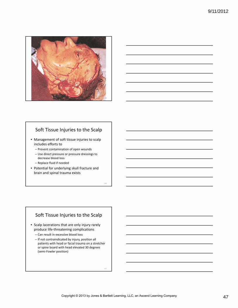
Soft Tissue Injuries to the Scalp
• Management of soft tissue injuries to scalp includes efforts to
– Prevent contamination of open wounds
– Use direct pressure or pressure dressings to decrease blood loss
– Replace fluid if needed
• Potential for underlying skull fracture and brain and spinal trauma exists
140
Soft Tissue Injuries to the Scalp
• Scalp lacerations that are only injury rarely produce life‐threatening complications
– Can result in excessive blood loss
– If not contraindicated by injury, position all patients with head or facial trauma on a stretcher or spine board with head elevated 30 degrees (semi‐Fowler position)
141
Copyright © 2013 by Jones & Bartlett Learning, LLC, an Ascend Learning Company

9/11/2012
48
Skull Fractures
• Skull fracture classifications
– Linear fractures
– Basilar fractures
– Depressed fractures,
– Open vault fractures
142
Skull Fractures
• Complications associated with injuries
– Cranial nerve injury
– Vascular involvement (e.g., meningeal artery and dural sinuses)
– Infection
– Underlying brain injury
– Dural defects caused by depressed bone fragments
• Consider possibility of spinal injury
– Proper spinal precautions should be maintained
143
144
Copyright © 2013 by Jones & Bartlett Learning, LLC, an Ascend Learning Company
9/11/2012
49
Linear Fractures
• Linear fractures (seen as straight lines on x‐ray film) account for 80 percent of all fractures to skull– Usually not depressed– Often occur without an overlying scalp laceration– As isolated injury, usually have low rate of complication
– If fracture is associated with scalp laceration, infection is possible
– Linear fractures that cross meningeal groove in temporal‐parietal area, midline, or occipital area may lead to epidural bleeding from middle cerebral artery
145
Will you be able to detect linear skull fractures during a
physical examination in the prehospital setting?
146
Basilar Skull Fractures
• Usually associated with major impact trauma
– May occur when mandibular condyles perforate into base of skull
– More commonly, result from extension of linear fracture into floor of anterior and middle fossae
– Can be difficult to see on x‐ray films
147
Copyright © 2013 by Jones & Bartlett Learning, LLC, an Ascend Learning Company

9/11/2012
50
Basilar Skull Fractures
• Usually diagnosed clinically by signs and symptoms– Ecchymosis over mastoid process resulting from fracture to temporal bone (Battle’s sign)
– Ecchymosis of one or both orbits caused by fracture of base of sphenoid sinus (raccoon’s eyes)
– Blood behind tympanic membrane caused by fractures of temporal bone (hemotympanum)
– Cerebrospinal fluid leakage, which can result in bacterial meningitis
148
149
Basilar Skull Fractures
• Other complications– Cranial nerve injuries
– Massive hemorrhage from vascular involvement of carotid artery
• Treatment– Bed rest
– In‐hospital observation
– Evaluation for hearing loss caused by acoustic nerve injury
150
Copyright © 2013 by Jones & Bartlett Learning, LLC, an Ascend Learning Company
9/11/2012
51
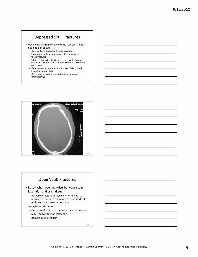
Depressed Skull Fractures
• Usually result from relatively small object striking head at high speed– Commonly associated with scalp lacerations
– Frontal and parietal bones most often affected by these fractures
– 30 percent of patients with depressed skull fractures estimated to have associated hematomata and cerebral contusions
– If depression is greater than thickness of skull, dural laceration also is likely
– Often require surgical removal of bone fragments (craniectomy)
151
152
Open Vault Fractures
• Result when opening exists between scalp laceration and brain tissue
– Because of nature of these injuries and force required to produce them, often associated with multiple trauma to other systems
– High mortality rate
– Exposure of brain tissue to external environment may lead to infection (meningitis)
– Require surgical repair
153
Copyright © 2013 by Jones & Bartlett Learning, LLC, an Ascend Learning Company

9/11/2012
52
Open Vault Fractures
• Prehospital management
– Limited to spinal immobilization
– Ventilatory support
– Efforts to prevent contamination
– Rapid transport
154
155
Cranial Nerve Injuries
• 12 pairs of cranial nerves leave brain and pass through openings in skull called foramina
• Injury to cranial nerves usually associated with skull fractures
• Signs and symptoms of common cranial nerve injuries
– Cranial nerve I (olfactory nerve)
• Loss of smell
• Impaired taste (dependent on food aroma)
• Hallmark of basilar skull fracture
156
Copyright © 2013 by Jones & Bartlett Learning, LLC, an Ascend Learning Company

9/11/2012
53
Cranial Nerve Injuries
• Signs and symptoms of common cranial nerve injuries
– Cranial nerve II (optic nerve)
• Blindness in one or both eyes
• Visual field defects
– Cranial nerve III (oculomotor nerve)
• Ipsilateral (same side), dilated, fixed pupil
• Especially compression by temporal lobe
• Mimics direct ocular trauma
157
Cranial Nerve Injuries
• Signs and symptoms of common cranial nerve injuries
– Cranial nerve VII (facial nerve)
• Immediate or delayed facial paralysis
• Basilar skull fracture
– Cranial nerve VIII (auditory nerve)
• Deafness
• Basilar skull fracture
158
Traumatic Brain Trauma
• Defined by Brain Injury Association as “a traumatic insult to the brain capable of producing physical, intellectual, emotional, social, and vocational change”
• Can be divided into two categories
– Primary brain injury
• Direct trauma to brain and to associated vascular injuries that occurred from initial injury
– Secondary brain injury
159
Copyright © 2013 by Jones & Bartlett Learning, LLC, an Ascend Learning Company
9/11/2012
54
• Secondary brain injury– Results from intracellular and extracellular derangements that probably were initiated at time of injury
– May include• Hypoxia• Hypocapnia• Hypercapnia from airway compromise• Aspiration of gastric contents, and thoracic injury• Anemia and hypotension from external and internal hemorrhage
• Hyperglycemia or hypoglycemia that can further injure ischemic brain tissue
Traumatic Brain Trauma
160
Traumatic Brain Trauma
• Adverse effects of secondary brain injury can be minimized
– Perhaps can be reversed if recognized and properly managed in prehospital setting
– Brain injuries can be classified as diffuse (moderate or severe) or focal
• Two forms are commonly found together
161
Traumatic Brain Trauma
• Diffuse Injuries (usually caused by acceleration–deceleration forces)
– Diffuse axonal injury (DAI)
– Hypoxic‐ischemic damage
– Meningitis
– Vascular injury
162
Copyright © 2013 by Jones & Bartlett Learning, LLC, an Ascend Learning Company

9/11/2012
55
Traumatic Brain Trauma
• Focal injuries (generally caused by contact)
– Scalp injury
– Skull fracture
– Surface contusions
– Brain hemorrhage
163
Diffuse Injuries
• Diffuse injuries
– Concussion
– Diffuse axonal injury
• Major cause of damage in diffuse injury is disruption of axons—neural processes that allow one axon to communicate with another
164
Concussion
• Sometimes called mild diffuse axonal injury– Caused by mild to moderate impact to skull, movement of brain within cranial vault, or both
– Occurs when function of brainstem (particularly reticular activating system) or both cerebral cortices is temporarily disturbed
• Results in brief altered level of consciousness, but not always loss of consciousness
• If loss of consciousness occurs, usually less than 5 minutes in duration
• May be serious injury (no concussion is minor)
• Can be assigned into one of three grades as defined by American Academy of Neurology (AAN)
165
Copyright © 2013 by Jones & Bartlett Learning, LLC, an Ascend Learning Company

9/11/2012
56
Concussion
• Grade 1
– Transient confusion
– NO loss of consciousness
– Concussion symptoms clear in less than 15 minutes
166
Concussion
• Grade 2
– Transient confusion
– NO loss of consciousness
– Concussion symptoms or mental status abnormalities last longer than 15 minutes
• Grade 3
– Any loss of consciousness, either brief (seconds) or prolonged (minutes)
167
Concussion • Altered level of consciousness or loss of consciousness usually is followed by periods of– Drowsiness
– Restlessness
– Confusion, with fairly rapid return to normal behavior
• Patient may have no recall of events before injury (retrograde amnesia)– Amnesia may exist after recovery of consciousness (anterograde amnesia)
– This short‐term memory loss may produce anxiety
168
Copyright © 2013 by Jones & Bartlett Learning, LLC, an Ascend Learning Company

9/11/2012
57
Concussion
• Patient may ask repetitive questions (e.g., “Where am I? What happened?”)
• Other signs and symptoms– Vomiting
– Combativeness
– Transient visual disturbances (e.g., light flashes and wavy lines)
– Defects in equilibrium and coordination
– Changes in BP, pulse rate, and respiration (rare)
• After physician evaluation, treatment usually consists of in‐hospital or home observation by reliable observer for 24 to 48 hours
169
Consider the patient with a new onset of retrograde or anterograde amnesia. Why should the patient
not be considered a reliable historian?
170
Concussion
• Concussion injury affects patient most severely at time of impact but is followed by improvement
• Most common type of brain injury
• Any patient whose condition worsens over time or whose level of consciousness deteriorates rather than improves must be suspected of having more serious injury
171
Copyright © 2013 by Jones & Bartlett Learning, LLC, an Ascend Learning Company
9/11/2012
58
Concussion
• Documentation of baseline measurements of level of consciousness, memory status, and neurological function (e.g., Glasgow Coma Scale or AVPU scale) in any victim of head injury is important
• If patient has loss of consciousness longer than 5 minutes, suspect more serious injury caused by contusion or hemorrhage
172
Moderate Diffuse Axonal Injury
• Head injury that results in minute petechialbruising of brain tissue
– Involvement of brainstem and reticular activating system leads to unconsciousness
– Accounts for 20 percent of all severe head injuries and 45 percent of all cases of diffuse injury
– Often will have basilar skull fracture
– Most patients will survive injury
– Permanent neurological impairment is common
173
Moderate Diffuse Axonal Injury
• Initially will be unconscious, followed by– Persistent confusion
– Disorientation
– Amnesia of event
• During recovery, patients often experience– Inability to concentrate
– Frequent periods of anxiety
– Uncharacteristic mood swings
– Sensorimotor deficits
174
Copyright © 2013 by Jones & Bartlett Learning, LLC, an Ascend Learning Company

9/11/2012
59
Moderate Diffuse Axonal Injury
• Managed as are those with concussion
– Frequent reassessments of level of consciousness
– Assurance of adequate airway and tidal volume
– If possible, patients should be moved to quiet, calm area
– Exposure to bright lights should be avoided
– Patients often are photophobic
– Constant reorientation of patient may be necessary
175
Severe Diffuse Axonal Injury
• Severest form of brain injury
– Once known as brainstem injury
– Involves severe mechanical shearing of many axons in both cerebral hemispheres extending to brainstem
– Occurs in approximately 16 percent of patients with severe head trauma
– Often are unconscious for prolonged periods
176
Severe Diffuse Axonal Injury
• May exhibit abnormal posturing and other signs of increased intracranial pressure (ICP)
• Prehospital care is focused on ensuring adequate airway and tidal volume
– Hypoxia must be prevented in all patients with head injury
• Helps to avoid secondary injury to brain tissue
177
Copyright © 2013 by Jones & Bartlett Learning, LLC, an Ascend Learning Company

9/11/2012
60
Can a patient with a diffuse axonal injury die as a result of that injury?
178
Focal Injury
• Specific, grossly observable brain lesions
– Lesions that result from
• Skull fracture
• Contusion
• Edema with associated increased ICP
• Ischemia
• Hemorrhage
– Brain occupies 80 percent of intracranial space
179
Focal Injury
• Brain is divided into four areas
– Brainstem (medulla, pons, and midbrain)
– Diencephalon (thalamus and hypothalamus)
– Cerebrum
– Cerebellum
180
Copyright © 2013 by Jones & Bartlett Learning, LLC, an Ascend Learning Company

9/11/2012
61
Focal Injury
• Intracranial contents consist of
– Brain water (58 percent)
– Brain solids (25 percent)
– Cerebrospinal fluid (7 percent)
– Intracranial blood (10 percent)
181
Cerebral Contusion
• Bruising of brain in area of cortex or deeper within frontal (most common), temporal, or occipital lobes – Produces structural change in brain tissue
– Results in greater neurological deficits and abnormalities than are seen with concussions
• Seizures
• Hemiparesis
• Aphasia
• Personality changes
182
183
Copyright © 2013 by Jones & Bartlett Learning, LLC, an Ascend Learning Company

9/11/2012
62
Cerebral Contusion
• If brainstem also is contused, patient may lose consciousness
• In some cases, comatose state may be prolonged
– May last hours to days or longer
• Of patients who die from head injury, majority have cerebral contusions at autopsy
184
Cerebral Contusion
• If applied force is enough to cause brain to be displaced against irregular surfaces of skull, tiny blood vessels in pia mater may rupture– Brain substance may be damaged locally at site of impact (coup)
– Brain may be damaged on opposite, or contralateral, side (contrecoup)
– Contrecoup injuries often are caused by deceleration of head
• May occur in fall or motor vehicle crash
185
Cerebral Contusion
• As a rule, cerebral contusions usually heal without intervention
– Patients usually improve
– Time to heal and level of improvement differ in these two conditions
186
Copyright © 2013 by Jones & Bartlett Learning, LLC, an Ascend Learning Company

9/11/2012
63
Cerebral Contusion
• As a rule, cerebral contusions usually heal without intervention– Most important complication associated with cerebral contusion is increased ICP manifested by
• Headache
• Nausea
• Vomiting
• Seizures
• Declining level of consciousness
– Signs usually are delayed responses to injury
– Usually not seen in prehospital emergency setting
187
Edema
• Major injuries to brain often result in swelling of brain tissue with or without associated hemorrhage
– Swelling results from humoral and metabolic responses to injury
• Leads to considerable increases in ICP
• Can lead to decreased cerebral perfusion or herniation
188
Ischemia
• Ischemia can result from
– Vascular injuries
– Secondary vascular spasm
– Increased ICP
• In any case, focal or more global infarcts can result
189
Copyright © 2013 by Jones & Bartlett Learning, LLC, an Ascend Learning Company

9/11/2012
64
Hemorrhage
• Same forces that result in concussion and contusion also may cause serious vascular damage– Damage may result in hemorrhage into or around brain tissue
– May cause epidural or subdural hematomata
– Hematomata compress underlying brain tissue, or produce intraparenchymal hemorrhage (bleeding directly into brain tissue)
– Bleeding often results from cerebral contusions and skull fractures
190
Cerebral Blood Flow
• Although brain accounts for only 2 percent of adult weight, 20 percent of total body oxygen use and 25 percent of total body glucose use are devoted to brain metabolism
• Oxygen and glucose delivery are controlled by cerebral blood flow
191
Cerebral Blood Flow
• Function of cerebral perfusion pressure (CPP) and resistance of cerebral vascular bed– Cerebral blood flow is determined by mean arterial pressure (MAP) (diastolic pressure + 1/3 pulse pressure) minus intracranial pressure (CPP = MAP – ICP)
– Normal mean arterial pressure ranges from 85 to 95 mm Hg
– Intracranial pressure is normally 10 to 15 mm Hg or less
– Normal CPP is 70 to 80 mm Hg
192
Copyright © 2013 by Jones & Bartlett Learning, LLC, an Ascend Learning Company

9/11/2012
65
Cerebral Blood Flow
• As ICP approaches mean arterial pressure, gradient for flow decreases and cerebral blood flow decreases– When ICP increases, CPP decreases
– As CPP decreases, vessels in brain dilate (cerebral vasodilation)
• Results in increased cerebral blood volume (increasing ICP) and further cerebral vasodilation
• In most EMS systems, CPP is not calculated because mean arterial pressure and ICP are not measured in prehospitalsetting
• Maintaining systolic BP of at least 90 mm Hg also may help maintain adequate mean arterial pressure
193
What happens to the flow of oxygen to the brain, and carbon dioxide from the brain to the capillaries, when intracranial
pressure is increasing and cerebral perfusion pressure is decreasing?
194
Cerebral Blood Flow
• Vascular tone in normal brain is regulated by
– Carbon dioxide pressure (PCO2), oxygen pressure (PO2), and autonomic and neurohumoral control
– PCO2 has greatest effect on intracerebral vascular diameter and subsequent resistance
195
Copyright © 2013 by Jones & Bartlett Learning, LLC, an Ascend Learning Company

9/11/2012
66
Intracranial Pressure
• Normal range of ICP is 10 to 15 mm Hg or less– When ICP rises above, body has difficulty maintaining adequate CPP, usually because of expanding mass or diffuse swelling
– Cerebral blood flow is diminished when CPP is not adequate
– As cranial vault continues to fill (because of brain edema or expanding hematoma), body tries to compensate for decline in CPP by rise in mean arterial pressure (Cushing reflex)
196
Intracranial Pressure
• Increase in cerebral blood flow further elevates ICP
– As pressure continues to increase, cerebrospinal fluid is displaced to make up for expansion
– If unresolved, brain substance may herniate over edge of tentorium
• One of three extensions of dura mater that separates cerebellum from occipital lobe of cerebrum
• May herniate through foramen magnum
197
198
Copyright © 2013 by Jones & Bartlett Learning, LLC, an Ascend Learning Company

9/11/2012
67
Intracranial Pressure
• Early signs and symptoms of increased ICP
– Nausea and vomiting
– Altered level of consciousness
– Widened pulse pressure
– Decrease in pulse and irregular respiratory pattern (Cushing’s triad)
– As volume continues to expand in cranial vault, herniation of temporal lobe of brain through tentorium may occur
199
Intracranial Pressure
• Herniation causes compression of cranial nerve III
– Produces dilated pupil and loss of light reflex on side of compression
– Patient rapidly becomes unresponsive to verbal and painful stimuli
– May exhibit ominous signs of decorticate posturing
• Extension of legs and flexion of arms at elbows
– May exhibit decerebrate posturing
• Extension of all four extremities
200
201
Copyright © 2013 by Jones & Bartlett Learning, LLC, an Ascend Learning Company

9/11/2012
68
Why is cranial nerve III affected by this shift in brain tissue?
202
Respiratory Patterns
• As ICP continues to rise, abnormal respiratory patterns may develop– Respiratory abnormalities associated with increased ICP and significant brainstem injury include
• Hypoventilation
• Cheyne‐Stokes breathing (which may accompany decorticate posturing)
• Central neurogenic hyperventilation (which may accompany decerebrate posturing)
• Ataxic breathing
203
Respiratory Patterns
• Clinical significance of decorticate (flexion) and decerebrate (extension) posturing and respiratory patterns are not of major clinical importance
– Identify need for intervention and treatment
• Intubation and consideration of immediate neurosurgical intervention
204
Copyright © 2013 by Jones & Bartlett Learning, LLC, an Ascend Learning Company
9/11/2012
69
Types of Brain Hemorrhage
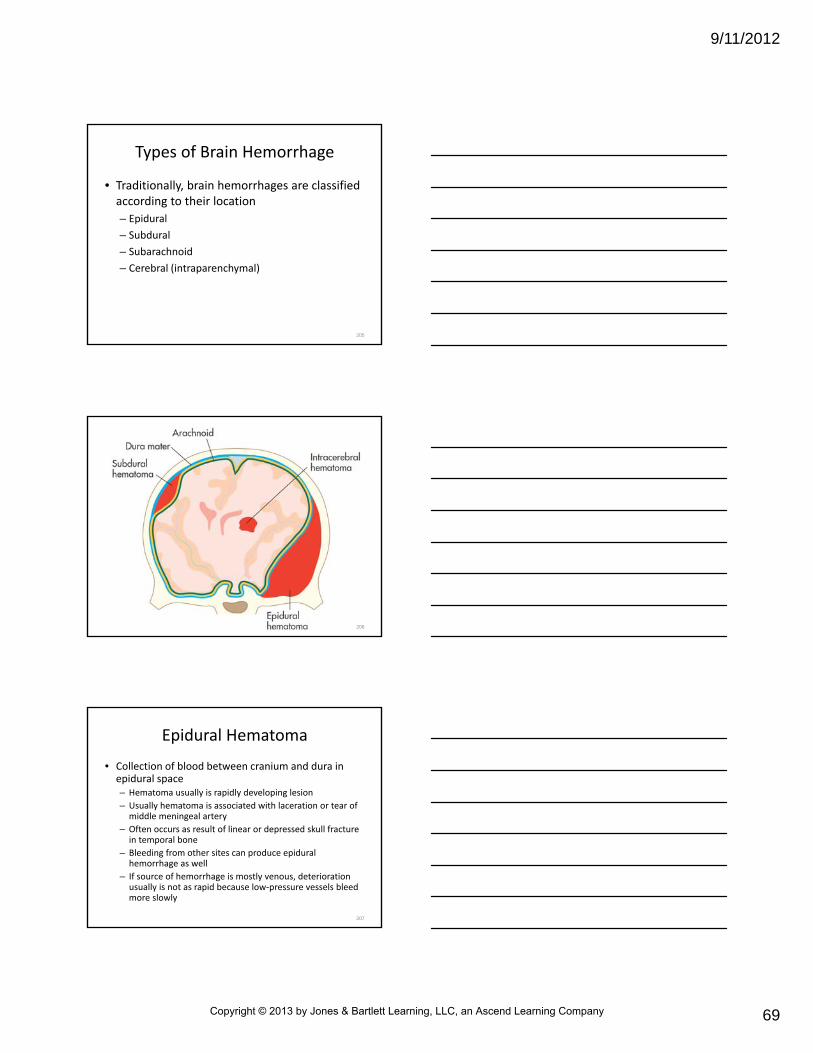
• Traditionally, brain hemorrhages are classified according to their location
– Epidural
– Subdural
– Subarachnoid
– Cerebral (intraparenchymal)
205
206
Epidural Hematoma
• Collection of blood between cranium and dura in epidural space– Hematoma usually is rapidly developing lesion
– Usually hematoma is associated with laceration or tear of middle meningeal artery
– Often occurs as result of linear or depressed skull fracture in temporal bone
– Bleeding from other sites can produce epidural hemorrhage as well
– If source of hemorrhage is mostly venous, deterioration usually is not as rapid because low‐pressure vessels bleed more slowly
207
Copyright © 2013 by Jones & Bartlett Learning, LLC, an Ascend Learning Company

9/11/2012
70
208
Epidural Hematoma
• 50 percent of patients with epidural hematoma have transient loss of consciousness, followed by lucid interval in which neurological status returns to normal– Remaining 50 percent of patients with acute epidural hematoma never recover consciousness
– Lucid interval usually lasts between 6 to 18 hours
– During this time, hematoma enlarges
– As ICP rises, patient develops headache with lethargy, decreasing level of consciousness, contralateralhemiparesis
209
Epidural Hematoma
• In early stages, patient may complain only of headache and drowsiness
– Definitive treatment includes immediate recognition and rapid transport to a proper facility for surgery
– Common causes
• Low‐velocity blows to head
• Violent altercations
• Deceleration injuries
– About 20 percent of these patients who are comatose die
210
Copyright © 2013 by Jones & Bartlett Learning, LLC, an Ascend Learning Company

9/11/2012
71
What could account for delays in surgical treatment, causing
subsequent death, in patients who have an epidural hematoma?
211
Subdural Hematoma
• Collection of blood between dura and surface of brain in subdural space
– Injury usually results from bleeding of veins that bridge subdural space
– Associated contusion or laceration of brain often is present
– Hematoma often results from blunt head trauma
– Commonly hematoma is associated with skull fracture
212
213
Copyright © 2013 by Jones & Bartlett Learning, LLC, an Ascend Learning Company
9/11/2012
72
Subdural Hematoma
• Classified as acute, subacute, and chronic
– Depends on time lapse between injury and development of symptoms
– Acute if symptoms occur within 24 hours
– Subacute between 2 and 10 days
– Chronic after 2 weeks
• More common than epidural hematomata
214
Subdural Hematoma
• Signs and symptoms– Headache
– Nausea and vomiting
– Decreasing level of consciousness
– Coma
– Abnormal posturing
– Paralysis
– In infants, bulging fontanelles• Findings may be subtle because of slow development of hematoma in subacute and chronic phases
215
Subdural Hematoma
• Definitive care
– Surgery to remove blood from hematoma
• Individuals at increased risk of developing subdural hematoma include older adults and patients with clotting deficiencies
– Patients with cortical atrophy
216
Copyright © 2013 by Jones & Bartlett Learning, LLC, an Ascend Learning Company
9/11/2012
73
Subarachnoid Hematoma
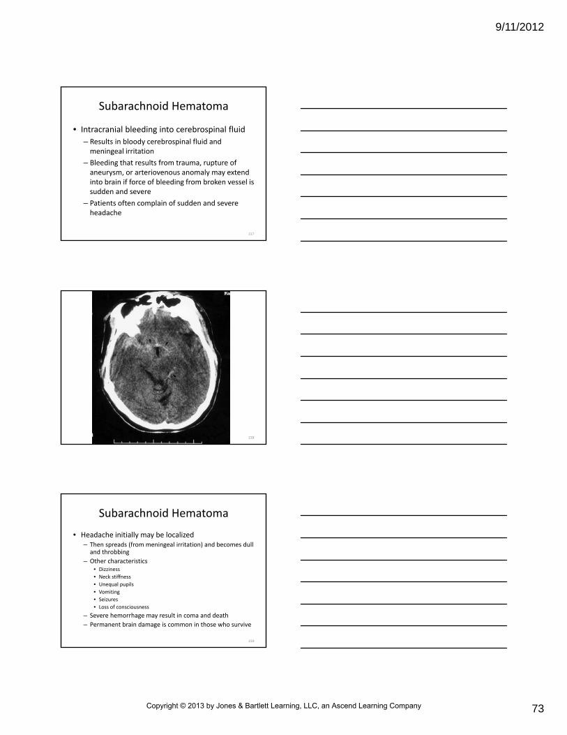
• Intracranial bleeding into cerebrospinal fluid
– Results in bloody cerebrospinal fluid and meningeal irritation
– Bleeding that results from trauma, rupture of aneurysm, or arteriovenous anomaly may extend into brain if force of bleeding from broken vessel is sudden and severe
– Patients often complain of sudden and severe headache
217
218
Subarachnoid Hematoma
• Headache initially may be localized– Then spreads (from meningeal irritation) and becomes dull and throbbing
– Other characteristics• Dizziness
• Neck stiffness
• Unequal pupils
• Vomiting
• Seizures
• Loss of consciousness
– Severe hemorrhage may result in coma and death
– Permanent brain damage is common in those who survive
219
Copyright © 2013 by Jones & Bartlett Learning, LLC, an Ascend Learning Company

9/11/2012
74
What causes the vomiting, seizures, and loss of consciousness in a patient with subarachnoid
hemorrhage?
220
Cerebral Hematoma
• Intracerebral hematoma may be defined as collection of more than 5 mL of blood somewhere within substance of brain, most commonly in frontal or temporal lobe– Can result from multiple lacerations produced by penetrating head trauma (gunshot wound)
– Injury also may result from high‐velocity deceleration injury (automobile crash) in which vessels are torn as brain moves across rough surfaces of skull
– Increased ICP can produce intracerebral hematoma as result of brain being compressed
221
Cerebral Hematoma
• Often associated with subdural hemorrhage and skull fracture
– Signs and symptoms may be immediate or delayed
– Depends on size and location of hemorrhage
– Once symptoms appear, patient usually deteriorates rapidly
– Mortality rate after surgical evacuation of hematoma (if possible) approaches 45 percent
222
Copyright © 2013 by Jones & Bartlett Learning, LLC, an Ascend Learning Company

9/11/2012
75
Penetrating Injury
• Penetrating injuries to brain usually are caused by missiles fired from handguns and stab wounds caused by sharp objects
– Less often, may result from falls and high‐velocity vehicle crashes
– Associated injuries
• Skull fracture
• Damage to cerebral arteries, veins, or venous sinuses
• Intracranial hemorrhage
223
Penetrating Injury
• Complications
– Infection and posttraumatic epilepsy
• Definitive care for these injuries requires neurosurgical intervention
224
Assessment and Neurological Evaluation
• Prehospital management of patient with head injury is determined by
– Mechanism and severity of injury
– Patient’s level of consciousness
• Associated injuries affect priorities of emergency care
225
Copyright © 2013 by Jones & Bartlett Learning, LLC, an Ascend Learning Company

9/11/2012
76
Airway and Ventilation
• Initial step in treating all patients with head trauma is to ensure open airway with spinal precautions
– Next step is to provide adequate ventilation with high‐concentration oxygen
– Airway management may include
• Oral or nasal adjuncts
• Multilumen devices
• Nasal or tracheal intubation to maintain and protect airway
– Tracheal intubation and ventilatory support usually recommended in all patients with head injuries who have Glasgow Coma Scale (GCS) score of 8 or lower
226
Imagine what a patient with a GCS score of 8 or lower would look like. Why should these patients be intubated?
What if the GCS score improves rapidly?
227
Airway and Ventilation
• Patients with head injuries are likely to vomit
– If patient has decreased level of consciousness after airway is secured, nasogastric tube should be inserted to empty stomach
228
Copyright © 2013 by Jones & Bartlett Learning, LLC, an Ascend Learning Company

9/11/2012
77
Airway and Ventilation
• Patients with head injuries are likely to vomit
– In presence of facial fractures, rhinorrhea (cerebrospinal fluid discharge from nose) or otorrhea (cerebrospinal fluid discharge from ear), an orogastric tube rather than a nasogastric tube should be inserted
• Use of this tube helps to avoid possible intubation of cranial cavity through fracture site
• Patient should be well stabilized on long spine board for safe repositioning
• Suction equipment with large‐bore suction catheters should be available
229
Airway and Ventilation
• Ventilatory support should be focused on maintaining adequate oxygenation and optimizing cerebral perfusion
– Capnography and pulse oximetry should be used to maintain oxygen saturation at level of 95 percent or greater
– Aggressive hyperventilation reduces carbon dioxide
• Can lead to secondary brain injury through cerebral vasoconstriction and decrease in cerebral blood flow
• Routine prophylactic hyperventilation should be avoided
230
Airway and Ventilation
• In absence of capnography to guide ventilatory support, normal ventilations should be provided at– 10 breaths/min for adults
– 20 breaths/min for children
– 25 breaths/min for infants
– With evidence of herniation patient should be hyperventilated at following rates
• 20 breaths/min for adults
• 30 breaths/min for children
• 35 breaths/min for infants
– These rates should yield a PCO2 of about 35 mm Hg
231
Copyright © 2013 by Jones & Bartlett Learning, LLC, an Ascend Learning Company

9/11/2012
78
Circulation
• After airway has been secured (maintaining spinal protection), support of patient’s cardiovascular function becomes next priority– Control major external bleeding
– Assess patient’s vital signs• Establishes baseline for future evaluations
– Cardiac monitor will detect changes in rhythm that can occur with increasing ICP and brainstem injury
– BP of every patient should be maintained at normal levels with fluid replacement (per medical direction)
232
Circulation
• Single episode of hypotension doubles mortality and increases morbidity in patient with traumatic brain injury
– Administer IV fluids to support oxygen delivery and to avoid hypertension or limit hypotension to shortest duration possible
233
Circulation
• Persistent hypotension from isolated head injury is rare and terminal event– Exception is head injury in infants and small children
– Closed head injury in adult does not produce hypovolemic shock
• Patient with head injuries who also is hypotensiveshould be evaluated for other injuries that could cause hemorrhage
• Evaluate for possibility of neurogenic shock from spinal cord trauma
234
Copyright © 2013 by Jones & Bartlett Learning, LLC, an Ascend Learning Company

9/11/2012
79
Circulation
• Infusion of isotonic fluids (lactated Ringer’s solution or normal saline) may be indicated for hemorrhagic shock
– These fluids should be used cautiously in patients with hypotension caused by neurogenic shock
• Vasopressors also may be helpful in maintaining BP
235
Circulation
• Neurogenic shock may be distinguished from hemorrhagic shock by
– Relatively bradycardic response (e.g., a pulse of 80 with a BP of 80 mm Hg)
– Skin that often is warm and dry (not cool and clammy)
– No evidence of significant blood loss or hypovolemia
– Paralysis and loss of spinal reflexes
236
Neurological Examination
• Conscious patients should be interviewed to determine memory status before and after injury and to learn of significant medical history
– History should include mechanism of injury and events that led up to injury
237
Copyright © 2013 by Jones & Bartlett Learning, LLC, an Ascend Learning Company

9/11/2012
80
Neurological Examination
• Evaluate motor skills of conscious patients
– Determines patient’s ability to follow commands and helps paramedic to note any paralysis
• Hemiparesis or hemiplegia, especially with a sensory deficit on the same side, indicates brain damage rather than spinal trauma
– If patient is unconscious on EMS arrival, interview bystanders about history of event
238
Neurological Examination
• Ask bystanders about length of time patient has been unconscious
• Most important indicator of increasing ICP is deterioration in patient’s sensorium
– Evaluate level of consciousness using GCS every 5 minutes
• Decrease of 2 points with GCS score of 9 or lower is significant
• Indicates significant injury
239
How reliable will patients be regarding the duration of their loss
of consciousness?
240
Copyright © 2013 by Jones & Bartlett Learning, LLC, an Ascend Learning Company

9/11/2012
81
Neurological Examination
• After patient has been resuscitated and stabilized, assess patient’s pupils for symmetry, size, and reactivity to light– Abnormal pupillary responses may indicate increase in ICP and cranial nerve involvement
– Asymmetric pupils differ more than 1 mm in size
– Dilated pupils are 4 mm or larger in adults
– Fixed pupil shows less than 1 mm change in response to bright light
• Evaluate pupil size every 5 minutes
241
Neurological Examination
• Alcohol and some other drugs can cause abnormal pupillary reactions
– Reactions commonly are bilateral (except for certain eye drops, if placed in one eye)
– If patient is conscious, evaluate extraocular movement
242
Fluid Therapy
• In absence of hypotension, fluid therapy normally should be restricted in patient with head injury to minimize cerebral edema
– If patient is hemodynamically stable, establish an IV line of crystalloid fluid to keep vein open
– If significant hypovolemia is present from another injury, give isotonic fluid bolus
• Guided by medical direction
243
Copyright © 2013 by Jones & Bartlett Learning, LLC, an Ascend Learning Company

9/11/2012
82
Fluid Therapy
• Transport rapidly to proper facility
– Injury causing hypovolemia usually is more immediately life threatening than head injury
– As rule, hypotension in presence of head injury initially should be managed with fluid boluses to maintain systolic BP of at least 90 mm Hg
244
Drug Therapy
• Prehospital use of drugs for treatment of head injuries is controversial– Drugs that may be prescribed by medical direction to decrease cerebral edema or circulating blood volume may include mannitol and hypertonic saline
– Medical direction may require insertion of indwelling urinary catheter for careful monitoring of urine output before starting diuretic therapy
– Hypotension leading to hypoperfusion may occur as complication of diuretic use in patients with head injuries
245
Drug Therapy
• Anticonvulsant agents such as lorazepam and diazepam are used to control seizure activity in head‐injured patients– As rule, these drugs are not used in initial management of head injuries because of their sedating effects
– Prevention of seizures avoids rise in ICP that often accompanies sudden seizure activity
– Intravenously administered lidocaine has been shown to control increases in ICP that normally occur during endotracheal intubation
246
Copyright © 2013 by Jones & Bartlett Learning, LLC, an Ascend Learning Company

9/11/2012
83
Drug Therapy
• Use of sedatives and paralytics for some patients with head injuries may be indicated for airway management
– Drugs also may be used to aid in transport of combative patients (especially in aeromedicaltransport)
– Follow local protocol and consult with medical direction regarding use of these drugs
247
Injury Rating System
• Used to
– Triage
– Guide patient care
– Predict patient outcome
– Identify changes in patient status
– Evaluate trauma care in epidemiological studies and quality assurance reviews
• Aid in determining patient care needs with reference to hospital resources
248
Glasgow Coma Scale
• GCS evaluates eye opening, verbal and motor responses, and brainstem reflex function – Scale is considered one of best indicators of eventual clinical outcome and should be part of any neurological examination for patients with head injury
– GCS score of 9 to 13 indicates moderate traumatic brain injury
– GCS score of lower than 8 indicates severe traumatic brain injury
249
Copyright © 2013 by Jones & Bartlett Learning, LLC, an Ascend Learning Company

9/11/2012
84
Glasgow Coma Scale
• Hypoxemia and hypotension have been shown to affect GCS scoring negatively
– GCS should be measured after primary survey
– Score should be measured after clear airway is established
– Should be measured after necessary ventilation and circulatory resuscitation have been performed
– Unresponsive patients with GCS score of 3 to 8 should be transported to trauma center with traumatic brain injury capabilities
250
• Trauma score was developed in 1980 to predict outcome for patients with blunt or penetrating injuries
– Has limited use in prehospital setting
– Does not predict adequately mortality for isolated, severe head injury
Trauma Score/Revised Trauma Score
251
• Revised Trauma Score was published in 1989– Uses GCS with measurements for systolic BP and respiratory rate that are divided into five intervals
– Range of values for these physiological measurements is assigned a number between 0 and 4
– These numbers then are added to give total between 0 and 12
• Score of 0 indicates most critical
• Score of 12 indicates least critical
Trauma Score/Revised Trauma Score
252
Copyright © 2013 by Jones & Bartlett Learning, LLC, an Ascend Learning Company

9/11/2012
85
• Calculating Revised Trauma Score en route to receiving hospital provides baseline measurements– Can be helpful to physician in managing patient’s care
– In some EMS systems, calculated after arrival at emergency department using data from radio reports and prehospital care report
Trauma Score/Revised Trauma Score
253
Pediatric Trauma Score
• Grades six characteristics commonly seen in pediatric trauma patients
– Size (weight)
– Airway
– Consciousness
– Systolic BP
– Fracture
– Cutaneous injury
254
Pediatric Trauma Score
• Has significant inverse linear relationship with patient mortality
– Child with score lower than 8 should be cared for in appropriate pediatric trauma center
255
Copyright © 2013 by Jones & Bartlett Learning, LLC, an Ascend Learning Company

9/11/2012
86
Pediatric Trauma Score
• Patient size (weight) is one of first parameters to assess
– The smaller the child, the greater the risk for severe injury because of increased ratio of body surface to volume
– Risk also is greater because of potential for limited physiological reserve
256
Pediatric Trauma Score
• Child’s airway is scored by potential difficulty in management
– Scoring is by type of care required to ensure adequate ventilation and oxygenation
– Respiratory failure is main cause of death in most pediatric patients
• Aggressive management to control airway should be started without delay
257
Pediatric Trauma Score
• As with adult patients, most critical factor in assessing CNS of child is change in level of consciousness
– Any change in level of consciousness will reduce score, no matter how brief the time
258
Copyright © 2013 by Jones & Bartlett Learning, LLC, an Ascend Learning Company

9/11/2012
87
Pediatric Trauma Score
• Assessment of systolic BP in pediatric patient is critical because circulating volume is notably less than adult
– Because of normal child’s healthy heart and excellent reserve capacity, children often do not show classic signs of shock until they have lost about 25 percent of their circulating volume
– Any child who has systolic BP lower than 50 mm Hg is in obvious jeopardy
259
Pediatric Trauma Score
• Child’s skeleton is more pliable than that of adult– Allows traumatic forces to be sent through body and to organs
– Fracture in pediatric patient is sign that serious injury likely has occurred
– Cutaneous injury in pediatric patient is potential contributor to mortality and disability
– Include open and visible wounds and penetrating trauma
260
Summary
• Major causes of maxillofacial trauma are motor vehicle crashes, home accidents, athletic injuries, animal bites, intentional violent acts, and industrial injuries
• With exception of compromised airway and potential for significant bleeding, damage to tissues of maxillofacial area is seldom life threatening– Some facial fractures are associated with basilar skull fracture
– Blunt trauma injuries may be classified as fractures to the mandible, midface, zygoma, orbit, and nose
261
Copyright © 2013 by Jones & Bartlett Learning, LLC, an Ascend Learning Company

9/11/2012
88
Summary
• Injury to ears, eyes, or teeth may be minor or may result in permanent sensory function loss and disfigurement– Trauma to the ear may include lacerations and contusions, thermal injuries, chemical injuries, traumatic perforation, and barotitis
– Evaluation of the eye should include thorough history
• Assessment also should include measurement of visual acuity, pupillary reaction, and extraocular movements
262
• Anterior neck injuries may result in damage to skeletal structures, vascular structures, nerves, muscles, and glands of the neck– Patient should be assessed for airway compromise, bleeding, and cervical spine injury
• Injuries to the skull may be classified as soft tissue injuries to the scalp and skull fractures– Skull fractures may be classified as linear fractures, basilar fractures, depressed fractures, and open vault fractures
Summary
263
Summary
• Categories of brain injury include diffuse axonal injury and focal injury
– Diffuse axonal injury may be mild (concussion), moderate, or severe
– Focal injuries are specific, grossly observable brain lesions
• Included in this category are lesions that result from skull fracture, contusion, edema with associated increased ICP, ischemia, and hemorrhage
264
Copyright © 2013 by Jones & Bartlett Learning, LLC, an Ascend Learning Company

9/11/2012
89
Summary
• Prehospital management of a patient with head injuries is determined by
– Mechanism of injury
– Severity of injury
– Patient’s level of consciousness
• Associated injuries affect priorities of care
265
Summary
• Several injury rating systems are used to triage, guide patient care, predict patient outcome, identify changes in patient status, and evaluate trauma care
– Rating systems commonly used in emergency care include the Glasgow Coma Scale, trauma score/Revised Trauma Score, and pediatric trauma score
266
Questions?
267
Copyright © 2013 by Jones & Bartlett Learning, LLC, an Ascend Learning Company