chapter 61 integumentary system. chapter 62 introduction a.organs are body structures composed of...
TRANSCRIPT

Chapter 6 1
Integumentary System

Chapter 6 2
IntroductionA.Organs are body structures composed of two or more
different tissues.B.The skin and its accessory organs make up the
integumentary system.
CopyrightThe McGraw-Hill Companies, Inc. Permission required for reproduction or display.

Chapter 6 3
Chapter 6 4
Skin and Its Tissues
A. The skin is a largest organ responsible for:*maintaining homeostasis through temperature regulation*protection of underlying tissues*retardation of water loss*housing sensory receptors* synthesizing certain chemicals*excreting wastes
CopyrightThe McGraw-Hill Companies, Inc. Permission required for reproduction or display.

Chapter 6 5
B. The skin consists of an outer epidermis and a dermis, connected to underlying tissue by the subcutaneous layer (hypodermis).
CopyrightThe McGraw-Hill Companies, Inc. Permission required for reproduction or display.

Chapter 6 6
Epidermis1. The epidermis is made up of stratified squamous epithelium and lacks blood vessels.2. The layer of reproducing cells (the stratum basale), which lies at the base of the epidermis, is well-nourished by dermal blood vessels.3. Cells are pushed outward as new cells are formed, and become keratinized as they die. 4 or 5 layers (STRATA) may be seen.
4.The epidermis is important because it protects against water loss, mechanical injury, chemicals, and microorganisms.
5.Melanocytes,which lie deep in the epidermis and underlying dermis, produce a pigment called melanin that protects deeper cells from the sun's ultraviolet rays.
6.Melanocytes pass melanin to nearby cells through cytocrine secretion.
CopyrightThe McGraw-Hill Companies, Inc. Permission required for reproduction or display.

Chapter 6 7
4 or 5 Layers (STRATA) of the Epidermis stratum basale always present DEEP
stratum spinosum always present
stratum granulosum always present
stratum lucidum found in the thicker palms and soles
stratum corneum always present Superficial
CopyrightThe McGraw-Hill Companies, Inc. Permission required for reproduction or display.

Chapter 6 8
Stratum Basale• Deepest single layer of
cells • Combination of
melanocytes, keratinocytes & stem cells that undergo mitosis
• Cells attached to each other & to basement membrane

Chapter 6 9
Stratum Spinosum
• 8 to 10 cell layers held together
• During slide preparation, cells shrink and look spiny
• Melanin taken in by nearby melanocytes

Chapter 6 10
Stratum Granulosum
• 3 - 5 layers of flat dying cells
• Show nuclear degeneration
• Contain dark-staining granules
• Contain granules that release lipid that repels water
Chapter 6 11
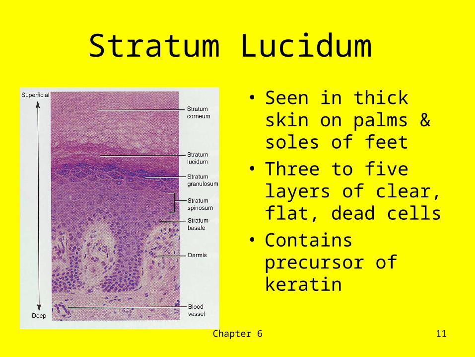
Stratum Lucidum
• Seen in thick skin on palms & soles of feet
• Three to five layers of clear, flat, dead cells
• Contains precursor of keratin

Chapter 6 12
Stratum Corneum
• 25 to 30 layers of flat dead cells filled with keratin and surrounded by lipids
• Continuously shed• Barrier to light, heat,
water, chemicals & bacteria
• Friction stimulates callus formation

Chapter 6 13

Chapter 6 14
Figure 06.02

Chapter 6 15
Dermis1. The dermis binds the epidermis to underlying
tissues. Epidermal ridges and dermal papillae cause the border to be uneven.
2. The dermis consists of connective tissue with collagen and elastic fibers within a gel-like ground substance.
3. Dermal blood vessels carry nutrients to upper layers of skin and help to regulate temperature.
4. The dermis also contains nerve fibers, sensory fibers, hair follicles, sebaceous glands, and sweat glands.
CopyrightThe McGraw-Hill Companies, Inc. Permission required for reproduction or display.
Chapter 6 16
• Dermis does not shed like epidermis• Can regenerate when injured• Starts to produce new tissue: will
become a scar• Cut or incision can make a lesser
scar if made in the same direction as cleavage lines
• Stretch marks (striae)- caused by rapid growth & tearing of elastic tissue; causes scarring
Dermal Growth & Repair
Chapter 6 17
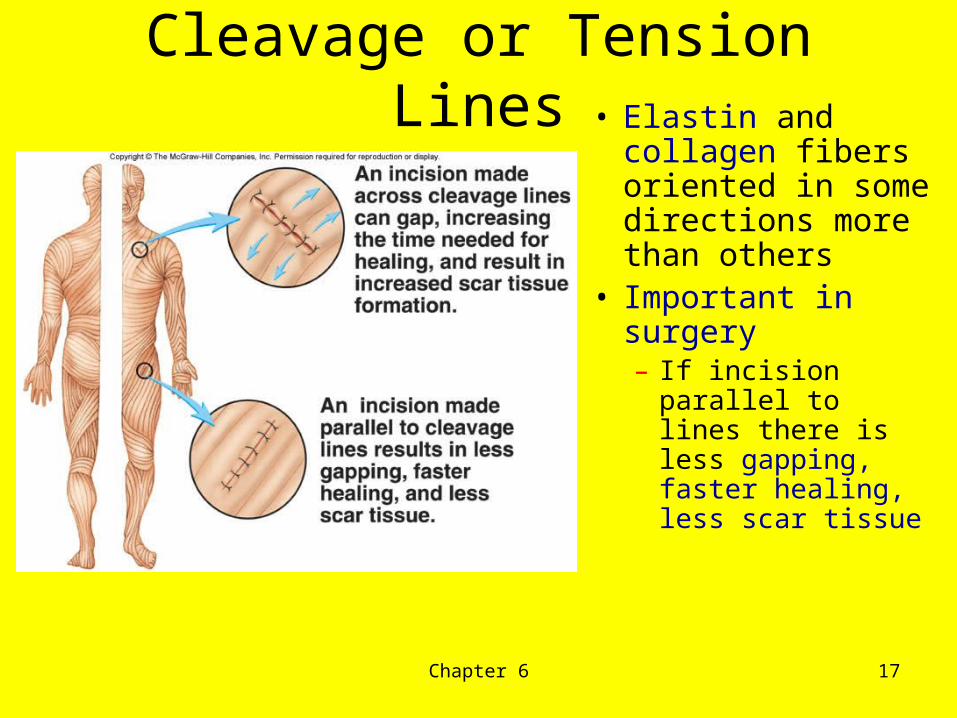
Cleavage or Tension Lines• Elastin and
collagen fibers oriented in some directions more than others
• Important in surgery– If incision
parallel to lines there is less gapping, faster healing, less scar tissue

Chapter 6 18
Skin Color
– Skin color results from a combination of genetic, environmental, and physiological factors.
• Melanin produced in epidermis by melanocytes– same number of melanocytes in everyone, but differing amounts
of pigment produced– results vary from yellow to tan to black color– melanocytes make melanin
» UV in sunlight increases melanin production• Clinical observations
– freckles or liver spots = melanocytes in a patch– albinism = inherited lack of pigment– vitiligo = autoimmune loss of melanocytes in areas of the skin
produces white patches• Carotene in dermis
– yellow-orange pigment (precursor of vitamin A)– found in stratum corneum & dermis
• Hemoglobin– red, oxygen-carrying pigment in blood cells– if other pigments are not present, epidermis is translucent so pinkness will be evident
CopyrightThe McGraw-Hill Companies, Inc. Permission required for reproduction or display.

Chapter 6 19
Subcutaneous Layer
1. The subcutaneous layer (hypodermis) is composed of loose connective tissue and insulating adipose tissue.
2. It binds the skin to underlying organs and contains the blood vessels that supply the skin.
3. No sharp boundary exists between the dermis and subcutaneous layer.
CopyrightThe McGraw-Hill Companies, Inc. Permission required for reproduction or display.

Chapter 6 20
Accessory Organs of the Skin
A. Nails 1. Nails are protective coverings over the ends of fingers and toes.2. Nails consist of stratified squamous epithelial cells overlying the nail bed, with the lunula as the most actively growing region of the nail root.3. As new cells are produced, older ones are pushed outward and become keratinized.
CopyrightThe McGraw-Hill Companies, Inc. Permission required for reproduction or display.
Chapter 6 21
CopyrightThe McGraw-Hill Companies, Inc. Permission required for reproduction or display.
B. Hair Follicles 1. Hair can be found in nearly all regions of the skin.2. Individual hairs develop from cells at the base of the
hair follicle, an invagination of the lower epidermis that dips down into the dermis.
3. As new cells are formed, old cells are pushed outward and become keratinized, and die forming the hair shaft.
4. A bundle of smooth muscle cells, called the arrector pili muscle, attaches to each hair follicle. These muscles cause goose bumps when cold or frightened.
5. Hair color is determined by genetics; melanin from melanocytes is responsible for most hair colors. Dark hair has eumelanin while blonde and red hair have pheomelanin.

Chapter 6 22
Did you know…
– Dark hair contains true melanin– Blond and red hair contain melanin with iron and sulfur
added– Graying hair is result of decline in melanin production– White hair has air bubbles in the medullary shaft

Chapter 6 23

Chapter 6 24

Chapter 6 25
C. Sebaceous Glands
1. Sebaceous glands (holocrine glands) are associated with hair follicles and secrete sebum that waterproofs and moisturizes the hair shafts.
CopyrightThe McGraw-Hill Companies, Inc. Permission required for reproduction or display.

Chapter 6 26
D. Sweat Glands 1. Sweat glands (sudoriferous glands) are either eccrine, which respond to body temperature, or apocrine, which respond to body temperature, stress, and sexual arousal.2. Modified sweat glands, called ceruminous glands, secrete wax in the ear canal.3. Mammary glands, another modified type of sweat glands, secrete milk.
CopyrightThe McGraw-Hill Companies, Inc. Permission required for reproduction or display.

Chapter 6 27
Regulation of Body Temperature A. Proper temperature regulation is vital to maintaining metabolic reactions.B. The skin plays a major role in temperature regulation with the hypothalamus controlling it.C. Active cells, such as those of the heart and skeletal muscle, produce heat.D. Heat may be lost to the surroundings from the skin through radiation.E. The body responds to excessive heat by dilation of dermal blood vessels and sweating.F. The body responds to excessive cooling by constricting dermal blood vessels, inactivating sweat glands, and shivering.
CopyrightThe McGraw-Hill Companies, Inc. Permission required for reproduction or display.

Chapter 6 28
Sensation is the conscious or subconscious awareness of external or internal stimuli. For a sensation to arise, four events typically occur:
i. a stimulus capable of activating specific sensory neurons must occurii. a sensory receptor or sense organ must respond to the stimulus and
transduce (convert) it into a nerve impulseiii. nerve impulses are conducted to the brain iv. a region of the brain must receive and integrate the nerve impulses,
producing a sensationSomatic Sensations: Somatic sensations arise from stimulation of sensory
receptors embedded in: skin or subcutaneous layeri. Some parts of the body are densely populated with receptors (e.g., tip of
tongue, lips, fingertips) and other parts of the body have few receptors (e.g., back of neck).
ii. Somatic sensations that arise from stimulation of the skin surface are called cutaneous sensations
Skin as a Sense Organ

Chapter 6 29
Skin as a Sense Organ
Touch receptors- can feel many stimuliTouch / pressure / position (mechanoreceptors): They are sensitive to stimuli that distort their cell membranes. They contain mechanically regulated ion channels, which open and close in response to movement. There are three classes: tactile, baroreceptors, and proprioceptors.
Tactile receptors provide the sensations of touch, pressure, and vibration. Distinctions between them are not well defined. Fine touch and pressure receptors provide detailed information about a source of stimulation, including the exact location, shape, size, texture, and movement. These receptors are extremely sensitive and have relatively narrow receptive fields. Crude touch and pressure receptors provide poor localization and information. Tactile receptors range in complexity from free nerve endings to specialized sensory complexes complete with accessory cells and supporting structures. There are at least six tactile receptors on the skin and called by various names according to the source:
Chapter 6 30
Free nerve endings are sensitive to touch and pressure. They are situated between epidermal cells and have no apparent differences in structure with those of the free nerve endings that provide temperature or pain sensations. Root hair plexus is made up of free nerve endings to detect hair movement. Merkel's discs are fine touch and pressure neurons located in the lower epidermal layer of the skin. Meissner's corpuscles are fine touch and pressure receptors located in the eyelids, lips, fingertips, nipples, and external genitalia. Pacinian corpuscles are large receptors sensitive to deep pressure and to pulsing or high-frequency vibrations. They are found in the skin, fingers, breasts, and external genitalia, as well as in joint capsules, mesenteries, the pancreas, and walls of the urinary bladder. Ruffini corpusles are located in the dermis of the skin and are sensitive to pressure and distortions of the skin.
Basically:Superficial layer of dermis (fine touch & 2 pt discrimination)Deeper in the dermis (crude touch)
Skin as a Sense Organ

Chapter 6 31
Pain receptors- can feel a change from touch to something different, it hurts. More nerves are involved in the sensation = free nerve endings
Superficial portions of the skin, in joint capsules, within the periostea of bones, around the walls of blood vessels, plus a few in deep tissues or most visceral organs. These can produce fast pain (prickling) or slow pain (burning or aching).
Temperature receptors- can detect both hot or coldThey are free nerve endings scattered immediately beneath the skin surface and located in skeletal muscles, the liver, and hypothalamus. Cold receptors are three to four times more numerous than warm receptors, but there are no known structural differences between the two.
Skin as a Sense Organ
Chapter 6 32
Healing of Wounds and Burns
A. Inflammation, in which blood vessels dilate and become more permeable, causing tissues to become red and swollen, is the body's normal response to injury.B. Superficial cuts are filled in by reproducing epithelial cells.C. Deeper cuts are closed off by clots, covered by scabs, and eventually filled in by fibroblasts, making connective tissue. Blood vessels extend into the area, injured tissues are replaced, and the scab falls off.D. Large wounds leave scars and healing may be accompanied by the formation of granulations.
CopyrightThe McGraw-Hill Companies, Inc. Permission required for reproduction or display.

Chapter 6 33
Skin Disorders
• Burns– Partial thickness burns: 1st & 2nd degree – 1st degree- only epidermis; red, painful, slight swelling– 2nd degree- epidermis and dermis; red, pain, swelling,
blistering– Full thickness burn: 3rd degree– 3rd degree- epidermis & dermis are completely
destroyed; no pain due to destruction of sense receptorsSkin Grafts may be needed:
• New skin can not regenerate if stratum basale and its stem cells are destroyed• Skin graft is covering of wound with piece of healthy skin
– autograft from self– isograft from twin– autologous skin
» transplantation of patients skin grown in culture

Chapter 6 34
• Photodamage– Ultraviolet light (UVA and UVB) both
damage the skin– Acute overexposure causes sunburn– DNA damage in epidermal cells can lead
to skin cancer– UVA damages collagen and elastic fibers
and lead to wrinkling of the skin

Chapter 6 35
• Pressure Sores– Decubitus ulcers– Caused by constant deficiency of blood
flow to tissue– Areas affected is skin over bony
prominence in bedridden patients– Preventable with proper care

Chapter 6 36
• Skin Cancer– 1 million cases diagnosed per year– 3 common forms of skin cancer
• basal cell carcinoma (rarely metastasize)• squamous cell carcinoma (may metastasize)• malignant melanomas (metastasize rapidly)
– most common cancer in young women– arise from melanocytes ----life threatening– key to treatment is early detection watch for changes
in symmetry, border, color and size– risks factors include-- skin color, sun exposure, family
history, age and immunological status

Chapter 6 37

Chapter 6 38
Do you know your ABCDE’s of Skin Cancer?
A- Asymmetry: Normal moles or freckles are completely symmetrical. If you were to
draw a line through a normal spot, you would have two symmetrical halves. In cases of skin cancer, spots will not look the same on both sides.
B- Border: A mole or spot with blurry and/or jagged edges. C- Color: A mole that is more than one hue is suspicious and needs to be
evaluated by a doctor. Normal spots are usually one color. This can include lightening or darkening of the mole.
D- Diameter: If it is larger than a pencil eraser (about 1/4 inch or 6mm), it needs to
be examined by a doctor. This is includes areas that do not have any other abnormalities (color, border, asymmetry).
E- Elevation: Elevation means the mole is raised above the surface and has an
uneven surface
Chapter 6 39