chapter 7 bioenergetics and mechanism of acid blue...
TRANSCRIPT
115
CHAPTER 7
BIOENERGETICS AND MECHANISM OF ACID BLUE 113
DEGRADATION BY STAPHYLOCOCCUS LENTUS
7.1 INTRODUCTION
In leather manufacturing, azo dyes are commonly used since they
are versatile in nature (Stolz et al 2001). During manufacturing and use of
azo dyes, it has been estimated that 10 % (wt) of the dyes used is released in
the environment (Vaidya et al 1982). Azo dyes contribute to 60-70 % of the
world market and manufactured in large quantities. Azo dye containing
effluent has been a desirable target for environmental distribution studies over
the past 20 years (Vajnhandl et al 2007). The process of leather
manufacturing involves a number of unit operations, including dyeing, that
utilizes large quantities of water. It has been estimated that nearly 40–45 L of
water per kilogram of raw-hides is used by tanneries for processing finished
leather (Sarkar et al 1997). While anaerobic decolorization of azo dyes is
well documented, not much information is available on the aerobic
degradation (Michaels et al 1986).
Biodegradation of sulfonated aromatic compounds has been studied
for many years (Feigel et al 1988, Goszczynski et al 1994 and Blumel et al
1998). Bacteria capable of degrading aromatic sulfonates have been isolated
from industrial sewage treatment plants (Zimmermann et al 1982). The
bacterial metabolism of azo dyes is initiated in most cases by a reductive
cleavage of the azo bond, which results in the formation of (usually colorless)
116
amines. These reductive processes have been associated with some aerobic
bacteria, which can grow with (rather simple) azo compounds. These
specifically adapted microorganisms synthesize true azoreductases, which
cleave the azo group in the resence of molecular oxygen (Vaidya et al 1982).
In the natural environment, azo dyes are degraded by a variety of
microorganisms including aerobic and anaerobic bacteria. S. aureus was able
to grow and reduce azo dyes, indicating that azoreductase was functionally
expressed in the bacterium (Suzuki et al 2001). A vast literature (Chapter 2)
on biological degradation of various dyes employing different kinds of
microorganism is available and one gets confused with the utility of the data.
It is because the studies are not comprehensive; in most of the reported
studies, either the chemical characterization of the metabolites of the dye nor
toxicity assessments are done to prove that the products of degradation do not
contain a toxic metabolite. Only a few of the studies on microbial azo dye
reduction included a clear demonstration of the total or partial, biodegradation
of the metabolites (Pinherio et al 2004). In recent reviews (Pinherio et al
2004 and Anjali et al 2007 ), the authors concluded that the treatment of azo-
dye containing waste waters presented a technical challenge and insisted on
the need to assess the extent of mineralization of aromatic amines, as many
amines can undergo auto-oxidation, leading to the formation of soluble
recalcitrant polymers, which are toxic. Therefore, there is a requirement of
microbial consortia that harbor genes for rapid degradation of mixtures of
aromatic amines.
The advent of bench-scale calorimeters over the last 20 years has
brought major improvements in its sensitivity, of which the isothermal mode
is most suited for biological studies (von Stockar et al 1989). Heat generation
is a universal feature of whole cell-catalyzed biological processes (Battley
et al 1987 and Kemp et al 2004). It is particularly valuable as a control

117
parameter because it can provide real time insights to rapid metabolic changes
(Duboc et al 1998 and Marison et al 1998). Respirometry is one of the
analytical tools generally employed for assessing the physiological behavior
of organism cultivated under aerobic conditions. Usually both respirometric
and calorimetric data provide similar information (Voisard et al 2002).
Several research studies are reported on monitoring the metabolic activity of
cell culture and enzyme secretion employing the biocalorimeter in different
kinds of bioprocess systems, and wastewater treatment systems (Senthilkumar
et al 2007, Senthilkumar et al 2008 a and Senthilkumar et al 2008 b). About
40 to 50 % of the energy stored in a carbon source is converted to biological
energy (ATP) during aerobic metabolism, and the rest is released in the form
of heat, CO2, H2O. For actively growing cells heat evolution is directly
related to their growth (Shuler and Kargi 2002).
Many biocalorimetric studies on different kinds of bioprocess
systems have been published (Surianarayanan et al 2011), yet no known
attempt has been done on monitoring the azo dye degradation process through
metabolic heat measurements. In this study, calorimetric experiments were
performed in ‘real time’ version of a calorimeter known ‘RTCal’ and
metabolic heat flow rate measurements from calorimeter have been used to
analyze the azo dye degradation process and study the physiological behavior
of the organism. Oxygen uptake studies were performed in varying oxygen
flow conditions and heat yield coefficient values determined for dye
degradation process. Finally, the metabolic degradation pathway is worked
out with the support of analytical instrumentation techniques such as FT-IR
and GC-MS. The results could be useful during industrial bioreactor scale-up
projections of a dye degradation process.

118
7.2 MATERIALS AND METHODS
7.2.1 Materials
The Azo dye Acid blue 113 used here has the molecular formula
C32H21N5O6Na2S2 (mol.wt 68.64). It was a commercial sample obtained from
one of the tanning industries located at Chennai India. NADH (N6879) was
purchased from Sigma Aldrich. All other chemicals and reagents were of
analytical grade, manufactured and procured from M/s. SD Fine Chemicals,
Bangalore, India.
7.2.2 Bacterial Strain Isolation and Culture Conditions
The organism used was S. lentus, which is a halotolerant bacterium.
Isolation details and culturing conditions were described in previous Chapters
(3&4).
7.2.3 Media for Dye Degradation
The contents of the mineral salt medium used in the degradation
study are given in Table 4.1. The pH of the medium was adjusted to 7.0.
The medium, without glucose, was sterilized at 121oC for 20 min. Glucose
was sterilized separately and added to the medium under aseptic condition.
4 % of inoculum (v/v) was used to inoculate 1 L of growth medium
containing 100 mg/L of Acid blue 113 in the biocalorimeter. A calibration
graph (Appendix 3) was plotted for different concentration of dye and the
same was used to determine the % dye degradation. Dye degradation was
monitored spectrophotometrically by withdrawing samples at different times.
The samples were centrifuged at 10,000×g (Sigma, 3-18 k model) at 4 C for
15 min to remove the biomass (or) any other sediment. The supernatant was
used for determining the degradation efficiency.
119
7.3 EXPERIMENTAL SETUP
7.3.1 Gas Chromatography-Mass spectrometry (GC-MS) Analysis
In this study, GC-MS is used to characterize the degradation
products of the dye acid blue 113. Samples withdrawn from shake flask and
biocalorimetric experiments were centrifuged. The supernatant was extracted
thrice with equal volume of ethyl acetate. The extract was dried over Na2SO4
and concentrated in a rotary evaporator. It was then subjected to GC-MS
analysis in a Perkin Elmer Autosystem XL GC with Turbomass MS
spectrometer after dissolving it in 1 mL of ethyl acetate. Identification of
metabolites was done by matching the fragmentation pattern with the NIST
chemistry web book (NIST Chemistry Web Book).
7.3.2 High Performance Liquid Chromatography (HPLC) Analysis
HPLC analysis was performed (Shimadzu Model CTO -10 AVP) to
monitor the progress of degradation compounds. The bacterial culture
medium along with the degradation products was centrifuged and filtered
through 0.2 µm filters and the supernatant extracted thrice with equal volume
of ethyl acetate. The extract was dried over Na2SO4, concentrated in a rotary
evaporator and equal amount of HPLC grade methanol was added to the
sample. About 25 µL of this filtrate was subjected to HPLC analysis using
Gemini C-6 phenyl mobile phase column with a solvent system consisting of
methanol and water.
7.3.3 Fourier Transform Infrared Spectroscopy (FTIR) Analysis
FT-IR analysis of the degraded samples was carried out using ABB
MB3000 Spectrometer. The culture medium containing the degradation
products was centrifuged and 5 µL of supernatant was sandwiched between
two plates of high purity potassium bromide (KBr) salt and the spectrum
recorded.
120
7.3.4 Glucose Analysis
Glucose analysis was performed by the Di-nitro-salicylic acid
method (Miller 1959).
7.3.5 Chemical Oxygen Demand
Chemical Oxygen Demand (COD) analysis was performed by the
closed reflux method and the measurement made calorimetrically, as per
APHA guidelines (APHA 1998).
7.3.6 Dye Decolourization Measurement
The residual colours in the control sample and treated samples were
analyzed by measuring the absorbance at 546 nm wavelength (absorbance
maxima of acid blue 113) using a UV visible spectrophotometer (Shimadzu,
Kyoto, Japan UV- 210 PC). The absorbance values were correlated to the
calibration graph. The percentage of dye degradation was then calculated as
follows:
i
fi
c
ccnDegradatio% (7.1)
where Ci is initial concentration and Cf is final concentration.
7.3.7 Azoreductase Assay
Azoreductase activity was assayed by the Zimmermann method
(Zimmermann et al 1982) using Acid blue 113 as the dye substrate. The assay
mixture contained 0.8 mL of 100 mM phosphate buffer (pH 7.0) with 0.2mM
of the dye Acid blue 113, 0.1 mL of 1mM NADH and 0.1 mL of enzyme in
1mL of reaction mixture. The reaction mixture without NADH was

121
preincubated for 4 min and the reaction started by the addition of NADH. Dye
decolorization was followed by monitoring the decrease in colour intensity at
565 nm at room temperature. One unit (U) of Azoreductase activity was
defined as the amount of enzyme required to reduce 1 µM of dye/min.mL
under the assay conditions.
7.3.8 Cytotoxicity Testing
The cytotoxicity testing of the metabolites collected at 72 h was
carried out according to the previously reported method (Adedayo et al 2004).
The bacterial culture mediums along the degradation products were
centrifuged. The supernatant was extracted with equal volume of ethyl acetate
thrice. The extract was dried over Na2SO4 and concentrated in a rotary
evaporator. The concentrated extract was used for cytotoxicity testing.
VERO South African monkey kidney cell lines was used to test the
cytotoxicity of the decolorized metabolites of acid blue 113 on the cultured
cells at concentrations of 0.039 to 5 mg/mL. On exposure of these samples to
cells up to 48 h. The percentage of surviving cells was determined by
counting the number of live and dead cells on a haemocytometer. At least
250 cells were counted for each measurement.
7.3.9 Heat Yield Calculation
Though power-time profiling depicts the metabolic shifts during a
growth process, quantitative information on relative consumption of
substrates YQ/S (kJ/g of glucose consumed), energy changes associated with
biomass growth YQ/X (kJ/g cell dry weight formed) and oxycalorific
coefficient YQ/O (kJ/mol of oxygen consumed) are evaluated by calculating
cumulative heat production values by integrating the power-time curve
(Surianarayanan et al 2010).

122
7.4 RESULTS AND DISCUSSION
Prior to calorimetric experiments physical parameters such as
temperature, pH, innoculum size and glucose concentration were optimized
for the growth of S. lentus at shake flask experiments. The optimized
parameters are summarized in Table 7.1.
Table 7.1 Optimized parameters for the growth of S. lentus at shake flask
S.No Optimized parameters Conditions
1.
2.
3.
4.
5.
pH
Temperature
Inoculum
Glucose
Acid Blue 113 (Dye)
7
37oC
4%
5 g/L
100 mg/L
7.4.1 Optimization of Process Variables in Shake Flask for Acid Blue
113 Degradation by S. lentus
The optimization of growth parameters for S. lentus performed in
shake flasks and biocalorimetry were reported in Chapter-4. Similar
optimization experiments were performed by incorporating the dye under
investigation at different concentrations in order to ascertain any changes in
optimization of process variables. Figure 7.1 reveals that the best medium for
dye degradation is MSM. Henceforth dye degradation experiments were
conducted in MSM media.
The effect of salinity for acid blue 113 degradation by S. lentus was
studied by varying the NaCl levels between 0.1- 0.5 % (w/v). The results are
presented in Figure 7.2. It was found that at 0.4% (w/v) NaCl concentration
maximum dye degradation of 90% occurs. Further dye degradation
experiments were conducted with 4% NaCl concentration.

123
0 10 20 30 40 50 60 70 80
0
20
40
60
80
100
Dy
e d
eg
rad
ati
on
(%
)
Time (h)
Figure 7.1 Effect of Medium for Acid blue 113 degradation:
( MSM (92%) ( ), BSM (75%) ( )).
0 10 20 30 40 50 60 70 80
0
10
20
30
40
50
60
70
80
90
100
Dy
e d
eg
rad
ati
on
(%
)
Time (h)
Figure 7.2 Effect of Salinity on 100ppm Acid blue 113 degradation by
S. lentus in MSM at pH 7, 37oC, 4% inoculum and 100ppm
dye concentration
(NaCl Concentration in % (w/v), (Dye degradation (%)): –
0.1 (60%), – 0.2 (71%), – 0.3 (85%), – 0.4 (90%) and
- 0.5 (56%)).
124
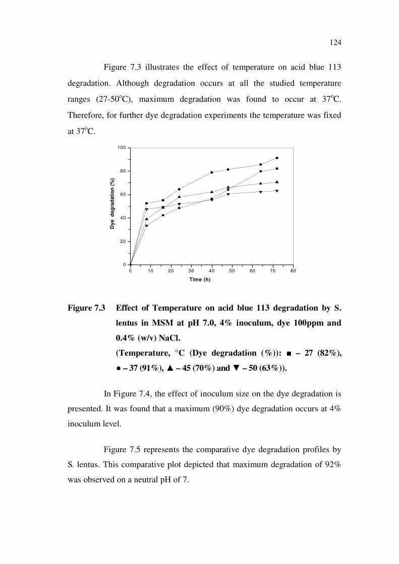
Figure 7.3 illustrates the effect of temperature on acid blue 113
degradation. Although degradation occurs at all the studied temperature
ranges (27-50oC), maximum degradation was found to occur at 37
oC.
Therefore, for further dye degradation experiments the temperature was fixed
at 37oC.
0 10 20 30 40 50 60 70 80
0
20
40
60
80
100
Dy
e d
eg
rad
ati
on
(%
)
Time (h)
Figure 7.3 Effect of Temperature on acid blue 113 degradation by S.
lentus in MSM at pH 7.0, 4% inoculum, dye 100ppm and
0.4% (w/v) NaCl.
(Temperature, °C (Dye degradation (%)): – 27 (82%),
– 37 (91%), – 45 (70%) and – 50 (63%)).
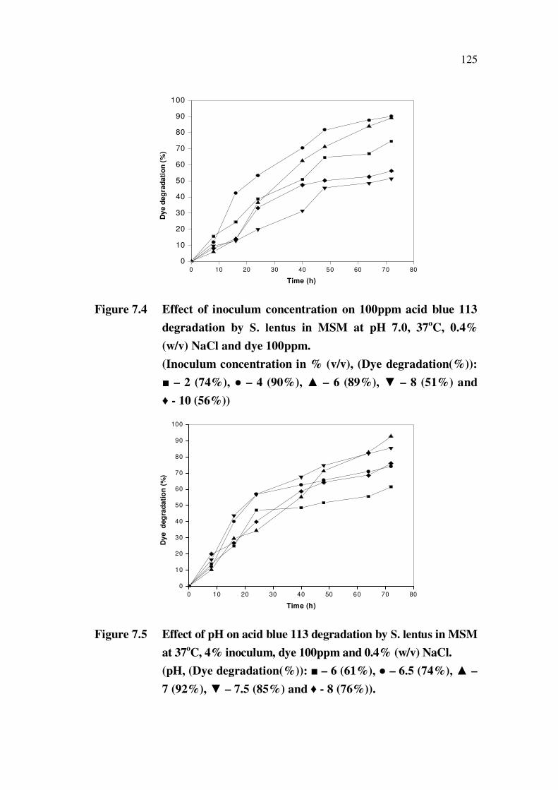
In Figure 7.4, the effect of inoculum size on the dye degradation is
presented. It was found that a maximum (90%) dye degradation occurs at 4%
inoculum level.
Figure 7.5 represents the comparative dye degradation profiles by
S. lentus. This comparative plot depicted that maximum degradation of 92%
was observed on a neutral pH of 7.
125
0 10 20 30 40 50 60 70 80
0
10
20
30
40
50
60
70
80
90
100
Dy
e d
eg
rad
ati
on
(%
)
Time (h)
Figure 7.4 Effect of inoculum concentration on 100ppm acid blue 113
degradation by S. lentus in MSM at pH 7.0, 37oC, 0.4%
(w/v) NaCl and dye 100ppm.
(Inoculum concentration in % (v/v), (Dye degradation(%)):
– 2 (74%), – 4 (90%), – 6 (89%), – 8 (51%) and
- 10 (56%))
0 10 20 30 40 50 60 70 80
0
10
20
30
40
50
60
70
80
90
100
Dy
e d
eg
rad
ati
on
(%
)
Time (h)
Figure 7.5 Effect of pH on acid blue 113 degradation by S. lentus in MSM
at 37oC, 4% inoculum, dye 100ppm and 0.4% (w/v) NaCl.
(pH, (Dye degradation(%)): – 6 (61%), – 6.5 (74%), –
7 (92%), – 7.5 (85%) and - 8 (76%)).

126
In order to select a suitable carbon source that maximum degrades
acid blue 113, several carbon sources were screened. The resulted are
presented in Figure 7.6. It was found that glucose is the best carbon source for
maximum degradation.
0 10 20 30 40 50 60 70 80
0
20
40
60
80
100
Dy
e d
eg
rad
ati
on
(%
)
Time (h)
Figure 7.6 Effect of carbon source on Acid blue 113 degradation by S.
lentus in MSM media 5 g/L of different carbon sources at
pH 7, 37oC, 4% inoculum concentration and 0.4% NaCl.
(Carbon Source (Dye degradation (%)): ( – Glucose (91%),
– Fructose (86%), – Sucrose (84%) and – Lactose
(82%))
It was found that incorporating dye in the growth process did not
alter the optimized conditions, excepting the notable difference in dye
degradation efficiency. With the optimized growth parameters, dye
degradation experiments were carried out by varying the initial dye
concentration from 25 to 500 ppm levels in order to verify the efficiency of
the organism under study. The shake flask experimental results are presented
in Figure 7.7.
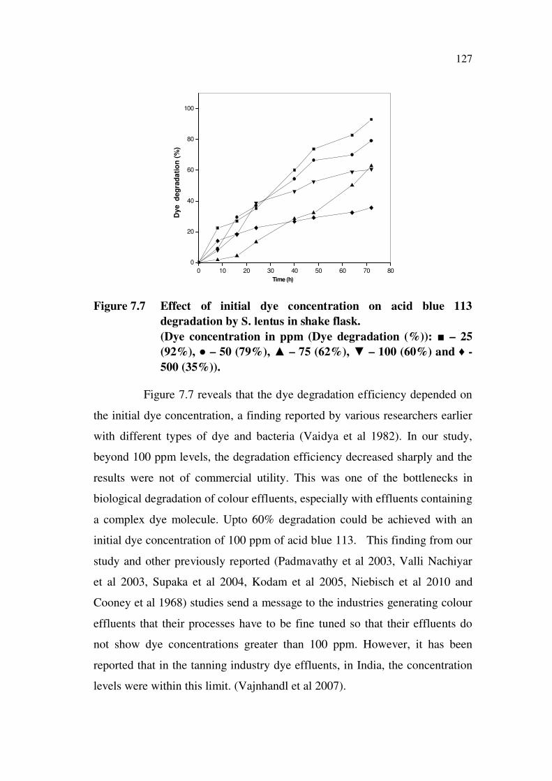
127
0 10 20 30 40 50 60 70 80
0
20
40
60
80
100
Dy
e d
eg
rad
ati
on
(%
)
Time (h)
Figure 7.7 Effect of initial dye concentration on acid blue 113
degradation by S. lentus in shake flask.
(Dye concentration in ppm (Dye degradation (%)): – 25
(92%), – 50 (79%), – 75 (62%), – 100 (60%) and -
500 (35%)).
Figure 7.7 reveals that the dye degradation efficiency depended on
the initial dye concentration, a finding reported by various researchers earlier
with different types of dye and bacteria (Vaidya et al 1982). In our study,
beyond 100 ppm levels, the degradation efficiency decreased sharply and the
results were not of commercial utility. This was one of the bottlenecks in
biological degradation of colour effluents, especially with effluents containing
a complex dye molecule. Upto 60% degradation could be achieved with an
initial dye concentration of 100 ppm of acid blue 113. This finding from our
study and other previously reported (Padmavathy et al 2003, Valli Nachiyar
et al 2003, Supaka et al 2004, Kodam et al 2005, Niebisch et al 2010 and
Cooney et al 1968) studies send a message to the industries generating colour
effluents that their processes have to be fine tuned so that their effluents do
not show dye concentrations greater than 100 ppm. However, it has been
reported that in the tanning industry dye effluents, in India, the concentration
levels were within this limit. (Vajnhandl et al 2007).

128
The shake flask optimized conditions (Table7.1) were employed in
further biocalorimetric experiments for determining the influence of aeration
and agitation rates on dye degradation.
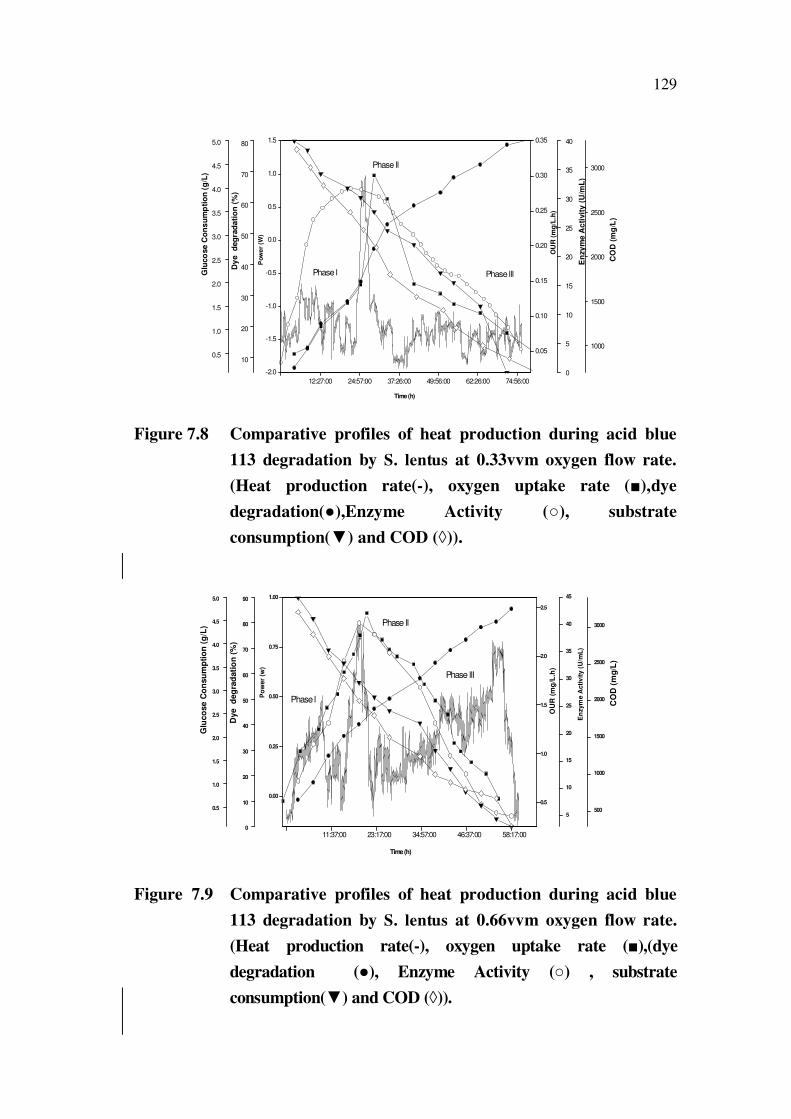
7.4.2 Effect of Aeration on Heat Release Rates and Dye Degradation
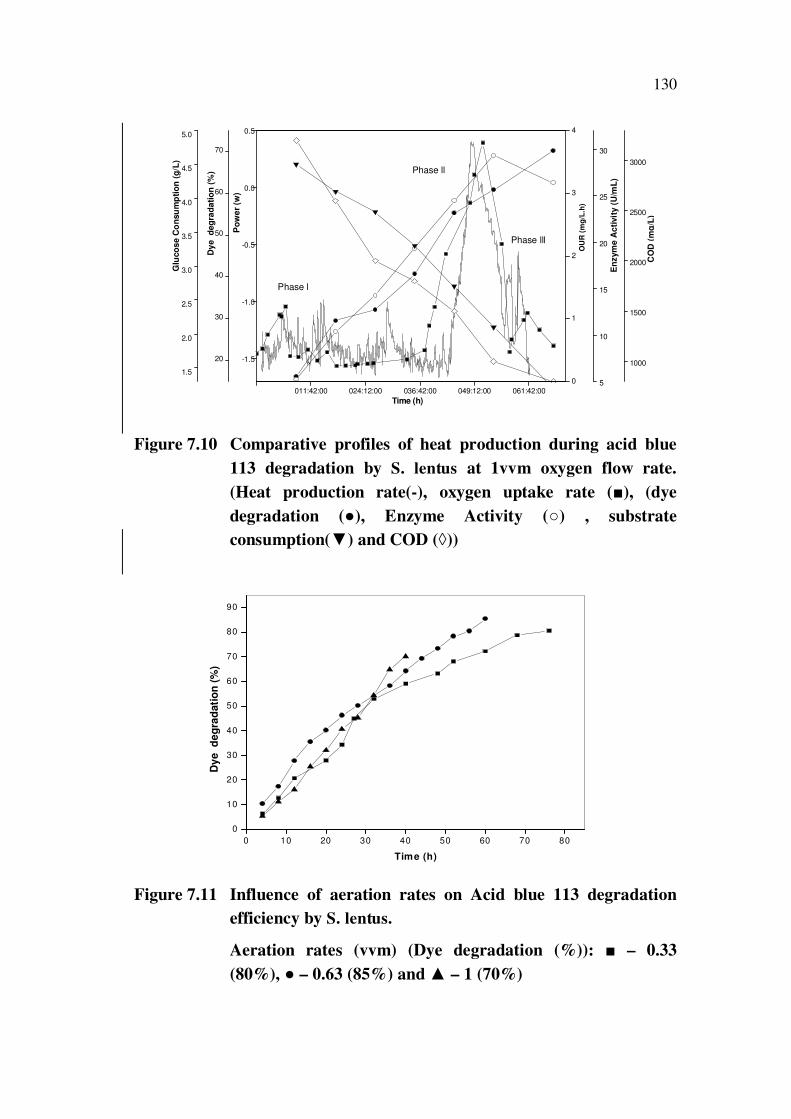
The comparative plots of biocalorimetric experiments for acid blue
113 degradation at varying aeration rates 0.33, 0.66, 1.0 vvm are presented in
Figures 7.8-7.10 and percent dye degraded alone are presented in Figure 7.11.
The figures showed that forced aeration certainly increased the dye
degradation efficiency from 60% levels in shaker flask to 85 % at 0.66vvm
oxygen flow (Figure 7.9). The COD values were found to reduce from 3200
ppm to 450 ppm as a result of dye degradation. Increase in aeration rates up
to 0.66 vvm resulted in the increase in dye degradation. Further increase in
aeration to 1vvm resulted in decrease of maximum degradation efficiency to
70 %. The peak heat release rates, OUR and enzyme release rates were
found to correspond to the degradation efficiency. As a result of growth, dye
degradation and substrate consumption, the power time curve exhibited three
distinct phases. In the endogenous phase the degradation rates were low
compared to the early and exponential phases.
Interestingly, one can see a clear shift in the peak heat release rates
(time shift) with change in aeration rates; at 0.33 vvm, the peak heat release
occurred between 20- 37 h, and at 0.66 vvm it occurred between 11 and 24 h
and at 1 vvm it was beyond 38 h. This observation was an indication of the
organism’s efficiency to adapt to various aeration rates; although the
organism was able to initiate its growth and cell multiplication by
consumption of glucose and dye, right from the beginning, at both 0.33 and
0.66 vvm, at higher O2 flow rates, it could not effectively carry out the
metabolism. This resulted in poor dye degradation efficiency and reflected in other
parameters such as OUR, enzyme activity and COD. So aeration rates were found
to be vital not only for growth but for efficient dye degradation also.
129
12:27:00 24:57:00 37:26:00 49:56:00 62:26:00 74:56:00
-2.0
-1.5
-1.0
-0.5
0.0
0.5
1.0
1.5
0.05
0.10
0.15
0.20
0.25
0.30
0.35
10
20
30
40
50
60
70
80
0
5
10
15
20
25
30
35
40
0.5
1.0
1.5
2.0
2.5
3.0
3.5
4.0
4.5
5.0
1000
1500
2000
2500
3000
Po
we
r (W
)
Time (h)
OU
R (
mg
/L.h
)
CO
D (
mg
/L)
Glu
co
se
Co
ns
um
pti
on
(g
/L)
Dy
e d
eg
rad
ati
on
(%
)
Phase l
Phase ll
Phase lll
En
zy
me
Ac
tivit
y (
U/m
L)
Figure 7.8 Comparative profiles of heat production during acid blue
113 degradation by S. lentus at 0.33vvm oxygen flow rate.
(Heat production rate(-), oxygen uptake rate ( ),dye
degradation( ),Enzyme Activity ( ), substrate
consumption( ) and COD ( )).
11:37:00 23:17:00 34:57:00 46:37:00 58:17:00
0.00
0.25
0.50
0.75
1.00
0.5
1.0
1.5
2.0
2.5
0
10
20
30
40
50
60
70
80
90
5
10
15
20
25
30
35
40
45
0.5
1.0
1.5
2.0
2.5
3.0
3.5
4.0
4.5
5.0
500
1000
1500
2000
2500
3000
Po
we
r (w
)
Time (h)
Dye d
eg
rad
ati
on
(%
)
Phase l
Phase ll
Phase lll
OU
R (
mg
/L.h
)
CO
D (
mg
/L)
Glu
co
se
Co
nsu
mp
tio
n (
g/L
)
En
zym
e A
cti
vit
y (
U/m
L)
Figure 7.9 Comparative profiles of heat production during acid blue
113 degradation by S. lentus at 0.66vvm oxygen flow rate.
(Heat production rate(-), oxygen uptake rate ( ),(dye
degradation ( ), Enzyme Activity ( ) , substrate
consumption( ) and COD ( )).
130
011:42:00 024:12:00 036:42:00 049:12:00 061:42:00
-1.5
-1.0
-0.5
0.0
0.5
0
1
2
3
4
20
30
40
50
60
70
5
10
15
20
25
30
1.5
2.0
2.5
3.0
3.5
4.0
4.5
5.0
1000
1500
2000
2500
3000
Po
we
r (w
)
Time (h)
Dy
e d
eg
rad
ati
on
(%
)
Phase l
Phase ll
Phase lll
OU
R (
mg
/L.h
)
CO
D (
mg
/L)
En
zym
e A
cti
vit
y (
U/m
L)
Glu
co
se C
on
su
mp
tio
n (
g/L
)
Figure 7.10 Comparative profiles of heat production during acid blue
113 degradation by S. lentus at 1vvm oxygen flow rate.
(Heat production rate(-), oxygen uptake rate ( ), (dye
degradation ( ), Enzyme Activity ( ) , substrate
consumption( ) and COD ( ))
0 10 20 30 40 50 60 70 80
0
10
20
30
40
50
60
70
80
90
Dy
e d
eg
rad
ati
on
(%
)
Time (h)
Figure 7.11 Influence of aeration rates on Acid blue 113 degradation
efficiency by S. lentus.
Aeration rates (vvm) (Dye degradation (%)): – 0.33
(80%), – 0.63 (85%) and – 1 (70%)

131
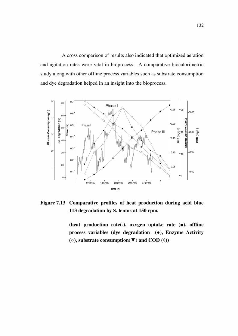
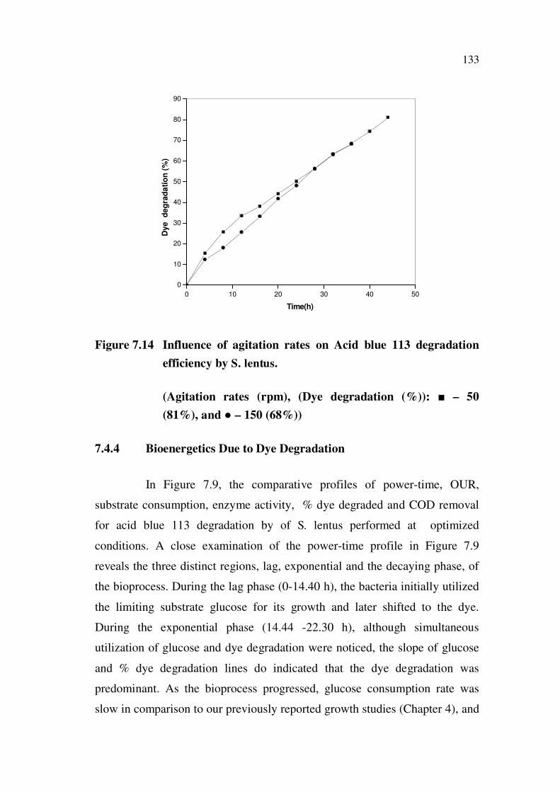
7.4.3 Effect of Agitation on Heat Release Rates and Dye Degradation
The effect of agitation on the acid blue 113 degradation can be seen
from the comparative heat profile plots presented in Figures 7.12 and 7.13 and
the degradation efficiency alone in Figure 7.14. These plots indicated that
increase in agitation rate from 50 to 100 rpm increased oxygen uptake rates
leading to increase in dye degradation efficiency. Further increase to 150 rpm
resulted both in decrease of OUR and dye degradation efficiency, therefore
the agitation did not have influence either the advancement (or) postponement
of the simultaneous growth and dye degradation process, unlike the effects of
aeration. Our studies led conclude that very high agitation was unnecessary
and not favourable to achieve maximum dye degradation efficiency
(Figure 7.13). The sample collected towards the end of the 50 h at 100 rpm
and 0.66 vvm aeration was almost colourless.
7:27:00 14:57:00 22:27:00 29:57:00 37:27:00 44:57:00
-0.5
0.0
0.5
1.0
0.2
0.4
0.6
0.8
1.0
10
20
30
40
50
60
70
80
10
20
30
1
2
3
4
5
1000
1500
2000
2500
3000O
UR
(m
g/L
.h)
CO
D (
mg
/mL
)
En
zym
e A
cti
vit
y (
U/m
L)
Po
wer
(w)
Time (h)
Phase l
Phase ll
Phase lll
Dye d
eg
rad
ati
on
(%)
Glu
co
se C
on
su
mp
tio
n (
g/L
)
Figure 7.12 Comparative profiles of heat production during acid blue
113 degradation by S. lentus at 50 rpm.
(Heat production rate(-), oxygen uptake rate ( ), offline
process variables (dye degradation ( ), Enzyme Activity
), substrate consumption( ) and COD ( ))
132
A cross comparison of results also indicated that optimized aeration
and agitation rates were vital in bioprocess. A comparative biocalorimetric
study along with other offline process variables such as substrate consumption
and dye degradation helped in an insight into the bioprocess.
07:27:00 14:57:00 22:27:00 29:57:00 37:27:00 --
0.1
0.2
0.3
0.4
0.5
0.6
0.7
0.05
0.10
0.15
0.20
0.25
10
20
30
40
50
60
70
5
10
15
20
1
2
3
4
5
1500
2000
2500
3000
OU
R (
mg
/L.h
)
Phase lll
Phase ll
Phase l
Time (h)
CO
D (
mg
/L)
En
zym
e A
cti
vit
y (
U/m
L)
Po
wer
(w)
Dye
de
gra
dati
on
(%
)
Glu
co
se C
on
su
mp
tio
n (
g/L
)
Figure 7.13 Comparative profiles of heat production during acid blue
113 degradation by S. lentus at 150 rpm.
(heat production rate(-), oxygen uptake rate ( ), offline
process variables (dye degradation ( ), Enzyme Activity
), substrate consumption( ) and COD ( ))
133
0 10 20 30 40 50
0
10
20
30
40
50
60
70
80
90
Dye
deg
rad
ati
on
(%
)
Time(h)
Figure 7.14 Influence of agitation rates on Acid blue 113 degradation
efficiency by S. lentus.
(Agitation rates (rpm), (Dye degradation (%)): – 50
(81%), and – 150 (68%))
7.4.4 Bioenergetics Due to Dye Degradation
In Figure 7.9, the comparative profiles of power-time, OUR,
substrate consumption, enzyme activity, % dye degraded and COD removal
for acid blue 113 degradation by of S. lentus performed at optimized
conditions. A close examination of the power-time profile in Figure 7.9
reveals the three distinct regions, lag, exponential and the decaying phase, of
the bioprocess. During the lag phase (0-14.40 h), the bacteria initially utilized
the limiting substrate glucose for its growth and later shifted to the dye.
During the exponential phase (14.44 -22.30 h), although simultaneous
utilization of glucose and dye degradation were noticed, the slope of glucose
and % dye degradation lines do indicated that the dye degradation was
predominant. As the bioprocess progressed, glucose consumption rate was
slow in comparison to our previously reported growth studies (Chapter 4), and
134
even after 40 hours, only 60 % glucose was consumed. Again after the 45th
hour, the bacteria reversed the trend to consume glucose faster. The
exponential phase was thus marked by simultaneous utilization of glucose and
dye degradation. It was noticed that the azo reductase enzyme release
enhanced the dye degradation resulting in peak heat release. After 50% dye
degradation, marked by heat release and OUR, the enzyme release also
decreased. Heat release and OUR pattern followed each other as reported
(Senthilkumar et al 2008 b).
As can be seen from the comparative graph, the power time curve
follows OUR pattern. There are three shifts in the heat curve due to (a)
utilization of glucose as primary carbon source initially, (b) simultaneous
utilization of glucose and dye, finally (c) dye degradation. Glucose
consumption decreased as enzyme activity increased up to 40 U/mL;
simultaneously dye degradation increased to 85 % and COD decreased. The
results suggested that power-time profile could be used as an indirect
parameter to measure COD removal and the application of calorimetry for
monitoring the dye degradation process was feasible. Quantitative
information on relative consumption of substrates, energy changes associated
with biomass growth and oxycalorific values were evaluated using cumulative
heat production values by integrating the power – time curve. The results are
discussed below.
7.4.5 Biomass Yield
Heat yield coefficient due to biomass, YQ/X (kJ heat evolved per
g cell dry weight formed) was determined from the plot between total heat
evolved by the culture (kJ/L and the biomass concentration (g/L). For dye
degradation process under varying aeration and agitation the values ranged
from 10-13 kJ/g (Table 7.2). Our results corroborated the earlier findings
(Vonstockar et al 1989).
135
7.4.6 Oxycalorific Coefficient
Oxycalorific coefficient was important to asses both the metabolic
efficiency and the aerobic nature of the organism under study. The theoretical
value for oxycalorific heat yield was 460 kJ/mol of oxygen consumed
irrespective of the nature of the microorganism, in substrate or product
(Cooney et al 1968). Several researchers have (Volesky et al 1982), claimed
YQ/O > 400 kJ/mol to be the behind the pure aerobic nature of the process.
However in our studies we obtained values from 368 to 651.2 kJ/mol (Table –
7.2) for different aeration and agitation rates. The deviation from the
theoretical values was reported to be due to the degree of aerobicity, partly
anaerobic nature of the organism well as to the inability to precisely measure
the low OUR occurring at low values of substrate reductions. In OUR studies
under optimized conditions, the YQ/O value was 460 kJ/mol.
7.4.7 Heat Yield Due to Substrate Consumption
Determining the heat yield due to substrate consumption (kJ heat
evolved per g of glucose consumed) could be helpful in understanding
metabolism of S. lentus. Quite interestingly, in our experiments the organism
was consuming glucose and dye for the entire bioprocess duration.
Assuming the growth to be mainly due to glucose , a simple comparison of
the heat yields due to substrate and biomass indicated that catabolic activity
contributed maximum heat evolution (YQ/S > YQ/X). Generally for an aerobic
process, when glucose acted as the substrate this value would be around 20
kJ/g. The value obtained now was higher in all cases and particularly at
optimized conditions 25.4 kJ/g.
The reason for such a high value and low
biomass values for 0.33 vvm and 1 vvm could be the shift of limiting carbon
source from glucose to the dye (Table 7.2). The detailed bioenergetics studies
presented here might help resolve the issues in design and scale up of a
suitable bioreactor for commercial dye degradation process.

136
Table 7.2 Influence of aeration and agitation rates on bioenergetics
during acid blue 113 degradation by S. lentus in BioRTCal
ExperimentsYQ/X
(kJ/g)
YQ/S
(kJ/g)
YQ/O
(kJ/mol)
YQ/cod
(kJ/mg)
0.33 vvm / 100 rpm 11.03 17 508.6 7.97
0.66 vvm/100 rpm 13.27 25.4 460 12.2
1 vvm/100rpm 10.16 14 651.2 10.3
50 rpm 12.33 20 426 11.09
150 rpm 9.12 13.2 368 5.43
7.4.8 Analysis of Decolorized Product of Acid Blue 113
So far there are no reported studies on the elucidation of
degradation pathways of acid blue 113 although carried out in other azo dyes
such as RHE 7B (kalyani et al 2008), Red BL1 (Satish et al 2007) RO (Sarayu
et al 2010), RY107, RB5, RR198 and DB71 (Elisangela et al 2009), reactive
blue 172 (Dhanve et al 2008) and acid red GR (Xu et al 2007).
7.4.9 HPLC Analysis
The HPLC Chromatogram of the degraded samples collected from
BioRTCal at the 24 h and 72 h along with the pure dye is in Figure 7.15. The
peak at 7.03 RT in Figure 7.15 (a) was due to the dye molecule (Xueheng et al
2007). As the degradation process went on, the intensity of the peak at
7.03 RT decreased. HPLC studies confirmed the efficiency of S. lentus to
degrade the complex molecule of Acid blue 113.

137
(a)
(b)
(c)
Figure 7.15 HPLC Chromatogram showing the progress of dye
degradation (a) pure dye (b) 24th
h (c) 72nd
h
138
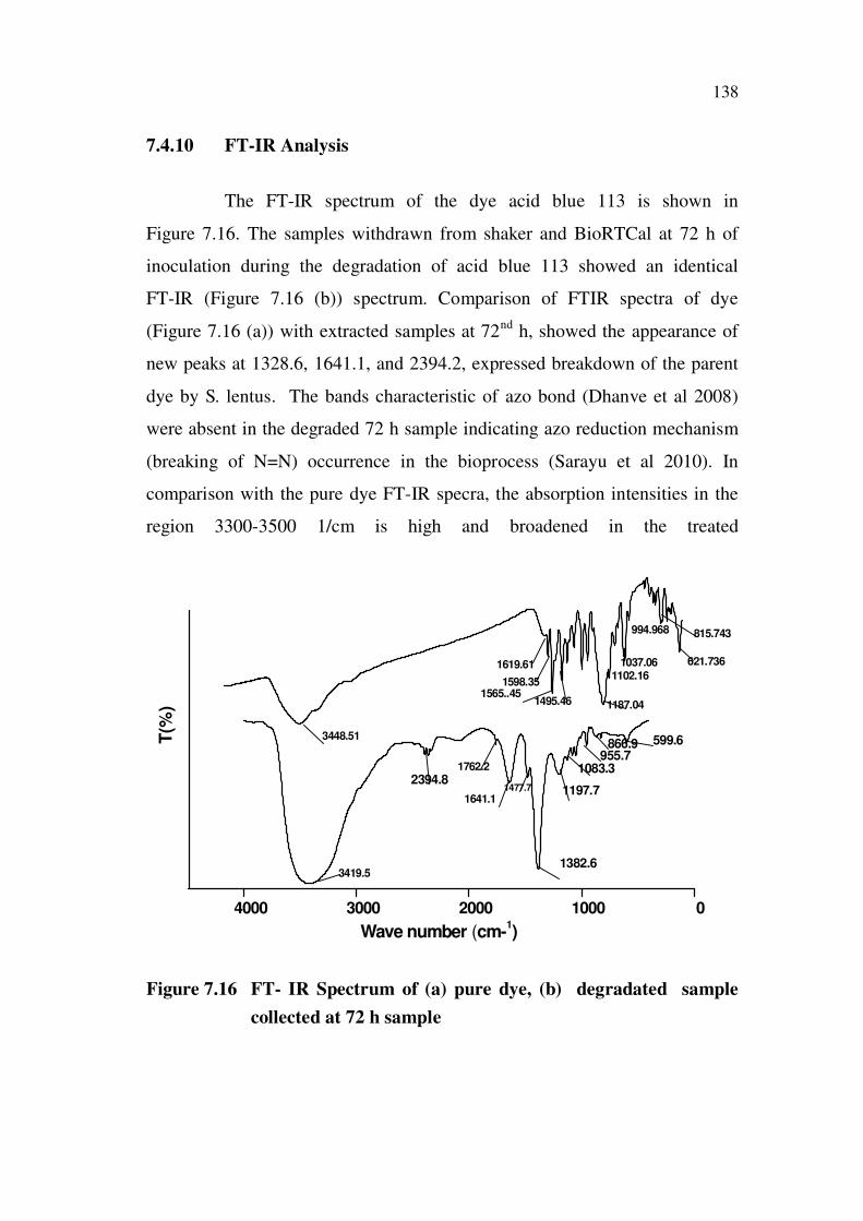
7.4.10 FT-IR Analysis
The FT-IR spectrum of the dye acid blue 113 is shown in
Figure 7.16. The samples withdrawn from shaker and BioRTCal at 72 h of
inoculation during the degradation of acid blue 113 showed an identical
FT-IR (Figure 7.16 (b)) spectrum. Comparison of FTIR spectra of dye
(Figure 7.16 (a)) with extracted samples at 72nd
h, showed the appearance of
new peaks at 1328.6, 1641.1, and 2394.2, expressed breakdown of the parent
dye by S. lentus. The bands characteristic of azo bond (Dhanve et al 2008)
were absent in the degraded 72 h sample indicating azo reduction mechanism
(breaking of N=N) occurrence in the bioprocess (Sarayu et al 2010). In
comparison with the pure dye FT-IR specra, the absorption intensities in the
region 3300-3500 1/cm is high and broadened in the treated
4000 3000 2000 1000 0
1382.6
599.6866.9955.7
1083.3
1197.71477.7
1641.1
1762.2
2394.8
3419.5
1187.041495.461565..45
1598.35
3448.51T(%
)
Wave number (cm-1)
621.736
815.743994.968
1037.061102.16
1619.61
Figure 7.16 FT- IR Spectrum of (a) pure dye, (b) degradated sample
collected at 72 h sample

139
sample, and this indicated the increase in the number of OH and NH groups
as a result of biodegradation. Broadening of IR spectra is found to be due to
destruction of the conjugated, aromatic structure of the dye upon microbial
treatment (Pourbabee et al 2006). A peak appearing at 1641.751/cm showed
the presence of compounds containing C (NH)=O. Also peaks at 1382.6 1/cm
represented N-H bending vibrations. Thus the FTIR spectrum of the sample
after decolourization showed characteristic change in peak positions as
compared to pure dye sample.
7.4.11 GC-MS Analysis
7.4.11.1 Decolorized Product from Shake Flask Experiments
The products identified from the GC-MS trace spectra (Appendix 4)
are listed in Table 7.3. The degradation pathway shown in Figure 7.17 was
worked out based on the product profile listed in Table 7.3. The poly
aromatic dye underwent azoreduction via ring cleavage, to yield aromatic
compounds and one sulphur – aniline derivative. While reporting the
degradation products of Navitan Fast Blue by pseudomonas aeruginosa
(Valli Nachiyar et al 2004 ) by GC-MS analysis, the authors also found the
ring cleavage of the aromatic dye to yield similar products as found in our
study. The formation of intermediates such as pthalic acid, long chain
alkanes and diethyl pthalate indicated a similar degradation approach for the
non – nitrogen moiety in the dye. In addition, products such as palmitic acid
and its corresponding unsaturated vinyl ester were also identified. Aerobic
biodegradation of aromatic compounds have several common features.
Structurally diverse compounds are degraded through many different
peripheral pathways to a few intermediates that are further channeled via a
few central pathways to the central metabolism of the cell. In the aerobic
catabolic funnel, most peripheral pathways involve oxygenation reactions
carried out by monooxygenases and hydroxylating dioxygenases that generate
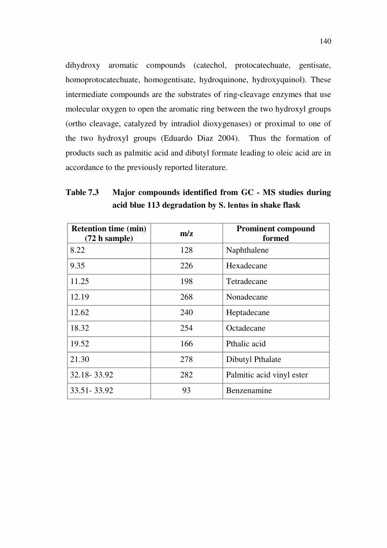
140
dihydroxy aromatic compounds (catechol, protocatechuate, gentisate,
homoprotocatechuate, homogentisate, hydroquinone, hydroxyquinol). These
intermediate compounds are the substrates of ring-cleavage enzymes that use
molecular oxygen to open the aromatic ring between the two hydroxyl groups
(ortho cleavage, catalyzed by intradiol dioxygenases) or proximal to one of
the two hydroxyl groups (Eduardo Diaz 2004). Thus the formation of
products such as palmitic acid and dibutyl formate leading to oleic acid are in
accordance to the previously reported literature.
Table 7.3 Major compounds identified from GC - MS studies during
acid blue 113 degradation by S. lentus in shake flask
Retention time (min)
(72 h sample)m/z
Prominent compound
formed
8.22 128 Naphthalene
9.35 226 Hexadecane
11.25 198 Tetradecane
12.19 268 Nonadecane
12.62 240 Heptadecane
18.32 254 Octadecane
19.52 166 Pthalic acid
21.30 278 Dibutyl Pthalate
32.18- 33.92 282 Palmitic acid vinyl ester
33.51- 33.92 93 Benzenamine

141
DODECANE TETRADECANE
HEXADECANE
(Reduction of C groups
intermediate R-CH2-COONa)
COOH
COOH
EsterificationOxidation toalcohols
COOC4H9
COOC4H9
DIBUTYL PTHALATE
CH3(CH2)7CH=CH(CH2)7COOH
OLEIC ACID
C11H23
HO
O
PALMITIC ACID
Esterification
C11H23
O
O
PALMITIC ACID VINYL ESTER
NH2
N
N
N
N
HN
SO3Na
SO3Na
Acic blue 113
SO3Na
HN
H2N
H2N
NH2
NH2
SO3Na
Desulfonation
Azo reduction
Oxidative Deamination
m/z:93
m/z : 226,198268,240 and 254
m/z: 166
m/z : 278
Figure 7.17 Degradation pathways of acid blue 113 by S. lentus in shake
flask

142
7.4.12 Decolorized Product from BioRTCal Experiments
The predominant degradation products identified from the GC-MS
trace spectra (Appendix 5) from the sample collected at 72 h of the
inoculation are listed in Table 7.4. The degradation pathway (Figure 7.18)
was worked out based on the product profile listed in Table 7.4. The dye
degradation did undergo azoreduction to yield fragmented aromatic products
consisting of three, two and one aromatic rings (Neill et al 2006),
Intermediates such as pthalic acid were obtained followed by corresponding
esters such as dibutyl phthalate. This could possibly due to formation of long
chain hydrocarbons followed by oxidation to alcohols. Aniline and pthalic
acid undergo oxidative deamination to resorcinol using molecular oxygen and
oxygenases that undergo further extra- ring oxidation to yield products such
as 1,3,5-benzenetriol.
The nitrogen moiety gets sequestered as benzeneamine via
desulphonation (progesterone, oleic acid) The presence of diazo derivative of
progesterone in the GC-MS analysis can be explained by the above
mechanism . The degradation of primary alcohol is carried out by the alcohol-
aldehyde dehydrogenase pathway to obtain the corresponding fatty acid
which explains the biosynthesis of oleic acid (Eduardo Diaz 2004). These
enzymes usually have a bound NAD (nicotine amine adenine dinucleotide),
and are induced by the presence of hydrocarbons (Robert et al 2003).
Examination of the degradation pathway undergone in shake flask and
BioRTCal, revealed that only partial oxidation of the dye molecule was
possible in shake flask experiments, although % decolourization was
satisfactory. In BioRTCal experiments the oxidative reactions continued even
after the primary azo reduction reactions leading to the biosynthesis of diazo
derivatives of progesterone and less toxic products. A vast literature on

143
biological degradation of various dyes employing different kinds of
microorganism is available and one gets confused with the utility of the data.
It is because the studies are not comprehensive; in most of the reported
studies, either the chemical characterization of the metabolites of the dye nor
toxicity assessments are done to prove that the products of degradation do not
contain a toxic metabolite. From this perspective the present study is
comprehensive to prove that the product of degradation is less toxic in nature.
Table 7.4 Major compounds identified from GC - MS studies during
acid blue 113 degradation by S. lentus in BioRTCal
Time(h)Retention
Time (min)m/z
Prominent Compound
Formed
24 16.13 166 Pthalic Acid
24 8.06 198 Tetradecane
24 17.91 278 Dibutyl Pthalate
36 30.20 282 Oleic acid
72 6.40 110 Resorcinol
72 12.39 126 1,3,5, Benzenetriol
72 33.26 340 Diazoprogesterone
72 34.4 144 2-naphthalenone

144
N
N
N
N
HN
SO3Na
SO3Na
ACID BLUE 113
HN
H2N
H2N
NH2
NH2
COOH
COOH
OH
OH
SO3Na
NH2
BENZENAMINE
Desulfonation
COOC4H9
COOC4H9
OH
HO OH
PTHALIC ACID
RESRCINOL
Further oxidation
1,3,5,BENZENETRIOLDIBUTYL PTHALATE
OLEIC ACID BIOSYNTHESIS ALCOHOL
NAD/NADH Fatty acid
Azo reduction
Oxidative Deamination
Oxidation to alcohols
m/z : 110
m/z : 166
m/z : 278
m/z : 126
m/z : 282
Figure 7.18 Degradation pathways of acid blue 113 by S. lentus in
BioRTCal

145
7.4.13 Heat of Reaction for the Degradative Pathway of Acid Blue 113
The bio-degradation of the acid blue 113 dye was carried out in the
bio-reaction calorimeter using the bacterial strain S. lentus. Here glucose was
used as the carbon source. Using the CHN analysis the empirical formula of
S. lentus was determined. The molecular structures of compounds in the
degradation pathway (Figure 7.19) were drawn in CHEMDRAW software
version 7.3 and incorporated into CHETAH software (Shanley et al 1995)
using smiley input. The experimental heat of reaction values obtained from
BioRTCal (7586.3kJ/mol) was due to simultaneous growth of the organism and
dye degradation. The heat of reaction values (7545.3 kJ/mol) determined from
the growth experiments performed at identical conditions were subtracted
from the dye degradation experiment to obtain the heat of reaction for
degradation alone (7258.23kJ/mol) The values were found to be close and
thus further validated the mechanism of acid blue 113 degradation.
Figure 7.19 CHETAH for biodegradation of acid blue 113

146
7.4.14 Cytotoxicity Assessment
Figure 7.20 the percent viability of Vero cells to the toxins are
shown. The IC50 values for the samples were found out to be 1.22 mg/mL.
Since the concentration levels used for cytotoxicity testing were very high in
our studies in comparison to many reported studies (Adedayo et al 2004), it
was thought appropriate to consider the IC90 values to assess the toxic nature.
The IC 90 values (78 mg/mL) shows 91 % cell viability and confirms less
toxic nature of the degradation products obtained in our studies. Moreover, in
actual effluents the concentration levels will be very less due to dilution of
large quantities of water. Oxidation of aromatic amines in the aerobic stage
was found to be responsible for less toxicity of the extracts (Elisangela et al
2009).
0 1 2 3 4 5
30
40
50
60
70
80
90
100
via
bilit
y (
%)
Concentration (mg/mL)
Figure: 7.20 MTT assay showing the cytotoxicity of Vero cells

147
7.5 CONCLUSION
Dye degradation of Acid blue 113 by the bacteria S. lentus
was successfully performed in a BioRTCal for the first time.
Power-time and OUR curve exhibited similar trends
suggesting that profiles could be used for monitoring the
biological degradation of color effluents.
The shifts observed in power- time profile indicated three
distinct phases of the bioprocess, and suggested simultaneous
utilization of glucose (primary) and dye (secondary carbon
source). Secretion of azoreductase enzyme enhanced the
degradation process.
Optimization of aeration and agitation rates were found to be
vital for efficient dye degradation. The degradative pathway
of acid blue 113 by S. lentus was worked by performing
HPLC, FT-IR, and GC–MS analysis.
Interestingly, the predominant products identified were less
toxic and the final end product was found to be
Diazoprogesterone.
The biochemical energetics and the detailed mechanistic
pathway presented will be useful for designing a suitable
bioreactor for degradation of dye effluent.
Moreover, this study proves the feasibility of application of
calorimetry as an in-line analytical tool for monitoring dye
degradation process. Biocalorimetric data for dye degradation
have been reported for the first time here.