chapter 7 membrane structure and function. you should now be able to: 1.define the following terms:...
TRANSCRIPT

Chapter 7
• Membrane Structure and Function
You should now be able to:
1. Define the following terms: amphipathic molecules, aquaporins, diffusion
2. Explain how membrane fluidity is influenced by temperature and membrane composition
3. Distinguish between the following pairs or sets of terms: peripheral and integral membrane proteins; channel and carrier proteins; osmosis, facilitated diffusion, and active transport; hypertonic, hypotonic, and isotonic solutions
Copyright © 2008 Pearson Education, Inc., publishing as Pearson Benjamin Cummings

4. Explain how transport proteins facilitate diffusion
5. Explain how an electrogenic pump creates voltage across a membrane, and name two electrogenic pumps
6. Explain how large molecules are transported across a cell membrane
Copyright © 2008 Pearson Education, Inc., publishing as Pearson Benjamin Cummings
Overview: Life at the Edge
• The plasma membrane is the boundary that separates the living cell from its surroundings
• The plasma membrane exhibits selective permeability, allowing some substances to cross it more easily than others
Copyright © 2008 Pearson Education, Inc., publishing as Pearson Benjamin Cummings
Concept 7.1: Cellular membranes are fluid mosaics of lipids and proteins
• Phospholipids are the most abundant lipid in the plasma membrane
• Phospholipids are amphipathic molecules, containing hydrophobic and hydrophilic regions
• The fluid mosaic model states that a membrane is a fluid structure with a “mosaic” of various proteins embedded in it
Copyright © 2008 Pearson Education, Inc., publishing as Pearson Benjamin Cummings
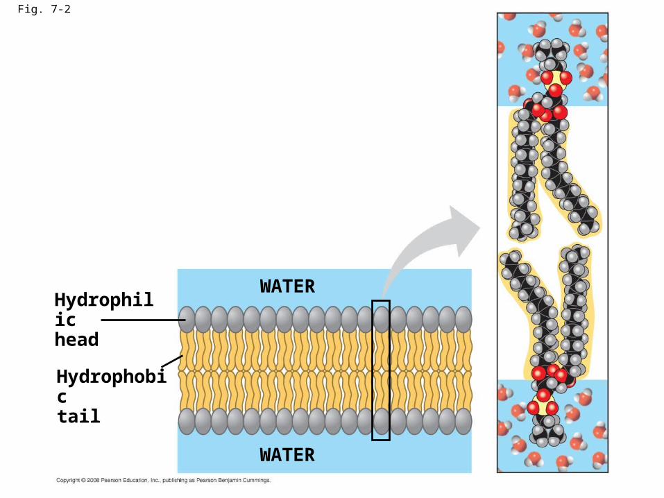
Fig. 7-2
Hydrophilichead
WATER
Hydrophobictail
WATER
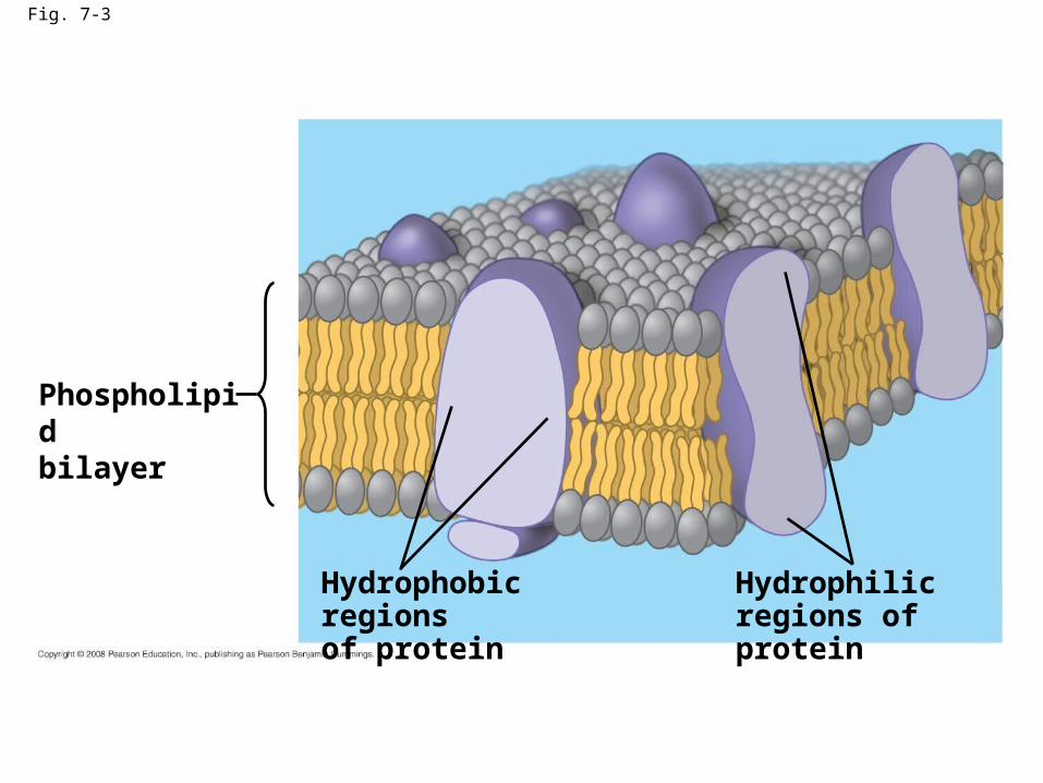
Fig. 7-3
Phospholipidbilayer
Hydrophobic regionsof protein
Hydrophilicregions of protein

Fig. 7-4
TECHNIQUE
Extracellularlayer
KnifeProteins Inside of extracellular layer
RESULTS
Inside of cytoplasmic layer
Cytoplasmic layerPlasma membrane
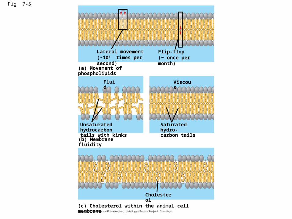
Fig. 7-5
Lateral movement(~107 times per second)
Flip-flop(~ once per month)
(a) Movement of phospholipids
(b) Membrane fluidity
Fluid Viscous
Unsaturated hydrocarbontails with kinks
Saturated hydro-carbon tails
(c) Cholesterol within the animal cell membrane
Cholesterol

Fig. 7-6
RESULTS
Membrane proteins
Mouse cellHuman cell
Hybrid cell
Mixed proteinsafter 1 hour
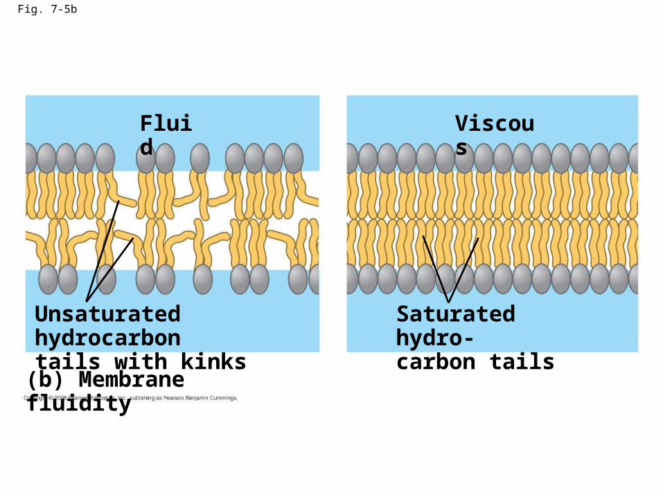
Fig. 7-5b
(b) Membrane fluidity
Fluid
Unsaturated hydrocarbontails with kinks
Viscous
Saturated hydro-carbon tails

Fig. 7-5c
Cholesterol
(c) Cholesterol within the animal cell membrane

Membrane Proteins and Their Functions
• A membrane is a collage of different proteins embedded in the fluid matrix of the lipid bilayer
• Proteins determine most of the membrane’s specific functions
Copyright © 2008 Pearson Education, Inc., publishing as Pearson Benjamin Cummings
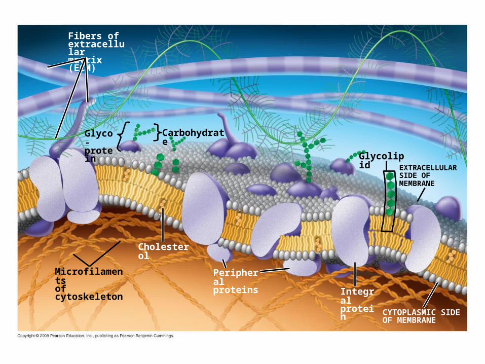
Fig. 7-7
Fibers ofextracellularmatrix (ECM)
Glyco-protein
Microfilamentsof cytoskeleton
Cholesterol
Peripheralproteins
Integralprotein
CYTOPLASMIC SIDEOF MEMBRANE
GlycolipidEXTRACELLULARSIDE OFMEMBRANE
Carbohydrate
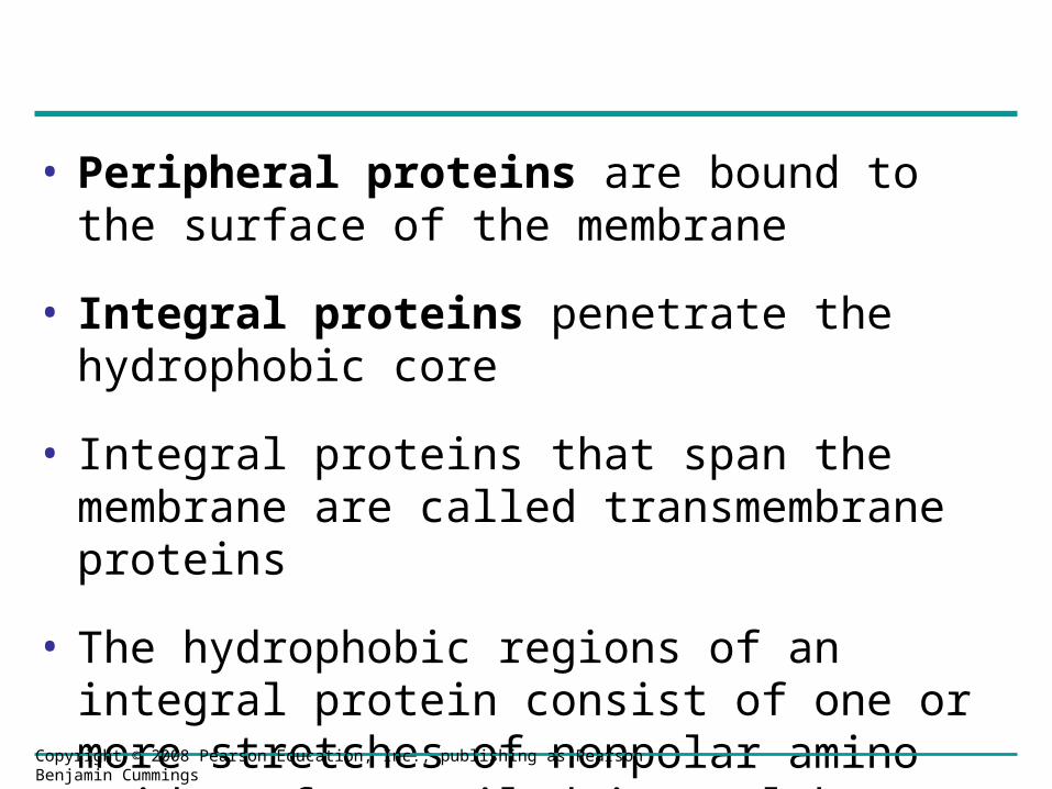
• Peripheral proteins are bound to the surface of the membrane
• Integral proteins penetrate the hydrophobic core
• Integral proteins that span the membrane are called transmembrane proteins
• The hydrophobic regions of an integral protein consist of one or more stretches of nonpolar amino acids, often coiled into alpha helices
Copyright © 2008 Pearson Education, Inc., publishing as Pearson Benjamin Cummings
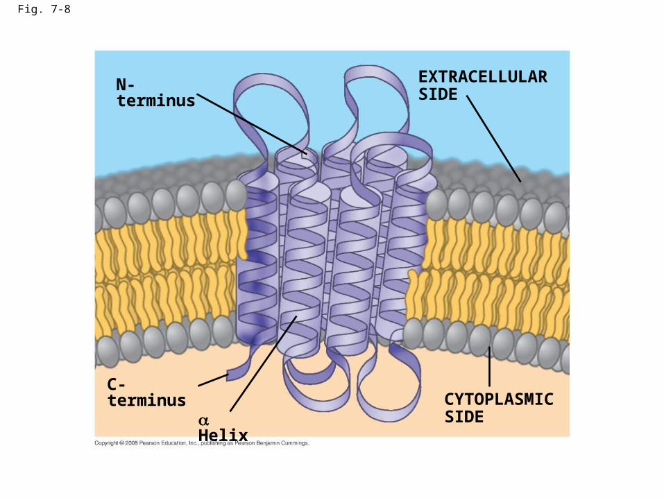
Fig. 7-8
N-terminus
C-terminus
HelixCYTOPLASMICSIDE
EXTRACELLULARSIDE

• Six major functions of membrane proteins:
– Transport
– Enzymatic activity
– Signal transduction
– Cell-cell recognition
– Intercellular joining
– Attachment to the cytoskeleton and extracellular matrix (ECM)
Copyright © 2008 Pearson Education, Inc., publishing as Pearson Benjamin Cummings
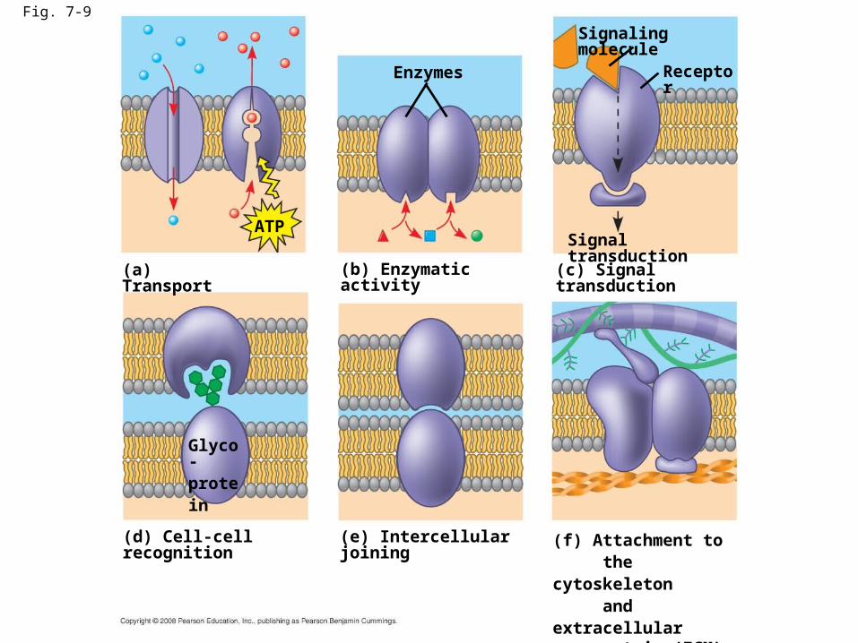
Fig. 7-9
(a) Transport
ATP
(b) Enzymatic activity
Enzymes
(c) Signal transduction
Signal transduction
Signaling molecule
Receptor
(d) Cell-cell recognition
Glyco-protein
(e) Intercellular joining (f) Attachment to the cytoskeleton and extracellular matrix (ECM)
The Role of Membrane Carbohydrates in Cell-Cell Recognition
• Cells recognize each other by binding to surface molecules, often carbohydrates, on the plasma membrane
• Membrane carbohydrates may be covalently bonded to lipids (forming glycolipids) or more commonly to proteins (forming glycoproteins)
• Carbohydrates on the external side of the plasma membrane vary among species, individuals, and even cell types in an individual
Copyright © 2008 Pearson Education, Inc., publishing as Pearson Benjamin Cummings

Fig. 7-10
ER1
Transmembraneglycoproteins
Secretoryprotein
Glycolipid
2Golgiapparatus
Vesicle
3
4
Secretedprotein
Transmembraneglycoprotein
Plasma membrane:
Cytoplasmic face
Extracellular face
Membrane glycolipid
Synthesis and Sidedness of Membranes
Concept 7.2: Membrane structure results in selective permeability
• A cell must exchange materials with its surroundings, a process controlled by the plasma membrane
• Plasma membranes are selectively permeable, regulating the cell’s molecular traffic
Copyright © 2008 Pearson Education, Inc., publishing as Pearson Benjamin Cummings
The Permeability of the Lipid Bilayer
• Hydrophobic (nonpolar) molecules, such as hydrocarbons, can dissolve in the lipid bilayer and pass through the membrane rapidly
• Polar molecules, such as sugars, do not cross the membrane easily
Copyright © 2008 Pearson Education, Inc., publishing as Pearson Benjamin Cummings
Transport Proteins
• Transport proteins allow passage of hydrophilic substances across the membrane
• Some transport proteins, called channel proteins, have a hydrophilic channel that certain molecules or ions can use as a tunnel
• Channel proteins called aquaporins facilitate the passage of water
Copyright © 2008 Pearson Education, Inc., publishing as Pearson Benjamin Cummings

• Other transport proteins, called carrier proteins, bind to molecules and change shape to shuttle them across the membrane
• A transport protein is specific for the substance it moves
Copyright © 2008 Pearson Education, Inc., publishing as Pearson Benjamin Cummings
Concept 7.3: Passive transport is diffusion of a substance across a membrane with no energy investment
• Diffusion is the tendency for molecules to spread out evenly into the available space
• Although each molecule moves randomly, diffusion of a population of molecules may exhibit a net movement in one direction
• At dynamic equilibrium, as many molecules cross one way as cross in the other direction
Animation: Membrane SelectivityAnimation: Membrane Selectivity Animation: DiffusionAnimation: Diffusion
Copyright © 2008 Pearson Education, Inc., publishing as Pearson Benjamin Cummings
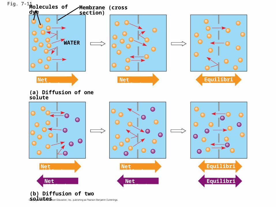
Fig. 7-11Molecules of dye Membrane (cross section)
WATER
Net diffusion Net diffusion Equilibrium
(a) Diffusion of one solute
Net diffusion
Net diffusion
Net diffusion
Net diffusion
Equilibrium
Equilibrium
(b) Diffusion of two solutes
• Substances diffuse down their concentration gradient, the difference in concentration of a substance from one area to another
• No work must be done to move substances down the concentration gradient
• The diffusion of a substance across a biological membrane is passive transport because it requires no energy from the cell to make it happen
Copyright © 2008 Pearson Education, Inc., publishing as Pearson Benjamin Cummings

Effects of Osmosis on Water Balance
• Osmosis is the diffusion of water across a selectively permeable membrane
• Water diffuses across a membrane from the region of lower solute concentration to the region of higher solute concentration
Copyright © 2008 Pearson Education, Inc., publishing as Pearson Benjamin Cummings
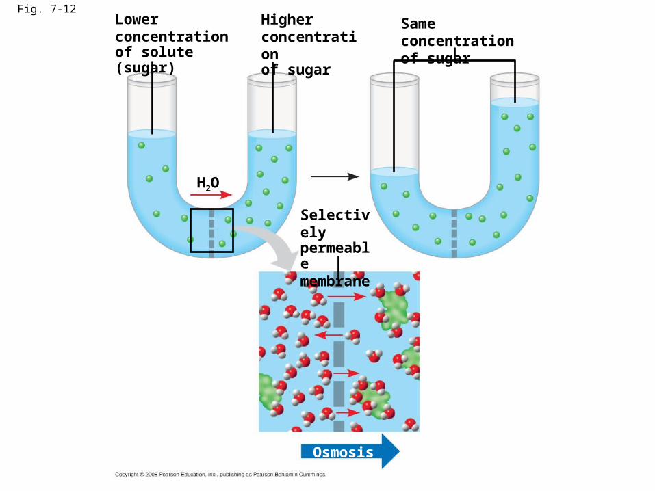
Lowerconcentrationof solute (sugar)
Fig. 7-12
H2O
Higher concentrationof sugar
Selectivelypermeablemembrane
Same concentrationof sugar
Osmosis
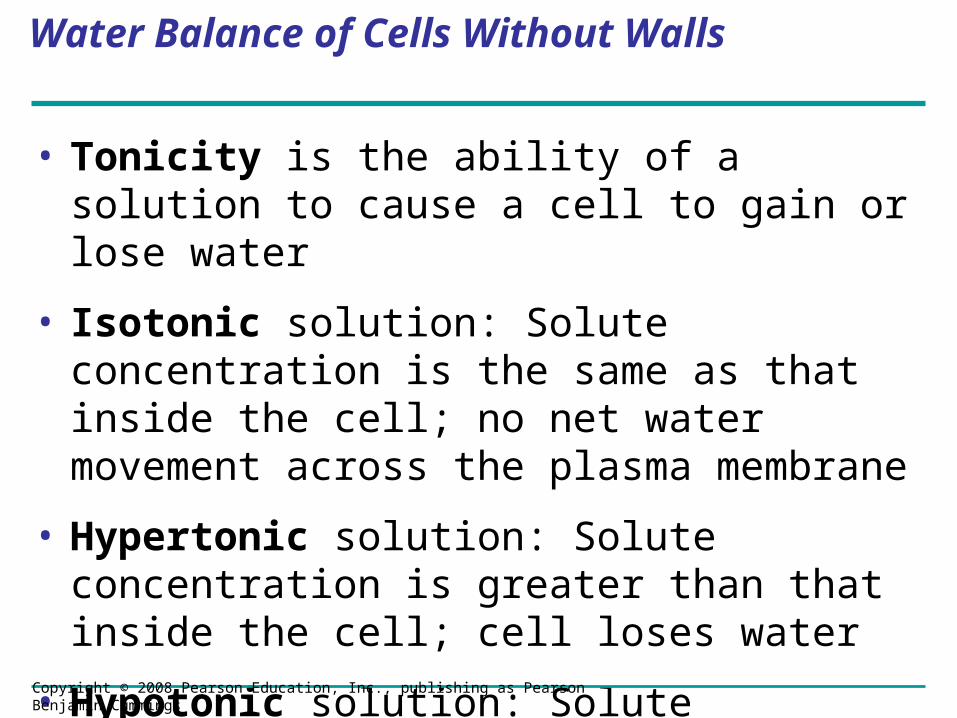
Water Balance of Cells Without Walls
• Tonicity is the ability of a solution to cause a cell to gain or lose water
• Isotonic solution: Solute concentration is the same as that inside the cell; no net water movement across the plasma membrane
• Hypertonic solution: Solute concentration is greater than that inside the cell; cell loses water
• Hypotonic solution: Solute concentration is less than that inside the cell; cell gains water
Copyright © 2008 Pearson Education, Inc., publishing as Pearson Benjamin Cummings
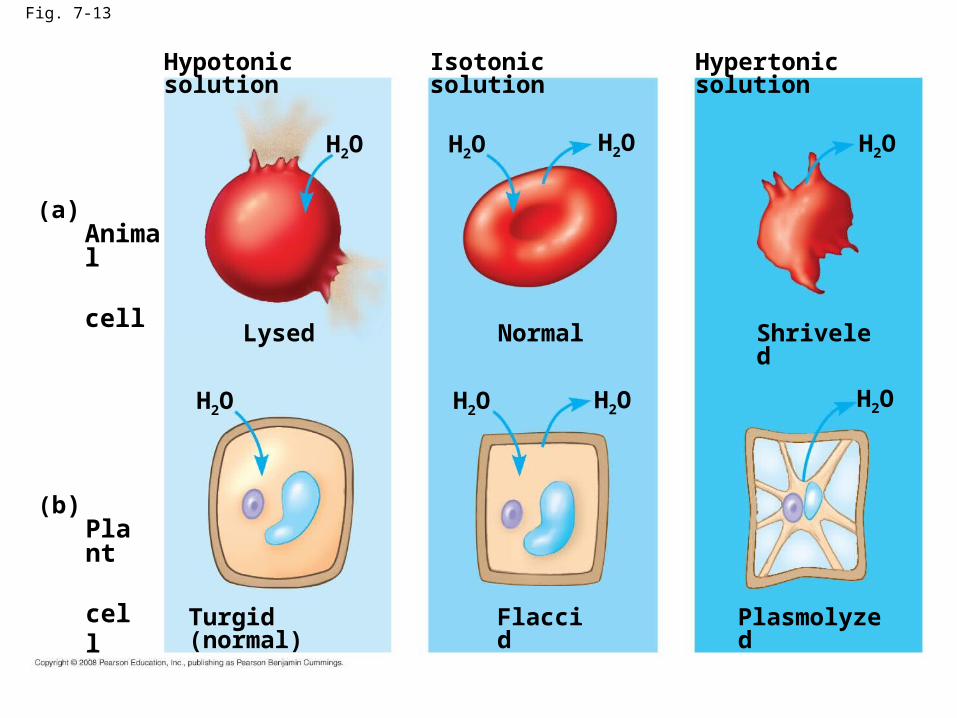
Fig. 7-13
Hypotonic solution
(a) Animal cell
(b) Plant cell
H2O
Lysed
H2O
Turgid (normal)
H2O
H2O
H2O
H2O
Normal
Isotonic solution
Flaccid
H2O
H2O
Shriveled
Plasmolyzed
Hypertonic solution
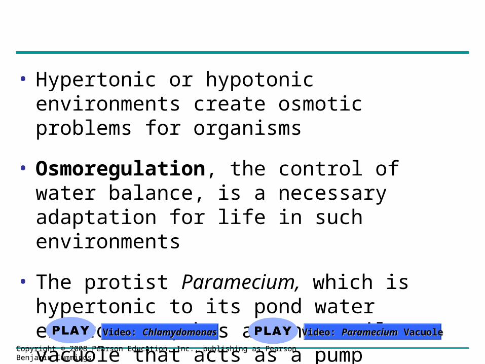
• Hypertonic or hypotonic environments create osmotic problems for organisms
• Osmoregulation, the control of water balance, is a necessary adaptation for life in such environments
• The protist Paramecium, which is hypertonic to its pond water environment, has a contractile vacuole that acts as a pump
Video: Video: ChlamydomonasChlamydomonas Video: Video: ParameciumParamecium Vacuole Vacuole
Copyright © 2008 Pearson Education, Inc., publishing as Pearson Benjamin Cummings
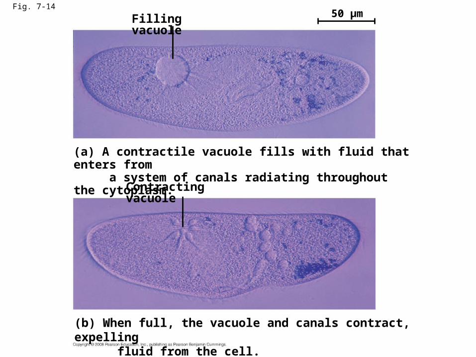
Fig. 7-14
Filling vacuole 50 µm
(a) A contractile vacuole fills with fluid that enters from a system of canals radiating throughout the cytoplasm.
Contracting vacuole
(b) When full, the vacuole and canals contract, expelling fluid from the cell.

Water Balance of Cells with Walls
• Cell walls help maintain water balance
• A plant cell in a hypotonic solution swells until the wall opposes uptake; the cell is now turgid (firm)
• If a plant cell and its surroundings are isotonic, there is no net movement of water into the cell; the cell becomes flaccid (limp), and the plant may wilt
Copyright © 2008 Pearson Education, Inc., publishing as Pearson Benjamin Cummings
Video: PlasmolysisVideo: Plasmolysis
Video: Turgid Video: Turgid ElodeaElodea
Animation: OsmosisAnimation: Osmosis
• In a hypertonic environment, plant cells lose water; eventually, the membrane pulls away from the wall, a usually lethal effect called plasmolysis
Copyright © 2008 Pearson Education, Inc., publishing as Pearson Benjamin Cummings
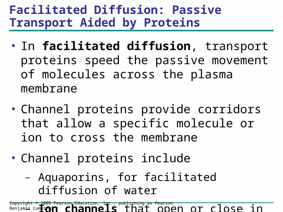
Facilitated Diffusion: Passive Transport Aided by Proteins
• In facilitated diffusion, transport proteins speed the passive movement of molecules across the plasma membrane
• Channel proteins provide corridors that allow a specific molecule or ion to cross the membrane
• Channel proteins include
– Aquaporins, for facilitated diffusion of water
– Ion channels that open or close in response to a stimulus (gated channels)
Copyright © 2008 Pearson Education, Inc., publishing as Pearson Benjamin Cummings

Fig. 7-15
EXTRACELLULAR FLUID
Channel protein
(a) A channel protein
Solute CYTOPLASM
Solute Carrier protein
(b) A carrier protein
Concept 7.4: Active transport uses energy to move solutes against their gradients
• Facilitated diffusion is still passive because the solute moves down its concentration gradient
• Some transport proteins, however, can move solutes against their concentration gradients
Copyright © 2008 Pearson Education, Inc., publishing as Pearson Benjamin Cummings
The Need for Energy in Active Transport
• Active transport moves substances against their concentration gradient
• Active transport requires energy, usually in the form of ATP
• Active transport is performed by specific proteins embedded in the membranes
Animation: Active TransportAnimation: Active Transport
Copyright © 2008 Pearson Education, Inc., publishing as Pearson Benjamin Cummings

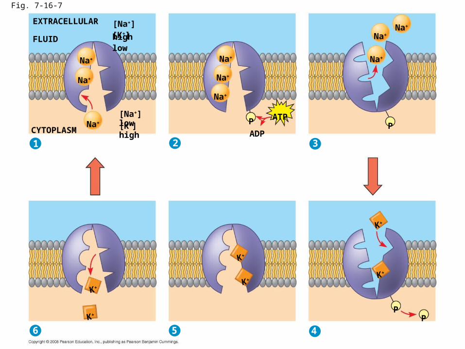
• Active transport allows cells to maintain concentration gradients that differ from their surroundings
• The sodium-potassium pump is one type of active transport system
Copyright © 2008 Pearson Education, Inc., publishing as Pearson Benjamin Cummings
2
EXTRACELLULAR
FLUID [Na+] high [K+] low
[Na+] low
[K+] high
Na+
Na+
Na+
Na+
Na+
Na+
CYTOPLASM ATP
ADP P
Na+ Na+
Na+
P 3
K+
K+ 6
K+
K+
5 4
K+
K+
P P
1
Fig. 7-16-7
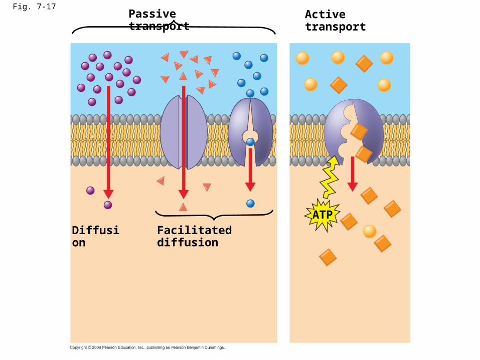
Fig. 7-17Passive transport
Diffusion Facilitated diffusion
Active transport
ATP

Fig. 7-19
Proton pump
–
–
–
–
–
–
+
+
+
+
+
+
ATP
H+
H+
H+
H+
H+
H+
H+
H+
Diffusionof H+
Sucrose-H+
cotransporter
Sucrose
Sucrose

Concept 7.5: Bulk transport across the plasma membrane occurs by exocytosis and endocytosis
• Small molecules and water enter or leave the cell through the lipid bilayer or by transport proteins
• Large molecules, such as polysaccharides and proteins, cross the membrane in bulk via vesicles
• Bulk transport requires energy
Copyright © 2008 Pearson Education, Inc., publishing as Pearson Benjamin Cummings

Exocytosis
• In exocytosis, transport vesicles migrate to the membrane, fuse with it, and release their contents
• Many secretory cells use exocytosis to export their products
Animation: ExocytosisAnimation: Exocytosis
Copyright © 2008 Pearson Education, Inc., publishing as Pearson Benjamin Cummings
Endocytosis
• In endocytosis, the cell takes in macromolecules by forming vesicles from the plasma membrane
• Endocytosis is a reversal of exocytosis, involving different proteins
• There are three types of endocytosis:
– Phagocytosis (“cellular eating”)
– Pinocytosis (“cellular drinking”)
– Receptor-mediated endocytosis
Animation: Exocytosis and Endocytosis IntroductionAnimation: Exocytosis and Endocytosis Introduction
Copyright © 2008 Pearson Education, Inc., publishing as Pearson Benjamin Cummings

Fig. 7-20PHAGOCYTOSIS
EXTRACELLULARFLUID
CYTOPLASM
Pseudopodium
“Food”orother particle
Foodvacuole
PINOCYTOSIS
1 µm
Pseudopodiumof amoeba
Bacterium
Food vacuole
An amoeba engulfing a bacteriumvia phagocytosis (TEM)
Plasmamembrane
Vesicle
0.5 µm
Pinocytosis vesiclesforming (arrows) ina cell lining a smallblood vessel (TEM)
RECEPTOR-MEDIATED ENDOCYTOSIS
Receptor Coat protein
Coatedvesicle
Coatedpit
Ligand
Coatprotein
Plasmamembrane
A coated pitand a coatedvesicle formedduringreceptor-mediatedendocytosis(TEMs)
0.25 µm

Fig. 7-20cRECEPTOR-MEDIATED ENDOCYTOSIS
Receptor Coat protein
Coatedpit
Ligand
Coatprotein
Plasmamembrane
0.25 µm
Coatedvesicle
A coated pitand a coatedvesicle formedduringreceptor-mediatedendocytosis(TEMs)
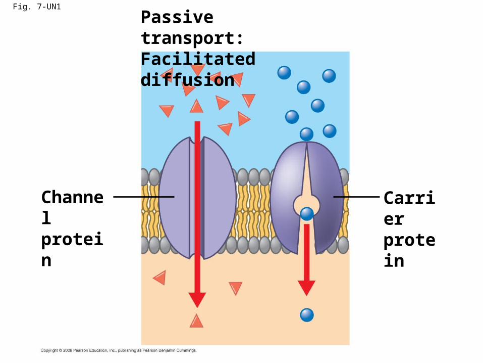
Fig. 7-UN1
Passive transport:Facilitated diffusion
Channelprotein
Carrierprotein
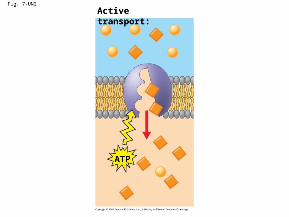
Fig. 7-UN2
Active transport:
ATP
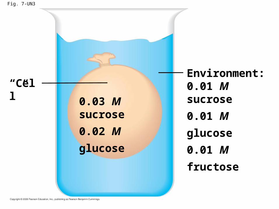
Fig. 7-UN3
Environment:0.01 M sucrose
0.01 M glucose
0.01 M fructose
“Cell”
0.03 M sucrose
0.02 M glucose
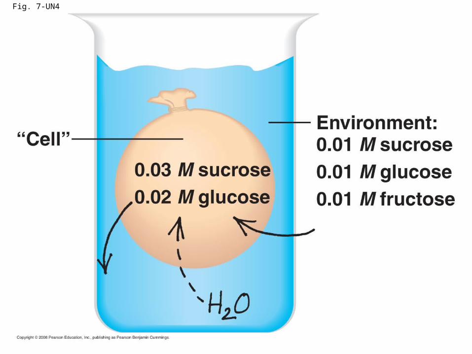
Fig. 7-UN4