chapter 9 catalytic strategies. 4 classes of enzymes. 1. protease 2. carbonic anhydrase 3....
TRANSCRIPT

Chapter 9
Catalytic Strategies
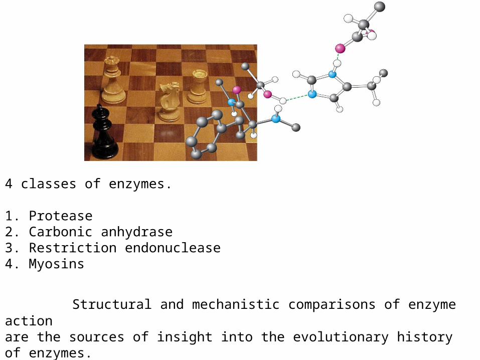
4 classes of enzymes.
1. Protease2. Carbonic anhydrase3. Restriction endonuclease4. Myosins
Structural and mechanistic comparisons of enzyme action are the sources of insight into the evolutionary history of enzymes.

9.1 Proteases facilitate a fundamentally difficult reaction
- Proteins must be degraded to that their constituent amino acids can be recycled for the synthesis of new proteins.

- In the absence of a catalyst, the half-life for the hydrolysis of a peptide at neutral pH is estimated to be between 10 and 1000 years.
- But, peptide bonds must be hydrolyzed within milliseconds in some biochemical processes.
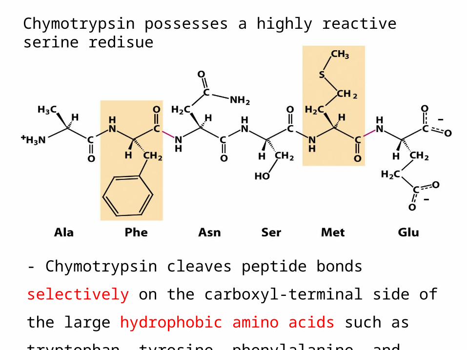
Chymotrypsin possesses a highly reactive serine redisue
- Chymotrypsin cleaves peptide bonds
selectively on the carboxyl-terminal side of
the large hydrophobic amino acids such as
tryptophan, tyrosine, phenylalanine, and
methionine.
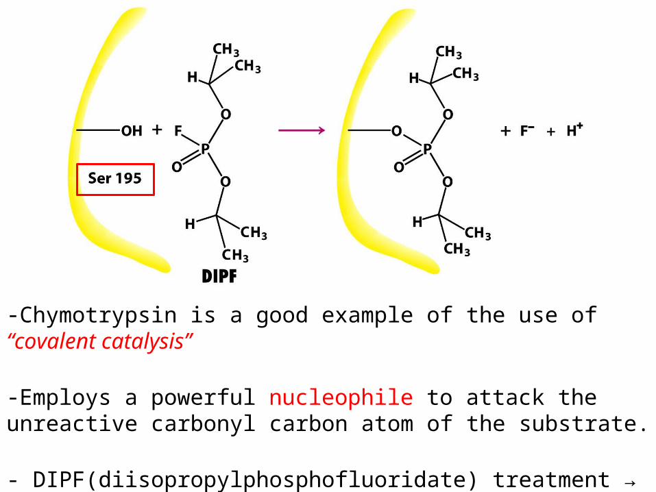
-Chymotrypsin is a good example of the use of “covalent catalysis”
-Employs a powerful nucleophile to attack the unreactive carbonyl carbon atom of the substrate.
- DIPF(diisopropylphosphofluoridate) treatment → Serine 195 modification → activity irreversibly
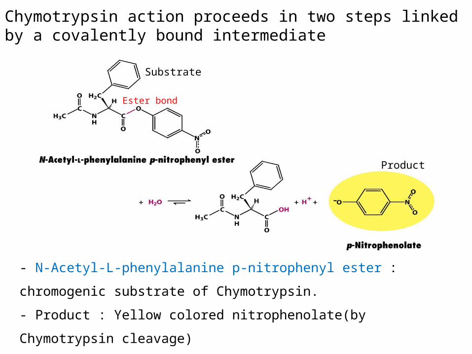
Chymotrypsin action proceeds in two steps linked by a covalently bound intermediate
- N-Acetyl-L-phenylalanine p-nitrophenyl ester :
chromogenic substrate of Chymotrypsin.
- Product : Yellow colored nitrophenolate(by
Chymotrypsin cleavage)
Substrate
Product
Ester bond

-Product measurement : absorbance of light(amount of p-
nitrophenolate)
-Under steady-state conditions, Km = 20μM, kcat = 77s-1
-Hydrolysis (by Chymotrypsin) proceeds in two steps.
-1). Burst phase (rapid)
-2). Steady-state phase

Burst phase Steady-state phase
-Burst phase : the acyl group of the substrate becomes covalently attached to the enzyme as p-nitrophenolate.
(acyl-enzyme intermediate)
-Steady-state phase : acyl-enzyme intermediate is hydrolyzed to release the carboxylic acid component of the substrate and regenerate the free enzyme; it takes longer for the enzyme to be reset.

Serine is part of a catalytic triad that also includes histidine and aspartate
-The three-dimensional structure of chymotrypsin was solved by David Blow in 1967.
-It is synthesized as a single polypeptide, termed Chymotrypsinogen, which is activated by the proteolytic cleavage to yield the three chains.[1GCT.pdb]

-The active site of chymotrypsin, marked by serine 195, lies in a cleft on the surface of the enzyme.
-This side chain of serine 195 is hydrogen bonded to the imidazole ring of histidine 57.
cleft
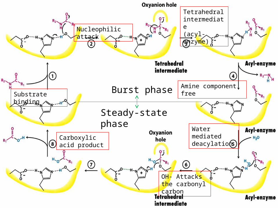
Substrate binding
Nucleophilic attack
Tetrahedral intermediate(acyl-enzyme)
Amine component free
Water mediated deacylation
OH- Attacks the carbonyl carbon
Carboxylic acid product
Burst phase
Steady-state phase
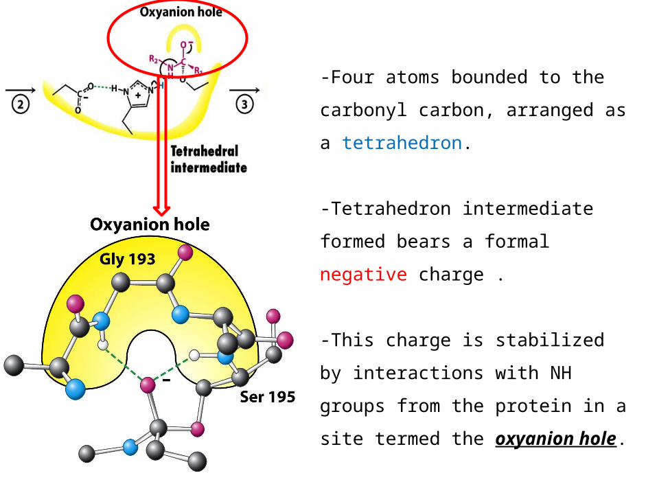
-Four atoms bounded to the
carbonyl carbon, arranged as
a tetrahedron.
-Tetrahedron intermediate
formed bears a formal
negative charge .
-This charge is stabilized
by interactions with NH
groups from the protein in a
site termed the oxyanion
hole.

-Preference for
cleaving the peptide
bonds just past
residues with large,
hydrophobic side
chains.
-Chymotrypsin has a
deep, hydrophobic
pocket, called the S1
pocket.
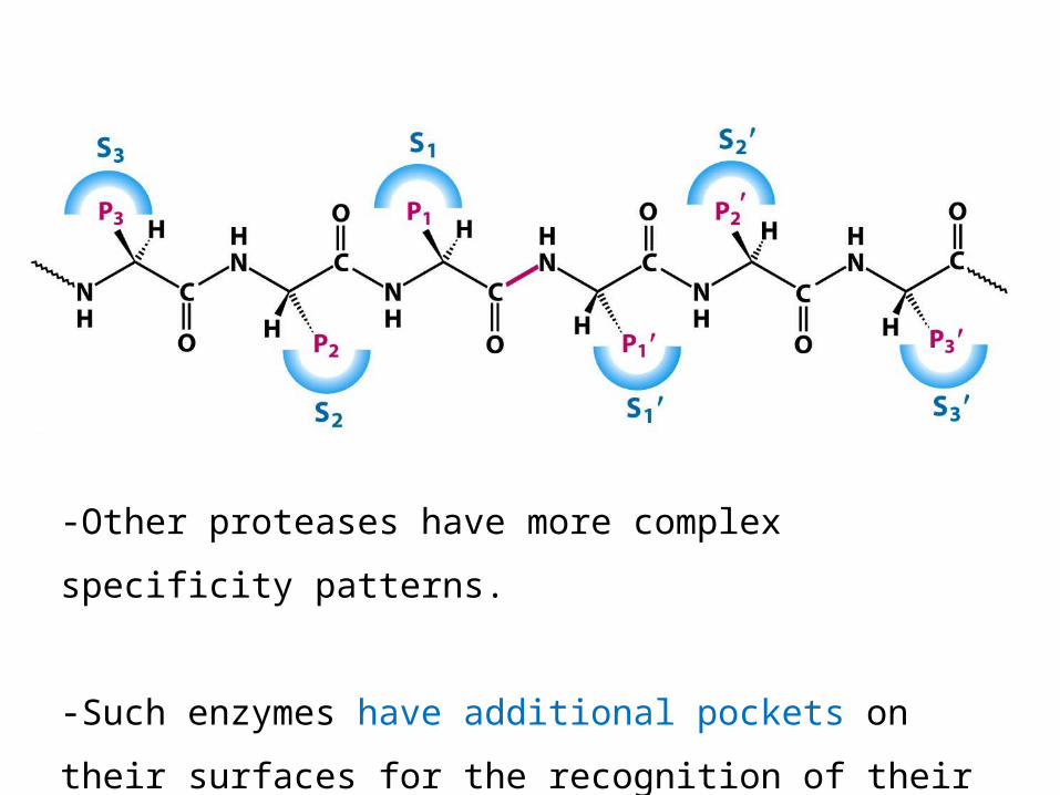
-Other proteases have more complex
specificity patterns.
-Such enzymes have additional pockets on
their surfaces for the recognition of their
residues in the substrate.
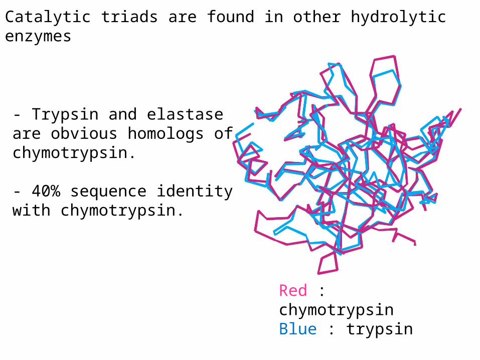
Catalytic triads are found in other hydrolytic enzymes
- Trypsin and elastase are obvious homologs of chymotrypsin.
- 40% sequence identity with chymotrypsin.
Red : chymotrypsinBlue : trypsin

Chymotrypsin- Cleaves after residues with an aromatic or long nonpolar side
chains.
Trypsin-Cleaves after residues with an long, positively charged side chains. (R.K)-Asp189 at bottom of the S1 pocket
Elastase-Cleaves after residues with a small
side chains. (A.S)-Val190 and Val216 at bottom of the S1 pocket
Similar structure
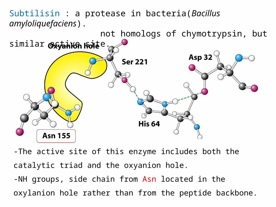
Subtilisin : a protease in bacteria(Bacillus amyloliquefaciens). not homologs of chymotrypsin, but similar active site.
-The active site of this enzyme includes both the catalytic triad
and the oxyanion hole.
-NH groups, side chain from Asn located in the oxylanion hole
rather than from the peptide backbone.
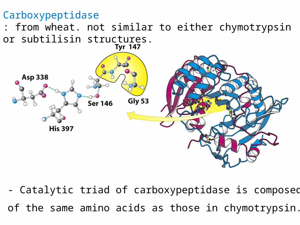
Carboxypeptidase : from wheat. not similar to either chymotrypsin or subtilisin structures.
- Catalytic triad of carboxypeptidase is composed
of the same amino acids as those in chymotrypsin.
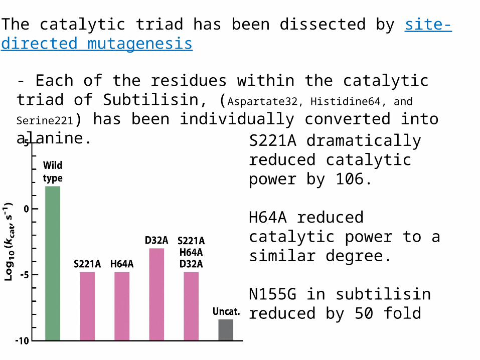
The catalytic triad has been dissected by site-directed mutagenesis
- Each of the residues within the catalytic triad of Subtilisin, (Aspartate32, Histidine64, and Serine221) has been individually converted into alanine. S221A dramatically
reduced catalytic power by 106.
H64A reduced catalytic power to a similar degree.
N155G in subtilisin reduced by 50 fold

Cysteine, Aspartyl, and Metalloproteases are other major classes of peptide-cleaving enzyme.
- A cys residue, activated by a his, plays the role of the nucleophile that attacks the peptide bond.
-Ex). Papain : purified from the fruit of the papaya. -Caspases

Cysteine, Aspartyl, and Metalloproteases are other major classes of peptide-cleaving enzyme.
- A pair of aspartic acid residues that act together to allow a water molecule to attack the peptide bond. One aspartic acid activates the attacking water molecule. The other polarizes the peptide carbonyl group.- Ex). Renin : having a role in the regulation of blood pressure. Pepsin : the digestive enzyme. HIV protease

HIV protease : a dimeric aspartyl protease
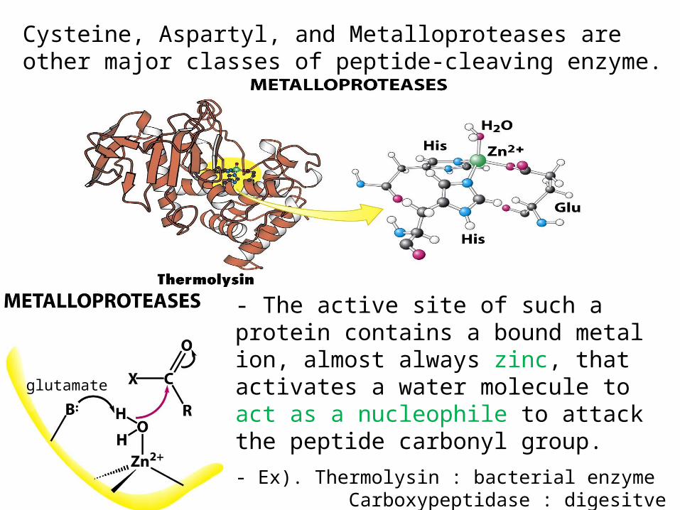
Cysteine, Aspartyl, and Metalloproteases are other major classes of peptide-cleaving enzyme.
- The active site of such a protein contains a bound metal ion, almost always zinc, that activates a water molecule to act as a nucleophile to attack the peptide carbonyl group.
- Ex). Thermolysin : bacterial enzyme Carboxypeptidase : digesitve enzyme
glutamate
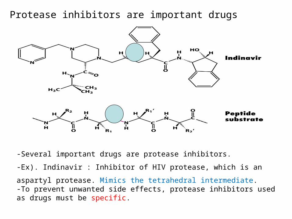
Protease inhibitors are important drugs
-Several important drugs are protease inhibitors.
-Ex). Indinavir : Inhibitor of HIV protease, which is an aspartyl
protease. Mimics the tetrahedral intermediate. -To prevent unwanted side effects, protease inhibitors used as drugs must be specific.
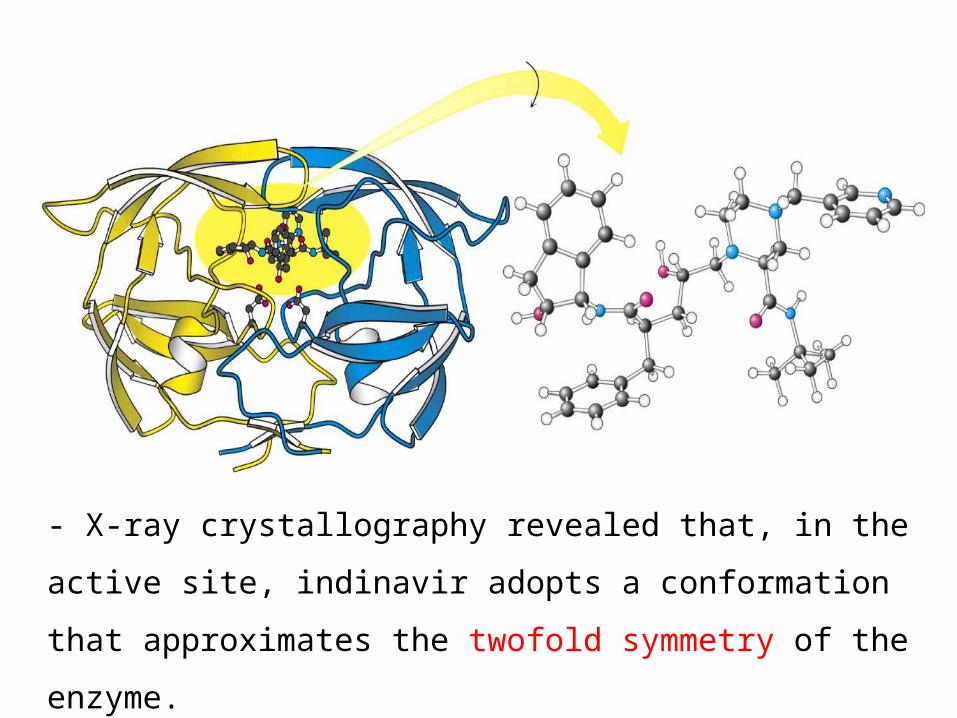
- X-ray crystallography revealed that, in the
active site, indinavir adopts a conformation
that approximates the twofold symmetry of the
enzyme.
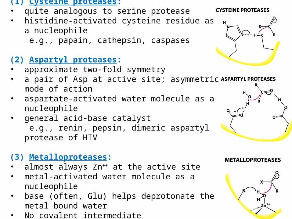
(1) Cysteine proteases: • quite analogous to serine protease • histidine-activated cysteine residue as a nucleophile e.g., papain, cathepsin, caspases
(2) Aspartyl proteases:• approximate two-fold symmetry• a pair of Asp at active site; asymmetric mode of action • aspartate-activated water molecule as a nucleophile • general acid-base catalyst e.g., renin, pepsin, dimeric aspartyl protease of HIV
(3) Metalloproteases: • almost always Zn++ at the active site • metal-activated water molecule as a nucleophile • base (often, Glu) helps deprotonate the metal bound
water • No covalent intermediate• e.g., matrix metalloproteases

Activation strategies for three classes of proteases
In each of these classes of enzymes,the active site includes features that allow for
(1) the activation of water or another nucleophile(2) the polarization of the peptide carbonyl group (3) subsequent stabilization of a tetrahedral intermediate
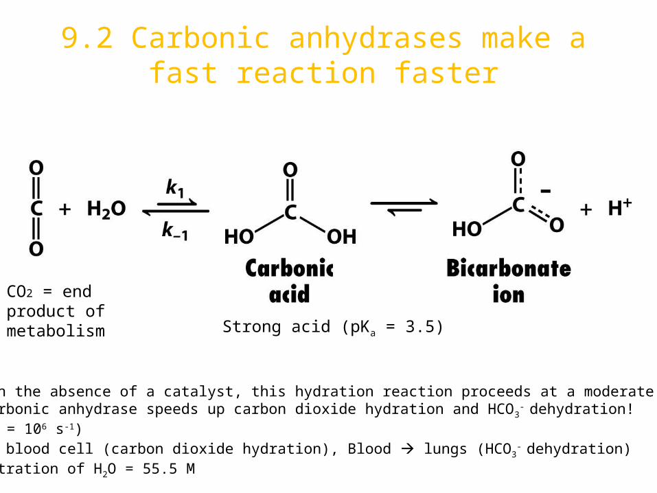
9.2 Carbonic anhydrases make a fast reaction faster
CO2 = end product of metabolism Strong acid (pKa = 3.5)
Even in the absence of a catalyst, this hydration reaction proceeds at a moderate rate.But carbonic anhydrase speeds up carbon dioxide hydration and HCO3
- dehydration! (Kcat = 106 s-1)In red blood cell (carbon dioxide hydration), Blood lungs (HCO3
- dehydration)Concentration of H2O = 55.5 M

Carbonic anhydrase contains a bound zinc ion essential for catalytic activity
- Carbonic anhydrase contains Zinc ion; first known zinc-containing
enzyme.
- Zinc ion is necessary for catalytic activity.
- At least 7 carbonic anhydrase in human.
- Carbonic anhydrase Ⅱ is the most extensively studied.
- Three coordination sites are occupied by the imidazole rings of three
His.
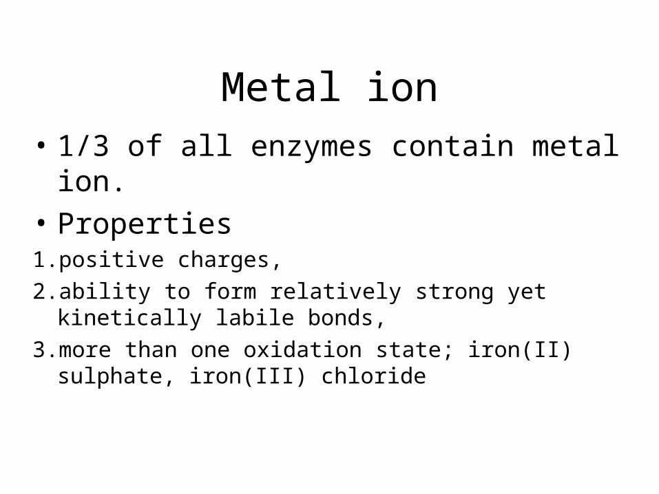
Metal ion• 1/3 of all enzymes contain metal ion.
• Properties 1.positive charges, 2.ability to form relatively strong yet kinetically labile bonds,
3.more than one oxidation state; iron(II) sulphate, iron(III) chloride
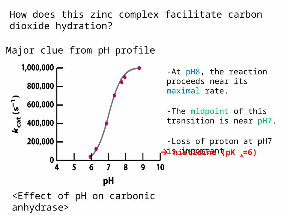
How does this zinc complex facilitate carbon dioxide hydration?
-At pH8, the reaction proceeds near its maximal rate.
-The midpoint of this transition is near pH7.
-Loss of proton at pH7 is important
<Effect of pH on carbonic anhydrase>
Major clue from pH profile
histidine (pK a=6)

- The binding of a water molecule to the positively
charged zinc center reduces the pKa of the water molecule
from 15.7 to 7.
- A zinc-bound hydroxide ion (OH-) is a potent nucleophile
able to attack carbon dioxide.
Nucleophile attack

Release of a proton from waterGenerate a hydroxide ion
CO2 bond to hydrophobic active site
HO- attacks CO2 converting it into HCO3-
Catalytic site is regenerated with the release of HCO3- and binding another water.
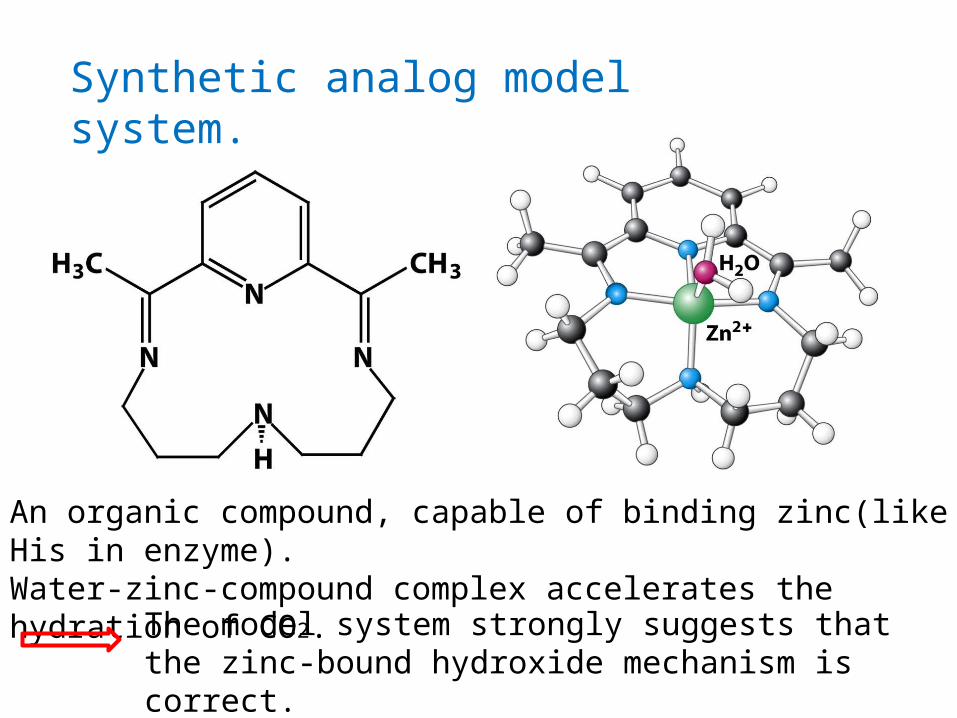
Synthetic analog model system.
An organic compound, capable of binding zinc(like His in enzyme).Water-zinc-compound complex accelerates the hydration of CO2. The model system strongly suggests that
the zinc-bound hydroxide mechanism is correct.

A proton shuttle facilitates rapid regeneration of the active form of the enzyme
In the first step of a CO2 hydration reaction, the zinc-bound water molecule must lose a proton to regenerate the active form of the enzyme. The rate of reverse reaction, the protonation of the zinc-bound hydroxide ion, is limited by the rate of proton diffusion
Equilibrium constant
K-1 = 1011 M-1s-1 because proton diffusion rate 10-11 M-1s-1 So, K1 = 104 M-1s-1 then, How carbon dioxide is hydrated at 106s-1 ?

The deprotonation of the zinc-bound water molecule in carbonic anhydrase is aided by buffer component B.

The rate of carbon dioxide hydration increases with the
concentration of the buffer 1,2-dimethylbenzimidazole.
The buffer enables the enzyme to achieve its high catalytic rates.
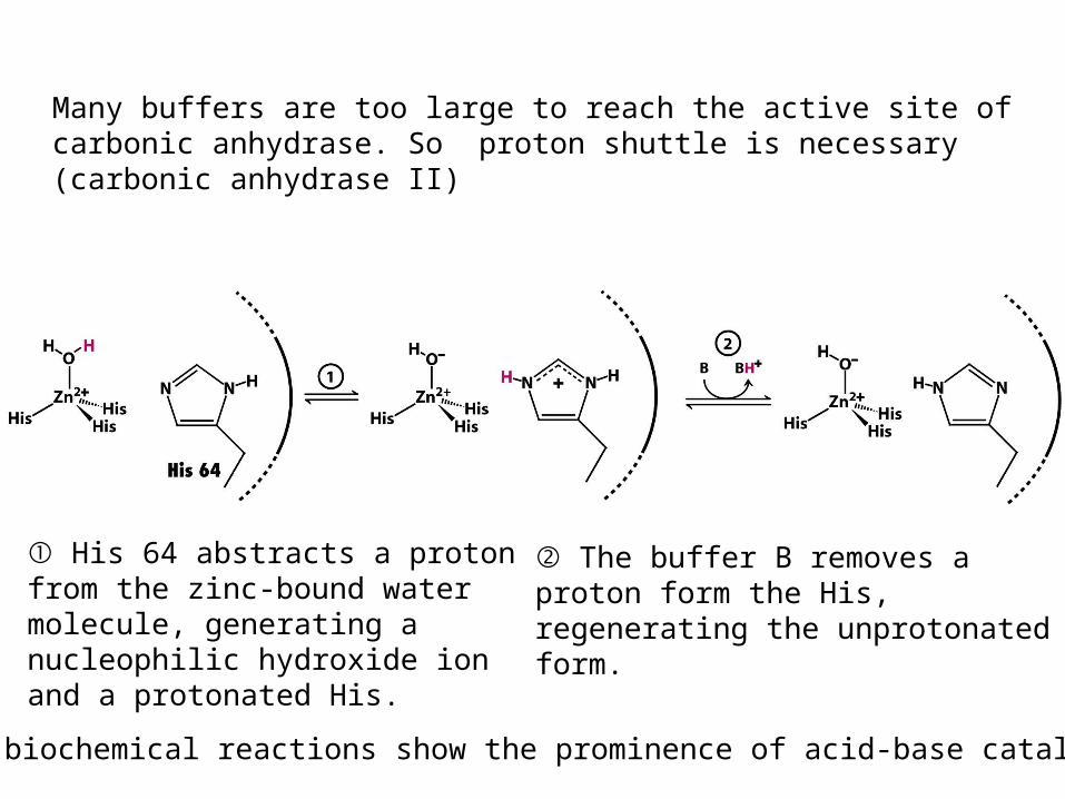
① His 64 abstracts a proton from the zinc-bound water molecule, generating a nucleophilic hydroxide ion and a protonated His.
② The buffer B removes a proton form the His, regenerating the unprotonated form.
Many buffers are too large to reach the active site of carbonic anhydrase. So proton shuttle is necessary (carbonic anhydrase II)
Many biochemical reactions show the prominence of acid-base catalysis

Convergent evolution has generated zinc-based active sites in different carbonic anhydrases
- In addition to a-carbonic anhydrases, two other families of carbonic
anhydrases have been discovered.
- β-carbonic anhydrases : found in higher plants and in many bacteria.
Zinc ion is bound by one His and two Cys.
- γ-carbonic anhydrases : identified in the archaeon Methanosarcina
thermophila. Three zinc sites similar to α-carbonic anhydrase. But zinc sites
lie at the interfaces between the three subunits of a trimeric enzyme.
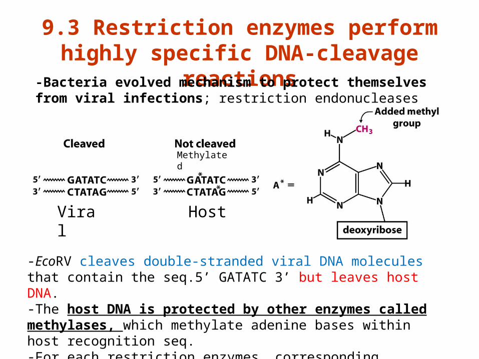
9.3 Restriction enzymes perform highly specific DNA-cleavage
reactions
-EcoRV cleaves double-stranded viral DNA molecules that contain the seq.5’ GATATC 3’ but leaves host DNA.-The host DNA is protected by other enzymes called methylases, which methylate adenine bases within host recognition seq.-For each restriction enzymes, corresponding methylases exist.
Methylated
Viral
Host
-Bacteria evolved mechanism to protect themselves from viral infections; restriction endonucleases
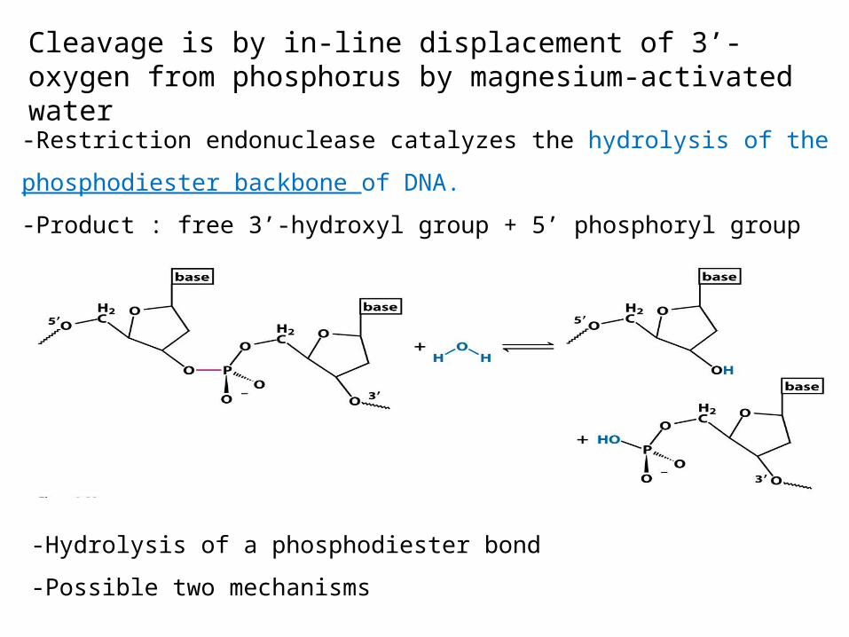
Cleavage is by in-line displacement of 3’-oxygen from phosphorus by magnesium-activated water
-Restriction endonuclease catalyzes the hydrolysis of the
phosphodiester backbone of DNA.
-Product : free 3’-hydroxyl group + 5’ phosphoryl group
-Hydrolysis of a phosphodiester bond
-Possible two mechanisms

Mechanism 1 (covalent intermediate) : analogy with chymotrypsin: two displacement, retained configuration

Mechanism 2 (direct hydrolysis): analogy with asparty protease and metalloproteases: one displacement, inverted configuration
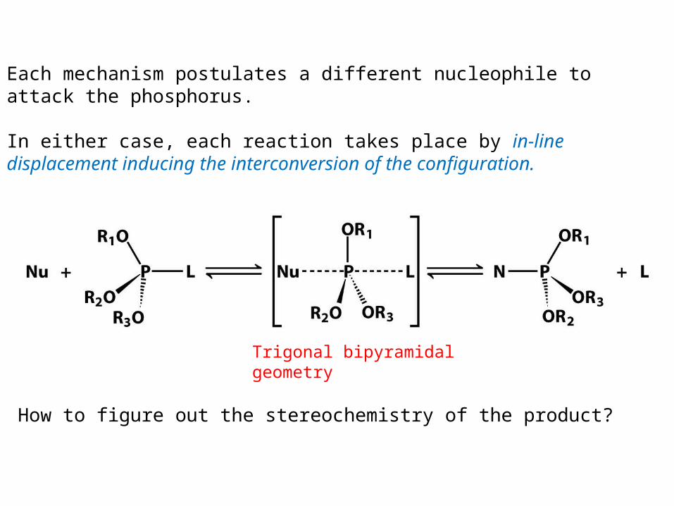
Each mechanism postulates a different nucleophile to attack the phosphorus.
In either case, each reaction takes place by in-line displacement inducing the interconversion of the configuration.
How to figure out the stereochemistry of the product?
Trigonal bipyramidal geometry
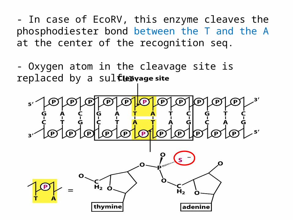
- In case of EcoRV, this enzyme cleaves the phosphodiester bond between the T and the A at the center of the recognition seq.
- Oxygen atom in the cleavage site is replaced by a sulfur.

Water 18O labeled
The hydrolysis takes place by water’s direct attack at the phosphorus atom.
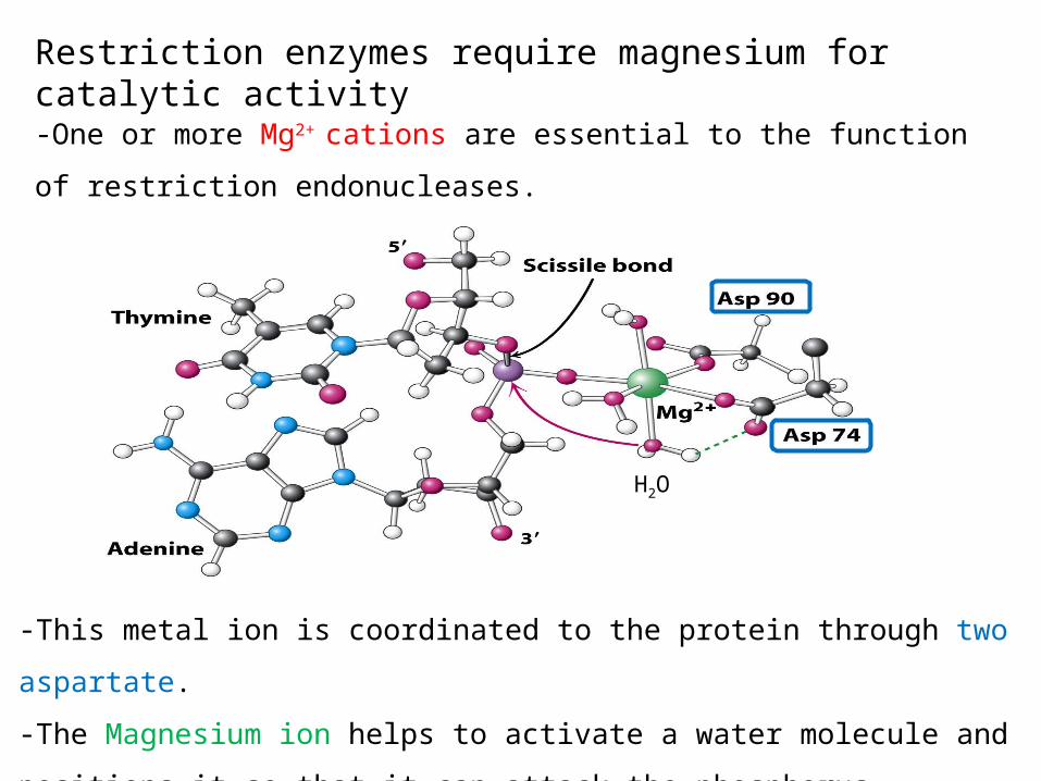
Restriction enzymes require magnesium for catalytic activity-One or more Mg2+ cations are essential to the function of
restriction endonucleases.
-This metal ion is coordinated to the protein through two aspartate.
-The Magnesium ion helps to activate a water molecule and positions it
so that it can attack the phosphorus.
H2O
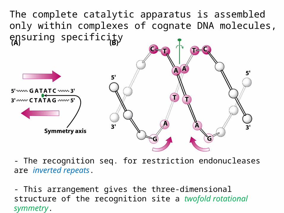
The complete catalytic apparatus is assembled only within complexes of cognate DNA molecules, ensuring specificity
- The recognition seq. for restriction endonucleases are inverted repeats.
- This arrangement gives the three-dimensional structure of the recognition site a twofold rotational symmetry.
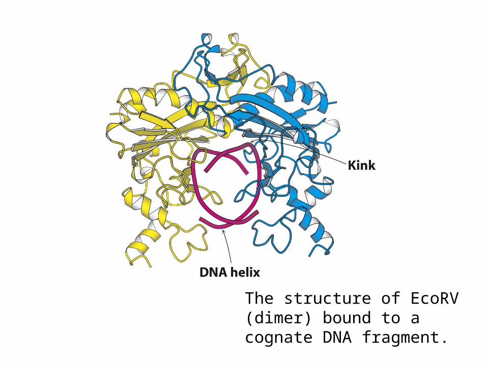
The structure of EcoRV (dimer) bound to a cognate DNA fragment.

5’ – G A T A T C – 3’
3’ – C T A T A G – 5’
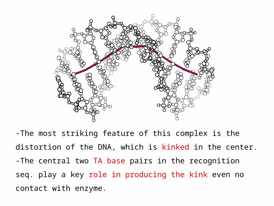
-The most striking feature of this complex is the distortion of
the DNA, which is kinked in the center.
-The central two TA base pairs in the recognition seq. play a
key role in producing the kink even no contact with enzyme.

- The noncognate DNA conformation is not substantially distorted.
cognatenon-cognate
- This lack of distortion has important consequences with regard to catalysis.-No phosphate is positioned sufficiently close to the active-site aspartate residues to complete a Mg2+ binding site.-DNA distortion and subsequent Mg2+ binding account for the catalytic specificity

-The distorted DNA makes
additional contacts with the
enzyme, increasing the binding
energy
- The increase is canceled by the
energetic cost of DNA distortions.
- Interactions that take place within the distorted substrate
complex stabilize the transition state leading to DNA
hydrolysis.
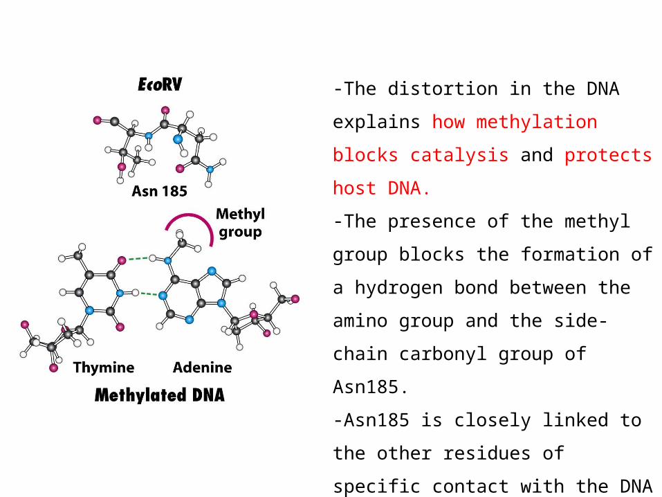
-The distortion in the DNA explains
how methylation blocks catalysis
and protects host DNA.
-The presence of the methyl group
blocks the formation of a
hydrogen bond between the
amino group and the side-chain
carbonyl group of Asn185.
-Asn185 is closely linked to the
other residues of specific contact
with the DNA
-Leading to the no distortion of
DNA

TypeⅡ restriction enzymes have a catalytic core in common and are probably related by horizontal gene transfer
No significant seq. similarity. However, a core structures are conserved. It includes strands (blue) that contain the aspartate residues of the Mg-binding sites.
Horizontal gene transfer: passing between species of pieces of DNA (plasmid).Representative example: antibiotics
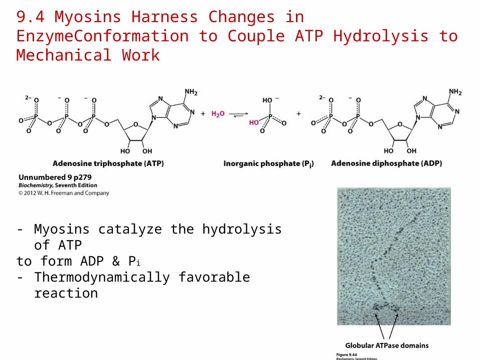
9.4 Myosins Harness Changes in EnzymeConformation to Couple ATP Hydrolysis to Mechanical Work
- Myosins catalyze the hydrolysis of ATP
to form ADP & Pi
- Thermodynamically favorable reaction
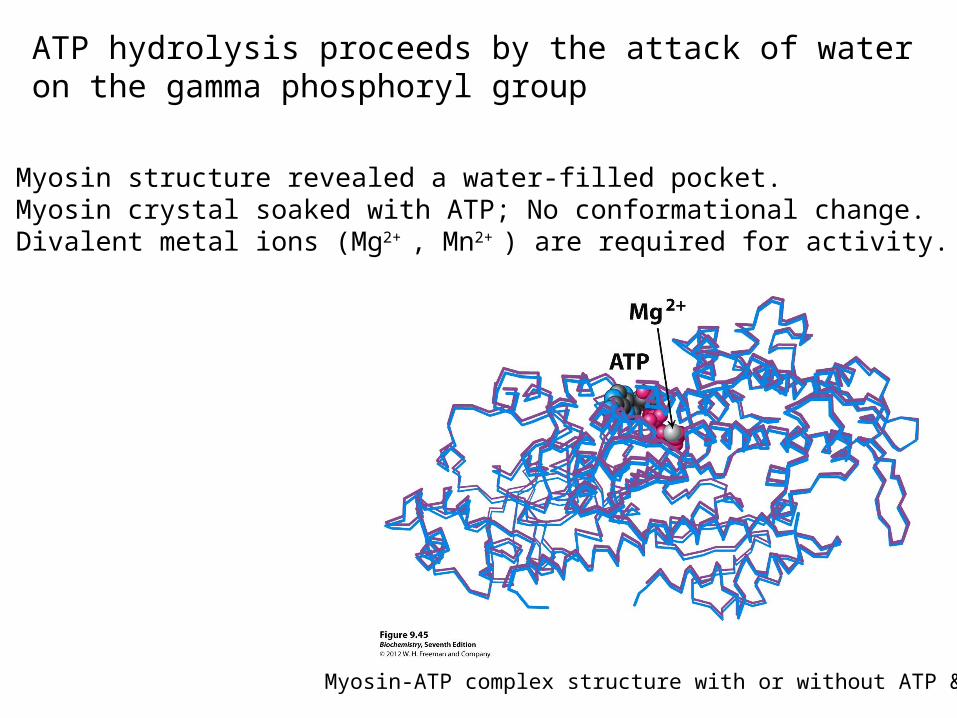
ATP hydrolysis proceeds by the attack of water on the gamma phosphoryl group
Myosin-ATP complex structure with or without ATP & Mg
• Myosin structure revealed a water-filled pocket.• Myosin crystal soaked with ATP; No conformational change.• Divalent metal ions (Mg2+ , Mn2+ ) are required for activity.

-Myosin are essentially inactive in the absence of metal ions
(Mg2+, Mn2+).
-Nucleotides bind these ions, and it is the metal ion-nucleotide
complex that is the true substrate for the enzymes.
-Kd of ATP-Mg2+ complex ~0.1mM, Mg2+ conc in cytosol is mM
range. So?
• Nucleophilic attack by a water requires a basic residue or bound metal to activate the water
• ATP hydrolysis includes a pentacoordinate transition state.
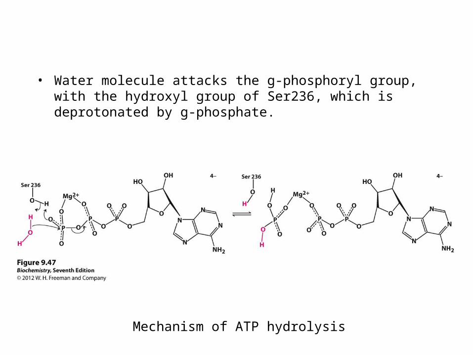
Mechanism of ATP hydrolysis
• Water molecule attacks the g-phosphoryl group, with the hydroxyl group of Ser236, which is deprotonated by g-phosphate.


NMP kinases are a family of enzymes containing P-loop structures
-Comparison of NMP kinase structures reveals that these enzymes
form a family of homologous proteins.
-The nucleoside triphosphate binding domain is a common feature
in these homologous nucleotide kinases and others.
Core domain

-Core domain of NMP kinases
-Consist of a central β sheet, surrounded on both sides by α helices.
-P loop : between the first β strand and the first α helix. Seq. : Gly-X-X-X-X-Gly-Lys Interact with NTP

- P loop interact with ATP
Phosphate

Magnesium or manganese complexes of nucleoside triphosphates are the true substrates for essentially all NTP-dependent enzymes.
-NMP kinases are essentially inactive in the absence of metal ions
(Mg2+, Mn2+).
-Nucleotides bind these ions, and it is the metal ion-nucleotide
complex that is the true substrate for the enzymes.
-Kd of ATP-Mg2+ complex ~0.1mM, Mg2+ conc in cytosol is mM
range. So?

-Magnesium ion is bound to the β and γ phosphoryl groups and to four water molecules at the remaining coordination position.
→ The magnesium ion provides additional points of interaction between the ATP-Mg2+ complex and the enzyme, thus increasing the binding energy; coordinated to six groups in an octahedral arrangement.-Mg2+ binding hold the nucleotide in a well-defined conformation that can be bound by an enzyme.
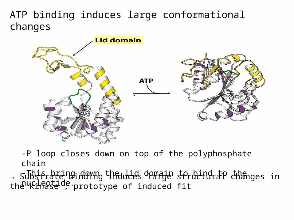
ATP binding induces large conformational changes
→ Substrate binding induces large structural changes in the kinase ; prototype of induced fit
-P loop closes down on top of the polyphosphate chain -This bring down the lid domain to bind to the nucleotide.
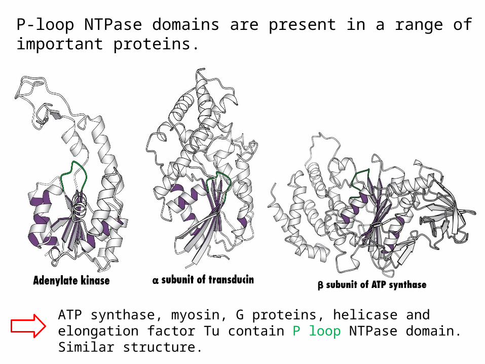
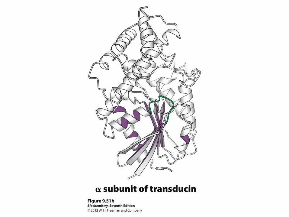
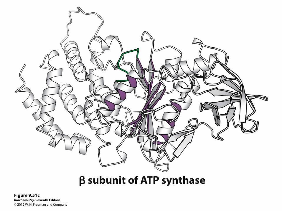
P-loop NTPase domains are present in a range of important proteins.
ATP synthase, myosin, G proteins, helicase and elongation factor Tu contain P loop NTPase domain.Similar structure.