chapter 9 head and neck tumors -...
TRANSCRIPT

1
Chapter 9
Head and Neck Tumors Peter Nthumba
Introduction
Lesions of any size in the head and neck may have a significant effect on cosmesis, speech, respiration, and alimentation and they can significantly impact a patient’s quality of life. Head and neck cancers include squamous cell carcinoma (SCC), basal cell carcinoma (BCC), and malignant melanoma, amongst others. Adenoid cystic carcinoma of salivary glands is a tumor with poor prognosis because of its mode of spread, and early lung metastases. Wide excision of lesions is a challenge in this region, as these margins would encroach on important structures. Prevention of local recurrence is a significant challenge. Frequent recurrence and regional nodal metastases lead to a poor prognosis, especially with aggressive tumors. Radiotherapy and chemotherapy are showing improved outcomes as primary treatment modalities, with results comparable to surgical resections. These are modalities that will continue to be largely inaccessible to many in the sub-Saharan region, and surgery, therefore, remains the main treatment modality for the majority of patients. Plastic surgeons are generally not involved in the treatment of thyroid, parathyroid or laryngeal cancers, and these will not be discussed in this chapter. Plastic surgeons will be involved in the management of salivary gland tumors, especially of the parotid gland, because of the defect that is left following resection of a malignancy and management of the facial nerve that may be involved in resection.
Physical examination
Studies have suggested that even in the West, patients with head and neck cancer make more than 10 hospital/physician visits prior to the initial diagnosis of a cancer. Sub-Sahara African patients frequently present with huge/extensive tumors, with a history of having presented to healthcare facilities or physicians at some point in the past. While early diagnosis of

2
malignancy improves outcomes, failure to recognize a malignancy will lead to increased morbidity and mortality associated with a late presentation.
A careful systematic history and physical examination will reveal head and neck pathology.
Scalp – look for nodules, ulcers, pigmented lesions etc.
Cranial nerves – form an important part of head and neck examination; occult malignancies may cause nerve palsies that make it easy for the physician to locate them. Facial nerve palsy in the background of a parotid mass makes it more likely to be a malignancy, rather than a benign tumor.
Oral cavity – halitosis, bad breath, will usually be present in patients with an upper aero-digestive malignancy. Bi-manual palpation of the oral cavity may reveal an indurated area that would be the seat of a lesion.
Neck – palpation reveals presence of any enlarged nodes or other masses. Palpate for thrills/auscultate for bruits.
Pearl – up to 5% of patients with head and neck cancer have a synchronous squamous cell carcinoma of the head and neck, esophagus or lungs, and where available, endoscopy should be performed (esophagoscopy, direct laryngoscopy and bronchoscopy) in patients with a known head and neck cancer. The tonsils, base of tongue and piriform sinuses form the most common sites for occult primary tumors.
Biopsy
Biopsies form a core part of the management of head and neck tumors, as they determine the operation needed, extent of resection and the need for neck dissection. As a rule, biopsies should be performed away from any necrotic or infected areas. They should include some normal adjacent tissue.
Fine needle aspirate (FNA) – although dependent on the physician’s accuracy and the pathologist’s experience in cytopathology, this is an excellent initial modality for many head and neck tumors. FNA in good hands has a false negative rate of less than 7%.
Punch biopsy – useful for mucosal lesions.
Core and open biopsies are invasive, and most authors recommend a resection soon after, or even immediately. These give more tissue, and, thus, the diagnosis is more definite, allowing for appropriate treatment.

3
Squamous cell carcinomas (SCC) are the most common head and neck cancers. While histologic grade (degree of differentiation) has been used to prognosticate in the past, perineural spread, lymphatic invasion, and tumor spread beyond the lymph node capsule have been found to more consistently predict prognosis.
Fig.1 Late presentation
Unresectable SCC
Tumors of the Head and Neck Region
Because of its anatomy, the head and neck region has a diversity of tumors, both benign and malignant. A number of these tumors are rare. Head and neck cancer is more common in males than in females, and occurs in older age groups.
Head and neck primary cutaneous malignancies constitute 20% of all skin malignancies in the African population; 27% of all primary skin squamous cell carcinomas occur in this region. In the USA, head and neck cancers consist of 2-3% of all cancers and about 50% of patients have metastatic disease at the time of presentation. Equivalent rates in sub-Saharan Africa are unknown, but given that most patients present late, the percentage of patients with metastatic disease would presumably be much higher.
In the US, head and neck cancer is associated with lower survival amongst Blacks when compared with Caucasians with equivalent tumor stages.
Risk factors for the development of head and neck cancers include tobacco and alcohol consumption (tumors of the aero-digestive tract), sunlight (UV-light with cutaneous malignancies especially BCC and SCC), viral infection (e.g. EBV with naso-pharyngeal carcinoma), and environmental carcinogen exposures (e.g. wood dust with nasal carcinoma). Diets rich in Vitamin A, α -tocopherol and β -carotene may reduce the risk of developing head and neck cancer.

4
75% of all cancers of the head and neck region are squamous cell carcinomas.
Treatment options
Surgical treatment of head and neck cancer should be offered to patients with:
1. Curative intent
2. Significant palliative benefit
Absence of supportive/adjuvant therapy should not form the basis for precluding surgical treatment for patients who would otherwise benefit from such care. Radiation therapy and chemotherapy remain difficult to access in most parts of sub-Sahara Africa; most facilities have to refer their patients to a central national site, straining resources, with the long waiting times between intervention and treatment often leading to recurrences.
Lymphatic drainage
The lymphatic drainage of the head and neck is predictable, and knowledge of patterns of drainage will help the clinician determine the primary tumor, in most instances, when a tumor first presents as a neck nodal metastasis.
Neck nodes have been grouped together in levels (I to VI):
I – Ia: Submental; Ib: Submandibular
II – IIa: Upper Jugular (anterior to XI cranial nerve); IIb: lower jugular (posterior to XI cranial nerve)
III – Middle Jugular nodes
IV – IVa: Lower jugular (clavicular); IVb: Lower jugular (sternal)
V – Va: Posterior triangle (below XI cranial nerve); Vb: Posterior triangle (transverse cervical)
VI – Central neck nodes

5
Fig 2 Fig 3
Lymph node metastasis with unknown primary: wide resection, bilateral lymph node dissection and closure with rotation of large deltopectoral flap
Level I
Marginal mandibular nerve
Avoid the marginal mandibular nerve – found 1cm anterior and inferior to the angle of the mandible. Locate the mandibular notch: identify the submandibular gland and the facial vein passing over it. The nerve is deep to the platysma muscle, but superficial to the facial vein. Identify the facial vein and ligate it. Dissect deep to the vein, raising it with fascia over the gland. The nerve will be safely out of harm’s way.
Hypoglossal nerve
Appears deep to the internal jugular vein and internal carotid artery, turns 90o and passes between these two structures.
Level II
Spinal accessory nerve
Found deep to internal carotid artery in 70% of patients; in 30% is superficial to the internal carotid artery.
Level IV
Phrenic nerve
Postero-lateral to carotid sheath, within prevertebral fascia, on anterior scalene muscle
The scalp

6
Fig. 4
Scalp SCC
Primary carcinomas of the scalp are common; 30% of all primary cutaneous malignancies occur in the scalp. Because of the robust vascularity of the scalp region, this is a common site for cutaneous metastasis of tumors from other parts the body.
Small lesions should be excised using elliptical incisions, parallel to skin lines. In order to define skin lines on the face and scalp, ask the patient to grimace; this will enhance the skin lines, ensuring an esthetic scar.
Skin grafts are best applied on those areas of the face/scalp that have the least motion: the parotid area, the temporal region, and laterally, on the bridge of the nose. The best donor sites for full thickness grafts for facial defects are the retro-auricular skin and the supra-clavicular fossa skin. Primary closure or closure with the use of local flaps, either random or axial in design, are based on the rich anastomoses between the branches of the external carotid artery and sturdy sub-dermal plexus.
Marjolin ulcer in unhealed post-burn wounds is commonly seen.
Fig 5 Fig 6 Fig 7 Rapidly growing Marjolin ulcer in post burn scar in older patient: Fig 5 at the time of biopsy,
Fig 6 is at time of surgery two weeks later showing rapid growth, and Fig 7 showing

7
reconstruction with the rotation of a large scalp flap with STSG to donor site. Tumor went down to galea but not to periosteum. Galea was removed with the tumor.
Fig 8 Fig 9
14 year old girl with unhealed burn wound of 12 years. Six months later she has a large Marjolin’s ulcer and bilateral neck metastasis
Conjunctiva
Fig 10
Lower lid conjunctival tumor
The conjunctiva is the mucous membrane that covers the anterior portion of the globe; the lining epithelium is both stratified squamous and columnar. Tumors may arise from either the epithelium or stroma. While small benign-appearing tumors may be managed by serial examination, larger tumors should be managed by either excisional or incisional biopsies. While many modalities of therapy exist, the main ones accessible to the majority of sub-Sahara African patients will be surgery and radiotherapy. Surgery may involve orbital exenteration, and general surgeons should be familiar with the steps of this operation. Topical chemotherapy may also be effective for small tumors. A

8
slit-lamp examination is an important screening tool, but may not be available outside major medical centers.
Common malignancies include squamous cell carcinoma, malignant melanoma and sebaceous gland carcinoma. Benign tumors include limbal dermoids, papilloma, and pyogenic granulomas. The circumscribed nevus is the commonest melanocytic tumor of the conjunctiva, with a less than 1% risk of malignant transformation into malignant melanoma.
Major salivary gland tumors
The major salivary glands include the parotid, submandibular, and sublingual glands. The proportion of tumors arising in these glands is 100:10:1 respectively. While most salivary gland tumors arise from the parotid gland, 80% are benign. Of all the tumors of the submandibular, sublingual and minor salivary glands, 40%, 60% and 80%, respectively, are malignant. Radiation exposure is a risk factor for the development of salivary gland tumors, especially mucoepidermoid tumors.
While fine needle aspiration with cytology is useful for diagnosis, many surgeons consider that the presence of salivary gland enlargement is sufficient reason for resection.
Pleomorphic adenoma, a benign mixed tumor, is the most common salivary gland tumor, usually occurring in the parotid gland. A superficial parotidectomy is sufficient treatment, with a recurrence rate of less than 1%.
Fig 11
Recurrent Parotid tumor
Mucoepidermoid carcinoma is the commonest malignant tumor of the parotid gland. Adenoid cystic carcinoma is an aggressive tumor with an affinity for perineural invasion, and early lung metastases. It is the second commonest malignant tumor of the parotid gland, and the most common in the submandibular and sublingual glands.

9
Pearl: Bilateral parotidomegaly should be investigated to rule out systemic conditions, such as HIV /AIDS, lymphoma, or inflammatory conditions.
Parotidectomy requires a good understanding of the anatomy of the facial nerve, to avoid facial nerve injury. While a superficial parotidectomy is sufficient for most benign tumors, a total parotidectomy must be performed for malignant tumors that involve the deep lobe as well. Facial nerve injury is then a real possibility and must be discussed with the patient/guardian. If recognized, the transected facial nerve should be repaired with the finest suture available and, if necessary, a nerve graft.
Although CT scans and MRI scans have different indications as investigative modalities, where available, they provide useful information that aids in the planning of surgery. They help differentiate between cystic and solid lesions and may suggest malignancy.
In performing a submandibulectomy, the surgeon must keep in mind the proximity of the marginal mandibular nerve, coursing in the surface of the gland, just above the facial vessels.
Oral tumors
Pyogenic granulomas
Can be confused for malignant lesions; these develop most often on the lips and gingivae, but can be found anywhere in the oral cavity. They usually develop in response to local irritation, trauma and/or hormonal changes, although the actual cause remains unknown. Mucosal and cutaneous lesions are probably etiologically different. These lesions may vary in size from a few millimeters to several centimeters. Their commonest mode of presentation is bleeding. Surgical excision is often sufficient as recurrence is extremely rare.
Fig 12
Pyogenic granuloma
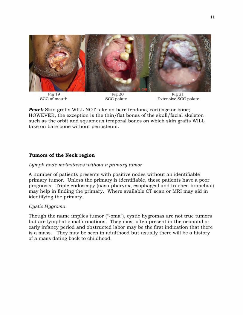
Squamous cell carcinoma

10
This is the commonest malignancy in the oral cavity. Squamous cell carcinoma (SCC) in the oral cavity may present with induration or an ulcer. Gingival lesions may present with loosening teeth or as a non-healing ulcer after a tooth extraction. Late presentation in sub-Saharan Africa is common as a result of misinformation, poverty, and illiteracy. Most patients will attribute an ulcer to some past event, and will wait for resolution for a long time, before presenting to the doctor (Fig. 21). Wide excision with or without neck dissection is the main mode of management. Where radiotherapy is accessible, surgery may be followed by radiotherapy of the neck. As noted previously, radiotherapy alone is equivalent to surgery for many lesions, without the attendant morbidity of surgical therapy.
Achieving adequate tumor resection in our environment may be more important than cosmesis; surgical reconstruction can be achieved upon referring the patient to centers where the expertise is available. The prognosis after tumor resection is not impacted at all by a waiting period prior to reconstruction; referring a patient for resection and reconstruction on the other hand may worsen the patient’s prognosis because of the waiting time to treatment.
Fig 13 Fig 14 Fig 15
Leukoplakia or early SCC can be treated by a lip peel: Excision of lesion and mucosal advancement
Fig 16 Fig 17 Fig 18
Lip SCC with wide resection and reconstruction with Karapandzic flaps
11
Fig 19 Fig 20 Fig 21 SCC of mouth SCC palate Extensive SCC palate Pearl: Skin grafts WILL NOT take on bare tendons, cartilage or bone; HOWEVER, the exception is the thin/flat bones of the skull/facial skeleton such as the orbit and squamous temporal bones on which skin grafts WILL take on bare bone without periosteum.
Tumors of the Neck region
Lymph node metastases without a primary tumor
A number of patients presents with positive nodes without an identifiable primary tumor. Unless the primary is identifiable, these patients have a poor prognosis. Triple endoscopy (naso-pharynx, esophageal and tracheo-bronchial) may help in finding the primary. Where available CT scan or MRI may aid in identifying the primary.
Cystic Hygroma
Though the name implies tumor (“-oma”), cystic hygromas are not true tumors but are lymphatic malformations. They most often present in the neonatal or early infancy period and obstructed labor may be the first indication that there is a mass. They may be seen in adulthood but usually there will be a history of a mass dating back to childhood.

12
Fig 22 Fig 23 Fig 24
Large macrocystic hygroma in newborn, wide excision with preservation of facial nerve and immediate postoperative photo
Surgical excision can be challenging in the infant with an extensive lesion that is intricately related to the facial nerve. Avoidance of facial nerve injury is critical. Macrocystic lesions (made up of large cysts which are more easily identifiable) have a much better prognosis whether managed surgically or medically. Microcystic cystic hygromas are very difficult to treat as it is difficult to identify all the small cysts for complete removal.
While watchful waiting may be used for a few lesions, medical management involves the use of sclerosants. Of the sclerosants used, absolute alcohol or tetracycline/doxycycline (10mg/ml solution) are the most readily available in sub-Saharan Africa.
Pearl: Although aspiration has been historically been frowned upon, some workers have reported successes with this technique.
Further reading: Other chapters in this book include other head and neck tumors and the reader is referred to these chapters:
Chapter 2 Chronic Wounds and Ulcers
Chapter 8 Cutaneous Lesions
Chapter 9 Jaw Tumors
Chapter 11 Nasoencephalocele
Chapter 14 Noma
Chapter 31 Neurofibromatosis