chapter 6shodhganga.inflibnet.ac.in/bitstream/10603/34582/11/11_chapter6.pdf · in a typical...
TRANSCRIPT

Chapter 6 Green synthesis and characterization of silver nanoparticles using Jatropha seedcake extract This part has been communicated as: Anjali Bose, Haresh Keharia and M. P. Deshpande (2012). Eco-friendly phyto-synthesis of silver nanoparticles using Jatropha seedcake extract. Industrial Crops and Products.
JSC for silver nanoparticle synthesis Chapter 6
116
6.1. Introduction
Nanoparticles (structures with at least one dimension approximately within 1 to 100
nm) have received considerable attention in both scientific and technological areas due to
their unique and unusual physico-chemical properties compared with that of bulk materials
(Bhushan 2004). Further, metal nanoparticles are particularly interesting because they can
be easily synthesized, modified chemically and applied for device fabrication (Feldheim
and Foss 2002).
Amongst various metal nanoparticles, silver nanoparticles (AgNPs) are perhaps most
widely recognized owing to its broad range of applications such as, in photonics (Gould et
al 2000), micro-electronics (Maekawa et al 2010), photocatalysis (Chen et al 2010),
lithography (Hulteen et al 1999), biosensing (Ngece et al 2011), etc. The properties and
applications of nanoparticles particularly depend on their morphology i.e. crystal structure
and dimensions. Consequently, significant research has been concentrated on developing
techniques to control their size, shape and distribution (Ozin 1992, Feldheim and Foss
2002). Different surface passivator reagents such as thiophenol, thiourea, mercapto-acetate
etc. have been employed in order to prevent the agglomeration of particles during and upon
their synthesis as well as to subsequently control their size (Li et al 2003, Stewart et al
2012). Unfortunately, these passivators are toxic (HSDB, TOXNET, U.S. NLM, NIH),
would raise environmental issues, if they are to be used in processes for large scale
synthesis of nanoparticles.
In addition, AgNPs have also been recognized as novel therapeutic agents extending
its use as antibacterial, antifungal, antiviral, anti-inflammatory and anti-cancerous agents
(Vaidyanathan et al 2009). The broad-spectrum antimicrobial properties of AgNPs
encourage its use in a large number of biomedical and environmental applications as well
as in cosmetics, clothing and different consumer products (Rai et al 2009). The
conventional synthesis of AgNPs involve number of chemical and physical methods that
are energy and capital intensive, employ toxic chemicals, thus limiting its biomedical
applications. Consequently, there has been growing need for synthesis of bio-compatible
metal nano-particles, without employing toxic chemicals in the synthesis protocols. In this
respect, use of microorganisms in biosynthesis of nano-particles has been demonstrated in
last two decades, particularly owing to their facile assembly of the nano-dimensioned

JSC for silver nanoparticle synthesis Chapter 6
117
particles. However, slow reaction rates, difficulty in maintaining culture and aseptic
conditions during synthesis, directed research towards the use of plant part and extract for
the synthesis of metal nanoparticles. Gardea-Torresdey and co-workers first reported the
formation of AgNPs by living plants (Gardea-Torresdey et al 2003). Later on, the synthesis
of AgNPs has been reported using plants (Iravani 2011) and plant products like green tea
(Camellia sinensis) (Vilchis-Nestor et al 2008), natural rubber (Abu Bakar et al 2007),
Aloe-vera plant extract (Chandran et al 2006), latex of Jatropha curcas (Bar et al 2009),
mesocarp of coconut (Roopan et al 2013) etc.
In the present chapter, we discussed the use of Jatropha seedcake as reducing and
capping agent for biogenesis of AgNPs. The AgNPs thus obtained were characterized by
Transmission Electron Microscopy (TEM), X-Ray Diffraction (XRD), and Fourier
Transform Infra-Red (FTIR) spectroscopy.
6.2. Materials and Methods
6.2.1. Materials
Jatropha seedcake (JSC) was obtained as gratis from the Food Processing and
Bioenergy division, Anand Agriculture University, Anand, Gujarat, India. The seedcake
was sun dried and powdered. 10 g powdered JSC was boiled in 100 mL distilled water for 1
h and then filtered to get the extract. This extract was used as reducing agent and stabilizer.
Silver nitrate (AgNO3) analytical grade was purchased from Hi-Media, India.
6.2.2. Synthesis of Silver nanoparticles
In a typical reaction procedure, a measured volume of JSC extract was added to
appropriate volume of aqueous AgNO3 solution to get a final volume of 10 mL, the mixture
was heated at 90°C and the resulting solution became reddish in color after 15 min of
heating. The reaction was stopped by plunging the tubes in ice-cold water.
Reactions were performed to determine the effect of varying concentrations of JSC
extract and AgNO3 on the AgNPs synthesis.

JSC for silver nanoparticle synthesis Chapter 6
118
6.2.3. Characterization of the nanoparticles
Formation and stability of AgNPs in aqueous colloidal solution was confirmed by
UV-Visible spectrophotometry (UV-Visible spectrophotometer, Shimadzu UV-1601) in the
wavelength range of 300-700 nm operated at resolution of 1 nm.
The size and morphology of the nanoparticles was determined by transmission
electron microscopy (Techai 20; Philips, Holland). AgNPs were mounted on TEM grids by
placing a drop of the particle solution on a carbon-coated copper grid and dried overnight.
The SAED was obtained by directing the electron beam perpendicular to one of the
nanoparticle. The crystalline nature of the metal nanoparticles was confirmed XRD using
Powder X-ray diffractometer (X’pert MPD; Philips, Holland) with Cu-Kα radiation and data
were obtained over the range of 0 to 100° (2θ) with a scanning rate of 0.005°s-1 and step
size of 0.02°. The XRD peaks were indexed and crystallographic lattice parameters were
determined by Powder X-diffraction analysis software (Dong 1999).
6.2.4. FTIR analysis
The functional groups responsible for reduction and stabilization of the bioreduced
AgNPs were analyzed using FTIR spectroscopy (Spectrum GX; Perkin-Elmer). Samples
were prepared by grinding 1 to 2 mg of nanoparticles along with 250 mg KBr and
pelletized using hydraulic press at 20,000 prf.
6.2.5. Evaluation of antibacterial activity of the bioreduced silver nanoparticles.
The antibacterial activity of AgNPs was measured by agar gel diffusion method using
following bacterial test cultures: Escherichia coli MTCC 40, Salmonella para-typhi MTCC
735, Bacillus subtilis ATCC 6051 and Staphylococcus aureus MTCC 87.
To evaluate the minimum inhibitory concentration (MIC), 50 µL of 108 cfu/mL test
culture was added to nutrient broth supplemented with varying concentrations (25 to 500
µg/mL) of silver nanoparticles. Control tubes contained only inoculated broth. The tubes
were incubated at 37°C for 24 h. The turbidity of the tubes was measured using visible
spectrophotometer (Systronics, Ahmedabad, India) at 600nm.

JSC for silver nanoparticle synthesis Chapter 6
119
6.3. Results and Discussion
6.3.1. Synthesis of silver nanoparticles
The formation of AgNPs in aqueous colloidal solution was marked by the change in
the colour of the reaction solution from pale yellow to deep red (Mulvaney 1996). The
characteristic surface plasmon resonance (SPR) band of colloidal silver was observed
between 424 to 438 nm, which is in good agreement with the earlier reports of bioreduced
AgNPs (Bar et al 2009, Kumar and Mamidyala 2011, Roopana et al 2013). Furthermore, it
was observed that with increase in volume fraction of JSC extract upto 0.1, the intensity of
SPR band increased and further increase in JSC extract volume fraction seized the reaction
(Fig. 6.1). The increase in the intensity of characteristic SPR band may be attributed to
higher concentration of AgNPs (Bar et al 2009). However, at higher volume fraction the
reaction mixture became hazy, probably due to the presence of excess biomaterial (Bar et al
2009).
Figure 6.1 UV-Visible absorption spectra of silver nanoparticles at varying volume fractions of JSC aqueous extract.
Figure 6.2 shows the effect of varying AgNO3 concentrations on AgNPs synthesis
when the reaction was carried out using fixed volume fraction (0.1) of JSC extract. The

JSC for silver nanoparticle synthesis Chapter 6
120
intensity of SPR band, which corresponds to concentration of AgNPs, increased with
increasing concentration of AgNO3 upto 1 mM. At concentration of AgNO3 above 1 mM,
no further increase in intensity of SPR band was observed, indicating the saturation of
biomolecules for the reduction of silver ions to silver. Such effect has been earlier reported
for synthesis of nanoparticles employing the extract of Cinnamon zeylanicum bark
(Satishkumar et al 2009) and Banana peel (Bankar et al 2010).
Figure 6.2 UV-Visible absorption spectra of silver nanoparticles at varying AgNO3 concentration.
6.3.2. Characterization of the bioreduced silver nanoparticles
The bioreduced AgNPs were found to be predominantly spherical in shape, well
dispersed with no agglomeration (Fig. 6.3). The average particle size of AgNPs observed in
TEM image was computed to be 10.48 ± 2.74 nm (Fig. 6.4). The similar size of AgNPs was
reported by Philip (2010) and Valodkar et al (2011) using leaf extract of Hibiscus rosa
sinensis and latex of Euphorbia nivulia, while AgNPs of larger size have been reported by
Mude et al (2009) and Santhoshkumar et al (2011) using callus extract of Carica papaya
and leaf extract of Nelumbo nucifera as reducing and stabilizing agents. Such size variation

JSC for silver nanoparticle synthesis Chapter 6
121
of metal nanoparticles synthesized using plant parts and products could be attributed to
difference in reductive potential and capping biomolecules from different plant origin.
Figure 6.3 TEM micrograph of uniformly distributed silver nanoparticles.
Figure 6.4 Particle size distribution of silver nanoparticles synthesized using JSC aqueous extract
The SAED pattern (Fig. 6.5) indicated the polycrystalline nature of bioreduced
AgNPs. Inter-planar spacing (also known as d-spacing) was calculated using Bragg
equation (Bragg and Bragg 1931):
RLd hkl
…………………………………………………………………...…………….. (1)
JSC for silver nanoparticle synthesis Chapter 6
122
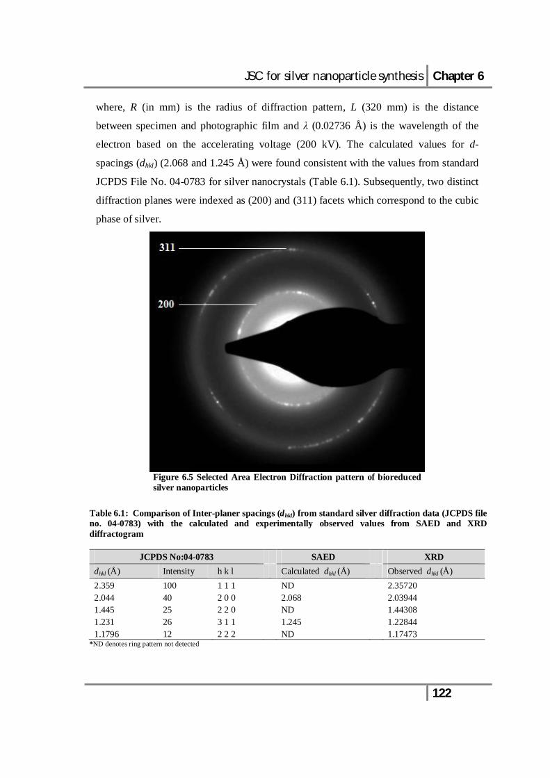
where, R (in mm) is the radius of diffraction pattern, L (320 mm) is the distance
between specimen and photographic film and λ (0.02736 Å) is the wavelength of the
electron based on the accelerating voltage (200 kV). The calculated values for d-
spacings (dhkl) (2.068 and 1.245 Å) were found consistent with the values from standard
JCPDS File No. 04-0783 for silver nanocrystals (Table 6.1). Subsequently, two distinct
diffraction planes were indexed as (200) and (311) facets which correspond to the cubic
phase of silver.
Figure 6.5 Selected Area Electron Diffraction pattern of bioreduced silver nanoparticles
Table 6.1: Comparison of Inter-planer spacings (dhkl) from standard silver diffraction data (JCPDS file no. 04-0783) with the calculated and experimentally observed values from SAED and XRD diffractogram
JCPDS No:04-0783 SAED XRD dhkl (Å) Intensity h k l Calculated dhkl (Å) Observed dhkl (Å) 2.359 100 1 1 1 ND 2.35720 2.044 40 2 0 0 2.068 2.03944 1.445 25 2 2 0 ND 1.44308 1.231 26 3 1 1 1.245 1.22844 1.1796 12 2 2 2 ND 1.17473
*ND denotes ring pattern not detected
JSC for silver nanoparticle synthesis Chapter 6
123
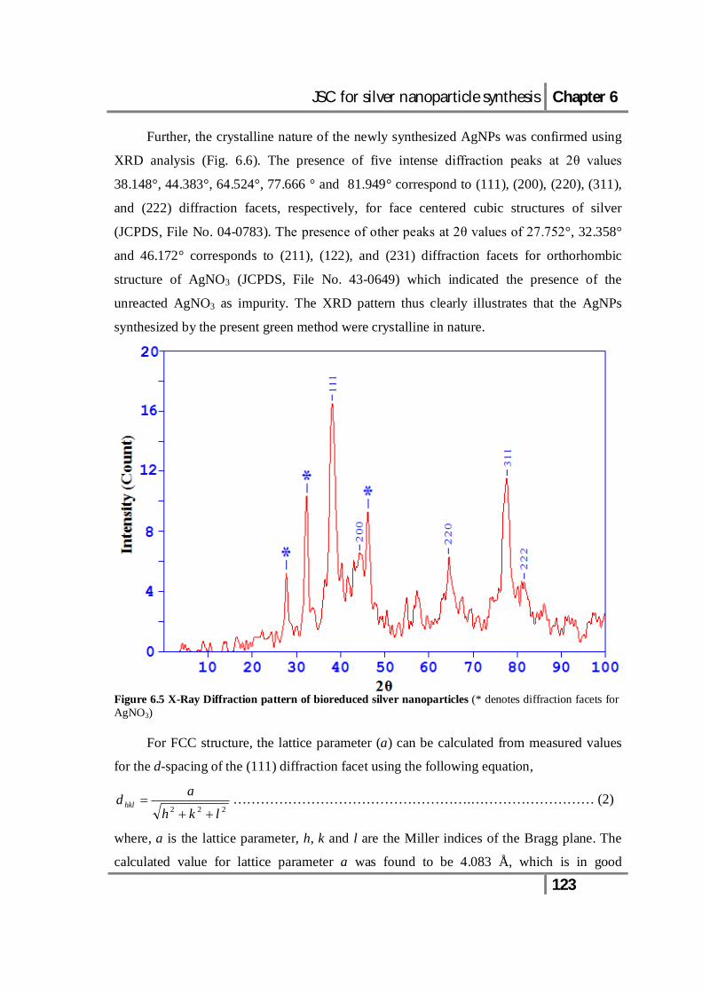
Further, the crystalline nature of the newly synthesized AgNPs was confirmed using
XRD analysis (Fig. 6.6). The presence of five intense diffraction peaks at 2θ values
38.148°, 44.383°, 64.524°, 77.666 ° and 81.949° correspond to (111), (200), (220), (311),
and (222) diffraction facets, respectively, for face centered cubic structures of silver
(JCPDS, File No. 04-0783). The presence of other peaks at 2θ values of 27.752°, 32.358°
and 46.172° corresponds to (211), (122), and (231) diffraction facets for orthorhombic
structure of AgNO3 (JCPDS, File No. 43-0649) which indicated the presence of the
unreacted AgNO3 as impurity. The XRD pattern thus clearly illustrates that the AgNPs
synthesized by the present green method were crystalline in nature.
Figure 6.5 X-Ray Diffraction pattern of bioreduced silver nanoparticles (* denotes diffraction facets for AgNO3)
For FCC structure, the lattice parameter (a) can be calculated from measured values
for the d-spacing of the (111) diffraction facet using the following equation,
222 lkh
ad hkl
…………………………………………….……………………… (2)
where, a is the lattice parameter, h, k and l are the Miller indices of the Bragg plane. The
calculated value for lattice parameter a was found to be 4.083 Å, which is in good

JSC for silver nanoparticle synthesis Chapter 6
124
agreement with the standard lattice parameter value (a=4.086 Å) from standard JCPDS File
No. 04-0783 for silver nanocrystals.
The crystallite size of the bioreduced AgNPs was calculated from broadening of the
diffraction peaks according to Scherrer equation (Scherrer 1981):
cos2
KT ……………………………………………………………………………. (3)
where, T is the crystallite size, K (~1) is the Scherrer’s constant related to the shape and
index (h.k.l) of the crystal, λ is the wavelength of the X-ray used (Cu-Kα, 1.54056 Å), θ is
the Bragg’s diffraction angle (in degree), and β2θ is broadening of diffraction lines
measured at half of its maximum intensity (in radian). The average crystallite size was
calculated to be 5.58 ± 1.29 nm (Table. 6.2).
Table 6.2: Calculation for crystallite size of bioreduced silver nanoparticles from XRD data using Scherrer equation
FWHM β2θ (°)
FWHM β2θ (radians)
2θ (°)
θ (radians)
cos θ (radians)
T * (Å) h k l
1.8 0.0314159 27.752 0.242181682 0.970817072 50.51166761 1.9 0.03316123 32.358 0.282376581 0.960395943 48.37240705 1.9 0.03316123 38.148 0.33290382 0.945097394 49.15542442 1 1 1 1.1 0.01919861 44.383 0.387314414 0.925926752 86.66271677 2 0 0 1.9 0.03316123 46.172 0.402926371 0.919917469 50.50090366 2.2 0.03839721 64.524 0.563077648 0.845616297 47.44665408 2 2 0 1.95 0.03403389 77.666 0.677763136 0.778977297 58.10883986 3 1 1 2.1 0.03665188 81.949 0.71513933 0.755001887 55.67167461 2 2 2 T = 55.80378601
* T = Kλ/β₂θ cosθ, where K = 1 and λ = 1.54056 Å
6.3.3. FTIR analysis
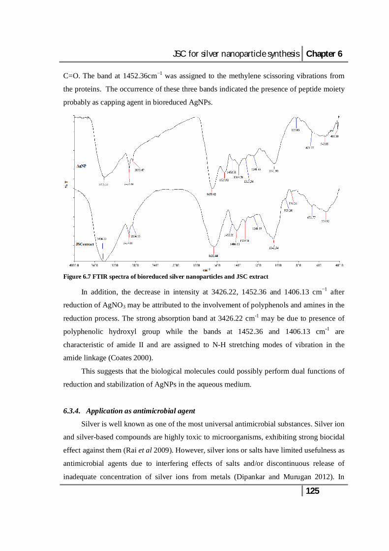
It was observed that the AgNPs in aqueous solution remained stable over one year
with little aggregation. It has been reported that the carbonyl groups from the amino acid
residues and peptides have a strong affinity to bind with metals and act as encapsulating
agent, thus preventing the nanoparticles from agglomeration (Mitra and Das 2008). In FTIR
spectrum of AgNP, the bands at 1650.42, 1452.36 and 1241.89 cm−1 (Fig. 6.7)
corresponded to characteristic amide I, II and III bands (Coates 2000). The amide band I
was assigned to the stretch mode of the carbonyl group coupled to the amide linkage (NH)-
JSC for silver nanoparticle synthesis Chapter 6
125
C=O. The band at 1452.36cm−1 was assigned to the methylene scissoring vibrations from
the proteins. The occurrence of these three bands indicated the presence of peptide moiety
probably as capping agent in bioreduced AgNPs.
Figure 6.7 FTIR spectra of bioreduced silver nanoparticles and JSC extract
In addition, the decrease in intensity at 3426.22, 1452.36 and 1406.13 cm−1 after
reduction of AgNO3 may be attributed to the involvement of polyphenols and amines in the
reduction process. The strong absorption band at 3426.22 cm-1 may be due to presence of
polyphenolic hydroxyl group while the bands at 1452.36 and 1406.13 cm-1 are
characteristic of amide II and are assigned to N-H stretching modes of vibration in the
amide linkage (Coates 2000).
This suggests that the biological molecules could possibly perform dual functions of
reduction and stabilization of AgNPs in the aqueous medium.
6.3.4. Application as antimicrobial agent
Silver is well known as one of the most universal antimicrobial substances. Silver ion
and silver-based compounds are highly toxic to microorganisms, exhibiting strong biocidal
effect against them (Rai et al 2009). However, silver ions or salts have limited usefulness as
antimicrobial agents due to interfering effects of salts and/or discontinuous release of
inadequate concentration of silver ions from metals (Dipankar and Murugan 2012). In

JSC for silver nanoparticle synthesis Chapter 6
126
contrast, these kinds of limitation can be overcome using AgNPs because they are highly
reactive species due to their extremely large surface area, which provides better contact
with micro-organisms. The AgNPs produced biologically are known to exhibit potent
antimicrobial activity (Sharma et al 2009).
Figure 6.8 Anti-bacterial activity of silver nanoparticles against (a) E. coli MTCC 40, (b) S. paratyphi MTCC 735, (c) B. subtilis ATCC 6051 and (d) S. aureus MTCC 87. (CON indicates reaction control and AgNP indicates bioreduced silver nanoparticles)
The AgNPs described in present study were found to exhibit antibacterial activity
against both Gram positive and Gram negative bacteria tested (Fig. 6.8). Similar
observations regarding antibacterial activity of AgNPs have been reported by Sondi and
Salopek-Sondi (2004), Satishkumar et al (2009), Kora et al (2010) and Soo-Hwan et al
(2011). In addition, the AgNPs exhibited lower MIC against the Gram-positive bacteria
JSC for silver nanoparticle synthesis Chapter 6
127
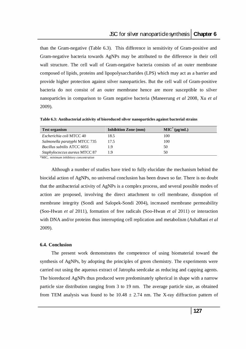
than the Gram-negative (Table 6.3). This difference in sensitivity of Gram-positive and
Gram-negative bacteria towards AgNPs may be attributed to the difference in their cell
wall structure. The cell wall of Gram-negative bacteria consists of an outer membrane
composed of lipids, proteins and lipopolysaccharides (LPS) which may act as a barrier and
provide higher protection against silver nanoparticles. But the cell wall of Gram-positive
bacteria do not consist of an outer membrane hence are more susceptible to silver
nanoparticles in comparison to Gram negative bacteria (Maneerung et al 2008, Xu et al
2009). Table 6.3: Antibacterial acitivity of bioreduced silver nanoparticles against bacterial strains
Test organism Inhibition Zone (mm) MIC* (µg/mL) Escherichia coli MTCC 40 18.5 100 Salmonella paratyphi MTCC 735 17.5 100 Bacillus subtilis ATCC 6051 1.9 50 Staphylococcus aureus MTCC 87 1.9 50
*MIC, minimum inhibitory concentration
Although a number of studies have tried to fully elucidate the mechanism behind the
biocidal action of AgNPs, no universal conclusion has been drawn so far. There is no doubt
that the antibacterial activity of AgNPs is a complex process, and several possible modes of
action are proposed, involving the direct attachment to cell membrane, disruption of
membrane integrity (Sondi and Salopek-Sondi 2004), increased membrane permeability
(Soo-Hwan et al 2011), formation of free radicals (Soo-Hwan et al 2011) or interaction
with DNA and/or proteins thus interrupting cell replication and metabolism (AshaRani et al
2009).
6.4. Conclusion
The present work demonstrates the competence of using biomaterial toward the
synthesis of AgNPs, by adopting the principles of green chemistry. The experiments were
carried out using the aqueous extract of Jatropha seedcake as reducing and capping agents.
The bioreduced AgNPs thus produced were predominately spherical in shape with a narrow
particle size distribution ranging from 3 to 19 nm. The average particle size, as obtained
from TEM analysis was found to be 10.48 ± 2.74 nm. The X-ray diffraction pattern of

JSC for silver nanoparticle synthesis Chapter 6
128
AgNPs indicated a face-centred cubic crystalline phase of silver nanoparticles with lattice
parameter estimated to be 4.083 Å. AgNPs prepared through this route were quiet stable
and remained intact for over one year at 4°C. The AgNPs synthesized in present study
exhibited potential antibacterial activity, suggesting its prospective applications in
antibacterial formulations.

JSC for silver nanoparticle synthesis Chapter 6
129
References
Abu Bakar ANHH, Ismail J, Abu Bakar M (2007) Synthesis and characterization of silver
nanoparticles in natural rubber. Mater Chem Phys 104: 276-283.
AshaRani PV, Mun GLK, Hande MP, Valiyaveettil S (2009) Cytotoxicity and genotoxicity
of silver nanoparticles in human cells. ACS Nano 3: 279-290.
Bankar A, Joshi B, Kumar AR, Zinjarde S (2010) Banana peel extract mediated novel route
for the synthesis of silver nanoparticles. Colloid Surf A 368: 58-63.
Bar H, Bhui DK, Sahoo GP, Sarkar P, De SP, Misra A (2009) Green synthesis of silver
nanoparticles using latex of Jatropha curcas. Colloid Surf A 339: 134-139.
Bhushan B (2004) Handbook of Nanotechnology. Heidelberg: Spinger-Verlag Berlin.
Bragg WH, Bragg WL (1913) The reflection of X-rays by crystals. Proc R Soc Lond A 88:
428-438.
Chandran SP, Chaudhary M, Pasricha R, Ahmad A, Sastry M (2006) Synthesis of gold
nanotriangles and silver nanoparticles using Aloe vera plant extract. Biotechnol Prog
22: 577-583.
Chen X, Zheng Z, Ke X, Jaatinen E, Xie T, Wang D, Guo C, Zhao J, Zhu H (2010)
Supported silver nanoparticles as photocatalysts under ultraviolet and visible light
irradiation. Green Chem 12: 414-419.
Coates J (2000) Interpretation of infrared spectra, a practical approach. In: Meyers RA (Ed)
Encyclopaedia of Analytical Chemistry. John Wiley & Sons Ltd, Chichester, pp.
10815-10837.
Dipankar C, Murugan S (2012) The green synthesis, characterization and evaluation of the
biological activities of silver nanoparticles synthesized from Iresine herbstii leaf
aqueous extracts. Colloid Surf B 98: 112-119.
Dong C (1999) Windows-95-based program for powder X-ray diffraction data processing. J
Appl Crystallography 32: 838-838.
Feldheim DL, Foss CA (2002) Metal Nanoparticles: Synthesis, Characterization and
Applications. New York: CRC Press, Marcel Dekker Inc.
Gardea-Torresdey GL, Gomez E, Peralta-Videa J, Parsons JG, Troiani HE, Jose-Yacaman
M (2003) Alfalfa sprouts: a natural source for the synthesis of silver nanoparticles.
Langmuir 19: 1357-1361.

JSC for silver nanoparticle synthesis Chapter 6
130
Gould IR, Lenhard JR, Muenter AA, Godleski SA, Farid S (2000). Two-electron
sensitization: a new concept for silver halide photography. J Am Chem Soc 122:
11934-11943.
HSDB (Hazardous Substances DataBank), TOXNET (TOXicology Data Network), U.S.
National Library of Medicine, National Institute of Health.
Hulteen J, Treichel DA, Smith MT, Duval ML, Jensen TR, Duyne RPV (1999) Nanosphere
lithography: size-tunable silver nanoparticle and surface cluster arrays. J Phys Chem
B 103: 3854-3863.
Iravani S (2011) Green synthesis of metal nanoparticles using plants. Green Chem 13:
2638-2650.
Kora AJ, Sashidhar RB, Arunachalama J (2010) Gum kondagogu (Cochlospermum
gossypium): A template for the green synthesis and stabilization of silver
nanoparticles with antibacterial application. Carbohydr Polym 82: 670-679.
Kumar CG, Mamidyala SK (2011) Extracellular synthesis of silver nanoparticles using
culture supernatant of Pseudomonas aeruginosa. Colloid Surf B 84: 462-466.
Li X, Zhang J, Xu W, Jia H, Wang X (2003) Mercaptoacetic acid-capped silver
nanoparticles colloid: formation, morphology, and SERS Activity. Langmuir 19:
4285-4290.
Maekawa K, Yamasaki K, Niizeki T, Mita M, Matsuba Y, Terada N, Saito H (2010) Laser
sintering of silver nanoparticles for electronic use. Mat Sci Forum 2085: 638-642.
Maneerung T, Tokura S, Rujiravanit R (2008) Impregnation of silver nanoparticles into
bacterial cellulose for antimicrobial wound dressing. Carbohydr Polym 72: 43-51.
Mitra RN, Das PK (2008) In situ preparation of gold nanoparticles of varying shape in
molecular hydrogel of peptide amphiphiles. J Phys Chem C 112: 8159-816.
Mude N, Ingle A, Gade A, Rai M (2009) Remove from marked Records Synthesis of silver
nanoparticles using callus extract of Carica papaya - a first report. J Plant Biochem
Biotechnol 18: 83-86.
Mulvaney P (1996) Surface plasmon spectroscopy of nanosized metal particles. Langmuir
12: 788-800.
Ngece RF, West N, Ndangili PA, Olowu RA, Williams A, Hendricks N, Mailu S, Baker P,
Iwuoha E (2011) A silver nanoparticle/poly (8-anilino-1-naphthalene sulphonic acid)
JSC for silver nanoparticle synthesis Chapter 6
131
bioelectrochemical biosensor system for the analytical determination of ethambutol.
Int J Electrochem Sci 6: 1820-1834.
Ozin GA (1992) Nanochemistry: synthesis in diminishing dimensions. Adv Mater 4: 612-
649.
Philip D (2010) Green synthesis of gold and silver nanoparticles using Hibiscus rosa-
sinensis. Physica E 42: 1417-1424.
Rai M, Yadav A, Gade A (2009) Silver nanoparticles as a new generation of antimicrobials.
Biotechnol Adv 27: 76-83.
Roopana SM, Madhumitha RG, Rahuman AA, Kamaraj C, Bharathi A, Surendra TV
(2013) A Low-cost and eco-friendly phyto-synthesis of silver nanoparticles using
Cocos nucifera coir extract and its larvicidal activity. Ind Crops Prod 43: 631-635.
Santhoshkumar T, Rahuman AA, Rajakumar G, Marimuthu S, Bagavan A, Jayaseelan C,
Zahir AA, Elango G, Kamaraj C (2011) Synthesis of silver nanoparticles using
Nelumbo nucifera leaf extract and its larvicidal activity against malaria and filariasis
vectors. Parasitol Res 108: 693-702.
Satishkumar M, Sneha K, Won SW, Cho CW, Kim S, Yun YS (2009) Cinnamon
zeylanicum bark extract and powder mediated green synthesis of nano-crystalline
silver particles and its antibacterial activity. Colloid Surf B 73: 332-338.
Scherrer P (1918) Bestimmung der Grösse und der Inneren Struktur von Kolloidteilchen
Mittels Röntgenstrahlen, Nachrichten von der Gesellschaft der Wissenschaften,
Göttingen. Mathematisch-Physikalische Klasse 2: 98-100.
Sharma VK, Yngard RA, Lin Y (2009) Silver nanoparticles: green synthesis and their
antimicrobial activities. Adv Colloid Interface Sci 145: 83-96.
Sondi I, Salopek-Sondi B (2004) Silver nanoparticles as antimicrobial agent: a case study
on E. coli as a model for Gram-negative bacteria. J Colloid Interface Sci 275: 177-
182.
Soo-Hwan K, Lee H-S, Ryu D-S, Choi, S-J, Lee D-S (2011) Antibacterial activity of silver-
nanoparticles against Staphylococcus aureus and Escherichia coli. Korean J
Microbiol Biotechnol 39: 77-85.

JSC for silver nanoparticle synthesis Chapter 6
132
Stewart A, Zheng S, McCourt MR, Bell SEJ (2012) Controlling assembly of mixed thiol
monolayers on silver nanoparticles to tune their surface properties. ACS Nano 6:
3718-3726.
Vaidyanathan R, Kalishwaralal K, Gopalram S, Gurunathan S (2009) Nanosilver-the
burgeoning therapeutic molecule and its green synthesis. Biotechnol Adv 27: 924-
937.
Valodkar M, Nagar PS, Jadeja RN, Thounaojam MC, Devkar RV, Thakore S (2011)
Euphorbiaceae latex induced green synthesis of non-cytotoxic metallic nanoparticle
solutions: A rational approach to antimicrobial applications. Colloid Surf A 384: 337-
344.
Vilchis-Nestor AR, Sanchez-Mendieta V, Camacho-Lopez MA, Gomez-Espinosa RM,
Camacho-Lopez MA, Arenas-Alatorre JA (2008) Solvent less synthesis and optical
properties of Au and Ag nanoparticles using Camellia sinensis extract. Mater Lett 62:
3103-3105.
Xu K, Wang J, Kang X, Chen J (2009) Fabrication of antibacterial monodispersed Ag-SiO2
core-shell nanoparticles with high concentration. Mater Lett 6: 31-33.