chapter biomedical implants for regenerative therapies
TRANSCRIPT

1
Chapter
Biomedical Implants for Regenerative TherapiesAndrea Domingues Goncalves, Wendy Balestri and Yvonne Reinwald
Abstract
Regenerative therapies aim to develop novel treatments to restore tissue function. Several strategies have been investigated including the use of biomedi-cal implants as three-dimensional artificial matrices to fill the defect side, to replace damaged tissues or for drug delivery. Bioactive implants are used to provide growth environments for tissue formation for a variety of applications including nerve, lung, skin and orthopaedic tissues. Implants can either be biodegradable or non-degradable, should be nontoxic and biocompatible, and should not trigger an immunological response. Implants can be designed to provide suitable surface area-to-volume ratios, ranges of porosities, pore inter-connectivities and adequate mechanical strengths. Due to their broad range of properties, numerous biomaterials have been used for implant manufacture. To enhance an implant’s bioactivity, materials can be functionalised in several ways, including surface modification using proteins, incorporation of bioactive drugs, growth factors and/or cells. These strategies have been employed to create local bioactive microenvironments to direct cellular responses and to promote tissue regeneration and controlled drug release. This chapter provides an overview of current bioactive biomedical implants, their fabrication and applications, as well as implant materials used in drug delivery and tissue regeneration. Additionally, cell- and drug-based bioactivity, manufacturing considerations and future trends will be discussed.
Keywords: biomaterials, bioactive biomedical implants, stem cells, drug delivery, manufacturing
1. Introduction to bioactive implants
Implants are man-made devices that are fabricated for the implantation inside body to replace or support a biological structure, together with delivering drugs and monitoring body functions. They can remain in the body temporarily or perma-nently [1]. To date, biomedical implants are used not only as sensory devices [2]; brain and neural devices including neuronal, cochlear and retinal implants [3, 4]; subcutaneous implants [5]; cardiovascular devices such as vascular grafts, stent, heart valves, pacemakers [3]; sutures and wound dressings [6]; spinal [7] and den-tal implants [8]; cosmetic [9] and structural implants [10] including rods, braces, craniofacial, hip and knee replacements; but also as ophthalmic devices including glasses and contact lenses as well as insulin delivery devices [6].

Biomaterials
2
In recent years, scaffolds made of synthetic or natural polymers were developed to regenerate damaged or deteriorated tissues, or to deliver drugs to specific locations. Scaffolds are three-dimensional (3D) structures that mimic the native extracellular matrix (ECM) of tissues and provide a substrate for cell adhesion and proliferation.
These biomedical implants can be made of bioactive materials. The term “bioac-tive” means that a material can affect its surrounding tissue biologically. Scaffolds can include molecules that promote a biological response in the region where they are implanted. Moreover, cells can be included in these scaffolds to promote healing and regeneration, as they naturally secrete growth factors and cytokines [11].
The risks related to the surgery during the placement or removal of the implant include infection and implant failure. Also, inflammation reaction against the material or rejection needs to be taken into consideration [1]. Here, we report what it is known about bioactive biomedical implants, their desired properties and their applications, focusing on the techniques and materials used for their fabrication. We further provide an overview of cell-based and drug-based implants, implant manufacture and its considerations.
2. Biomaterials for implants in drug delivery and regenerative therapies
To assist native tissue regeneration or/and replacement, implants are made of biomaterials, which support cell and tissue growth through cell adhesion, prolifera-tion and differentiation, prevent unwanted cell and tissue growth, tailor tissue response and prevent immunological responses [12].
Biomaterials have been used for controlled drug delivery systems, sutures and adhesives including biodegradable and non-biodegradable materials, cardiovascular grafts, reconstructive and orthopaedic implants, ophthalmic devices such as cor-neas and contact lenses, and dental implants [13]. Various types of materials have been used to produce biomedical implants. These include bioceramics, polymers, metals and composites, which are further discussed below. Table 1 summarises current biomedical applications for biomaterials.
Application Material References
Ophthalmic applications (contact lenses, intraocular lenses)
Silicones, hydrogels [14]
Cardiovascular applications (vascular prostheses, artificial valves, stents, cardiac-assisted pumps, blood bags and catheters)
Polymers, metals and ceramics; polyurethane (PU); polyesters (PE); polybutesters (PBE); polypropylene (PP) and PTFE; stainless steel
[15–21]
Central nervous system and peripheral nervous system (scaffolds for nerve regeneration)
Polycaprolactone (PCL), silk, collagen [11, 22–26]
Orthopaedic applications (total hip replacement, hip arthroplasty, total knee arthroplasty, bone screws, orthodontic brackets and wires; bone fillers and scaffolds as bone replacements)
Chromium, cobalt, molybdenum, nickel, titanium and zirconium alloys, ultrahigh molecular weight polyethylene (UHMWPE), Ti-6Al-4V, ceramic-coated steels, stainless steel, copper; natural polymers like collagen, chitosan, alginates, synthetic polymers, ceramics like bioglasses, hydroxyapatite and beta-TCP; poly(l-lactic acid) (PLLA); poly(lactic acid), poly(lactic-co-glycolic acid) (PLGA), polycaprolactone (PCL)
[11, 12, 14, 27–33]
Table 1. Examples of biomedical applications for currently used biomaterials.

3
Biomedical Implants for Regenerative TherapiesDOI: http://dx.doi.org/10.5772/intechopen.91295
2.1 Bioceramics
Ceramics are chemically inert and possess low thermal and electric conductivity as well as physical properties, which make them a suitable material glass for bio-medical implants [34, 35]. Bioceramics are osteoinductive and osteoconductive and possess mechanical properties like native bone. Their use as biomedical implants prevents the transmission of diseases and immunogenicity. To date, bioceramics are utilised for dental, periodontal, maxillofacial and orthopaedic applications [36]. In comparison to non-resorbable bioceramics, degradable ceramics exhibit lower mechanical strength. Their chemical and physical composition determines their biological response [37].
Ceramics produced from aluminium, zirconium and titanium oxides possess bending, tensile and compressive strength at least 3 times higher than natural bone and are used mainly for pin-type dental implants and root- and endosteal plate forms [38]. The first zirconia implants were reported in the 1970s. These implants exhibited the ability to integrate into bone tissue, accumulate less plaque and provide improved aesthetics compared to titanium implants [39, 40]. Hence, titanium-zirconium alloys, also called Straumann Roxolid or Roxolit (TiZr1317), are often used as dental implants due to their enhanced mechani-cal properties and osseointegrative properties that are often used as dental implants [41].
Calcium phosphate-based bioceramics such as tricalcium phosphate (TCP) are similar in chemical composition to the inorganic phase natural bone tissue. TCP exhibits better biodegradation, restorability and bioactivity in vivo than hydroxyapatite and is commonly used for orthopaedic, dental and maxillofacial applications. Complete resorption of orthopaedic implants fabricated from TCP was reported after up to 2 years in the rat tibia and for the formation of cancellous bone [42].
Amorphous or low crystalline hydroxyapatite (HAp) is bioactive and bioresorb-able. The preparation of synthetic HA at high temperatures results in high crystal-linity. Biodegradation and resorbability of HAp are very slow. HAp bioceramics are commonly used for small defects in the case of bone loss or fractures of the tibia, calcaneus and vertebra. HAp is not employed for load-bearing bone applications because of its poor mechanical properties. The modification of HAp with stron-tium, magnesium and silicon ions resulted in enhanced mechanical and biologi-cal properties [43]. Improved bioresorbability was achieved by zinc—[44] and manganese—[45] substitution of HAp.
Dicalcium phosphates (DCP) are biodegradable ceramics composed of calcium phosphates and water. DCPs are widely added to material compositions to modify their physical properties. Dehydrated DCP is known as brushite, which is used in tibial plate and distal metaphysis bone fractures [46].
Historically, ceramics have been used as dental and orthopaedic implant mate-rials. However, compared to other material classes, ceramics have not been used extensively as implant materials due to their limited load-bearing capacity [14].
2.2 Polymers
Polymers are macromolecules that consist of covalently bonded repeating units, which can be of the same (homopolymers) or different (co-polymers) molecule type [27]. A variety of natural and synthetic polymers are used as soft tissue trans-plants, facial prostheses, denture, hip and joint replacements as well as medical adhesives, sealants and coatings [14].

Biomaterials
4
Polymers are commonly selected based on their physical characteristics, compo-sition, and mechanical properties; how easily they can be modified and moulded; their heat and electric conductivity as well as their ability to integrate into and attach to native tissue [47].
2.2.1 Natural polymers
Natural polymers possess similar properties to native tissues. They are non-toxic and exhibit protein binding-sites and biochemical moieties that are important for tissue regeneration. However, natural polymers are often associated with immu-nological reactions, low mechanical strength and degradation at body temperature limiting their usability [14].
One of the most commonly used natural polymers is collagen. More than twenty different collagens are known in connective tissues such as bone, tendon, skin, cartilage and ligaments of the ECM of different species. Collagen type I is the main component in bone, skin and tendon, whereas type II is found in articular cartilage. Because of its abundance in nature, its importance for tissue homeostasis and growth, collagen has been investigated as material for bone, cartilage, tendon, skin and blood vessel regeneration [11]. In the clinic, collagen is used for the generation of dermal tissue, neo-tissue formation and wound healing [14]. Further natural polymers are chitosan, hyaluronic acid, fibrin and silk.
Silk, or silk fibroin, is a naturally occurring polymeric protein produced by insects and worms. The protein content gives rise to silk’s biocompatibility and its high tensile strength making it an ideal biomaterial for biomedical applications as gels, sponges and films [11, 48–53]. Silk composites fabricated from silk-chitosan and silk-hydroxyapatite have been used to improve silk’s elasticity, degradation and porosity [54, 55].
Hyaluronic acid (HA), a non-adhesive glycosaminoglycan (GAG), occurs mostly in connective, epithelial, and neural tissue [56]. HA is commonly used as hydro-gel for the regeneration of bone, cartilage and the vascular system and for drug delivery [11].
Chitosan is a biodegradable polysaccharide produced through partial deacety-lation of chitin. Chitosan scaffolds exhibit similar properties to naturally occurring GAGs, leading to their bioactivity, and support cellular adhesion [11, 57]. It has been investigated as scaffold material in combination with collagen and HA, as well as PCL for bone, cartilage and nerve regeneration [11].
2.2.2 Synthetic polymers
Synthetic polymers were developed with tailored physical and chemical prop-erties depending on the desired application to overcome limitations of natural polymers. Synthetic polymers are linear, branched or cross-linked depending on their molecular arrangement [58] and possess amorphous or crystalline struc-tures [27]. In addition, synthetic polymers are cheaper in production and enable improved functionality [11]. Commonly used synthetic polymers are poly(lactic acid-co-glycolic acid) (PLGA), polyglycolic acid (PGA), polylactic acid (PLA), polycaprolactone (PCL) and poly-hydroxybutyrate (PHB) [11, 27, 59, 60].
PGA, PLA and PCL are used for sutures, interference screws, fixation plates for meniscal repair and craniomaxillofacial fixtures and 3D scaffolds. However, they are known to induce inflammatory responses and are limited in mechanical integrity and controlled degradation. Hence, metal/polymer composites such as Mg/PCL have been developed [27]. Biodegradable synthetic polymers

5
Biomedical Implants for Regenerative TherapiesDOI: http://dx.doi.org/10.5772/intechopen.91295
are chosen based on the required physical, chemical and mechanical material characteristics (Table 2).
2.3 Metals and alloys
Due to their mechanical properties, the ease of their processing and the pos-sibility to sterilise them, metals and alloys are ideal materials for biomedical implants [34].
Metals are commonly used as load-bearing orthopaedic implants such as wires, screws, fixation plates, artificial joints for hips, knees, shoulders and ankles, as well as for dental, cardiovascular and craniofacial applications [14].
Novel magnesium alloys have been investigated for orthopaedic and cardiovas-cular applications [84, 85]. Combining magnesium alloys with aluminium or rare earth metals improves their mechanical properties [86, 87]. However, the accumu-lation of these elements is associated with neurotoxicity and hepatotoxicity [88]; hence, these alloys are not suitable for biomedical applications. Instead, extensive research is carried out to develop nontoxic magnesium alloys [89], such as Mg-Si and Mg-Sr alloys [90].
Titanium alloys are among the most commonly used metal alloys [91] due to their biocompatibility [92] and corrosion resistance [93]. Their composition
Synthetic polymer Application References
Poly(l-lactic acid) (PLLA); poly (d-lactic acid) (PDLA)
Sutures, drug delivery, vascular grafts, bone screws, fixation pins, dermal filler for facial atrophy (Scultra™)
[11, 61]
Poly(lactic-co-glycolic acid) (PLGA) Drug delivery [11, 62]
Polycaprolactone (PCL) Long-term implant, maxillo-cranial facial implant; drug release
[11, 63]
PCL-gelatin, PCL-chitosan, PCL-collagen
Tissue regeneration [60, 64, 65]
Poly-para-dioxanone (PPD) Internal fracture fixation, medical implant as films, foams and moulded scaffolds
[47, 66, 67]
Low-density polyethylene (LDPE), high-density polyethylene (HDPE)
Total hip arthroplasty and treatment of osteolysis as polymer-ceramic composites; rhinoplasty surgery
[68–71]
Poly(methyl methacrylate) (PMMA) Orbital medical implants, rhinoplasty, cranioplasty, bone cement in hip joint replacement, dental implant for restoration and aesthetics
[72–76]
Polydimethylsiloxane (PDMS) Enclosing implantable electronic devices and sensors, medical implants, oesophagus substitutes, catheters, shunts, blood pumps and peacemakers
[77–80]
Polyamides (PA), e.g., nylon and nylon-composites
Sutures, fabrication of dentures; scaffold materials and nanofillers for bone regeneration
[81, 82]
Carbon nanotubes (CNT) and composites
Metal coatings for load-bearing musculoskeletal implants to improve surface porosity, reduce metal ionisation and promote the formation of hydroxyapatite
[83]
Table 2. Synthetic polymers commonly used for the fabrication of biomedical implants.

Biomaterials
6
and microstructure vary depending on their elemental composition [94]. The mechanical properties of B-titanium alloys have a Young’s modulus like bone but possess a low fatigue strength. Their mechanical properties can be enhanced through the addition of silicon dioxide, zirconium dioxide and Yttrium oxide. Furthermore, to increase their wear resistance, titanium alloys are surface treated. Pure titanium alloys are used in pacemaker cases, ventricular devices, implantable drug pumps, screws and staples in spinal surgery, dental implants and craniofacial implants. Ti-6Al-4V alloys are used in hip and knee replacements and dental implants. Due to the release of aluminium and vanadium ions, which can cause neurological conditions such as Alzheimer’s, Ti-6Al-4V alloys are not considered safe for long-term use. β-Titanium alloys substituted with stabilising elements like zirconium, tantalum and molybdenum are safer compared to Ti-6Al-4V [27], and alternative titanium alloys, vanadium free Ti-6Al-7Nb and Ti-5Al-2.5Fe, are being developed [95].
Titanium has become the material of choice for implants; however, com-ponents of prosthetics are still manufactured from gold alloys, stainless steel, nickel-chromium alloys and cobalt-chromium alloys [35]. Cobalt chromium alloys enable the fabrication of customised grafts including subperiosteal implants. They are mainly composed of cobalt, chromium and molybdenum, which give rise to corrosion resistance and mechanical properties [96, 97]. Stainless steel alloys such as iron-chromium-nickel-based alloys are used as orthopaedic implants such as ramus blade, ramus frame, stabiliser pins and some mucosal inserts. Due to its nickel content, these alloys possess a low corrosion resistance and induce immunological reactions in patients with nickel allergies [34, 38].
3. Implant properties
Implant materials should possess adequate chemical and physical properties to allow for host tissue infiltration and nutrient transport; biocompatibility to avoid immunological responses; and corrosion resistance, degradation and biore-sorbability to enable normal cellular activity and controlled implant degradation [14, 27, 98]. In addition, temporary implants should possess a highly interconnected porous structure to allow cell migration and nutrient and waste transport, provide suitable surface topography to support cell adhesion and growth, as well as allow for the release of bioactive molecules if applicable [5, 11, 12].
Mechanical properties like Young’s Modulus, tensile, compressive and shear strength, yield strength and fatigue strength are required to ensure uniform stress distribution, to minimise the movement or fracture of the implant [34].
3.1 Surface properties
Surface properties influence cell adhesion and cellular and tissue responses. Surface tension determines the wettability by a wetting fluid, such as blood or water [11, 34]. Implant surfaces are also categorised by roughness, texture and the orientation of irregularities [38, 99]. The surface textures can vary from concave or convex. Concave surface textures occur due to additive treatments such as hydroxy-apatite coatings, whereas convex surfaces are created through etching and blast-ing. Furthermore, implant surfaces can either be isotropic, meaning that implant properties are independent from the measurement direction, or anisotropic, which means properties are directionally dependent [12, 34].

7
Biomedical Implants for Regenerative TherapiesDOI: http://dx.doi.org/10.5772/intechopen.91295
3.2 Corrosion and degradation of implants
3.2.1 Metal corrosion
Corrosion is the involuntary breakdown of metals by an electrochemical reac-tion and through the loss of ions from the metal surface in an acidic, an alkaline or a neutral environment. It is one of the most common reasons for implant failure [98]. Table 3 summarises the types of corrosions that have been observed in metal implants [98–100].
Magnesium for example corrodes faster with an increase of impurities such as nickel, copper and iron [101]. The higher the purity of magnesium, the slower its corrosion rate. However, pure magnesium is not suitable for medical implants due to its mechanical characteristics. Instead, calcium is used for the grain refinement in magnesium alloys [102]. Orthopaedic implants fabricated from Mg-Ca alloys were observed to corrode over a 3-month period after bone formation [103]. Magnesium’s mechanical properties can also be enhanced through Mg-Zn with calcium, manga-nese, yttrium or zirconium [104, 105]. Mg-Zn alloys withstand galvanic corrosion and biocorrosion in vitro; however, biocorrosion in vivo resulted in a 2 mm/year reduction of a Mg-Zn alloy used as rods in femur shafts [106].
3.2.2 Polymer degradation
Polymer degradation, or biodegradation, occurs through a process called hydro-lysis. The polymer surface is attacked by organisms, which secrete enzymes break-ing down ester bonds in macromolecules. The resulting smaller polymer molecules are further converted into carbon dioxide and water. The process of biodegradation varies for each polymer [27, 107, 108]; however, all polymers lose their mechani-cal integrity. To date, PGA, PLA and PLGA among others have been explored for biomedical implants [27]. Their degradation into non-toxic by-products made them favourable materials for temporary biomedical implants [109]. Poly(l-lactic) acid
Corrosion type Explanation Biomedical implants
Crevice corrosion
• Occurs in narrow regions
• Metal ions create localised positive charge in the crevice
• Interfaces between screws/plates and bone
Pitting corrosion
• Occurs in implants with small surface pit
• Metal ions react with chloride ions resulting in rough surfaces
• Orthopaedic and dental implants
Galvanic corrosion
• Occurs due to electrical gradient between Co-Cr alloys, Ni-Cr, Ag-Pd, Au-ternary Ti
• Oral/dental implants
• Screws and nuts
Corrosion fatigue and fretting
• Occurs due to cyclic stress • Bone cement
• Femoral implants
• Bone plates and screws at the bone-stem interface
• Stem-cement interfaces of modular hip implant
Table 3. Types of corrosions observed in metallic biomedical implants.

Biomaterials
8
(PLLA) has been shown to induce inflammatory responses in vivo upon degradation due to its high crystallinity; hence, poly(d, l-lactic acid) (PDLA) was synthesised [110, 111].
PLGA degrades into acidic moieties, which in higher concentrations can affect the microenvironment of the implant’s surrounding tissue. This can be especially important for drug delivery applications, where pH-sensitive drugs are used [11]. By increasing the amount of poly(glycolic acid) (PGA) compared to poly(lactic acid) (PLA) in PLGA, the degradation rate is reduced; hence, less acidic by-products are formed [11].
3.3 Biocompatibility
Biocompatibility indicates a desired response of the implant to its biological surrounding [34] and depends on biodegradability and corrosion. The ISO 10993 standard series is used to assess biocompatibility of medical grade materials and medical devices [112]. Test categories investigate the materials’ cytotoxicity, sensitization, irritation, toxicity, implantation and biodegradation [71]. Materials that meet these criteria include noble metals and titanium, their alloys, cobalt-based alloys, but also alumina, zirconia, quartz, fused silica, bioglass, silicon, biocompat-ible polymers like epoxies, silicones, polyurethanes, polyimides, silicon-polyimides, polycyclic-olefins, silicon-carbons, and liquid crystal polymers [113].
3.4 Foreign body response
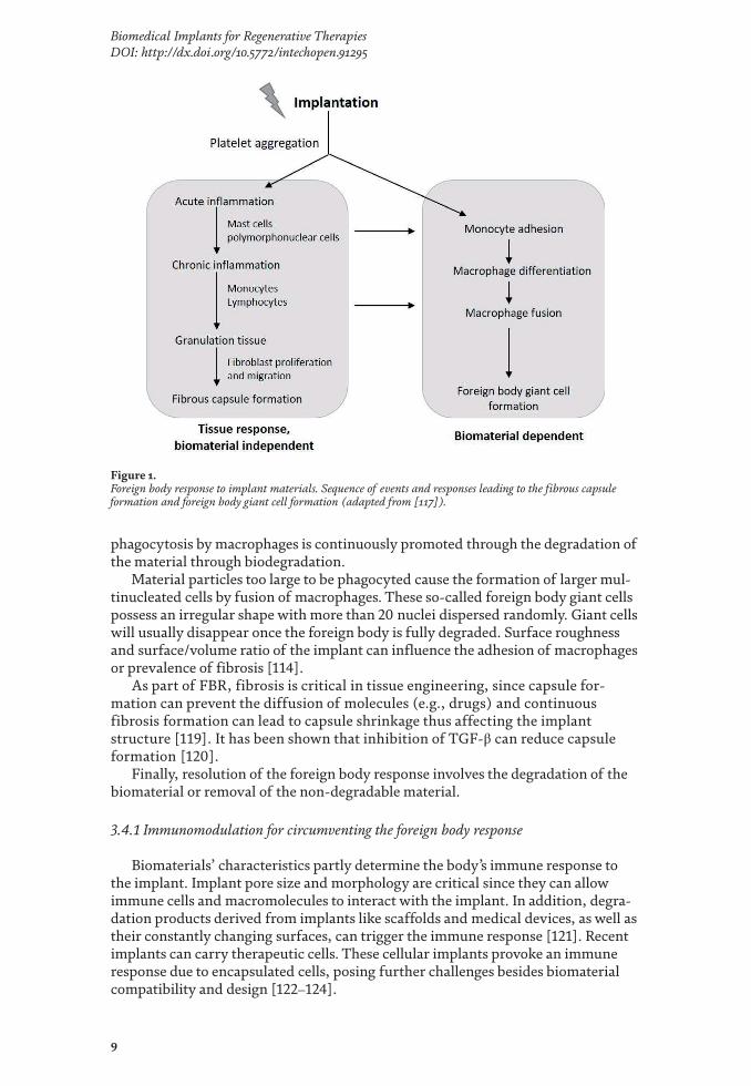
Foreign body response (FBR) is a non-specific immune reaction of the body to implanted materials. This inflammatory reaction can happen in response to surgical implantation of biodegradable or non-biodegradable materials present in medical devices or implants [114, 115]. FBR can modulate the safety and/or function of the implanted material. FBR is characterised by distinct phases, namely onset, progres-sion and resolution [116] (Figure 1). The onset starts with the surgical implantation of the biomaterial, for example, subcutaneously, which causes local tissue damage [117]. Upon tissue damage, vessel permeation to cells and proteins increases and coagulation occurs where inflammatory mediators like vascular endothelial growth factor (VEGF) plays an important role along with neutrophils and macrophages to initiate the wound healing process. In parallel, angiogenic factors stimulate local vasculature.
FBR comprises of a biomaterial-dependent and biomaterial-independent reac-tion (Figure 1). If biodegradable materials are present, the FBR will persist until the material is fully degraded. With non-degradable or long-term implants, a fibrotic capsule creating a barrier between the material and the body will form.
Progression of FBR depends on the material’s surface chemistry and wettabil-ity [118], where protein, antibody and macrophage adsorption can vary due to the material’s intrinsic properties. Additionally, fibrinogen can be adsorbed by the implant altering its structure. During FBR’s progression, leukocyte extraver-sion occurs from the blood vessels. These migrate towards the foreign body. Consequently, polymorphonuclear neutrophils (PMNs) are activated, which recruit cells including macrophages to the site. Macrophage activation leads to the recruitment of fibroblasts, monocytes and more PMNs [116], which ultimately increases production of extracellular matrix and hence implant encapsulation and fibrosis.
Phagocytosis occurs from the onset when antibodies are non-specifically adsorbed by the biomaterials, thus recruiting phagocytes. During progression,
9
Biomedical Implants for Regenerative TherapiesDOI: http://dx.doi.org/10.5772/intechopen.91295
phagocytosis by macrophages is continuously promoted through the degradation of the material through biodegradation.
Material particles too large to be phagocyted cause the formation of larger mul-tinucleated cells by fusion of macrophages. These so-called foreign body giant cells possess an irregular shape with more than 20 nuclei dispersed randomly. Giant cells will usually disappear once the foreign body is fully degraded. Surface roughness and surface/volume ratio of the implant can influence the adhesion of macrophages or prevalence of fibrosis [114].
As part of FBR, fibrosis is critical in tissue engineering, since capsule for-mation can prevent the diffusion of molecules (e.g., drugs) and continuous fibrosis formation can lead to capsule shrinkage thus affecting the implant structure [119]. It has been shown that inhibition of TGF-β can reduce capsule formation [120].
Finally, resolution of the foreign body response involves the degradation of the biomaterial or removal of the non-degradable material.
3.4.1 Immunomodulation for circumventing the foreign body response
Biomaterials’ characteristics partly determine the body’s immune response to the implant. Implant pore size and morphology are critical since they can allow immune cells and macromolecules to interact with the implant. In addition, degra-dation products derived from implants like scaffolds and medical devices, as well as their constantly changing surfaces, can trigger the immune response [121]. Recent implants can carry therapeutic cells. These cellular implants provoke an immune response due to encapsulated cells, posing further challenges besides biomaterial compatibility and design [122–124].
Figure 1. Foreign body response to implant materials. Sequence of events and responses leading to the fibrous capsule formation and foreign body giant cell formation (adapted from [117]).

Biomaterials
10
Polymers such as collagen, alginate, chitosan, polyethylene glycol, polyvinyl alcohol and polyurethane are used in several implantable products that may have an inherent biocompatibility. Understanding how these polymer’s chemical and physi-cal properties can be used to either avoid immune response or modulate it, while improving their functionality, is crucial for the advancement of these systems [121].
Strategies to circumvent the FBR include changing the biomaterial’s surface properties like wettability, its chemical moieties, and surface charge, because they affect protein adhesion to the biomaterial [121, 125].
To create more hydrophilic surfaces, monolayers of hydrophilic polymers such as polyethylene glycol (PEG) and polyethylene oxide (PEO) are added, thus prevent-ing protein adsorption altogether [126]. The deposition of chemical moieties like amino (▬NH2), carboxyl (▬COOH), hydroxyl (▬OH), and methyl (▬CH3) groups allows the modulation of cellular adhesion influencing inflammatory cell infiltra-tion and macrophage response affecting the fibrotic capsule thickness around the implant [121]. Surface charge is important for the FBR immunomodulation. There have been contradicting reports on how exactly neutral, positive or nega-tive charges reduce the inflammatory response connected to the FBR. Generally, negatively charged surfaces tend to inhibit the immune response through reduced cell adhesion [127].
Moreover, implant topography including texture, shape and size has shown to trigger an FBR [121]. Therefore, several manufacturing techniques like particles, assembled monolayers and photolithography are used to create variety of shapes, sizes and surface topographies [128, 129]. Surface roughness at the nanoscale can modulate protein adsorption [130], while variations in surface roughness at microscale affect cells directly [131]. For example, the inflammatory response of titanium used for dental or orthopaedic applications can be decreased by altering its surface nano-and microstructures via physical or chemical procedures [121].
Macrophage interaction with differently shaped biomaterials demonstrated preferred internalisation of nanorods via pinocytosis compared to nanospheres. Additionally, sharper cornered surfaces led to more acute immune responses than smoother surfaces [121, 132]. Moreover, spherical alginate capsules of 1.5 mm or greater were reported to be more biocompatible than their smaller, non-spherical comparators, demonstrating that larger, rounder, smoother capsules could diminish the FBR [133].
The use of decellularised ECM as scaffolds by removing immunogenic compo-nents to avoid an acute response but keeping the original structure has been stud-ied. While decellularised ECMs contribute to a pro-regenerative environment [134], it has been discussed that the immune response modulation still depends on the original tissue from which the ECMs have been obtained. Therefore, this option still presents a potential solution with more research needed to advance its understand-ing, manufacturing and impact [134].
The incorporation of bioactive molecules such as adhesion molecules, drugs and growth factors to promote immunological interaction with the host attenuating its response has been investigated. Bioactive molecules bound to the biomaterial for controlled release aiding tissue regeneration [125] include proinflammatory molecules like prostaglandins [135] and anti-inflammatory molecules like cytokines [136]. Combining their delivery with glucocorticoids improved tissue regenera-tion and attenuation of inflammation [137]. In recent years, the encapsulation of immune cells that act as producers or inducers of specific biological responses to reduce inflammation and/or induce repair has been investigated as immunomodula-tion strategy [125]. Examples include the encapsulation of MSCs to decrease the fibrosis in FBR [138] or the encapsulation of macrophages to mediate pro-angiogenic activation [139].

11
Biomedical Implants for Regenerative TherapiesDOI: http://dx.doi.org/10.5772/intechopen.91295
Overall, understanding the fundamental biological systems associated with FBR and the structural, physical and chemical properties of biomaterials will lead to new designs and strategies allowing to circumvent or work together with the natural body’s response towards implants.
4. Bioactive implants
Implants can be bioactive, inducing an alteration to the surrounding tissue, by their own biomaterials imparting this alteration to the surrounding tissue, by releasing a drug (or drugs) inducing bioactivity, or by containing cells that can produce bioactive molecules. In the following sub-sections, we discuss the implant bioactivity induced by drugs and cells implicating in drug delivery and in tissue regeneration.
4.1 Bioactive implantable and injectable drug delivery systems
Bioactive implants may incorporate active substances including small chemicals, peptides, proteins, hormones and even cells, which will have a therapeutic function in the human body. For drug delivery, these systems are commonly administered via parenteral route by injection or implantation. There are also implantable drug deliv-ery systems that can be administered via ocular administration or via surgical pro-cedures such as brain implants (e.g., Gliadel®). Implantable drug delivery systems are designed to slowly release the active substance(s) that they carry, thus avoiding repetitive injection. The active substance is delivered at a consistent predictable rate creating a drug release profile. This avoids peaks and troughs in the drug-blood level, which is common for non-long acting injectable products (e.g., intravenous solutions). Implantable drug delivery systems can be also injected subcutaneously, intramuscular or via other sites including intra-articular. They include implants and suspensions of micro- or nano-particles. Typically, these systems are preferred when the active substance has a poor absorption by other means of administration or a short half-life. The major advantages of such systems are improved pharmaco-kinetics, control of the drug release rate, and enhanced patient acceptability due to the reduction of side effects by maintaining the drug-blood level constant and by decreasing administration frequency [140, 141].
Sustained drug release is obtained via diffusion of the active substance through a biomaterial matrix, or released through biomaterial biodegradation, or a combina-tion of both mechanisms. To date, commonly used biomaterials for drug delivery are either biodegradable like PCL and PLA or non-biodegradable like polydimeth-ylsiloxane, polyethyl vinyl acetate, or titanium alloy [141]. Several approaches have been developed to produce implantable drug delivery systems [142] and to control the drug release. These include (i) using diffusion via membrane permeation, either porous or semi-porous membranes; (ii) controlling drug release by matrix diffusion using porous polymers; (iii) reservoir systems, where the drug is encapsulated in an inner reservoir; and (iv) actively releasing the drug from the implant via osmotic pressure, electric current, vapour pressure, hydrolysis or ultrasound activation.
Typically, simple rod-like solid implants, produced by hot melt extrusion processes using biodegradable polymers like PLA, PCL, PLGA and PEVA, often display a biphasic drug release kinetics showing a burst release due to the drug being deposited on the surface or near the surface of the implant, fol-lowed by a zero-order kinetics reflected by drug diffusion, matrix erosion, or a combination of both depending on the polymeric biomaterial used. Table 4 summarises drug release systems that are currently commercially available or

Biomaterials
12
Syst
emPr
oduc
tD
rug
Man
ufac
ture
rIn
dica
tion
Clin
ical
stat
us
Impl
ants
[143
–148
]Zo
lade
x® (P
LGA
solid
rod,
1 ×
10 m
m)
Gos
erel
in (u
p to
3 m
onth
re
leas
e)A
stra
Zene
caPr
osta
te ca
ncer
App
rove
d by
FDA
Nex
plan
on®
(rad
iopa
que P
EVA
solid
ro
d)Et
onog
estr
el (r
elea
se u
p to
3
year
s)M
erck
Cont
race
ptio
nA
ppro
ved
by F
DA
ITCA
650
(Med
ici t
echn
olog
y, fo
rmer
D
uros
®)
Exen
atid
e (re
leas
e up
to
2 ye
ars)
Ipse
nTy
pe 2
dia
bete
sCl
inic
al P
hase
III/
NDA
MK-
8591
(PCL
solid
impl
ant)
EFdA
(lon
g-te
rm re
leas
e)M
erck
HIV
trea
tmen
t and
pr
even
tion
Pre-
clin
ical
/Pha
se I
Mic
ropa
rtic
les
[149
–151
]Ri
sper
dal C
onst
a® (P
LGA
mic
rosp
here
s)Ri
sper
idon
eJa
nsse
nA
ntip
sych
otic
App
rove
d by
FDA
Dec
apep
tyl S
R® (P
LGA
mic
rosp
here
s)Tr
ipto
relin
Deb
ioph
arm
/Fer
ring/
Ipse
nPr
osta
te ca
ncer
App
rove
d by
FDA
Sand
osta
tin L
AR®
(PLG
A m
icro
sphe
res)
Oct
reot
ide
Nov
artis
Acr
omeg
aly
App
rove
d by
FDA
Bydu
reon
® (P
LGA
mic
rosp
here
s)Ex
enat
ide
Am
ylin
/Ast
raZe
neca
Type
2 d
iabe
tes
App
rove
d by
FDA
Viv
itrol
® (P
LGA
mic
rosp
here
s)N
altr
exon
eA
lker
mes
Opi
oid/
alco
hol d
epen
denc
eA
ppro
ved
by F
DA
In si
tu h
ydro
gels
[152
, 15
3]El
igar
d® (A
trig
el®
tech
nolo
gy)
Leup
rolid
e ace
tate
Sano
fi-Av
entis
Pros
tate
canc
erA
ppro
ved
by F
DA
Posid
in®
(Sab
re®
tech
nolo
gy)
Bupi
vaca
ine
Dur
ect/
Sand
ozPo
stop
erat
ive p
ain
Clin
ical
Pha
se II
I/N
DA
Reld
ay®
(Sab
re®
tech
nolo
gy)
Risp
erid
one
Dur
ect/
Zoge
nix
Schi
zoph
reni
a/bi
pola
r di
sord
erPh
ase I
Subl
ocad
e® (A
trig
el®
tech
nolo
gy)
Bupr
enor
phin
eIn
divi
orSe
vere
opi
oid
use d
isord
erA
ppro
ved
by F
DA
Sum
mar
ised
are c
urre
nt sy
stem
s tha
t are
com
mer
cial
ly a
vaila
ble o
r und
er d
evelo
pmen
t. Ta
ble w
as a
dapt
ed fr
om [1
40, 1
41, 1
48, 1
51, 1
53].
Tabl
e 4.
D
rug d
elive
ry sy
stem
s as i
mpl
anta
ble a
nd in
jecta
ble d
epot
s.

13
Biomedical Implants for Regenerative TherapiesDOI: http://dx.doi.org/10.5772/intechopen.91295
under development. There are numerous advantages to using implants in drug delivery such as the possibility of removal after treatment, the consistent and predictable drug release, and versatility in manufacture using various bioma-terials. However, there are potential disadvantages of this dosage form, where often a specialised device (e.g., trocar) and technique are needed for implanta-tion and removal requires minor surgical procedures. Additionally, there may be complications in locating the implant for removal since it can migrate from its original location. From a commercialisation point of view, this type of bioactive implant may require complex regulatory and commercial strategies for market approval [154].
Injectable drug delivery systems, such as particulate suspensions or hydrogels like in situ forming gel depots, are designed from biodegradable biomaterials, injected (e.g., subcutaneous, intramuscular), form a depot, erode when in contact with body fluids, and release the drug by diffusion and erosion [149]. Injectable depots are not designed to be retrieved. Examples of injectable depots are micro- or nano-scale particles, where the drug is encapsulated within the polymer matrix. The polymeric particles are commonly prepared from biodegradable materials (e.g., PLGA, PCL, or silica) since the intent is to deliver the depot system once by injec-tion, let it erode and release the drug with time.
Choosing the polymer grade, type and combining polymer types can help tune the drug release as necessary [149, 154]. Key points in preparing these bioactive depots are the choice of the biomaterial (biodegradable/erodible), the physico-chemical properties of drug to be encapsulated (i.e., hydrophobic or hydrophilic), the drug loading needed to deliver the therapeutic dose, and the inherent phar-macokinetics of the drug. This will inform the choice of manufacturing methods, often by emulsification. Common polymers used in these preparations are PLGA and PLA, where their long safety records deem these polymers as preferred, even though some minor inflammatory responses can still be reported [150].
Hydrogels, prepared from different types of biomaterials (e.g., hyaluronic acid, polyesters and chitosan) have been extensively investigated as carriers for sustained drug release [152, 155]. In situ forming hydrogels as injectable depots pose major advantages over other drug release systems since they allow for rapid, painless and easier administration through smaller needle sizes. These biodegradable in situ depots are of low viscosity prior injection and solidify into a gel or solid depot after injection, typically due to a specific trigger depending on the chemistry of the chosen biomaterial [153].
4.2 Bioactive cell-based implants as drug delivery systems
Before commercialising a cellular implant, it needs to be approved by FDA’s Cellular, Tissue and Gene Therapies Advisory Committee. The Committee evaluates the safety and effectiveness of cellular implants for the reconstruction, repair or replacement of damaged tissues [156].
Cell-based drug delivery systems can be defined as technologies capable of treating diseases using living cells to deliver the therapeutic bioactive molecules in the body, as either transport system or as production units [157]. Some examples of commercially available or under development cell-based implants are shown in Table 5. These cell-based drug delivery systems are used as constant producers of bioactive molecules in the form of implant devices. Judging by the current developments in this technology, a major driving force behind this type of delivery is the improvement in treatment of insulin-dependent diabetes mellitus. The biggest challenge in cell-based drug delivery systems is avoiding immune response.

Biomaterials
14
Com
pany
Prod
uct
App
licat
ion
Met
hod
of ac
tion
Man
ufac
ture
Neu
rote
chEn
caps
ulat
ed ce
ll th
erap
y (E
CT)
Oph
thal
mol
ogy
Cilia
ry n
euro
trop
hic f
acto
r (CN
TF) h
as n
euro
prot
ectiv
e eff
ects
on
phot
orec
epto
rs.
Enca
psul
ated
hum
an ce
lls p
rodu
cing
CN
TF in
to th
e bac
k of
the e
ye.
VIa
Cyte
Enca
ptra
®St
em ce
ll de
liver
y fo
r tr
eatm
ent o
f dia
bete
s m
ellit
us
Hum
an st
em ce
lls ar
e iso
late
d an
d di
ffer
entia
ted
into
β is
let c
ells
cont
aine
d in
to a
pouc
h, w
hich
is im
plan
ted.
PTFE
por
ous m
embr
ane d
evic
e fill
ed
with
cells
.
Sern
ova
Cell
Pouc
hTM w
ith
Sert
olin
TMD
iabe
tes/
haem
ophi
lia/
thyr
oid
dise
ase
Ther
apeu
tic ce
lls ar
e ins
erte
d in
to a
pouc
h m
ade o
f med
ical
-gra
de
mat
eria
ls in
sert
ed su
bcut
aneo
usly
; Ser
tolin
® is
a pa
tent
ed im
mun
e pr
otec
tion
syst
em.
Pouc
h m
ade o
f med
ical
-gra
de
mat
eria
ls.
Phar
maC
yte
Cell-
in-a
-box
®Pa
ncre
atic
canc
er/b
reas
t ca
ncer
/dia
bete
sU
ses c
otto
n ce
llulo
se to
enca
psul
ate c
ells.
Sing
le ce
ll en
caps
ulat
ion
in
prop
rieta
ry p
olym
er, f
reez
e-dr
ying
pr
oces
s to
keep
cells
via
ble i
n th
e lon
g te
rm.
Beta
-O2
βAir®
bio
artif
icia
l pan
crea
sD
iabe
tes,
adre
nal
insu
ffic
ienc
yD
evic
e usin
g al
gina
te—
high
gul
uron
ic ac
id an
d hi
gh m
annu
roni
c ac
id—
to en
caps
ulat
e cel
ls an
d im
preg
nate
a PT
FE p
orou
s m
embr
ane,
resp
ectiv
ely.
Also
com
prise
s an
oxyg
en-p
rovi
ding
ch
ambe
r, w
hich
nee
ds re
fillin
g.
Sing
le ce
ll en
caps
ulat
ion
in
prop
rieta
ry p
olym
er, f
reez
e-dr
ying
pr
oces
s to
keep
cells
via
ble i
n th
e lon
g te
rm.
Sigi
lon
Afib
rom
er™
Dia
bete
sH
uman
stem
cells
diff
eren
tiate
d to
β is
lets
enca
psul
ated
in
mod
ified
algi
nate
sphe
res,
whi
ch su
ppre
ss im
mun
e sys
tem
re
spon
se an
d FB
R.
Enca
psul
ifeEn
caps
ulat
ion
syst
em fo
r th
e im
mun
oiso
latio
n of
liv
ing
cells
Dia
bete
sCe
llulo
se-b
ased
pol
ymer
enca
psul
atio
n of
cells
.Pa
ncre
atic
isle
ts en
caps
ulat
ed ar
e st
imul
ated
to p
rodu
ce in
sulin
.
Org
anog
enes
is in
corp
orat
edG
INTU
ITM
ucog
ingi
val
cond
ition
sKe
ratin
ocyt
es an
d fib
robl
asts
pro
duce
cyto
kine
s and
gro
wth
fa
ctor
s tha
t pro
mot
e hea
ling
and
rege
nera
tion
of th
e tiss
ue.
Allo
gene
ic k
erat
inoc
ytes
and
fibro
blas
t in
bovi
ne co
llage
n.
Tabl
e 5.
Ex
ampl
es o
f com
mer
cial
ly a
vaila
ble c
ell-b
ased
impl
ants
for d
rug d
elive
ry.

15
Biomedical Implants for Regenerative TherapiesDOI: http://dx.doi.org/10.5772/intechopen.91295
4.3 Bioactive cellular implants as tissue replacements
When damages due to disease, injury or trauma lead to the degeneration of tissues, it is necessary to provide support for their repair, replacement or regeneration. Common approaches include tissue transplantation, both from the patient’s own body (autograft) and from a donor (allograft). However, harvesting autografts is expensive and invasive and the patient may experience infections and hematomas. While for the allografts, there are risks of rejection along with the infections due to the surgery or the transplanted tissue [158]. With tissue engineering, biological implants are developed that restore, maintain and improve the tissue function [159]. Implants provide the environment for cell adhesion and proliferation to grow new tissues. They can also include active substances like growth factors and drugs as well as cells to aid tissue regeneration [11]. Cell-based scaffolds are either cultured in vitro with the aim of synthetizing tissues that can be implanted, or to be implanted directly in the damaged region [158]. Table 6 summarises some of the recent studies about cell-based implants tested on in vivo models.
4.3.1 Primary cells versus stem cells
The advantage of using cell-based scaffolds is the possibility to customise the construct using cells derived from the patient (primary cells). In this way, there is no risk of rejection due to immunological incompatibility. Cells can be isolated from biopsies and then seeded on the scaffold (Figure 2). However, primary cells are dif-ferentiated, post-mitotic cells. This leads to a limited lifespan, where after a limited number of cell doubling, they will enter in senescence and stop dividing, but are still viable [166, 167]. Moreover, primary cell types are difficult to culture, because they have difficulties adhering and proliferating in vitro [168].
To overcome problems associated with primary cells, stem cells have been be used. Stem cells are present in most if not all tissues and, according to their origin, they can be classified into embryonic and adult stem cells. Stem cells are able to both duplicate (self-renew) and differentiate in one or more cell types [167].
Embryonic stem cells (ESCs) are isolated from inner cell mass of embryo at the blastocyst stage. They can differentiate in any cell type (pluripotency) and have a high rate of self-renewal. Unfortunately, they can cause an immune response, as they are derived from a different body, so immunosuppression is necessary to avoid rejection. Moreover, the injection of undifferentiated ESC can lead to the formation of teratoma [169].
Adult stem cells (ASCs) are multipotent cells that can differentiate in a limited number of cell types, which reside in a specific microenvironment, the stem cell niche. Their role is to replace damaged and dead cells in the tissue to maintain homeostasis [170].
ASCs can be isolated from bone marrow, blood, adipose tissue, liver and skin [169]. Compared to ESCs, ASCs proliferate more slowly and have limited expan-sion capacity in vitro. Like primary cells, they can enter in senescence [171]. With age, their regenerative potential, growth and divisions are affected [172]. The most commonly used type of ASCs is mesenchymal stem cells (MSCs). These cells can differentiate into musculoskeletal cells, marrow and other cells of connective tissue, and they can provide trophic support and modulate the immune response [173]. They can migrate to a damaged region and promote healing by secreting molecules involved in angiogenesis and cell proliferation and inhibit oxidative stress and apoptosis [174, 175].

Biomaterials
16
Application Cell type Implant Outcome Reference
Tendon regeneration
Rat tendon stem/progenitor cells
Asymmetric chitosan-based sponges
• Tenogenic specific genes expression and protein production in vitro and in vivo.
• Formation of aligned collagen fibres in vivo.
[160]
Neural tissue engineering
Schwann cells, human bone marrow mesenchymal stem cells (BMSCs)
Polyvinyl alcohol (PVA)/sulphate alginate nanofibers
• Metabolic active cells adhered to scaffold.
• Mesenchymal stem cells differentiate in neuronal cells.
[161]
Wound healing and skin regeneration
Wharton’s jelly mesenchymal stem cells (MSCs)
Poly(ε-caprolactone) (PCL)/gelatin nanofibers
• Nanofibrous biode-gradable scaffolds.
• Cells were metabolic active and proliferative after 21 days in culture.
• MSCs on the scaffolds reduced the presence of denaturised proteins in vitro, possible anti-inflammatory response.
[162]
Mandible defects repair
Endothelial progenitor cells (EPCs), BMSCs
Biodegradable bioactive glass ceramic scaffold
• Expression of osteo-genesis and angiogen-esis markers in vitro.
• After 9 months post-implantation in vivo, the defects were nearly completely recovered, and angiogenesis was promoted.
[163]
Acute kidney injury
Human placenta-derived mesenchymal stem cells (hP-MSCs)
Self-assembling peptide hydrogel
• hP-MSCs niche, cell survival and angiogen-esis were promoted in in vivo.
• Renal functions were ameliorated.
[164]
Spinal cord regeneration
Neural stem cells (NSCs)
Elastic poly(sebacoyl diglyceride) (PSeD) scaffolds coated with poly(sebacoyl diglyceride)-isoleucine-lysinevaline-alanine-valine-serine (PSeD-IKVAVS)
• Graft-host integration in spinal cord in vivo.
• NSCs exhibited neuro-nal differentiation.
• Inflammatory cells infiltrated the lesion site, functional recov-ery after 4 weeks.
• Degradation products of PSeD-IKVAVS promoted NSCs differentiation, inhibited neuronal apoptosis and allevi-ated inflammation.
[165]
Table 6. Examples of recent studies on cell-based implants.

17
Biomedical Implants for Regenerative TherapiesDOI: http://dx.doi.org/10.5772/intechopen.91295
Induced pluripotent stem cells (iPSCs) originate from fully differentiated somatic cells, which are dedifferentiated to form iPSCs by a process called repro-gramming. The methodology was developed in 2006 [176] and involves the stimu-lation of genes that are active during the embryogenesis. Thanks to the cell derivation, the implantation of these cells does not lead to rejection. However, as with ESCs, iPSCs can form teratoma. Moreover, some of the genes that are activated are also associated with tumour development [177].
Figure 2. Source of cells used for cell-based implants. Image adapted from [168].

Biomaterials
18
5. Implant manufacture
To generate tissue replacements, it is essential to resemble the native extra-cellular matrix (ECM). Therefore, the matrix composition, shape and physical properties are crucial. Nanofibers, sponges or gels have been fabricated using numerous different techniques or combinations of techniques to mimic the native ECMs. Some of these techniques and their biomedical applications are presented in Table 7.
5.1 Solvent casting particulate leaching
Solvent casting particulate leaching is a technique developed in 1993 by Mikos et al., where a polymer is dissolved in an organic solvent and the polymer solution is mixed with an insoluble porogen. The solvent is evaporated by solvent casting or freeze-drying techniques. The evaporation leads to a porogen-polymer com-pound, which is washed to remove the porogen leaving a porous polymer matrix behind [187, 188]. This method is relatively easy to use and inexpensive [189]. Pore size, porosity and interconnectivity can be controlled selecting the right polymer, porogen and their concentration [190].
5.2 Phase separation
Phase separation employs temperature changes that separate the polymeric solu-tion in two phases: the lean phase (low polymer concentration) and the rich phase (high polymer concentration). Briefly, the polymer is dissolved in a solvent, then, the temperature is rapidly decreased to have a liquid-liquid separation and two-phase solid is formed [191]. Finally, the liquid is removed by extraction, evapora-tion or sublimation [192].
5.3 Freeze-drying
Freeze-drying technique or lyophilisation [193] is based on a sublimation pro-cess that will produce a porous scaffold. A polymer is added to a mixture of water and organic solvent and moved into a mould. The mixture is quickly frozen and, by lowering the pressure to few millibars, the water and the organic solvent sublimate. The complete removal of the liquid phase takes place under vacuum [189, 194, 195]. To control porosity and pore size, polymer/water ratio, ionic concentration, viscosity and pH, together with freezing rate and temperature, can be changed [194, 195].
5.4 Electrospinning
Electrospinning is used to produce micro- and nanofibers. It is widely used as it can produce matrix that can resemble the native ECMs. Nanofiber scaffolds offer mechanical support and a nanoscale environment for the cells [196, 197]. A polymer solution is added to a syringe. Then, high voltage is applied, and the solution accelerates to a collector of opposite charge. The solution-air inter-face changes from rounded to conical, due to the repulsive electrostatic forces between the polymer molecules in solution and the attractive force between the polymer solution and the collector. The polymer solution is ejected from the syringe when the electrostatic forces are higher than the surface tension of the solution. Then, the solvent evaporates, and the solid polymer is deposited

19
Biomedical Implants for Regenerative TherapiesDOI: http://dx.doi.org/10.5772/intechopen.91295
Technique Material Application Outcome References
Solvent casting—particulate leaching
Poly(l-lactic acid) (PLLA) and matrilin-3 (MATN3)
Articular cartilage regeneration
• Nanofibrous porous scaffold.
• Cell hypertrophy and endochon-dral ossification prevented in vivo.
• Chondrogenesis is promoted in vivo.
[178]
Phase separation—particulate leaching
Poly(lactic acid) (PLA)
Bone tissue engineering
• Porous scaffold.
• Osteosarcoma cells (MG63) were metabolic active and viable after 14 days in culture.
[179]
Freeze-drying Poly(ε-caprolactone) (PCL) and zein
Drug delivery
• Porous and degradable scaffold.
• Degradation rate increases with the concentration of zein.
[180]
Freeze-drying and self-assembly
Collagen Tissue engineering
• Aligned collagen scaffolds.
• Rat fibroblasts and neurons elongate along aligned fibres.
[181]
Freeze-drying Silk fibroin-chitosan
Cartilage regeneration
• Porous scaffold.
• MSCs were metabolic active, viable and differentiate after 21 days in culture.
[182]
Electrospinning Poly(ε-caprolactone) (PCL)/poly(d, l-lactide-co-glycolide) (PLGA)/gelatin
Vascular tissue engineering
• Dual-oriented/bilayer hydro-philic nanofibers.
• Smooth muscle cells and endothe-lial cells were viable after 7 days; orientation along the fibres.
[183]
Electrospinning SiO2CaO Wound healing
• Cotton wool-like, fibrous and porous scaffold.
• Human fibroblast seeded on top were metabolic active and proliferative after 7 days in culture, and produced vascular endothelial growth factor.
[184]
3D printing Copper/tetrakis (4-carboxyphenyl) porphyrin/β-tricalcium phosphate (Cu-TCPP-TCP)
Bone tumour ablation and osteogenesis
• Metal-organic photothermal nanosheets.
• Promoted osteosarcoma cell death in vitro, ablation of subcutaneous bone tumour tissue in vivo.
• Adhesion of bone marrow MSCs HUVEC in vitro.
• MSCs differentiated in osteocytes.
• HUVEC expressed angiogenesis markers in vitro.
• Enhanced bone regeneration in vivo.
[185]
E-jet 3D printing
Poly(lactic-co-glycolic acid) and drugs (5-fluorouracil and NVP-BEZ235)
Drug delivery in orthotopic breast cancer
• Long-term drug release near the tumour site.
• Less risk for normal tissue.
• No need for several administrations.
[186]
Table 7. Techniques used for the fabrication of bioactive implants.
Biomaterials
20
on the collector. By changing the voltage, collector, polymer concentration and solvent, it is possible to control the size of the fibres [196, 198].
5.5 Additive manufacturing techniques
Additive manufacturing (AM) techniques, or solid freeform fabrications (SFFs), are based on the use of computer-aided design (CAD) to fabricate scaf-folds. The CAD controls the layer-by-layer deposition of material. The advantage of these methods is the full control of the topography of the construct [196, 198].
Three-dimensional (3D) printing is a commonly used AM technique that was developed at the Massachusetts Institute of Technology in 1990s [198, 199]. The CAD is converted in a stereo lithography (STL) file and exported to the 3D printer to control the movement and deposition of the material.
This technique allows the inclusion of cells within the scaffold, as high tem-perature or solvents are not required for its production [200]. In recent years, 3D printing has been used to produce scaffolds and anatomically customised implants based on MRI and CT scans. The AM can be classified in three different approaches [201], namely laser-based (stereolithography, selective laser sintering, electron beam melting and binder jetting) [202–204], nozzle-based (fused deposition modelling and melt electrospinning writing) [204–208] and indirect 3D printing [209–211].
5.6 Injection moulding
Injection moulding is one of the most commonly used techniques for large-scale production of thermoplastic items. The plastic is melted and injected into a mould of desired shape. When the material solidifies, the mould is removed, and the finished part is extracted [212]. Metal constructs can also be fabricated with this technique. Metal injection moulding uses fine metal powders mixed with a binder and is injected with a conventional thermoplastic moulding machine. The binder is then removed, and the product is formed. This method allows the production of constructs with a sophisticate shape and higher mechanical properties [213].
5.7 Self-assembly
Self-assembly is the spontaneous formation of molecular units in supramolecu-lar structures, without external intervention. These molecules interact through hydrogen bonding, van der Waals and electrostatic forces. Due to their biocom-patibility and biodegradability, peptides are commonly used for self-assembly. Specific structure can be created by modifying the amino-acidic composition of the peptides [189, 214]. These nanostructures can be used in drug delivery and tissue engineering [215].
5.8 Manufacturing considerations
The manufacturing of bioactive implants, whether these are for tissue engi-neering or for drug delivery purposes, includes several common aspects. These include the manufacturing methods that are employed, the biomaterial source, use of solvents, scalability, the need for aseptic facilities or if final product sterilisa-tion is preferred, and if a specifically designed device is needed to administer the implant.

21
Biomedical Implants for Regenerative TherapiesDOI: http://dx.doi.org/10.5772/intechopen.91295
Most importantly, the regulatory strategy for filing needs to be in place soon in product development to define all the data that are needed for submission. It is important to define if the bioactive implant will be considered a drug product, a medical device, or a combination product (drug-device), and in which markets should it be launched, as different markets have different regulations and requisites for the different categories.
Comprehensive reviews and discussions on the regulatory aspects for filing medical devices, and combination products in the biomedical field were reviewed [216–218]. Ragelle et al. provide an excellent perspective for nanoparticle-based bio-materials, its manufacture and regulatory outlook for biomedical engineering [219].
To enable the use in animals and humans, the sterilisation process is impor-tant. If the manufacturing method is simplified enough, the use of aseptic technique for manufacturing, using filters and a particulate-free environment, will be possible although costly and complex for significantly large-scale manu-facturing. For devices where metals are used, sterilisation by moist or dry heat may be possible.
However, for implants that involve polymers or heat-sensitive bioactive mol-ecules, the preferred sterilisation method is gamma-irradiation. The drawback of using this technique is the potential risk to polymer and/or bioactive molecule degradation, changing the release rate and potentially compromising the efficacy of the implant [150]. Depending on the biomaterial and bioactive molecule involved, there may be ways to avoid degradation upon gamma-irradiation, such as using an antioxidant mixed with the drug. Apart from aseptic conditions, gamma-irradiation of the final product remains the best solution but exposes one of the disadvantages of developing bioactive implants as it is still a costly technique bringing its own risks [220].
6. Summary
In this chapter, we reviewed the current literature about bioactive biomedical implants applicable to regenerative therapies and used in drug delivery. To gener-ate new biomedical implants, biomaterials are continuously developed, either through entirely new or by combining advantageous properties of well-known and safe biomaterials to improve the application and effectiveness of implants. Thus, materials that enhance the natural response of the body and simultane-ously provide support for cell adhesion and proliferation are required. Another field that has developed in recent years is cell-induced bioactivity, where cells are used in implants for tissue regeneration and disease treatment. While there are several manufacturing techniques to create application-specific bioactive implants, new technologies, such as additive manufacturing, bring advantages and versatility to the field.

Biomaterials
22
Author details
Andrea Domingues Goncalves1, Wendy Balestri2 and Yvonne Reinwald2*
1 Pharmaceutical Development—Oral and Inhaled, Product Development and Supply, GlaxoSmithKline, Ware, United Kingdom
2 Department of Engineering, Nottingham Trent University, Nottingham, United Kingdom
*Address all correspondence to: [email protected]
© 2020 The Author(s). Licensee IntechOpen. This chapter is distributed under the terms of the Creative Commons Attribution License (http://creativecommons.org/licenses/by/3.0), which permits unrestricted use, distribution, and reproduction in any medium, provided the original work is properly cited.

23
Biomedical Implants for Regenerative TherapiesDOI: http://dx.doi.org/10.5772/intechopen.91295
References
[1] Regulation of medical implants. House of Commons—Evidence—HC 163-i. 2019. Available from: http://www.publications.parliament.uk/pa/cm201213/cmselect/cmsctech/uc163-i/uc16301.htm
[2] Huang Y, Van Dessel J, Martens W, Lambrichts I, Zhong WJ, Ma GW, et al. Sensory innervation around immediately vs. delayed loaded implants: A pilot study. International Journal of Oral Science. 2015;7:49
[3] Arsiwala A, Desai P, Patravale V. Recent advances in micro/nanoscale biomedical implants. Journal of Controlled Release. 2014;189(2014):25-45
[4] Takmakov P, Ruda K, Phillips KS, Isayeva IS, Krauthamer V, Welle CG. Rapid evaluation of the durability of cortical neural implants using accelerated aging with reactive oxygen species. Journal of Neural Engineering. 2015;12:026003
[5] Guo R, Merkel AR, Sterling JA, Davidson JM, Guelcher SA. Substrate modulus of 3D-printed scaffolds regulates the regenerative response in subcutaneous implants through the macrophage phenotype and Wnt signaling. Biomaterials. 2015;73:85-95
[6] Li J, Stachowski M, Zhang Z. Application of responsive polymers in implantable medical devices and biosensors. In: Switchable and Responsive Surfaces and Materials for Biomedical Applications. Cambridge, UK: Elsevier; 2015. pp. 259-298
[7] Bagga C, Erbe EM, Murphy JP, Freid JM, Pomrink GJ. Bioactive Spinal Implants and Method of Manufacture Thereof. U.S. Patent 8,715,353; 2014
[8] Klein MO, Schiegnitz E, Al-Nawas B. Systematic review on
success of narrow-diameter dental implants. The International Journal of Oral & Maxillofacial Implants. 2014;29:43-54
[9] Yahyavi-Firouz-Abadi N, Menias CO, Bhalla S, Siegel C, Gayer G, Katz DS. Imaging of cosmetic plastic procedures and implants in the body and their potential complications. American Journal of Roentgenology. 2015;204:707-715
[10] Silva VV, Domingues RZ, Lameiras FS. Microstructural and mechanical study of zirconia-hydroxyapatite (ZH) composite ceramics for biomedical applications. Composites Science and Technology. 2001;61:301-310
[11] Stratton S, Shelke NB, Hoshino K, Rudraiah S, Kumbar SG. Bioactive polymeric scaffolds for tissue engineering. Bioactive Materials. 2016;1(2):93-108
[12] Dhandayuthapani B, Sakthikumar D. Biomedical Applications of Polymeric Materials and Composites. 1st ed. Weinheim, Germany: Wiley; 2017. pp. 1-20
[13] Jones AJ, Denning NT. Polymeric Biomaterials: Bio- and Ecocompatible Polymers, A Perspective for Australia. Department of Industry, Technology and Commerce; 1988. ISBN: 0642134618, 9780642134615
[14] ASM International. Overview of biomaterials and their use in medical devices. Handbook of Materials for Medical Devices. Cleveland, OH, USA: ASM International, Inc; 2003
[15] Zdrahala RJ, Zdrahala IJ. Biomedical applications of polyurethanes: A review of past promises, present realities, and a vibrant future. Journal of Biomaterials Applications. 1999;14(1):67-90

Biomaterials
24
[16] Daebritz SH, Fausten B, Hermanns B, Schroeder J, Groetzner J, Autschbach R, et al. Introduction of a flexible polymeric heart valve prosthesis with special design for the aortic position. Circulation. 2003;108(10-Suppl. 1):II-135-II-139
[17] Zhu Y, Gao C, He T, Shen J. Endothelium regeneration on luminal surface of polyurethane vascular scaffold modified with diamine and covalently grafted with gelatin. Biomaterials. 2004;25(3):423-430
[18] Yuan Y, Ai F, Zang X, Zhuang W, Shen J, Lin S. Polyurethane vascular catheter surface grafted with zwitterionic sulfobetaine monomer activated by ozone. Colloids and Surfaces B. 2004;35(1):1-5
[19] Lin W-C, Tseng C-H, Yang M-C. In-vitro hemocompatibility evaluation of a thermoplastic polyurethane membrane with surfaceimmobilized water-soluble chitosan and heparin. Macromolecular Bioscience. 2005;5:1013-1021
[20] Topol EJ, Serruys PW. Frontiers in interventional cardiology. Circulation. 1802-1820;1998:98
[21] Whelan DM, van der Giessen WJ, Krabbendam SC, van Vliet EA, Verdouw PD, Serruys PW, et al. Biocompatibility of phosphorylcholine coated stents in normal porcine coronary arteries. Heart. 2000;83:338-345
[22] Horner PJ, Gage FH. Regenerating the damaged central nervous system. Nature. 2000;407:963-970
[23] Scheib J, Hoke A. Advances in peripheral nerve regeneration. Nature Reviews. Neurology. 2013;9:668-676
[24] Cao J, Xiao Z, Jin W, Chen B, Meng D, Ding W, et al. Induction of rat
facial nerve regeneration by functional collagen scaffolds. Biomaterials. 2013;34:1302-1310
[25] White JD, Wang S, Weiss AS, Kaplan DL. Silke-tropoelastin protein films for nerve guidance. Acta Biomaterialia. 2015;14:1-10
[26] Xie J, MacEwan MR, Liu W, Jesuraj N, Li X, Hunter D, et al. Nerve guidance conduits based on double-layered scaffolds of electrospun nanofibers for repairing the peripheral nervous system. ACS Applied Materials & Interfaces. 2014;6:9472-9480
[27] Prakasam M, Locs J, Salma- Ancane K, Dagnija Loca D, Largeteau A, Berzina-Cimdina L. Biodegradable materials and metallic implants—A review. Journal of Functional Biomaterials. 2017;8:44
[28] Zimmerli W. Clinical presentation and treatment of orthopaedic implant-associated infection. Journal of Internal Medicine. 2014;276:111-119
[29] Bellini H, Moyano J, Gil J, Puigdollers A. Comparison of the superelasticity of different nickel–titanium orthodontic archwires and the loss of their properties by heat treatment. Journal of Materials Science. Materials in Medicine. 2016;27:158
[30] Hutmacher DW, Garcia AJ. Scaffold-based bone engineering by using genetically modified cells. Gene. 2005;347(1):1-10
[31] Liu YD, Hu J, Zhuang XL, et al. Synthesis and characterization of novel biodegradable and electroactive hydrogel based on aniline oligomer and gelatin. Macromolecular Bioscience. 2011;12:241-250
[32] Guo BL, Glavas L, Albertsson AC. Biodegradable and electrically

25
Biomedical Implants for Regenerative TherapiesDOI: http://dx.doi.org/10.5772/intechopen.91295
conducting polymers for biomedical applications. Progress in Polymer Science. 2013;38:1263-1286
[33] Guo BL, Finne-Wistrand A, Albertsson AC. Versatile functiona-lization of polyester hydrogels with electroactive aniline oligomers. Journal of Polymer Science Part A: Polymer Chemistry. 2011;49:2097-2105
[34] Saini M, Singh Y, Arora P, Arora V, Jain K. Implant biomaterials: A comprehensive review. World Journal of Clinical Cases. 2015;3(1): 52-57
[35] Sykaras N, Iacopino AM, Marker VA, Triplett RG, Woody RD. Implant materials, designs, and surface topographies: Their effect on osseo-integration. A literature review. The International Journal of Oral & Maxillofacial Implants. 2000;15: 675-690
[36] LeGeros RZ, Lin S, Rohanizadeh R, Mijares D, LeGeros JP. Biphasic calcium phosphate bioceramics: Preparation, properties and applications. Journal of Materials Science. Materials in Medicine. 2003;14:201-209
[37] DeGroot K. Clinical applications of calcium phosphate biomaterials: A review. Ceramics International. 1993;19:363-366
[38] Wennerberg A, Albrektsson T. On implant surfaces: A review of current knowledge and opinions. The International Journal of Oral & Maxillofacial Implants. 2010;25:63-74
[39] Hoffmann O, Angelov N, Gallez F, Jung RE, Weber FE. The zirconia implant-bone interface: A preliminary histologic evaluation in rabbits. The International Journal of Oral & Maxillofacial Implants. 2008;23:691-695
[40] Özkurt Z, Kazazoğlu E. Zirconia dental implants: A literature review.
The Journal of Oral Implantology. 2011;37:367-376
[41] Chiapasco M, Casentini P, Zaniboni M, Corsi E, Anello T. Titanium-zirconium alloy narrow-diameter implants (Straumann Roxolid(®)) for the rehabilitation of horizontally deficient edentulous ridges: Prospective study on 18 consecutive patients. Clinical Oral Implants Research. 2012;23:1136-1141
[42] Teramoto H, Kawai A, Sugihara S, Yoshida A, Inoue H. Resorption of apatite-wollastonite containing glass-ceramic and beta-tricalcium phosphate in vivo. Acta Medica Okayama. 2005;59:201-207
[43] Boanini E, Gazzano M, Bigi A. Ionic substitutions in calcium phosphates synthesized at low temperature. Acta Biomaterialia. 2010;6:1882-1894
[44] Thian ES, Konishi T, Kawanobe Y, Lim PN, Choong C, Ho B, et al. Zinc-substituted hydroxyapatite: A biomaterial with enhanced bioactivity and antibacterial properties. Journal of Materials Science. Materials in Medicine. 2013;24:437-445
[45] Mayer I, Jacobsohn O, Niazov T, Werckmann J, Iliescu M, Richard-Plouet M, et al. Manganese in precipitated hydroxyapatites. European Journal of Inorganic Chemistry. 2003:1445-1451
[46] Tamimi F, Le Nihouannen D, Eimar H, Sheikh Z, Komarova S, Barralet J. The effect of autoclaving on the physical and biological properties of dicalcium phosphate dihydrate bioceramics: Brushite vs. monetite. Acta Biomaterialia. 2012;8:3161-3169
[47] Hodosh M, Povar M, Shklar G. The dental polymer implant concept. The Journal of Prosthetic Dentistry. 1969;22:371-380

Biomaterials
26
[48] Wang Y, Blasioli DJ, Kim H-J, Kim HS, Kaplan DL. Cartilage tissue engineering with silk scaffolds and human articular chondrocytes. Biomaterials. 2006;27:4434-4442
[49] Vepari C, Kaplan DL. Silk as a biomaterial. Progress in Polymer Science. 2007;32:991-1007
[50] Yang Y, Ding F, Wu J, Hu W, Liu W, Liu J, et al. Development and evaluation of silk fibroin-based nerve grafts used for peripheral nerve regeneration. Biomaterials. 2007;28:5526-5535
[51] Hakimi O, Knight DP, Vollrath F, Vadgama P. Spider and mulberry silkworm silks as compatible biomaterials. Composites. Part B, Engineering. 2007;38:324-337
[52] Etienne O, Schneider A, Kluge JA, Bellemin-Laponnaz C, Polidori C, Leisk GG, et al. Soft tissue augmentation using silk gels: An in vitro and in vivo study. Journal of Periodontology. 2009;80:1852-1858
[53] Kardestuncer T, McCarthy M, Karageorgiou V, Kaplan DL, Gronowicz G. RGD-tethered silk substrate stimulates the differentiation of human tendon cells. Clinical Orthopaedics and Related Research. 2006;448:234-239
[54] Cai Z-X, Mo X-M, Zhang K-H, Fan L-P, Yin A-L, He C-L, et al. Fabrication of chitosan/silk fibroin composite nanofibers for wound-dressing applications. International Journal of Molecular Sciences. 2010;11:3529-3539
[55] Okabayashi R, Nakamura M, Okabayashi T, Tanaka Y, Nagai A, Yamashita K. Efficacy of polarized hydroxyapatite and silk fibroin composite dressing gel on epidermal recovery from full-thickness skin wounds. Journal of Biomedical
Materials Research Part B: Applied Biomaterials. 2009;90B:641-646
[56] Necas J, Bartosikova L, Brauner P, Kolar J. Hyaluronic acid (hyaluronan): A review. Veterinární Medicína. 2008;53:397-411
[57] Laurencin CT, Jiang T, Kumbar SG, Nair LS. Biologically active chitosan systems for tissue engineering and regenerative medicine. Current Topics in Medicinal Chemistry. 2008;8:354-364
[58] He S, Yaszemski MJ, Yasko AW, Engel PS, Mikos AG. Injectable biodegradable polymer composites based on poly (propylene fumarate) crosslinked with poly (ethylene glycol)-dimethacrylate. Biomaterials. 2000;21:2389-2394
[59] Shelke NB, Anderson M, Idrees S, Nip MJ, Donde S, Yu X, et al. Handbook of Polyester Drug Delivery Systems. Singapore: Pan Stanford; 2016. pp. 595-649
[60] Narayanan G, Gupta BS, Tonelli AE. Enhanced mechanical properties of poly(ε-caprolactone) nanofibers produced by the addition of nonstoichiometric inclusion complexes of poly(ε-caprolactone) and a-cyclodextrin. Polymer. 2015;76:321-330
[61] Karst D, Yang Y. Molecular modeling study of the resistance of PLA to hydrolysis based on the blending of PLLA and PDLA. Polymer. 2006;47:4845-4850
[62] Gentile P, Chiono V, Carmagnola I, Hatton PV. An overview of poly (lactic co-glycolic) acid (PLGA)-based biomaterials for bone tissue engineering. International Journal of Molecular Sciences. 2014;15:3640-3659
[63] Woodruff MA, Hutmacher DW. The return of a forgotten polymer

27
Biomedical Implants for Regenerative TherapiesDOI: http://dx.doi.org/10.5772/intechopen.91295
polycaprolactone in the 21st century. Progress in Polymer Science. 2010;35:1217-1256
[64] Nada AA, James R, Shelke NB, Harmon MD, Awad HM, Nagarale RK, et al. A smart methodology to fabricate electrospun chitosan nanofiber matrices for regenerative engineering applications. Polymers for Advanced Technologies. 2014;25:507-515
[65] Chang KY, Hung LH, Chu I, Ko CS, Lee YD. The application of type II collagen and chondroitin sulfate grafted PCL porous scaffold in cartilage tissue engineering. Journal of Biomedial Materials Research Part A. 2010;92:712-723
[66] Tan L, Yu X, Wan P, Yang K. Biodegradable materials for bone repairs: A review. Journal of Materials Science and Technology. 2013;29:503-513
[67] Pezzin AP, Ekenstein V, Alberda GO, Zavaglia CA, Ten Brinke G, Duek EA. Poly (para-dioxanone) and poly (L-lactic acid) blends: Thermal, mechanical, and morphological properties. Journal of Applied Polymer Science. 2003;88:2744-2755
[68] Amanatullah DF, Landa J, Strauss EJ, Garino JP, Kim SH, Di Cesare PE. Comparison of surgical outcomes and implant wear between ceramic-ceramic and ceramic-polyethylene articulations in total hip arthroplasty. Journal of Arthroplasty. 2011;26(6):72-77
[69] Green JM, Hallab NJ, Liao Y-S, Narayan V, Schwarz EM, Xie C. Anti-oxidation treatment of ultra high molecular weight polyethylene components to decrease periprosthetic osteolysis: Evaluation of osteolytic and osteogenic properties of wear debris particles in a murine calvaria model. Current Rheumatology Reports. 2013;15(5):1-5
[70] Zhou J, Huang X, Zheng D, Li H, Herrler T, Li Q. Oriental nose elongation using an L-shaped polyethylene sheet implant for combined septal spreading and extension. Aesthetic Plastic Surgery. 2014;38(2):295-302
[71] Teo AJT, Mishra A, Park I, Kim YJ, Park WT, Yoon YJ. Polymeric biomaterials for medical implants and devices. ACS Biomaterials Science & Engineering. 2016;2:454-472
[72] Pérez-Merino P, Dorronsoro C, Llorente L, Durán S, Jiménez- Alfaro I, Marcos S. In vivo chromatic aberration in eyes implanted with intraocular lenses. Investigative Ophthalmology & Visual Science. 2013;54(4):2654-2661
[73] Terrada C, Julian K, Cassoux N, Prieur A-M, Debre M, Quartier P, et al. Cataract surgery with primary intraocular lens implantation in children with uveitis: Long-term outcomes. Journal of Cataract & Refractive Surgery. 2011;37(11):1977-1983
[74] Kim B-J, Hong K-S, Park K-J, Park D-H, Chung Y-G, Kang S-H. Customized cranioplasty implants using three-dimensional printers and polymethyl-methacrylate casting. Journal of Korean Neurosurgical Association. 2012;52(6):541-546
[75] Rivkin A. A prospective study of non-surgical primary rhinoplasty using a polymethylmethacrylate injectable implant. Dermatologic Surgery. 2014;40(3):305-313
[76] Shklar G, Hodosh M, Povar M. Tissue reactions to polymer-coated vitallium pin implants. Journal of Prosthetic Dentistry. 1970;24:636-645
[77] Qin Y, Howlader MM, Deen MJ, Haddara YM, Selvaganapathy PR. Polymer integration for packaging of implantable sensors. Sensors and Actuators, B: Chemical. 2014;202:758

Biomaterials
28
[78] Lachhman S, Zorman C, Ko W. Multi-layered polydimethylsiloxane as a non-hermetic packaging material for medical MEMS. In: 2012 Annual International Conference of the IEEE Engineering in Medicine and Biology Society (EMBC). Piscataway, NJ: IEEE; 2012. pp. 1655-1658
[79] Rahimi A, Mashak A. Review on rubbers in medicine: Natural, silicone and polyurethane rubbers. Plastics Rubber and Composites. 2013;42(6):223-230
[80] Kappel RM, Klunder AJ, Pruijn GJ. Silicon chemistry and silicone breast implants. European Journal of Plastic Surgery. 2014;37(3):123-128
[81] Kubyshkina G, Zupančič B, Štukelj M, GroŠelj D, Marion L, Emri I. Sterilization effect on structure, thermal and time-dependent properties of polyamides. In: Mechanics of Time-Dependent Materials and Processes in Conventional and Multifunctional Materials. Vol. 3. New York: Springer; 2011. pp. 11-19
[82] Cruz F. Fabrication of HA/PLLA composite scaffolds for bone tissue engineering using additive manufacturing technologies. In: Elnashar M, editor. Biopolymers. Rijeka, Croatia: INTECH Open Access; 2010. pp. 227-242. (Chapter 11)
[83] Li X, Liu X, Huang J, Fan Y, Cui, F.-z. Biomedical investigation of CNT based coatings. Surface and Coating Technology. 2011;206(4):759-766
[84] Li N, Zheng Y. Novel magnesium alloys developed for biomedical application: A review. Journal of Materials Science and Technology. 2013;29:489-502
[85] Witte F, Fischer J, Nellesen J, Crostack HA, Kaese V, Pisch A, et al. In vitro and in vivo corrosion
measurements of magnesium alloys. Biomaterials. 2006;27:1013-1018
[86] Tang A, Pan F, Yang M, Cheng R. Mechanical properties and microstructure of magnesium-aluminum based alloys containing strontium. Materials Transactions. 2008;49:1203-1211
[87] Tekumalla S, Seetharaman S, Almajid A, Gupta M. Mechanical properties of magnesium-rare earth alloy systems: A review. Metals. 2015;5:1-39
[88] Taïr K, Kharoubi O, Taïr OA, Hellal N, Benyettou I, Aoues A. Aluminium-induced acute neurotoxicity in rats: Treatment with aqueous extract of Arthrophytum (Hammada scoparia). Journal of Acute Disease. 2016;5:470-482
[89] Gu X-N, Zheng Y-F. A review on magnesium alloys as biodegradable materials. Frontiers of Materials Science in China. 2010;4:111-115
[90] Zhang E, Yang L, Xu J, Chen H. Microstructure, mechanical properties and bio-corrosion properties of Mg–Si(–Ca, Zn) alloy for biomedical application. Acta Biomaterialia. 2010;6:1756-1762
[91] Kulkarni M, Mazare A, Gongadze E, Perutkova Š, Kralj-Igli V, Milošev I, et al. Titanium nanostructures for biomedical applications. Nanotechnology. 2015;26:062002
[92] Kopova I, Stráský J, Harcuba P, Landa M, Janeček M, Bačákova L. Newly developed Ti–Nb–Zr–Ta–Si–Fe biomedical beta titanium alloys with increased strength and enhanced biocompatibility. Materials Science and Engineering: C. 2016;60: 230-238
[93] Wang S, Liu Y, Zhang C, Liao Z, Liu W. The improvement of wettability,

29
Biomedical Implants for Regenerative TherapiesDOI: http://dx.doi.org/10.5772/intechopen.91295
biotribological behavior and corrosion resistance of titanium alloy pre-treated by thermal oxidation. Tribology International. 2014;79:174-182
[94] Li Y, Yang C, Zhao H, Qu S, Li X, Li Y. New developments of Ti-based alloys for biomedical applications. Materials. 2014;7:1709-1800
[95] Gepreel MA, Niinomi M. Biocompatibility of Ti-alloys for long-term implantation. Journal of the Mechanical Behavior of Biomedical Materials. 2013;20:407-415
[96] Arvidson K, Cottler-Fox M, Hammarlund E, Friberg U. Cytotoxic effects of cobalt-chromium alloys on fibroblasts derived from human gingiva. Scandinavian Journal of Dental Research. 1987;95:356-363
[97] Phillips RW. Skinner’s Science of Dental Materials. 8th ed. Philadelphia: WB Saunders; 1982
[98] Manivasagam G, Dhinasekaran D, Rajamanickam A. Biomedical implants: Corrosion and its prevention—A review. Recent Patents on Corrosion Science. 2010;2:40-54
[99] Chaturvedi TP. An overview of the corrosion aspect of dental implants (titanium and its alloys). Indian Journal of Dental Research. 2009;20:91-98
[100] Adya N, Alam M, Ravindranath T, Mubeen A, Saluja B. Corrosion in titanium dental implants: Literature review. Journal of Indian Prosthodontic Society. 2005;5:126-131
[101] Song GL, Atrens A. Corrosion mechanisms of magnesium alloys. Advanced Engineering Materials. 1999;1:11-33
[102] Lee YC, Dahle AK, StJohn DH. Grain refinement of magnesium. In: Essential Readings in Magnesium Technology. Berlin, Germany: Springer; 2016. pp. 247-254
[103] Lee JW, Han HS, Han KJ, Park J, Jeon H, Ok MR, et al. Long-term clinical study and multiscale analysis of in vivo biodegradation mechanism of Mg alloy. Proceedings of the National Academy of Sciences of the United States of America. 2016;113:716-721
[104] Zhang S, Zhang X, Zhao C, Li J, Song Y, Xie C, et al. Research on an Mg–Zn alloy as a degradable biomaterial. Acta Biomaterialia. 2010;6:626-640
[105] Wang J, Zhang J, Zong X, Xu C, You Z, Nie K. Effects of Ca on the formation of LPSO phase and mechanical properties of Mg-Zn-Y-Mn alloy. Materials Science and Engineering A. 2015;648:37-40
[106] He Y, Tao H, Zhang Y, Jiang Y, Zhang S, Zhao C, et al. Biocompatibility of bio-Mg-Zn alloy within bone with heart, liver, kidney and spleen. Chinese Science Bulletin. 2009;54:484-491
[107] David C, De Kesel C, Lefebvre F, Weiland M. The biodegradation of polymers: Recent results. Macromolecular Materials and Engineering. 1994;216:21-35
[108] Premraj R, Doble M. Biodegradation of polymers. Indian Journal of Biotechnology. 2005;4:186-193
[109] Hule RA, Pochan DJ. Polymer nanocomposites for biomedical applications. MRS Bulletin. 2007;32: 354-358
[110] Lasprilla AJ, Martinez GA, Lunelli BH, Jardini AL, Maciel Filho R. Polylactic acid synthesis for application in biomedical devices: A review. Biotechnology Advances. 2012;30:321-328
[111] Fukushima K, Kimura Y. An efficient solid-state polycondensation method for synthesizing

Biomaterials
30
stereo-complexed poly (lactic acid) s with high molecular weight. Journal of Polymer Science Part A: Polymer Chemistry. 2008;46:3714-3722
[112] Sastri VR. Plastics in Medical Devices: Properties, Requirements, and Applications. Cambridge, UK: William Andrew; 2013
[113] Joung Y-H. Development of implantable medical devices: From an engineering perspective. International Neurourology Journal. 2013;17(3):98-106
[114] Anderson J. Inflammatory response to implants. ASAIO Transactions. 1988;34(2):101-107
[115] Kenneth Ward W. A review of the foreign-body response to subcutaneously-implanted devices: The role of macrophages and cytokines in biofouling and fibrosis. Journal of Diabetes Science and Technology. 2008;2(5):768-777
[116] Luttikhuizen D, Harmsen M, Luyn M. Cellular and molecular dynamics in the foreign body reaction. Tissue Engineering. 2006;12(7):1955-1970
[117] Anderson J, Rodriguez A, Chang D. Foreign body reaction to biomaterials. Seminars in Immunology. 2008;20(2):86-100
[118] Wilson C, Clegg R, Leavesley D, Pearcy M. Mediation of biomaterial-cell interactions by adsorbed proteins: A review. Tissue Engineering. 2005;11(1-2):1-18
[119] Kuhn A, Singh S, Smit P, Ko F, Falcone R, Lyle W, et al. Periprosthetic breast capsules contain the fibrogenic cytokines TGF-beta1 and TGF-beta2, suggesting possible new treatment approaches. Annals of Plastic Surgery. 2000;44(4):387-391
[120] Mazaheri M, Schultz G, Blalock T, Caffee H, Chin G, Lineaweaver W. Role of connective tissue growth factor in breast implant elastomer capsular formation. Annals of Plastic Surgery. 2003;50(3):263-268
[121] Mariani E, Lisignoli G, Borzì R, Pulsatelli L. Biomaterials: Foreign bodies or tuners for the immune response? International Journal of Molecular Sciences. 2019;20(3):636-678
[122] Orive G, De Castro M, Kong H, Hernández R, Ponce S, Mooney D, et al. Bioactive cell-hydrogel microcapsules for cell-based drug delivery. Journal of Controlled Release. 2009;135(3):203-210
[123] Orive G, Santos E, Poncelet D, Hernández R, Pedraz J, Wahlberg L, et al. Cell encapsulation: Technical and clinical advances. Trends in Pharmacological Sciences. 2015;36(8):537-546
[124] Major M, Wong V, Nelson E, Longaker M, Gurtner G. The foreign body response: At the Interface of surgery and bioengineering. Plastic and Reconstructive Surgery. 2015;135(5):1489-1498
[125] Vishwakarma A, Bhise N, Evangelista M, Rouwkema J, Dokmeci M, Ghaemmaghami A, et al. Engineering immunomodulatory biomaterials to tune the inflammatory response. Trends in Biotechnology. 2016;34(6):470-482
[126] Drury J, Mooney D. Hydrogels for tissue engineering: Scaffold design variables and applications. Biomaterials. 2003;24(24):4337-4351
[127] Kakizawa Y, Lee J, Bell B, Fahmy T. Precise manipulation of biophysical particle parameters enables control of proinflammatory cytokine production in presence of TLR 3
31
Biomedical Implants for Regenerative TherapiesDOI: http://dx.doi.org/10.5772/intechopen.91295
and 4 ligands. Acta Biomaterialia. 2017;15(57):136-145
[128] Bota P, Collie A, Puolakkainen P, Vernon R, Sage E, Ratner B, et al. Biomaterial topography alters healing in vivo and monocyte/macrophage activation in vitro. Journal of Biomedical Materials Research. 2010;95(2):649-657
[129] Mohiuddin M, Pan H, Hung Y, Huang G. Control of growth and inflammatory response of macrophages and foam cells with nanotopography. Nanoscale Research Letters. 2012;7:394
[130] Scopelliti P, Borgonovo A, Indrieri M, Giorgetti L, Bongiorno G, Carbone R, et al. The effect of surface nanometre-scale morphology on protein adsorption. PLoS One. 2010;5(7):e11862
[131] Mrksich M, Whitesides G. Using self-assembled monolayers to understand the interactions of man-made surfaces with proteins and cells. Annual Review of Biophysics and Biomolecular Structures. 1996;25:55-78
[132] Bartneck M, Keul H, Singh S, Czaja K, Bornemann J, Bockstaller M, et al. Rapid uptake of gold nanorods by primary human blood phagocytes and immunomodulatory effects of surface chemistry. ACS Nano. 2010;4(6):3073-3086
[133] Veiseh O, Doloff J, Ma M, Vegas A, Tam H, Bader A, et al. Size- and shape-dependent foreign body immune response to materials implanted in rodents and non-human primates. Nature Materials. 2015;14:643-651
[134] Badylak S, Dziki J, Sicari B, Ambrosio F, Boninger M. Mechanisms by which acellular biologic scaffolds promote functional skeletal
muscle restoration. Biomaterials. 2016;103:128-136
[135] Toyoda H, Terai H, Sasaoka R, Oda K, Takaoka K. Augmentation of bone morphogenetic protein-induced bone mass by local delivery of a prostaglandin E EP4 receptor agonist. Bone. 2005;37:555-562
[136] Wynn T, Vannella K. Macrophages in tissue repair, regeneration, and fibrosis. Immunity. 2016;44(3):450-462
[137] Tsianakas A, Varga G, Barczyk K, Bode G, Nippe N, Kran N, et al. Induction of an anti-inflammatory human monocyte subtype is a unique property of glucocorticoids, but can be modified by IL-6 and IL-10. Immunobiology. 2012;217(3):329-335
[138] Swartzlander M, Blakney A, Amer L, Hankenson K, Kyriakides T, Bryant S. Immunomodulation by mesenchymal stem cells combats the foreign body response to cell-laden synthetic hydrogels. Biomaterials. 2015;41:79-88
[139] Dohle E, Bischoff I, Bose T, Marsano A, Banfi A, Unger R, et al. Macrophage-mediated angiogenic activation of outgrowth endothelial cells in co-culture with primary osteoblasts. European Cells and Materials. 2014;27:149-164
[140] Tiwari G, Tiwari R, Bannerjee S, Bhati L, Pandey S, Pandey P, et al. Drug delivery systems: An updated review. International Journal of Pharmaceutical Investigation. 2012;2(1):2
[141] Koduri R, Mahalakshmi K, Maheswara Rao U. Implantable drug delivery systems: A review on parenteral implants. International Journal of Innovative Pharmaceutical Sciences and Research. 2015;3(9): 1406-1418
[142] Agrawal M, Limbachiya M, Sapariya A, Patel G. A review on

Biomaterials
32
parenteral controlled drug delivery system. IJPSR. 2012;3(10):3657-3669
[143] Citrin D, Resnick M, Guinan P, Al-Bussam N, Scott M, Gau T, et al. A comparison of Zoladex@ and DES in the treatment of advanced prostate cancer: Results of a randomized multicenter trial. The Prostate. 1991;18:139-146
[144] Royer P, Jones K. Progestins for contraception: Modern delivery systems and novel formulations. Clinical Obstetrics and Gynecology. 2014;57(4):644-658
[145] Mommers E, Blum G, Gent T, Peters K, Sordal T, Marintcheva-Petrova M. Nexplanon, a radiopaque etonogestrel implant in combination with a next-generation applicator: 3-year results of a noncomparative multicenter trial. American Journal of Obstetrics and Gynecology. 2012;207(5): 388.e1-388.e6
[146] Intarcia. n.d. Available from: https://www.intarcia.com/media.html
[147] Rohloff C, Alessi T, Yang B, Dahms J, Carr J, Lautenbach S. DUROS ® technology delivers peptides and proteins at consistent rate continuously for 3 to 12 months. Journal of Diabetes Science and Technology. 2008;2(3):461-467
[148] Barrett S, Teller R, Forster S, Li L, Mackey M, Skomski D, et al. Extended-duration MK-8591-eluting implant as a candidate for HIV treatment and prevention. Antimicrobial Agents and Chemotherapy. 2018;62:1058-1076
[149] Zaki M, Patil S, Baviskar D, Jain D. Implantable drug delivery system: A review. International Journal of Pharm Tech Research. 2012;4(1):280-292
[150] Hu L, Zhang H, Song W. An overview of preparation
and evaluation sustained-release injectable microspheres. Journal of Microencapsulation. 2013;30(4):369-382
[151] Park E, Amatya S, Kim M, Park J, Seol E, Lee H, et al. Long-acting injectable formulations of antipsychotic drugs for the treatment of schizophrenia. Archives of Pharmacal Research. 2013;36(6):651-659
[152] Hoffman A. Hydrogels for biomedical applications. Advanced Drug Delivery Reviews. 2012;64:18-23
[153] Kempe S, Mäder K. In situ forming implants—An attractive formulation principle for parenteral depot formulations. Journal of Controlled Release. 2012;161(2):668-679
[154] Flexner C. Antiretroviral implants for treatment and prevention of HIV infection. Current Opinion in HIV and AIDS. 2018;13(4):374-380
[155] Ruel-Gariépy E, Leroux J. In situ-forming hydrogels—Review of temperature-sensitive systems. European Journal of Pharmaceutics and Biopharmaceutics. 2004;58(2):409-426
[156] Available from: https://www.fda.gov/advisory-committees/blood-vaccines-and-other-biologics/cellular-tissue-and-gene-therapies-advisory-committee [Accessed: 28 November 2019]
[157] Fliervoet LAL, Mastrobattista E. Drug delivery with living cells. Advanced Drug Delivery Reviews. 2016;106(part A):63-72
[158] O’Brien FJ. Biomaterials & scaffolds for tissue engineering. Materials Today. 2011;14(3):88-95
[159] Atala A. Tissue engineering and regenerative medicine: Concepts for clinical application. In: Rejuvenation

33
Biomedical Implants for Regenerative TherapiesDOI: http://dx.doi.org/10.5772/intechopen.91295
Research. Vol. 7. Mary Ann Liebert Inc.; 2004. pp. 15-31
[160] Chen E, Yang L, Ye C, Zhang W, Ran J, Xue D, et al. An asymmetric chitosan scaffold for tendon tissue engineering: In vitro and in vivo evaluation with rat tendon stem/progenitor cells. Acta Biomaterialia. 2018;73:377-387
[161] Hazeri Y, Irani S, Zandi M, Pezeshki-Modaress M. Polyvinyl alcohol/sulfated alginate nanofibers induced the neuronal differentiation of human bone marrow stem cells. International Journal of Biological Macromolecules. 2019;147:946-953
[162] Kannaiyan J, Khare S, Narayanan S, Mahuvawalla F. Fabrication of electrospun polycaprolactone/gelatin composite nanofibrous scaffolds with cellular responses. American Journal of Nano Research and Applications. 2019;7(2):11
[163] Xu F, Ren H, Zheng M, Shao X, Dai T, Wu Y, et al. Development of biodegradable bioactive glass ceramics by DLP printed containing EPCs/BMSCs for bone tissue engineering of rabbit mandible defects. Journal of the Mechanical Behavior of Biomedical Materials. 2020;103:103532
[164] Wang H, Shang Y, Cheng X, Wang Z, Zhu D, Liu Y, et al. Delivery of MSCs with a hybrid β-sheet peptide hydrogel consisting IGF-1C domain and D-form peptide for acute kidney injury therapy. SSRN Electronic Journal; 2019;39. DOI: 10.2139/ssrn.3485132 [Epub ahead of print]
[165] Gong Z, Lei D, Yu C, Wang C, Xia K, Shu J, et al. Fast degrading bioactive elastic scaffold loaded with neural stem cells promote rapid spinal cord regeneration. SSRN Electronic Journal. 2019. [Epub ahead of print]
[166] Ryu W-S. Diagnosis and methods. In: Molecular Virology of Human
Pathogenic Viruses. Cambridge, UK: Elsevier; 2017. pp. 47-62
[167] Buttery LDK, Bishop AE. Introduction to tissue engineering. In: Biomaterials, Artificial Organs and Tissue Engineering. Cambridge, UK: Elsevier Inc.; 2005. pp. 193-200
[168] Kengla C, Kidiyoor A, Murphy SV. Bioprinting complex 3D tissue and organs. In: Kidney Transplantation, Bioengineering, and Regeneration: Kidney Transplantation in the Regenerative Medicine Era. Cambridge, UK: Elsevier Inc.; 2017. pp. 957-971
[169] Rippon HJ, Bishop AE. Embryonic stem cells. Cell Proliferation in Basic and Clinical Sciences. Feb 2004;37(1):23-34
[170] Seo BJ, Hong YJ, Do JT. Cellular reprogramming using protein and cell-penetrating peptides [Internet]. International Journal of Molecular Sciences. 2017;18:552
[171] Carpenedo RL, McDevitt TC. Stem Cells: Key Concepts. In: Biomaterials Science: An Introduction to Materials. 3rd ed. Cambridge, UK: Elsevier Inc.; 2013. pp. 487-495
[172] Ahmed ASI, Sheng MH, Wasnik S, Baylink DJ, Lau K-HW. Effect of aging on stem cells. World Journal of Experimental Medicine. 2017;7(1):1
[173] Beyer Nardi N, Da Silva Meirelles L. Mesenchymal stem cells: Isolation, in vitro expansion and characterization. Handbook of Experimental Pharmacology. 2006;174:249-282
[174] Los MJ, Skubis A, Ghavami S. Stem cells. In: Stem Cells and Biomaterials for Regenerative Medicine. Cambridge, UK: Elsevier; 2019. pp. 5-16
[175] Atala A, Lanza R, Thomson JA, Nerem RM. Principles of Regenerative Medicine. Cambridge, UK: Academic Press (Elsevier); 2018. p. 1454

Biomaterials
34
[176] Takahashi K, Yamanaka S. Induction of pluripotent stem cells from mouse embryonic and adult fibroblast cultures by defined factors. Cell. 2006;126(4):663-676
[177] Okita K, Ichisaka T, Yamanaka S. Generation of germline-competent induced pluripotent stem cells. Nature. 2007;448(7151):313-317
[178] Liu Q , Wang J, Chen Y, Zhang Z, Saunders L, Schipani E, et al. Suppressing mesenchymal stem cell hypertrophy and endochondral ossification in 3D cartilage regeneration with nanofibrous poly(L-lactic acid) scaffold and matrilin-3. Acta Biomaterialia. 2018;76:29-38
[179] Salerno A, Fernández- Gutiérrez M, San Román Del Barrio J, Domingo C. Bio-safe fabrication of PLA scaffolds for bone tissue engineering by combining phase separation, porogen leaching and scCO2 drying. Journal of Supercritical Fluids. 2015;97:238-246
[180] Fereshteh Z, Fathi M, Bagri A, Boccaccini AR. Preparation and characterization of aligned porous PCL/zein scaffolds as drug delivery systems via improved unidirectional freeze-drying method. Materials Science and Engineering: C. 2016;68:613-622
[181] Lowe CJ, Reucroft IM, Grota MC, Shreiber DI. Production of highly aligned collagen scaffolds by freeze-drying of self-assembled, fibrillar collagen gels. ACS Biomaterials Science & Engineering. 2016;2(4):643-651
[182] Vishwanath V, Pramanik K, Biswas A. Optimization and evaluation of silk fibroin-chitosan freeze-dried porous scaffolds for cartilage tissue engineering application. Journal of Biomaterials Science, Polymer Edition. 2016;27(7):657-674
[183] Li X, Huang L, Li L, Tang Y, Liu Q , Xie H, et al. Biomimetic dual-oriented/bilayered electrospun scaffold for vascular tissue engineering. Journal of Biomaterials Science. Polymer Edition. 2020;31(4):1-15
[184] Elizabeth N, Carolina RR, Gowsihan P, Joshua PC, Qun J, Akiko O, et al. Electrospinning 3D bioactive glasses for wound healing. Journal of Biomedical Materials Research. 2020;15:015014
[185] Dang W, Ma B, Li B, Huan Z, Ma N, Zhu H, et al. 3D printing of metal-organic framework nanosheets-structured scaffolds with tumor therapy and bone construction. Biofabrication. 2019;12(2):025005
[186] Yang Y, Qiao X, Huang R, Chen H, Shi X, Wang J, et al. E-jet 3D printed drug delivery implants to inhibit growth and metastasis of orthotopic breast cancer. Biomaterials. 2019;13:119618
[187] Sola A, Bertacchini J, D’Avella D, Anselmi L, Maraldi T, Marmiroli S, et al. Development of solvent-casting particulate leaching (SCPL) polymer scaffolds as improved three-dimensional supports to mimic the bone marrow niche. Materials Science and Engineering: C. 2019;96:153-165
[188] Prasad A, Sankar MR, Katiyar V. State of art on solvent casting particulate leaching method for orthopedic scaffoldsfabrication. In: Materials Today: Proceedings. Cambridge, UK: Elsevier Ltd; 2017. pp. 898-907
[189] Yadegari A, Fahimipour F, Rasoulianboroujeni M, Dashtimoghadarm E, Omidi M, Golzar H, et al. Specific considerations in scaffold design for oral tissue engineering. In: Biomaterials for Oral and Dental Tissue Engineering

35
Biomedical Implants for Regenerative TherapiesDOI: http://dx.doi.org/10.5772/intechopen.91295
[Internet]. Cambridge, UK: Woodhead Publishing; 2017. pp. 157-183
[190] Cannillo V, Chiellini F, Fabbri P, Sola A. Production of Bioglass® 45S5—Polycaprolactone composite scaffolds via salt-leaching. Composite Structures. 2010;92(8):1823-1832
[191] Eberli D. Tissue Engineering [Internet]. InTech; 2010. p. 536. Available from: http://www.intechopen.com/books/tissue-engineering
[192] Mikos AG, Lu L, Temenoff JS, Tessmar JK. Synthetic bioresorbable polymer scaffolds. In: Biomaterials Science: An Introduction to Materials in Medicine. 2nd ed. Cambridge, UK: Academic Press; 2004. pp. 735-753
[193] Pang WQ , Xu Y. Synthesis and purification at low temperatures. In: Modern Inorganic Synthetic Chemistry. 2nd ed. Cambridge, UK: Elsevier Inc.; 2017. pp. 45-71
[194] Walker JL, Santoro M. Processing and production of bioresorbable polymer scaffolds for tissue engineering. In: Bioresorbable Polymers for Biomedical Applications: From Fundamentals to Translational Medicine. Cambridge, UK: Elsevier; 2016. pp. 181-203
[195] Wahid F, Khan T, Hussain Z, Ullah H. Nanocomposite scaffolds for tissue engineering; properties, preparation and applications. In: Applications of Nanocomposite Materials in Drug Delivery. Cambridge, UK: Elsevier; 2018. pp. 701-735
[196] Girth D, Webster TJ. Matrices for tissue engineering and regenerative medicine [Internet]. Biomaterials for Artificial Organs. Chapter 10. Cambridge, UK: Woodhead Publishing; 2011:270-286
[197] Zafar M, Najeeb S, Khurshid Z, Vazirzadeh M, Zohaib S, Najeeb B, et al. Potential of electrospun nanofibers for
biomedical and dental applications. In: Materials. Vol. 9. Basel, Switzerland: MDPI AG; 2016
[198] Hutmacher DW, Woodfield TBF, Dalton PD. Scaffold design and fabrication. In: Tissue Engineering. 2nd ed. Cambridge, UK: Elsevier Inc.; 2014. pp. 311-346
[199] Ma PX, Elisseeff J. Scaffolding in tissue engineering. In: Scaffolding in Tissue Engineering. Boca Raton, FL, USA: CRC Press; 2005. pp. 1-639
[200] Liu J, Yan C. 3D printing of scaffolds for tissue engineering. In: 3D Printing [Internet]. Rijeka: IntechOpen; 2018. Available from: http://www.intechopen.com/books/3d-printing/3d-printing-of-scaffolds-for-tissue-engineering
[201] Billiet T, Vandenhaute M, Schelfhout J, Van Vlierberghe S, Dubruel P. A review of trends and limitations in hydrogel-rapid prototyping for tissue engineering. Biomaterials. 2012;33:6020-6041
[202] Skoog SA, Goering PL, Narayan RJ. Stereolithography in tissue engineering. Journal of Materials Science: Materials in Medicine. 2014;25:845-856
[203] Kumar S. Selective laser sintering: A qualitative and objective approach. Journal of Minerals, Metals and Materials Society. 2003;55(10):43-47
[204] Ambrosi A, Pumera M. 3D-printing technologies for electrochemical applications. Chemical Society Reviews and Royal Society of Chemistry. 2016;45(10):2740-2755
[205] Yan X, Gu P. A review of rapid prototyping technologies and systems. In: CAD Computer Aided Design. Vol. 28. Cambridge, UK: Elsevier Ltd; 1996. pp. 307-318
[206] Masood SH. Advances in fused deposition modeling. In: Comprehensive

Biomaterials
36
Materials Processing. Cambridge, UK: Elsevier Ltd; 2014. pp. 69-91
[207] Dayan CB, Afghah F, Okan BS, Yıldız M, Menceloglu Y, Culha M, et al. Modeling 3D melt electrospinning writing by response surface methodology. Materials and Design. 2018;148:87-95
[208] Tourlomousis F, Ding H, Kalyon DM, Chang RC. Melt electrospinning writing process guided by a “Printability Number”. Journal of Manufacturing Science and Engineering, Transactions of the ASME. 2017;139(8):081004-1 to 081004-15
[209] Do A-V, Smith R, Acri TM, Geary SM, Salem AK. 3D printing technologies for 3D scaffold engineering. In: Deng Y, Kuiper J, editors. Functional 3D Tissue Engineering Scaffolds. Cambridge, UK: Elsevier; 2018. pp. 203-234
[210] Taboas JM, Maddox RD, Krebsbach PH, Hollister SJ. Indirect solid free form fabrication of local and global porous, biomimetic and composite 3D polymer-ceramic scaffolds. Biomaterials. 2003;24(1):181-194
[211] Hernández-Córdova R, Mathew DA, Balint R, Carrillo- Escalante HJ, Cervantes-Uc JM, Hidalgo-Bastida LA, et al. Indirect three-dimensional printing: A method for fabricating polyurethane-urea based cardiac scaffolds. Journal of Biomedical Materials Research Part A. 2016;104(8):1912-1921
[212] Ebnesajjad S. Injection molding. Fluoroplastics. 2015;2:236-281
[213] Huang B, Liang S, Qu X. The rheology of metal injection molding. Journal of Materials Processing Technology. 2003;137(1-3):132-137
[214] Ghalia MA, Dahman Y. Advanced nanobiomaterials in tissue engineering:
Synthesis, properties, and applications. In: Nanobiomaterials in Soft Tissue Engineering: Applications of Nanobiomaterials. Cambridge, UK: Elsevier Inc.; 2016. pp. 141-172
[215] Habibi N, Kamaly N, Memic A, Shafiee H. Self-assembled peptide-based nanostructures: Smart nanomaterials toward targeted drug delivery. Nano Today. Elsevier B.V. 2016;11(1):41-60
[216] Greenbaum J. Regulation of drug–device combination products in the USA. In: Greenbaum J, Lewis A, editors. Drug-Device Combination Products. 1st ed. Cambridge, UK: Woodhead Publishing; 2010. pp. 496-529
[217] Leppard S. Regulation of drug–device combination products in Europe. In: Leppard S, Lewis A, editors. Drug-Device Combination Products. 1st ed. Cambridge, UK: Woodhead Publishing.; 2010. pp. 464-495
[218] Ratner B, Hoffman A, Schoen F, Lemons J, editors. Voluntary standards, regulatory compliance, and non-technical issues. In: Biomaterials Science. 3rd ed. Cambridge, UK: Elsevier; 2012. pp. 1387-1472
[219] Ragelle H, Danhier F, Préat V, Langer R, Anderson D. Nanoparticle-based drug delivery systems: A commercial and regulatory outlook as the field matures. Expert Opinion on Drug Delivery. 2017;14(7):851-864
[220] Lambert B, Martin J. Sterilization of implants and devices. In: Lambert B, Martin J, Ratner B, Hoffman A, Schoen F, Lemons J, editors. Biomaterials Science. 3rd ed. London, UK: Elsevier; 2013. pp. 1339-1353