chapter-v novel blend microspheres of poly(vinyl alcohol...
TRANSCRIPT

98
CHAPTER-V
Novel Blend Microspheres of Poly(vinyl alcohol) and Succinyl Chitosan for
Controlled Release of Nifedipine
Abstract: Water soluble succinyl chitosan (SCS) was synthesized by reacting
succinic anhydride with –OH and –NH2 reactive groups of chitosan (CS). The blend
hydrogel microspheres were prepared from SCS with poly(vinyl alcohol) (PVA) by
water-in-oil (w/o) emulsion cross-linking using glutaraldehyde (GA) as the cross-
linking agent. Nifedipine (NFD), an antihypertensive drug having a plasma half-life
of 2 h, was encapsulated giving encapsulation efficiency up to 92 % and its release
was extended up to 12 h. Scanning electron microscopy (SEM) confirmed the
spherical nature and smooth surfaces of the microspheres, while Fourier transform
infrared spectroscopy (FTIR) confirmed succinylation of CS and chemical stability of
NFD in the matrix. Thermogravimetry (TGA) and differential scanning calorimetry
(DSC) characterized the SCS and the blend hydrogel microspheres. X-ray diffraction
(XRD) and DSC were also used to study the crystalline or amorphous nature of NFD.
Swelling and in vitro release experiments performed in pH 1.2 and 7.4 buffer media
showed a dependence of blend composition, extent of cross-linking and pH of the
media. The mechanism of drug release as analyzed by an empirical equation,
suggested non-Fickian trends.
(i) Results of this chapter are presented in 49th
Annual Convention of Chemists
2012 organized by Indian Chemical Society held at Department of Applied
Science, NITTTR, Bhopal, India.
(ii) Results of this chapter are published in Polymer Bulletin, 70 (2013) 3387-3406.

99
V.1. Introduction
Utilization of polymer blends in drug delivery has been accelerating at a rapid
speed due to their versatility with which these are being used as controlled release
(CR) devices [1]. In particular, hydrogels of polymeric blends in various physical
states and forms such as discs, powders or microspheres by interlocking the polymer
network either by physical interactions or by chemical cross-linking have been the
attractive devices in CR applications [2]. Such blend hydrogels are generally glassy in
their dehydrated state, but swell to a large extent upon contact with water [3]. In CR
area, encapsulated drug diffuses through the swollen network into the surrounding
media, but it is necessary to control the release of drug to overcome its toxicity
problems. The blend hydrogel microspheres would regulate the supply of active
ingredients, thereby allowing a slow and continuous release to reduce the dosage
frequency and maintain drug’s concentration for balancing between toxicity and
effectiveness [4]. The drug-loaded microspheres distribute uniformly in the
gastrointestinal tract (GIT), resulting in a uniform absorption, avoiding the effects of
irritation and toxicity due to high local drug concentration of the single unit dosage
forms [5].
Chitosan (CS), a naturally occurring polysaccharide obtained from
deacetylation of chitin, is abundantly available in marine crustaceans, insects and
fungi. The biocompatibility, non-antigenicity, non-toxicity and biodegradability of CS
make it a good candidate for CR applications [6-10]. However, CS is insoluble in
either water or many organic solvents. Even though CS is soluble in aqueous diluted
acids, its poor solubility is a major limiting factor to develop as CR devices [11]. If
water-soluble CS derivative could be prepared by a simple method, it is expected that
biological and physiological potentials of CS would be exploited dramatically. In this

100
respect, succinyl chitosan was synthesized to improve the solubility of CS in both
neutral and alkaline pH media [12,13].
Poly(vinyl alcohol) (PVA), due to its hydrophilicity and semicrystalline nature
as well as its availability in highly hydrolyzed (degree of hydrolysis above 98.5%)
and partially hydrolyzed grades (degree of hydrolysis of 80.0 to 98.5%), exhibits
different chemical properties, solubility, and crystallizability [14,15]. PVA is a widely
used polymer in CR applications because of its hydrophilic nature and ease of
processability in addition to its pH and temperature stabilities [16]. Many attempts
have been made in the earlier literature to develop the pH-sensitive CR systems of
PVA for oral delivery of drugs. Grafting of PVA has been one of the known methods
to induce pH sensitivity to PVA, but it was reported that poly(acrylic acid) (PAA) can
be used as a side chain on the main PVA backbone to produce pH-sensitive hydrogels
by in situ hydrolysis of acrylamide [17]. On the other hand, blending of PVA with pH
sensing synthetic polymer like PAA or with biopolymers such as sodium alginate,
gellan gum, chitosan or with synthetically converted cousins of biopolymers such as
poly(acrylic acid)-grafted-guar gum, carboxymethyl cellulosics, etc., have led to
novel types of pH-sensitive devices for the CR drugs [18-21].
Nifedipine (NFD), a calcium channel blocker, has been used in the treatment
of hypertension, angina and myocardial infarction, but the bioavailability of NFD
with its plasma half-life of 2 h is low as it undergoes the first-pass hepatic metabolism
when administered orally. The conventional formulations of NFD have not been
successful due to rapid elimination with significant fluctuations in plasma drug
concentrations. Self-poisoning of a calcium channel blocker is the common cause of
in-hospital death. Doses of only 2 - 3 times the therapeutic dose have caused profound
toxicity and side effects in susceptible individuals [22,23]. Therefore, it is desirable to

101
develop the CR dosages of NFD to reduce the side effects, prevent toxicity, extend its
plasma life-time and thereby improve patient compliance. Recently, NFD-loaded
alginate-methyl cellulose blend hydrogel microspheres [24], itaconic acid-grafted-
alginate microsphere [25] and alginate-N-succinyl chitosan hydrogel beads [26] have
been investigated as CR devices. In efforts to further contribute in this area, we now
report the blend hydrogel microspheres of SCS and PVA for investigating the CR of
NFD.
V.2. Results and Discussion
In this research, water soluble succinyl chitosan (SCS) was synthesized by
substituting succinic anhydride onto CS as per the mechanism of succinylation of
chitosan shown in Figure II.4. The hydrogel microspheres of pure PVA and its
blends with 25 % (PSC1), 50 % (PSC2), 75 % w/w (PSC3) of SCS and that of pure
SCS were prepared by w/o emulsion cross-linking method as per the formulation
details given in Table II.3. NFD was successfully encapsulated in the blend hydrogel
microspheres of PVA and SCS by cross-linking the matrix with GA. However,
toxicity due to unreacted GA was reduced by deactivating the free aldehydic groups
by repeatedly washing the microspheres with 0.1M glycine solution, which eventually
converted the amine group of glycine to imine bond with the aldehydic group of
unreacted GA that was deactivated. The absence of unreacted GA was confirmed by
the Brady’s test, suggesting extremely low level concentrations of GA, indicating
their safe applications.
V.2.1. Encapsulation Efficiency
For any new formulation development to be used successfully, it is necessary
to achieve high encapsulation efficiency (EE), but these data depend on process

102
variables like blend composition, extent of cross-linking, temperature and speed of
stirring. In the present study, plain PVA hydrogel microspheres showed 66 % of EE,
whereas for SCS hydrogel microspheres, EE increased to as high as 92 % (see Table
V.1). The low % EE of PVA hydrogel microspheres is due to the presence of cross-
linked polymer debris along with the NFD-loaded microspheres i.e., possibly during
the preparation of hydrogel microspheres, certain amount of PVA fails to form the
cross-linked spherical microstructure, which remains as a cross-linked spongy waste
without or with negligible amount of NFD, thus leading to low value of EE.
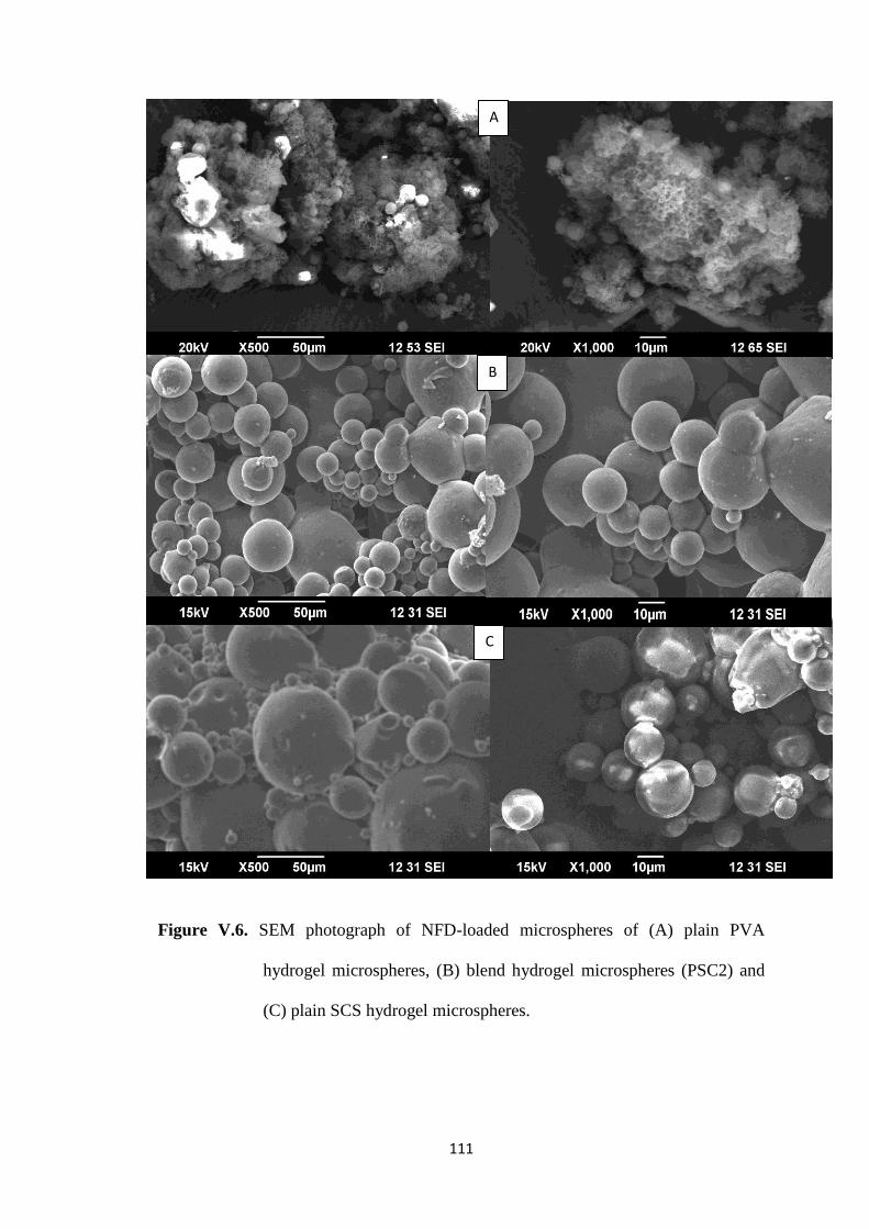
SEM micrographs shown in Figure V.6(A) indicate the presence of cross-
linked spongy waste along with the hydrogel microspheres. In case of SCS hydrogel
microspheres, high EE was observed because most of the loaded drug gets entrapped
into the microspheres, except a small loss of NFD, which was leached out along with
water during cross-linking. However, the blend hydrogel microspheres PSC1, PSC2
and PSC3 showed 74, 82 and 87 % of EE, respectively; indicating a high
encapsulating ability of SCS was imparted due to the blend matrix. The % EE
depends upon the extent of cross-linking as well as hydrophilic nature of SCS. As the
concentration of cross-linking agent increased from 2.5 (PSC4), 5 (PSC2) to 7.5 mL
(PSC5), the EE value also increased from 78, 82 to 84 %, suggesting the formation of
a rigid network structure at high concentration of GA, thereby retaining more of drug
particles in the microspheres.
V.2.2. FTIR Analysis
From the FTIR spectra of CS and SCS presented in Figure V.1, the CS shows
O-H and N-H stretching frequency at 3411 cm-1
for CS, whereas the peaks at 1652
cm-1
, 1553 cm-1
and 1424 cm-1
correspond to C=O (amide I), N-H (amide II) and C-N
(amide III) stretching frequencies, respectively. The bridged-O stretching is observed

103
at 1153 cm-1
, whereas C-O-C vibration frequency appeared at 1052 cm-1
. However,
primary O-H group attached -CH2OH shows the C-O stretching vibration at 1076 cm-
1. In the case of SCS, a new peak at 1717 cm
-1 corresponding to ester C=O stretching
vibrations confirms succinylation at the primary O-H group of CS, as confirmed by a
peak at 1076 cm-1
, whose intensity decreased compared to that of CS. Increase in the
intensity of amide I (C=O) and amide III (C-N) peaks and a decrease in the intensity
of amide II (N-H) peak confirms the succinylation reaction on –NH2 group of CS.
Thus, the reactive primary O-H and –NH2 groups of CS are the succinylated sites.
FTIR spectra of (A) NFD, (B) NFD-loaded microspheres and (C) placebo
microspheres presented in Figure V.2 confirm the chemical stability of NFD in the
hydrogel microspheres. In case of NFD, a band at 3334 cm-1
is due to N–H stretching
vibrations. Bands at 3068, 2932, 2887 and 2826 cm-1
are attributed to both aromatic
and aliphatic C–H stretching vibrations. A peak at 1680 cm-1
represents C=O
stretching vibrations, whereas the characteristic peak of pyridine is observed at 1624
cm-1
. The -NO2 stretching vibrations are observed at 1530 cm-1
and N–H bending as
well as C-N stretching vibrations are observed at 1497 cm-1
and 1227 cm-1
,
respectively. In case of drug-loaded microspheres, along with the peaks corresponding
to placebo microspheres, characteristic peaks of NFD at ~ 1680 cm-1
, 1631 cm-1
and
1531 cm-1
are observed. Presence of unmodified functional groups of NFD in NFD-
loaded microspheres indicates the chemical stability of NFD after encapsulation into
the polymer matrix.

104
Figure V.1. FTIR spectra of chitosan (CS) and succinyl chitosan (SCS).

105
Figure V.2. FTIR spectra of placebo blend hydrogel microspheres, NFD-laded blend
hydrogel microspheres and NFD.
V.2.3. Thermal Analysis
DSC-TGA thermograms of CS and SCS are presented in Figure V.3. DSC
thermogram of CS showed an endothermic peak at 94°C corresponding to the loss of
moisture present in the sample and an exothermic peak at 296°C, indicating the
decomposition of CS. In the case of SCS, along with the peaks corresponding to

106
moisture loss and polymer degradation, an endothermic peak is observed at 408°C,
which may be due to electrostatic interaction between the –COO-
group of succinic
acid and –NH3+ group of CS. Succeeding endothermic transitions may be due to the
degradation of succinic acid side group.
TGA curve of CS showed two stage weight losses. The initial 11 % of weight
loss at 60-98°C is due to the loss of trace amount of water in the sample, while the
second stage of weight loss (51 %) at 293-330°C corresponds to CS degradation. The
SCS showed a moisture weight loss of 4 % around 40-94°C and 10 % weight loss due
to polymer degradation around 221-300°C. A new stage of weight loss of 24 % is
observed for SCS at 462-503°C, due to degradation of succinic acid group of SCS.
Thus, the presence of additional endothermic transition at 408°C in the DSC
thermogram as well as third zone of weight loss in TGA curves of SCS confirmed
succinylation reaction with CS.
DSC thermograms of (A) CS, (B) SCS, (C) PVA, (D) placebo blend hydrogel
microspheres, (E) NFD-loaded blend hydrogel microspheres and (F) pure NFD are
presented in Figure V.4. In case of plain PVA, two endothermic peaks are observed,
one at 192°C, which corresponds to the melting process, and the other at 326°C due to
the thermal decomposition. Placebo microspheres exhibit two endothermic peaks, one
at 95°C corresponding to loss of trace water present in the sample and the other at
318°C due to thermal decomposition of polymers. In case of placebo blend hydrogel
microspheres, endothermic transition at 192°C that corresponds to the melting of PVA
is absent, indicating that PVA has lost its crystallinity after the formation of cross-
linked blend hydrogel matrix. For pure NFD, an endothermic peak appeared at 173°C
due to melting of the drug. Similarly, an endothermic peak observed at 171°C in the
NFD-loaded blend hydrogel microspheres indicates the crystalline dispersion of NFD
in the polymer matrix.

107
Figure V.3. TGA/DSC thermograms of CS and SCS

108
Figure V.4. DSC thermograms of (A) CS, (B) SCS, (C) PVA, (D) placebo blend
hydrogel microspheres, (E) NFD-loaded blend hydrogel microspheres
(PSC2) and (C) NFD

109
V.2.4. XRD Analysis
XRD spectra recorded for (A) placebo blend hydrogel microspheres, (B)
NFD-loaded blend hydrogel microspheres and (C) pure NFD presented in Figure V.5
are used to investigate the crystallinity of NFD in the hydrogel microspheres. NFD
has characteristic intense peaks between 2θ of 8 and 28° due to its crystalline nature,
but for placebo microspheres, broad peak due to the amorphous polymer matrix has
appeared at 2θ of 18°, but no intense peaks were observed between 2θ of 8 and 28°.
However, in case of NFD-loaded microspheres, intense peaks are observed at 2θ of
8.1°, 10.5°, 12°, 19.4° and 26.7° along with that of polymer matrix, indicating the
crystalline nature of NFD after encapsulation into the microspheres.
V.2.5. SEM Analysis
To assess the shape and surface morphology of the hydrogel microspheres,
SEM images of NFD-loaded PVA, SCS and their blend hydrogel microspheres cross-
linked with 5 mL of GA (PSC2) were taken at 500 and 1000x magnifications are
presented in Figure V.6. SEM images of plain PVA hydrogel microspheres (A)
shows spherical shapes of about 10 μm size surrounded by some spongy material.
During the formulation process, certain amount of PVA fails to form spherical
microstructure, which remains as the cross-linked spongy waste. SEM image (C)
suggests that hydrogel microspheres of SCS are spherical with smooth surfaces.
However, blend hydrogel microspheres (PSC2) as shown in image (B) represent the
fused spheres with wrinkled surfaces that are due to the interaction between blend
components i.e., -OH group of PVA has hydrogen bonding interaction with the –
COO- group of succinic acid moiety of SC, leading to high viscous solution. This
increases the particle size of the blend hydrogel microspheres compared to individual
components.

110
Figure V.5. XRD patterns of (A) placebo blend hydrogel microspheres, (B) NFD-
loaded blend hydrogel microspheres and (C) NFD.
0
2000
4000
6000
8000
10000
3 13 23 33 43 53
Lin
(C
ou
nts
)
2-Theta-Scale
(C)
0
500
1000
1500
2000
2500
3000
3 13 23 33 43 53
Lin
(C
ou
nts
)
2-Theta-Scale
(B)
0
500
1000
1500
2000
2500
3000
3500
4000
3 8 13 18 23 28 33 38 43 48 53 58
Lin
(C
ou
nts
)
2-Theta-Scale
(A)
111
Figure V.6. SEM photograph of NFD-loaded microspheres of (A) plain PVA
hydrogel microspheres, (B) blend hydrogel microspheres (PSC2) and
(C) plain SCS hydrogel microspheres.
A
C
B

112
V.2.6. Equilibrium Swelling (ES) Study
The % ES of the blend hydrogel microspheres has an effect on drug release
characteristics. Hence, to know the swelling capacity of the blend hydrogel
microspheres, ES experiments were performed in gastric and intestinal pH conditions
at 37°C (see Table V.1). Pure PVA hydrogel microspheres showed 160 and 162 % of
ES in gastric (pH 1.2) and intestinal (pH 7.4) swelling media, respectively. However,
in pH 1.2 media, the SCS hydrogel microspheres showed 210 % of ES, whereas in pH
7.4 phosphate buffer media, it is 291 %. This indicates that PVA hydrogel
microspheres exhibit pH independent swelling, whereas SCS hydrogel microspheres
show pH dependent swelling. On the other hand, formulations PSC1, PSC2 and PSC3
exhibit the ES values of 168, 172 and 183 %, respectively in acidic media, whereas in
alkaline buffer, these values are higher i.e., 209, 227 and 237 %. This suggests that
blending of SCS with PVA might have induced pH sensing nature to the polymer
matrix, but increasing the SCS concentration of the blend matrix might increase the
ES of the blend hydrogel microspheres, due to the presence of substituent succinic
acid on CS backbone.
In alkaline pH condition, the repulsion between -COO- groups of succinic acid
leads to high swelling of the matrix, but in acidic media, all the carboxylate groups
are protonated, due to charge repulsion related swelling. The latter is because of the
more hydrophilic nature of SCS component of the blend and the presence of succinic
acid moiety is responsible for inducing hydrophilicity. Swelling capacity of the blend
hydrogel microspheres decreased with the increasing concentration of GA from 2.5 to
7.5 mL (i.e., formulations PSC4, PSC2 and PSC5), but the ES values decrease from
179 to 166 % in acidic pH, whereas in pH 7.4 buffer media, ES decrease from 232 to

113
218 %, due to the formation of highly cross-linked rigid network structure at high
concentration of GA.
V.2.7. In Vitro Release Study
Effect of Blend Composition: To understand the drug release behavior of PVA,
SCS and their blend hydrogel microspheres, in vitro release experiments were
performed at 37°C in pH 1.2 and in pH 7.4 buffer solution to mimic the respective
gastric and intestinal pH conditions. The % cumulative release vs. time plots for
NFD-loaded PVA, PSC1, PSC2, PSC3 and SCS microspheres are compared in
Figure V.7(A) to investigate the effect of blend composition. The % cumulative
release is higher for SCS than for PVA hydrogel microspheres i.e., after 8 h of
dissolution, 100 % NFD was released from the SCS hydrogel microspheres, whereas
only 70 % of NFD was released from the PVA hydrogel microspheres even after 12 h
of dissolution.
In the case of blend hydrogel microspheres, drug release decreased with
increasing PVA concentration of the blend i.e., formulations containing PSC3, PSC2
and PSC1 with 25, 50 and 75 % (w/w) of PVA released 98, 86 and 73 % NFD,
respectively at the 8th
hour. This reveals that PVA content of the blend retards the
drug release to increase cumulative release with increasing amount of SCS in the
blend matrix. Since drug release from the hydrogel matrix depends on their swelling
characteristics under different pH conditions, the equilibrium swelling of SCS
hydrogel microspheres is higher than that of PVA hydrogel microspheres, but the
blend hydrogel microspheres showed the intermediary values of swelling. Thus,
increased water uptake capacity of the microspheres consequently increases the
matrix swelling, which would influence the drug release characteristics of the
hydrogel microspheres.

114
Figure V.7. (A) Effect of blend composition on in vitro release of NFD from
hydrogel microspheres in gastric (pH 1.2) and intestinal (pH 7.4)
conditions at 37°C.
Effect of Cross-linking: To study the effect of cross-linking on in vitro release
rates, we have used the formulations PSC4, PSC2 and PSC5 (prepared with 2.5, 5 and
7.5 mL of GA, respectively) and their release profiles are presented in Figure V.7(B).
The % cumulative release is higher in case of microspheres cross-linked with 2.5 mL
of GA, the least % cumulative release is observed with the microspheres cross-linked
with 7.5 mL of GA, while the intermediary values are observed for microspheres
cross-linked with 5 mL of GA. In other words, the release is slower for a formulation
containing higher amount of GA compared to that with the lower amount of cross-
linker. This may be due to the fact that at higher cross-linking, free volume of the
matrix will be reduced, thereby hindering easy transport of drug molecules through
the matrix.
(A)

115
Figure V.7. (B) effect of cross-linking on in vitro release of NFD from hydrogel
microspheres in gastric (pH 1.2) and intestinal (pH 7.4) conditions at
37°C.
Effect of pH: In vitro release experiments were performed under both gastric
(pH 1.2) and intestinal (pH 7.4) pH conditions separately for formulation PVA, PSC2
and SCS to assess the effect of pH of the release media on the in vitro NFD release.
Figure V.8 shows that NFD release from the PVA hydrogel microspheres is
independent of pH of the release media, whereas SCS and PSC2 hydrogel
microspheres showed a pH-dependent NFD release i.e., release is higher at pH 7.4
than at pH 1.2 for both the formulations. Hence, succinic acid group if present on the
CS makes it to sense the changes in pH of the external release media. Then,
formulation PSC2 responds to the change in pH due to the presence of SCS in the
blend. In all the formulations, however, the initial burst release was observed because
of the presence of drug on the surface of the microspheres.
(B)

116
Figure V.8. Effect of pH on in vitro drug release from PVA, PSC2 and SCS hydrogel
microspheres at 37°C.
0
10
20
30
40
50
60
70
80
90
100
0 1 2 3 4 5 6 7 8 9 10 11 12
% C
um
ula
tive
Re
leas
e
Time (h)
pH 1.2
PVA
PSC2
SCS
0
10
20
30
40
50
60
70
80
90
100
110
0 1 2 3 4 5 6 7 8 9 10 11 12
% C
um
ula
tive
Re
leas
e
Time (h)
pH 7.4
PVA
PSC2
SCS
117
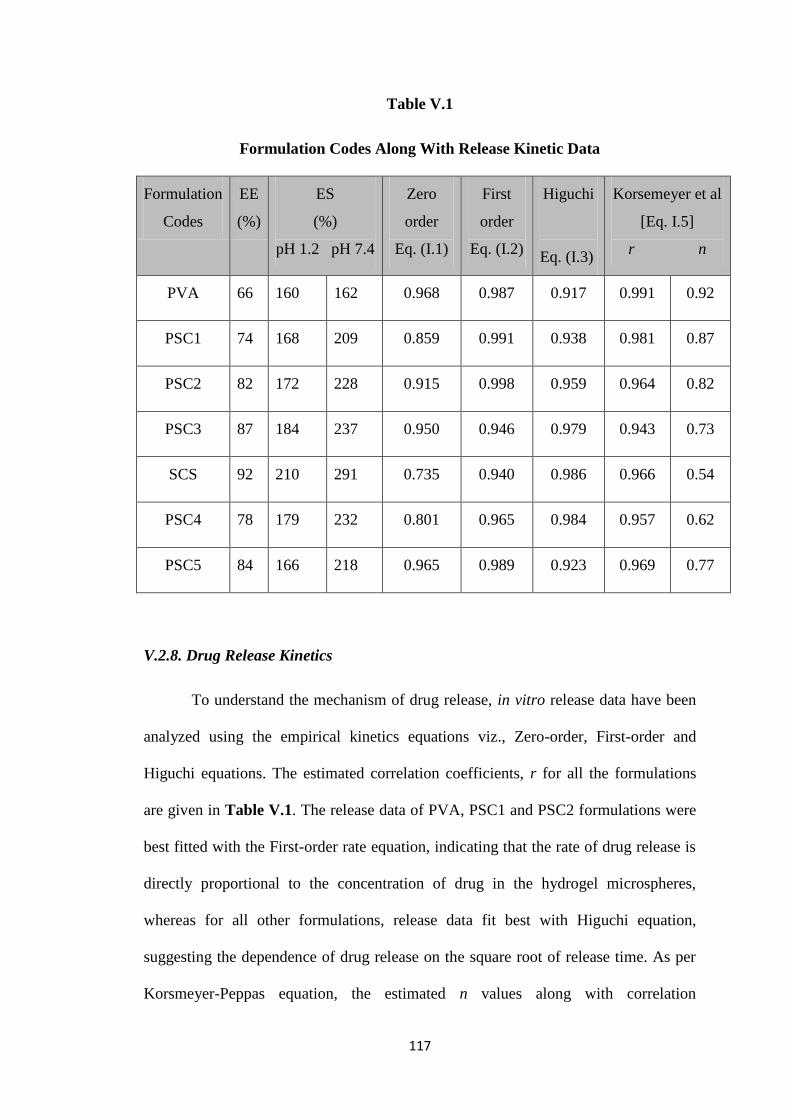
Table V.1
Formulation Codes Along With Release Kinetic Data
Formulation
Codes
EE
(%)
ES
(%)
pH 1.2 pH 7.4
Zero
order
Eq. (I.1)
First
order
Eq. (I.2)
Higuchi
Eq. (I.3)
Korsemeyer et al
[Eq. I.5]
r n
PVA 66 160 162 0.968 0.987 0.917 0.991 0.92
PSC1 74 168 209 0.859 0.991 0.938 0.981 0.87
PSC2 82 172 228 0.915 0.998 0.959 0.964 0.82
PSC3 87 184 237 0.950 0.946 0.979 0.943 0.73
SCS 92 210 291 0.735 0.940 0.986 0.966 0.54
PSC4 78 179 232 0.801 0.965 0.984 0.957 0.62
PSC5 84 166 218 0.965 0.989 0.923 0.969 0.77
V.2.8. Drug Release Kinetics
To understand the mechanism of drug release, in vitro release data have been
analyzed using the empirical kinetics equations viz., Zero-order, First-order and
Higuchi equations. The estimated correlation coefficients, r for all the formulations
are given in Table V.1. The release data of PVA, PSC1 and PSC2 formulations were
best fitted with the First-order rate equation, indicating that the rate of drug release is
directly proportional to the concentration of drug in the hydrogel microspheres,
whereas for all other formulations, release data fit best with Higuchi equation,
suggesting the dependence of drug release on the square root of release time. As per
Korsmeyer-Peppas equation, the estimated n values along with correlation

118
coefficients are presented in Table V.1. The estimated value of n for PVA and PSC1
hydrogel microspheres is > 0.85, indicating a swelling controlled release mechanism,
whereas the value of n for all other hydrogel microspheres ranged from 0.54 to 0.85,
indicating the anomalous (non-Fickian) nature of the drug release.
V.3. Conclusions
Succinyl chitosan was successfully synthesized and its blend hydrogel
microspheres were prepared with PVA to investigate the CR of NFD, an
antihypertensive drug. About 92 % of NFD was encapsulated into the hydrogel
microspheres prepared by emulsion cross-linking method using GA as a cross-linker.
Succinylation of chitosan and chemical stability of NFD within the blend matrix was
confirmed by FTIR. Thermal analysis, XRD and DSC analyses revealed the presence
of NFD crystals within the blend hydrogel microspheres. The microspheres were
characterized for their surface morphology, shape and size using SEM. The %
equilibrium swelling and in vitro release study performed in pH 1.2 and pH 7.4 buffer
media showed a dependence on blend composition, concentration of cross-linking
agent as well pH of the release media. In vitro release kinetics of NFD was described
by empirical equations, which suggest that hydrogel microspheres released NFD in a
swelling controlled manner, whereas all the other formulations followed the
anomalous trend i.e., NFD release from the hydrogel microspheres followed swelling
as well as diffusion controlled mechanisms.

119
V.4. Literature Cited
[1] Y.H. Bae, S.W. Kim, Adv. Drug. Deliv. Rev. 11 (1993)109.
[2] P.B. Kajjari, L.S. Manjeshwar, T.M. Aminabhavi, Ind. Eng. Chem. Res. 50 (2011)
7833.
[3] Y.F. Tang, Y.M. Du, X.W. Hu, X.W. Shi, J.F. Kennedy JF, Carbohydr Polymer.
67 (2007) 491.
[4] T. Takahashi, Int. J. Clin. Oncol. 7 (2002) 206.
[5] H. Zheng, Z. Zhou, Y. Chen, J. Huang, F. Xiong, J. Appl. Polymer. Sci. 106
(2007) 1034.
[6] S.A. Agnihotri, T.M. Aminabhavi, Drug. Dev. Ind. Pharm. 33 (2007) 1254.
[7] X. Zhang, D. Teng, Z. Wu, X. Wang, Z. Wang, D. Yu, C. Li, J. Mater. Sci. Mater.
Med. 19 (2008) 3525.
[8] T. Dai, M. Tanaka, Y.Y. Huang, M.R. Hamblin, Expert. Rev. Anti. Infect. Ther. 9
(2011) 857.
[9] A.A. Riccardo, Muzzarelli, Carbohydr Polymer 29 (1996) 309.
[10] L. Illum, Pharmaceut. Res. 15 (1998) 1326.
[11] C. Zhang, Q.E. Ping, H.J. Zhang, J. Shen, Eur. Polymer. J. 39 (2003) 1629.
[12] Y. Kato, H. Onishi, Y. Machida, Biomaterials. 25 (2004) 907.
[13] X.J. Zhu, W. Yuan, P. Li, X. Liu, J.Q. He, Drug. Dev. Ind. Pharm. 36 (2010)
1463.
[14] S.A. Agnihotri, T.M. Aminabhavi, Drug. Dev. Ind. Pharm. 31 (2005) 491.
[15] A.G. Sullad, L.S. Manjeshwar, T.M. Aminabhavi, Ind. Eng. Chem. Res. 50
(2011) 11778.
[16] A.G. Sullad, L.S. Manjeshwar, T.M. Aminabhavi, Ind. Eng. Chem Res. 49 (2010)
7323.

120
[17] O. Sanli, N. Ay, N. Isiklan, Eur. J. Pharm. Biopharm. 65 (2007) 204.
[18] M.D. Kurkuri, T.M. Aminabhavi, J. Contr. Release. 96 (2004) 9.
[19] U.K Parida, A.K. Nayak, B.K. Binhani, P.L. Nayak, J. Biomaterial.
Nanobiotechn. 2 (2011) 414.
[20] L.C. Wang, X.G. Chen, L.J. Yu, P.W. Li, Polymer. Eng. Sci. 47 (2007) 1373.
[21] K.M. Rao, B. Mallikarjuna, K.S.V. Krishna Rao, M.N. Prabhakar, K. Chowdoji
Rao, M.C.S. Subha, Polymer. Bull. 68 (2012) 1905.
[22] J. Huang, R.J. Wigent, C.M. Bentzley, J.B. Schwartz, Int. J. Pharm. 319 (2006)
44.
[23] P. Li, Y.N. Dai, J.P. Zhang, A.Q. Wang, Q. Wei, Int. J. Biomed. Sci. 4 (2008)
221.
[24] V. Ramesh Babu, M. Sairam, K.M. Hosamani, T.M. Aminabhavi, Carbohydr.
Polymer. 69 (2008) 241.
[25] N. Isiklan, M. Inal, F. Kursun, G. Ercan, Carbohydr. Polymer. 84 (2011) 933.
[26] Y.N. Dai, P. Li, J.P. Zhang, A.Q. Wang, Q. Wei, Biopharm. Drug. Dispos. 29
(2008) 173.
[27] C. Mura, M. Manconi, D. Valenti, M.L. Manca, O.D. Sales, G. Loy, A.M. Fadda,
Carbohydr. Polymer. 85 (2011) 578.
[28] S. Dash, P.N. Murthy, L. Nath, P. Chowdhury, Acta. Pol. Pharm. Drug. Res. 67
(2010) 217.
[29] R.W. Korsmeyer, S.R. Lustig, N.A. Peppas, J. Polymer. Sci. B. Polymer. Phys.
24 (1986) 395.