characterisation of laser - induced physical alterations of
TRANSCRIPT

e-PRESERVATIONScience
Rui Bordalo1*, Paulo J. Morais2, Christina R.T. Young3, Luís F. Santos4,Rui M. Almeida4
published by
CHARACTERISATION OF LASER - INDUCEDPHYSICAL ALTERATIONS OF PIGMENTED OILLAYERS
47
SCIENTIFIC PAPER
This paper is based on a presentation at
the 2nd International Conference “Matter
and Materials in / for Cultural Heritage”
(MATCONS 2011) in Craiova, Romania,
24-28 August 2011.
Guest editor:
Prof. Dr. Elena Badea
1. Universidade Portucalense, R. Dr.
António Bernardino Almeida, 541, 4200-
072 Porto, Portugal
2. Instituto de Soldadura e Qualidade,
Taguspark-Oeiras, Av. Prof. Dr. Cavaco
Silva 33, 2740-120 Porto Salvo, Portugal
3. Conservation and Technology
Department, Courtauld Institute of Art,
Somerset House, Strand, London, WC2R
ORN, United Kingdom
4. Departamento de Engenharia de
Materiais / ICEMS, Instituto Superior
Técnico / TU Lisbon, Av. Rovisco Pais,
1049-001 Lisboa, Portugal
corresponding author:
received: 31.12.2011
accepted: 04.11.2012
key words:
laser cleaning, excimer, pigments, oil,
SEM, AFM
e-PS, 2012, 9, 47-59
ISSN: 1581-9280 web edition
ISSN: 1854-3928 print edition
www.Morana-rtd.com
© by M O R A N A RTD d.o.o.
There has been a growing interest since the mid-1990s inthe use of lasers as an alternative method of cleaning ofpainted surfaces to surpass the possible problems of irre-versible modification caused by chemical or mechanicalmethods. The objective of this research is to extend thecurrent understanding of excimer laser-induced discol-oration effects on pigments mixed in oil medium. The studyfocuses on the response of samples of pure pigments andtheir mixtures widely used by painters in the nineteenthcentury, to different laser parameters. Characterisation ofphysical changes was performed using optical microscopy,colorimetry, scanning electron microscopy, and atomicforce microscopy. It has been found that materials have dif-ferent sensitivity to irradiation at 248 nm and that even ifan ultraviolet wavelength is used, the photo-chemicalmechanism may not be the predominant effect. The effectof irradiation on a pigment mixture cannot be predictedfrom the effect of the irradiation on the individual pig-ments.
1 Introduction
Over the last decades, laser cleaning has been widely studied and hasstarted to be used in the conservation of works of art.1-4 Since the mid-1990s, there has been a growing interest in the use of lasers in the clea-ning of painted surfaces and hence considerable research has takenplace.5-7 However, these studies have revealed that the paint can degra-de or change its surface morphology by laser radiation, due to its multip-le layers and heterogeneous nature. The greatest disadvantage of lasercleaning is that under laser radiation, painting materials can experienceundesirable physical and chemical alterations such as pigment discolora-tion or medium degradation.8-9 The constituent materials react to laserradiation in different ways, depending on their chemistry. Laser-matterinteraction depends both on the properties of laser radiation and on theintrinsic properties of the material surface. Artwork cleaning by laser inter-action can be explained by three major mechanisms: thermal, photo-mechanical and photo-chemical, which may occur at the same time alt-hough one is normally predominant depending mainly from the wave-length used.10
In varnish removal, it has been observed that the high energy pulse of theKrF excimer laser (248 nm) is adequate for material ablation since theenergy results in a photochemical action that breaks the atomic bonds of
M O R A N A RTD d.o.o.

organic materials. In fact, research has shown that248 nm is the most promising wavelength for varnishand overpaint layer removal.11,12 UV radaition isstrongly absorbed by the traditional varnish function-al groups and its degradation products. In partialremoval there is a minimal thickness that shouldremain intact to prevent any radiation to be absorbedby the underlying layers. However, further researchinto the discoloration of the paint layer in case ofdirect laser irradiation has yet to be carried out.Previous research has focused mainly on direct laserirradiation of real paintings or of pigment samples,either in the form of powder or mixed within a mediumbut not in a systematic way.7,13-17 The present studycompares the physical changes of pigment mixturesto those of pure pigments. This approach was chosenin order to better understand the behaviour of thepaint samples which more closely represent realpaintings. Previous studies of the laser effects onmost of these pigments have been limited.
2 Experimental
2.1 Sample Preparation
A comprehensive list of inorganic pigments in use inthe 19th century was compiled from different sources.Next, the pigments were categorised according totheir colour and chemical nature. From among thepigments used during the nineteenth century, prioritywas given to those that were also used in previouscenturies, which was desirable in order to obtainresults as broad and useful as possible. Also, the pig-ments had to belong to different colour families(black, white, yellow, red and green), so that theirmixtures would be unique and visually distinguisha-ble. Finally, the pigments had to be as different aspossible in their chemical nature in order to achieve awide representativity of the chemical pigment fami-lies. A fourth condition, desirable but non-compuls-ory, was the lack of previous studies on the lasereffects. Initially, a set of seven inorganic pigmentswere considered but an organic one was added toprovide a potentially useful behaviour comparisonbetween organic and inorganic pigments and becau-se laser-induced effects in organic pigments are stillan important gap in the current state-of-the-art. Afterconsidering a large list of pigments used in the 19th
century, the following were chosen: lead white, yellowochre, chrome yellow, vermilion, viridian green,Prussian blue, bone black and rose madder. The bin-ding medium was cold-pressed linseed oil. The pig-ments were produced according to historic recipesand obtained from Kremer Pigmente (Germany) andAP Fitzpatrick (United Kingdom).
The samples were made according to the traditionaltechnique of oil paint preparation by grinding pigmentin oil on a stone plate. Linseed oil was added in theratio of typically 3-4 ml of oil per 2 g of pigment, toobtain a mixture fluid enough to be easily spread onthe support. The paint was deposited over a flexiblesubstrate (polyester film sheets 3M PP2280) with amanual film applicator (Neurtek) producing a 200 µmthick wet layer. Pure and mixed samples were prepa-red: a total of eight pure pigments and 16 binary mix-tures (in a 1:1 ratio).
The samples were then left to dry for two months innormal ambient conditions and then placed in aphoto-degradation chamber equipped with UV-filteredfluorescent daylight tubes to simulate ageing in amuseum. The samples were left in these conditionsfor 8 months during which the ageing is assumed tobe equivalent to approximately 110 years.
2.2 Laser System
The samples were then irradiated with a KrF laserLambda Physic COMPex 110. This laser operates ata wavelength of 248 nm, with 30 ns pulse duration at2 Hz frequency. After emission, the laser beam wasshaped by a mask of 11 x 5 mm and focused by a 150mm focal length quartz plano-convex lens. For easycontrol of the samples, they were placed on a manu-al X-Y translation stage.
Before irradiation, preliminary tests were performedin order to determine the sensitivity and to make anearly prediction of the behaviour of the samples.Based on the results, the samples were irradiated atvarying fluence (F), from F = 0.001 J/cm2 to F = 0.4J/cm2, at 1, 3 and 5 pulses, and occasionally 10 pul-ses to determine the discoloration and ablation thres-holds.
2.3 Analytical Techniques
The samples were analysed before and after laserirradiation using several analytical techniques fortheir chemical and physical characterisation. In parti-cular, the samples were analysed by optical micro-scopy, colorimetry, scanning electron microscopy,and atomic force microscopy. Due to the largeamount of collected data per sample, only the mostrelevant results will be discussed.
Visual analysis was performed with a high resolutionCarl Zeiss Axiotech 100HD-3D microscope. A CanonPowerShot G5 digital camera was coupled to themicroscope. The photographs were taken with shutterspeed 1/60 s, lens aperture F/4 and focal length 16mm.
The colour was measured by a Konica-Minolta CM-2600d spectrophotometer under normal ambient con-ditions and with the following operating conditions:specular component included, UV excluded, illumi-nant D65 and 10° standard observer. The data wasanalysed with SpectraMagic NX software. The repor-ted CIELab coordinates are the average of threemeasurements on each sample.
Scanning electron microscopy in combination withenergy dispersive X-ray analysis (SEM-EDX) wasperformed on a Hitachi S-2400 scanning electronmicroscope with a standard EDX detector fromBruckër (ex-Rontec). The samples were gold-coatedto improve surface conduction, and the images weretaken at 25 kV with a spot size of ~1 nm and usingsoftware Multi Image 2.1.
The topographic characterization of some of the filmsamples was made using a Veeco DI CP-II AtomicForce and Scanning Tunneling Microscope. The sys-tem is equipped with a 50 x 50 µm scanner stage and
www.e-PRESERVATIONScience.org
Laser-induced Physical Alteration of Pigmented Oil Layers, e-PS, 2012, 9, 47-59
48

a motorized Z stage, and it was connected to a PCenabling computer control and data acquisition withsoftware Proscan 2.1.
3 Results
One of the first objectives was the determination ofthe discoloration and ablation thresholds. The disco-loration threshold was first identified by naked eyeand then quantified using a spectrophotometer todetermine its colour change (ΔE CIE 1976). The abla-tion threshold was determined by optical microscopy,analysing the surface at 50x and 500x. The samplesrevealed gradual laser-induced effects and thus athreshold range is determined. The threshold wasfound by irradiating the samples at consecutively hig-her fluences, each one also at increasing number ofpulses (1, 3, 5, and 10) to determine a smallest rangein combination with varying fluence. The exact abla-tion thresholds of each sample have been publishedelsewhere17. The set of eight pigments can be distri-buted in three ranges according to their ablationthresholds: a) 0.18 < F < 0.2 J/cm2, 1 to 5 pulses; b)0.26 < F < 0.29 J/cm2, 1 to 5 pulses; and c) F > 0.4J/cm2. The lowest fluence range is where most of thepigments (five) have their ablation threshold. Parallelto the samples, a sample of oil without pigments wasirradiated and it was verified that the ablation of oil ispromoted by the presence of pigments.
3.1 Lead White (LW100)
Lead white is one of the pigments with the lowest dis-coloration threshold and very sensitive to becominggrey or black. Lead white is an unusual pigment withrespect to its behaviour upon laser irradiation. It isknown that the discoloration of lead white diminisheswith time but literature does not report the degree ofrecovery. It was found that the colour recovery isdependent on the energy incident on the surface.Immediately after irradiation, the sample exhibited acolour change of up to ΔE = 30 although a permanentdiscoloration of ΔE = 13 was measured after twoweeks. Although colour variation less than ΔE ≤ 1 is
considered hardly visible by the human eye, most ofthe permanent discolorations of the sample were per-ceived.
Figure 1 shows an area irradiated using differentlaser parameters (vertical shadows in the centre),after colour conversion, which was found to be per-manent after two weeks. SEM analysis was perfor-med but the comparison between the non-irradiatedand the irradiated area at F = 0.36 J/cm2 did not reve-al any changes even at higher magnifications of up to15,000x. The absence of distinctive differencesbetween the non-irradiated and the irradiated areassuggests that the colour change induced by laserradiation in lead white is not accompanied by anyphysical change, leading to the conclusion that it is ofa purely photo-chemical nature.
3.2 Yellow Ochre (YO100)
Yellow ochre is the only earth pigment among the setof pigments in the study. As expected, it revealed hig-her resistance to laser-induced alterations in compa-rison with the other pigments.
SEM imaging confirmed that laser irradiation at lowfluence increased surface roughness, even though itmay be invisible to the naked eye. Figure 2 shows thedifference between the original surface and the irra-diated area at F = 0.36 J/cm2. The non-irradiated areais smooth and shows little topography (Figure 2A)while the irradiated area is quite uneven (Figure 2B).The precise photo-mechanic mechanism of this alte-ration is unknown although it could be due to meltingor due to a surface rearrangement mechanism.
© by M O R A N A RTD d.o.o.
Laser-induced Physical Alteration of Pigmented Oil Layers, e-PS, 2012, 9, 47-59
49
Figure 1: Micrograph of an area of LW100, permanently affected by
laser irradiation at F = 0.1 J/cm2 and 5 pulses, in reflected-light bright
field (magnification 200x).
Figure 2: Scanning electron micrograph of the characteristic surface
of a non-irradiated area (A) and of an irradiated area at F=0.36 J/cm2
(B) of yellow ochre (YO100). Reproduced with permission from ref.
17.
A
B

3.3 Chrome Yellow (CY100)
When observed through the optical microscope, theirradiation tests were only perceptible in bright fieldmode. After laser irradiation, only the SEM image at15,000x revealed a perceptible alteration. A compa-rative analysis of the highlights of individual areascan be seen in Figure 3. In the non-irradiated areathe pigment particles were well embedded in the oilmatrix, while in the irradiated area the particles wereat the surface because of the removal of a superficialoil layer.
3.4 Viridian Green (VG100)
Viridian green has one of the highest discolorationthresholds of all pigments as shown by the relativelylow colour change of ΔE = 1.7 at a fluence of F = 0.15J/cm2 and 3 pulses.
Comparative analysis between areas before and afterlaser irradiation seems to indicate that no ablativeprocess took place since both surfaces are similarlysmooth (Figure 4). However, the irradiated areashows a layer of particles with an approximate size of1-5 µm deposited over the surface. These were depo-sited onto the surface after laser irradiation althoughtheir origin is not clear since there are no surface cra-ters or other modifications that can explain the sour-ce of the particles. Figure 5 shows the general topo-graphy of the irradiated (left) and non-irradiated(right) areas. The presence of the particles is almostimperceptible here, in comparison with the sampletopography.
3.5 Vermilion (V100)
Upon laser irradiation, vermilion typically turns togrey and black. The pure pigment sample (V100)showed a discoloration threshold of F=0.03 J/cm2,which was the lowest in comparison with all the otherpigments but presented the highest colour variation,with ΔE = 17.8 at F = 0.36 J/cm2.
Two different areas of V100, a non-irradiated and anirradiated one, were analysed by AFM. Figure 6shows 3D micrographs of the surface of V100 beforeand after laser irradiation at F = 0.36 J/cm2. The esti-mated root mean square roughness (Rrms) was 400nm and 349.5 nm for the non-irradiated and the irra-diated areas, respectively. However, this difference is
www.e-PRESERVATIONScience.org
Laser-induced Physical Alteration of Pigmented Oil Layers, e-PS, 2012, 9, 47-59
50
Figure 5: Scanning electron micrograph at 80º of the irradiated and
non-irradiated areas of viridian green (VG100). Reproduced with per-
mission from ref. 17.
Figure 3: Scanning electron micrographs at 15,000 x of chrome yel-
low (CY100): non-irradiated area (A), and irradiated area (B).
Reproduced with permission from ref. 17.
A
B
Figure 4: Scanning electron micrograph of viridian green (VG100):
non-irradiated area (A), and irradiated area (B) at F = 0.36 J/cm2.
A
B
not conclusive as it may be due to the different loca-tions where the analyses were performed.
3.6 Rose Madder (RM100)
Rose madder is the only organic pigment from the setand it was expected to show good resistance to laserirradiation due to its organic composition. The colourchange of the pigment is one the lowest among thepigments studied, with a colour variation that rangesfrom ΔE = 2.1 to ΔE = 2.6. The irradiated area is verysimilar to the non-irradiated one except for a higherconcentration of particles deposited on its surface(Figure 7).
Rose madder has a high discoloration threshold,undergoing discoloration at a fluence of approximate-ly 0.28 J/cm2. The laser-induced alteration was diffi-cult to assess. Optical microscopy and SEM imagessuggested only slight discoloration.
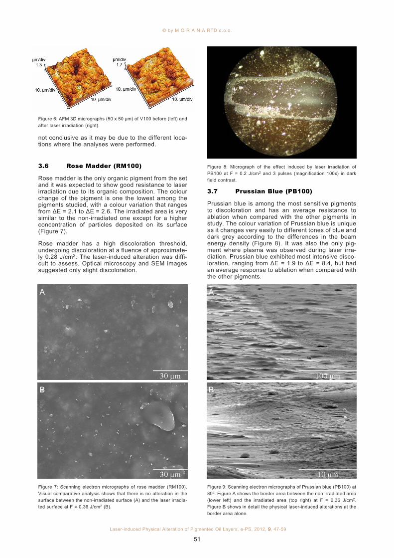
3.7 Prussian Blue (PB100)
Prussian blue is among the most sensitive pigmentsto discoloration and has an average resistance toablation when compared with the other pigments instudy. The colour variation of Prussian blue is uniqueas it changes very easily to different tones of blue anddark grey according to the differences in the beamenergy density (Figure 8). It was also the only pig-ment where plasma was observed during laser irra-diation. Prussian blue exhibited most intensive disco-loration, ranging from ΔE = 1.9 to ΔE = 8.4, but hadan average response to ablation when compared withthe other pigments.
© by M O R A N A RTD d.o.o.
Laser-induced Physical Alteration of Pigmented Oil Layers, e-PS, 2012, 9, 47-59
51
Figure 8: Micrograph of the effect induced by laser irradiation of
PB100 at F = 0.2 J/cm2 and 3 pulses (magnification 100x) in dark
field contrast.
Figure 6: AFM 3D micrographs (50 x 50 µm) of V100 before (left) and
after laser irradiation (right).
Figure 7: Scanning electron micrographs of rose madder (RM100).
Visual comparative analysis shows that there is no alteration in the
surface between the non-irradiated surface (A) and the laser irradia-
ted surface at F = 0.36 J/cm2 (B).
A
B
Figure 9: Scanning electron micrographs of Prussian blue (PB100) at
80º. Figure A shows the border area between the non irradiated area
(lower left) and the irradiated area (top right) at F = 0.36 J/cm2.
Figure B shows in detail the physical laser-induced alterations at the
border area alone.
A
B

SEM imaging of the control sample revealed asmooth surface where the pigment particles areembedded into the medium and with some particlesat the surface. However, significant alteration of thesurface is observed. Unlike other samples, visualcomparison between two different areas shows a sig-nificant difference. Figure 9A shows a small area ofPB100 irradiated at F = 0.36 J/cm2 oriented at 80ºand focused at the interface of the irradiated area.Laser ablation is obvious in this area, as can beobserved in the detail in Figure 9B. However, a simi-lar effect was not observed for the irradiated area,despite the fact that the surface seems smoother andmore regular than that of the non-irradiated area.
AFM analysis was performed on PB100 in order toassess the topographic alterations that laser irradia-tion induced to the sample surface. For the analysis,two areas were selected from nearby the borderlinebetween both zones. Figure 10 shows 2D and 3Dmicrographs from the surface of PB100 before andafter laser irradiation at F = 0.36 J/cm2. The topo-graphical difference after laser irradiation is obviousin both 2D and 3D micrographies. The surface rough-ness (Rrms) for the non-irradiated surface (492 nm)decreased after laser irradiation (357 nm).
3.8 Bone Black (BB100)
Bone black revealed to be the least sensitive pigmentto laser irradiation with higher discoloration and abla-tion thresholds than any other pigment in the presentstudy. At a relatively high fluence such as F = 0.2J/cm2 the colour variation was still under ΔE < 1. Thediscoloration is towards a dark grey and the colourvariation is small, ranging from ΔE = 0.2 to ΔE = 3.2.SEM imaging show no changes on the sample sur-face even at a high fluence such as F = 0.36 J/cm2.
3.9 Lead White-Yellow Ochre(LW50-YO50)
SEM imaging confirmed that the oil medium has beenremoved leaving a surface of unprotected pigment
particles. Figure 11 shows the difference between theoriginal surface (Figure 11A) and the irradiated areaat F = 0.36 J/cm2 (Figure 11B) of LW50-YO50. Themost obvious difference between the micrographs isthe removal of the most superficial layer of the medi-um, apparently leaving the pigment particles withoutthe binder. This difference, however, is not wellappreciated at lower magnifications.
3.10 Lead White-Chrome Yellow(LW50-CY50)
Direct comparison using SEM imaging of the non-irra-diated and the irradiated areas at 80º (Figure 12) sug-gests that this alteration is due to the lifting of smallflakes instead of the oil layer removal. Figure 12Bshows several particles of different sizes and shapesscattered over the irradiated surface. It is not possi-ble to assess if the particles remained on the surfaceafter selective removal of the binder or if they weredeposited on the surface during or after the irradia-tion process.
The comparison between this mixture and the purepigments in its composition does not reveal any spe-cial similarity. SEM imaging of LW100 did not revealany laser-induced effect although CY100 revealed acomparable situation with that shown in Figure 12.However, the pigment particles that are observable inthe present case are much bigger than in case ofCY100. Thus, there seems to be no particular laser-induced effect on the pigment.
www.e-PRESERVATIONScience.org
Laser-induced Physical Alteration of Pigmented Oil Layers, e-PS, 2012, 9, 47-59
52
Figure 10: AFM 2D and 3D micrographs (50 x 50 µm) of PB100 befo-
re (above) and after laser irradiation (below).
Figure 11. Scanning electron micrograph of the characteristic sur-
face of a non-irradiated area (A) and of an irradiated area at
F=0.36 J/cm2 (B) of LW50-YO50.
A
B

3.11 Lead White-Viridian Green(LW50-VG50)
The mixture of lead white with viridian green did notundergo chemical degradation and colorimetryshowed that the mixture is less sensitive than its purepigments. The reason why a certain mixture has ahigher discoloration threshold than its component pig-ments could not be explained but it is clearly pigmentdependent. Colorimetry analysis concluded that themixture has its discoloration threshold in the rangebetween the respective thresholds of its pure pig-ments.
Figure 13 shows a SEM micrograph where the irra-diated (A) and non-irradiated (B) borderline is high-lighted. Further analysis at a higher magnificationshowed that the reason for the perceptible differenceis the removal of the binder at the surface.Nevertheless, there is no evidence that the pigmentwas affected. The mixture presents a different beha-viour than that of the pure pigments alone. While leadwhite did not show any physical alteration, viridiangreen revealed only some particles that were appa-rently deposited on the surface, leaving the surfacetop layer undamaged.
3.12 Lead White-Vermilion (LW50-V50)
Although the mixture contains lead white and vermi-lion, which are the most sensitive pigments in theensemble, the mixture was relatively unaffected bylaser irradiation. Indeed, the mixture (discolorationthreshold at F = 0.1 J/cm2) is less sensitive than thepure pigments as lead white and vermilion have dis-coloration thresholds at F = 0.05 J/cm2 and at F =0.03 J/cm2, respectively. This is confirmed by colori-metry, which upon irradiation at F = 0.085 J/cm2
showed a negligible discoloration of ΔE = 0.4.
The comparison of the surface between the non-irra-diated and the irradiated area at F = 0.36 J/cm2 canbe observed in Figure 14. The topographic aspect ofthe surface prior to laser irradiation (Figure 14A) isvery smooth with only a few regularly distributed ori-fices that may be related probably to the release of airbubbles while drying. On the other hand, the irradia-ted area (Figure 14B) reveals that ablation took placeleaving a highly rough and irregular surface.
© by M O R A N A RTD d.o.o.
Laser-induced Physical Alteration of Pigmented Oil Layers, e-PS, 2012, 9, 47-59
53
Figure 13: Scanning electron micrograph of LW50-VG50. The image
highlights the borderline between the non-irradiated and the laser
irradiated surface at F = 0.36 J/cm2.
Figure 12: Scanning electron micrograph at 80º of the characteristic
surface of a non-irradiated area (A) and an irradiated area at
F=0.36 J/cm2 (B) of LW50-CY50.
A
B
Figure 14: Scanning electron micrographs of LW50-V50. Figure A
shows the topographic aspect of the surface prior to laser irradiation
while Figure B shows the physical changes that the sample undergo
after laser irradiation at F = 0.36 J/cm2.
A
B

3.13 Lead White-Rose Madder(LW50-RM50)
In the case of rose madder, the mixture with leadwhite increased the sensitivity of the organic pigment.Laser radiation had a singular effect on LW50-RM50at microscopic level, observable by SEM imaging.Figure 15 shows the borderline between the non-irra-diated (left) and the irradiated (right) areas. Thenatural roughness of the surface can be observed inthe left part whereas the irradiated part is presentedas a flatter surface. A detail of the borderline can beseen in Figure 16 where it is clear that the laser radia-tion induced localized bursts resulting in the altera-tion of the surface although the exact alterationmechanism is unknown. This effect, unlike the reac-tions of either rose madder or lead white alone, isprobably a photo-mechanical mechanism. A similarreaction was also observed in other mixtures such asLW50-BB50.
3.14 Lead White-Prussian Blue(LW50-PB50)
The laser-induced change to Prussian blue is verycharacteristic. However, unlike the behaviour of thepure pigment, its mixture with lead white produces adifferent discoloration towards a darker tone, undoub-tedly due to the presence of lead white. The discolo-ration threshold of this mixture is within the rangebetween the pure pigment thresholds.
SEM imaging of the laser-induced alteration betweenthe non-irradiated and the irradiated areas at F = 0.36J/cm2 (Figure 17) is evident even if the borderline isnot very clear. It is evident that ablation took place inthe upper left corner of the micrograph, leaving thesurface uneven and rough. The irradiated area wasobserved at higher magnification revealing numerouscraters which are not visible in the non-irradiatedarea corresponding to the selective removal of the oil(Figure 18). The creation of this crater was inducedby laser irradiation, which seems to preferentiallydamage the surface of the binder rather than the pig-ment particles. It should also be noted that the effecton the medium seems to have a direct connection tothe presence of pigment. In the micrograph (Figure18) it can be observed that the areas with more cra-ters are also those with a higher concentration of pig-ment particles while the smoother areas correspondto a higher concentration of the binder. The neigh-bouring areas appeared similar. A suitable explana-tion for this mechanism could not be found.
3.15 Lead White-Bone Black(LW50-BB50)
As expected, the discoloration threshold of this mix-ture is between those of the pure pigments. However,the mixture exhibits higher discoloration for lower flu-ences than the ones of bone black. This means thatits behaviour is closer to that of lead white than to thatof bone black.
www.e-PRESERVATIONScience.org
Laser-induced Physical Alteration of Pigmented Oil Layers, e-PS, 2012, 9, 47-59
54
Figure 15: Scanning electron micrograph of LW50-RM50. It is possi-
ble to observe the interface area between the non-irradiated area
(left) and the irradiated (right) area at F=0.36 J/cm2 (B).
Figure 17: Comparative scanning electron micrograph of the charac-
teristic surface of an irradiated area at F = 0.36 J/cm2 (upper left) and
of a non-irradiated area (lower right) of LW50-PB50.
Figure 16. Scanning electron micrograph of LW50-RM50, detail of
the interface from Figure 15.
Figure 18: Macro view of the irradiated area of LW50-PB50 at F =
0.36 J/cm2 showing numerous craters, not visible in the non-irradia-
ted area, corresponding to the selective removal of the oil.

Figure 19 compares the non-irradiated and the irra-diated areas of the mixture at F = 0.36 J/cm2. Theirradiated surface appears to be very rough and irre-gular as the laser beam would have lifted some of thematerial in a burst.
3.16 Yellow Ochre-Prussian Blue(YO50-PB50)
The mixture shows a discoloration threshold that cor-responds to the pigment with the lower threshold(Prussian blue, F = 0.08 J/cm2) rather than an inter-mediate one between the discoloration thresholds forthe pure pigments. Despite the lower discolorationthreshold, the mixture shows an increasing resistan-ce to higher fluences in comparison to the pure pig-ments alone.
SEM imaging showed that the laser radiation inducedthe removal of the top oil layer. The non-irradiatedsurface of the sample can be observed in Figure 20Awhere the pigment is regularly mixed in the binderand the surface is evenly smooth. After laser irradia-tion at F = 0.36 J/cm2 (Figure 20B), it appears that themedium was partially and non-selectively removedfrom the surface, revealing the pigment particlesunderneath and leaving behind several flake-likeareas of medium, giving a rough and uneven aspectto the surface.
3.17 Yellow Ochre-Bone Black(YO50-BB50)
As standard procedure, a view at low magnificationand 80º was attempted but it was insufficient to obtaina perceptible view of the laser-induced effects. Thesurface of the non-irradiated and the irradiated areasare compared in Figure 21. The non-irradiated areaappears as a flat and smooth surface where the pig-ment is embedded in the binder below the surface,except for some particles dispersed by the surface(Figure 21A). After irradiation the surface is unevenand characterized by a series of craters of differentsizes created by the removal of the binder, leavingthe pigment underneath uncovered (Figure 21B).Once again, the response of this mixture to laser irra-diation has no similarity to the individual effects onthe pure pigments in its composition.
3.18 Chrome Yellow-Vermilion(CY50-V50)
The mixture shows better resistance to laser radiationthan its pure pigments. Although the differencebetween the non-irradiated and the irradiated areas isnot strong, it is noticeable by the localized cratersthat characterize the irradiated area. The differencemay be seen in more detail in Figure 22. In fact, theirradiated area is very similar to the non-irradiatedone except for the localized craters that partiallyremoved the binder leaving the pigment particlesbeneath uncovered. The craters are clean and nomaterial is deposited around them or at the samplesurface. The reason why they are so scattered is notknown but it is suggested that the laser impact couldlead to stress on the surface causing material to be
© by M O R A N A RTD d.o.o.
Laser-induced Physical Alteration of Pigmented Oil Layers, e-PS, 2012, 9, 47-59
55
Figure 19: Scanning electron micrograph at 80º of the characteristic
surface of a non-irradiated area (A) and of an irradiated area at F =
0.36 J/cm2 (B) of LW50-BB50.
A
B
Figure 20: Scanning electron micrograph of YO50-PB50 aspect of
the surface before (A) and after (B) laser irradiation at F = 0.36
J/cm2.
A
B

ejected in points of structural weakness. It shouldalso be underlined, as shown in Figure 22B and byother details (images not shown), that all craters aresurrounded by a halo.
3.19 Vermilion-Rose Madder (V50-RM50)
This mixture contains pigments with divergent sensi-tivity to laser radiation: rose madder has a high dis-coloration threshold while vermilion has the lowestdiscoloration threshold among the pigments in thisstudy. Experimentally, it has been observed that thecorresponding mixture has a slightly increased resi-stance to laser radiation.
Comparison of the SEM images of the irradiated andnon-irradiated areas showed the main difference tobe the presence of isolated craters on the top surfaceof the binder, similar to those observed in sampleCY50-V50. The surface presents isolated puncturesin the top layer of the binder while the pigment seemsunaffected.
3.20 Vermilion-Prussian Blue(V50-PB50)
The discoloration of the mixture resembles that ofpure Prussian blue. The mixture exhibits the samediscoloration threshold as the most sensitive pigmentin the mixture, vermilion, i.e. F = 0.05 J/cm2.Nevertheless, the mixture shows higher resistance tolaser radiation than vermilion at other fluences.
www.e-PRESERVATIONScience.org
Laser-induced Physical Alteration of Pigmented Oil Layers, e-PS, 2012, 9, 47-59
56
Figure 21: Scanning electron micrographs of YO50-BB50. Figure A
shows the topographic aspect of the surface prior to laser irradiation
while Figure B shows the physical changes of the surface after laser
irradiation at F = 0.36 J/cm2.
A
B
Figure 22. Scanning electron micrographs of CY50-V50. Figure A
shows the topographic aspect of the surface prior to laser irradiation
while Figure B shows the physical changes that the sample under-
went after laser irradiation at F = 0.36 J/cm2.
A
B
Figure 23: Scanning electron micrographs of V50-PB50 of the inter-
face between the irradiated and the non-irradiated areas after laser
irradiation at F = 0.36 J/cm2. Figure A shows the topographic aspect
of the surface at 80º while Figure B shows the same area from a per-
pendicular perspective.
A
B

SEM imaging showed that the laser-induced effect islimited to the superficial layers of the surface. Thefeatures shown in Figure 23 suggest that a superficiallayer of the binder is altered or removed, leavingbehind an almost identical surface with the exceptionof a few craters of minimum depth. This effect seemslimited to the binder layer and the disrupted aspectallows the possibility of thermally induced degrada-tion. The pigment seems to be unaffected by laserradiation.
3.21 Vermilion-Bone Black (V50-BB50)
The mixture is made of pigments with the lowest andhighest discoloration thresholds among the pigmentsin the study. The laser-induced discoloration of thismixture is towards a ‘burnt’ darker tone. The compa-rison of the thresholds indicates a small influence ofvermilion. Despite the fact that the mixture has alower discoloration threshold than that of bone black,the results suggest that the mixture withstands betterlaser radiation at higher fluences.
Figure 24 compares two SEM micrographs from anon-irradiated and an irradiated area. The first(Figure 24A) is comparable with many other samplesalready analysed, where it is possible to see the pig-ment particles embedded in the binder and underne-ath the surface. On the other hand, the irradiatedarea shows a very disrupted surface (Figure 24B): thebinder seems to have been removed leaving the par-ticles underneath uncovered. The top of the pigmentparticles appears to have been affected as well. Upon
laser irradiation at F = 0.36 J/cm2, the top layer of thebinder was removed leaving uncovered the pigmentparticles that underwent a severe disruption..
3.22 Bone Black-Rose Madder(BB50-RM50)
This mixture includes pigments with the highest dis-coloration thresholds. Nevertheless, the mixture ofboth pigments presents a discoloration thresholdbelow the threshold of any of the pure pigments. Thelaser-induced alteration of the sample surface can beappreciated in the borderline between the non-irra-diated (lower right side) and the irradiated (upper leftside) area at F = 0.36 J/cm2 in Figure 25. The non-
© by M O R A N A RTD d.o.o.
Laser-induced Physical Alteration of Pigmented Oil Layers, e-PS, 2012, 9, 47-59
57
Figure 24: Scanning electron micrographs of V50-BB50. Figure A
shows the topographic aspect of the surface prior to laser irradiation
while Figure B shows the physical changes of the surface after laser
irradiation at F = 0.36 J/cm2.
A
B
Figure 26. Scanning electron micrograph of BB50-RM50 at 80º of the
characteristic surface of a non-irradiated area (A) and of an irradia-
ted area at F = 0.36 J/cm2 (B).
A
B
Figure 25. Scanning electron micrograph of BB50-RM50 at 80º of the
interface between the irradiated (lower) and the non-irradiated
(upper) areas.

irradiated area appears smooth and flat as opposedto the irradiated one.
The irradiated surface (Figure not shown) showssevere damage: although the binder layer is still visi-ble, the surface is disrupted by several particles,most probably pigment and binder elements. This iseven more obvious at a higher magnification in Figure26, where a detailed comparison of the laser-inducedalteration is shown at a closer look at 80º. In particu-lar, Figure 26B illustrates how the laser beam indu-ced a burst of the top layer without ejection of thefractured material.
4 Discussion
According to Bordalo,18 a sample can physicaly exhi-bit one of three possible stages upon laser irradiation:no perceptible modification, colour or morphologicalchange on the surface, and ablation of the material.In a linear model, two thresholds would divide thethree stages where one stage could evolve to thenext one by increasing the laser fluence. However,this model is not accurate as it depends on materialproperties. In fact, one stage does not necessarilyprecede another. Therefore, there are two mainthresholds, normally referred to in the literature, thatshould be considered and assessed: the discolorationand ablation thresholds. These thresholds are defi-ned as the minimum amount of laser energy incidenton the sample necessary to induce colour alterationor material removal from its surface, respectively.However, there is also another threshold that shouldalso be considered for this specific application, whichis the minimum amount of laser energy incident onthe sample necessary to induce any physical altera-tion of the material surface.
One of the initial surprising effects was that many ofthe laser induced effects on the surface were visibleby the naked eye but were not immediately obviousby optical microscopy. This is because of the scatte-ring properties of the pigment and the angle of inci-dence of the light. It was necessary to observe manyof the irradiated areas in bright field, dark field andpolarized contrast in order to obtain the most accura-te information.
Analysing the discoloration and ablation thresholds, itwas also observed that the reaction of the mixturesdoes not follow a linear relationship with the reactionof their pure pigments. It has been observed thatamong the mixtures three types of reactions may takeplace; 1) the first and the expected reaction is whena mixture has the same or nearly the same ablationthreshold as the pure pigments in its composition; 2)when the mixture shows a lower ablation thresholdthan that of its pure pigments; 3) when the mixtureshows a higher ablation threshold than that of its pig-ments. The discoloration thresholds for all pigmentsare at relatively low fluences, between F = 0.03 J/cm2
and F = 0.18 J/cm2, except for bone black (BB100).The most sensitive pigments are lead white and ver-milion which revealed similar behaviour: both under-go discoloration at a low fluence near F = 0.05 J/cm2
and ablation near F = 0.20 J/cm2. Chrome yellow wasfound to be the pigment with the smallest differencein the fluence between thresholds of only 0.05 J/cm2.Among all the pigments, bone black proved to be the
most resilient pigment to λ = 248 nm laser irradiation.Its discoloration threshold is slightly above the abla-tion threshold of the other pigments. Bone black(BB100) was irradiated at the maximum fluence pos-sible with the laser set-up used (F = 0.40 J/cm2) andstill no ablation was observed.
The results also suggest that in some cases the addi-tion of pigments, such as viridian green, to other pig-ments decreases the sensitivity of the mixture tolaser-induced degradation. This was evident, forexample, in the mixture of yellow ochre with viridiangreen which showed less discoloration even whenirradiated at higher fluences. However, this fact wouldhave to be studied further in a more comprehensivestudy involving several pigments.
5 Conclusion
This research presents a study of the physical effectsof KrF excimer laser (248 nm) on a selected group oforganic and inorganic pigments extensively used inthe 19th century. The study aimed at a more compre-hensive understanding of the physical and chemicaleffects, by analysing the pigments and their mixturesbefore and after irradiation. Optical microscopy,scanning electron microscopy, and atomic forcemicroscopy were used to analyse the samples beforeand after the irradiation. The experimental results areexpected to serve as a basis to further research andlaser applications on laser cleaning of paintings aswell as to help to establish working procedures andsafety limits in future applications.
SEM analysis was a valuable characterisation techni-que because it allowed detailed observation of thelaser-induced effects on the sample surface, evenwhen the effects were not visible by naked eye. FromSEM analysis one can conclude that laser irradiationhad different effects at very different levels on thesurface, depending on the sample. These differencesranged from minimum changes to a complete modifi-cation of the topography and roughness of the sur-face. The most common effect was removal of the toplayer of the binder, leaving behind a surface of unpro-tected pigment particles. Interestingly, there wereseveral samples that revealed quite unexpectedeffects such as, e.g., the mixture LW50-PB50 whichshowed laser-induced craters on its surface.
The mixtures show a behaviour which is very differentfrom the pure pigments, suggesting that each pig-ment has a different influence on the behaviour of themixture. Several material or physical factors mayinfluence the physical effect that laser radiation hason the surface of the samples. Particle size is one ofthose factors. For example, the difference betweenYO100 and CY100 is consistent with the different par-ticle sizes for both pigments.
The predominant degradation mechanism of organicmaterials by ultraviolet radiation is photo-chemical,inducing scission of covalent bonds. It is suggestedthat most of the differences observed by SEM are dueto different degradation mechanisms. It has beenreported that the photo-chemical mechanism is thepredominant one with UV lasers. However, SEM ana-lysis suggests that the photo-mechanical and photo-thermal mechanisms are also present. While most
www.e-PRESERVATIONScience.org
Laser-induced Physical Alteration of Pigmented Oil Layers, e-PS, 2012, 9, 47-59
58

samples do not show any sign of predominant ther-mal processes, some samples showed a type of dis-ruption of the surface which is associated withmechanical stress during laser irradiation. Thisphoto-mechanical degradation was much greaterthan expected.
The results for the mixtures of pure pigments showedthat their behaviour may not be assumed to be anaverage of their pure pigments. In fact, either physi-cally or chromatically, the mixtures showed differentand sometimes unexpected behaviour.
6 Acknowledgments
Fundação para a Ciência e Tecnologia (BD/16759) isgratefully acknowledged for funding the PhD research(Courtauld Institute of Art, London). The participantsin the national project LASARTE (“Tecnologia Laserem Materiais Pictóricos”, project nº 70/00343), fun-ded by ADI, are also acknowledged.
7 References
1. J. F. Asmus, G. Guattari, L. Lazzarini, G. Musumeci, R. F.
Wuerker, Holography in the Conservation of Statuary, Stud.
Conserv. 1973, 18, 49-63.
2. J. F. Asmus, C. Murphy, W. Munk, Studies on the Interaction of
Laser Radiation with Art Artifacts, Proc. Soc. Photo-opt. Instr. Eng.
1973, 41, 19-27.
3. L. Lazzarini, J. F. Asmus, The application of laser radiation to
the cleaning of statuary, Bull. Am. Inst. Conserv. Hist. Art. Works
1973, 13, 39-49.
4. J. F. Asmus, More light for Art Conservation, IEEE Circuits and
Devices Magazine, 1986, 2, 6-14.
5. C. Fotakis, E. Hontzopoulos, I. Zergioti, V. Zafiropulos, M.
Doulgeridis, T. Friberg, Laser Applications in Painting Conservation,
in: 1995 Paintings Specialty in 1995 AIC Paintings Specialty Group
Postprints, J.H. Gorman (ed.), The American Institute for
Conservation of Historic and Artistic Works, Saint Paul, 1996, pp.
36-42.
6. I. Zergioti, A. Petrakis, V. Zafiropulos, C. Fotakis, A. Fostiridou,
M. Doulgeridis, Laser Applications in Painting Conservation, in:
Restauratorenblätter, Sonderband – Lacona I, Laser in the
Conservation of Artworks, E. König, W. Kautek (ed.), Verlag Mayer
& Comp., Vienna, 1997, pp. 57-60.
7. R. Bordalo, P. J. Morais, H. Gouveia, C. Young, Laser Cleaning
of Easel Paintings: An Overview, Las. Chem., 2006, Article ID
90279.
8. V. Zafiropulos, T. Stratoudaki, A. Manousaki, K. Melesanaki, G.
Orial, Discoloration of pigments induced by laser irradiation, Surf.
Eng. 2001, 17, 249-252.
9. M. Castillejo, M. Martin, M. Oujja, J. Santamaria, D. Silva, R.
Torres, A. Manousaki, V. Zafiropulos, O.F. van den Brink, R.M.A.
Heeren, R. Teule, A. Silva, Evaluation of the chemical and physical
changes induced by KrF laser irradiation of tempera paints, J. Cult.
Her., 2003, 4, 257s–263s.
10. M. I. Cooper, Laser Cleaning in Conservation: An Introduction,
in: Laser Cleaning in Conservation: an Introduction, M. Cooper
(ed.), Butterworth-Heinemann, Oxford, 1998.
11. V. Zafiropulos, C. Fotakis, Lasers in the conservation of painted
artworks, in: Laser Cleaning in Conservation: an Introduction, M.
Cooper (ed.), Butterworth-Heinemann, Oxford, 1998.
12. C. Theodorakopoulos, The excimer laser ablation of pictur evar-
nishes: an evaluation with reference to light-induced deterioration,
PhD Thesis, RCA/AMOLF, London, UK, 2005.
13. A. Athanassiou, A. E. Hill, T. Fourrier, L. Burgio, R.J.H. Clark,
The effects of UV laser light radiation on artists’ pigments, J. Cult.
Her. 2000, 1, S209-S213.
14. P. Pouli, D.C. Emmony, The effect of Nd:YAG laser radiation on
medieval pigments, J. Cult. Her. 2000, 1, S181–S188.
15. A. Sansonetti, M. Realini, Nd:YAG laser effects on inorganic
pigments, J. Cult. Her. 2000, 1, S189-S198.
16. A. Andreotti, P. Bracco, M. P. Colombini, A. deCruz, G.
Lanterna, K. Nakahara, F. Penaglia, Novel applications of the
Er:YAG laser cleaning of old paintings, in: Proceedings of the 6th
International Conference on Lasers in the Conservation of Artworks
(Lacona VI ‘05), J. Nimmrichter, W. Kautek, M. Schreiner (ed.), vol.
116 of Springer Proceedings in Physics, Vienna, Austria, 2005, pp.
269-279.
17. R. Bordalo, P. J. Morais, C. R. T. Young, L. F. Santos, R. M.
Almeida, The Effect of Excimer Laser Irradiation on Selected
Pigments Used in the 19th Century, ICOM-CC Triennial Meeting
Lisbon, September 2011.
18. R. Bordalo, Characterization of the Alterations Induced by UV
Laser Radiation on the Pigmented Oil layers in Nineteenth Century
Paintings on Canvas, PhD thesis, Courtauld Institute of Art,
University of London, 2011.
© by M O R A N A RTD d.o.o.
Laser-induced Physical Alteration of Pigmented Oil Layers, e-PS, 2012, 9, 47-59
59