characterization of a human lymphocyte … human lymphocyte surface sialoglycoprotein possible link...
TRANSCRIPT

C H A R A C T E R I Z A T I O N O F A H U M A N L Y M P H O C Y T E
S U R F A C E S I A L O G L Y C O P R O T E I N T H A T IS D E F E C T I V E I N
W I S K O T T - A L D R I C H S Y N D R O M E
BY EILEEN REMOLD-O'DONNELL,* DIANNE M. KENNEY,* ROBERTSON PARKMAN, ~ LLOYD CAIRNS, ! BEVERLEY SAVAGE,* AND
FRED S. ROSEN**
From **The Center for Blood Research, and from I**the Division of Immunology and the *'**Department of Medicine, Children's Hospital and the I**Departments of Pediatrics and
*Biological Chemistry, Harvard Medical School, Boston, Massachusetts 02115; and the ~Division of Research Immunology and Bone Marrow Transplantation, Children's Hospital of
Los Angeles, Los Angeles, California 90027
T h e Wiskott-Aidrich syndrome is an X-linked recessive disorder character ized by reduced T lymphocyte function including decreased ant ibody product ion to carbohydra te antigens, eczema, and th rombocytopenia with platelets o f reduced size and function (2, 3). Allogeneic histocompatibil i ty-matched bone-marrow transplantation can completely correct the Wiskott-Aldrich syndrome by the engra f tment o f normal hematopoiet ic and lymphoid stem cells; partial engraft- ment in two cases demons t ra ted that the immune defects are due to a pr imary T lymphocyte defect (4). Splenectomy has t ransformed both platelet size and number to normal in Wiskott-Aldrich syndrome patients, demonst ra t ing that these abnormalit ies are secondary manifestations o f the disease (5).
In an earlier study, we described structural defects in surface components of both lymphocytes and platelets in the Wiskott-Aldrich syndrome (6). In lympho- cytes, a previously unde tec ted surface glycoprotein o f apparent mol wt 115,000 called g p L l l 5 , 1 which was present in all normal donors examined, was not detectable in three Wiskott-Aldrich patients; whereas in platelets o f the one Wiskott-Aldrich syndrome pat ient examined, r educed amounts and restricted heterogenei ty were detected o f the well-characterized glycoprotein Ib (GPIb, re fe rence 7), the Factor VIII-related von Willebrand recep tor involved in platelet adhesion (8).
T h e deficiency o f the lymphocyte surface glycoprotein gpL1 15 represents a
This work was supported by March of Dimes Birth Defects Foundation grant 6-370 and by NIH grants RR 02172, AI 21163, and CA 35971. A preliminary report has appeared (1). E.R.-O. is the recipient of National Cancer Institute Research Career Development Award CA 00620. L.C. is a fellow of the Arthritis and Rheumatism Foundation of New Zealand.
1 Abbreviations used in this paper: CEM, also known as CCRF-CEM, a lymphoblastoid cell line (13); DFP, diisopropylfluorophosphate; DME, Dulbecco's minimal essential medium; FCS, fetal calf serum; gpL 115, surface glycoprotein of human peripheral lymphocytes of apparent tool wt 115,000 on SDS electrophoresis which is defective in lymphocytes of Wiskott-Aldrich patients; HBSS, Ca+*/ Mg ÷÷ free Hanks' balanced salt solution; L10, L4, M3, monoclonal antibody-producing hybrid cell lines (see Materials and Methods); LSGP, leukocyte sialoglycoprotein; NP-40, the detergent Nonidet P40; SDS, sodium dodecyl sulfate; PBS, phosphate-buffered saline.
J. ExP. MED.© The Rockefeller University Press • 0022-1007/84/06/1705/19 $1.00 1 705 Volume 159 June 1984 1705-1723

1706 HUMAN LYMPHOCYTE SURFACE SIALOGLYCOPROTEIN
possible link between an X-chromosome defect and a broad functional immune deficiency. Questions arose about the nature and function of gpL 115.
In the current study we present substantial characterization of the gpL115 molecule from peripheral lymphocytes of normal individuals and from a lym- phoblastoid cell line. Cumulatively, the characterization indicates that gpL 115 is not identical to platelet GPIb, but shares with GPIb and with glycophorin of erythrocytes, the property of being a sialoglycoprotein with high content of O- linked (mucin-type acidic-type) carbohydrate moieties, gpL115 also shares char- acteristics with leukocyte sialoglycoprotein (LSGP), the rat and mouse thymocyte surface component responsible for peanut lectin agglutinability which consists of 60% O-linked carbohydrate residues (9, 10).
Materials and Methods Materials. The sources of materials are as follows: galactose oxidase, soybean trypsin
inhibitor, and /3-galactosidase from Escherichia coil (Worthington Biochemical Corp., Freehold, NJ); sialidase from Vibrio cholerae, 20 IU per mg protein, and lactoperoxidase (Calbiochem Behring Corp., American Hoechst Corp., San Diego, CA); myosin from chick muscle (Bethesda Research Laboratories, Inc., Gaithersburg, MD); sodium dodecyl sulfate (SDS) and Nonidet P40 (NP-40) (BDH Chemicals, Ltd., Poole, England); ultrapure urea and Coomassie Brilliant Blue (Schwarz/Mann Div., Becton, Dickinson and Co., Orangeburg, NJ); sodium [~5]iodide and sodium [SH]borohydride (Amersham Corp., Arlington Heights, IL); L-[sSS]methionine (New England Nuclear, Boston, MA); leupeptin (acetyI-L-leucyl-L-leucyl-argininal), 8-azaguanine, polyethylene glycol (approximate mol wt of 1,000), dithiothreitoi, N-acetyi-~glucosamine, D-galactose, a-methyI-D-mannoside, phosphorylase a, albumin, carbonic anhydrase, 2-mercaptoethanoi, diisopropylfluoro- phosphate (DFP), sodium periodate (Sigma Chemical Co., St. Louis, MO); x-ray film (X- Omat, Eastman Kodak Co., Rochester, NY); and intensifying screens (Cronex Lightning Plus; E.I. Dupont de Nemours Co., Wilmington, DE); acrylamide (Bio-Rad Laboratories, Richmond, CA); 2,5-diphenyloxazole (Fisher Scientific Co., Fair Lawn, NJ); creatine kinase (Boehringer Mannheim Biochemicals, Indianapolis, IN); pristane (2,6,10,14-tetra- methylpentadecane), iodoacetamide, and cyanogen bromide (Aldrich Chemical Co., Inc., Milwaukee, WI); formaldehyde-fixed, heat-killed Staphylococcus aureus (The Enzyme Cen- ter, Boston, MA); Ficoll-Paque and Sepharose-6B (Pharmacia, Inc., Piscataway, NJ); rabbit antisera to mouse whole IgG (Miles Laboratories, Inc., Elkhart, IN); rabbit antisera to mouse IgG 1, IgG2a, IgG2b, and IgG3 (Litton Bionetics, Charleston, SC) and acid-citrate- dextrose, NIH formula A (Fenwal Laboratories, Div. of Travenol Labs., Inc., Deerfield, IL).
Ca++/Mg ++ free Hanks' balanced salt solution (HBSS), Dulbecco's Modified Eagle's Medium (DME), penicillin, streptomycin, L-methionine, and fetal bovine serum were from M. A. Bioproducts, Walkersville, MD; fetal bovine sera were from Sterile Systems, Inc., Logan, UT; Gibco Laboratories, Grand Island, NY; and M.A. Bioproducts. All sera were heat-inactivated (56°C for 1 h). Fetal bovine serum for biosynthesis experiments was dialyzed for 24 h at 4 °C against several changes of HBSS and sterilized by filtration (0.22- #m Millipore filter). Eagle's Modified Minimum Essential Medium (Earle's salts) lacking methionine was from Flow Laboratories, McLean, VA.
Lentil lectin (Lens culinaris lectin) was purified to homogeneity from lentil beans (Peak brand, D and D Bean Co., Greeley, CO) (11) and was coupled at 2.5 mg/ml resin in 0.1 M NaHCO~, pH 8.4, 0.1 M a-methyl-D-mannoside to Sepharose-6B activated by CNBr (12). Wheat germ lectin (Triticum vulgaris lectin) (Bethesda Research Labs, Inc.) was coupled at 1.0 mg/ml in 0.1 M NaHCOa, pH 8.4, 0.1 M N-acetyl-~glucosamine to CNBr- activated Sepharose-6B and peanut lectin (Arachis hypogaea lectin) (Sigma Chemical Co.) was coupled at 1.0 mg/ml in 0.1 M NaHCOa, pH 8.4, 0.1 M galactose.
Lymphocytes. Peripheral blood was collected from normal individuals and from male children clinically diagnosed as having the Wiskott-Aldrich syndrome (persistent throm-
REMOLD-O'DONNELL ET AL. 1707
bocytopenia with 20,000-50,000 platelets/#l, reduced platelet size, eczema, and variable immunodeficiency). Acid-citrate-dextrose was used as anticoagulant and the platelet-rich plasma was removed after centrifugation at 150 g for 8 rain. Unless otherwise specified, all procedures were done at room temperature. The pelleted cells were diluted 1:1 with Ca++/Mg ÷+ free Hanks' balanced salt solution (HBSS) containing 20 #g/ml of the protease inhibitor leupeptin and were separated by FicolI-Hypaque centrifugation. The interface mononuclear cells were diluted with HBSS containing 2% fetal calf serum (FCS), pelleted at 300 g x 10 min, and incubated at 5 x 10⁶/ml in RPMI 1640 with 2% FCS for 1 h at 37 °C in plastic tissue culture flasks. The nonadherent cells were pelleted and, if necessary, washed by suspending and pelleting in HBSS with 2% FCS (1-3 cycles) to reduce platelet contamination to negligible levels as judged by visual examination (opalescence) and/or microscopic examinations. The lymphocytes were washed twice in cold HBSS before radioiodination or in cold PBS before SH-iabeling of surface carbohydrates. In the earliest experiments, heparin was used as anticoagulant and the platelet-depleted cell pellet was suspended in HBSS for Ficolt-Hypaque centrifugation. Adherent cells were removed by 1 h culture in 10% human serum and the nonadherent cells were then washed three times by pelleting in HBSS.
Cell Lines. The human T-lymphoblastoid cell lines HSB (13) and CCRF-CEM (14) were donated by Dr. H. Lazarus (Univ. of Miami Medical School) and grown in DME with 4.5 mg/ml glucose, 100 U/ml penicillin, 100/~g/ml streptomycin, and 10% fetal calf serum and washed before use by pelleting three times in HBSS or PBS. The nonsecreting mouse myeloma cell line NS-1 used for cell fusions was donated by Dr. D. McMahon Pratt (Brigham and Women's Hospital) and was grown in the above medium.
Radioiodination of Surface Protein Moieties. Using a modified method (15), lymphocytes, 7 125 CEM, or HSB cells were suspended at 2 X 10 /ml in HBSS with 10 #M Na I (200 #Ci/
ml for lymphocytes, 100 #Ci/ml for HSB or CEM cells) and 10 #g/ml lactoperoxidase at room temperature. H~O~ was added in five portions (each 12 #l/ml of 0.015%) over 10 min. The reaction was terminated by adding 5-10 volumes of cold HBSS containing 0.1 mM NaI, and the cells were washed by pelleting.
Radiolabeling of Surface Carbohydrate Moieties. In a modified method (16) to introduce 3H-label into surface sialic acid residues, lymphocytes and CEM cells at 2-5 x 10⁷ in 1 ml were incubated in 1 mM sodium periodate in PBS at 4°C in the dark for 30 min and washed twice by pelleting in cold PBS. [3H]NaBH4 (50 #Ci in 5 #i cold 0.01 M NaOH) was added to the cells suspended in 1 ml PBS. After 20 min at room temperature the radiolabeled cells were washed by pelleting in cold HBSS. To introduce "H-label into desialylated surface moieties (17), lymphocytes and CEM cells at 2-5 X 10⁷ in 1 ml were incubated with 0.1 IU/ml sialidase (Vibrio cholerae) and 25 #g/ml galactose oxidase (70- 90 U/mg) for 30 min at room temperature and treated as described above.
Biosynthesis of [*SS]Methionine-labeled Protein. Lymphocytes or CEM cells were prein- cubated for 15 min in methionine-free medium, pelleted, and cultured at 1-4 x 10⁷/ml in Eagle's Minimum Essential Medium lacking nonradioactive methionine with 100 U/ml of penicillin, 100 #g/ml of streptomycin, 5% dialyzed fetal bovine serum, and 80-160 #Ci/ml [35S]methionine (900-1,100 Ci/mmol) for 1-2 h (CEM cells) or 18 h (lymphocytes).
To quantify [sSS]methionine incorporation into total cell protein, duplicate 10-t*l samples were pipetted onto 2-cm squares of No. 3MM filter paper (Whatman) that were processed with trichloroacetic acid as described (18) and evaluated by scintillation count- ing.
Preparation of Monoclonal Antibodies. Two BALB/cByJ female mice (The Jackson Laboratory, Bar Harbor, ME) received intravenous injections of 2 x 10⁶ CEM cells in HBSS followed after 3 wk by 0.5 X 106 CEM cells and after further 5 wk by 2 x 106 CEM cells. 3 d later, the mice were sacrificed and their spleen cells (1.4 and 2.2 x l0 s cells) mixed at a 10:1 ratio with NS-1 myeloma cells and 35% polyethylene glycol and hybridized as described (19). After 15 d, culture supernatants from the 53 largest colonies chosen from the 960 microtiter wells were assayed by immunoprecipitation (described below) with NP-40 extracts of 12SI-labeled CEM cells as antigen, followed by SDS-electrophoresis and autoradiography to identify the precipitated antigens. One colony, which secreted
1708 HUMAN LYMPHOCYTE SURFACE SIALOGLYCOPROTEIN
antibody that immunoprecipitates 125I-gpL115CEM, was cloned twice at limiting dilution to yield the cell line L10. Antibody isotypes were determined by double diffusion in Ouchterlony plates with isotype-specific antisera. L10, an IgG1, has also been grown as ascites in pristane-treated BALB/cByJ mice. Other cell lines from the same fusions are L2, which secretes an anti-gpL115 antibody, and L1, L8, L4, and L9, which secrete antibodies recognizing other CEM surface proteins. As negative controls in immunopre- cipitation, we used L4 antibody, an IgG1 that immunoprecipitates an unidentified CEM- surface protein o f -160 ,000 daltons and M3, a mouse monoclonai IgG1 generated against guinea pig macrophage surface antigens, (E. Remold-O'Donnell et al., to be published) which does not immunoprecipitate l~SI-labeled human lymphocyte or CEM antigens.
Lectin Affinity Chromatography. Lymphocytes or CEM were extracted with 0.5% NP- 40, 10 mM Tris-HCI, pH 7.4, 150 mM NaC1, I mM diisopropylfluorophosphate, 3 mM iodoacetamide (1 ml extract per 1-8 × 10⁷ lympbocytes or 1-2 × 10⁷ CEM cells) for 3 min at room temperature and 8 rain at 4°C. Insoluble material was removed at 12,000 gm~x × 15 rain. The "NP-40 extracts" (0.8-2 ml) were applied to 1-ml columns of lentil lectin-Sepharose, wheat germ lectin-Sepharose or peanut lectin-Sepharose, which were washed with 0.3% NP-40, 10 mM Tris-HCI pH 7.4, 150 mM NaCI (0.3% N.T.S.). For each column, two fractions were collected, the "nonadherent" fraction and the "adherent fraction," the latter being the fraction eluted with 0.1 M a-methylmannoside in the case of lentil lectin, 0.4 M N-acetylglucosamine in the case of wheat germ lectin, and 0.4 M galactose in the case of peanut lectin in 0.3% N.T.S.
Sialidase Treatment. l~I-labeled lymphocytes or CEM cells at 1-2 × 10⁷/ml in HBSS or NP-40 extracts of ~eSI-labeled cells were incubated for 30 min at room temperature with 2 mM diisopropylfluorophosphate without or with 0.02 IU/ml Vibrio cholerae siali- dase.
lmmunoprecipitation. Washed formaldehyde-fixed S. aureus were incubated for 30 min at room temperature with rabbit antiserum to mouse IgG (20 #l antiserum/6 mg of bacteria) and the resulting bacteria-antibody complexes washed as described (20) by pelleting once in Buffer B (10 mM Tris-HCl, pH 8.6, 0.1% SDS, 0.05% NP-40, 300 mM NaCl) and once in Buffer A (12 mM sodium phosphate, pH 7.4, 200 mM NaCI). The complexes were incubated with hybridoma culture supernatant (200 #1/6 mg bacteria) for 11/2 h at room temperature and the washing procedure was repeated. The ternary complexes were incubated for 1-3 h with 50-200 #1 x2~I-labeled or [35S]methionine- labeled cell fractions. The resulting bacteria-second antibody-antibody-antigen complexes were washed twice and extracted for 2 min at 100°C with 2% SDS in 60 mM Tris-glycine buffer, pH 6.8, with or without 2% 2-mercaptoethanol. Fixed bacteria were removed by centrifugation.
In a simpler variation used to screen hybridoma colonies, 75 #1 of hybridoma culture supernatant were incubated with 75 #1 of NP-40 extract of l~5I-labeled CEM for 2 h at room temperature and then combined with 6 mg fixed S. aureus containing bound rabbit antibodies to mouse IgG. Incubation, washing, and extraction were as above.
Two-dimensional Electrophoresis. Two-dimensional isoelectrofocusing-SDS-electropho- resis analysis was performed according to a modified procedure (2I, 22). The first dimension isoelectrofocusing tube gel was composed of 3.3% polyacrylamide, 9.2 M urea, and 3.2% Triton X-100 with 0.4% ampholytes (Ampholine, LKB) of range pH 2.5-4, 2% of range pH 3.5-5, 2% of range pH 5-7, and 0.6% of pH 3.5-10. The sample dissolved in 9.5 M urea, 0.2% NP-40, and 60 mM dithiothreitol was applied to the basic end and focused for 16 h at 400 V and 2 h at 1,000 V. The pH values are averages measured after elution of 5 mm slices of duplicate gels with water.
Polyacrylamide Gel Electrophoresis and Autoradiography. NP-40 extracts and purified cell fractions were solubilized by heating with an equal volume of 2% SDS in 60 mM Tris- glycine buffer, pH 6.8, with or without 2% 2-mercaptoethanol for 2 min at 100°C. SDS electrophoresis (23) and autoradiography conditions were as described (24) with gels of 7% polyacrylamide or of 7.5-14% exponential polyacrylamide gradients. The standard proteins myosin, /3-galactosidase, phosphorylase a, albumin, creatine kinase, carbonic anhydrase, and soybean trypsin inhibitor indicated mol wt of 200,000, 130,000, 94,000,
REMOLD-O'DONNELL ET AL. 1709
68,000, 40,000, 29,000, and 22,000, respectively. Polyacrylamide gels containing 35S- or 3H-labeled proteins were prepared for fluorography by impregnating with 10 w/v% 2,5- diphenyioxazole in dimethyl sulfoxide as described (25).
Resu l t s
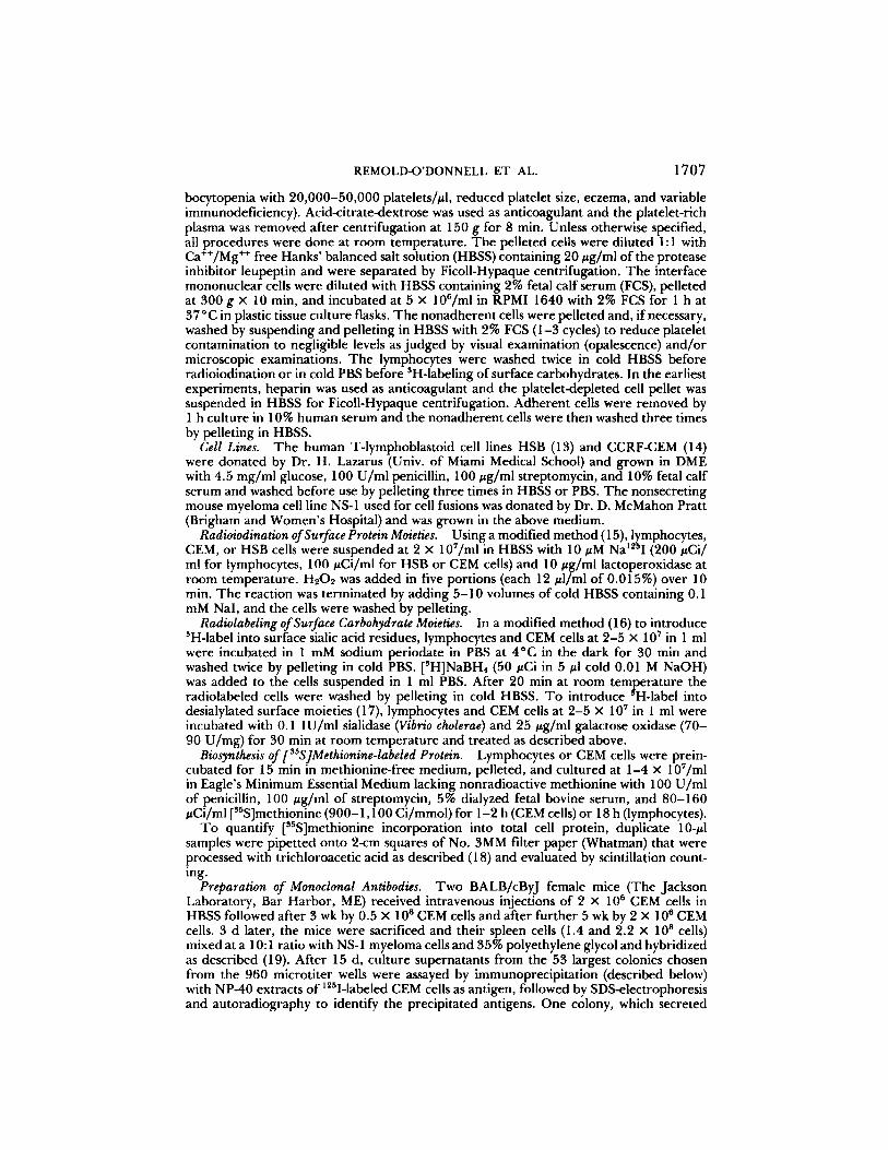
Lentil Lectin Chromatography of gpLll5 from Lymphocytes and Lymphoblastoid Cell Lines. T o characterize its carbohydrate moieties, gpL115 was analyzed by affinity chromatography on columns of lentil lectin-Sepharose. Lentil lectin shows specificity for mannose (26), a core component of asparagine-linked (N- linked) carbohydra te units, l~SI-labeled-gpL115 of normal lymphocytes passed unre ta rded through lentil lectin-Sepharose columns (Fig. 1, left), indicating that gpL115 contains little or no exposed mannose residues and suggesting the absence of N-linked carbohydrate .
T w o long-term lymphoblastoid cell lines HSB (14) and CEM (13) were surface radioiodinated. Both contain ~SI-labeled components that co-migrate on SDS- electrophoresis with gpL 115 of normal peripheral lymphocytes (not shown). The component of apparent mol wt 115,000 in CEM cells is the most prominent l~sI- labeled cell surface protein. On chromatography of extracts o f 125I-labeled CEM cells, the major radiolabeled 115,000 mol wt component was found to be lentil l ec t in-nonadherent (Fig. 1, right) suggesting its identity with gpL 115 of normal lymphocytes.
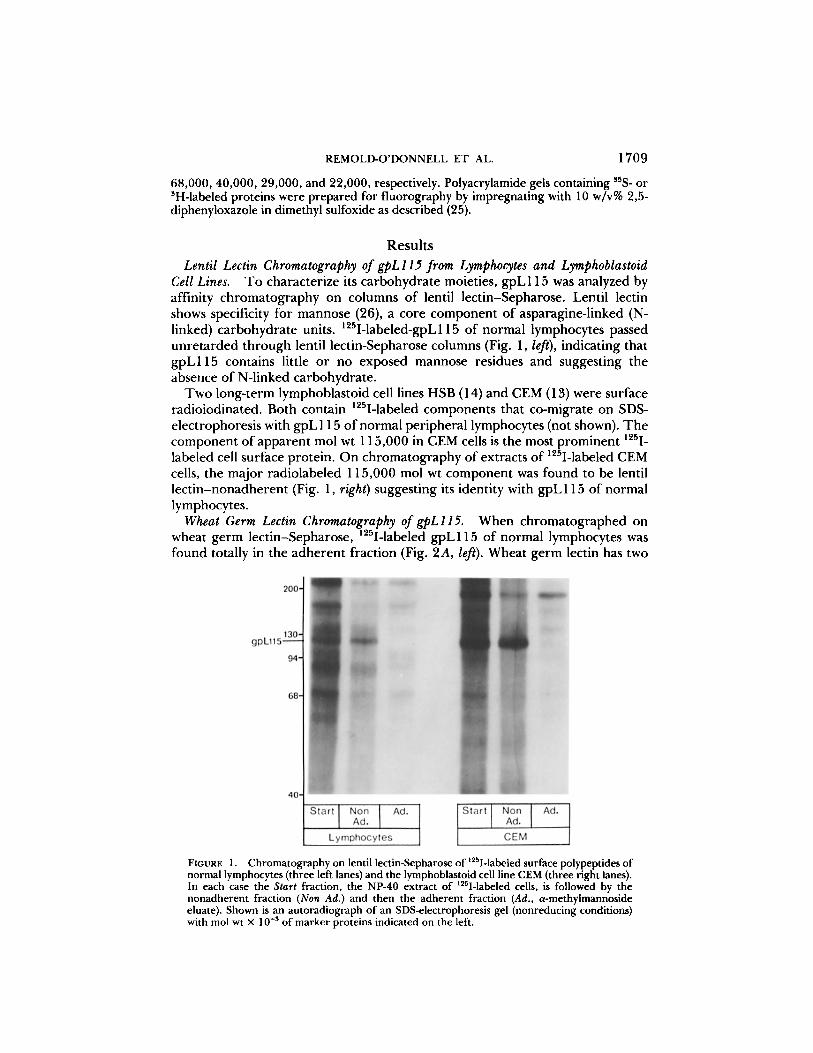
Wheat Germ Lectin Chromatography of gpLll5. When chromatographed on wheat germ iect in-Sepharose, 12~I-labeled gpL115 of normal lymphocytes was found totally in the adherent fraction (Fig. 2A, le~). Wheat germ lectin has two
FIGURE 1. Chromatography on lentil lectin-Sepharose of ~25I-labeled surface polypeptides of normal lymphocytes (three left lanes) and the lymphoblastoid cell line CEM (three right lanes). In each case the Start fraction, the NP-40 extract of 125I-labeled cells, is followed by the nonadherent fraction (Non Ad.) and then the adherent fraction (Ad., a-methylmannoside eluate). Shown is an autoradiograph of an SDS-electrophoresis gel (nonreducing conditions) with tool wt × 10 -3 of marker proteins indicated on the left.

1710 HUMAN LYMPHOCYTE SURFACE SIALOGLYCOPROTEIN
FIGURE 2. (A) Chromatography on wheat germ lectin-Sepharose of ~25I-labeled surface polypeptides of normal lymphocytes without (Native) and with (AsiMo) sialidase treatment of the starting fraction, an NP-40 extract. Shown is an autoradiograph of an SDS-electrophoresis gel (nonreducing conditions). The left three lanes show chromatography of a native extract, with the starting fraction (Start), the nonadherent fraction (Non Ad.), and the adherent fraction (Ad; N-acetylglucosamine eluate) indicated. The three lanes on the right show chromatography of a sialidase-treated extract. Positions of mol wt marker proteins and gpL 115 are indicated on the left. The shift in electrophoretic mobility of gpL115 after sialidase treatment can be noted by comparing Native and Asialo starting fractions. Asialo-gpL115, designated by the arrows on the right, migrates with apparent mol wt 150,000; its identity as a species of gpL 115 was verified by immunoprecipitation (shown below)? Note that native gpL115 from lympho- cytes is wheat germ lectin-adherent and asialo-gpL 115 is nonadherent. (B) Chromatography on wheat germ lectin-Sepharose of l~5I-labeled surface polypeptides of CEM cells. Details and findings are as in A,

REMOLD-O'DONNELL ET AL. 1711
specificities, binding both to N-acetylglucosamine (27, 28) and to clustered sialic acid residues (29, 30). To determine which of these specificities mediate the binding of g p L l l 5 , the starting fraction, an NP-40 extract of 1~I-labeled lymphocytes, was treated with sialidase to remove sialic acid. Sialidase treatment was found, first of all, to reduce the electrophoretic mobility of gpL115 from apparent mol wt 115,000 to apparent mol wt 150,000 (Fig. 2A, comparison of "Native" and "Asialo" starting fractions). This large reduction in electrophoretic mobility suggests that gpL 115 has significant sialic acid content. On chromatog- raphy, asialo-gpL115 was found in the wheat germ lect in-nonadherent fraction (Fig. 2A, right), indicating that native gpL115 adheres to wheat germ lectin via sialic acid residues and does not adhere via N-acetylglucosamine residues.
The native 115,000 mol wt 125I-labeled molecule from CEM cells, like lym- phocyte gpL115, was found to be wheat germ lectin-adherent (Fig. 2B, le~). Sialidase treatment shifted this molecule to apparent mol wt 150,000 and converted it to a wheat germ lect in-nonadherent form (Fig. 2 B, right), strongly suggesting that this CEM surface molecule is identical to gpL 115 of lymphocytes.
Radiolabeling of the Carbohydrate Moieties of gpL115. Surface carbohydrate moieties were labeled by treating lymphocytes with periodate followed by [3H]- NaBH4, which converts terminal 9-carbon sialic acid residues to 3H-labeled 7- carbon 5-acetamido-3,5-dideoxy-L-arabino-2-heptulosonic acid residues (31). 3H- label was found in several lymphocyte surface glycoproteins, including a com- ponent of apparent mol wt 115,000, presumably gpL115 (Fig. 3, le~). Lympho- cytes were also treated with sialidase-galactose oxidase followed by [SH]NaBH4, which yields 3H-labeled asiaio-glycoproteins. The 3H-labeled asialo-glycoproteins of lymphocytes migrate with apparent tool wt _150,000 (Fig. 3). After similar treatments of CEM cells, the major 3H-labeled native surface glycoprotein has apparent mol wt 115,000 and the major ~H-labeled asialo-glycoprotein has apparent mol wt 150,000 (Fig. 3).
Isoelectrofocusing ofgpLll5. On two-dimensional analysis (isoelectrofocusing- SDS electrophoresis) of partially purified fractions, l~SI-labeled g p L l l 5 from CEM cells was found as a broad band or "streak," primarily in the extreme acidic region at pH 4.1 (Fig. 4, top). On the other hand, sialidase-treated gpL115 focuses as a defined spot at pH ~5.0 (Fig. 4, bottom). Identical behavior on isoelectrofocusing was found for native and sialidase-treated 125I-labeled gpL 115 from lymphocytes (not shown). The large difference in the behavior of native and sialidase-treated gpL115 on isoelectrofocusing indicates a prominent role for multiple sialic acid residues in determining the physiochemical characteristics of gpL115 (see Discussion).
Peanut Lectin Chromatography ofgpL115. Peanut lectin shows specificity for the disaccharide Gal/31-3GalNAc (32), a sequence found in mucin-type O-linked saccharides (33). When extracts of l~SI-labeled lymphocytes were chromato- graphed on peanut lectin-Sepharose, gpL115 was found to be peanut lectin- nonadherent (Fig. 5A, left). Sialidase-treated gpL115 from lymphocytes, how- ever, was found to be peanut lectin-adherent (Fig. 5A, right). 125I-labeled native gpL 115 from CEM cells was also found to be peanut lectin-nonadherent, and the sialidase-treated form to be peanut lectin-adherent (Fig. 5 B), which provides further evidence that the gpL115 molecules from CEM cells and lymphocytes

1712 HUMAN LYMPHOCYTE SURFACE SIALOGLYCOPROTEIN
FIGURE 3. SH-labeled surface glycopeptides of normal peripheral lymphocytes (two left lanes) and CEM cells (two right lanes). Intact cells were reacted with [~H]NaBH4 after periodate treatment to generate reactive carbonyl groups in terminal sialic acid residues (Native) or after treatment with sialidase and galactose oxidase (Asialo). Shown is a fluorograph of NP-40 cell extracts fractionated by SDS-electrophoresis under nonreducing conditions with mol wt (×10 -s) indicated on the right. Arrow on the left indicates the position of native lymphocyte gpL 115 and the major co-migrating SH-labeled native glycopeptide of CEM. Arrow on the right indicates the position of the major CEM SH-labeled asialo-glycopeptide which migrates with apparent mol wt 150,000.
are identical. T h e peanut lectin chromatography findings indicate that gpL1 15 contains the sequence sialic acid-Gall31-3GalNAc, a sequence that is characteristic for O-linked, mucin-type saccharide residues.
Generation of Anti-gpLl l5 Monoclonal Antibody. Hybr idoma technique was used to genera te a cell line secreting monoclonal ant ibody to gpL 115. T h e cell line, L 10, was genera ted using CEM cells as the immunizing agent. L 10 ant ibody is an IgG1 molecule that immunoprecipi ta tes lzSI-labeled native and sialidase- t reated gpL1 15 (Fig. 6). Both gpL1 15 f rom lymphocytes and CEM cells are immunoprecipi ta ted by L 10 ant ibody (Fig. 6), fu r the r demonstra t ing their iden- tity.
Synthesis of gpL115 by Lymphocytes. Since detect ion o f gpL 1 15 relied exclu- sively on surface labeling techniques, we sought to de termine whether gpL 1 15 is a biosynthesis p roduc t o f lymphocytes and CEM cells or an acquired surface component . Lymphocytes f rom five normal donors cul tured with [35S]methionine at 2 - 4 × 10⁷/ml were found to incorporate 21% + 3% of added radioactivity into tr ichloroacetic acid-precipi table protein in 18 h. CEM cells, in contrast,

REMOLD-O'DONNELL ET AL. 1713
FIGURE 4. Two-dimensional analysis of native and sialidase-treated1251-1abeled gpL l15 from CEM cells. Shown is an autoradiograph of two-dimensional gels with isoelectrofocusing on the horizontal axis (pH gradient is indicated) and SDS-electrophoresis under reducing conditions on the vertical axis (tool wt marker positions indicated on the right). NP-40 extracts of 1~sI- labeled CEM cells were incubated without (Native) or with (Asialo) sialidase. The former fraction was then purified by wheat germ iectin chromatography and the latter by peanut lectin chromatography. Note that sialidase treatment shifts gpL 115 to an isoelectric point that
1 ',n3 T is less acidic and more sharply defined. Identical results were obtained when I-labeled gpL1 15 was first purified (by lentil lectin and wheat germ lectin chromatography) and then incubated without (Native) or with (Asialo) sialidase (not shown).
incorpora ted > 6 0 % within 2 h. On immunoprec ip i ta t ion , L10 ant ibody precip- itates a [SSS]methionine-labeled c o m p o n e n t o f appa ren t mol wt 115,000 f rom lymphocytes and CEM cells that was not found in immunoprec ip i ta tes o f two control monoclonal IgG1 ant ibodies (Fig. 7). T h e [SSS]methionine-labeled com- ponen t precipi ta ted by L10 ant ibody co-migrates with 125I-gpL115 unde r reduc- ing and nonreduc ing conditions (not shown), thus establishing that lymphocytes and CEM cells synthesize gpL 115.
Defect in gpLI l5 in Lymphocytes of Wiskott-Aldrich Syndrome Patients Lymphocytes o f patients with the Wiskott-Aldrich syndrome and normal indi-
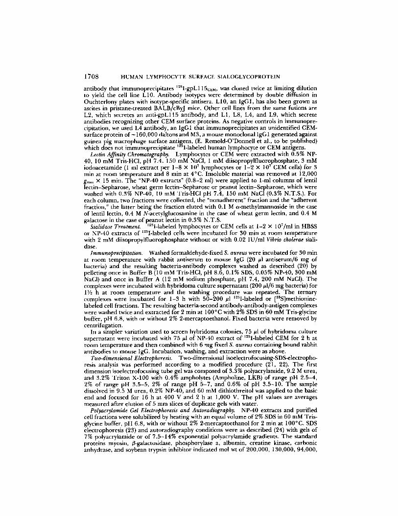
viduals were radioiodinated and their surface componen t s examined by SDS electrophoresis and au torad iography . In all, lymphocytes f rom 8 patients were

1714 HUMAN LYMPHOCYTE SURFACE SIALOGLYCOPROTEIN
FIGURE 5. (A and B) Chromatography on peanut lectin-Sepharose of l~5I-labeled surface polypeptides of normal lymphocytes (A) and CEM cells (B) without (Native, three left lanes) or with (Asialo, three right lanes) sialidase treatment. Details as in Fig. 2, A and B. Note that native gpL 115 of lymphocytes and CEM cells (arrow on the left) is peanut lectin-nonadherent and that asialo-gpL115 (arrows on the right) is the major 1~SI-labeled peanut lectin-adherent protein.
e x a m i n e d in 12 e x p e r i m e n t s , lzSI-labeled gpL1 15 was f o u n d to be def ic ien t in l y m p h o c y t e s o f all pat ients . H o w e v e r , the levels o f 125I-gpLl15 var ied , be ing n o n d e t e c t a b l e in pa t i en t 1 (Fig. 1 o f r e f e r e n c e 6) pa t i en t s 2, 3, a n d 5 (Fig. 8) a n d de tec t ab le , bu t d e c r e a s e d re la t ive to levels in n o r m a l lymphocy tes , in pa t ien ts 4, 6, 7, 8 (Fig. 8).

REMOLD-O'DONNELL ET AL. 1715
FIGURE 6. Immunoprecipitation of native and sialidase-treated t25I-labeled gpL 115 by L 10 monoclonal antibody. NP40 extracts of normal lymphocytes and CEM cells were incubated without (Native) or with (Asialo) sialidase and immunoprecipitated with hybridoma culture supernatant of L l0 cells (lanes 1, 3, 5, 7) or M3 cells (lanes 2, 4, 6, 8). Shown is an autoradiograph of an SDS electrophoresis gel (reducing conditions) with tool wt X 10 -~ of marker proteins indicated on the left.
FIGURE 7. Immunoprecipitation of gpL115 labeled biosynthetically with [sSS]methionine. Normal lymphocytes and CEM ceils were cultured with [sSS]methionine. gpLl 15-containing fractions were prepurified by NP-40 extraction and wheat germ lectin chromatography and immunoprecipitated with L10, (lanes I and 4) or L4 (lanes 2 and 5) or M3 (lanes 3 and 6). Shown is a fluorograph of an SDS electrophoresis gel (reducing conditions) with the position indicated of m~sI-labeled gpL 115 which was electrophoresed in parallel.
FIG
UR
E 8
. ~5
I-la
bele
d su
rfac
e pr
otei
ns o
f ly
mph
ocyt
es o
f si
x no
rmal
ind
i-
vidu
als
(N2-
NT)
and
eig
ht W
isko
tt-A
ldri
ch s
yndr
ome
pati
ents
(P
1-P
8). S
how
n is
a c
ompo
site
of
auto
radi
ogra
phs
of S
DS
ele
ctro
phor
etic
sep
arat
ions
of
NP
- 40
ext
~ct
s of
a~I
-lab
eled
lym
phoc
ytes
fro
m s
ix e
xper
imen
ts.
Ele
ctro
phor
etic
se
para
uons
wer
e of
non
redu
ced
extr
acts
in
four
exp
erim
ents
(ei
ght
lane
s st
arti
ng f
rom
the
lef
t) a
nd r
educ
ed e
xtra
cts
in t
wo
expe
rim
ents
(si
x la
nes
on
the
righ
t).
Wit
hin
each
of
the
six
expe
rim
ents
, no
rmal
and
pa
tien
t w
ere
mat
ched
for
cel
l nu
mbe
r,
radi
oiod
inat
ion
cond
itio
ns,
elec
trop
hore
sis,
and
auto
radi
ogra
phy
cond
itio
ns.
Arr
ows
on t
he l
eft i
ndic
ate
gpL
115
. The
pat
ient
s in
dica
ted
by P
1, P
2, a
nd P
3 ha
ve b
een
prev
ious
ly d
escr
ibed
(6)
. PI,
T in
dica
tes
pati
ent
1 ex
amin
ed 2
6 m
o af
ter
bone
mar
row
tra
nspl
ant;
pri
or t
o tr
ansp
lant
hi
s ly
mph
ocyt
es h
ad n
onde
tect
able
lev
els
of ~
I-la
bele
d kr
oL11
5 (6
) N
ote
12
5
. v
~
. .
."
that
I-
gpL
11.
5 le
vels
are
red
uced
or
nond
etec
tabl
e m
all
pau
ents
. N
ote
that
so
me
pati
ents
(P
3, P
6, a
nd p
ossi
bly
P2)
hav
e an
l~S
I-la
bele
d co
mpo
nent
of
elec
trop
hore
tic
mob
ilit
y sl
ower
tha
n gp
L11
5 (i
ndic
ated
by
dots
on
the
left
) w
hich
is
not
foun
d in
nor
mal
ind
ivid
uals
.
z O O © .<
c~
© O
REMOLD-O'DONNELL ET AL. 1717
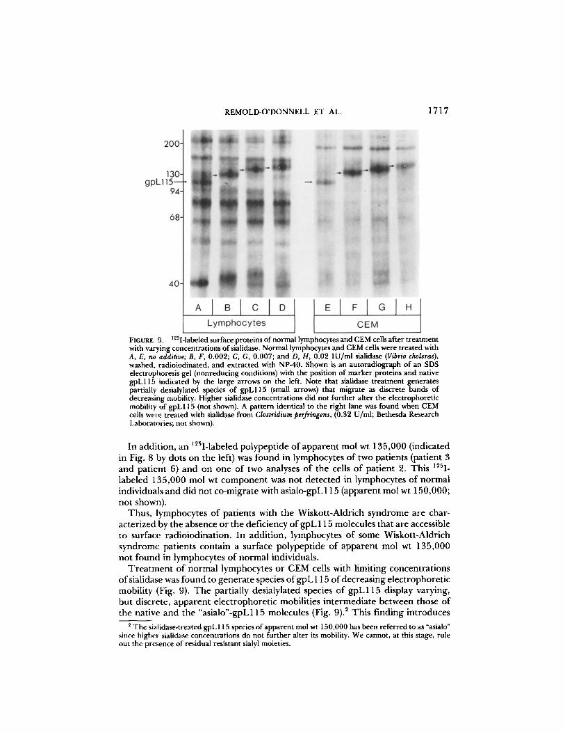
FIGURE 9. ~251-1abeledsurfaceproteinsofnormallymphocytesandCEMcellsaftertreatment with varying concentrations of sialidase. Normal lymphocytes and CEM cells were treated with A, E, no additive; B, F, 0.002; C, G, 0.007; and D, H, 0.02 IU/ml sialidase (Vibrio cholerae), washed, radioiodinated, and extracted with NP-40. Shown is an autoradiograph of an SDS electropboresis gel (nonreducing conditions) with the position of marker proteins and native gpL115 indicated by the large arrows on the left. Note that sialidase treatment generates partially desialylated species of gpL115 (small arrows) that migrate as discrete bands of decreasing mobility. Higher sialidase concentrations did not further alter the electrophoretic mobility of gpL115 (not shown). A pattern identical to the right lane was found when CEM cells were treated with sialidase from Clostridium perfringens, (0.32 U/ml; Bethesda Research Laboratories; not shown).
In addition, an ~25I-labeled polypept ide o f apparent mol wt 135,000 (indicated in Fig. 8 by dots on the left) was found in lymphocytes o f two patients (patient 3 and patient 6) and on one of two analyses o f the cells of patient 2. This 125I- labeled 135,000 mol wt componen t was not detected in lymphocytes of normal individuals and did not co-migrate with asialo-gpL 115 (apparent mol wt 150,000; not shown).
Thus , lymphocytes o f patients with the Wiskott-Aldrich syndrome are char- acterized by the absence or the deficiency o f gpL 115 molecules that are accessible to surface radioiodination. In addition, lymphocytes o f some Wiskott-Aldrich syndrome patients contain a surface polypeptide of apparent mol wt 135,000 not found in lymphocytes of normal individuals.
T r e a t m e n t o f normal lymphocytes or CEM cells with limiting concentrat ions o f sialidase was found to genera te species o f g p L 115 o f decreasing electrophoret ic mobility (Fig. 9). T h e partially desialylated species o f gpL115 display varying, but discrete, apparent e lect rophoret ic mobilities in termediate between those of the native and the "asialo"-gpL115 molecules (Fig. 9). 2 This finding introduces
2 The sialidase-treated gpL 115 species of apparent mol wt 150,000 has been referred to as "asialo" since higher sialidase concentrations do not further alter its mobility. We cannot, at this stage, rule out the presence of residual resistant sialyl moieties.

1718 HUMAN LYMPHOCYTE SURFACE SIALOGLYCOPROTEIN
the possibility that the abnormal 135,000 mol wt component found in lympho- cytes of some Wiskott-Aidrich syndrome patients is a partially desialylated var- iant(s) of gpL 115.
Discussion gpL115, the lymphocyte surface glycoprotein that is deficient in Wiskott-
Aldrich syndrome, is a sialoglycoprotein that can be radiolabeled in protein moieties by lzSI-lactoperoxidase and in carbohydrate moieties by [SH]NaBH4. gpL 115 can be labeled by culturing lymphocytes with [SSS]methionine, indicating that it is synthesized by lymphocytes.
Comparison of radioiodinated surface proteins of lymphocytes and the lym- phoblastoid cell line CEM by electrophoresis, isoelectrofocusing, sialidase treat- ment, lectin affinity chromatography, and immunoprecipitation established that gpL115 is also present on the surface of the CEM cells, a finding used to advantage in the generation of a monoclonal antibody to gpL115. The designa- tion of CEM cells as a suitable source establishes the opportunity for isolation and chemical analysis of gpL 115. Through the use of surface marker antigens, CEM has been shown to be a cell line of early thymocyte origin (34), thus implying that thymocytes, which were not examined in this study, also express surface gpLl 15. 3
gpL115 was identified through its deficiency in Wiskott-Aldrich syndrome patients (6) and has also been found defective in lymphocytes of two non-Wiskott immunodeficient patients. 4 ~25I-labeled gpL115 was not immunoprecipitated by monocional antibodies directed against the surface antigen CALLA (references 35, 36; common acute lymphoblastic leukemia antigen; antibodies supplied by Dr. J. Ritz, Dana Farber Cancer Institute), thus establishing the nonidentity of gpL 115 and CALLA. Likewise, gpL 115 was not immunoprecipitated by mono- clonal anti-T12 antibody (reference 37; supplied by Dr. S. Schlossman, Dana Farber Cancer Institute), demonstrating the nonidentity of the surface antigens T12 and gpL115.
The quantity of sialic acid on gpL115 is not known, but appears to be large since desialylation dramatically alters the physical behavior of the molecule. On SDS electrophoresis, desialylation shifts gpL115 from a mobility corresponding to tool wt 115,000 to a mobility corresponding to mol wt 150,000. Good correlation of mol wt with electrophoretic mobility in the presence of SDS has been established for a wide range of polypeptides (38) and the behavior of gpL 115 is, thus, atypical. Anomalous behavior on SDS electrophoresis is a feature of glycoproteins with high content of carbohydrate and is caused by the inability of carbohydrate to bind SDS (39, 40). 5
On isoelectrofocusing, sialidase treatment shifts gpL115 from a very broad band or "streak," primarily in the extreme acidic region (pH 4.1) to a well- defined "spot" at pH 5. We do not know why the native molecule is found as a broad streak. Heterogeneity at the level of sialylation is a possibility, but would not explain the finding that the streak of native gpL115 extends to pH regions
Using fluorescent techniques, LI0 anti-gpL115 monoclonal antibody stains 99% of normal lymphocytes and thymocytes (R. Parkman et al., to be published).
4 R. Parkman et al., manuscript in preparation. 5 We have at present no estimate of the true molecular weight of gpL 115.

REMOLD-O'DONNELL ET AL. 1719
less acidic than the spot of sialidase-treated gpL115 (observed in five of five experiments). We favor the explanation that native gpL115 is incompletely focused, i.e., that the rate of focusing of native gpL 115 is atypically slow due to its elongated conformation enforced by its high sialic acid content.
Lymphocytes of patients with the Wiskott-Aldrich syndrome are characterized by a defect in gpL115. 12SI-labeled gpL115 was deficient in eight patients; the extent of the deficiency varied from nondetectable levels (four patients) to levels that are decreased relative to normal individuals (four patients). In addition, an l~I-labeled surface protein of apparent mol wt 135,000, which was not found in normal individuals, was detected in three of eight Wiskott-Aldrich patients. We speculate that this abnormal component is a variant of gpL115. 6 g p L l l 5 of normal individuals can be converted to apparent mol wt 135,000 by treating intact lymphocytes with limiting sialidase concentrations (Fig. 9).
The thesis that a single defective gene in Wiskott-Aldrich syndrome codes for both the defective lymphocyte and platelet surface components gpL115 and GPIb is unlikely since the molecules are not identical, i.e., they do not co-migrate on electrophoresis (not shown). We also did not detect partial identity, in that GPIb (l~SI-labeled and [3H]NaBH4-1abeled) was not immunoprecipitated by L 10 anti-gpL115 monoclonal antibody and 125I-gpL115 was not immunoprecipitated by a polyclonal antibody to GPIb/glycocalicin (not shown; rabbit antisera supplied by Dr. G. A. Jamieson, The American National Red Cross, Bethesda, MD).
A more likely hypothesis is that the concomitant defect of gpL 115 and GPIb in Wiskott-Aldrich syndrome results from a single defective processing element, possibly a synthesis enzyme such as a glycosyi transferase, or a degradation enzyme. The current study indicates that gpL115, like GPIb, is one of a small group of glycoproteins with high content of O-linked carbohydrate units (see below); and it is, thus, likely that they share some biosynthesis and/or degradation steps.
Glycoprotein carbohydrate moieties are of two types. The N-linked "complex" and "high mannose" units consist typically of 8-14 monosaccharide residues (sialic acid, galactose, fucose, mannose, and N-acetylglucosamine) linked to as- paragine via N-glycosidic bonds, gpL 115 is lentil lectin-nonadherent, suggesting the absence of N-linked carbohydrate. The second type, the "O-linked" units, also called "mucin-type" or "acidic type" because of the high sialic acid content, are linked to serine or threonine via O-glycosidic bonds. These units are generally small, consisting of three, four, or six residues. For example, in glycophorin O- linked units consist of galactose, N-acetylgalactosamine, and two terminal sialic acid residues (41) and in platelet GPIb, they also contain N-acetylglucosamine (42). Typically, mannose is not found.
A few glycoproteins consist of >50% O-linked carbohydrate; these include erythrocyte glycophorin (43), platelet GPIb (7), "masking" glycoproteins on certain tumor cells (44, 45), and LSGP (leukocyte sialoglycoprotein; 9), a rat T lymphocyte and thymocyte surface component. LSGP may be the counterpart in the rat of gpL115. LSGP displays the same unusual behavior as gpL115 on lentil lectin (46) and peanut lectin chromatography (10); its asialo-form is the
6 When lymphocytes from a suitable patient become available, this hypothesis will be tested by immunoprecipitation with L 10 antibody.
1720 HUMAN LYMPHOCYTE SURFACE SIALOGLYCOPROTEIN
major peanut iectin-adherent surface glycoprotein of rat thymocytes and T lymphocytes (10).
gpL115 adheres to wheat germ lectin, but asialo-gpL115 does not, indicating that wheat germ lectin-adherence is due to clustered sialic acid residues on the native molecule. Clustered sialic acid is a feature of glycoproteins with high content of O-linked sugar, gpL115 does not adhere to peanut lectin, but asialo- gpL 115 does - -a very important finding since peanut lectin shows specificity for the disaccharide galactose/3(1-3)-N-acetylgalactosamine. These findings indicate that native gpL 115 contains the sequence sialic acid-galactose/3(1-3)-N-acetylga- lactosamine, a sequence particular to O-linked, mucin-type carbohydrate units.
This study establishes similarity of the carbohydrate portions of the lymphocyte and platelet components altered in Wiskott-Aldrich syndrome. Like gpL115, GPIb and its soluble form glycocalicin (8) adhere to wheat germ lectin (7) and asialo-GPIb adheres to peanut lectin (8). GPIb (glycocalicin) consists to 60% of carbohydrate that appears to be exclusively mucin-type units linked to serine and threonine (7, 42).
Due to sialic acid content, glycoproteins with high O-linked carbohydrate content are thought to adopt extended conformation and to confer negative charge characteristics required in cell-cell interactions (9). In certain mouse adenocarcinomas a surface sialoglycoprotein with >50% O-linked carbohydrate, referred to as a "masking" glycoprotein, is thought to confer on the cells the capacity to invade allogeneic and xenogeneic hosts (44, 45). Similarly, sialidase treatment of mouse lymphoma cells exposes cryptic tumor antigens on glycolipid molecules that are, themselves, devoid of sialic acid (47). Finally, removal of sialic acid from lymphocytes has been shown to induce their loss from the circulation (48).
The cumulative findings suggest that the defect(s) in the sialoglycoprotein gpL 115 in the Wiskott-Aldrich syndrome adversely affect(s) the ability of the lymphocytes to interact with other cells and/or to survive in circulation.
S u m m a r y gpL1 15 is a lymphocyte surface component that is deficient in patients with
the X-chromosome-linked immune deficiency Wiskott-Aldrich syndrome (6). The glycoprotein nature of gpL 1 15 is demonstrated through labeling in carbo- hydrate moieties by [SH]NaBH4 and its synthesis by lymphocytes through labeling with [35S]methionine. Native gpL1 15 adheres to wheat germ lectin-Sepharose and sialidase-treated gpL1 15 does not adhere, indicating that native gpL1 15 adheres via clusters of sialic acid residues. When tested on peanut lectin, which shows specificity for the disaccharide Galt31-3GalNAc, gpL1 15 is nonadherent and sialidase-treated gpL 1 15 is adherent, indicating the presence of the sequence sialic acid-Gal~l-3GalNAc, which is characteristic for O-linked (mucin-type, acidic-type) carbohydrates. A surface glycoprotein with all the above character- istics was found on the lymphoblastoid cell line CEM. CEM cells were used as immunogen to generate the monoclonal antibody L10, an IgG~, which binds native and sialidase-treated gpL 1 15. Sialidase-treatment of ~ L 115 signifiCantly alters its physical properties, reducing its electropboretic mobility and changing its behavior on isoelectrofocusing. Cumulatively, these findings indicate that gpL 1 15, like glycophorin of erythrocytes and GPIb of platelets, is a sialoglyco-

REMOLD-O'DONNELL ET AL. 1721
protein with significant quantities of O-linked carbohydrate. On treatment with limiting sialidase concentrations, gpL 115 of normal lym-
phocytes is transformed into a series of partially desialylated species of decreasing electrophoret ic mobility. This finding resembles the situation with lymphocytes of some Wiskott-Aldrich syndrome patients. Lymphocytes o f eight Wiskott- Aldrich syndrome patients were found to be deficient in l~I-labeled gpL115. Lymphocytes f rom three of these patients displayed an abnormal ~25I-component of apparent mol wt 135,000.
We gratefully acknowledge the vital contributions of the patients and their families, the physicians, nurses, and phlebotomists and our co-workers who served as blood donors. We also thank Drs. G. A. Jamieson, Herbert Lazarus, David Liu, Diane McMahon Pratt, Jerome Ritz, and Stuart Schlossman for material and/or advice, Ms. Mary Lou Koehn and Ms. Anandi Mehta for skillful assistance, and Ms. Rachelle Rosenbaum for preparation of the manuscript.
Received for publication 24 January 1984.
Note added in proof'. After submission of the manuscript, we became aware of a report describing F10-44-2, an LSGP-like glycoprotein of tool wt 105,000 on human blood mononuclear cells (Dalchau, R., J. Kirkley, and J. w. Fabre. 1980. Monoclonal antibody to a human brain-granulocyte-T lymphocyte antigen probably homologous to the W 3/ 13 antigen of the rat. Eur. J. Immunol. 10:745). This molecule may be identical to gpL 115.
References 1. Remold-O'Donnell, E., R. Parkman, D. M. Kenney, and F. S. Rosen. 1983. Properties
of gpL115, a surface glycoprotein lacking in lymphocytes of Wiskott-Aldrich syn- drome patients. Fed. Proc. 42:1335.
2. Wiskott, A. 1937. Famili~irer, eingeborener Morhus Werlhoff. Monatsschr. Kinder- heilkd. 68:212.
3. Cooper, M. D., H. P. Chase, J. T. Lowman, W. Krivit, and R. A. Good. 1968. Wiskott-Aldrich syndrome: an immunologic deficiency disease involving the afferent limb of immunity. Am. J. Med. 44:499.
4. Parkman, R., J. M. Rappeport, R. Geha, J. Belli, R. Cassady, R. Levey, D. G. Nathan, and F. S. Rosen. 1978. Complete correction of the Wiskott-Aldrich syndrome by allogeneic bone marrow transplantation. N. Engl. J. Med. 298:921.
5. Lum, L. G., D. G. Tubergen, L. Corash, and R. M. Blaese. 1980. Splenectomy in the management of the thrombocytopenia of the Wiskott-Aidrich syndrome. N. Engl. J. Med. 302:892.
6. Parkman, R., E. Remold-O'Donnell, D. M. Kenney, S. Perrine, and F. S. Rosen. 1981. Surface protein abnormalities in lymphocytes and platelets from patients with Wiskott-AIdrich syndrome. Lancet. ii: 1387.
7. Okumura, T., C. Lombart, and G. A. Jamieson. 1976. Platelet glycocalicin. II. Purification and characterization. J. Biol. Chem. 251:5950.
8. Ciemetson, K. J., H. J. Naim, and E. F. Liischer. 1981. Relationship between glycocalicin and glycoprotein Ib of human platelets. Proc. Natl. Acad. Sci. USA. 78:2712.
9. Brown, W. R. A., A. N. Barclay, C. A. Sunderland, and A. F. Williams. 1981. Identification of a glycophorin-like molecule at the cell surface of rat thymocytes. Nature (Lond.). 289:456.
10. Brown, W. R. A., and A. F. Williams. 1982. Lymphocyte cell surface glycoproteins which bind to soybean and peanut lectins. Immunology. 46:713.
1722 HUMAN LYMPHOCYTE SURFACE SIALOGLYCOPROTEIN
11. Hayman, M. J., and M. J. Crumpton. 1972. Isolation of glycoproteins from pig lymphocyte plasma membrane using Lens culinaris phytohemagglutinin. Biochem. Biophys. Res. Commun. 47:923.
12. Cuatrecasas, P. 1970. Protein purification by affinity chromatography. J. Biol. Chem. 245:3059.
13. Foley, G. E., H. Lazarus, S. Farber, B. G. Uzman, B. A. Boone, and R. E. McCarthy. 1965. Continuous culture of human lymphoblasts from peripheral blood of a child with acute leukemia. Cancer 18:522.
14. Lazarus, H., E. F. Barell, S. Oppenheim, and A. Krishan. 1974. Divergent properties of two human lymphocytic cell lines isolated from a single specimen of peripheral blood. In Vitro. 9:303.
15. Morrison, M. 1974. The determination of exposed proteins on membranes by the use of lactoperoxidase. Methods Enzymol. 32:103.
16. Gahmberg, C. G., and L. C. Andersson. 1977. Selective radioactive labeling of cell surface sialoglycoproteins by periodate-tritiated borohydride.J. Biol. Chem. 252:5888.
17. Andersson, L. C., and C. G. Gahmberg. 1978. Surface glycoproteins of human white blood cells. Analysis by surface labelling. Blood. 52:57.
18. Roberts, B. E., and B. M. Paterson. 1973. Efficient translation of tobacco mosaic virus RNA and rabbit globin 9S RNA in a cell-free system from commercial wheat germ. Proc Natl. Acad. Sci. USA. 70:2330.
19. Kennett, R. H., K. A. Denis, A. S. Tung, and N. R. Klinman. 1978. Hybrid plasmacytoma production: fusions with adult spleen cells, monoclonal spleen frag- ments, neonatal spleen cells and human spleen cells. In Lymphocyte Hybridomas. F. Melchers, M. Potter, and N. Warner, editors. Springer-Verlag, Berlin. p. 77.
20. Kaplan, G., J. C. Unkeless, and Z. A. Cohn. 1979. Insertion and turnover of macrophage plasma membrane protein. Proc. Natl. Acad. Sci. USA. 76_'3824.
21. Linck, R. W., D. F. AIbertini, D. M. Kenney, and G. L. Langevin. 1982. Tektin filaments: chemically unique filaments of sperm flagellar microtubules. Cell Motility (Suppl.). 1:127.
22. O'Farrell, P. H. 1975. High resolution two-dimensional electrophoresis of proteins. J. Biol. Chem. 250:4007.
23. Laemmli, U. K. 1970. Cleavage of structural proteins during the assembly of the head of bacteriophage T4. Nature (Lond.) 227:680.
24. Heck, L. W., E. Remold-O'Donnell, and H. G. Remold. 1978. DFP-sensitive poly- peptides of the guinea pig peritoneal macrophage. Biochem. Biophys. Res. Commun. 83:1576.
25. Bonner, W. M., and R. A. Laskey. 1974. A film detection method for tritium-labelled proteins and nucleic acids in polyacrylamide gels. Eur. J. Biochem. 46:83.
26. Young, N. M., M. A. Leon, and T. Takahashi. 1971. Studies on a phytohemagglutinin from the lentil. III. Reaction of lens culinaris hemagglutinin with polysaccharides, glycoproteins, and lymphocytes. J. Biol. Chem. 246:1596.
27. Allen, A. K., A. Neuberger, and N. Sharon. 1973. The purification, composition and specificity of wheat-germ agglutinin. Biochem. J. 131 : 155.
28. Nagata, Y., and M. M. Burger. 1974. Wheat germ agglutinin: molecular character- istics and specificity for sugar binding.J. Biol. Chem. 249:3116.
29. Bhavanandan, V. P., and A. W. Katlic. 1979. The interaction of wheat germ agglutinin with sialoglycoproteins. The role of sialic acid. J. Biol. Chem. 254:4000.
30. Peters, B. P., S. Ebisu, I.J. Goldstein, and M. Flashner. 1979. Interaction of wheat germ agglutinin with sialic acid. Biochemistry. 18:5505.
31. Lenten, L. V., and G. Ashwell. 1971. Studies on the chemical and enzymatic modification of glycoproteins. J. Biol. Chem. 246:1889.
32. Lotan, R., E. Skutelsky, D. Danon, and N. Sharon. 1975. The purification, compo-
REMOLD-O'DONNELL ET AL. 1723
sition and specificity of the anti-T lectin from peanut (Arachis hypogaea). J. Biol. Chem. 250:8518.
33. Kornfeld, R., and S. Kornfeld. 1980. Structure of glycoproteins and their oligosac- charide units. In The Biochemistry of Glycoproteins and Proteoglycans. W. J. Lennarz, editor. Plenum Press, New York. p. 1.
34. Reinherz, E. L., P. C. Kung, G. Goldstein, R. H. Levey, and S. F. Schlossman. 1980. Discrete stages of human intrathymic differentiation: analysis of normal thymocytes and leukemic lymphoblasts of T-cell lineage. Proc. Natl. Acad. Sci. USA. 77:1588.
35. Pesando, J. M., J. Ritz, H. Levine, C. Terhorst, H. Lazarus, and S. F. Schlossman. 1980. Human leukemia-associated antigen: relation to a family of surface glycopro- teins. J. Immunol. 124:2794.
36. Ritz, J., J. M. Pesando, J. Notis-McConarty, H. Lazarus, and S. F. Schlossman. 1980. A monoclonal antibody to human acute lymphoblastic leukaemia antigen. Nature (Lond.). 283:583.
37. Reinherz, E. L., R. Geha, J. M. Rappeport, M. Wilson, A. C. Penta, R. E. Hussey, K. A. Fitzgerald, J. F. Daley, H. Levine, F. S. Rosen, and S. F. Schlossman. 1982. Reconstitution after transplantation with T-lymphocyte-depleted HLA haplotype- mismatched bone marrow for severe combined immunodeficiency. Proc. Natl. Acad. Sci. USA. 79:6047.
38. Weber, K., and M. Osborn. 1969. The reliability of molecular weight determinations by dodecyi sulfate-polyacrylamide gel electrophoresis.J. Biol. Chem. 244:4406.
39. Bretscher, M. S. 1971. Major human erythrocyte glycoprotein spans the cell mem- brane. Nature New Biol. 231:229.
40. Segrest, J. P., R. L. Jackson, E. P. Andrews, and V. T. Marchesi. 1971. Human erythrocyte membrane glycoprotein: a re-evaluation of the molecular weight as determined by SDS polyacrylamide gel electrophoresis. Biochem. Biophys. Res. Com- mun. 44:390.
41. Thomas, D. B., and R. J. Winzler. 1969. Structural studies on human erythrocyte glycoproteins. Alkali-labile oligosaccharides.J. Biol. Chem. 244:5943.
42. Tsuji, T., S. Tsunehisa, Y. Watanabe, K. Yamamoto, H. Tohyama, and T. Osawa. 1983. The carbohydrate moiety of human platelet glycocalicin. The structure of the major Ser/Thr-linked sugar chain.J. Biol. Chem. 258:6335.
43. Furthmayr, H., M. Tomita, and V. T. Marchesi. 1975. Fractionation of the major sialoglycopeptides of the human red blood cell membrane. Biochem. Biophys. Res. Commun. 65:113.
44. Codington, J. F., B. H. Sanford, and R. W. Jeanloz. Glycoprotein coat of the TA3 cell. Isolation and partial characterization of a sialic acid containing glycoprotein fraction. Biochemistry. 11:2559.
45. Sherblom, A. P., R. L. Buck, and K. L. Carraway. 1980. Purification of the major sialoglycoproteins of 13762 MAT-BI and MAT-C 1 rat ascites mammary adenocar- cinoma cells by density gradient centrifugation in cesium chloride and guanidine hydrochloride.J. Biol. Chem. 255:783.
46. Standring, R., W. R. McMaster, C. A. Sunderland, and A. F. Williams. 1978. The predominant heavily glycosylated glycoproteins at the surface of rat lymphoid cells are differentiation antigens. Eur. J. Immunol. 8:832.
47. Urdal, D. L., and S. Hakomori. 1983. Characterization of tumor-associated ganglio- N-triaosylceramide in mouse lymphoma and the dependency of its exposure and antigenicity on the sialosyl residues of a second glycoconjugate. J. Biol. Chem. 258:6869.
48. Kolb, H., A. Kriese, V. Kolb-Bachofen, and H.-A. Kolb. 1978. Possible mechanism of entrapment of neuraminidase-treated lymphocytes in the liver. Cell. Immunol. 40:457.