characterization of a new antifungal chitin-binding peptide from
TRANSCRIPT

Plant Physiol. (1997) 113: 83-91
Characterization of a New Antifungal Chitin-Binding Peptide from Sugar Beet Leaves
Klaus K. Nielsen*, john E. Nielsen, Susan M. Madrid, and jorn D. Mikkelsen
Danisco Biotechnology, Langebrogade 1 , P.O. Box 17, DK 1 O01 Copenhagen K, Denmark
The intercellular washing fluid (IWF) from leaves of sugar beet (Befa vulgaris L.) contains a number of proteins exhibiting in vitro antifungal activity against the devastating leaf pathogen Cercospora beticola (Sacc.). Among these, a potent antifungal peptide, desig- nated IWF4, was identified. The 30-amino-acid residue sequence of IWF4 is rich in cysteines (6) and glycines (7) and has a highly basic isoelectric point. IWF4 shows homology to the chitin-binding (hev- ein) domain of chitin-binding proteins, e.g. class I and IV chitinases. Accordingly, IWF4 has a strong affinity to chitin. Notably, it binds chitin more strongly than the chitin-binding chitinases. A full-length IWF4 cDNA clone was obtained that codes for a preproprotein of 76 amino acids containing an N-terminal putative signal peptide of 21 residues, followed by the mature IWF4 peptide of 30 residues, and an acidic C-terminal extension of 25 residues. IWF4 mRNA is expressed in the aerial parts of the plant only, with a constitutive expression in young and mature leaves and in young flowers. No induced expression of IWF4 protein or mRNA was detected dur- ing infection with C. beficola or after treatment with 2,6- dichloroisonicotinic acid, a well-known inducer of resistance in plants.
Plants produce a large number of defense-related pro- teins believed to be important in protecting them against pathogen infection. Many of the hitherto characterized pro- teins belong to the group of so-called pathogenesis-related proteins, a heterogeneous group separable into at least five different families and induced in many plants during in- fection with viral, bacterial, or fungal pathogens (Cutt and Klessig, 1992). A prominent and extensively studied family of pathogenesis-related proteins are the chitinases (EC 3.2.1.14), which have becn shown to be capable of inhibit- ing fungal growth by the degradation of chitin in growing fungal hyphae (for a review, see Collinge et al., 1993). At least six classes of plant chitinases have been proposed on the basis of their primary structure (Meins et al., 1994). Of these, classes I and IV, as well as some members of class V, contain an N-terminal chitin-binding domain of approxi- mately 40 amino acid residues containing 8 conserved Cys residues and separated from the catalytic domain by a variable hinge region. The chitin-binding class I chitinases show stronger specific activity on chitin, a p-1,4-linked polymer of GlcNAc, and possess significantly stronger in vitro antifungal activity than their non-chitin-binding counterparts, the class I1 chitinases (Legrand et al., 1987;
* Corresponding author; e-mail [email protected]; fax 45-32- 66-21-67.
Sela-Buurlage et al., 1993). These superior activities are presumably due to the ability of class I chitinases to both bind and hydrolyze the substrate.
The capability of binding chitin has been found for a wide array of otherwise unrelated plant proteins, including wheat germ agglutinin and other Gramineae lectins, hevein, wound-inducible proteins, stinging nettle lectin, and So- lanaceae lectins (reviewed by Raikhel et al., 1993). These proteins contain one or more copies of the chitin-binding domain, the so-called "hevein" domain, with the eight conserved Cys residues involved in four intradomain di- sulfide bridges. Most, if not all, of these proteins may be involved in plant defense, and antifungal properties have indeed been demonstrated for hevein (Van Parijs et al., 1991) and stinging nettle lectin (Broekaert et al., 1989). Finally, two nearly identical antimicrobial proteins, Ac- AMPl and Ac-AMP2, have been isolated from seeds of Amarantkus caudatus (Broekaert et al., 1992). These peptides of 29 and 30 residues, respectively, lack the C-terminal part of the full domain, including two Cys residues, and thus make up truncated versions of the chitin-binding domain, although they are still capable of binding chitin.
We are investigating the interaction between sugar beet and the fungal pathogen Cercospora beticola (Sacc.), which causes leaf spot disease. From sugar beet leaves infected with C. beticola we have isolated severa1 chitinases belong- ing to four different classes, of which only the chitin- binding class IV chitinase Ch4 shows good in vitro anti- fungal activity against the fungus (Mikkelsen et al., 1992; Nielsen et al., 1993, 1994a, 1994b, 1994c; Berglund et al., 1995; Susi et al., 1995). In addition, we have isolated two small (46 amino acids) Cys-rich antifungal proteins, AX1 and AX2 (Kragh et al., 1995), which are related to the plant defensins (Broekaert et al., 1995). Presently we are engaged in isolating and characterizing antifungal proteins located in the intercellular space of sugar beet leaves. From this environment we have recently identified two proteins, IWFl and IWF2, that show homology to the family of nonspecific lipid transfer proteins (Nielsen et al., 1996a). In the present paper we report the purification and character- ization of a new highly potent antifungal peptide from the IWF. This peptide, designated IWF4, shows homology to the chitin-binding domains present in many plant proteins.
Abbreviations: IC,,, concentration producing 50% growth inhibition; INA, 2,6-dichloroisonicotinic acid; IWF, intercellular washing fluid; RACE, rapid amplification of cDNA ends; RP, reverse-phase.
83
Dow
nloaded from https://academ
ic.oup.com/plphys/article/113/1/83/6070909 by guest on 23 N
ovember 2021

84 Nielsen et al. Plant Physiol. Vol. 113, 1997
The highest leve1 of homology is found with the Ac-AMP peptides from Amaranthus, which are the only hitherto reported truncated chitin-binding peptides. We also de- scribe the cloning and characterization of a cDNA clone encoding IWF4. Furthermore, we report the tissue specific- ity and expression patterns of IWF4, which are strikingly different from those of the Ac-AMPs.
MATERIALS A N D METHODS
Biological Materials and Bioassay
For the purification of antifungal proteins, plants of sugar beet (Beta vulgaris L. cv Monova) were grown in growth chambers as described previously (Nielsen et al., 1993). The inoculation with Cercospora beticola Sacc. isolate "FC573" (the gift of Dr. Earl G. Ruppel, U.S. Department of Agriculture, Fort Collins, CO) and chemical induction of resistance by treatment with INA (kindly supplied by Dr. Helmut Kessmann, Plant Protection Division, Ciba, Basel, Switzerland) were performed as described previously (Nielsen et al., 1993, 1994a).
The antifungal activity of protein fractions against spore cultures of C. beticola was assessed using the microtiter plate bioassay described previously, in which the growth of submerged funga1 cultures is measured as an increase in A,,, (Nielsen et al., 1993). The growth inhibition was also analyzed microscopically.
Purification of Extracellular Antifungal Protein IWF4
IWF was isolated from 500 to 700 g of leaves of 6-week- old sugar beet plants as described previously (Kragh et al., 1995) and purified as follows. In the initial step the IWF was fractionated by cation-exchange chromatography on a 10-mL CM-Sepharose column (Pharmacia LKB) equili- brated in 20 mM acetic acid, pH 4.5, at 4°C. The flow rate was 25 mL/ h. After extensive washing with the Hac buffer, bound proteins were eluted in a stepwise manner by in- creasing the salt concentration in the starting buffer: 0.1, 0.3, and 0.5 M NaC1. Subsequently, discrete protein peaks detected at 280 nm were tested for antifungal activity against C. beticola. The 0.1 M fraction contained strong antifungal activity and was further fractionated by gel filtration on a Sephadex G-75 column (Pharmacia LKB; 2.5 X 70 cm) equilibrated in 50 mM Mes, pH 6.0, with a flow rate of 20 mL/h. Fractions of 10 mL were collected. Protein fractions containing antifungal activity were further puri- fied by cation-exchange chromatography on a Mono-S fast protein liquid chromatography column (Pharmacia LKB; HR 5 / 5) equilibrated in 50 mM Mes, pH 6.0, containing 5% (w/v) betain (buffer A). After washing, bound proteins were eluted with a linear gradient from O to 0.5 M NaCl in 30 mL of the buffer A at a flow rate of 1 mL/min. Discrete protein peaks were subjected to a final purification by RP-HPLC on a Vydac C, column (The Separations Group, Hesperia, CA). The solvent system was: A, 0.1% (v/v) TFA in water; and B, 0.1% (v/v) TFA in acetonitrile. Proteins were eluted with a linear gradient from 5 to 45% of the buffer B from O to 18 min after sample loading, followed by 60% buffer B for 2 min. The flow rate was 0.7 mL/min. Protein was detected at 214 and 280 nm. Discrete protein
peaks were freeze-dried, washed twice with water, freeze- dried again, and then resolved in water for the analysis of purity and antifungal activity.
Amino Acid Analyses, Protein Determination, and MS
Amino acid composition and protein concentration of purified IWF4 protein were determined according to the method of Barkholt and Jensen (1989) to obtain a correla- tion coefficient between UV absorbance (280 nm) and pro- tein concentration, which would be used to estimate the IWF4 concentration in homogenous samples, as described previously (Kragh et al., 1995). Protein concentrations in nonhomogenous samples were determined according to the method of Bradford (1976), using a protein assay kit (Bio-Rad) with y-globulin as the standard. Laser desorp- tion MS was performed according to the method of Vorm and Roepstorff (1994). The IWF4 protein was carboxy- methylated and subjected to RP-HPLC on a Vydac C, column, after which the entire amino acid sequence was determined by N-terminal sequencing as described previ- ously (Nielsen et al., 1993).
SDS-PAGE, Antibodies, and lmmunoblotting
Separation of proteins by Tricine SDS-PAGE (16.6% [w / v] gels; Schagger and von Jagow, 1987) on the "Mighty Small" system (Hoefer Scientific, San Francisco, CA) was performed as described previously (Nielsen et al., 1993). IEF was performed on the Phast system (Pharmacia LKB) as described previously (Nielsen et al., 199413). For anti- body production in rabbits, IWF4 was conjugated to diph- theria toxoid according to the method of Marcussen and Poulsen (1991) before immunization. Proteins separated by Tricine SDS-PAGE were immunoblotted using a semidry blotting system (JKA-Biotech, Copenhagen, Denmark) (Kyhse-Andersen, 1984) as described previously (Nielsen et al., 1993). Blots were incubated overnight with the rabbit anti-IWF4 antibodies (diluted 1:200) and visualized using alkaline phosphatase-conjugated secondary antibody (pig anti-rabbit IgG; Dakopatts, Copenhagen, Denmark) and nitroblue tetrazolium (Kyhse-Andersen, 1984).
Chitin-Binding Activity
The ability of purified IWF4 protein to bind chitin was investigated. Regenerated chitin was prepared as described by Molano et al. (1977) and modified by Kragh et al. (1990). A 2-mL chitin column was equilibrated in 10 mM Tris-HC1, pH 8. After 100 pg of homogenous IWF4 protein was loaded and incubated for 1 h at 4"C, the column was washed with the starting buffer and eluted with 20 mM acetic acid at stepwise, decreasing pH values: 3.2, 2.8, and 2.0, 10 mL of each. The eluates were subjected to RP-HPLC on a Vydac C, column as described above. Finally, the presence of IWF4 protein was analyzed by immunoblotting and MS.
Cloning of cDNA
A cDNA clone encoding IWF4 was generated by a com- bination of 3' and 5' RACE. The template was mRNA from sugar beet leaves infected with C. beticola at d 6 after
Dow
nloaded from https://academ
ic.oup.com/plphys/article/113/1/83/6070909 by guest on 23 N
ovember 2021
Antifungal Chitin-Binding Peptide from Sugar Beet Leaves
inoculation. For this, total RNA was isolated according to the method of Collinge et al. (1987).
3’ RACf
First-strand cDNA synthesis followed by two consecu- tive PCR amplifications were performed as described pre- viously (Nielsen et al., 1993). Oligonucleotide number 403
TTCGGGA(T),,-3‘), comprising three restriction sites (BamHI, SalI, and ClaI) followed by an oligo(dT), was used as a primer in reverse transcription, whereas primer num- ber 401 (5’-AAGGATCCGTCGACATC-3’; BamHI and SalI) and nested primer no. 402 (5’-GACATCGATAATACGAC; ClaI) were used as downstream primers in the first and second PCR amplification, respectively. The degenerated upstream primer UP1 (CGCTCTAGAGA(AG)TG(TC)AA- (CT)ATGTA(TC)GG, position 135 to 151 in the cDNA shown in Fig. 7), extended with nine bases containing an XbaI site, was used in the first PCR amplification and either of the two nested primers UP2A (CGCCTGCAGATG- TA(TC)GG(TCAG)CG(TCAG)TG(TC)CC, position 144-160, PstI) and UP2B CGCCTGCAGATGTA(TC)GG(TCAG)AG- (GA)TG(TC)CC, same position, PstI) was used in the second PCR amplification. The annealing temperature was 42°C in both amplifications. UP2 was split into the two primers A and B with 64 and 32 permutations, respectively, to increase the specificity of the reactions. After the second PCR, distinct DNA products were digested with ClaIIPstI, cloned into pBluescript SK+, and sequenced according to the method of Sanger et al. (1977).
(5‘-GAAGGATCCGTCGACATCGATAATACGACTGAA-
5‘ RACE
The 5’ end of the IWF4 mRNA was obtained by 5’ RACE essentially as described previously (Nielsen et al., 1994b) using the 5’ RACE system from GIBCO-BRL. One micro- gram of total RNA and 2 pmol of a gene-specific down- stream primer GSPl (AAGCATGTAGCAAGCTAGGC, po- sition 372-353 in the cDNA sequence in Fig. 7) were used for reverse transcription. Two PCR amplifications on the dC-tailed first-strand cDNA were performed as described previously (Nielsen et al., 1994b), using the nested gene- specific primers GSP2 (CTTATTAGGTAACCCATGCC, position 340-321) and GSP3 (TGCCATTACGATTACCT- GGC, position 324-305) in the first and second amplifica- tions, respectively. The annealing temperature was 55°C in both amplifications. After the second PCR amplification, distinct DNA products were cloned into pT7Blue (Nova- gen, Madison, WI) and sequenced.
Southern and Northern Blotting Hybridization
From sugar beet leaves of cv Monova, genomic DNA was extracted according to the method of Dellaporte et al. (1983) and total RNA was extracted according to the method of Collinge et al. (1987). Southern and northern blotting hybridizations were performed as described pre- viously (Nielsen et al., 1993) using a 324-bp 32P-labeled IWF4 cDNA clone obtained by 5’ RACE as a probe in both hybridizations.
85
RESULTS
Purification and Biochemical Characterization of IWF4
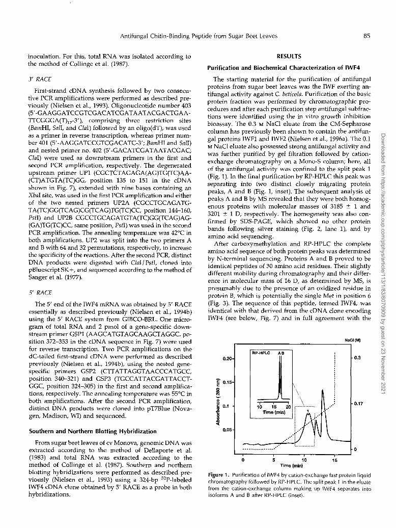
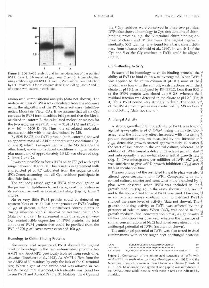
The starting material for the purification of antifungal proteins from sugar beet leaves was the IWF exerting an- tifungal activity against C. beticola. Purification of the basic protein fraction was performed by chromatographic pro- cedures and after each purification step antifungal subfrac- tions were identified using the in vitro growth inhibition bioassay. The 0.3 M NaCl eluate from the CM-Sepharose column has previously been shown to contain the antifun- gal proteins IWFl and IWF2 (Nielsen et al., 1996a). The 0.1 M NaCl eluate also possessed strong antifungal activity and was further purified by gel filtration followed by cation- exchange chromatography on a Mono-S column; here, a11 of the antifungal activity was confined to the split peak 1 (Fig. 1). In the final purification by RP-HPLC this peak was separating into two distinct closely migrating protein peaks, A and B (Fig. 1, inset). The subsequent analysis of peaks A and B by MS revealed that they were both homog- enous proteins with molecular masses of 3185 -+. 1 and 3201 4 1 D, respectively. The homogeneity was also con- firmed by SDS-PAGE, which showed no other protein bands following silver staining (Fig. 2, lane l), and by amino acid sequencing.
After carboxymethylation and RP-HPLC the complete amino acid sequence of both protein peaks was determined by N-terminal sequencing. Proteins A and B proved to be identical peptides of 30 amino acid residues. Their slightly different mobility during chromatography and their differ- ence in molecular mass of 16 D, as determined by MS, is presumably due to the presence of an oxidized residue in protein B, which is potentially the single Met in position 6 (Fig. 3). The sequence of this peptide, termed IWF4, was identical with that derived from the cDNA clone encoding IWF4 (see below, Fig. 7) and in full agreement with the
NaCl (M)
0.20.
- E 0.15.
Q - o
0.1 -e 8 s
0.05
3P-HPLC A B I 0.3
0.17
O I
O 10 15 Time (min)
Figure 1. Purification of IWF4 by cation-exchange fast protein liquid chromatography followed by RP-HPLC. The split peak 1 in the eluate from the cation-exchange column making up IWF4 separates into isoforms A and B after RP-HPLC (inset).
Dow
nloaded from https://academ
ic.oup.com/plphys/article/113/1/83/6070909 by guest on 23 N
ovember 2021
86 Nielsen et al. Plant Physiol. Vol. 113, 1997
kDa
8-
6
2.5-
Figure 2. SDS-PAGE analysis and immunodetection of the purifiedIWF4. Lane 1, Silver-stained gel; lanes 2 and 3, immunoblottingusing antibody against IWF4. + and —, With and without reductionby DTT treatment. One microgram (lane 1) or 250 ng (lanes 2 and 3)of protein was loaded in each lane.
amino acid compositional analysis (data not shown). Themolecular mass of IWF4 was calculated from the sequenceusing the algorithms of the PC/Gene software (IntelliGe-netics, Mountain View, CA). If we assume that all six Cysresidues in IWF4 form disulfide bridges and that the Met isoxidized in isoform B, the calculated molecular masses forthe two isoforms are (3190 - 6) = 3184 D (A) and (3190 -6 + 16) = 3200 D (B). Thus, the calculated molecularmasses coincide with those determined by MS.
By SDS-PAGE, the IWF4 protein (both isoforms) showedan apparent mass of 2.5 kD under reducing conditions (Fig.2, lane 3), which is in agreement with the MS data. On theother hand, under nonreduced conditions a higher molec-ular mass band of approximately 4.5 kD was observed (Fig.2, lanes 1 and 2).
It was not possible to focus IWF4 on an IEF gel with a pHrange between 3.0 and 9.0. This result is in agreement witha predicted pi of 9.7 calculated from the sequence data(PC/Gene), assuming that all Cys residues participate indisulfide linkages.
The antibody raised against IWF4 after conjugation ofthe protein to diphtheria toxoid recognized the protein inits reduced as well as nonreduced stage (Fig. 2, lanes 2and 3).
No or very little IWF4 protein could be detected onwestern blots of crude leaf homogenates or IWFs loading20 jug of protein, either in unstressed control plants orduring infection with C. beticola or treatment with INA(data not shown). In agreement with this apparent verylow, noninducible expression of IWF4 protein, the totalamount of IWF4 protein that could be purified from theIWF of 700 g of leaves never exceeded 100 jug.
Homology to Chitin-Binding Proteins
The amino acid sequence of IWF4 showed the highestlevel of homology to the two antimicrobial proteins Ac-AMP1 and Ac-AMP2, previously isolated from seeds of A.caudatus (Broekaert et al., 1992). Ac-AMPl differs from theAc-AMP2 of 30 residues by only the lack of the C-terminalArg. When a gap of one amino acid was allowed in Ac-AMP2 for optimal alignment, 66% identity was found be-tween IWF4 and Ac-AMP2 (Fig. 3). Notably, the 6 Cys and
the 7 Gly residues were conserved in these two proteins.IWF4 also showed homology to Cys-rich domains of chitin-binding proteins, e.g. the N-terminal chitin-binding do-main of class I and IV chitinases. The highest degree ofsimilarity, 55% identity, was found for a basic class I chiti-nase from tobacco (Shinshi et al., 1990), in which 4 of theCys and 5 of the Gly residues in IWF4 could be aligned(Fig. 3).
Chitin-Binding Activity
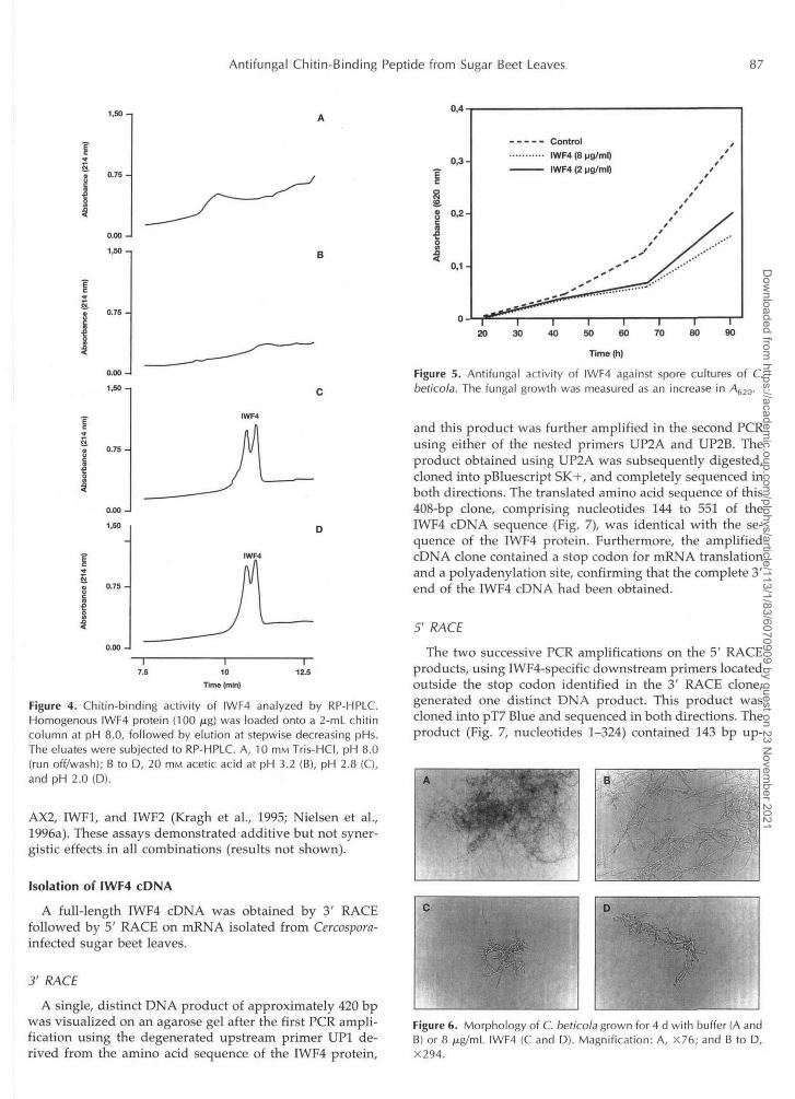
Because of its homology to chitin-binding proteins theability of IWF4 to bind chitin was investigated. When IWF4was applied to the chitin column at pH 8.0, none of theprotein was found in the run off/wash fractions or in theeluate at pH 3.2, as analyzed by RP-HPLC. Less than 50%of the IWF4 protein was eluted at pH 2.8, whereas theresidual fraction was detected in the eluate at pH 2.0 (Fig.4). Thus, IWF4 bound very strongly to chitin. The identityof the IWF4 protein peaks was confirmed by MS and im-munoblotting (data not shown).
Antifungal Activity
A strong growth-inhibiting activity of IWF4 was foundagainst spore cultures of C. beticola using the in vitro bio-assay, and the inhibitory effect increased with increasingprotein concentrations. As measured by the increase inA620, detectable growth started approximately 40 h afterthe start of incubation in the control culture, whereas theaddition of IWF4 caused a delay in detectable growth startand subsequently a somewhat slower initial growth rate(Fig. 5). Two micrograms per milliliter of IWF4 (0.7 JUM)was sufficient to give a50% growth inhibition (IC50) after80 h of incubation time.
The morphology of the restricted fungal hyphae was alsoaltered upon treatment with IWF4. Compared with thecontrol culture, shorter and somewhat more branched hy-phae were observed when IWF4 was included in thegrowth medium (Fig. 6). In the assay shown in Figures 5and 6, the nonoxidized form of IWF4 was used. However,in comparative assays oxidized and nonoxidized IWF4showed the same level of activity (data not shown). Thegrowth-inhibiting activity of IWF4 was affected by thepresence of calcium ions. When CaCl2 was added to thegrowth medium (final concentration 5 ITIM), a significantlyweaker inhibition was observed, whereas the presence ofsimilar concentrations of NaCl had no adverse effect on theantifungal potential of IWF4 (results not shown).
The antifungal potential of IWF4 was also tested in dualcombinations with other sugar beet antifungal proteins:
IWF4 SGECNMYGRCPPGYCCSKFGYCGVGRAYCG 30AC-AMP2 V***VR-****S*M***Q*****K*PK***R 30CHIT-TOB EQC*SQAGGA***S*L******N**NTND***PGNCQSQCP. . . 41Figure 3. Comparison of the amino acid sequence of IWF4 withAc-AMP2 from seeds of A. caudatus (Broekaert et al., 1992) and theN-terminal Cys-rich domain of class I tobacco chitinase (Shinshi etal., 1987). To optimize the alignment one gap (-) was introduced inAc-AMP2. Amino acids identical with those in IWF4 are indicated byasterisks.
Dow
nloaded from https://academ
ic.oup.com/plphys/article/113/1/83/6070909 by guest on 23 N
ovember 2021
Antifungal Chitin-Binding Peptide from Sugar Beet Leaves 87
1,50 n
I&
0.00
1,SO.
0,4
Ec*s
0.00
1,60
Si0, 0.75
0.00 -I
1,50
~Z 0.75 -
I7.5
I10
Time (min)
Figure 4. Chitin-binding activity of IWF4 analyzed by RP-HPLC.Homogenous IWF4 protein (100 jug) was loaded onto a 2-mL chitincolumn at pH 8.0, followed by elution at stepwise decreasing pHs.The eluates were subjected to RP-HPLC. A, 10 mM Tris-HCI, pH 8.0(run off/wash); B to D, 20 HIM acetic acid at pH 3.2 (B), pH 2.8 (C),and pH 2.0 (D).
AX2, IWF1, and IWF2 (Kragh et al., 1995; Nielsen et al.,1996a). These assays demonstrated additive but not syner-gistic effects in all combinations (results not shown).
Isolation of IWF4 cDNA
A full-length IWF4 cDNA was obtained by 3' RACEfollowed by 5' RACE on mRNA isolated from Cercospora-infected sugar beet leaves.
3' RACE
A single, distinct DNA product of approximately 420 bpwas visualized on an agarose gel after the first PCR ampli-fication using the degenerated upstream primer UP1 de-rived from the amino acid sequence of the IWF4 protein,
0,3-
0,2-
0,1-
----- Control........... |WF4 (8 M9/ml)———— IWF4 (2 |jg/ml)
20 30 40 50 60 70I
80I
90
Time (h)
Figure 5. Antifungal activity of IWF4 against spore cultures of C.beticola. The fungal growth was measured as an increase in A620-
and this product was further amplified in the second PCRusing either of the nested primers UP2A and UP2B. Theproduct obtained using UP2A was subsequently digested,cloned into pBluescript SK+, and completely sequenced inboth directions. The translated amino acid sequence of this408-bp clone, comprising nucleotides 144 to 551 of theIWF4 cDNA sequence (Fig. 7), was identical with the se-quence of the IWF4 protein. Furthermore, the amplifiedcDNA clone contained a stop codon for mRNA translationand a polyadenylation site, confirming that the complete 3'end of the IWF4 cDNA had been obtained.
5' RACE
The two successive PCR amplifications on the 5' RACEproducts, using IWF4-specific downstream primers locatedoutside the stop codon identified in the 3 RACE clone,generated one distinct DNA product. This product wascloned into pT7 Blue and sequenced in both directions. Theproduct (Fig. 7, nucleotides 1-324) contained 143 bp up-
Figure 6. Morphology of C. beticola grown for 4 d with buffer (A andB) or 8 |u.g/mL IWF4 (C and D). Magnification: A, X76; and B to D,X294.
Dow
nloaded from https://academ
ic.oup.com/plphys/article/113/1/83/6070909 by guest on 23 N
ovember 2021

88 Nielsen et al. Plant Physiol. Vol. 113, 1997
ACTCAACAAATTCAGAAAAAAACAGAAGCAAAAAAAGTTTATTGAAAGAGTAAGTTGAGGTGAAA 6 5
ATGATGAAAAGCTTTGTGATAGTTATGTTGGTCATGTCCATGATGGTGGCTACATCTATGGCA 128M M K S F V I V M L V M S M M V A T S M A 2 1
AGTGGTGAATGCAATATGTATGGTCGATGCCCCCCAGGGTATTGTTGTAGCAAGTTTGGCTAC 191S G E C W M Y G R C P P G Y C C S K F G Y 4 2
TGTGGTGTCGGACGCGCCTATTGTGGCGATGCTGAGCAGAAGGTTGAAGATCATCCATCTAAT 254C G V G R A Y C G D A E Q K V E D H P S N 6 3
GATGCTGATGTTCCTGAGTTTGTTGGAGCTGGTGCCCCATOATGCTCGAAGCCAGGTAATCGT 317D A D V P E F V G A G A P 7 6
AATGGCATGGGTTACCTAATAAGTAAACTCATTGTGCCTAGCTTGCTACATGCTTATCCACTA 380HJSPJ
TAAATAAGCTCCTACAGGAGTTGTGTTTTTCTTTTAATTTTGTAATCAAGGGTTTGACTTTAA 443
TTAATGAGACCAATGTATACTTGCATGTCGGATAAATATATGAACTAAGCCACTCGTATTGGT 506
TTATTATAAAACTACTATAAAAAAAAAAAAAAAAAAAAAAAAAAA 551
Figure 7. cDNA sequence and deduced amino acid sequence ofIWF4. Start and stop codons for mRNA translation and putativepolyadenylation signal are marked by bold letters. The amino acidsequence identical with the mature IWF4 protein is underlined. Thelocations of the degenerate primers used for 3' RACE (UP1 and UP2)and the gene-specific primers used for 5' RACE (GSP1-3) are markedwith arrows indicating polarity 5' to 3'.
stream of the IWF4 sequence obtained by 3' RACE, fol-lowed by 181 bp, showing 100% identity with the 3' RACEclone. The 5' RACE product was inferred to contain theentire IWF4 cDNA sequence due to the presence of ATGstart (Fig. 7, position 66) and TGA stop codons (position294) for mRNA translation, as well as a characteristicadenosine-rich sequence upstream of the start codon(Kozac, 1984; Lutcke et al., 1987). In agreement with this,the translated amino acid sequence contained the entireIWF4 protein.
Characterization of the IWF4 cDNA and Deduced AminoAcid Sequence
The obtained full-length IWF4 cDNA contained 551 bp,with an open reading frame of 228 bp. The 5' adenosine-rich region of 65 bp was followed by an ATG start codon atposition 66, a TGA stop codon at position 294, and a 3'noncoding region of 254 bp, containing a polyadenylationsignal at position 383 and a polyadenylation site at position525 (Fig. 7).
A polypeptide chain of 76 amino acid residues was en-coded by the IWF4 cDNA. From this it is inferred that IWF4is encoded as a preproprotein containing an N-terminalputative signal peptide of 21 residues processing all thecharacteristics of signal sequences (von Heijne, 1983), fol-lowed by the mature IWF4 protein of 30 residues and aC-terminal extension of 25 residues. The C-terminal prose-quence differed from the mature IWF4 protein by the lackof Cys residues (6 in IWF4) and by its acidic nature, with apredicted pi of 3.8 (pi 9.7 for IWF4).
The primary structure of the IWF4 preproprotein de-duced from the cDNA sequence was comparable to that ofAc-AMP2 (De Bolle et al., 1993). Both sequences containthree domains: the putative N-terminal signal peptide, fol-lowed by the mature protein domain, and a C-terminalpeptide. The entire IWF4 preprosequence was 10 aminoacid residues shorter than Ac-AMP because of shorterN-terminal signal peptide and C-terminal peptide domains
(25 and 31 amino acid residues, respectively, in Ac-AMP2).No sequence similarities were found between the N-terminal signal peptide and C-terminal peptide domains ofthe two proteins. Notably, however, the N-terminal signalpeptide of both IWF4 and Ac-AMP2 are rich in Met resi-dues (7 in both). The calculated pis of the mature proteindomains are comparable, showing pis of 9.7 and 10.9 forIWF4 and Ac-AMP2, respectively, whereas the C-terminalpeptide domain of Ac-AMP2 differs from the IWF4C-terminal peptide in having a slightly basic pi of 7.8, incontrast to the highly acidic pi of the IWF4 C-terminalpeptide. Finally, a putative N-glycosylation site present inthe C-terminal peptide of Ac-AMP2 (De Bolle et al., 1993)was not found in IWF4.
Genomic Diversity
Genomic DNA of sugar beet cv Monova was digestedwith the three restriction enzymes Hmdlll, EcoRI, and Xbal,of which only Hindlll cuts within the IWF4 cDNA (Fig. 7,position 74). Southern blots using these digests were probedwith the 324-bp IWF4 cDNA clone obtained by 5' RACE,followed by a high-stringency wash. Two to three stronglyhybridizing fragments were present in all three digests, andthe EcoRI digest also contained a few weakly hybridizingfragments (Fig. 8). The smallest fragment (800 bp) was foundin the Hwdlll digest, confirming the presence of a recogni-
kb
21.2-
1 2 3
Figure 8. Southern blots. Genomic DNA from sugar beet cv Monovawas digested with H/ndlll (lane 1), EcoRI (lane 2), and Xbal (lane 3),probed with a 324-bp IWF4 cDNA clone, and washed at highstringency.
Dow
nloaded from https://academ
ic.oup.com/plphys/article/113/1/83/6070909 by guest on 23 N
ovember 2021

Antifungal Chitin-Binding Peptide from Sugar Beet Leaves 89
tion site for this enzyme within the gene. The presence ofmore than one strongly hybridizing fragment in all digestsmay indicate either that the fragments carry the IWF4 genein alternate loci or that they make up a small gene familyencoding two or a few closely related isoforms. A parallelgenomic Southern blot using DNA from another sugar beetcultivar, +Tol (Danisco Seed; Nielsen et al., 1993), gave thesame hybridization pattern as seen for cv Monova (notshown), demonstrating that no polymorphism exists be-tween the two cultivars with respect to this gene.
Expression of IWF4 mRNA
The expression of IWF4 mRNA in different organs andtissues of sugar beet plants of cv Monova was analyzedusing RNA blots. The analysis included leaves and petioles(young and mature), young and mature flowers, and cen-tral and outer root tissue, all tissues being isolated fromnonstressed 6-week-old plants. A high accumulation ofIWF4 mRNA was found in the leaf tissue, both in youngand mature leaves (Fig. 9, lanes 4 and 7), whereas in thepetioles a very low level of mRNA expression was ob-served in young petioles only (Fig. 9, lane 1). Expression ofIWF4 mRNA was also found in flower tissue, with a fairlyhigh level of transcript accumulation in young flowers (Fig.9, lane 6) and a very low, barely detectable accumulation inmature flowers (Fig. 9, lane 5). No expression of IWF4mRNA was detected in root tissue (Fig. 9, lanes 2 and 3).
Time-course analyses were performed to investigate theaccumulation of IWF4 transcript in mature leaves of 6-week-old plants after treatment with IN A and / or during infectionwith C. beticola. In these experiments a constitutive level ofmRNA accumulation was found in nontreated leaves,whereas no increased expression was observed followinginducer treatment or fungal infection (results not shown).
To summarize, IWF4 transcripts accumulated only in theaerial parts of the plant. A strong, constitutive, noninduc-ible mRNA accumulation was found in the leaves regard-less of age, and additional expression was observed inyoung flowers.
DISCUSSION
IWF4 was purified to homogeneity from the IWF of sugarbeet leaves. IWF4 shows homology to the Cys-/Gly-rich domain of chitin-binding proteins, of which themajority contain single or multiple chitin-binding domainsof 40 to 43 residues, with 8 Cys residues involved in four
1 2 3 4 5 6 7 8Figure 9. RNA blots. Fifteen micrograms of total RNA, isolated fromdifferent sugar beet tissues, was loaded in each lane. The blot wasprobed with a 324-bp IWF4 cDNA clone and washed at high strin-gency. Tissue: Lane 1, petiole (young leaf); lanes 2 and 3, root(central and outer tissue, respectively); lane 4, young leaf; lanes 5and 6, flower (mature and young, respectively); lane 7, mature leaf;and lane 8, petiole (mature leaf).
intradomain disulfide bridges. Compared with these, the30-amino-acid residue sequence of IWF4 lacks theC-terminal part of the "full" domain, including the twoterminal Cys residues that form a separate disulfide bridge(Beintema, 1994). Furthermore, the distance between the twoN-terminal Cys residues is smaller in IWF4. In spite of thesedeletions IWF4 shows a strong chitin-binding activity. In-deed, the present study demonstrated that IWF4 binds evenmore strongly to chitin than does the class IV sugar beetchitinase Ch4, which is readily elutable at pH 3.2 (Mikkelsenet al., 1992). Other chitinases, in both classes I and IV, can bereleased from the chitin affinity column at pH 3.2 (Boiler etal., 1983; Broekaert et al., 1988; Jacobsen et al., 1990; Rasmus-sen et al., 1992; Kragh et al., 1993; Sela-Buurlage et al., 1993).
IWF4 shows the highest degree of similarity to the anti-microbial proteins Ac-AMPl and Ac-AMP2 of 29 and 30amino acid residues, respectively, isolated from seeds ofamaranth (Broekaert et al., 1992). The Ac-AMPs are theonly previously reported antimicrobial peptides consistingof a sole truncated chitin-binding domain (Raikhel et al.,1993). Recently, an antifungal protein Pn-AFP of 41 aminoacids was isolated from seeds of Pharbitis nil (Koo et al.,1995). Pn-AFP represents a nontruncated version of thechitin-binding domain containing all 8 Cys residues and isthus more similar to the hevein of 43 amino acid residues(Van Parijs et al., 1991).
The apparent mass of IWF4 determined by SDS-PAGE,4.5 kD in the nonreduced versus 2.5 kD in the reducedstage, may indicate that IWF4 is a dimeric protein. How-ever, the result of the MS analysis, as well as the stronghomology to thoroughly characterized chitin-binding do-mains, strongly indicates that all Cys residues are involvedin intramolecular disulfide bridge formation, makingdimer formation less likely. The low mobility of the unre-duced protein in SDS-PAGE is probably instead due to theerratic behavior of such a small, compact, and basic mole-cule in the SDS-gel. A similar difference in mobility ofunreduced versus reduced proteins was observed in theAc-AMPs (Broekaert et al., 1992).
IWF4 exerts a strong growth-inhibiting activity againstC. beticola, with an IC50 of <2 ng/mL (0.7 JUM) after 80 h ofincubation time. This activity is comparable to that of theAc-AMPs, with IC50 values varying between 1 and 10/n,g/mL (0.3-3 JAM), depending on the fungal species (Broe-kaert et al., 1992). The Pn-AFP shows IC50 values in thisrange, varying from 0.6 to 18 (xg/mL (0.1-4 JUM) whentested against eight different fungi (Koo et al., 1995). No-tably, the antifungal potential of hevein is significantlyweaker, showing IC50 values of 90 to 1250 ^.g/rnL (20-280/J.M; eight fungi; Van Parijs et al., 1991). In addition thestinging nettle lectin, consisting of two chitin-binding do-mains, shows lower antifungal activity than the truncated,single-domain peptides, with IC50 values of 20 to 150jMg/mL (2-18 JJ.M; seven fungi; Broekaert et al., 1989). Theobserved calcium salt sensitivity of IWF4 is in agreementwith observations in other families of small, Cys-rich anti-fungal proteins (including the Ac-AMPs), all showing re-duced antifungal activity when tested in growth mediumsupplemented with CaCl2 (Cammue et al., 1994).
Dow
nloaded from https://academ
ic.oup.com/plphys/article/113/1/83/6070909 by guest on 23 N
ovember 2021

90 Nielsen et al. Plant Physiol. Vol. 1 1 3 , 1997
The IWF4 cDNA encodes a preproprotein with N- and C-terminal sequences flanking the mature protein. The 21- amino-acid N-terminal sequence shows the characteristics of signal peptides facilitating protein secretion, which is in agreement with the presence of IWF4 in the IWF. The acidic C-terminal extension of 25 residues shows no homology to any published protein sequence (Swiss-Prot, release 31). The Ac-AMP2 cDNA encodes a C-terminal prosequence that shows little homology to that of IWF4. Moreover, the Ac-AMP2 C-terminal peptide is longer (31 amino acid residues), has a slightly basic pI, and contains an N-glycosylation site not present in the IWF4 C-terminal sequence. The functional roles of these prosequences are not known. Acidic C-terminal propeptides are also present in the family of type 1 thionins, which are small, Cys-rich antimicrobial proteins present in the endosperm of various monocotyledons (Florack and Stiekema, 1994). Florack et al. (1994) studied the expression of a hordothionin in to- bacco using different constructs, some of which lacked the sequence encoding the acidic C-terminal peptide. They found that inclusion of the peptide enhanced the protein expression significantly but had no effect on the subcellular (intracellular) localization of the protein. The study indi- cated that the acidic peptide was not involved in sorting and suggested that it might facilitate transport through membranes (Florack et al., 1994). Thus, these propeptides may increase the stability of the mature protein during transport and processing. However, C-terminal prose- quences of some other proteins have been shown to facil- itate the transport of the proteins to the vacuole (Bednarek and Raikhel, 1992; Nakamura and Matsuoka, 1993).
Whereas the N- and C-terminal peptides of IWF4 show little homology to those of Ac-AMP2, the mature IWF4 and Ac-AMP proteins resemble each other in size, pI, amino acid sequence, chitin-binding ability, and antifungal poten- tials. However, striking differences exist with respect to tissue specificity and expression patterns. Our studies demonstrated a strong expression of IWF4 in vegetative, aerial tissue, i.e. leaves and flowers, whereas no root ex- pression was observed. Furthermore, no pathogen- or INA- induced expression of IWF4 was found, either on mRNA or on the protein level. In contrast, the Ac-AMPs have been shown by northern blotting experiments to be seed- specific; they are expressed in near-mature seeds of ama- ranth only and have no homologs that are expressed in vegetative tissue, i.e. leaves and roots (De Bolle et al., 1993). Moreover, the expression of the Ac-AMPs is induced by pathogen infection and other stress factors. The genes en- coding the hevein-like Pn-AFPs in P. nil show expression patterns very similar to the Ac-AMPs in that they are expressed during seed maturation and germination but not in the aerial vegetative parts of the plant (Koo et al., 1995).
Because of the apparent lack of induction by infection with C. beticola IWF4 cannot be regarded as a “classical” pathogenesis-related protein. We have not attempted to estimate the concentration of the IWF4 protein in the IWF. However, there is no doubt that the peptide is present in cv Monova at concentrations below those required for good antifungal activity, which is consistent with the high sus-
ceptibility of this cultivar to C. beticola.. Furthermore, IWF4 is not induced upon treatment with INA, suggesting that this peptide is not important for the resistance mediated by this chemical.
In spite of the low, noninducible levels of IWF4 found in the sugar beet leaves, the strong antifungal potential of the protein suggests that it has a role in plant defense. More- over, immunohistochemical studies of Cercospora-infected leaf tissue indicated that IWF4, like AX2 (Kragh et al., 1995) and the antifungal chitinase Ch4 (Nielsen et al., 1996b), is primarily present in extracellular ”protein bodies,” presum- ably involved in defense (J. Nielsen, unpublished data). Whether, as has been suggested for the Ac-AMPs (Raikhel et al., 1993), IWF4 acts by simply “sticking” to nascent chitin fibers in the apices of growing fungal hyphae, thereby dis- turbing cross-linking and assembling of the fungal cell wall, still remains to be demonstrated. The strong chitin-binding affinity observed for IWF4, compared with the chitin- binding chitinases, may support this hypothesis. On the other hand, the sensitivity of both IWF4 and the Ac-AMPs to calcium salt does not point to an interaction with chitin for their antifungal action. We have investigated the influence of calcium on the chitin-binding activity of IWF4. At CaCI, concentrations 10 times higher than required for the com- plete abolishment of the growth-inhibiting activity of IWF4 the protein showed no reduced affinity to chitin (K.K. Nielsen, unpublished data). Finally, the strikingly different expression patterns of IWF4 and the Ac-AMPs may suggest different roles for these proteins in the plant. We are now in the progress of producing transgenic sugar beet plants over- expressing IWF4 to test its potential as an efficient agent for increasing resistance to C. beticola.
ACKNOWLEDCMENTS
We wish to thank Susanne Dreboldt and Jette Skovsgaard for excellent technical assistance and Anne Kroll Kristensen for per- forming MS.
Received July 12, 1996; accepted October 2, 1996. Copyright Clearance Center: 0032-0889/ 971 113/0083/09.
LITERATURE ClTED
Barkholt V, Jensen AL (1989) Amino acid analysis: determination of cysteine plus half-cystine in proteins after hydrochloric acid hydrolysis with a disulfide compound as additive. Anal Bio- chem 177: 318-322
Bednarek SY, Raikhel NV (1992) Intracellular trafficking of secre- tory proteins. Plant MOI Biol 20: 133-150
Beintema JJ (1994) Structural features of plant chitinases and chitin-binding proteins. FEBS Lett 350: 159-163
Berglund L, Brunstedt J, Nielsen KK, Chen Z, Mikkelsen JD, Marcker KA (1995) A proline-rich chitinase from Beta vulgaris. Plant MOI Biol 27: 211-216
Boller T, Gehri A, Mauch F, Vogeli U (1983) Chitinase in bean leaves: induction by ethylene, purification, properties, and pos- sible function. Planta 157: 22-31
Bradford MM (1976) A rapid and sensitive method for the quan- titation of microgram quantities of protein utilizing the principle of protein-dye binding. Anal Biochem 7 2 248-254
Broekaert WF, Marien W, Terras FRG, De Bolle MFC, Proost P, Van Damme J, Dillen L, Claeys M, Rees SB, Vanderleyden J, Cammue BPA (1992) Antimicrobial peptides from Amaranthus caudatus seeds with sequence homology to the cysteine / glycine-
Dow
nloaded from https://academ
ic.oup.com/plphys/article/113/1/83/6070909 by guest on 23 N
ovember 2021

Antifungal Chitin-Binding Peptide from Sugar Beet Leaves 91
rich domain of chitin-binding proteins. Biochemistry 31: 4308- 4314
Broekaert WF, Terras FRG, Cammue BPA, Osborn RW (1995) Plant defensins. Nove1 antimicrobial peptides as components of the host defense system. Plant Physiol 108: 1353-1358
Broekaert WF, Van Parijs J, Allen AK, Joos H, Peumans WJ (1989) A chitin-binding lectin from stinging nettle rhizomes with anti- funga1 properties. Science 245: 1100-1102
Broekaert WF, Van Parijs J, Allen AK, Peumans WJ (1988) Com- parison of some molecular, enzymatic and antifungal properties of chitinases from thorn-apple, tobacco and wheat. Physiol Mo1 Plant Pathol 33: 319-331
Cammue BPA, De Bolle MFC, Schoofs HME, Terras FRG, Thevissen K, Osborn RW, Rees SB, Broekaert WF (1994) Gene- encoded antimicrobial peptides from plants. 1994 antimicrobial peptides. Ciba Found Symp 186: 91-106
Collinge DB, Kragh KM, Mikkelsen JD, Nielsen KK, Rasmussen U, Vad K (1993) Plant chitinases. Plant J 3: 31-40
Collinge DB, Milligan DE, Dow JM, Scofield G, Daniels MJ (1987) Gene expression in Brassica cnmpestris showing a hyper- sensitive response to the incompatible pathogen Xanthomonns campestris pv. vitians. Plant Mo1 Biol 8: 405-414
Cutt JR, Klessig DF (1992) Pathogenesis-related proteins. In T Boller, F Meins, eds, Genes Involved in Plant Defense. Springer- Verlag, New York, pp 209-243
De Bolle MFC, David KMM, Sarah BR, Vanderleyden J, Cam- mue BPA, Broekaert WF (1993) Cloning and characterization of a cDNA encoding an antimicrobial chitin-binding protein from amaranth, Anzaranthus cnudatus. Plant Mo1 Biol 22: 1187-1190
Dellaporte SL, Wood J, Hicks JB (1983) A plant DNA miniprepa- ration: version 11. Plant Mo1 Biol Rep 1: 19-21
Florack DEA, Dirkse WG, Visser B, Heidekamp F, Stiekema WJ (1994) Expression of biologically active hordothionins in to- bacco. Effects of pre- and pro-sequences at the amino and car- boxy termini of the hordothionin precursor on mature protein expression and sorting. Plant Mo1 Biol 24: 83-96
Florack DEA, Stiekema WJ (1994) Thionins: properties, possible biological roles and mechanisms of action. Plant Mo1 Biol 26:
Jacobsen S, Mikkelsen JD, Hejgaard J (1990) Characterization of two antifungal endochitinases from barley grain. Physiol Plant 79: 554562
Koo JC, Lee SY, Chun HJ, Park SH, Lee BL, Cho MJ (1995) Sequential expression of two identical hevein homologs with a potent antifungal activity during maturation and germination of the seeds of Pkarbilitis nil (abstract). Presented at the 4th Inter- national Workshop on Pathogenesis-Related Proteins in Plants: Biology and Biotechnological Potential, Kloster Irsee, Germany September 3-7, 1995
Kozac M (1984) Compilation and analysis of sequences upstream from the translational start site in eucaryotic mRNAs. Nucleic Acids Res 12: 857-872
Kragh KM, Jacobsen S, Mikkelsen JD (1990) Induction, purifica- tion and characterization of barley leaf chitinase. Plant Sci 71:
Kragh KM, Jacobsen S, Mikkelsen JD, Nielsen KA (1993) Tissue specificity and induction of class I, I1 and 111 chitinases in barley (Hordeum vulgare). Physiol Plant 89: 490498
Kragh KM, Nielsen JE, Nielsen KK, Dreboldt S, Mikkelsen JD (1995) Characterization and localization of new antifungal cysteine-rich proteins from Beta vulgaris L. Mo1 Plant-Microbe Interact 8: 424434
Kyhse-Andersen J (1984) Electroblotting of multible gels: a simple apparatus without buffer tank for rapid transfer of proteins from polyacrylamide to nitrocellulose. J Biochem Biophys Methods
Legrand M, Kauffmann S, Geoffroy P, Fritig B (1987) Biological functions of 'pathogenesis-related proteins': four tobacco PR- proteins are chitinases. Proc Natl Acad Sci USA 8 4 6750-6754
Liitcke HA, Chow KC, Mickel KA, Kern HF, Scheele GA (1987) Selection of AUG initiation codons differ in plants and animals. EMBO J 6: 4348
25-37
55-68
10: 203-209
Marcussen J, Poulsen C (1991) A nondestructive method for pep- tide bond conjugation of antigen haptens to a diphtheria toxoid carrier, exemplified by two antisera specific to acetolactase syn- thase. Anal Biochem 198: 318-323
Meins F Jr, Fritig 8, Linthorst HJM, Mikkelsen JD, Neuhaus JM, Ryals J (1994) Plant chitinase genes. Plant Mo1 Biol Rep 12 (2, suppl): S22-S28
Mikkelsen JD, Berglund L, Nielsen KK, Christiansen H, Bojsen K (1992) Structure of endochitinase genes from sugar beets. In C Brine, SA Sandford, JP Zikakis, eds, Advances in Chitin and Chitosan. Elsevier Applied Science, Amsterdam, The Nether- lands, pp 344-353
Molano J, Duran A, Cabib E (1977) A rapid and sensitive assay for chitinase using tritiated chitin. Anal Biochem 83: 648-656
Nakamura K, Matsuoka K (1993) Protein targeting to the vacuole in plant cells. Plant Physiol 103: 1-5
Nielsen KK, Bojsen K, Collinge DB, Mikkelsen JD (1994a) In- duced resistance in sugar beet against Cercospora beticola: induc- tion by dichloroisonicotinic acid is independent of chitinase and /3-1,3-glucanase transcript accumulation. Physiol Mo1 Plant Pathol 45: 89-99
Nielsen KK, Bojsen K, Roepstorff P, Mikkelsen JD (1994b) A hydroxyproline-containing class IV chitinase of sugar beet is glycosylated with xylose. Plant Mo1 Biol 25: 241-257
Nielsen KK, Jergensen P, Mikkelsen JD (1994~) Antifungal activity of sugar beet chitinase against Cercospora beticola. An autoradio- graphic study on cell wall degradation. Plant Pathol 43: 979-986
Nielsen KK, Mikkelsen JD, Kragh KM, Bojsen K (1993) An acidic class 111 chitinase in sugar beet: induction by Cercospora beticola, characterization, and expression in transgenic tobacco plants. Mo1 Plant-Microbe Interact 6: 495-506
Nielsen KK, Nielsen JE, Madrid SM, Mikkelsen JD (1996a) New antifungal proteins from sugar beet (Beta vulgaris L.) showing homology to non-specific lipid transfer proteins. Plant Mo1 Biol
Nielsen JE, Nielsen KK, Mikkelsen JD (1996b) Immunohisto- logical localization of a basic class 1V chitinase in Beta vulgaris leaves after infection with Cercospora beticola. Plant Sci 119: 191 -202
Raikhel NV, Lee H-I, Broekaert WF (1993) Structure and function of chitin-binding proteins. Annu Rev Plant Physiol Plant Mo1 Biol 44: 591-615
Rasmussen U, Giese H, Mikkelsen JD (1992) Induction and pu- rification of chitinase in Brassica napus L. aap. oleifera infected with Phoma lingam. Planta 187: 328-334
Sanger F, Nicklen S, Coulson AR (1977) DNA sequencing with chain-determinating inhibitors. Proc Natl Acad Sci USA 74:
Schagger H, von Jagow G (1987) Tricine-sodium dodecyl sulfate- polyacrylamide gel electrophoresis for the separation of proteins in the range from 1 to 100 kDa. Anal Biochem 166: 368-379
Sela-Buurlage MB, Ponstein AS, Bres-Vloemans SA, Melchers LS, van den Elzen PJM, Cornelissen BJC (1993) Only specific tobacco (Nicotiana tabacum) chitinases and /3-1,3-glucanases ex- hibit antifungal activity. Plant Physiol 101: 857-863
Shinshi H, Neuhaus JM, Ryals J, Meins F (1990) Structure of a tobacco endochitinase gene: evidence that different chitinase genes can arise by transposition of sequences encoding cysteine- rich domain. Plant Mo1 Biol 14: 357-368
Susi A, Mikkelsen JD, von Weissenberg K, Nielsen KK (1995) Sugar beet chitinase inhibits the growth of a spruce pathogen. Eur J For Pathol 25: 61-64
Van Parijs J, Broekaert WF, Goldstein IJ, Peumans WJ (1991) Hevein: an antifungal protein from rubber-tree (Hevea brasilien- sis) latex. Planta 183: 258-264
Von Heijne G (1983) Patterns of amino acids near signal-sequence cleavage sites. Eur J Biochem 133: 17-21
Vorm O, Roepstorff P (1994) Peptide sequence information de- rived by partia1 acid hydrolysis and matrix-assisted laser de- sorption/ionization mass spectrometry. Biol Mass Spectrom 23:
31: 539-552
5463-5467
734-740
Dow
nloaded from https://academ
ic.oup.com/plphys/article/113/1/83/6070909 by guest on 23 N
ovember 2021