characterization of cytokeratin 20 expression in...
TRANSCRIPT

Characterization of Cytokeratin 20 Expression in Pancreatic andColorectal Cancer1
Stefan Wildi, Jorg Kleeff, Haruhisa Maruyama,Christoph A. Maurer, Helmut Friess,Markus W. Buchler, Arthur D. Lander, andMurray Korc 2
Departments of Medicine, Biological Chemistry, and Pharmacology[S. W., J. K., H. M., M. K.] and Department of Developmental andCell Biology and Developmental Biology Center [A. D. L.],University of California, Irvine, California 92697, and Department ofVisceral and Transplantation Surgery, University of Bern, 3010 Bern,Switzerland [C. A. M., H. F., M. W. B.]
ABSTRACTCytokeratin 20 belongs to the epithelial subgroup of the
intermediate filament family. Because of its restricted rangeof expression in humans, it has become an important tool fordetecting and identifying metastatic cancer cells by immu-nohistochemistry and by PCR analysis. Despite its wide-spread diagnostic use in colorectal cancer and occasional usein pancreatic cancer, little is known about the expression ofCK 20 in these tumors in vivo. Therefore, in the presentstudy we characterized CK 20 expression in pancreatic andcolorectal cancer by comparison with its expression in thenormal pancreas and colon. Tissue samples from 24 patientswith pancreatic cancer and from 41 patients with colorectalcancer were examined for CK 20 expression by Northernblot analysis, immunohistochemistry, andin situ hybridiza-tion. CK 20 expression was observed in the cancer cells ofboth cancer types. A subgroup of the pancreatic cancersexhibited a 3.2-fold increase in CK 20 mRNA by comparisonwith respective normal controls. In contrast, colon cancersunderexpressed CK 20 mRNA by comparison with the re-spective controls. In the normal tissues, CK 20 immunore-activity was relatively faint and sparse in the pancreaticductal cells but intense and abundant in the apical portionsof the colonic mucosa. CK 20 immunoreactivity was alsoevident in the ductal cells from the chronic pancreatitis-like
lesions adjacent to the cancer cells. Furthermore, distantmetastases from pancreas carcinomas exhibited strong CK20 immunoreactivity. It is concluded that CK 20 is overex-pressed in pancreatic cancer and that it can serve as anexcellent marker for metastatic pancreatic cancer.
INTRODUCTIONCKs3 are a family of proteins that form the intermediate
filament cytoskeleton of epithelial cells. They consist of twodifferent groups, the type I acidic keratins and the type II basickeratins (1). The first step in keratin intermediate filamentassembly is the formation of heterodimers between a type I anda type II keratin (2, 3). To date, 20 distinct polypeptides, whichbelong to this family, have been identified in various humanepithelial cell types (4–7). The differential expression of theseproteins is closely linked with specific programs of differenti-ation (8–11). Thus, cytokeratin expression correlates with dif-ferent epithelial cell lineages, allowing for the classification ofepithelial cells into numerous subtypes (12–16). The most re-cently described member of this family is the 46-kb cytoskeletalprotein CK 20.
CK 20 is consistently detected in human intestinal mucosa,urothelium, and Merkel cells (17). It is expressed preferentiallyin differentiated enterocytes and goblet cells of the intestinalvilli (17). In contrast, in crypts, only a few of the undifferenti-ated cells are CK 20 positive (17). CK 20 has been detected inthe pancreata of rat and pig (18, 19). In the rat, CK 20 expres-sion was restricted to the duct cells (19). In addition, CK 20 maybe considered as a “maturation” marker of the biliary tree in theliver of the rat (20). There is one report of CK 20 immunore-activity in primary pancreatic cancers (21) and a recent report ofits presence in pancreatic cancer metastases to the liver (22).The expression of several other cytokeratins (CK 7, CK 8, CK18, and CK 19) has also been reported in the human pancreas(18). Because of the lack of immunological cross-reactivity withother cytokeratins, CK 20 has become an important tool fordelineating the origin of metastatic human adenocarcinomasarising from an unknown primary source (23–33). In addition,several reports have highlighted the importance of detecting CK20 by the use of the PCR as a marker for the spread of tumorcells into blood, bone marrow, and lymph nodes of colorectaland other gastrointestinal cancers such as gastric and pancreaticcancers (34–40).
Colorectal cancer is the second most common cause ofcancer death and the fourth most prevalent carcinoma in theWestern world. In the United States, there were;131,000 newcases of colorectal cancer and 55,000 deaths from this disease in1997 (41). Similarly, pancreatic cancer is a devastating disease
Received 5/27/99; revised 8/3/99; accepted 8/5/99.The costs of publication of this article were defrayed in part by thepayment of page charges. This article must therefore be hereby markedadvertisementin accordance with 18 U.S.C. Section 1734 solely toindicate this fact.1 This work was supported by USPHS Grant CA-40162 awarded by theNational Cancer Institute (to M. K.). S. W. was the recipient of post-doctoral trainee awards from the Ursula Zindel-Hilti-Foundation, theCiba-Geigy-Anniversary Foundation, and the Department of Visceraland Transplantation Surgery of the University Hospital of Bern. J. K.was the recipient of a fellowship award from the University of Califor-nia Research and Education Grant on Gene Therapy for Cancer.2 To whom requests for reprints should be addressed, at Division ofEndocrinology, Diabetes and Metabolism, Medical Sciences I, C240,University of California, Irvine, CA 92697. Phone: (949) 824-6887;Fax: (949) 824-1035. 3 The abbreviation used is: CK, cytokeratin.
2840Vol. 5, 2840–2847, October 1999 Clinical Cancer Research
Research. on July 16, 2018. © 1999 American Association for Cancerclincancerres.aacrjournals.org Downloaded from

that is the fifth most common gastrointestinal tract cancer inmales and the seventh most common in females. Its high mor-tality rate makes it the fourth most common cause of death fromcancer in the general population. To date, the level of expressionof CK 20 in either colon or pancreatic cancer by comparisonwith the respective normal tissues has not been reported. Fur-thermore, the exact site of CK 20 expression in either malig-nancy has not been described. Therefore, in this study wecompared the expression of CK 20 in human colorectal andpancreatic cancer with its expression in the corresponding nor-mal tissues. We now report that CK 20 is expressed at highlevels in a subgroup of pancreatic cancers but not in coloncancers, and that CK 20 expression in pancreatic cancer occurspredominantly in the cancer cells.
MATERIALS AND METHODSMaterials. The following materials were purchased: fetal
bovine serum, DMEM, RPMI, Leibovitz L-15, trypsin solution,and penicillin-streptomycin solution from Irvine Scientific(Santa Ana, CA); restriction enzymes and the random primedlabeling kit from Boehringer-Mannheim (Indianapolis, IN);Sequenase Version 2.0 DNA Sequencing from UnitedStates Biochemical Corp. (Cleveland, OH); [a-32P]dCTP,[g-35S]dATP from Amersham (Arlington Heights, IL); andpBluescript-IISK1 from Stratagene (La Jolla, CA). A highlyspecific monoclonal mouse anti-human CK 20 antibody wasfrom Dako (Carpinteria, CA). All other chemicals and reagentswere purchased from Sigma Chemical Corp. (St. Louis, MO).ASPC-1, Capan-1, Panc-1, and Mia-PaCa-2 human pancreaticcell lines as well as CaCo2, SW 837, and SW 1463 humancolorectal cancer cell lines were obtained from American TypeCulture Collection (Rockville, MD). COLO-357 and T3M4human pancreatic cancer cell lines were a gift from Dr. R. S.Metzger (Durham, NC).
Tissue Samples. Normal human pancreatic tissue sam-ples (n5 18) were obtained through an organ donor program.Pancreatic cancer tissues (n 5 24) were surgical specimensobtained from pancreatic cancer patients. Normal colon tissuesamples (n5 41) as well as cancer samples (n 5 41) wereobtained from patients undergoing colorectal cancer surgery.The normal tissue was taken from the same patients at a distanceof at least of 10 cm from the tumor site. According to theTumor-Node-Metastasis classification of the Union Internatio-nale Contre le Cancer, there were 9 stage I, 3 stage II, and 12stage III pancreatic cancers and 13 stage I, 7 stage II, 7 stage III,and 14 stage IV colorectal cancers. Freshly removed tissuesamples were fixed in Bouin or 10% formaldehyde solution andparaffin-embedded for histological analysis. In addition, tissuesamples were frozen in liquid nitrogen immediately upon sur-gical removal and stored at280°C until use for RNA extraction.All studies were approved by the Ethics Committee of theUniversity of Bern and by the Human Subjects Committee ofthe University of California, Irvine.
RNA Extraction and Northern Blot Analysis. TotalRNA was extracted by the single-step acid guanidinium thiocy-anate-phenol-chloroform method (42). RNA was size fraction-ated on 1.2% agarose/1.8M formaldehyde gels, electrotrans-ferred onto nylon membranes, and cross-linked by UV
irradiation. Blots were prehybridized and hybridized with CK 20cDNA probe and washed under high stringency conditions asreported previously (43). Blots were then exposed at280°C toKodak BiomaxMS films, and the resulting autoradiographs werescanned to quantify the intensity of the radiographic bands. ABamHI 190-kb fragment of mouse 7S cDNA that hybridizeswith human cytoplasmatic RNA was used to confirm equalRNA loading and transfer (43).
Immunoblotting. Cells were washed with PBS (4°C)and solubilized in Tris-buffered saline (TBS) containing 20 mM
Tris, 150 mM NaCl, 1 mM EDTA, 1% NP40, 1 mM EGTA, 2.5mM NaPO4, 1 mM sodium vanadate, 50 mM sodium fluoride, 1mM b-glycerophosphate, and 10mg/ml leupeptin. Proteins weresubjected to SDS-PAGE and transferred to Immobilon P mem-branes. Membranes were incubated for 60 min with anti-CK 20antibodies, washed, and incubated with a secondary antibodyagainst mouse for 60 min. After washing, visualization wasperformed by enhanced chemiluminescence. Membranes werethen washed twice with TBS containing 0.1% Tween 20 (TTBS)for 30 min and reprobed with anti-ERK2 antibody to confirmequivalent loading of lanes.
Immunohistochemistry. A highly specific, affinity-pu-rified mouse monoclonal antibody to human CK 20 was used forimmunohistochemistry (11). Paraffin-embedded sections (4mm) were subjected to immunostaining using the streptavidin-peroxidase technique. Sections then were boiled for 5 min in 10mM citrate buffer. Endogenous peroxidase activity was blockedby incubation for 30 min with 0.3% hydrogen peroxide inmethanol. Tissue sections were incubated for 15 min (roomtemperature) with 10% normal goat serum and then incubatedfor 16 h at 4°C with CK 20 antibody (5 ng/ml) in PBS contain-ing 1% BSA. Bound antibodies were detected with biotinylatedgoat anti-mouse IgG secondary antibodies and streptavidin-
Fig. 1 Expression of CK 20 in cultured pancreatic cancer cells.A,Northern blotting. Total RNA (20mg/lane) was prepared from theindicated cell lines and hybridized with32P-labeled cDNA probes(500,000 cpm/ml) specific for CK 20. A 7S ribosomal cDNA probe(50,000 cpm/ml) was used as a loading and transfer control. Exposuretime was 12 h for CK 20 and 4 h for 7S.B, Western blotting. Lysatesfrom six pancreatic cancer cell lines were probed with a highly specificCK 20 antibody (see “Materials and Methods”). Immunoblotting withanti-ERK2 antibodies was used as a loading and transfer control. Ex-posure times were 2 min for CK 20 and 1 min for ERK2.
2841Clinical Cancer Research
Research. on July 16, 2018. © 1999 American Association for Cancerclincancerres.aacrjournals.org Downloaded from

peroxidase complex, using diaminobenzidine tetrahydrochlorideas the substrate. Sections were counterstained with Mayer’shematoxylin. Sections incubated without primary antibody didnot yield positive immunoreactivity.
In Situ Hybridization. To carry outin situ hybridizationanalysis, tissue sections (4mm thick) were placed on 3-amin-opropyl-methoxysilane-coated slides, deparaffinized, and incu-bated at 23°C for 20 min with 0.2N HCl and at 37°C for 15 minwith 40 mg/ml (10mg/ml for colon) proteinase K. The sectionswere then postfixed for 5 min in PBS containing 4% paraform-aldehyde and incubated briefly twice with PBS containing 2
mg/ml glycine and once in 50% (v/v) formamide/23 SSC (13SSC5 150 mM NaCl, 15 mM nacitrate, pH 7.0) for 1 h prior toinitiation of the hybridization reaction by the addition of 100mlof hybridization buffer. The hybridization buffer contained 0.6M NaCl, 1 mM EDTA, 10 mM Tris-HCl (pH 7.6), 0.25% SDS,200 mg/ml yeast tRNA, 13Denhart’s solution, 10% dextransulfate, 40% formamide, and the digoxigenin-labeled riboprobe.The CK 20 was labeled with digoxgenin-UTP by T3 or T7 RNApolymerase using the Genius 4 RNA labeling kit. Hybridizationwas performed in a moist chamber for 16 h at 50°C. Thesections were then washed sequentially with 50% formam-ide/23 SSC for 20 min at 42°C, 23 SCC for 20 min at 42°C,and 0.23SSC for 20 min at 42°C. The Genius 3 nonradioactivenucleic acid detection kit was used for immunological detection.The sections were washed briefly with buffer 1 solution (100mM Tris-HCl and 150 mM NaCl, pH 7.5) and incubated with 1%(w/v) blocking reagents in buffer 1 solution for 60 min at 23°C.The sections then were incubated for 30 min at 23°C with a1:2000 dilution of an alkaline phosphatase-conjugated poly-clonal sheep anti-digoxigenin Fab fragment antibody, washedtwice for 15 min at 23°C with buffer 1 solution containing 0.2%Tween 20, and equilibrated with buffer 3 solution (100 mM
Tris-HCl, 100 mM NaCl, and 50 mM MgCl2, pH 9.5) for 2 min.The sections were then incubated with color solution containingnitroblue tetrazolium and X-phosphate in a dark box for 2–3 h.After the reaction was stopped with TE buffer (10 mM Tris-HCl,1 mM EDTA, pH 8.0), the sections were mounted in aqueousmounting medium.
Fig. 2 CK 20 immunoreactivityin cultured cancer cell lines. Mod-erate to strong CK 20 immuno-staining was visible in MiaPaCa-2(A) and Panc-1 (B) pancreatic can-cer cells and in small foci ofCOLO-357 pancreatic cancer cells(C). SW 1463 colorectal cancercells also exhibited CK 20 immu-noreactivity (D).Bar, 25 mm.
Fig. 3 Expression of CK 20 mRNA in pancreatic tissues by Northernblotting. Total RNA (20mg/lane) was prepared from pancreatic tissuesand hybridized with32P-labeled cDNA probes (500,000 cpm/ml) spe-cific for CK 20. A 7S ribosomal cDNA probe (50,000 cpm/ml) was usedas a loading and transfer control. Exposure times were 12 h for CK 20and 4 h for 7S.
2842CK 20 Expression in Pancreatic and Colorectal Cancer
Research. on July 16, 2018. © 1999 American Association for Cancerclincancerres.aacrjournals.org Downloaded from
RESULTSCK 20 mRNA Expression in Pancreatic Cell Lines and
Tissues. Northern blot analysis of total RNA isolated from sixpancreatic cancer cell lines revealed a moderate CK 20 mRNAsignal (1.8 kb) in MiaPaCa-2 and Panc-1 cells and a weak signalin ASPC-1, Capan-1, and Colo-357 cells (Fig. 1A). In contrast,the CK 20 mRNA transcript was below the level of detection inT3M4 pancreatic cancer cells (Fig. 1A). Western blot analysisrevealed a distinct band (Mr ;46,000), corresponding to CK 20in Panc-1 and MiaPaCa-2 cancer cells, whereas this band wasnot detected in T3M4, COLO-357, Capan-1, or ASPC-1 cells(Fig. 1B). Immunohistochemical analysis of the cell lines re-vealed strong CK 20 immunoreactivity in many MiaPaCa-2(Fig. 2A) and Panc-1 (Fig. 2B) cells. In contrast, in COLO-357cells, there were foci of cells that were CK 20 positive, whereasmany other cells were negative (Fig. 2C). T3M4 cells werecompletely devoid of CK 20 immunostaining (not shown).Thus, there was good concordance between the presence of highlevels of CK 20 mRNA and protein as detected by immunoblot-ting when the majority of the cells expressed CK 20. In contrast,immunostaining was even more sensitive than immunoblotting,because it confirmed the presence of CK 20 in a subpopulationof COLO-357 cells.
Northern blot analysis of total RNA isolated from thenormal human pancreas revealed that the CK 20 mRNA moiety
was present at weak to moderate levels in 11 of 18 samples.Some cancer samples also exhibited weak CK 20 mRNA levels.However, 11 of 24 pancreatic cancer tissues showed moderate tohigh levels of CK 20 mRNA. Two examples of these Northernblots are shown in Fig. 3. Densitometric analysis of all of theNorthern blots revealed a 1.8-fold increase in CK 20 mRNAlevels in pancreatic cancer samples in comparison with thenormal samples. However, when the results from the 11 samplesthat overexpressed CK 20 were calculated, there was a 3.2-foldincrease (P5 0.01) in CK 20 mRNA levels.
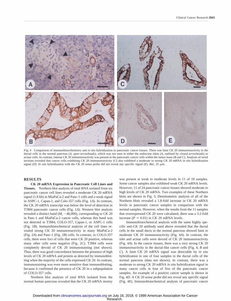
Immunohistochemical analysis with the same highly spe-cific anti-CK 20 antibody used above revealed that the ductalcells in the small ducts in the normal pancreas showed faint tomoderate CK 20 immunoreactivity (Fig. 4A). In contrast, theislet and acinar cells were devoid of CK 20 immunoreactivity(Fig. 4A). In the cancer tissues, there was a very strong CK 20immunoreactivity in the ductal-like cancer cells (Fig. 4,B andC). A faint CK 20 mRNA signal was detectable byin situhybridization in one of four samples in the ductal cells of thenormal pancreas (data not shown). In contrast, there was amoderate to strong CK 20 mRNAin situ hybridization signal inmany cancer cells in four of five of the pancreatic cancersamples. An example of a positive cancer sample is shown inFig. 4D. A CK 20 sense probe did not reveal any specific signal(Fig. 4E). Immunohistochemical analysis of pancreatic cancer
Fig. 4 Comparison of immunohistochemistry andin situ hybridization in pancreatic cancer tissues. There was faint CK 20 immunoreactivity in theductal cells in the normal pancreas (A, open arrowheads), which was not seen in either the endocrine islets (A, outlined byclosed arrowheads) oracinar cells. In contrast, intense CK 20 immunoreactivity was present in the pancreatic cancer cells within the tumor mass (B andC). Analysis of serialsections revealed that cancer cells exhibiting CK 20 immunoreactivity (C) also exhibited a moderate to strong CK 20 mRNAin situ hybridizationsignal (D).In situ hybridization with the CK 20 sense probe did not reveal any specific signal (E). Bar, 25 mm.
2843Clinical Cancer Research
Research. on July 16, 2018. © 1999 American Association for Cancerclincancerres.aacrjournals.org Downloaded from

metastases was performed in two pulmonary samples, revealingstrong CK 20 immunoreactivity in the metastatic cancer cells(Fig. 5).
CK 20 mRNA Expression in Colon Cancer Cell Linesand Tissues. Northern blot analysis of poly(A)1 RNA re-vealed a strong CK 20 mRNA signal in SW 1463 cells (Fig. 6A).In contrast, CK 20 was below level of detection in CaCo2 andSW 837 colorectal cancer cells (Fig. 6A). Similarly, Westernblot analysis showed a distinct band ofMr 46,000 correspondingto the size of the CK 20 protein in SW 1463, whereas SW 837and CaCo2 did not reveal the same band (Fig. 6B). There was
strong CK 20 immunoreactivity in SW 1463 cells (Fig. 2D). Incontrast, CaCo2 and SW 837 did not show a CK 20 staining(data not shown).
Northern blot analysis of RNA isolated from the normalportion of the colon revealed moderate to high levels of CK 20mRNA in 25 of 28 samples (Fig. 7). In contrast, in the corre-sponding cancer samples, CK 20 mRNA levels were weak tomoderate in 13 of 28 samples (Fig. 7). Only 1 cancer sample instage I disease and one cancer sample in stage IV diseaseexhibited high levels of CK 20 mRNA. Thus, densitometric
Fig. 5 CK 20 immunoreactivity in lung metastasis originating from pancreatic cancer. Strong CK 20 immunostaining is clearly evident in thepancreatic cancer cells within the lung parenchyma.A, low power view.Solid arrow,region shown under high power view in panelB. Bar, 25 mm.
Fig. 6 Expression of CK 20 in cultured colorectal cancer cells.A,Northern blotting. Poly(A)1-RNA (2 mg/lane) was prepared from theindicated colorectal cancer cell lines and hybridized with32P-labeledprobes (500,000 cpm/ml) specific for CK 20. A 7S ribosomal cDNAprobe (50,000 cpm/ml) was used as a loading and transfer control.Exposure times were 12 h for CK 20 and 4 h for 7S.B, Western blotting.Lysates from three colorectal cancer cell lines were probed with a highlyspecific CK 20 antibody (see “Materials and Methods”). ERK2 wasused as a loading and transfer control. Exposure times were 2 min forCK 20 and 1 min for ERK2.
Fig. 7 Expression of CK 20 in colon tissues by Northern blotting.Total RNA (20mg/lane) was prepared from colonic tissues from stageI through IV disease and hybridized with32P-labeled cDNA probes(500,000 cpm/ml) specific for CK 20. A 7S ribosomal cDNA probe(50,000 cpm/ml) was used as a loading and transfer control. Exposuretimes were 12 h for CK 20 and 4 h for 7S.
2844CK 20 Expression in Pancreatic and Colorectal Cancer
Research. on July 16, 2018. © 1999 American Association for Cancerclincancerres.aacrjournals.org Downloaded from
analysis revealed that CK 20 mRNA levels were 5-fold (P 50.009) higher in normal samples from stage I disease and3.3-fold (P 5 0.009) higher in normal samples from stage IVdisease by comparison to the corresponding cancer samples.There was no significant difference in CK 20 expression be-tween the four disease stages.
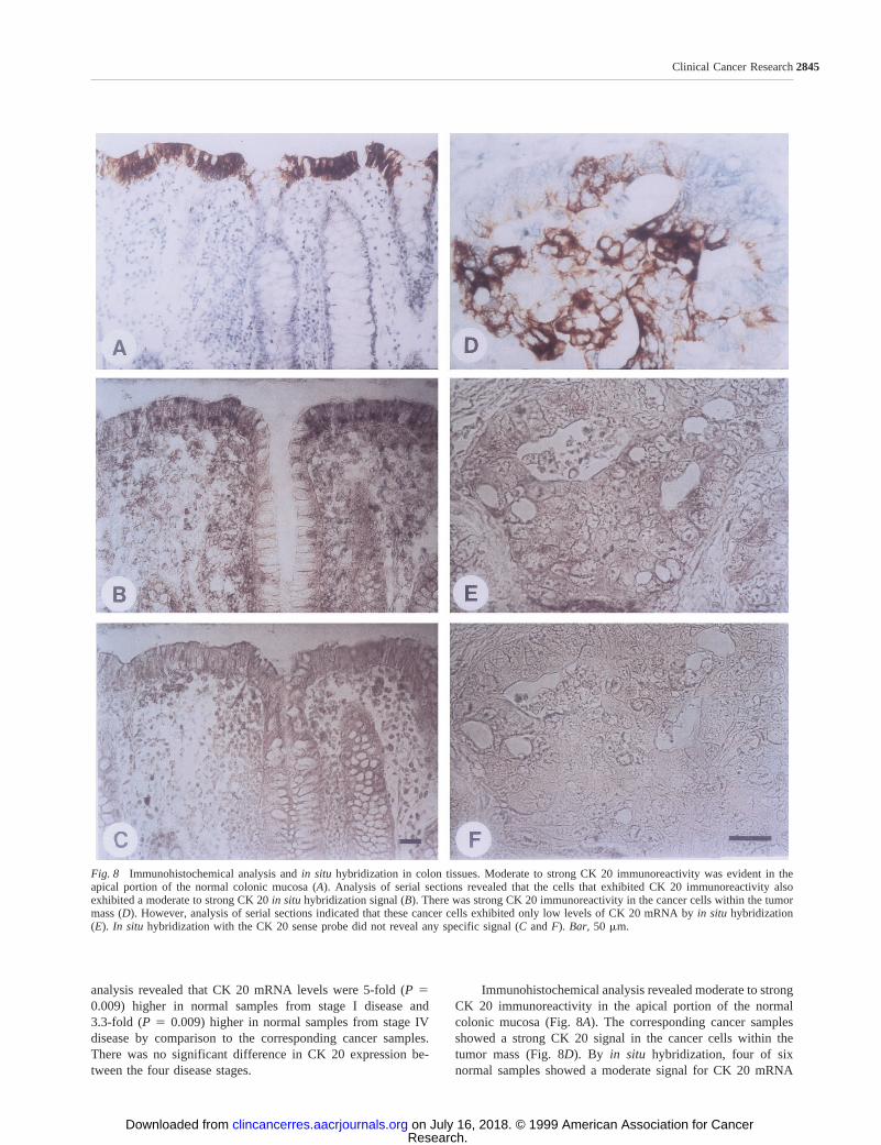
Immunohistochemical analysis revealed moderate to strongCK 20 immunoreactivity in the apical portion of the normalcolonic mucosa (Fig. 8A). The corresponding cancer samplesshowed a strong CK 20 signal in the cancer cells within thetumor mass (Fig. 8D). Byin situ hybridization, four of sixnormal samples showed a moderate signal for CK 20 mRNA
Fig. 8 Immunohistochemical analysis andin situ hybridization in colon tissues. Moderate to strong CK 20 immunoreactivity was evident in theapical portion of the normal colonic mucosa (A). Analysis of serial sections revealed that the cells that exhibited CK 20 immunoreactivity alsoexhibited a moderate to strong CK 20in situ hybridization signal (B). There was strong CK 20 immunoreactivity in the cancer cells within the tumormass (D). However, analysis of serial sections indicated that these cancer cells exhibited only low levels of CK 20 mRNA byin situ hybridization(E). In situ hybridization with the CK 20 sense probe did not reveal any specific signal (C andF). Bar, 50 mm.
2845Clinical Cancer Research
Research. on July 16, 2018. © 1999 American Association for Cancerclincancerres.aacrjournals.org Downloaded from

that was present in the surface epithelium and the mucosalcrypts (Fig. 8B). In contrast, two of six colon cancer samplesexhibited a weak CK 20in situhybridization signal in the cancercells (Fig. 8E), although most of these samples exhibited mod-erately intense CK 20 immunoreactivity. This discrepancy be-tween thein situ hybridization signal and the intensity of theimmunostaining raises the possibility that the CK 20 protein hasa long half-life. A CK 20 sense probe did not reveal any specificsignal (Fig. 8,C andF).
DISCUSSIONSince its detection in 1992 (11) and the subsequent report
of the complete nucleotide sequence 1 year later (17), CK 20 hasbecome an important tool for the characterization of primaryand metastatic human adenocarcinomas. It has been shown thatCK 20 has a restricted range of expression in human tissues suchas intestinal epithelium, gastric foveolar epithelium, urothelium,and Merkel cells in the skin. Furthermore, CK 20 has noimmunological cross-reactivity with other members of the CKfamily, therefore allowing for a highly specific analysis oftissues by immunohistochemistry.
A large number of studies designed to detect disseminatedtumor cells by using RT-PCR techniques based on the expres-sion of CK 20 mRNA and other members of this family (34,36–40). These reports showed a clear correlation between tu-mor cell spread in bone marrow, venous blood, and lymphnodes, respectively, and stage of the disease in colorectal cancer.Furthermore, Weitzet al.(38) reported a dissemination of tumorcells in venous blood from patients undergoing surgery forcolorectal cancer disease. RT-PCR analysis using CK 20 as amarker has been especially successful in colorectal cancer (36),whereas in gastric and pancreatic cancer the findings were farless convincing (37).
In the present study, we characterized CK 20 expression inpancreatic and colonic tissues. In the normal pancreas, CK 20immunoreactivity was faintly evident in the ductal cells. Thesefindings contrast with previous immunohistochemical studies,which failed to demonstrate CK 20 immunoreactivity in normalpancreatic tissues (17, 18). However, in agreement with ourresults, CK 20 immunoreactivity has been detected in the ductalcells of the rat pancreas (18–21). Furthermore, in the presentstudy, the expression of CK 20 mRNA in the normal pancreaswas confirmed by Northern blotting. In contrast to the relativelylow expression of CK 20 in the normal pancreas, the normalcolon tissue samples exhibited a strong CK 20 immunoreactiv-ity, which was restricted to the upper layer of the colonicmucosa and the mucosal crypts. These results were consistentwith previous immunohistochemical studies (17, 33). In addi-tion, in the present study,in situ hybridization confirmed thatCK 20 mRNA was expressed in the same cells in the normalcolon mucosa as shown by immunohistochemistry.
In pancreatic cancer, CK 20 immunoreactivity was ex-tremely strong in the duct-like cancer cells. This is in contrast toprevious immunohistochemical studies, which reported thatonly a small number of pancreatic cancers exhibited a signifi-cant level of CK 20 immunoreactivity (11, 17, 33). Our immu-nohistochemical findings in pancreatic cancer are supported bytwo observations: (a) CK 20 mRNA levels by Northern blot
analysis were increased in a subgroup of pancreatic cancertissues by comparison to the normal pancreas; (b) in situ hy-bridization analysis revealed increased CK 20 mRNA signals inthe cancer cells within the pancreatic tumor mass. However,CP-like lesions adjacent to the cancer also revealed an increasedexpression of CK 20, indicating that elevated CK 20 expressionwas not specific to the cancer cells in this malignancy.
There was a good correlation between CK 20 expression byNorthern blot analysis and immunoblotting in the colorectalcancer cell lines. However, in contrast to the findings in pan-creatic cancer, in the colorectal cancer samples, CK 20 mRNAwas decreased by comparison with the normal colon samples.This decrease was observed in all four stages and was confirmedby the relatively low CK 20 mRNAin situ hybridization signalin the cancer cells within the tumor mass. In contrast, normalsamples exhibited markedly increased levels of CK 20 in im-munohistochemistry andin situ hybridization in the apicalmucosa. The relative abundance of CK 20 in the normal gas-trointestinal mucosa by immunohistochemistry andin situ hy-bridization and its relative low abundance in the colorectalcancer cells are consistent with the observation that CK 20expression is increased with differentiation. Nonetheless, thecolon cancer cells are capable of expressing CK 20, which is thereason why CK 20 has been a useful marker for colorectalcancer (11, 17, 22–24, 31–33).
Metastatic cancer of unknown origin occasionally presentssignificant clinical dilemmas with respect to diagnosis and ther-apy. The ability of metastatic cancer cells to retain the capacityto express markers of differentiation such as CK 20 has beenhelpful in establishing clinically the primary source of meta-static lesions. The present study documents that CK 20 isoverexpressed in a significant number of pancreatic cancers.Furthermore, we show that metastatic lesions from pancreaticcancer exhibit strong CK 20 immunoreactivity. To our knowl-edge, this is the first report of CK 20 overexpression in anyhuman malignancy. We propose, therefore, that CK 20 may alsobe a more valuable marker for metastatic pancreatic cancer thanhas been appreciated previously.
REFERENCES1. Eichner, R., Bonitz, P., and Sun, T. T. Classification of epidermalkeratins according to their immunoreactivity, isoelectric point, andmode of expression. J. Cell. Biol.,98: 1388–1396, 1984.2. Hatzfeld, M., and Weber, K. The coiled coil ofin vitro assembledkeratin filaments is a heterodimer of type I and II keratins: use ofsite-specific mutagenesis and recombinant protein expression. J. Cell.Biol., 110: 1199–1210, 1990.3. Steinert, P. M. The two-chain coiled-coil molecule of native epider-mal keratin intermediate filaments is a type I-type II heterodimer.J. Biol. Chem.,265: 8766–8774, 1990.4. Moll, R., Franke, W. W., Schiller, D., Geiger, B., and Krepler, R. Thecatalog of human cytokeratins: patterns of expression in normal epithe-lia, tumors and cultured cells. Cell,31: 11–24, 1982.5. Moll, R., Schiller, D. L., and Franke, W. W. Identification of proteinIT of the intestinal cytoskeleton as a novel type I cytokeratin withunusual properties and expression patterns. J. Cell. Biol.,111:567–580,1990.6. Van Muijen, G. N. P., Ruiter, D. J., Franke, W. W., Achtstaetter, T.,Haasnoot, W. H. B., Ponec, M., and Warnaar, S. O. Cell type hetero-geneity of cytokeratin expression in complex epithelia and carcinomas
2846CK 20 Expression in Pancreatic and Colorectal Cancer
Research. on July 16, 2018. © 1999 American Association for Cancerclincancerres.aacrjournals.org Downloaded from
as demonstrated by monoclonal antibodies specific for cytokeratins nos.4 and 13. Exp. Cell. Res.,162: 97–113, 1986.7. Wu, Y. J., Parker, L. M., Binder, N. E., Beckett, M. A., Sinard, J. H.,Griffiths, C. T., and Rheinwald, J. G. The mesothelial keratins: a newfamily of cytoskeletal proteins identified in cultured mesothelial cellsand nonkeratinizing epithelia. Cell,31: 693–703, 1982.8. Osborn, M., and Weber, K. Tumor diagnosis by intermediate fila-ment typing: a novel tool for surgical pathology. Lab. Investig.,48:372–394, 1983.9. Nagle, R. B. Intermediate filaments: a review of the basic biology.Am. J. Surg. Pathol.,12 (Suppl. 1):4–16, 1988 .10. Nagle, R. B. Intermediate filaments. Efficacy in surgical pathologicdiagnosis. Am. J. Clin. Pathol.,91(Suppl. 1):14–18, 1989 .11. Moll, R., Loewe, A., Laufer, J., and Franke, W. W. Cytokeratin 20in human carcinomas. Am. J. Pathol.,140: 427–447, 1992.12. Fuchs, E., and Green, H. Changes in keratin gene expression duringterminal differentiation of the keratinocytes. Cell,19: 1033–1042, 1980.13. Franke, W. W., Schiller, D. L., Moll, R., Winter, S., Schmid, E.,Engelbrecht, I., Denk, H., Krepler, R., and Plazter, B. Diversity ofcytokeratins. Differentiation specific expression of cytokeratin polypep-tides in epithelial cells and tissues. J. Mol. Biol.,153: 933–959, 1981.14. Tseng, S. C. G., Jarvinen, M. J., Nelson, W. G., Huang, J. W.,Woodcock-Mitchell, J., and Sun, T. T. Correlation of specific keratinswith different types of epithelial differentiation: monoclonal antibodystudies. Cell,30: 361–372, 1982.15. Fuchs, E., Tyner, A. L., Giudice, G. J., Marchuk, D., Ray-Chaudhury, H., and Rosenberg, M. The human keratin genes and theirdifferential expression. Curr. Top. Dev. Biol.,22: 5–34, 1987.16. Cooper, D., Schermer, A., and Sun, T. T. Classification of humanepithelia and their neoplasma using monoclonal antibodies to keratins:strategies, applications and limitations. Lab. Investig.,52: 243–256, 1985.17. Moll, R., Zimbelmann, R., Goldschmidt, M. D., Keith, M., Laufer, J.,Kasper, M., Koch, P. J., and Franke, W. W. The human gene encodingcytokeratin 20 and its expression during fetal development and in gastro-intestinal carcinomas. Differentiation (Camb.),53: 75–93, 1993.18. Bouwens, L., Lu, W. G., De Krijger, R., and Pipeleers, D. Keratinsas markers of duct-to-islet cytodifferentiation in fetal pancreas. Diabe-tologia,39: A63, 1996.19. Bouwens, L., Braet, F., and Heimberg, H. Identification of ratpancreatic duct cells by their expression of cytokeratins 7, 19 and 20invivo and after isolation and culture. J. Histochem. Cytochem.,43:245–253, 1995.20. Faa, G., Van Eyken, P., Roskams, T., Miyazaki, H., Serreli, S.,Ambu, R., and Desmet, V. J. Expression of cytokeratin 20 in developingrat liver and in experimental models of ductular and oval cell prolifer-ation. J. Hepatol.,29: 628–633, 1998.21. Bouwens, L. Cytokeratins and cell differentiation in the pancreas.J. Pathol.,184: 234–239, 1998.22. Tot, T. Adenocarcinomas metastatic to the liver: the value ofcytokeratins 20 and 7 in the search for unknown primary tumors. Cancer(Phila.),85: 171–177, 1999.23. Lagendijk, J. H., Mullink, H., Van Diest, H., Meijer, G., and Meijer,C. J. L. M. Tracing the origin of adenocarcinomas with unknown primaryusing immunohistochemistry. Hum. Pathol.,29: 491–497, 1998.24. Maeda, T., Kajiyama, K., Adachi, E., Takenaka, K, Sugimachi, K.,and Tsuneyoshi, M. The expression of cytokeratins 7, 19, and 20 inprimary and metastatic carcinomas of the liver. Mod. Pathol.,9: 901–909, 1996.25. Berezowski, K., Stastny, J. F., and Kornstein, M. J. Cytokeratins 7and 20 and carcinoembryonic antigen in ovarian and colonic carcinoma.Mod. Pathol.,9: 426–429, 1996.26. Sack, M. J., and Roberts, S. A. Cytokeratins 20 and 7 in thedifferential diagnosis of metastatic carcinoma in cytologic specimens.Diagn. Cytopathol.,16: 132–136, 1997.27. Loy, T. S., and Calaluce, R. D. Utility of cytokeratin immuno-staining in separating pulmonary adenocarcinomas from colonic adeno-carcinomas. Am. J. Clin. Pathol.,102: 764–767, 1994.
28. Young, R. H., and Hart, W. R. Metastatic intestinal carcinomassimulating primary ovarian clear cell carcinoma and secretory endo-metrioid carcinoma: a clinicopathologic and immunohistochemicalstudy of five cases. Am. J. Surg. Pathol.,22: 805–815, 1998.
29. Longatto Filho, A., Bisi, H., Alves, V. A., Kanamura, C. T.,Oyafuso, M. S., Bortolan, J., and Lombardo, V. Adenocarcinoma infemales detected in serous effusions. Cytomorphologic aspects andimmunocytochemical reactivity to cytokeratins 7 and 20. Acta Cytol.,41: 961–971, 1997.
30. Ronnett, B. M., Kurman, R. J., Shmookler, B. M., Sugarbaker,P. H., and Young, R. H. The morphologic spectrum of ovarian metas-tases of appendiceal adenocarcinomas: a clinicopathologic and immu-nohistochemical analysis of tumors often misinterpreted as primaryovarian tumors or metastatic tumors from other gastrointestinal sites.Am. J. Surg. Pathol.,21: 1144–1155, 1997.
31. Loy, T. S., Calaluce, R. D., and Keeney, G. L. Cytokeratin immu-nostaining in differentiating primary ovarian carcinoma from metastaticcolonic adenocarcinoma. Mod. Pathol.,9: 1040–1044, 1996.
32. Litle, V. R., Warren, R. S., Moore, D., II, and Pallavicini, M. G.Molecular cytogenetic analysis of cytokeratin 20-labeled cells in pri-mary tumors and bone marrow aspirates from colorectal carcinomapatients. Cancer (Phila.),79: 1664–1670, 1997.
33. Miettinen, M. Keratin 20: immunohistochemical marker for gastro-intestinal, urothelial, and Merkel cell carcinoma. Mod. Pathol.,8: 384–388, 1995.
34. Wyld, D. K., Selby, P., Perren, T. J., Jonas, S. K., Allen-Mersh,T. G., Wheeldon, J., and Burchill, S. A. Detection of colorectal cancerin peripheral blood by reverse-transcriptase polymerase chain reactionfor cytokeratin 20. Int. J. Cancer,79: 288–293, 1998.
35. Bostick, P. J., Chatterjee, S., Chi, D. D., Huynh, K. T., Giuliano,A. E., Cote, R., and Hoon, D. S. Limitations of specific reverse-transcriptase polymerase chain reaction markers in the detection ofmetastases in the lymph nodes and blood of breast cancer patients.J. Clin. Oncol.,16: 2632–2640, 1998.
36. Soeth, E., Roeder, C., Juhl, H., Krueger, U., Kremer, B., andKalthoff, H. The detection of disseminated tumor cells in bone marrowfrom colorectal-cancer patients by a cytokeratin-20-specific nested re-verse-transcriptase-polymerase-chain reaction is related to the stage ofdisease. Int. J. Cancer,69: 278–282, 1996.
37. Soeth, E., Vogel, I., Roeder, C., Juhl, H., Marxsen, J., Krueger, U.,Henne-Bruns, D., Kremer, B., and Kalthoff, H. Comparative analysis ofbone marrow and venous blood isolates from gastrointestinal cancerpatients for the detection of disseminated tumor cells using reversetranscription PCR. Cancer Res.,57: 3106–3110, 1997.
38. Weitz, J., Kienle, P., Lacroix, J., Willeke, F., Benner, A., Lehnert,T., Herfarth, C., and Von Knebel Doeberitz, M. Dissemination of tumorcells in patients undergoing surgery for colorectal cancer. Clin. CancerRes.,4: 343–348, 1998.
39. Funaki, N. O., Tanaka, J., Ohshio, G., Onodera, H., Maetani, S., andImamura, M. Cytokeratin 20 mRNA in peripheral venous blood ofcolorectal carcinoma patients. Br. J. Cancer,77: 1327–1332, 1998.
40. Futamura, M., Takagi, Y., Koumura, H., Kida, H., Tanemura, H.,Shimokawa, K., and Saji, S. Spread of colorectal cancer micrometasta-ses in regional lymph nodes by reverse transcriptase-polymerase chainreactions for carcinoembryonic antigen and cytokeratin 20. J. Surg.Oncol.,68: 34–40, 1998.
41. Parker, S. L., Tong, T., Bolden, S., and Wingo, P. A. Cancerstatistics. CA Cancer J. Clin.,47: 5–27, 1997.
42. Chomczynski, P., and Sacchi, N. Single-step method of RNA iso-lation by acid guanidinium thiocynate-phenol-chloroform extraction.Anal. Biochem.,162: 156–159, 1987.
43. Korc, M., Chandrasekar, B., Yamanaka, Y., Friess, H., Buechler,M. W., and Beger, H. G. Overexpression of the epidermal growth factorin human pancreatic cancer is associated with concomitant increase inthe levels of epidermal growth factor and transforming growth factora.J. Clin. Investig.,90: 1352–1360, 1992
2847Clinical Cancer Research
Research. on July 16, 2018. © 1999 American Association for Cancerclincancerres.aacrjournals.org Downloaded from

1999;5:2840-2847. Clin Cancer Res Stefan Wildi, Jörg Kleeff, Haruhisa Maruyama, et al. and Colorectal CancerCharacterization of Cytokeratin 20 Expression in Pancreatic
Updated version
http://clincancerres.aacrjournals.org/content/5/10/2840
Access the most recent version of this article at:
Cited articles
http://clincancerres.aacrjournals.org/content/5/10/2840.full#ref-list-1
This article cites 39 articles, 7 of which you can access for free at:
Citing articles
http://clincancerres.aacrjournals.org/content/5/10/2840.full#related-urls
This article has been cited by 7 HighWire-hosted articles. Access the articles at:
E-mail alerts related to this article or journal.Sign up to receive free email-alerts
Subscriptions
Reprints and
To order reprints of this article or to subscribe to the journal, contact the AACR Publications
Permissions
Rightslink site. Click on "Request Permissions" which will take you to the Copyright Clearance Center's (CCC)
.http://clincancerres.aacrjournals.org/content/5/10/2840To request permission to re-use all or part of this article, use this link
Research. on July 16, 2018. © 1999 American Association for Cancerclincancerres.aacrjournals.org Downloaded from