characterization of desulfovibrio salinus sp. nov., a
TRANSCRIPT
HAL Id: hal-01981629https://hal-amu.archives-ouvertes.fr/hal-01981629
Submitted on 15 Jan 2019
HAL is a multi-disciplinary open accessarchive for the deposit and dissemination of sci-entific research documents, whether they are pub-lished or not. The documents may come fromteaching and research institutions in France orabroad, or from public or private research centers.
L’archive ouverte pluridisciplinaire HAL, estdestinée au dépôt et à la diffusion de documentsscientifiques de niveau recherche, publiés ou non,émanant des établissements d’enseignement et derecherche français ou étrangers, des laboratoirespublics ou privés.
Distributed under a Creative Commons Attribution| 4.0 International License
Characterization of Desulfovibrio salinus sp. nov., aslightly halophilic sulfate-reducing bacterium isolated
from a saline lake in TunisiaZouhaier Ben Ali Gam, Abdoulaye Thioye, Jean-Luc Cayol, Manon Joseph,
Guy Fauque, Marc Labat
To cite this version:Zouhaier Ben Ali Gam, Abdoulaye Thioye, Jean-Luc Cayol, Manon Joseph, Guy Fauque, et al.. Char-acterization of Desulfovibrio salinus sp. nov., a slightly halophilic sulfate-reducing bacterium isolatedfrom a saline lake in Tunisia. International Journal of Systematic and Evolutionary Microbiology,Microbiology Society, 2018, 68 (3), pp.715-720. �10.1099/ijsem.0.002567�. �hal-01981629�
Downloaded from www.microbiologyresearch.org by
IP: 147.94.134.146
On: Tue, 15 Jan 2019 09:56:31
Characterization of Desulfovibrio salinus sp. nov., a slightlyhalophilic sulfate-reducing bacterium isolated from a salinelake in Tunisia
Zouhaier Ben Ali Gam,1 Abdoulaye Thioye,1,2 Jean-Luc Cayol,1 Manon Joseph,1 Guy Fauque1 and Marc Labat1,*
Abstract
A novel slightly halophilic sulfate-reducing bacterium, designated strain P1BSRT, was isolated from water of a saline lake in
Tunisia. Strain P1BSRT had motile (single polar flagellum), Gram-negative, rod-shaped, non-spore-forming cells, occurring
singly or in pairs. Strain P1BSRT grew at temperatures between 15 and 45�
C (optimum 40�
C), and in a pH range between 6
and 8.5 (optimum pH 6.7). The strain required NaCl for growth (1% w/v), and tolerated high NaCl concentration (up to 12%
w/v) with an optimum of 3% (w/v). Sulfate, thiosulfate and sulfite served as terminal electron acceptors, but not elemental
sulfur, fumarate, nitrate and nitrite. Strain P1BSRT utilized lactate, pyruvate, formate, D-fructose and glycerol as carbon and
energy sources. The main cellular fatty acid was C16 : 0 (50.8%). The genomic DNA G+C content was 47.7mol%. Phylogenetic
analysis of 16S rRNA gene sequence similarity indicated that strain P1BSRT was affiliated to the genus Desulfovibrio, with
the type strains Desulfovibrio salexigens (96.51%), Desulfovibrio zosterae (95.68%), Desulfovibrio hydrothermalis (94.81%) and
Desulfovibrio ferrireducens (94.73%) as its closest phylogenetic relatives. On the basis of genotypic, phenotypic and
phylogenetic characteristics, it is proposed to assign strain P1BSRT to a novel species of the genus Desulfovibrio,
Desulfovibrio salinus sp. nov. The type strain is P1BSRT (=DSM 101510T=JCM 31065T).
Sulfate-reducing prokaryotes (SRPs) are mainly chemohe-terotrophs, both Bacteria and Archaea (250 species of 65genera), that can use sulfate as a terminal electron acceptorin their energy metabolism [1, 2]. Owing to their broad met-abolic capacities, sulfate-reducing bacteria (SRB) areimportant in the mineralization of organic matter in anoxicmarine sediments. Dissimilatory sulfate reduction has beenobserved in various hypersaline environments such as salt-erns, the Dead Sea and the Great Salt Lake [3]. However,most of the halophilic SRPs isolated so far are marine orslightly halophilic microorganisms [with optimum salinityranging from 1 to 4% (w/v) NaCl] belonging to several gen-era of SRPs, including Desulfovibrio, Desulfonatronovibrioand Desulfonatronobacter species [3–6]. The first SRB spe-cies belonging to the genus Desulfovibrio isolated from ahypersaline environment was Desulfovibrio salexigens,which did not grow at NaCl concentrations higher than12% (w/v) [7]. Cord-Ruwish [8] then isolated severalstrains of SRBs from hypersaline oilfield water containingabout 10% (w/v) NaCl. One isolate grew slowly up to 27%(w/v) NaCl but has not been described in more detail since.
One year later, Trüper and Galinski [9] isolated a few SRBstrains from hot brines in the Red Sea that were similar toDesulfovibrio halophilus, a moderately halophilic sulfatereducer isolated by Caumette et al. [10] from the hypersa-line Solar Lake in Sinai. The latter isolate grew in salinityranging from 3 to 18% (w/v), and optimally at 6–7% (w/v)NaCl. However, since 1991, no novel SRB species belongingto the genus Desulfovibrio has been characterized that growsin salt concentrations above 10% NaCl (w/v). No pure cul-ture of extreme halophilic SRB (i.e. growing under saturat-ing-salt conditions) has been isolated until now.
In this study, we report the isolation and characterization ofa mesophilic, slightly halophilic SRB isolated from watersamples of a Tunisian saline lake that is able to grow in upto 12% NaCl (w/v), but grows optimally at 3% (w/v). ThisSRB isolate is proposed to represent a novel species of thegenus Desulfovibrio.
Water samples were collected from a continental salinelake located in the middle-east of Tunisia and transportedto the laboratory at ambient temperature. Bacteria were
Author affiliations:1Laboratoire de Microbiologie IRD, Aix-Marseille Universit�e, Universit�e du Sud Toulon-Var, CNRS/INSU, IRD, MI0 UM110, 163
avenue de Luminy, case 925, F-13288 Marseille cedex 9, France; 2Laboratoire de Microbiologie Appliqu�ee et de G�enie Industriel, Ecole Sup�erieurePolytechnique, Universit�e Cheikh Anta Diop, BP 5005 Dakar-Fann, Dakar, S�en�egal.*Correspondence: Marc Labat, [email protected]: Desulfovibrio; slightly halophilic; sulfate-reducing bacterium; saline lake; Tunisia.Abbreviations: SRB, sulfate-reducing bacteria; SRP, sulfate-reducing prokaryote.The Genbank/EMBL/DDBJ accession number for the 16S rRNA gene sequence of strain P1BSRT is KT767983.
TAXONOMIC DESCRIPTION
Ben Ali Gam et al., Int J Syst Evol Microbiol 2018;68:715–720
DOI 10.1099/ijsem.0.002567
002567 ã 2018 IUMS
715
Downloaded from www.microbiologyresearch.org by
IP: 147.94.134.146
On: Tue, 15 Jan 2019 09:56:31
isolated and cultivated under strict anaerobiosis accordingto the Hungate technique [11], modified for use with syrin-ges [12, 13].
The basal culture medium (BM) for isolation included (perlitre): 0.3 g KH2PO4, 0.2 g K2HPO4, 0.3 g NH4Cl, 30 g NaCl,0.5 g KCl, 0.1 g CaCl2, 0.4 g MgCl2, 0.5 g cysteine HCl, 0.5 gyeast extract (Difco), 1ml mineral element solution and1ml 0.1% (w/v) resazurin [14]. The pH was adjusted to 7.2with 10 M KOH solution and the medium was boiled undera stream of O2-free N2 gas and cooled to room temperature.Aliquots of 5ml were dispensed into Hungate tubes underN2/CO2 (80 : 20, v/v) and subsequently sterilized byautoclaving at 120
�
C for 20min. Before inoculation, 0.1ml10% (w/v) NaHCO3, 0.1ml 2% (w/v) Na2S.9H2O, 20mMlactate and 20mM sulfate were injected from sterile stocksolutions into the tubes. Na2S.9H2O as a reducing agent wasomitted when H2S production was determined [8].
Enrichments were performed in Hungate tubes or serumbottles inoculated with 10% (v/v) sample and incubated at30
�
C. The culture was purified by repeated use of the Hun-gate roll tube method, using agar solid medium (0.8%w/v),and transferred to liquid medium.
The pH, temperature and NaCl concentration ranges forgrowth were determined in duplicate experiments using BMsupplemented with lactate (20mM) as electron donor andsulfate (20mM) as previously described [15]. The pH (from5 to 9) of the culture medium was adjusted by injecting ali-quots of anaerobic stock solution into Hungate tubes con-taining 100mM HCl for low pH or either 10% NaHCO3
(w/v) or 8% Na2CO3 (w/v) for high pH. Water baths wereused to incubate bacterial cultures from 15 to 50
�
C. NaClrequirement was determined by directly weighing NaCl intothe Hungate tubes before dispensing the medium. Cultureswere subcultured at least twice under the same experimentalconditions before determination of growth rates.
Bacterial growth was monitored by measuring the increasein turbidity at 580 nm after inserting Hungate tubes into thecuvette holder of a Cary 50 UV–vis spectrophotometer(Varian). H2S production was determined photometricallyas colloidal CuS following the method described in [8].
Morphological characteristics and purity of strains werechecked under an Optiphot (Nikon) phase-contrast micro-scope. Presence of spores was analysed by phase-contrastmicroscopy observations of cultures and after pasteurizationtests performed for 10 and 20min at 80, 90 and 100
�
C.
Culture of the isolate (strain P1BSRT) was stopped at theend of the exponential phase and sent to the DeutscheSammlung von Mikroorganismen und Zellkulturen(DSMZ) for cellular fatty acid analysis. Fatty acids wereextracted using the Miller method [16] as modified byKuykendall et al. [17], and the cellular fatty acid profile wasanalysed by gas chromatography using the Microbial Identi-fication System (MIDI, Sherlock Version 6.1; database,TSBA40; gas chromatograph, model 6890N; Agilent
Technologies). G+C content was determined by HPLC asdescribed by [18].
Substrate utilization was tested in the presence of 0.5 gyeast extract l�1. Substrates (20mM except where indi-cated) included lactate, pyruvate, acetate, formate, fuma-rate, malate, succinate, propionate, methanol (0.1%w/v),ethanol (0.1%w/v), butanol (0.1%w/v), glycerol (0.1 %w/v), D-fructose, D-glucose and H2/CO2 (20 : 80 v/v). Ele-mental sulfur (1%w/v), sulfate (20mM), thiosulfate(20mM), sulfite (2mM), fumarate (10mM), nitrate(10mM) and nitrite (2mM) were tested as potential termi-nal electron acceptors.
Extraction and purification of total DNA followed by ampli-fication and sequencing of the 16S rRNA gene were per-formed as previously described [15]. The 16S rRNA genesequence was then compared with available sequencesfound in GenBank using the BLASTN search [19]. Evolution-ary history was inferred using the neighbour-joiningmethod [20]. A multiple alignment was built using theMUSCLE program [21] implemented in MEGA6 [22]. Sequencepositions with alignment uncertainty and gaps were omittedfrom the analysis. Evolutionary analyses were conducted inMEGA6 using the neighbour-joining method. Evolutionarydistances were computed using the maximum-composite-likelihood method [23]. The final dataset consisted of a totalof 1213 positions. Branch robustness was estimated by thenon-parametric bootstrap procedure implemented inMEGA6 (1000 replicates of the original dataset). The tree was
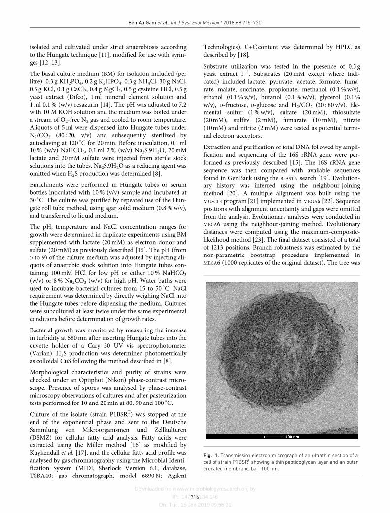
Fig. 1. Transmission electron micrograph of an ultrathin section of a
cell of strain P1BSRT showing a thin peptidoglycan layer and an outer
crenated membrane; bar, 100 nm.
Ben Ali Gam et al., Int J Syst Evol Microbiol 2018;68:715–720
716
Downloaded from www.microbiologyresearch.org by
IP: 147.94.134.146
On: Tue, 15 Jan 2019 09:56:31
drawn to scale, with branch lengths measured in number ofsubstitutions per site [24].
Several colonies developed after incubation at 30�
C and
were picked separately. Colonies were black and circular
with diameters ranging from 1.0 to 2.0mm after 1 week of
incubation at 30�
C. The serial dilution process was
repeated several times until the isolates were deemed to be
axenic. Purity of the isolates was checked by microscopy
and inoculation in sulfate-free medium containing yeast
extract and D-glucose to confirm the absence of contamina-
tion by fermentative microorganisms. Several strains were
isolated that showed similar morphology, size and meta-
bolic profiles, and the same phylogenic inference was
obtained for all of them. One strain, designated P1BSRT,
was selected and used for further metabolic and physiologi-
cal characterization.
Cells of strain P1BSRT were Gram-stain-negative rods (0.7–
0.9�2.3 µm) when grown on medium containing lactate as
an electron donor and sulfate as a terminal electron accep-
tor. Ultrathin sections showed a typical Gram-negative cell
wall with a thin peptidoglycan layer and a crenated outer
membrane (Fig. 1).
The optimal temperature for growth was 40�
C (range 15–45
�
C) and the optimum pH was 6.7 (range pH 6–8.5). Thestrain required a minimum of 1% (w/v) NaCl for growth,and tolerated up to 12% (w/v); optimum growth occurredwith 3% (w/v) NaCl. All substrates were used by strainP1BSRT with incomplete oxidation. Amongst the substratestested, only lactate, pyruvate, formate, glycerol and D-fruc-tose were used as electron donors. End products of pyru-vate, fumarate and malate were not fermented. Thiosulfate,sulfate and sulfite, but not fumarate, elemental sulfur,nitrate or nitrite served as terminal electron acceptors.Malate, succinate, methanol, ethanol, butanol, D-glucose,acetate, propionate and H2/CO2 did not support growth.The presence of yeast extract in minimal medium with lac-tate as the only energy and carbon source was required forgrowth.
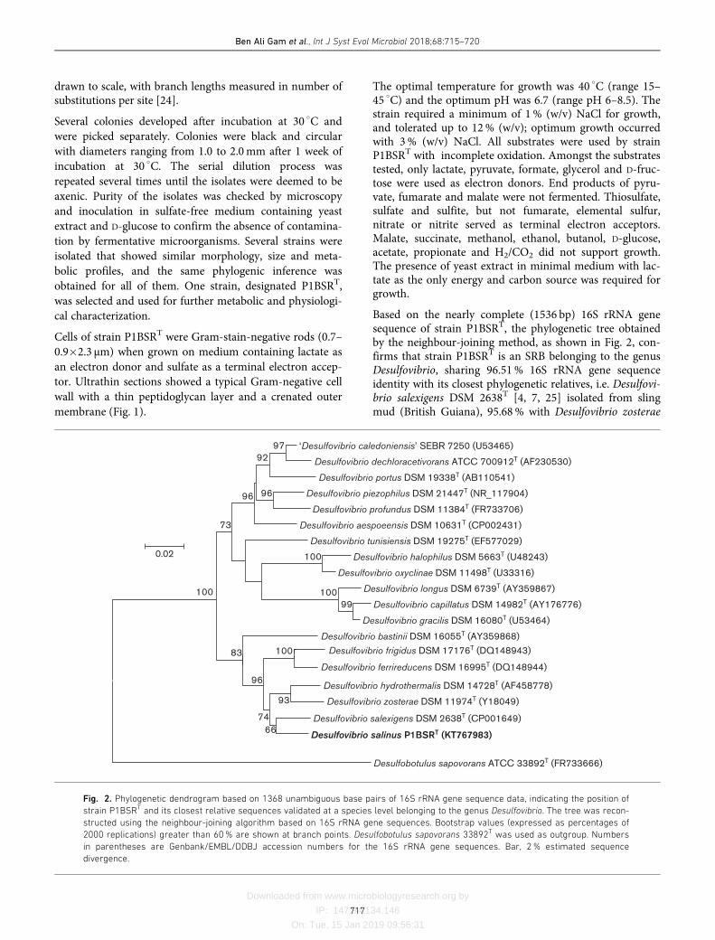
Based on the nearly complete (1536 bp) 16S rRNA genesequence of strain P1BSRT, the phylogenetic tree obtainedby the neighbour-joining method, as shown in Fig. 2, con-firms that strain P1BSRT is an SRB belonging to the genusDesulfovibrio, sharing 96.51% 16S rRNA gene sequenceidentity with its closest phylogenetic relatives, i.e. Desulfovi-brio salexigens DSM 2638T [4, 7, 25] isolated from slingmud (British Guiana), 95.68% with Desulfovibrio zosterae
‘Desulfovibrio caledoniensis’ SEBR 7250 (U53465)
Desulfovibrio dechloracetivorans ATCC 700912T (AF230530)
Desulfovibrio portus DSM 19338T (AB110541)
Desulfovibrio piezophilus DSM 21447T (NR_117904)
Desulfovibrio profundus DSM 11384T (FR733706)
Desulfovibrio aespoeensis DSM 10631T (CP002431)
Desulfovibrio tunisiensis DSM 19275T (EF577029)
Desulfovibrio halophilus DSM 5663T (U48243)
Desulfovibrio oxyclinae DSM 11498T (U33316)
Desulfovibrio longus DSM 6739T (AY359867)
Desulfovibrio capillatus DSM 14982T (AY176776)
Desulfovibrio gracilis DSM 16080T (U53464)
Desulfovibrio bastinii DSM 16055T (AY359868)
Desulfovibrio frigidus DSM 17176T (DQ148943)
Desulfovibrio ferrireducens DSM 16995T (DQ148944)
Desulfovibrio hydrothermalis DSM 14728T (AF458778)
Desulfovibrio zosterae DSM 11974T (Y18049)
Desulfovibrio salexigens DSM 2638T (CP001649)
Desulfovibrio salinus P1BSRT (KT767983)
Desulfobotulus sapovorans ATCC 33892T (FR733666)
99100
100
97
100
66
74
96
83
73
96
92
96
93
0.02
100
Fig. 2. Phylogenetic dendrogram based on 1368 unambiguous base pairs of 16S rRNA gene sequence data, indicating the position of
strain P1BSRT and its closest relative sequences validated at a species level belonging to the genus Desulfovibrio. The tree was recon-
structed using the neighbour-joining algorithm based on 16S rRNA gene sequences. Bootstrap values (expressed as percentages of
2000 replications) greater than 60% are shown at branch points. Desulfobotulus sapovorans 33892T was used as outgroup. Numbers
in parentheses are Genbank/EMBL/DDBJ accession numbers for the 16S rRNA gene sequences. Bar, 2% estimated sequence
divergence.
Ben Ali Gam et al., Int J Syst Evol Microbiol 2018;68:715–720
717
Downloaded from www.microbiologyresearch.org by
IP: 147.94.134.146
On: Tue, 15 Jan 2019 09:56:31
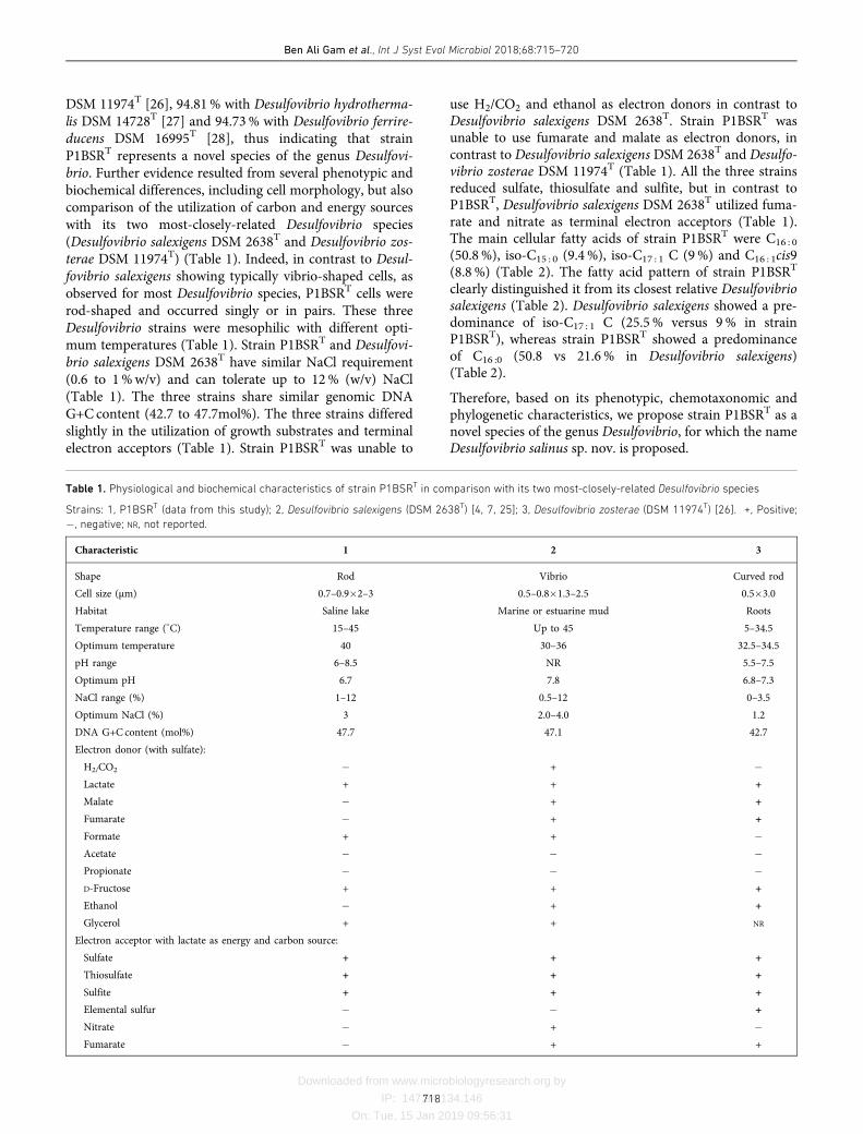
DSM 11974T [26], 94.81% with Desulfovibrio hydrotherma-lis DSM 14728T [27] and 94.73% with Desulfovibrio ferrire-ducens DSM 16995T [28], thus indicating that strainP1BSRT represents a novel species of the genus Desulfovi-brio. Further evidence resulted from several phenotypic andbiochemical differences, including cell morphology, but alsocomparison of the utilization of carbon and energy sourceswith its two most-closely-related Desulfovibrio species(Desulfovibrio salexigens DSM 2638T and Desulfovibrio zos-terae DSM 11974T) (Table 1). Indeed, in contrast to Desul-fovibrio salexigens showing typically vibrio-shaped cells, asobserved for most Desulfovibrio species, P1BSRT cells wererod-shaped and occurred singly or in pairs. These threeDesulfovibrio strains were mesophilic with different opti-mum temperatures (Table 1). Strain P1BSRT and Desulfovi-brio salexigens DSM 2638T have similar NaCl requirement(0.6 to 1%w/v) and can tolerate up to 12% (w/v) NaCl(Table 1). The three strains share similar genomic DNAG+C content (42.7 to 47.7mol%). The three strains differedslightly in the utilization of growth substrates and terminalelectron acceptors (Table 1). Strain P1BSRT was unable to
use H2/CO2 and ethanol as electron donors in contrast toDesulfovibrio salexigens DSM 2638T. Strain P1BSRT wasunable to use fumarate and malate as electron donors, incontrast to Desulfovibrio salexigens DSM 2638T and Desulfo-vibrio zosterae DSM 11974T (Table 1). All the three strainsreduced sulfate, thiosulfate and sulfite, but in contrast toP1BSRT, Desulfovibrio salexigens DSM 2638T utilized fuma-rate and nitrate as terminal electron acceptors (Table 1).The main cellular fatty acids of strain P1BSRT were C16 : 0
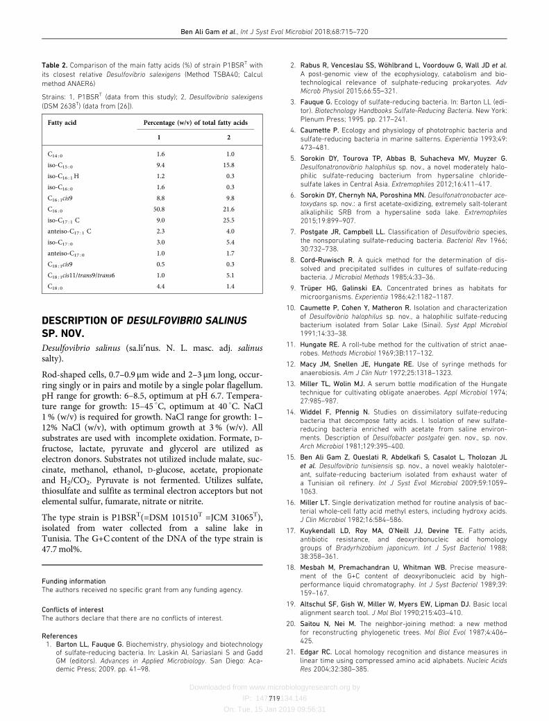
(50.8%), iso-C15 : 0 (9.4%), iso-C17 : 1 C (9%) and C16 : 1cis9(8.8%) (Table 2). The fatty acid pattern of strain P1BSRT
clearly distinguished it from its closest relative Desulfovibriosalexigens (Table 2). Desulfovibrio salexigens showed a pre-dominance of iso-C17 : 1 C (25.5% versus 9% in strainP1BSRT), whereas strain P1BSRT showed a predominanceof C16 :0 (50.8 vs 21.6% in Desulfovibrio salexigens)(Table 2).
Therefore, based on its phenotypic, chemotaxonomic andphylogenetic characteristics, we propose strain P1BSRT as anovel species of the genus Desulfovibrio, for which the nameDesulfovibrio salinus sp. nov. is proposed.
Table 1. Physiological and biochemical characteristics of strain P1BSRT in comparison with its two most-closely-related Desulfovibrio species
Strains: 1, P1BSRT (data from this study); 2, Desulfovibrio salexigens (DSM 2638T) [4, 7, 25]; 3, Desulfovibrio zosterae (DSM 11974T) [26]. +, Positive;
�, negative; NR, not reported.
Characteristic 1 2 3
Shape Rod Vibrio Curved rod
Cell size (µm) 0.7–0.9�2–3 0.5–0.8�1.3–2.5 0.5�3.0
Habitat Saline lake Marine or estuarine mud Roots
Temperature range (�
C) 15–45 Up to 45 5–34.5
Optimum temperature 40 30–36 32.5–34.5
pH range 6–8.5 NR 5.5–7.5
Optimum pH 6.7 7.8 6.8–7.3
NaCl range (%) 1–12 0.5–12 0–3.5
Optimum NaCl (%) 3 2.0–4.0 1.2
DNA G+C content (mol%) 47.7 47.1 42.7
Electron donor (with sulfate):
H2/CO2 � + �
Lactate + + +
Malate � + +
Fumarate � + +
Formate + + �
Acetate � � �
Propionate � � �
D-Fructose + + +
Ethanol � + +
Glycerol + + NR
Electron acceptor with lactate as energy and carbon source:
Sulfate + + +
Thiosulfate + + +
Sulfite + + +
Elemental sulfur � � +
Nitrate � + �
Fumarate � + +
Ben Ali Gam et al., Int J Syst Evol Microbiol 2018;68:715–720
718
Downloaded from www.microbiologyresearch.org by
IP: 147.94.134.146
On: Tue, 15 Jan 2019 09:56:31
DESCRIPTION OF DESULFOVIBRIO SALINUS
SP. NOV.
Desulfovibrio salinus (sa.li¢nus. N. L. masc. adj. salinussalty).
Rod-shaped cells, 0.7–0.9 µm wide and 2–3 µm long, occur-ring singly or in pairs and motile by a single polar flagellum.pH range for growth: 6–8.5, optimum at pH 6.7. Tempera-ture range for growth: 15–45
�
C, optimum at 40�
C. NaCl1% (w/v) is required for growth. NaCl range for growth: 1–12% NaCl (w/v), with optimum growth at 3% (w/v). Allsubstrates are used with incomplete oxidation. Formate, D-fructose, lactate, pyruvate and glycerol are utilized aselectron donors. Substrates not utilized include malate, suc-cinate, methanol, ethanol, D-glucose, acetate, propionateand H2/CO2. Pyruvate is not fermented. Utilizes sulfate,thiosulfate and sulfite as terminal electron acceptors but notelemental sulfur, fumarate, nitrate or nitrite.
The type strain is P1BSRT(=DSM 101510T =JCM 31065T),isolated from water collected from a saline lake inTunisia. The G+C content of the DNA of the type strain is47.7mol%.
Funding information
The authors received no specific grant from any funding agency.
Conflicts of interest
The authors declare that there are no conflicts of interest.
References
1. Barton LL, Fauque G. Biochemistry, physiology and biotechnologyof sulfate-reducing bacteria. In: Laskin AI, Sariaslani S and GaddGM (editors). Advances in Applied Microbiology. San Diego: Aca-demic Press; 2009. pp. 41–98.
2. Rabus R, Venceslau SS, Wöhlbrand L, Voordouw G, Wall JD et al.
A post-genomic view of the ecophysiology, catabolism and bio-technological relevance of sulphate-reducing prokaryotes. Adv
Microb Physiol 2015;66:55–321.
3. Fauque G. Ecology of sulfate-reducing bacteria. In: Barton LL (edi-
tor). Biotechnology Handbooks Sulfate-Reducing Bacteria. New York:
Plenum Press; 1995. pp. 217–241.
4. Caumette P. Ecology and physiology of phototrophic bacteria and
sulfate-reducing bacteria in marine salterns. Experientia 1993;49:473–481.
5. Sorokin DY, Tourova TP, Abbas B, Suhacheva MV, Muyzer G.
Desulfonatronovibrio halophilus sp. nov., a novel moderately halo-philic sulfate-reducing bacterium from hypersaline chloride-sulfate lakes in Central Asia. Extremophiles 2012;16:411–417.
6. Sorokin DY, Chernyh NA, Poroshina MN. Desulfonatronobacter ace-
toxydans sp. nov.: a first acetate-oxidizing, extremely salt-tolerantalkaliphilic SRB from a hypersaline soda lake. Extremophiles
2015;19:899–907.
7. Postgate JR, Campbell LL. Classification of Desulfovibrio species,
the nonsporulating sulfate-reducing bacteria. Bacteriol Rev 1966;30:732–738.
8. Cord-Ruwisch R. A quick method for the determination of dis-
solved and precipitated sulfides in cultures of sulfate-reducingbacteria. J Microbiol Methods 1985;4:33–36.
9. Trüper HG, Galinski EA. Concentrated brines as habitats for
microorganisms. Experientia 1986;42:1182–1187.
10. Caumette P, Cohen Y, Matheron R. Isolation and characterization
of Desulfovibrio halophilus sp. nov., a halophilic sulfate-reducingbacterium isolated from Solar Lake (Sinai). Syst Appl Microbiol
1991;14:33–38.
11. Hungate RE. A roll-tube method for the cultivation of strict anae-
robes. Methods Microbiol 1969;3B:117–132.
12. Macy JM, Snellen JE, Hungate RE. Use of syringe methods for
anaerobiosis. Am J Clin Nutr 1972;25:1318–1323.
13. Miller TL, Wolin MJ. A serum bottle modification of the Hungate
technique for cultivating obligate anaerobes. Appl Microbiol 1974;27:985–987.
14. Widdel F, Pfennig N. Studies on dissimilatory sulfate-reducing
bacteria that decompose fatty acids. I. Isolation of new sulfate-reducing bacteria enriched with acetate from saline environ-ments. Description of Desulfobacter postgatei gen. nov., sp. nov.Arch Microbiol 1981;129:395–400.
15. Ben Ali Gam Z, Oueslati R, Abdelkafi S, Casalot L, Tholozan JL
et al. Desulfovibrio tunisiensis sp. nov., a novel weakly halotoler-
ant, sulfate-reducing bacterium isolated from exhaust water ofa Tunisian oil refinery. Int J Syst Evol Microbiol 2009;59:1059–1063.
16. Miller LT. Single derivatization method for routine analysis of bac-
terial whole-cell fatty acid methyl esters, including hydroxy acids.J Clin Microbiol 1982;16:584–586.
17. Kuykendall LD, Roy MA, O’Neill JJ, Devine TE. Fatty acids,
antibiotic resistance, and deoxyribonucleic acid homologygroups of Bradyrhizobium japonicum. Int J Syst Bacteriol 1988;38:358–361.
18. Mesbah M, Premachandran U, Whitman WB. Precise measure-
ment of the G+C content of deoxyribonucleic acid by high-performance liquid chromatography. Int J Syst Bacteriol 1989;39:159–167.
19. Altschul SF, Gish W, Miller W, Myers EW, Lipman DJ. Basic local
alignment search tool. J Mol Biol 1990;215:403–410.
20. Saitou N, Nei M. The neighbor-joining method: a new method
for reconstructing phylogenetic trees. Mol Biol Evol 1987;4:406–425.
21. Edgar RC. Local homology recognition and distance measures in
linear time using compressed amino acid alphabets. Nucleic Acids
Res 2004;32:380–385.
Table 2. Comparison of the main fatty acids (%) of strain P1BSRT with
its closest relative Desulfovibrio salexigens (Method TSBA40; Calcul
method ANAER6)
Strains: 1, P1BSRT (data from this study); 2, Desulfovibrio salexigens
(DSM 2638T) (data from [26]).
Fatty acid Percentage (w/v) of total fatty acids
1 2
C14 : 0 1.6 1.0
iso-C15 : 0 9.4 15.8
iso-C16 : 1H 1.2 0.3
iso-C16 : 0 1.6 0.3
C16 : 1cis9 8.8 9.8
C16 : 0 50.8 21.6
iso-C17 : 1 C 9.0 25.5
anteiso-C17 : 1 C 2.3 4.0
iso-C17 : 0 3.0 5.4
anteiso-C17 : 0 1.0 1.7
C18 : 1cis9 0.5 0.3
C18 : 1cis11/trans9/trans6 1.0 5.1
C18 : 0 4.4 1.4
Ben Ali Gam et al., Int J Syst Evol Microbiol 2018;68:715–720
719
Downloaded from www.microbiologyresearch.org by
IP: 147.94.134.146
On: Tue, 15 Jan 2019 09:56:31
22. Tamura K, Stecher G, Peterson D, Filipski A, Kumar S. MEGA6:molecular evolutionary genetics analysis version 6.0. Mol Biol Evol2013;30:2725–2729.
23. Tamura K, Nei M, Kumar S. Prospects for inferring very largephylogenies by using the neighbor-joining method. Proc Natl Acad
Sci USA 2004;101:11030–11035.
24. Felsenstein J. Confidence limits on phylogenies: an approachusing the bootstrap. Evolution 1985;39:783–791.
25. Kuever J. The family Desulfovibrionaceae. In: Rosenberg E, Delong E,Stackebrandt E and Thompson F (editors). The Prokaryotes,
Deltaproteobacteria and Epsilonproteobacteria. Heidelberg: Springer;2014. pp. 107–133.
26. Nielsen JT, Liesack W, Finster K. Desulfovibrio zosterae sp. nov., a
new sulfate reducer isolated from surface-sterilized roots of theseagrass Zostera marina. Int J Syst Bacteriol 1999;49:859–865.
27. Alazard D, Dukan S, Urios A, Verh�e F, Bouabida N et al. Desulfovi-
brio hydrothermalis sp. nov., a novel sulfate-reducing bacteriumisolated from hydrothermal vents. Int J Syst Evol Microbiol 2003;53:173–178.
28. Vandieken V, Knoblauch C, Jørgensen BB. Desulfovibrio frigidus
sp. nov. and Desulfovibrio ferrireducens sp. nov., psychrotolerant
bacteria isolated from Arctic fjord sediments (Svalbard) withthe ability to reduce Fe(III). Int J Syst Evol Microbiol 2006;56:681–685.
Ben Ali Gam et al., Int J Syst Evol Microbiol 2018;68:715–720
720
Five reasons to publish your next article with a Microbiology Society journal
1. The Microbiology Society is a not-for-profit organization.
2. We offer fast and rigorous peer review – average time to first decision is 4–6 weeks.
3. Our journals have a global readership with subscriptions held in research institutions aroundthe world.
4. 80% of our authors rate our submission process as ‘excellent’ or ‘very good’.
5. Your article will be published on an interactive journal platform with advanced metrics.
Find out more and submit your article at microbiologyresearch.org.