characterization of gellan-like polymers for new ... · characterization of gellan-like polymers...
TRANSCRIPT

Characterization of gellan-like polymers for new
biotechnological applications
Ana Alexandra Navarro Rodrigues
Thesis to obtain the Master of Science Degree in
Bioengineering and Nanosystems
Supervisors:Professor Doctor Leonilde Fátima Morais Moreira;
Professor Doctor Frederico Castelo Alves Ferreira
Examination Committee
Chairperson: Professor Doctor Luis Joaquim.Pina da Fonseca
Supervisor: Professor Doctor Leonilde Fátima Morais Moreira
Member of the committee: Doctor Inês Nunes Silva
December 2014

ii
“A person who never made a mistake never tried anything new”
Albert Einstein

iii
Acknowledgments
First of all I would like to acknowledge Doctor Leonilde Moreira for giving me the
opportunity of working of BSRG during this year and half. Also thanks for the support during the
development of the experimental work and for your patience when things went less well.
I am also thankful to Doctor Frederico Ferreira for the guidance during my work. Even
with your busy schedule, Professor demonstrated interest about my work, even though
unfortunately the results were not expected and cannot develop the required tests in TagusPark.
I am would like to express my gratitude to my colleagues at IST, Inês and Sofia who
always had a helping hand to give. Especially Mário for all the teachings, patience, understanding
and all support during my entire work.
To Filipa, Telma, Jorge, Vanessa, Gabriel and all others who crossed my path during
these three years, who stood by my side throughout my adventure in IST and in my life. To
“famiglia erasmus”, Cátia, Joana, Sara, Filipe e Diogo for every moments and for sharing with me
the best and greatest adventures of my life.
To old friends that are still here, friends of now, just by being part of my life and being
there for me at every moment and for being my greatest joys. To Lili, Xana, Sofia, Joana, Soraia,
Ana Teresa, Gonçalo, Daniel, Rui Pedro, Luis, Alexandre, Elisabete, Rúben Afonso and Catarina
Trabulo. To my best friends Andreia e Fábio for they know me like no one, for never giving up on
me and be my support always, and at all hours. The really brothers of a another mother.
To Bé, Zé, Márcia, Carla and Anita, my family's heart, for having created me, for loving
me and for being an unconditional support in every moment of my life.
To Rui, that despite everything, proved to be a true mate of every hour. Thank you for all
that we have been building, thanks for the support, patience and kindness and for giving an
additional meaning to everything.
Grateful to all my family, my grandparents, uncle Xande, aunt Paula and Clara for all
support, advices and love. But the most important, one big, deep and special thanks to my parents
and brother for supporting me over all these years, always sustaining my decisions and giving me
the best advices. Thank you for all the patience, strength, encouragement and love. For guiding
me through this journey that helped me being the person you I am today. Thank you with all my
heart for everything that you made and make for me and for always being by my side.

iv
Resumo
O estudo dos mecanismos moleculares subjacentes à síntese de exopolissacáridos (EPS)
bacterianos têm despertado grande interesse, sendo a bactéria não-patogénica Sphingomona
elodea ATCC31461 um excelente exemplo. Esta bactéria produz gelano, um EPS
tradicionalmente usado nas indústria alimentar e farmacêutica e mais recentemente, também em
engenharia de tecidos. O desenvolvimento de suportes para a proliferação de condrócitos, requer
que o polissacarídeo se mantenha liquído acima da temperatura corporal, mas que gelifique a
37ºC criando uma estrutura 3D. Isto desencadeou a nossa pesquisa de derivados do gelano que
cumpram estas características. Para tal, criaram-se diversas mutações na proteína GelE de
S.elodea e estudou-se o seu efeito nas propriedades do polímero. As mutações na folha-β
existente em C-terminal e em Y198F levaram à obtenção de polímeros com baixas viscosidades,
enquanto as mutações N99A e Y209F, conduziram a maiores viscosidades. O gelano nativo e
os produzidos na presença das mutações N99A, C75K, V231E, e Y198F na proteina GelE foram
purificados com o solvente orgânico clorofórmio. O gelano nativo foi o único que gelificou na
presença de Ca2+, mas estes géis revelaram-se instáveis no meio de cultura para condrócitos.
Para encontrar novos polímeros bacterianos com potenciais aplicações estudaram-se isolados
do género Massilia. Estes produziram um polímero extracelular com composição química
desconhecida, mas com propriedades reológicas diferentes do gelano. No geral, confirmou-se a
possibilidade de obter derivados do gelano através da modificação de proteínas que regulam o
comprimento das cadeias de EPS. Dado que estes polímeros apresentam propriedades
reológicas distintas, estudos adicionais deverão ser feitos para encontrar novas aplicações.
Palavras-Chave:
Goma de gelano; Gelificação; Shingomonas elodea; Autocinase GelE; Propriedades reológicas

v
Abstract
Studies on the mechanisms underlying the synthesis of bacterial exopolysaccharides (EPS)
have raised great interest, being the non-pathogenic bacterium Sphingomona elodea
ATCC31461 one of the best examples. This bacterium produces gellan gum, traditionally used in
food and pharmaceutical industries and more recently also in 3D-scaffold material for tissue
engineering. To prepare scaffolds for chondrocyte proliferation, a polysaccharide that remains
liquid slightly above body temperature, but jellifies at 37ºC creating a 3D-structure is needed. Our
aim was to search for gellan-like polymers fitting these requirements. To achieve that, mutations
in the autokinase GelE of S. elodea were introduced and their effect on gellan properties
determined. Mutations in the C-terminal β-strand and in Y198F led to lower viscosity polymers,
while mutations N99A and Y209F led to EPS solution with higher viscosity. Native gellan and
gellan-like polymers produced in the presence of GelE mutations N99A, C75K, V231E, and
Y198F were further purified with organic solvents, being chloroform the best choice. Native gellan
was the only one forming gels in the presence of Ca2+, but these gels were unstable in standard
chondrocytes growth medium. In an attempt to find new bacterial polymers with potential
applications, soil isolates of the genus Massilia were studied. These produced an extracellular
polymer of unknown chemical composition, but with very different rheology from gellan. Overall,
this study confirms the possibility to obtain gellan-like polymers by modifying proteins involved in
EPS chain-length regulation. Since these polymers display different rheological properties,
additional characterization should be made to find new potential applications.
Key-words:
Gellan gum; Gelation; Sphingomonas elodea; GelE autokinase; Rheological properties

vi
Contents
Resumo iv
Abstract v
Abbreviations vii
List of Figures viii
List of Tables ix
1. Introduction 1
- Gellan gum 1
1.1.1 - Genes and enzymes involved in gellan biosynthesis 3
1.1.2 - Environmental and genetic engineering of gellan production 6
1.1.3 - Physicochemical properties of gellan 6
1.2 - Gellan gum applications 9
2 Use of hydrogels in tissue engineering 11
- Gellan gum in tissue engineering 16
3 Aims of this study 19
4 Materials and Methods 20
4.1 - Strains and plasmids, media and culture conditions 20
4.2 - Preparation of competent cells and electrotransformation 20
4.3 - Gellan production and purification 21
4.4 - Viscosity measurements 21
4.5 - Methods for protein quantification 21
4.6 - Gelation of modified gellan gum samples 23
5 Results 24
5.1 - Production and rheological properties of gellan-like polysaccharides 24
5.2 - Purification and gelling properties of the gellan-like polymers 29
5.3 - Assessment of polymer production in strains of the Massilia genus 32
6 Discussion 34
7 References 36

vii
Abbreviations
α-MEM Minimum Essential Medium Eagle
BSA Bovine Serum Albumin
DMEM Dulbecco´s Modified Eagle Medium
EPS Exopolysaccharide
Glc Glucose
GlcA Glucuronic acid
Glucose-6-P Glucose-6-Phosphate
HEMA 2-Hydroxyethyl methacrylate)
IN Inner Membrane
ON Outer membrane
PEO Poly (ethylene oxide)
PgmG Phosphoglucomutase
PHB-free gellan Polyhydroxybutyrate free gellan
PVA Poly (vinyl alcohol)
Rha Rhamnose
UDP-D-Glucose Uridine diphosphate glucose
UgpG UDP-glucose pyrophosphorylase
UgdG UDP-glucose dehydrogenase

viii
List of figures
Figure 1 - Chemical structure of the repeat-unit of gellan gum (adapted from Jasson et al 1983).
1
Figure 2 - Typical fermentation process for gellan production and its purification at laboratory
scale (adapted from Fialho et al, 2008). 2
Figure 3 - Physical organization and putative functions of the gel cluster of genes involved in
gellan biosynthesis in Sphingomonas elodea ATCC 31461 (adapted from Moreira et al, 2004). 3
Figure 4 - Proposed pathway leading to the nucleotide-sugar precursors involved in gellan
biosynthesis with glucose as the substrate (adapted from Fialho et al 2008, Sá-Correia et al,
2002) 4
Figure 5 - A model for biosynthesis and assembly of gellan (adapted from Fialho et al, 2008). 4
Figure 6 - Sol-gel transition occurring in a gellan gum solution containing CaCl2 (adapted from
Oliveira et al, 2010a). 7
Figure 7 - Schematic illustration of typical tissue engineering 11
Figure 8 Photograph shows the self-supporting nature of gellan gum cylinders (adapted from
Mota, 2010 and Smith et al, 2007). 16
Figure 9 The versatility of Gellan gum structures that can be formed using simple polymer
processing technologies: (A) discs; (B) membranes; (C) fibers; (D) particles; (E) and (F) 3D
lyophilized scaffolds (adapted from Oliveira et al, 2010a). 17
Figure 10 - Graphical representation of the calibration curve used for the determination of protein
concentration by the Bradford method. 23
Figure 11 – Morphology of S. elodea ATCC (A) or ∆gelE mutant (B) grown for 3 days at 30ºC in
solid S medium. 24
Figure 12 - Point mutations introduced into GelE coding sequencing and respective amino acid
exchange. The effect of these mutations on gellan recovery from the growth culture is also
indicated as + or -. 24
Figure 13 – Growth medium viscosity (shear rate 12 s-1) of S. elodea ATCC 31461 and ∆gelE
mutant complemented with the empty pBBR1MCS vector or with gelE carrying the indicated point
mutations, after 48 hours of growth in S medium at 30ºC with 250 rpm agitation. Data are average
of three biological replicates. 25
Figure 14 – Viscosity at 12 s-1 (A) or at different shear rates (B), of the aqueous solutions prepared
with 1 g/l of the ethanol-precipitated polymers isolated from the supernatants of the indicated
strains, grown for 48h at 30ºC in S medium. These bars (curves) are based on the median values
of three viscosity measurements of polymer solutions obtained from three independent growth
experiments. 27
Figure 15 – Gellan production assessed by dry weight of the ethanol precipitated polysaccharide
in the wild-type strain and when the mutant genes are expressed in the S. elodea ∆gelE mutant.
28
Figure 16 – Modified Lowry Protein Quantification Standard Curve prepared with serum bovine
albumine (BSA). 29
Figure 17 – Images showing attempts of gelation on V231E (A), C75K (B) and wild-type (C)
polymers with CaCl2. 31
Figure 18 – Images showing the S.elodea ATCC 31461 native gellan after incubation with CaCl2
and gelation (A) and after gel incubation in the presence of DMEM supplemented with serum
overnight at 37ºC (B). 31
Figure 19 – (A) Comparison of growth curves of RAG-1 and RAG-2 grown at 30ºC in LB or LBG
media; (B) exopolysaccharide production, assessed based on the ethanol-precipitated
supernatant; (C) growth medium viscosity at 1.2 s-1shear rate; (D) viscosity at different shear rates
of the aqueous solutions prepared with the same concentration (1g/l) of the ethanol-precipitated
material isolated from different cultures grown, for 96 hours, at 30ºC. These curves are based on
the medium values from triplicate experiments. Strains legend for all the graphics is in (D). 33

ix
List of Tables
Table 1 - Select list of worldwide-issued and published patents covering the use of gellan gum
categorized into applications field adapted from Fialho et al 2008 ............................................ 10
Table 2 – Examples of natural and synthetic hydrogels used in tissue engineering .................. 13
Table 3 Preparation of Diluted Albumin (BSA) standards. ........................................................ 22
Table 4 – Protein concentration in µg/ml of EPS samples. ....................................................... 30
Table 5 – Protein concentration in µg/ml of the EPS samples by using the Bradford method. ... 30

1. Introduction
- Gellan gum
Bacterial exopolysaccharides (EPS) have diverse biological roles, as virulence factors in plant
and animal pathogens, signaling molecules in bacteria–plant interaction and contributing to cell
protection from environmental aggression. Some of these EPS are potential or accepted products
of Biotechnology (Vartack et al, 1995). Among those is gellan, discovered in 1977 through a
scrupulous screening of more than 30000 bacterial isolates in the search for new polysaccharides
that could provide high solutions viscosity.
This commercial gelling agent, gellan, is an extracellular EPS produced by the non-pathogenic
bacterium Sphingomonas elodea ATCC 31461, formerly known as Pseudomonas elodea or
Sphingomonas paucimobilis. It is aerobic, rod-shaped with a single flagellum and forms round
mucoid yellow pigmented colonies in a defined medium (Kang and Veeder, 1981). The mentioned
heteropolysaccharide has an estimated molecular mass of 500 kDa (Fialho et al, 2008). This
straight-chain is composed of a repeat-unit of four monosaccharide molecules, i.e., two D-
glucose, one D-glucuronic acid and one L-rhamnose. The native form of gellan also presents two
lateral carboxyl groups on its glucose residue immediately after production: one L-glycerate and
one of O-acetate (1 mol and 0.5 mol, respectively, per repeat-unit) (Omoto et al, 1999; Mota,
2010) as shown in Figure 1.
Figure 2 shows the typical fermentation process for gellan production and its purification at
laboratory scale.(Figure 2, A1). The viscosity of the culture medium increases during the
exponential and stationary phases and reaches, at the end of the process, a very high value
(Figure 2, A2). Gellan production is growth-associated with a maximum production of 12 g/l. This
increase of viscosity creates a great problem in terms of separating gellan from cells and before
precipitation, it is necessary the dilution of the culture broth with saline solution in order to try
reduce the viscosity, followed by centrifugation to separate de cells. The supernatant is
precipitated with cold isopropyl alcohol (Figure 2, A3), dried and resuspended in water, followed
by dialysis and lyophilization (Figure 2, A4). Finally gellan can be resuspended in water to produce
a gel (Figure 2 A5) (Fialho et al, 2008).
Figure 1 - Chemical structure of the repeat-unit of gellan gum (adapted from Jasson et al 1983).

2
Gellan belong to a family of sugar polymers known by sphingans including welan, rhamsan,
gellan, diutan, S-7, S-88, and S-198 (Fialho et al, 2008). The physical properties of sphingans
vary considerably, depending on the nature and location of the side chains and in the presence
of certain acyl groups. Relatively to the chemical structure of gellan, while acetyl esters are
common on EPS, the glyceryl group is an uncommon substituent that has not been reported as
a component of other polysaccharides (Fialho et al, 2008). The two acyl substituents control the
gel-like properties of this polysaccharide, drastically affecting the rheology of the gels formed with
various cations. Chemical deacylation of the native form results in a change from soft (weak),
elastic thermoreversible gels to harder (firmer, strong) and more brittle gels (Sá-Correia and
Fialho, 2002; Chandrasekran and Radha, 1995).
Figure 2 - Typical fermentation process for gellan production and its purification at laboratory scale (adapted from Fialho et al, 2008).

3
1.1.1 - Genes and enzymes involved in gellan biosynthesis
Many studies were made to identify and characterized the genes involved in the biosynthesis
of gellan. Among these is the work of Harding et al (2004) which isolated a cluster of 18 genes
homologous to the genes required for the synthesis of sphingan polysaccharide. This cluster of
genes encode several proteins that are essential for cellular metabolism and formation of gellan
tetrasaccharide unit (Figure 3).
The gellan biosynthetic pathway is a multi-step process that can be divided into three
sequential steps: intracellular synthesis of activated sugar-nucleotide precursors, assembly of the
tetrasaccharide repeat-units linked to the inner membrane, and translocation of the repeat-units
to the periplasmic space followed by their polymerization and export through the outer membrane
(Fialho et al, 2008). The first step starts with the cytosolic formation of activated nucleotide-sugar
precursors and three of seven enzymes required for the synthesis of these sugar nucleotides
which are phosphoglucomutase (PgmG), UDP-glucose pyrophosphorylase (UgpG) and UDP-
glucose dehydrogenase (UgdG), have been biochemically characterized (Fialho et al, 2008).
Figure 4 shows all the chemical reactions required for the synthesis of UDP-D-glucose, dTDP-
L-Rhamnose and UDP-D-glucuronic acid. These nucleotide sugar precursors are the donors of
monomers for the formation of the tetrasaccharide repeat-unit. While the rmlABCD genes
involved in dTDP-L-rhamnose are located in the gel cluster, genes pgmG, ugpG and ugdG are
somewhere in the S. elodea genome.
Figure 3 - Physical organization and putative functions of the gel cluster of genes involved in gellan biosynthesis in Sphingomonas elodea ATCC 31461 (adapted from Moreira et al, 2004).

4
The synthesis of sugar nucleotide precursors is followed by the formation of the repeat-unit by
sequential transfer of the sugar donors to an activated lipid carrier by committed
glycosyltransferases (Figure 5).
Figure 4 - Proposed pathway leading to the nucleotide-sugar precursors involved in gellan biosynthesis with glucose as the substrate (adapted from Fialho et al 2008, Sá-Correia et al,
2002)
Figure 5 - A model for biosynthesis and assembly of gellan (adapted from Fialho et al, 2008).

5
The undecaprenyl pyrophosphate-linked tetrasaccharide repeat-unit is assembled at the
interface between the cytoplasm and the inner membrane by sequential activity of GelB, GelK,
GelL, and GelQ glycosyltransferases. These newly synthesized undecaprenyl pyrophosphate
linked repeat-units are then translocated across the membrane in a process requiring GelS, and
provide the substrate for GelG polymerization and also require GelC/GelE proteins, possibly by
forming oligomers that connect polymerization to export and regulate polysaccharide chain
length. Gellan chains are then exported by GelD, which may act as a channel (reviewed in Fialho
et al, 2008).
Genes gelC and gelE encode two polypeptides homologous to the activator domain and to the
kinase domain, respectively, of bacterial tyrosine kinase proteins (Moreira et al, 2004). As
opposed to what would be expected, the pair GelC/GelE exhibit a genetic organization similar to
Gram-positive bacteria, being composed of two independent polypeptides instead of a single
polypeptide of gram-negative bacteria. Moreira et al (2004) studied further the characteristics of
these two important genes, namely in gellan biosynthesis. These authors showed that the deletion
of gelC or gelE genes from S. elodea resulted in a non-mucoid phenotype and the total absence
of gellan from the culture supernatant. The absence of gellan biosynthesis in the gelE deletion
mutant also suggests an important role for this protein, probably regulate gelC activity. In fact,
GelE has ATP-binding activity, but the introduction of several point mutations in the Walker A or
Walker B ATP-binding motifs did not prevent wild-type levels of gellan biosynthesis. Despite the
several attempts, it was not possible to assess a tyrosine autokinase activity to GelE (Moreira et
al, 2004). However, the tyrosine residue at position 198 was shown to be essential for the
synthesis of high-molecular weight gellan as compared to the wild-type. Both the amount and the
viscosity of the EPS produced were much lower. Nevertheless, the authors were not been able
to clarify if the tyrosine residue at position 198 of GelE is important for structure/stability in vivo or
to phosphorylation. It was also found, at computational level, that GelE structure has an
amphipathic helix at the C-terminal region, which may indicate that is involved in association with
the plasma membrane and thus possibly interacts with GelC (Moreira et al, 2004).

6
1.1.2 - Environmental and genetic engineering of gellan production
Although the production yields, composition, structure and properties of the gellan produced
by S. elodea ATCC 31461 are genetically determined, it is possible to influence these factors by
modifying culture conditions such as temperature (Martins and Sá-Correia, 1994), oxygen transfer
and growth medium composition, in particular the carbon and the nitrogen source (Fialho and
Martins, 1999). Gellan gum biosynthesis is temperature dependent, with a maximal production
yield at 20 – 25°C, which is well below the optimal range for growth (30–35°C) and for maximal
activities of gellan enzymes in the producing cells (30–35°C). In addition, the biopolymer
synthesized at 20°C gives rise to solutions with maximal viscosity. Despite the uncertainties
characteristics of biopolymers synthesized at various temperatures, it was proposed that a more
rapid turnover of the lipid carrier, at temperatures causing higher specific growth rates, may lead
to an earlier release of a polymer with a shorter chain length (Martins and Sá-Correia, 1994).
Comparison of gellan biosynthesis by S. elodea ATCC 31461 in a synthetic medium containing
glucose or lactose (5–30 g/l) and in diluted sweet cheese whey (Fialho and Martins, 1999)
indicates that alteration of the growth medium can markedly affect the polysaccharide yield, acyl
substitution level, polymer rheological properties and susceptibility to degradation. In addition, it
was shown that a number of complex organic and inorganic nitrogen sources support gellan
production by S. elodea ATCC 31461, increasing production yield when a complex nitrogen
source is present instead of ammonium sulfate (West and Strohfus, 1998). The role of fermentor
hydrodynamics on gellan fermentation kinetics and the rheological properties of the EPS
synthesized were also studied, increasing gellan production when oxygen transfer capacity is
improved (Kang and Veeder, 1981).
1.1.3 - Physicochemical properties of gellan
Gelation of gellan solutions occurs abruptly upon heating and cooling of gellan gum solutions
in the presence of cations. Such sol-gel transitions are considered as phase transition. The
gelation of gellan gum is a function of polymer concentration, temperature, and presence of
monovalent and divalent cations in solution (Yuguchi et al, 1993). At low temperature gellan forms
an ordered helix of double strands, while at high temperature a single-stranded polysaccharide
occurs, which significantly reduces the viscosity of the solution. The transition temperature is
approximately 35°C, but can range from 30–50°C. Below transition temperature, a stiff structure
is obtained (setting point), and results in gel formation (Bajaj et al, 2007). The gelation mechanism
involves the formation of double helical junction zones followed by aggregation of the double
helical segments to form a three-dimensional network by compilation with cations and hydrogen
bonding with water (Moris et al, 2012). Addition of monovalent or divalent cations during cooling
markedly increases the number of salt bridges at junction zone, thereby improving the gelling
potential of gellan gum (Bajaj et al, 2007) as showed in Figure 6.

7
Acetyl content, type and concentration of ions, pH and presence of hydrophilic ingredients
affect the gel strength. The study developed by Mao et al, (2000) proved that the most important
factor that affect the gel strength is the acetyl content. Gellan gum with different acetyl content
gives gels with different properties. Native gellan gum provides soft, elastic, thermoreversible
gels, and is very weak because of bulky acetyl and glyceryl groups that prevent close association
between gellan polymers packing of the cross-linked double helix. Deacetylated gellan gum forms
firm, brittle and thermoreversible gel because of the absence of acetyl and glyceryl groups (Bajaj
et al 2007, Mao et al, 2000). The different concentration of ions in addition to having an impact
on gel strength, also has in brittleness. Gellan does not form gel in deionized water, but the
addition of salts of calcium, potassium, sodium, and magnesium causes an increase in these two
properties. Divalent cations such as calcium and magnesium are more effective in achieving this,
even in gellan gels of very low concentration a high strength (Huang et al, 2004). Change in pH
does not alter the setting point of the gel, but affects melting temperature in some cases. Gels
prepared with very low levels of monovalent ions melt at around 70°C at neutral pH, but at pH=3.5
the melting temperature is slightly increased (Sanderson et al, 1984 ).
Addition of hydrophilic ingredients like sucrose tends to decrease the ion concentration
required for optimal gellan gel strength. Tang et al, (2002) proved that gelling temperatures of
gellan solutions generally increase with the addition of sucrose, whereas addition of fructose had
no effect. Incorporation of fructose and sucrose markedly increased the gel clarity. Effect of
sucrose on gel strength was dependent on cation concentration. At low cation concentrations,
sucrose strengthened the gels; but at high cation concentrations, sucrose weakened them (Tang
et al, 2002).
Figure 6 - Sol-gel transition occurring in a gellan gum solution containing CaCl2 (adapted from Oliveira et al (a), 2010).

8
Gellan gum is stable at higher temperatures and maintains its strength at 90°C. The melting
temperature can be below or above 100°C, depending on the conditions of gel formation. The
most important factor responsible for the flexibility of the melting point is concentration of cations
in the gels, because monovalent and divalent cations markedly increase the number of junction
zones in gels and make them more resistant to temperature. Modification of the melting point can
successfully replace other conventional thickeners/stabilizers, while used in much lower
concentration (Bajaj et al, 2007; Giavasis et al, 2000).
Several studies were developed to find out what happened with textural properties of gellan
gum when mixed with other food hydrocolloids. The solutions with sodium alginate dissolved in
calcium chloride show a sharp increase in rigidity on cooling, and convert to permanent gels on
storage at low temperature, but did not significantly change their elasticity, indicating that the
gellan acts as strong ’filler’ in an alginate matrix (Papageorgiou et al, 1994). Hardness, brittleness,
cohesiveness and springiness were measured on mix gellan-gelatin gels and ion calcium
concentration, in tests developed by Lau et al, 2000. The results suggested that there was a weak
positive interaction between gellan and gelatin when no calcium was added. At higher
concentrations, gellan formed a continuous network and gelatin the discontinuous phase.
Hardness was dependent on the concentration of gellan gum in the mixture, whereas brittleness,
springiness and cohesiveness were very sensitive to low levels of calcium but less sensitive to
higher calcium, concentrations and gellan/gelatin ratio (Bajaj et al, 2007; Lau et al, 2000). Studies
were also made in gellan-carrageenan and gellan-xanthan mixtures with aim to determine the
contribution of both polysaccharides to the viscoelastic behavior of this mix. It was observed that
strength of gellan alone was the highest, and gel strength of the two-component gels decreased
as the proportion of gellan was reduced. Mixed gels having a gellan concentration equal to or
lower than 50 % mass of the total concentration were less stiff and brittle, hence were more elastic
(Rodríguez-Hernández and Tecante, 1999). The textural properties of gellan gum in the presence
of chelatants was studied too. The effect of different concentrations of sodium citrate, sodium
metaphosphate and EDTA on gellan gel setting temperature and rheological properties was
performed by Camelin et al (1993). Results showed that at lower concentrations temperature
setting decreased progressively for all chelatants. This effect was accompanied by a significant
decrease of gel strength, and might be attributed to the binding of divalent cations required for
chain association during gelation by chelatants.
9
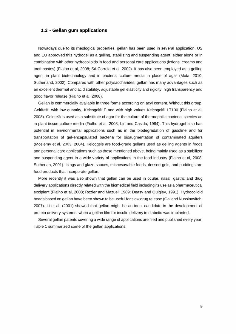
1.2 - Gellan gum applications
Nowadays due to its rheological properties, gellan has been used in several application. US
and EU approved this hydrogel as a gelling, stabilizing and suspending agent, either alone or in
combination with other hydrocolloids in food and personal care applications (lotions, creams and
toothpastes) (Fialho et al, 2008; Sá-Correia et al, 2002). It has also been employed as a gelling
agent in plant biotechnology and in bacterial culture media in place of agar (Mota, 2010;
Sutherland, 2002). Compared with other polysaccharides, gellan has many advantages such as
an excellent thermal and acid stability, adjustable gel elasticity and rigidity, high transparency and
good flavor release (Fialho et al, 2008).
Gellan is commercially available in three forms according on acyl content. Without this group,
Gelrite®, with low quantity, Kelcogel® F and with high values Kelcogel® LT100 (Fialho et al,
2008). Gelrite® is used as a substitute of agar for the culture of thermophilic bacterial species an
in plant tissue culture media (Fialho et al, 2008; Lin and Casida, 1984). This hydrogel also has
potential in environmental applications such as in the biodegradation of gasoline and for
transportation of gel-encapsulated bacteria for bioaugmentation of contaminated aquifers
(Moslemy et al, 2003, 2004). Kelcogels are food-grade gellans used as gelling agents in foods
and personal care applications such as those mentioned above, being mainly used as a stabilizer
and suspending agent in a wide variety of applications in the food industry (Fialho et al, 2008,
Sutherlan, 2001). Icings and glaze sauces, microwavable foods, dessert gels, and puddings are
food products that incorporate gellan.
More recently it was also shown that gellan can be used in ocular, nasal, gastric and drug
delivery applications directly related with the biomedical field including its use as a pharmaceutical
excipient (Fialho et al, 2008; Rozier and Mazuel, 1989; Deasy and Quigley, 1991). Hydrocolloid
beads based on gellan have been shown to be useful for slow drug release (Gal and Nussinovitch,
2007). Li et al, (2001) showed that gellan might be an ideal candidate in the development of
protein delivery systems, when a gellan film for insulin delivery in diabetic was implanted.
Several gellan patents covering a wide range of applications are filed and published every year.
Table 1 summarized some of the gellan applications.

10
Table 1 - Select list of worldwide-issued and published patents covering the use of gellan gum categorized into applications field (adapted from Fialho et al, 2008).

11
2 Use of hydrogels in tissue engineering
Since the pioneering work of Wichterle and Lim, (1960) on cross-linked HEMA (poly (2-
hydroxyethyl methacrylate)) hydrogels, and because of their hydrophilic character and potential
to be biocompatible, hydrogels have been of great interest to biomaterial scientists for many
years. In the same decade it was demonstrated the successful application of calcium alginate
microcapsules for cell encapsulation (Lin and Sum, 1980) and was tested the incorporation of
natural polymers such as collagen and shark cartilage into hydrogels for use as artificial burn
dressings (Yannas et al, 1989). Nowadays, hydrogels based on natural and synthetic polymers
have a special interest for cell encapsulation, drug delivery, serve as adhesives or barriers
between tissue and material surfaces, and tissue engineering as matrix for repairing and for
regenerating a wide variety of tissue and organs due unique biocompatibility, flexible methods of
synthesis, range of constituents, and desirable physical characteristics (reviewed in Hoffman,
2012). Every year millions of patients suffer the loss or failure of an organ or tissue as a result of
accidents or diseases. Tissue or organ transplantation is a generally accepted therapy to treat
these patients. However, this approach is extremely limited by a donor shortage. So the various
developments made in the biomedical field are an exciting and revolutionary strategy to treat
patients who need new organs or tissues. The main steps of this strategy are shown in Figure 7.
Figure 7 - Schematic illustration of typical tissue engineering approaches (adapted from Hoffman, 2012).

12
In this strategy, tissue-specific cells are isolated from a small tissue biopsy from the patient
and harvested in vitro. The cells are subsequently incorporated into 3D polymer scaffolds that act
as analogues to the natural extracellular matrices found in tissues. These scaffolds deliver the
cells to the desired site in the patient body, provide a space for new tissue formation, and
potentially control the structure and function of the engineered tissue (Lee and Mooney, 2001).
Nowadays a variety of tissues are being engineered using this approach including fabricated
artery, bladder, skin, cartilage, bone, ligament, and tendon.
The critical element of tissue engineering approach is the polymer scaffold. This polymer
potentially mimics many roles of extracellular matrixes found in tissues like bringing cells together
and control the tissue structure, regulate the function of the cells and allow the diffusion of
nutrients, metabolites and growth factors and must be taken in account the biocompatibility of this
material. This has to have the ability to exist within the body without damaging adjacent cells or
lead to significant scarring or otherwise elicit a response that detracts from its desired functions.
This may be especially problematic as the inflammatory response to a hydrogel can affect the
immune response toward the transplanted cells and vice-versa. However, the use of these types
of polymer scaffolds requires the surgeon to make incisions sufficiently large to enable placement
of the polymer/cell constructions. An alternative approach to cell delivery for tissue engineering is
the use of hydrogels that can be injected into the body. This approach enables the clinician to
transplant the cell and polymer combination in a minimally invasive manner (Lee and Mooney,
2001).
Various types of polymers have been studied and utilized in tissue engineering field. Hydrogels
are hydrophilic polymer networks which may absorb from 10-20% up to thousands of times their
dry weight in water. This polymers may be chemically stable or they may degrade and eventually
disintegrate and dissolve (reviewed Hoffman, 2012). Biological hydrogels have been formed from
agarose, alginate, chitosan, hyaluronan, fibrin, and collagen, and many others. The most used in
tissue engineering approach are the hydrogels from natural polymers. However, limitations of gels
from natural polymers have motivated approaches to modify these polymers as well as to use
various synthetic polymers. A wide range of synthetic polymers may potentially have suitable
chemical and physical properties for these applications. Beside the biocompatibility, the controlled
degradation of hydrogels is also critical in tissue engineering, whether the gels are originated from
natural resources or are synthetically created. Degradation of hydrogels can be due to hydrolysis,
the action of enzymes, and/or dissolution. The interactions of cells with hydrogels significantly
affects their adhesion as well as migration and differentiation and this issue is also important take
into account. The adhesion may be cell-type specific and is dependent on the interaction of
specific cell receptors with ligands that are a component or adsorbed onto the materials.
Inappropriate interactions could cause undesirable tissue formation (Lee and Mooney, 2001).

13
Table 2 – Examples of natural and synthetic hydrogels used in tissue engineering
Table 2 shows the polymers used in tissue engineering in nowadays. All of this network
structures have specific properties in common that are essential for their use in recent
applications. The physical structure is characterized by junctions or tie points, which may be
formed from strong chemical linkages (such as covalent and ionic bonds), permanent or
temporary physical entanglements, micro-crystallite formation, and weak interactions (such as
hydrogen bonds) (Slaughter, 2009). In terms of ionic charge, hydrogels can be neutral, cationic,
anionic, or ampholytic as determined by pendant groups incorporated into the gel backbone. With
regard of rubber elasticity, hydrogels under mechanical stress can exhibit a range of responses
from rapid, elastic recovery following an applied stress or strain to a time-dependent recovery
approaching viscous behavior. Effective solute transport is one of the most critical design
parameters for these hydrogels. Mass transport parameters determine how nutrients, gasses,
waste products, and bioactive agents, such as growth factors that stimulate natural tissue growth
are exchanged within scaffolds or are delivered by the gel. Diffusion alone is regarded as the
driving transport phenomenon.
Collagen is the most widely used tissue-derived natural polymer, and it is a main component
of extracellular matrices of mammalian tissues including skin, bone, cartilage, tendon, and
ligament. Physically formed collagen gels are thermally reversible and offer a limited range of
mechanical properties. However, the weakness of the gels has been a problem, and a number of
chemical modification methods have been investigated in order to improve the mechanical
properties of gelatin gels (Slaughter, 2009).
Hyaluronate is one of the glycosaminoglycan components in natural extracellular matrices and
plays a significant role in wound healing. This hydrogel has shown excellent potential for tissue
engineering applications such as artificial skin, facial intradermal implants, wound healing and soft
tissue augmentation. Hyaluronate requires thorough purification to remove impurities and
endotoxins that may potentially transmit disease or act as an adjuvant in eliciting an immune
response which is a disadvantage of this polymer. In addition, hyaluronate gels typically possess
low mechanical properties that limited the applications of this hydrogel (Slaughter, 2009).
Hydrogels from Natural Polymers Hydrogels from Synthetic Polymers
Collagen and Gelatin Poly(acrylic acid) and Its Derivatives
Hyaluronate Poly(ethylene oxide) and Its Copolymers
Fibrin Poly(vinyl alcohol)
Alginate Polyphosphazene
Agarose Polypeptides
Chitosan

14
Fibrin has been used as a sealant and an adhesive in surgery as it plays an important role in
natural wound healing. Fibrin gels can be produced from the patient’s own blood and can be used
as an autologous scaffold for tissue engineering. No toxic degradation or inflammatory reactions
are expected from this natural component of the body. This gels might promote cell migration,
proliferation, and matrix synthesis through the incorporation of platelet-derived growth factors.
This polymer has been utilized to engineer tissues with skeletal muscle cells, smooth muscle cells
and chondrocytes. However, fibrin gels are limited in mechanical properties which limits it is
application (Slaughter, 2009).
Alginate is a well-known biomaterial obtained from brown algae and is widely used for drug
delivery and in tissue engineering due to its biocompatibility, low toxicity, relatively low cost, and
simple gelation with divalent cations such as Ca2+, Mg2+, Ba2+, and Sr2+. Alginate has found uses
to date as an injectable cell delivery vehicle as well as wound dressing, dental impression, and
immobilization matrix. Alginate gel beads have also been prepared and used for transplantation
of chondrocytes, hepatocytes, and islets of Langerhans to treat diabetes. Despite its
advantageous features, alginate itself may not be an ideal material because it degrades via a
process involving loss of divalent ions into the surrounding medium, and subsequent dissolution.
Another potential limitation in using alginate gels in tissue engineering is the lack of cellular
interaction. This polymer cannot interact with mammalian cells due to hydrophilic character, so
modified alginate polymers with specific binding were made. These modified alginate gels have
been demonstrated to provide for the adhesion, proliferation, and expression of differentiated
phenotype of skeletal muscle cells (Slaughter, 2009).
Agarose is another type of marine algal polysaccharide, but unlike alginate it forms thermally
reversible gels. The physical structure of the gels can be mainly controlled by using a range of
agarose concentrations, which results in various pore sizes. The large pores and low mechanical
stiffness of the gels at low concentrations of agarose may enable the migration and proliferation
of cells (Slaughter 2009).
Chitosan, due to its biocompatibility, low toxicity, structural similarity to natural
glycosaminoglycans, and degradation by enzymes such as chitosanase and lysozyme appear in
various tissue engineering applications. However, chitosan is easily soluble in the presence of
acid, and generally insoluble in neutral conditions as well as in most organic solvents due to the
existence of amino groups and the high crystallinity what compromised it is use in this applications
of this field (Slaughter, 2009).
As regards synthetic polymer hydrogels one of the most studied is the hydrolytically stable
cross-linked HEMA. The permeability and hydrophilicity of these gels are dependent on the
crosslinking agents. Poly(HEMA) has been used for ophthalmic uses including contact lens, as
well as in many drug delivery applications. These polymers are also being investigated as an
injectable delivery vehicle for cartilage and pancreas engineering. This hydrogel has non-
degradable cross-links which is a limitation (Slaughter, 2009).

15
Poly (ethylene oxide) (PEO) has been used for several medical applications due to its
biocompatibility and low toxicity. These gels may be useful in tissue engineering as they can be
easily formulated with protein drugs or cells at room temperature or lower, and subsequently
delivered to the desired site in a minimally invasive manner (Slaughter, 2009).
Poly (vinyl alcohol) (PVA) is a hydrophobic and soluble hydrogel. In most of physiological
situations PVA is not degradable and that is a limitation. PVA hydrogels have been utilized in
tissue engineering for regeneration of artificial articular cartilage, hybrid-type artificial pancreas
and bone-like apatite formation (Slaughter, 2009).

16
- Gellan gum in tissue engineering
Smith et al, (2007) reported the first evaluation of gellan gum as a material for tissue
engineering applications. Using 1% gellan gum solution cross-linked with Minimum Essential
Medium Eagle (α-MEM) it was also shown the self-supporting nature of such constructs and that
these were sufficiently robust to permit handing with forceps (Mota, 2010). They also
demonstrated cell viability with in these constructs and the rheological potential of gellan in tissue
engineering. The variations on acyl form on the structure of this polymer lead directly with a
variation of rheological properties that makes possible the use of gellan gum both as an injectable
material and indirectly by culturing the cells in vitro and then implanting them. Another
characteristic of gellan gum comes from the optical clarity of gel formed gels, which allows an in
depth visualization of the encapsulated cells using conventional microscopy (Mota, 2010).
Oliveira et al, (2010a) showed the versatility of gellan gum by processing it into different
structures through temperature and pH dependence reactions (Figure 9). These researchers
introduced gellan gum as a new biomaterial in cartilage tissue engineering and showed that gellan
gum discs possess suitable material properties to be used in this field. This material has similar
mechanical properties to the other hydrogel used in identical cartilage regenerative approaches.
Gellan has an elastic nature, some damping capability and also presents the advantage of gelling
at physiological conditions and being able to efficiently encapsulate human nasal chondrocytes
with a homogeneous distribution. These hydrogels are noncytotoxic, maintain cell viability and
allow them to encompass active cell division (Oliveira et al, 2010a).
Figure 8 - Photograph shows the self-supporting nature of gellan gum cylinders (adapted from Smith et al, 2007).

17
The injectable approach using gellan gum is a challenging one since its gelation temperature
is too high. Normally the gelation temperature of commercially available and unmodified gellan is
over 42ºC and must be decreased in order to suspend the mammalian cells at physiological
temperature (~37.5ºC). Optimization of gelling point was performed by Gong et al, (2009) by
adjusting the molecular weight of gellan gum. They assume that a molecular weight decrease
would make assemble and aggregation of gellan molecular chains more difficult, lowering the
gelation temperature. This way, they demonstrated a increasedr performance of these modified
gellan gels in long-term in vitro in comparison with agarose (Oliveira et al, 2010).
Figure 9 - The versatility of Gellan gum structures that can be formed using simple polymer processing technologies: (A) discs; (B) membranes; (C) fibers; (D) particles; (E) and (F) 3D lyophilized scaffolds (adapted
from Oliveira et al, 2010a).
18
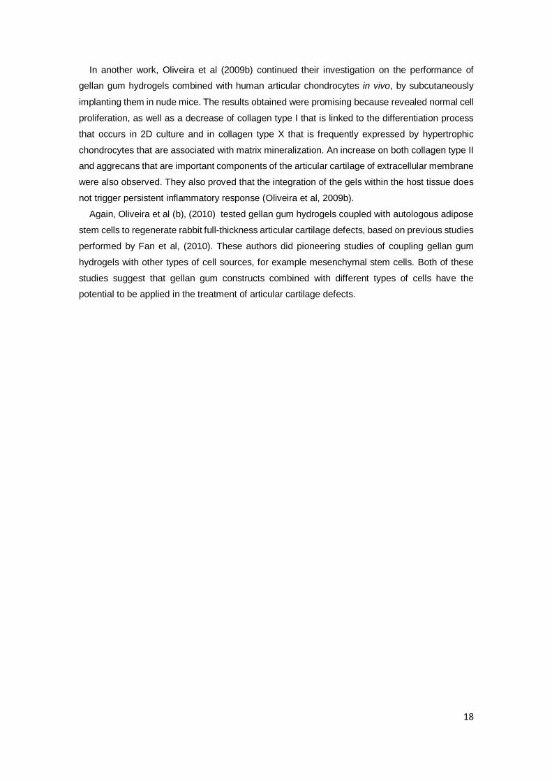
In another work, Oliveira et al (2009b) continued their investigation on the performance of
gellan gum hydrogels combined with human articular chondrocytes in vivo, by subcutaneously
implanting them in nude mice. The results obtained were promising because revealed normal cell
proliferation, as well as a decrease of collagen type I that is linked to the differentiation process
that occurs in 2D culture and in collagen type X that is frequently expressed by hypertrophic
chondrocytes that are associated with matrix mineralization. An increase on both collagen type II
and aggrecans that are important components of the articular cartilage of extracellular membrane
were also observed. They also proved that the integration of the gels within the host tissue does
not trigger persistent inflammatory response (Oliveira et al, 2009b).
Again, Oliveira et al (b), (2010) tested gellan gum hydrogels coupled with autologous adipose
stem cells to regenerate rabbit full-thickness articular cartilage defects, based on previous studies
performed by Fan et al, (2010). These authors did pioneering studies of coupling gellan gum
hydrogels with other types of cell sources, for example mesenchymal stem cells. Both of these
studies suggest that gellan gum constructs combined with different types of cells have the
potential to be applied in the treatment of articular cartilage defects.

19
3 Aims of this study
In the last years many studies have been developed to better understand the molecular
mechanisms and regulatory processes underlying the synthesis of bacterial exopolysaccharides
(EPS) as well as their potential applications. In the search solutions for biotechnological
applications, gellan gum produced by Sphingomonas elodea ATCC 31461 emerges as a new
promising extracellular polysaccharide due to its importance as commercial gelling agent. It is
used in food and pharmaceutical industries and more recently in others fields, like biomedicine
applications. The rheological proprieties of this hydrogel showed that it can be used in several
applications, such as construction of scaffolds in tissue engineering due to its ability as a cellular
support or its multi-functional capacity like synergy with other polysaccharides, which allows the
production of wide range of textures.
So the principal aim of this work was to test mutant polymers derived from gellan and verify
if their properties could improve those of the gellan gum and consequently explore their ability to
work as scaffold, drug delivery or encapsulating gel. To do that, the several gellan-like polymers
were produced, quantified and viscosity measurement of a polymer solution was determined. For
the selected ones, purification protocols were established and several tests were performed to
assess gelation properties.
During this work it was also tested the ability of strains from the Massilia genus and their
characteristics. The unknown polymer obtained from this strain was subjected to various tests,
namely rheological tests with aim of understand in order to know if it could be used in
biotechnological applications.

20
4 Materials and Methods
4.1 - Strains and plasmids, media and culture conditions
Strains used in this study were the native strain S. elodea ATCC 31461 that is the major
producer of gellan; S. elodea △gelE mutant strain (SpLM21-4) with the recombinant plasmid
containing the gelE gene (pHA010-3) and other mutant plasmids with Y189F, D179N, V231E,
C75K, G74A, Y121F, Y235F, Y209F, YF1F4, C75A, pBBR1MCS, pLM51-1, YF1F3F4, V227D,
I228D, N99A, D79A, L232E. Strains form the genus Massilia were designated RAG-1 and RAG-
2.
S. elodea strains were grown in S medium (Moreira et al, 2004) supplemented with the
appropriated antibiotic. Strains RAG-1 and RAG-2 were grown in Lennox broth (LB) with or
without of 5 g/l glucose (LBG). All strains were incubated at 30ºC with 250 rpm if in liquid culture.
4.2 - Preparation of competent cells and electrotransformation
S. elodea △gelE mutant was inoculated into S medium and grown overnight at 30ºC with stirring
of 250 rpm. The pre-inoculum was added to 150 ml of S medium and grown until O.D. 600 nm of
1.2 was reached. After that, cultures were cooled on ice during 15 minutes and centrifuged during
15 minutes at 8000 rmp at 4°C. Supernatant was discarded and cell pellet was resuspend in 200
ml of deionized, cold and sterile H2O. This process was repeat four times, followed by different
glycerol 10% volumes, 40 ml, 8 ml and 4 ml, respectively. The final cellular suspension was
aliquoted in 100 μl fractions and kept at -80°C. Prior to electrotransformation, plasmids were
extracted using the ZR Plasmid MiniPrep Kit according to manufacture’s instructions. After that,
cells and plasmid DNA were mixed and an electrical shock was applied. The cells were incubated
with 1 ml of LB medium during 3 hours at 30°C with stirring and plated on to solid S medium with
chloramphenicol. Plates were then incubated during 4 days at 30°C.

21
4.3 - Gellan production and purification
The strains under study were inoculated in 250 ml of S medium and grown at 30 °C for 48
hours. After that time, the culture was diluted with NaCl to facilitate cell removal by centrifugation
at 8000 rpm during 15 minutes at room temperature. The supernatant was precipitated with cold
ethanol 96%, followed by successive washes with ethanol to remove cell debris. The
polysaccharide was dried overnight and then weighted.
To further purify the polysaccharide it was necessary to test other approaches, such as treat
the samples with chloroform, phenol and both. For that, 1g/l solutions of the different polymers
were prepared in water. Then, to each 5 ml of sample were added 1.5 ml of ammonium acetate.
Then, 5 ml of chloroform (or phenol) were added and the mixture centrifuged for 5 minutes at
13000 rpm. The aqueous phase containing the polysaccharide was recovered and stored at 4ºC.
4.4 - Viscosity measurements
The viscosity of each solution was determined at 30 °C using the viscometer Brookfield model
DV-II with the spindle n. 40. Results are the means of at least three independent viscosity
determinations.
4.5 - Methods for protein quantification
The quantification of proteins was determined by two different approaches: the method
Bradford and the method of Lowry.
Lowry method: Preparation of diluted albumin (BSA) standards were performed as indicated in
Table 3.

22
40 µL of each standard on unknown sample was pipetted into microplate well and added 200
µL of Modified Lowry Reagent and mixed for 30 seconds. Microplate was then incubated at room
temperature for 10 minutes, followed by the addition 20 µL of 1X Folin-Ciocalteu Reagent to each
well. The microplate with the samples was agitated for 30 seconds and incubated at room
temperature for 30 minutes. After that period, the absorbance at 750 nm was measured.
Vial Volume of Diluent Volume and Source of BSA Final BSA concentration
A 250 µL 750 µL of Stock 1500 µg/mL
B 625 µL 625 µL of Stock 1000 µg/mL
C 310 µL 310 µL of vial A dilution 750 µg/mL
D 625 µL 625 µL of vial B dilution 500 µg/mL
E 625 µL 625 µL of vial D dilution 250 µg/mL
F 625 µL 625 µL of vial E dilution 125 µg/mL
G 800 µL 200 µL of vial F dilution 25 µg/mL
H 800 µL 200 µL of vial G dilution 5 µg/mL
I 800 µL 200 µL of vial H dilution 1 µg/mL
J 1000 µL 0 0 µg/mL
Table 3 - Preparation of diluted albumin (BSA) standards.

23
Bradford Method: Preparation of reagent Coomassie: It was added to 100 mg of Coomassie
Brilliant Blue G-50 (Sigma), 50 ml of 95% ethanol,100 ml of phosphoric acid 85% (w/v) and
distilled water to a final volume of 1L. The reactant was filtered before being stored in a dark bottle
at 4ºC. In a test tube was mixed 2.5 ml of reagent and 50 µl of extract to be analyzed and stirred
vigorously. Absorbance at 595 nm was read between 5-15 minutes after shaking the sample. To
plot the calibration curve (Figure 10) aqueous solutions of BSA (Bovine Serum Albumin) were
used.
4.6 - Gelation of modified gellan gum samples
The selected samples of gellan-like polymers were mixed with sterile distillated water under
constant stirring at room temperature. The solution was progressively heated at 90ºC until
homogeneous dispersion and maintained at that temperature for 15-20 minutes. Different
concentrations of several salts, such as NaCl, CaCl2, and BaCl2.2H20 were added to the solution
and progressively decreased the temperature until 40ºC.
To test the gelation of the mutants in Dulbecco's Modified Eagle's medium (DMEM) sample
were kept in an incubator (37ºC, 5% CO2).
Figure 10 - Graphical representation of the calibration curve used for the determination of protein concentration by the Bradford method.

24
5 Results
5.1 - Production and rheological properties of gellan-like
polysaccharides
Sphingomonas elodea produces gellan gum in high yield, giving rise to mucoid yellow
colonies in plates containing S medium (Figure 11-A). Contrastingly, the deletion of the gelE gene
encoding a putative tyrosine autokinase produces colonies that are slightly orange and dry (Figure
11-B).
Analyses to GelE secondary structure evidenced several conserved regions, such as the ATP-
binding pocket composed of the walker A, walker B and Mg2+-binding region, an amphipathic
helice, a C-terminal β-strand, and several tyrosine residues (Figure 12).
Figure 11 – Morphology of S. elodea ATCC (A) or ∆gelE mutant (B) grown for 3 days at 30ºC in solid S medium.
A B
Figure 12 - Point mutations introduced into GelE coding sequencing and respective amino acid exchange. The effect of these mutations on gellan recovery from the growth culture is also indicated as + or -.

25
To evaluate the role of these regions in GelE activity regarding gellan production, several point
mutations were previously introduced (Moreira et al, unpublished). The six mutations introduced
in the ATP-binding pocket of GelE (G74A, C75A, C75K, D97A, N99A and D179N) still
complement the gellan deficient phenotype of the ∆gelE mutant (Figure 12). Contrastingly, the
three simultaneous mutations in the amphipathic helice did not restore gellan production. When
single mutations were introduced in the C-terminal β-strand domain (V227D, I228D, V231E and
L232E) gellan was still produced, but the simultaneous deletion of two amino acids (VI or VL)
prevent gellan production. As GelE is a putative tyrosine autokinase, 4 tyrosine residues were
mutated (Y121F, Y198F, Y209F and Y235F). All of this mutations restored gellan biosynthesis by
the complemented ∆gelE mutant, although Y198F mutation gave rise to a lower amount of
polymer and drastic changes in its rheology (Moreira et al, 2004).
To understand whether these mutations were giving rise to gellan-like polymers with different
rheological properties, viscosity of the growth medium and of a polymer solution, and quantity of
produced polymers were determined. For that, the ∆gelE mutant was transformed by
electroporation with the empty vector (pBBR1MCS), with this vector carrying native gelE gene
(pHA010-3), with pLM51-1 having the two last amino acids of GelE deleted, and plasmids carrying
all the mutations of Figure 12 that still complement the EPS-producing phenotype. Three different
colonies were chosen for each strain and grown in S liquid medium for 48 hours. After that period,
growth medium viscosity was measured and data presented in Figure 13.
Figure 13 – Growth medium viscosity (shear rate 12 s-1) of S. elodea ATCC 31461 and ∆gelE mutant complemented with the empty pBBR1MCS vector or with gelE carrying the indicated point mutations, after 48 hours of growth in S medium at 30ºC with 250 rpm agitation. Data are average of three biological replicates.

26
As expected, the growth medium of ∆gelE mutant carrying pBBR1MCS does not show
significant viscosity, but when complemented with gelE gene (pHA010-3), viscosity is restored to
a level similar to the S. elodea wild type strain. Regarding the mutations in the different domains
of GelE, the ones with higher impact in viscosity reduction were in the C-terminal β-strand (V227D,
I228D and V231E) and in all containing the Y198F mutation ((Y198F; Y121F/Y235F (YF1F4)
Y121F/Y209F/Y235F (YF1F3F4)) (Figure 13). This analysis showed that although all GelE
variants restore gellan production by the ∆gelE mutant, their effect on medium viscosity is
variable, suggesting the present of polymers with different rheological properties.
To assess whether growth medium viscosity was comparable with the viscosity of the
precipitated/dissolved polymer, the polysaccharide present in the culture medium of each strain
was precipitated with ethanol, washed several times to remove cell debris and air dried. Then, 1
g/l solution of the gellan precipitates were prepared and viscosity was measured. Data here
obtained confirmed previous results of Moreira et al (2004) since ∆gelE supernatant precipitate
does not show viscosity and the viscosity of the complemented mutant (pHA010-3) precipitated
gellan does not reach the value of the wild-type S. elodea ATCC 31461 (Figure 14-A ). In
comparison to the viscosity of the polymer of the native GelE complemented strain, ATP-binding
site mutation C75A, D179N, N99A and D79A have a positive effect in polymer viscosity (Figure
14-A). Mutations in the β-strand C-terminal region had different effects depending on the
mutation. V231E, V227D and pLM51-1 had a negative effect on gellan viscosity while I228D and
L232E led a positive effect. Regarding the tyrosine residues, Y198F mutation had a negative
effect, but Y209F mutation displayed a strong increase of gellan solution viscosity (Figure 14-A).

27
Figure 14 – Viscosity at 12 s-1 (A) or at different shear rates (B), of the aqueous solutions prepared with 1 g/l of the ethanol-precipitated polymers isolated from the supernatants of the indicated strains, grown for 48h at 30ºC in S medium. These bars (curves) are based on the median values of three viscosity measurements of polymer solutions obtained from three independent growth experiments.

28
The aim of the experiment shown in Figure 14-A was to identify strains producing polymers
with different viscosities to assess properties such as gelation as it will be further described.
Therefore, we were choosing for further analysis strain N99A producing a polymer with a viscosity
comparable to the wild-type ATCC 31461, C75K with polymer display 30% of the wild type and
V231E and Y198F for lower polymer viscosity. Figure 14-B shows the viscosity at different shear
rates for the above mentioned strains, confirming the different rheologies.
Next step was to assess if the strains producing the chosen gellans were good producers by
determining the dry weight of the ethanol-precipitated polymer. Data shown in Figure 15
demonstrate that V231E and C75K mutations give rise to 8.3 and 7.4 g/l, respectively, of polymer
while Y198F produces approximately 4.4 g/l. The value for this last mutations confirms results
previously published (Moreira et al, 2004). Due to unknown reason, no EPS was recovered from
the strain with N99A mutation in this particular experiment and therefore no value can be shown.
Figure 15 – Gellan production assessed by dry weight of the ethanol precipitated polysaccharide in the wild-type strain and when the mutant genes are expressed in the S. elodea ∆gelE mutant.
29
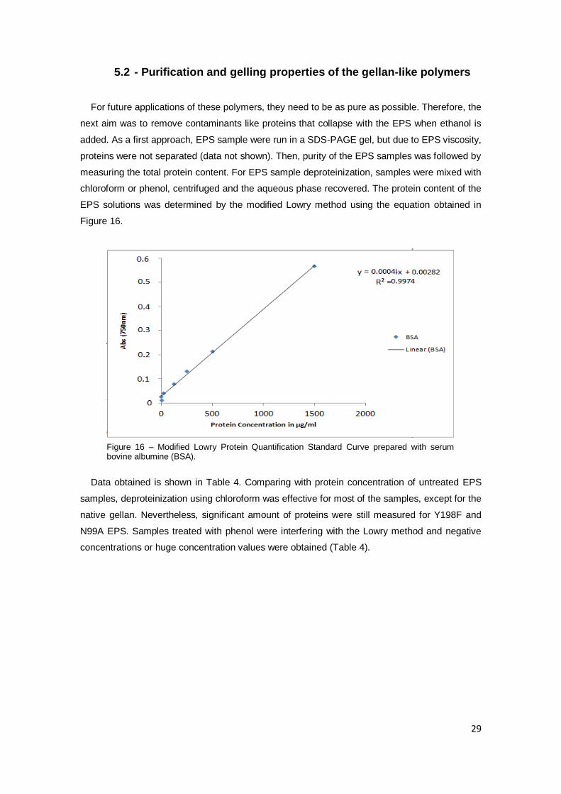
5.2 - Purification and gelling properties of the gellan-like polymers
For future applications of these polymers, they need to be as pure as possible. Therefore, the
next aim was to remove contaminants like proteins that collapse with the EPS when ethanol is
added. As a first approach, EPS sample were run in a SDS-PAGE gel, but due to EPS viscosity,
proteins were not separated (data not shown). Then, purity of the EPS samples was followed by
measuring the total protein content. For EPS sample deproteinization, samples were mixed with
chloroform or phenol, centrifuged and the aqueous phase recovered. The protein content of the
EPS solutions was determined by the modified Lowry method using the equation obtained in
Figure 16.
Data obtained is shown in Table 4. Comparing with protein concentration of untreated EPS
samples, deproteinization using chloroform was effective for most of the samples, except for the
native gellan. Nevertheless, significant amount of proteins were still measured for Y198F and
N99A EPS. Samples treated with phenol were interfering with the Lowry method and negative
concentrations or huge concentration values were obtained (Table 4).
Figure 16 – Modified Lowry Protein Quantification Standard Curve prepared with serum bovine albumine (BSA).

30
Table 4 – Protein concentration in µg/ml of EPS samples.
*-negative values were obtained.
Due to interference of phenol with the Lowry method it was used instead the Bradford method
to estimate protein concentration. After using BSA as standard for the calibration curve (Figure
10), the protein concentration are shown in Table 5.
Table 5 – Concentration in µg/ml of the proteins present in EPS samples by using the Bradford method.
The Bradford method seemed to be more suitable to determine EPS-samples protein
contamination, since consistent values were obtained. According to this data the best compound
for deproteinization of EPS samples was chloroform since it was observed, at least, a reduction
of 50% in the amount of contaminant proteins (Table 5).
After gellan-like polymers purification protocol was established, the following step was to
determine the best conditions for gelation. It was known from previous published data that NaCl,
CaCl2 or sucrose could help in the gelation of gellan (Morris et al, 2012). But as sucrose could
have detrimental effect or stem cell viability, only NaCl and CaCl2 were tested. Using the native
gellan produced by S. elodea ATCC 31461 and the ∆gelE mutant expressing mutant GelE
(V231E) in presence of NaCl, none of the samples made a gel (data not shown). When
concentrations of CaCl2 ranging from 0 to 200 mM were added to the EPS solutions, it was
observed the formation of a gellified solution for the native polymer, especially to the
concentrations around 20 mM CaCl2. An example of the type of gel formed is shown in Figure 17.
Strain Without treatment Chloroform Phenol
ATCC 31461 617.4 717.1 *
Y198F 1577.4 666.3 *
N99A 1036.6 674.6 *
V231E 522.2 71.5 7118.7
C75K 858.9 64.7 5407
Strain Without treatment Chloroform Phenol
ATCC 31461 468.2 19.2 211.2
Y198F 497.7 275.2 591.2
N99A 438.2 245.2 269.2
V231E 778.2 239.2 739.2
C75K 959.7 223.2 456.2
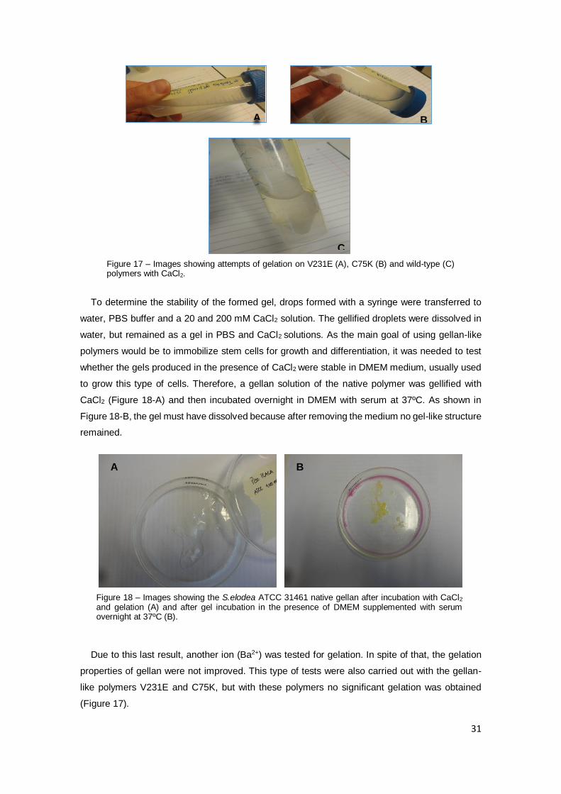
31
To determine the stability of the formed gel, drops formed with a syringe were transferred to
water, PBS buffer and a 20 and 200 mM CaCl2 solution. The gellified droplets were dissolved in
water, but remained as a gel in PBS and CaCl2 solutions. As the main goal of using gellan-like
polymers would be to immobilize stem cells for growth and differentiation, it was needed to test
whether the gels produced in the presence of CaCl2 were stable in DMEM medium, usually used
to grow this type of cells. Therefore, a gellan solution of the native polymer was gellified with
CaCl2 (Figure 18-A) and then incubated overnight in DMEM with serum at 37ºC. As shown in
Figure 18-B, the gel must have dissolved because after removing the medium no gel-like structure
remained.
Due to this last result, another ion (Ba2+) was tested for gelation. In spite of that, the gelation
properties of gellan were not improved. This type of tests were also carried out with the gellan-
like polymers V231E and C75K, but with these polymers no significant gelation was obtained
(Figure 17).
A B
C
Figure 17 – Images showing attempts of gelation on V231E (A), C75K (B) and wild-type (C) polymers with CaCl2.
Figure 18 – Images showing the S.elodea ATCC 31461 native gellan after incubation with CaCl2 and gelation (A) and after gel incubation in the presence of DMEM supplemented with serum overnight at 37ºC (B).
A B

32
5.3 - Assessment of polymer production in strains of the Massilia
genus
With the aim of finding new carbohydrate polymers with potential biotechnological applications,
we started the characterization of two soil isolates, which after several days grown on LB plates,
produce purple and highly mucoid colonies. These two isolates, named RAG-1 and RAG-2, do
not have a species attributed, but they may belong to the genus Massilia (António Veríssimo,
personal communication).
The first experiment was to assess growth rate in two different media, LB and LB supplemented
with glucose (LBG). Data shown in Figure 19-A indicate that the growth rate is similar for both
strains in both media, but while RAG-1 maintained the same optical density in stationary phase,
RAG-2 showed a slight decrease, suggestive of cell lysis. To quantify mucoidy strains were
cultivated in liquid medium, from which several samples were taken, and ethanol precipitated.
Precipitated dry-weight increases along the growth period with RAG-1-LB having a maximum at
72 hours, RAG-1-LBG at 96 hours, and RAG-2 at 120 hours in both media (Figure 19-B). To
evaluate if this polymer increased the viscosity of the growth media over time, we measured
growth medium viscosity at different shear rates. Figure 19-C shows the results for the shear rate
1.2 s-1. Despite some fluctuations, the general trend is an increase in viscosity, especially for
strain RAG-1. Possibly due to the cell lysis observed in stationary phase, RAG-2 growth media
shows lower viscosity when compared to RAG-1. As a final experiment to assess the rheology of
these polymers, the ethanol-precipitable material produced by the two strains in both media were
dissolved in water (1 g/l solution) and viscosity was measured. Data represented in Figure 19-D
shows that the most viscous polymer at 96 hours of growth is from RAG-1 grown in LB. Although
the medium viscosity for RAG-1-LBG at 96 hours is similar to RAG-1-LB, it shows considerably
lower viscosity, implying that the RAG-1-LB polymer has higher molecular mass.

33
Figure 19 – (A) Comparison of growth curves of RAG-1 and RAG-2 grown at 30ºC in LB or LBG media; (B) exopolysaccharide production, assessed based on the ethanol-precipitated supernatant; (C) growth medium viscosity at 1.2 s-1shear rate; (D) viscosity at different shear rates of the aqueous solutions prepared with the same concentration (1g/l) of the ethanol-precipitated material isolated from different cultures grown, for 96 hours, at 30ºC. These curves are based on the medium values from triplicate experiments. Strains legend for all the graphics is in (D).

34
6 Discussion
In this study it was evaluated whether different polymers produced by the ∆gelE deletion mutant
of S.elodea complemented with mutated variants of gelE gene could have different rheological
properties suitable for cell tissue engineering approaches. GelE is expected to interact with
membrane protein GelC (named activator domain) and regulate gellan chain length. Previous
studies have shown that deletion of gelC or gelE gene leads to gellan production abrogation, but
when a mutated variant of GelE having a phenylalanine residue instead of a tyrosine at position
198, gellan is still produced although in lower amount and with lower molecular mass (Moreira et
al, 2004). This suggests that mutations putatively affecting the structure/function of GelE may
result in different interaction with GelC and possibly to polymers with different rheological
properties. To investigate this hypothesis, mutations of GelE conserved motifs were introduced
by site-directed mutagenesis (Moreira, unpublished). Here, we analyzed the effect of these GelE
mutations in the viscosity of the gellan-like polymers produced. Regarding the viscosity of the
growth medium after 48 hours of incubation, the mutations with higher impact in medium viscosity
were in the C-terminal β-strand and Y198F which gave rise to lower viscosity and possibly to EPS
with lower-molecular mass. Contrastingly, mutation affecting Y209F seems to indicate that an
EPS with higher molecular mass is being produced. The C-terminal region of GelE seems to be
determinant for gellan rheology, but the reason for this is unknown. An hypothesis is that this
region is perhaps important for interaction with GelC, regulating then the activity of the
polysaccharide polymerase enzyme GelG and consequently the size of gellan chains.
When viscosity of the gellan-like polymers 1g/l solutions were determined, there was a good
correlation with the growth medium viscosity. Exceptions are the mutations that lack the two last
amino acids of GelE (pLM51-1) and I228D, with opposite effects. As representative polymers for
further studies we were choosing mutation giving rise to polymers with high viscosity (N99A),
intermediate (C75K) and low (V231E, Y198F). In addition we had determined the amount of EPS
production in the presence of these mutations. With the exception Y198F mutation, all the others
mutations do not interfere significantly with the amount of EPS produced. This is an important
feature regarding possible biotechnological applications.
For putative application in tissue engineering, a gellan-like polymer needs to have the right
rheological properties for cell growth and differentiation, but also requires a high degree of purity,
not to be toxic to eukaryotic cells. Due to gellan viscosity it is not always possible to remove cells
by centrifugation and quite often cells are precipitated with the polymer by the addition of ethanol.
These samples are highly contaminated with bacterial proteins that have to be removed. In this
work we tested whether organic solvents like phenol and chloroform, known for deproteinization
could be used.

35
Our data showed that chloroform was more effective in reducing the protein content of the EPS
solutions, but still not all protein contaminants were removed. Phenol does not seem to be a good
choice, because it is very difficult to remove from the EPS sample and is toxic to cells.
Nevertheless, some extra work can be done for EPS sample deproteinization, such as incubation
with a protease, followed by dialysis and lyophilization, or purification by chromatography.
Since an important goal was to test gelation of the different polymers, several conditions were
tested. The aim was to obtain a polymer that is in the liquid state at temperatures higher than
37ºC, but at body temperature makes a gel able to be used as scaffold for eukaryotic cell growth.
With that in view we tested the native gellan in the presence of divalent cations such as Ca2+,
being able to produce a gel. When this gel was tested for stability in solutions containing salts
(PBS, CaCl2) the gel was stable, but in the medium for stem cell growth, apparently the gel was
dissolving. In addition, some of gellan-like polymers did not gelify under the tested conditions and
other conditions have to be tested. One possibility is to crosslink these polymers with other
compounds, to obtain a stable network of fibers with a tridimensional structure. Despite our
results, this gellan-like polymers should not be left without consideration in further studies,
because gellan is one of the few bacterial gums with gelling properties and one of the most
promising tools for new biotechnological applications.
With the goal of searching for new bacterial exopolysaccharides with putative interest in the
biotechnological field, we did the first preliminary studies with two soil isolates from the genus
Massilia which comprises Gram-negative bacteria. RAG-1 and RAG-2 isolates produce a polymer
which gives a mucoid morphotype to the purple colonies and that after cell-free supernatant
precipitation gave a white material, not so dense as gellan. Viscosity measurements of the growth
media or of a polymer solution showed very low values when compared to gellan, indicating that
these polymers have different rheological properties. The nature of these polymers is unknown,
and further studies including sugar content determination and structure determination should be
conducted. From the two tested isolates, RAG-1 seems to be more promising since it has a
highest production and viscosity when compared to RAG-2. Nevertheless, these are preliminary
studies and new media, temperature of growth, pH should be tested. One important difference
between S. elodea ATCC 31461 and RAG-1/ RAG-2 is that gellan production is stimulated by the
high carbon/nitrogen ratio (S medium) while here we did not see significant differences between
LB and LBG. A medium like the S medium was not suitable for growth of neither RAG-1 nor RAG-
2 isolates.
Overall this work showed that mutations in proteins regulating polysaccharide chain length are
good strategies to obtain polymers with different rheological properties. In addition, we started the
characterization of other polymer producing microorganisms, hoping to find new interesting
features for possible future applications.

36
7 References
Bajaj, I., Survase, S., Saudagar, P., & Singhal, S. (2007). Gellan Gum: Fermentative Production,
Downstream Processing and Applications. Food, Technololy and Biotechnology, 45, 341-
354.
Camelin, I., Lacroix, C., Prevost, Provost, H., Cachon, R., & Divies, C. (1993). Effect of chelatants
on gellan gel rheological properties and setting temperature for immobilization of
Bifidobacteria. Biotechnology Progress, 9, 291-297.
Chandrasekaran, R., & Radha, A. (1995). Molecular architectures and funtional properties of
gellan gum and related polysaccharides. Trends in Food Science & Technology , 6.
Deasy, P., & Quigley, K. (1991). Rheological evaluation of deacetylated gellan gum (Gelrite) for
pharmaceutical use. Internacional Journal of Pharmaceutics, 73, 117-123.
Dreveton, E., Monot, F., Ballerini, D., Lecourtier, J., & Choplin, L. (1994). Effect of mixing and
mass transfer conditions on gellan production by Auromonas elodea. Journal of
Fermentation and Bioengineering, 6, 642-649.
Drury, J., & Mooney, D. (2003). Hydrogels for tissue engineering: scaffold design variables and
applications. Biomaterials, 24, 4337-4351.
Fan, J., Gong, Y., Ren, L., Varshney, R., Cai, D., & Wang, D. (2010). In vitro engineered cartilage
using synovium-derived mesenchymal stem cells with injectable gellan hydrogels. Acta
Biomaterialia, 6, 1178-1185.
Fialho, A., Moreira, L., Granja, A., Popescu, A., Hoffmann, K., & Sá-Correia, I. (2008). Occurence,
production and applications of gellan: curren state and perspectives. Applied
Microbiology and Biotecnhology, 79, 889-900.
Fialho, A.; Martins, L.; Donval, M.; Leitão, J.; Ridout, M.; Jay, A.; Morris, V.; Sa-Correia, I. (1999).
Structures and Properties of Gellan Polymers Producted by Shingomonas paucimobilis
ATCC 31461 from Lactose Compared with Those Produced from Glucose and from
Cheese Whey. Applied and Enviromental Microbiology, 2485-2491.
Gal, A., & Nussinovitch, A. (2007). Hydrocolloid carriers with filler inclusion for diltiazem
hydrochloride release. Journal Pharmaceutical Sciences, 96, 168-178.
Giavasis, I., Harvey, L., & McNeil, B. (2000). Gellan gum. Critical Reviews in Biotechnology, 20,
177-211.
Gong, Y., Wang, C., Lai, R., Su, K., Zhang, F., & Wang, D. (2009). An improved injectable
polysaccharide hydrogel: modified gellan gum for long-term cartilage regeneration in
vitro. Journal of Materials Chemistry, 19, 1968-1977.
Harding, N., Patel, Y., & Coleman, R. (2004). Organization of genes required for gellan
polysaccharide biosynthesis in Sphingomonas elodea ATCC 31461. Journal of Industrial
Microbiology and Biotechnology, 31, 70-82.
Hoffman, A. (2012). Hydrogels for Biomedical Applications. Advanced Drug Delivery Reviews,
64, 18-23.
Huang, Y., Singh, P., Tang, J., & Swanson, B. (2004). Gelling temperatures of high acyl gellan as
affected by monovalent and divalent cations with dynamic rheological analysis.
Carbohydr Polym, 56, 27-33.
Jasson, P., & Lindberg, B. (1983). Structural studies of gellan gum, an extracellular
polysaccharide elaborated by Pseudomonas elodea. Carbohydrate Research, 124, 135–
139.

37
Kang, S., & Veeder, T. (1981). Patent No. 4,377,636.
Lau, M., Tanga, J., & Paulson, A. (2000). Texture profile and turbidity of gellan/gelatin mixed gels.
Food Research International, 33, 665-671.
Lee, K., & Mooney, D. (2001). Hydrogels for Tissue Engineering. Chemical reviews, 101.
Li, J., Kamath, K., & Dwivedi, C. (2001). Gellan film as an implant for insulin delivery. Journal of
Biomaterials Applications, 15, 321-343.
Lim, F., & Sun, A. (1980). Microencapsulated islets as bioartifitial pancreas. Science, 210, 908-
910.
Lin, C., & Casida, L. (1984). GELRITE as a gelling agent in media for the growth of thermophilic
microorganisms. Applied and Environmental Microbiology, 47, 427-429.
Mao, R., Tanga, J., & Swanson, B. (2000). Texture properties of high and low acyl mixed gellan
gels. Carbohydrate Polymers, 41, 331-338.
Martins, L., & Sá-Correia, I. (1994). Temperature profiles of gellan gum synthesis and activities
of biosynthetic enzymes. Biochemistry, 20, 385-395.
Moreira , L., Hoffmann, K., Albano, H., Becker , A., Niehaus, K., & Sá-Correia, I. (2004). The
Gellan Gum Biosynthetic Genes gelC and gelE Encode Two Separate Polypeptides
Homologous to the Activator and the Kinase Domains of Tyrosine Autokinases. Journal
of Molecular Microbiology and Biotechnology, 43-57.
Morris, E., Nishinari, K., & Rinaudo, M. (2012). Gelation of gellan - A review. Food Hydrocolloids,
28, 373-411.
Moslemy, P., Guiot, S., & Neufeld, R. (2004). Activated sludge encapsulation in gellan gum
microbeads for gasoline biodegradation. Bioprocess and Biosystems Engineering, 26,
197-204.
Moslemy, P., Neufeld, R., Millette, D., & Guiot , S. (2003). Transport of gellan gum microbeads
through sand: an experimental evaluation for encapsulated cell bioaugmentation. Journal
of Environmental Management, 69, 249-259.
Mota, B. (2010). Investigating the use of modified gellan polymers as scaffolding materials for
cartilage tissue engineering. MSc thesis in Biomedical Engineering. Instituto Superior
Técnico.
Oliveira , J.; Martins, L.; Picciochi , R.; Malafaya , P.; Sousa , R.; Neves N.; Mano , J.; Reis, R.
(2010a). Injectable Gellan Gum Hydrogels with Autologous Cells for the Treatment of
Rabbit Articular Cartilage Defects. Journal of Orthopaedic Research, 28, 1193-1199.
Oliveira , J.; Santos , T.; Martins , L.; Silva , M.; Marques , A.; Castro , A.; Neves , N.; Reis, R.;.
(2009). Performance of new gellan gum hydrogels combined with humam articular
chondrocytes for cartilage regenaration when subcutaneously implanted in nude mice.
Journal of Tissue Engineering and Regenerative Medicine, 3, 493-500.
Oliveira, J.; Martins, L.; Picciochi , R.; Malafaya, P.; Sousa , R.; Neves , N.; Mano , J.; Reis, R.
(2010b). Gellan gum: a new biomaterial for cartilage tissue engineering applications.
Journal of Biomedical Materials Research Part A, 93, 852-863.
Oliveira,J.; Santos,T.; Martins,L.; Picciochi , R.; Marques , A.; Castro , A.; Neves , N.; Mano , J.;
Reis, R.;. (2010c). Gellan GGum Injectable Hydrogels for Cartilage Tissue Engineering
Applications: In Vitro Studies and Preliminary In Vivo Evaluation. Tissue Engineering Part
A, 16.
Omoto, T., Uno, Y., & Asai, I. (1999). The latest technologies for the application of gellan gum.
Progress in Colloid and Polymer Science , 144, 123-126.

38
Papageorgiou, M., Gothard, M., Willoughby, L., Kasapis, S., Richardson, K., & Morris, E. (1994).
Rheology and Structure of Gellan-Alginate Co-Gels. In Gums and Stabilisers for the Food
Industry (pp. 345-356). Oxford: Oxford University Press.
Rozier, A., Mazuel, C., Grove, J., & Plazonnet, B. (1989). Gelrite: a novel, ion-activated, in-situ
gelling polymer for ophthalmic vehicles. Effect on bioavailability of timolol. International
Journal of Pharmaceutics, 57, 163-168.
Sá-Correia, I., Fialho, A., Videira, P., Moreira, L., Marques, A., & Albano, H. (2002). Gellan gum
biosynthesis in Sphingomonas paucimobilis ATCC 31461: genes, enzymes and
exopolysaccharide production engineering. Journal of Industrial Microbiology and
Biotechnology, 29, 170-176.
Sanderson, G., & Clark, R. (1984). Gellan Gum, A New Gelling Polysaccharide. In Gums and
Stabilisers for the Food Industry (pp. 201-209). Oxford: Oxford University Press.
Slaughter , B., Khurshid, S., Fisher, O., Khademhosseini , A., & Peppas, N. (2009). Hydrogels in
Regenerative Medicine. Advanced Materials, 21, 3307–3329.
Smith, A., Shelton, R., Perrie, Y., & Harris, J. (2007). An initial evaluation of gellan gum as a
material for tissue engineering applications. Journal of Biomaterials Applications, 22, 241-
524.
Strohfus, B., & West, T. (1998). Effect of complex nitrogen sources upon gellan production by
Sphingomonas paucimobilis ATCC 31461. Microbios, 94, 145-152.
Sutherland , I. (2002). A sticky business. Microbial polysaccharides: current products and future
trends. Microbiology Today, 29, 70-71.
Sutherland, I. (2001). Microbial polysaccharides from Gram-negative bacteria. International Dairy
Journal, 11, 663-674.
Tang, J., Mao, R., Tung, M., & Swansonc, B. (2001). Gelling temperature, gel clarity and texture
of gellan gels containing fructose or sucrose. Elsevier, 44(Carbohydrate Polymers), 197-
209.
Tecante, A., & Rodríguez-Hernández, A. (1999). Dynamic viscoelastic behavior of gellan-
carrageenan and gellan-xanthan gels. Food Hydrocolloids Journal , 13, 59-64.
Vartak, N., Lin, C., Clear, J., Fagan, M., & Saier, Jr., M. (1995). Glucose metabolism in
'Sphingimonas elodea': pathway enginnering via construction of glucose-6-phosphate
dehydrogenase insertion mutant. Microbiology, 141, 2339-2350.
Wichterle, O., & Lim, D. (1960). Hydrophilic gels in biologic use. Nature, 185, 117.
Yannas, I., Lee, E., Orgill, D., Skrabut, E., & Murphy, G. (1989). Synthesis and characterization
of a model extracellular matrix that induces partial regeneration of adult mammalian skin.
Proceedings of the National Academy of Sciences, 86, 933-937.
Yuguchi, Y.; Mimuraa, M.; Kitamura, S.; Urakawaa, H.; Kajiwara, K. (1993). Structural
characteristics of gellan in aqueous solution. Food Hydrocolloids Journal, 7, 373-385.