characterization of hydroxylamine-cytochrome c … · characterization of hydroxylamine-cytochrome...
TRANSCRIPT
THE JOURNAL OF BIOLOGICAL CHEMISTRY Vol. 240, No. 10, October 1965
Printed in U.S.A.
Characterization of Hydroxylamine-Cytochrome c Reductase
from the Chemoautotrophs Nitrosomonas
europaea and Nitrosocystis oceanus*
ALAN B. HOOPER AND ALVIN NASON
From the Department of Zoology, University of Minnesota, Minneapolis, Minnesota, and the McCollum-Pratt Institute, The Johns Hopkins University, Baltimore 18, Maryland
(Received for publication, March 30, 1965)
Until recently studies on the mechanism of nitrification by the chemoautotrophic bacterium Nitrosomonas have been limited for the most part by the difficulty in obtaining large numbers of cells in pure culture. Lees (I), using a growth medium contain- ing a precipitate of the phosphates and carbonates of calcium, magnesium, and iron, showed that several chelators (including salicylaldoxime, sodium diethyldithiocarbamate, and allyl- thiourea) inhibited the production of nitrite from ammonia by whole cells. Later, Hofman and Lees (2) implicated hydroxyl- amine as an intermediate in the nitrification process by demon- strating in tivo that hydroxylamine was converted to nitrite and that, in the presence of hydrazine which inhibited this conversion, hydroxylamine accumulated from ammonia. The subsequent development by Engel and Alexander (3) of an entirely soluble growth medium giving appreciable yields of Nitrosomonas has made possible more extensive investigations of this organism.
Nicholas and Jones (4) reported that cell-free extracts pre- pared by sonic oscillation of Nitrosomonas catalyzed the hy- drazine-inhibited conversion of hydroxylamine to nitrite in the presence of such electron acceptors as mammalian cytochrome c or phenazine methosulfate. Hydrazine in the presence of extracts also reduced cytochrome c. Falcone, Shug, and Nicholas (5) later observed that a particulate Nitrosomonas fraction sedimenting after 10 hours at 100,000 x g catalyzed nitrite formation from hydroxylamine in the presence of oxygen and catalytic amounts of cytochrome c. Under anaerobic condi- tions 2 moles of cytochrome c were reduced per mole of hydroxyla- mine added, and nitrous oxide, rather than nitrite, was evolved. The presence in the particulate preparation of cytochromes of the b, C, and a type as well as a flavin was also indicated. The fact that atebrin inhibited cytochrome c reduction by NHzOH and that this effect was prevented by flavin adenine dinucleotide implicated a flavin involvement. Anderson (6) reported the anaerobic conversion of hydroxylamine to NO and N20 by N&osomonas extracts in the presence of methylene blue. In addition, the extracts catalyzed the uptake of NO in the presence of ferricyanide. Burge, Malavolta, and Delwiche (7) observed that hydroxylamine stimulates 32P-labeled phosphate incorpora- tion into adenosine triphosphate as catalyzed by the 100,000 x g
* Contribution No. 447 of the McCollum-Pratt Institute. This investigation was supported in part by a predoctoral fellowship (A. B. H.) and by Research Grants GM-02332 and GM-11814 from the National Institutes of Health, United States Public Health Service.
supernatant solution of Nitrosomonas extracts prepared by sonic oscillation.
The present work compares the properties of the electron transport systems involved in hydroxylamine oxidation by the chemautotrophs Nitrosomonas europaea and Nitrosocystis oceanus. It includes the partial purification and characterization of the hydroxylamine-cytochrome c reductase system from each or- ganism. The corresponding systems from the two organisms appear to be similar in most of the properties examined. A preliminary summary of this work has already been reported (8).
EXPERIMENTAL PROCEDURE
Growth of Bacteria-Nitrosomonas europaea (originally obtained from Dr. B. Pramer of Rutgers University) was routinely grown at 25” in 15 l-liter volumes of nutrient medium according to a modification of the procedure of Engel and Alexander (3). A volume of 15 liters of growth medium, pH 8.0, containing, per liter, 13.4 g of Na2HP01, 0.77 g of KH2P04, 0.5 g of NaHC03, and 2.5 g of (NH&.SO( was made up in a 20-liter glass or Nalgene carboy and autoclaved at 121” for 1 hour. A 30-ml volume of a separately autoclaved solution of Mg$O~-7HZ0 (250 g per liter), CaClz.2H20 (9.2 g per liter), and Chel-138 HFe, (Geigy Chemical) (50 mg per ml) was added to the I5-liter solution immediately before it was inoculated with a l-liter culture of bacteria. To prepare the l-liter inoculum, a loo-ml volume of complete growth medium was first inoculated with 0.1 to 0.5 ml of solution from another loo-ml culture which was already in the log phase of growth. When the recipient loo-ml culture attained the log phase of growth (in approximately 3 days), it was trans- ferred into 1 liter of growth medium in a 2.3-liter Fernbach flask and again incubated until the log phase of growth was reached. Cultures were vigorously aerated by shaking at 60 rpm on a reciprocating shaker (New Brunswick Scientific) in the case of the Erlenmeyer and Fernback flasks, and, in the carboys by forced aeration through sintered glass tips. The pH of the I5- liter batch cultures was maintained between 7.5 and 8 during growth by the addition of 50% K&O, which had been sterilized by passage through a millipore filter. Growth was followed by nitrite production as assayed calorimetrically in successive O.I-ml aliquots taken from the culture medium during the course of incubation.
The cells were usually harvested 6 days after inoculation when the amount of nitrite nitrogen produced (final nitrite concentra-
4044
by guest on July 9, 2018http://w
ww
.jbc.org/D
ownloaded from
October 1965 A. B. Hoopey and A. Nason 4045
tion, 0.02 M) was equivalent to half of the original ammonia nitrogen in the growth medium. Cells from 90 liters of growth medium were then sedimented in a Sharples refrigerated con- tinuous flow centrifuge with a flow rate of 20 to 30 liters per hour and washed by suspension in 500 ml of 0.1 M Tris buffer, pH 8.0 Sigma) and centrifugation at 10,000 X g for 15 min at 4O.r The washing procedure was repeated to yield a characteristic brownish red pellet of approximately 12 g (fresh weight), which was stored at -4”.
Batch cultures of Nitrosocystis oceanus sp. n. (9) were grown and harvested by Dr. S. W. Watson at the Woods Hole Oceano-- graphic Institution in a salt medium similar to that used for Nitrosomonas except that sea water made sterile by millipore filtration was employed in place of distilled water. An automatic continuous titration apparatus was employed to maintain the pH at 7.5. Cells were harvested by continuous flow centrifuga- tion (Servall); washed twice with 3% NaCl in 0.05 M phosphate, pH 7.5; collected by centrifugation for 15 min at 10,000 X g; suspended in a minimum of 0.01 M phosphate, pH 7.5; freeze- dried; and stored at -4”. The average yield of approximately 55 g, fresh weight, per 90 liters of culture medium was several fold greater than that obtained for Nitrosomonas. The con- tamination, which occurred with Nitrosocystis cultures of high cell density, was est,imated by Dr. Watson to be less than 1%. Engel and Alexander (3) found by plating cultures on nutrient broth that heterotrophic contamination of less than 1 y0 occurred in Nitrosomonas europaea grown essentially as described here.
Substrates, Cofactors, and Other Xubstances-Separate solutions of hydroxylamine hydrochloride (Baker “analyzed” reagent) and hydrazine hydrochloride (Fisher certified) were prepared daily by dilution from 0.1 M stock solutions, which were kept at O-4” for no longer than 3 weeks and which showed no decomposi- tion or trace of nitrite. Ammonium sulfate and sodium or potassium nitrite (Baker analyzed reagent) solutions were prepared weekly and stored at 4”. Aqueous solutions of DC12 (sodium salt, Eastman) and potassium ferricyanide (Baker analyzed reagent) were prepared daily. All substrates were employed without neutralization except when used at levels high enough to alter the final pH of the reaction mixture.
Unless otherwise noted, most of the studies employed aqueous solutions of type III horse heart cytochrome c (95 to 100% pure, from Sigma) which were prepared weekly and stored at 4”. Type II horse heart cytochrome c (60 to 70% pure, Sigma) was used in assays during purification of the enzymes. Type II cytochrome c was reduced by the hydrogen-palladium asbestos procedure as described by Smith (10). NADH (98 to 100% pure), NADPH (98% pure), NADP (94y0 pure), FAD (90 to 94% pure), FMN (100 % pure), and pyridoxal phosphate (99% pure) were obtained from Sigma; NAD from Pabst; and crystal- line bovine serum albumin from the Mann Research Laboratories. Antimycin A and sodium Amytal were supplied by the Wisconsin Alumni Research Foundation and Eli Lilly, respectively; the compound, 4-hydroxy-2-n-heptyl quinoline N-oxide, was kindly provided by Dr. Lester Packer. Iodoacetic acid, sodium ar- senate and arsenite, and 2,4-dinitrophenol were obtained from
1 During the first wash a white precipitate denser than the cells was sometimes observed from which the cells could be separated easily with a rubber policeman.
2 The abbreviations used are: DCI, 2,6-dichlorophenolindo- phenol; BSA, bovine serum albumin; HMB, p-hydroxymercuri- benzoate.
Distillation Products, Ltd., Merck, and Mathieson, Coleman and Bell, respectively; atebrin was a gift of Dr. Leslie Hellerman. Calcium phosphate gel was prepared according to the procedure of Keilin and Hartree (11) except that distilled water was used. Sephadex G-200 was purchased from Pharmacia, and hydroxyl- apatite from the Clarkson Chemical Company as Hypatite-C. Whatman DE-50 DEAE-cellulose was washed and prepared for use in 40-g batches by successive suspension as follows: (a) several times in water followed by decantation of the “fines,” (b) twice in 1 liter of 0.25 N NaOH followed by water, (c) twice in 500 ml of 0.1 N HCI followed by water, (d) twice in 0.25 N
NaOH, (e) in water until the pH approached 7.0, and (f) in the appropriate buffer until properly equilibrated.
Assay Procedures
Hydroxylamine, Nitrite, and Protein-The hydroxylamine content of 1 to 2 ml of solution contained in a test tube (15 x 125 mm) was assayed by the method of Frear and Burrell (12). Nitrite did not affect color formation in this assay. Ferric chloride and horse heart cytochrome c caused increases of ab- sorbance but, did not interfere with evaluation of differences in hydroxylamine levels. Although the standard curve of hy- droxylamine concentration with respect to absorbance was always linear, the intensity of color formation per mole of hy- droxylamine varied over a 2-fold range which required that a standard curve be run with each experiment.
Nitrite was assayed calorimetrically by the diazo-coupling procedure essentially as described by Nicholas and Nason (13). Neither DCI, whether oxidized or reduced, nor allylthiourea had an effect on color formation in the assay. Reduced and oxidized cytochrome c absorbed equally under the conditions of the assay but apparently did not interfere with the evaluation of changes in nitrite levels.
Samples for protein assay during enzyme purification were prepared by precipitating approximately 200 pg of protein at 0” in 1 ml of 7% trichloroacetic acid. The resulting pellet was resuspended in 2 ml of 2% Na2C03 in 0.1 N NaOH, and suitable aliquots were assayed for protein by the Lowry method as de- scribed by Layne (14) with crystalline BSA as the standard. For any particular purification step the absorbance at 280 rnp was considered a satisfactory estimation of relative protein concentra- tion in the collected fractions provided the ratio of absorbance at 280 rnp to absorbance at 260 rnK was the same and greater than 1.0 in all such fractions.
Difference spectra were measured in a Cary model 14 recording spectrophotometer with the use of the sensitive slide wire (0 to 0.2 absorbance unit) and a l-cm light path.
Standard Enzyme Assays-All enzyme assays were conducted at room temperature. The rates of enzymatic reduct,ion by hydroxylamine or hydrazine of cytochrome c, DCI, and fer- ricyanide, and oxidation of reduced mammalian cytochrome c by molecular oxygen were measured in a Beckman DU spec- trophotometer with a l-cm light path. Absorbance readings were typically taken 30 set after the start of the reaction and at 30-set intervals thereafter for 3 min with a level of enzyme giving an absorbance change within the range of 0.05 to 0.20 unit/2 min. The rate of a control reaction mixture from which enzyme had been omitted was measured simultaneously and subtracted from that in the presence of enzyme to correct for any nonenzy- matic reaction. The rate of an additional control reaction
by guest on July 9, 2018http://w
ww
.jbc.org/D
ownloaded from
4046 Hydroxylamine-Cytochrome c Reductase from Nitrosomonas and Nitrosocystis Vol. 240, No. 10
mixture from which hydroxylamine or hydrazine was excluded was also used, where required, to correct for endogenous reduc- tion of electron acceptor. The rate of change in absorbance was linear during the time observed and proportional to the amount of enzyme added. Anaerobic experiments were conducted under NS gas (purified by bubbling twice through a solution of 2 mM methylene blue containing an excess of powdered metallic zinc) in Thunberg cuvettes which had first been evacuated to approxi- mately 5 mm of Hg for at least 3 min. The reaction was started by tipping enzyme in from the side arm.
The cytochrome c reductase reaction was started by the addi- tion of 0.05 ml of a 2 mM solution of either NHzOH or NHzNHz to a cuvette containing 0.05 ml of 1.5 mM type III horse heart cytochrome c, 0.01 to 0.2 ml of enzyme solution, and enough 0.05 M glycine-NaOH buffer, pH 9.6, to give a total volume of 1.0 ml. The resulting rate of increase in absorbance at 550 mp was measured as described above. Under these conditions there was little or no endogenous or nonenzymatic reduction of cyto- chrome c. Appreciable nonenzymatic reduction did occur, however, at progressively higher concentrations of substrate and cytochrome c. Changes in concentration of cytochrome c expressed on a molarity basis were calculated from an extinction coefficient (reduced minus oxidized) of 2.10 x lo4 M+ cm-i (15) which was found not to vary over the pH range studied. One unit of NHzOH-cytochrome c reductase activity is defined as that amount of enzyme which catalyzes the reduction of 1.0 pmole of mammalian cytochrome c per min at room temperature. Specific activity is expressed as units of enzyme activity per mg of pro- tein.
The concomitant disappearance of hydroxylamine and reduc- tion of cytochrome c was followed in a lo-ml reaction mixture (contained within a larger cuvette) with the same concentrations of constituents as described above except that the initial con- centration of hydroxylamine ranged from 20 to 200 MM depending upon the experiment. The rate of hydroxylamine disappearance was measured in 0.5- to 2-ml aliquots pipetted from the above reaction mixture into test tubes (15 X 125 mm) containing 0.2 ml of 12% TCA at regular intervals during the course of the reaction. The rate of disappearance of hydroxylamine in the absence of enzyme was measured and subtracted from the rate with enzyme.
The assay of DCI-reductase was essentially the same as that for cytochrome c reductase described above except that 0.01 M
potassium phosphate buffer, pH 7.0; 0.05 ml of 2 X 10-s M
DCI; and 0.05 ml of lo-* M NHzOH were used in place of glycine buffer, cytochrome c, and 0.05 ml of 2 X 10-Z M NH,OH, respec- tively. The resulting rate of decrease in absorbance was meas- ured at 600 mN.
The potassium ferricyanide reductase assay was also essentially the same as that for cytochrome c reductase except that glycine buffer, pH 8.5, and 0.05 ml of 0.02 M potassium ferricyanide were used in place of buffer, pH 9.6, and cytochrome c, respectively. The resulting rate of decrease in absorbance was measured at 420 mp.
The cytochrome c oxidase reaction was started by the addition of 0.01 to 0.2 ml of enzyme solution to a cuvette containing 0.02 ml of 2.5 X 10v3 M type II reduced horse heart cytochrome c and 0.05 M glycine-NaOH (pH 9.6) or potassium phosphate (pH 7.5 or 6.8) to give a total volume of 1.0 ml. The resulting rate of decrease in absorbance was measured at 550 mp.
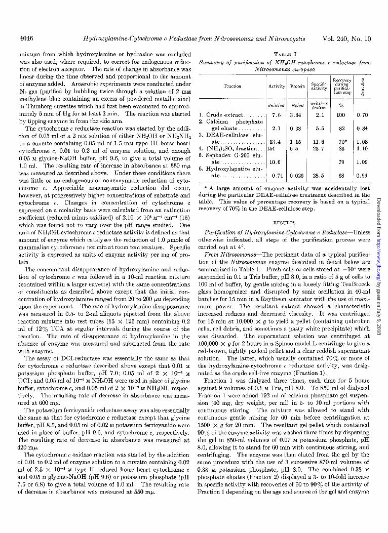
TABLE I summary of purijication of NHSOH-cytochrome c reductase from
Nitrosomonas europaea
Fraction Activity
____
zL?Iits/ml
1. Crude extract.. 7.6 2. Calcium phosphate
gel eluate. . 2.1 3. DEAE-cellulose elu-
ate................. 13.4 4. (NH,) rS0, fraction. . 154 5. Sephadex G-200 elu-
ate................. 10.6 6. Hydroxylapatite elu-
ate................. 0.74
-
Protein Specific activity
w/fd
3.64
0.38
1.15 6.5
?mits/nzg protein
2.1
5.5
11.6 23.7
0.026 28.5
1 R.%OW> during purifica- tion stq
% 100
82
70* 83
79
68
,
I
% a
f -4
0.70
0.84
1.08 1.10
1.09
0.94
* A large amount of enzyme activity was accidentally lost during the particular DEAE-cellulose treatment described in the table. This value of percentage recovery is based on a typical recovery af 70% in the DEAE-cellulose step.
RESULTS
Pur$cation of Hydroxylamine-Cytochrome c Reductase-Unless otherwise indicated, all steps of the purification process were carried out at 4”.
From Nitrosomonas-The pertinent data of a typical purifica- tion of the Nitrosomonas enzyme described in detail below are summarized in Table I. Fresh cells or cells stored at - 10” were suspended in 0.1 M Tris buffer, pH 8.0, in a ratio of 5 g of cells to 100 ml of buffer, by gentle mixing in a loosely fitting TenBroeck glass homogenizer and disrupted by sonic oscillation in 40.ml batches for 15 min in a Raytheon sonicator with the use of maxi- mum power. The resultant extract showed a characteristic increased redness and decreased viscosity. It was centrifuged for 15 min at 10,000 X g to yield a pellet (containing unbroken cells, cell debris, and sometimes a pasty white precipitate) which was discarded. The supernatant solution was centrifuged at 100,000 x g for 2 hours in a Spinco model L centrifuge to give a red-brown, tightly packed pellet and a clear reddish supernatant solution. The latter, which usually contained 70% or more of the hydroxylamine-cytochrome c reductase activity, was desig- nated as the crude cell-free enzyme (Fraction 1).
Fraction 1 was dialyzed three times, each time for 5 hours against 9 volumes of 0.1 M Tris, pH 8.0. To 850 ml of dialyzed Fraction 1 were added 192 ml of calcium phosphate gel suspen- sion (40 mg, dry weight, per ml) in 5- to lo-ml portions with continuous stirring. The mixture was allowed to stand with continuous gentle mixing for 60 min before centrifugation at 1500 x g for 20 min. The resultant gel-pellet which contained 90 y0 of the enzyme activity was washed three times by dispersing the gel in 850-ml volumes of 0.07 M potassium phosphate, pH 8.0, allowing it to stand for 60 min with continuous stirring, and centrifuging. The enzyme was then eluted from the gel by the same procedure with the use of 3 successive 870-ml volumes of 0.38 M potassium phosphate, pH 8.0. The combined 0.38 M
phosphate eluates (Fraction 2) displayed a 3- to lo-fold increase in specific activity with recoveries of 50 to 90% of the activity of Fraction 1 depending on the age and source of the gel and enzyme
by guest on July 9, 2018http://w
ww
.jbc.org/D
ownloaded from
October 1965 A. B. Hooper and A. Nason 4047
preparation. It was found necessary on the basis of preliminary trials with I- to 5ml portions of Fraction 1 to modify the ratio of gel to protein and the number and buffer molarity of elutions as different batches of gel and crude enzyme were used.
The potassium phosphate concentration of Fraction 2 was changed from 0.38 M to an estimated 1 .O mM by successive dialysis against distilled water over periods of 10 to 12 hours. Dialyzed Fraction 2 was then added at the rate of 3 drops per min to a chromatography column (4 X 40 cm) which had been packed under pressure with washed DEAE-cellulose and previously equilibrated with 1.0 mM potassium phosphate, pH 7.5. A red-brown area which apparently corresponded to the enzyme activity remained at the upper half of the column during subse- quent perfusion with 1 liter of 1.0 mM potassium phosphate at pH 7.5. A linear gradient elution procedure was then employed at the rate of 3 drops per min between 2 liters of 0.01 M phosphate, pH 7.5, and 2 liters of 0.5 M KC1 in 10-Z M phosphate at pH 7.5. In this manner enzyme activity was eluted in a slightly asym- metric peak beginning at approximately 0.2 M KCl. The DEAE- cellulose eluate fractions were pooled to form a red solution (Fraction 3), containing 70% of the Fraction 2 enzyme units with a specific activity 1.5 to 2.5 times that of Fraction 2.
Fraction 3 was dialyzed twice for 4 hours, each time against 100 volumes of 0.01 M potassium phosphate, pH 7.5. Successive amounts of solid ammonium sulfate were added slowly to Frac- tion 3 to give the following concentrations in grams per ml: 0.15, 0.2, 0.3, 0.4, 0.5, and 0.6. Each precipitate was collected by centrifugat.ion after standing for 1 hour at 0”. The red precipi- tates (collected between 0.3 and 0.5 g of (NH&SO4 per ml) containing 80 to 90% of the activity of Fraction 3 were resus- pended in a minimum of 0.01 M potassium phosphate, pH 7.5, and centrifuged to yield a clear supernatant solution (Fraction 4).
For the next purification step a column of Sephadex G-200 (1 X 20 cm) was prepared as follows. A suspension of 2 g of Sephadex G-200 dispersed in 250 ml of 0.1 M KCl-0.01 M potas- sium phosphate solution, pH 7.5, which had been decanted free of fine particles and equilibrated for 2 to 3 days, was allowed to settle in the column until a 2-cm layer had formed at the bot- tom. The phosphate-KCl-Sephadex suspension was then al- lowed to flow through the column at a rate of 5 drops per min. When the column had been fully packed and had equilibrated for 6 hours, a relatively firm 5-mm layer of Sephadex G-25 suspended in the same buffer was deposited at the top of the Sephadex G-200 in order to facilitate the subsequent addition of enzyme without disturbing the packing of Sephadex G-200. The column was equilibrated by allowing phosphate-KC1 solution to flow through overnight. Solid KC1 and sucrose were dissolved in Fraction 4 to a final concentration of 0.1 M and 25%, respectively, and 0.4 ml of this solution was layered on top of the Sephadex G-25 through the overlying 2 to 3 cm of phosphate-KC1 solution. As the phosphate-KC1 solution flowed through the column at 3 drops per min, the enzymatic activity moved in a single red band. The enzyme was eluted after the passage of a volume of effluent approximately equivalent to the void volume of the column to yield Fraction 5, with total and specific activities of 80 and 114%, respectively, of Fraction 4.
Fraction 5 was dialyzed for 10 hours against 500 volumes of 2 mM potassium phosphate buffer, pH 7.5, added at the rate of 2 drops per min to a gravity-packed Hypatite column (1 x 20 cm) which had been previously equilibrated with 2 mM potassium
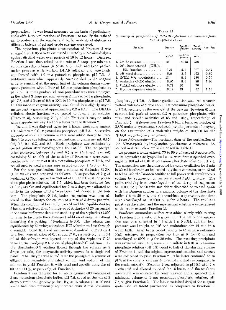
TABLE II summary of purijkation of NHnOH-cytochrome c reductase from
Nitrosocystis oceanus
Fraction
1. Crude extract.. . 2. 70” heat-treated (NH,)2.
SO4 fraction, 3. pH precipitate. 4. (NH,) zSO4 precipitate. 5. Sephadex G-200 eluate 6. DEAE-cellulose eluate. 7. Hydroxylapatite eluate.
1.5 2.0 167 5.0 3.6 162
13 9.5 186 0.95 9.0 86 0.71 10 76 0.14 13 52
310
0.42 0.60 0.70 1.06
1.10
phosphate, pH 7.0. A linear gradient elution was used between 250-ml volumes of 2 mM and 1.0 M potassium phosphate buffer, pH 7.5, resulting in the removal of the enzyme (Fraction 6) in a symmetrical peak at around 0.3 M potassium phosphate, with total and specific activities of 68 and lOS%, respectively, of Fraction 5. Nitrosomonas Fraction 6 had a turnover number of 2,850 moles of cytochrome c reduced per min per mole of enzyme, on the assumption of a molecular weight of 100,000 for the NH%OH-cytochrome c reductase.
From NitrosocystisThe pertinent data of the purification of the Nitrosocystis hydroxylamine-cytochrome c reductase de- scribed in detail below are summarized in Table II.
To prepare a crude extract, 70 to 90 g of washed Nitrosocystis,
or its equivalent as lyophilized cells, were first suspended over- night in 100 ml of 0.01 M potassium phosphate solution, pH 7.5. The suspension was then disrupted by sonic oscillation for 5 min in 30-ml batches in an ice-cooled Raytheon sonicator, or in 15-ml batches with the Branson sonifier at full power with simultaneous cooling by submersion in an ice-ethanol-NaCl mixture. The pellet resulting from centrifugation of the disrupted suspension at 20,000 X g for 20 min was either discarded or treated again with the Branson sonifier in a minimal volume of the phosphate buffer (15 to 20 ml), and the combined supernatant solutions were centrifuged at 100,000 x g for 2 hours. The resultant pellet was discarded, and the supernatant solution was designated as the crude extract (Fraction 1).
Powdered ammonium sulfate was added slowly with stirring to Fraction 1 in a ratio of 4 g per ml. The pH of the suspen- sion was then adjusted to 6.3 with 1 N NaOH, and the tem- perature was brought to 70” and maintained for 14 min in a water bath. After being cooled rapidly to 0” in an ice-ethanol- NaCl mixture, the preparation was kept at 0” for 60 min and centrifuged at 5000 X g for 30 min. The resulting precipitate was extracted with 32yo ammonium sulfate in 0.01 M potassium phosphate solution (pH 6.3) equal to half of the starting volume of Fraction 1, and the original supernatant solution and extract were combined to yield Fraction 2. The latter contained 55 to 91 y. of the activity and was 5- to ‘i-fold purified (as compared to the crude extract). Fraction 2 was adjusted to pH 5.0 with 2 N
acetic acid and allowed to stand for 15 hours, and the resultant precipitate was collected by centrifugation and suspended in a minimum volume of 1 mM potassium phosphate solution, pH 7.5, to give Fraction 3. The latter contained 54% of the enzyme units with an 8-fold purification as compared to Fraction 1.
by guest on July 9, 2018http://w
ww
.jbc.org/D
ownloaded from
Hydroxylamine-Cytochrome c Reductuse from Nitrosomonas and Nitrosocystis Vol. 240, No. 10
Ammonium sulfate was added to Fraction 3 in the ratio of 0.58 g per ml, and the mixture was allowed to stand at O-4” for 1 hour with intermittent stirring. The resultant heavy red pre- cipitate was sedimented by centrifugation and resuspended in about 3 ml of 0.01 M potassium phosphate-O.01 M KC1 solution (pH 7.5) to constitute Fraction 4, which possessed 60% of the total enzyme units with a 22-fold purification as compared to Fraction 1.
by hydrazine. Under optimum conditions the maximum rate was one-half the maximum rate of cytochrome c reduction by hydroxylamine with the same purified enzyme preparation. The two activities were not additive, for when saturating levels of both hydrazine and hydroxylamine were present in the standard reaction mixture the rate of cytochrome c reduction was no greater than that with either substrate alone.
Fracbion 4 was passed through a Sephadex G-200 column (2.4 x 24 cm) essentially as described for the Nitrosomonas enzyme to yield three elution peaks of 280 rnp absorbance. The small first peak and the large third peak had little or no activity and an Azso : ASeO ratio of 0.6, indicating a high nucleic acid con- tent. The second and largest peak, which followed closely after the first, contained the activity and had an Azso: ASG0 ratio of slightly more than 1.0. The pooled eluates of the active peak (Fraction 5) had 28% of the enzyme units, and Fraction 5 was 21-fold purified as compared to Fraction 1.
Fraction 5 was dialyzed for 6 hours against three changes of 100 volumes of 1 mM potassium phosphate solution (pH 7.5) and added to a DEAE-cellulose column (1.1 X 13 cm) previously packed under pressure and equilibrated with 1 mM phosphate, pH 7.5. Hydroxylamine-cytochrome c reductase activity ad- hered as a red-colored band in the upper portion of the column. A small amount of 280 rnp absorbing material was removed by the successive elution with 15- to 40-ml volumes of potassium phosphate solution (pH 7.5): 1 mM, 10 mM, 20 mM, 50 mM, 0.1 M,
0.15 M, and 0.2 M. A linear gradient elution procedure with the use of 100.ml volumes of 0.2 to 1.0 M KC1 in 1 mM phosphate at pH 7.5 resulted in elution of the enzyme (Fraction 6) between 0.25 and 0.3 M KCl-phosphate with a recovery of 17% of the enzyme units and a specific activity 30-fold that of Fraction 1.
In Nitrosocystis Preparations-Although crude extracts of Nitrosocystis contained NADH oxidase and cytochrome c oxidase activity, neither of these activities nor hydroxylamine- NAD, -NADP, -FAD, or -FMN reductase (assayed at pH 6.8 and 9.6) was present in the purified Nitrosocystis preparation. Hydrazine-cytochrome c reductase activity was present but at a level of 5% or less of the hydroxylamine-cytochrome c reductase activity. When the Nitrosocystis and Nitrosomonas purified enzymes were mixed and assayed under conditions of substrate saturation, the rates of hydrazine-cytochrome c reductase were additive, indicating that no inhibitor was present in the Nitroso- cystis preparation. The purified fractions from either organism contained no nitrite- or ammonium sulfate-cytochrome c reduc- tase.
Fraction 6 was dialyzed twice against 400 volumes of 2 mM potassium phosphate (pH 7.5) for a total of 12 hours, diluted to 1:2, and processed through a hydroxylapatite column (1 x 20 cm) as described for the Nitrosomonas enzyme to yield an eluate (Fraction 7) containing 17% of the original hydroxylamine- cytochrome c reductase units with an over-all 30-fold purification.
As shown in a later section, ferric ions stimulate the enzyme activity 2- to a-fold in the DEAE-cellulose and hydroxylapatite eluates (Fractions 6 and 7) but not in the crude extract. An adjustment for this stimulation has not been made on the data of Table II. The hydroxylamine-cytochrome c reductase of Nitrosocystis Fraction 7 had a turnover number of 1,300 (2,600 to 3,900 in the presence of FeClJ moles of cytochrome c reduced per min per mole of enzyme on the assumption of a molecular weight of 100,000 for the enzyme.
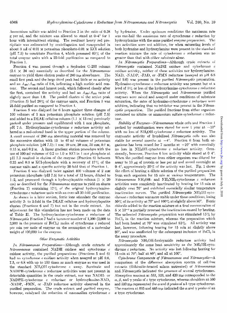
Stability of Enzymes-Nitrosomonas whole cells and Fraction 1 packed by centrifugation were stored for over 1 year at -10” with no loss of NHzOH-cytochrome c reductase activity. The enzymatic activity of lyophilized Nitrosocystis cells was also stable for several months at -4”. Fraction 6 from both or- ganisms has been stored for 7 months at -10” with essentially no loss in NHzOH-cytochrome c reductase activity. Occa- sionally, however, Fraction 6 lost activity while stored at O-4”. When the purified enzyme from either organism was diluted for assay to 10 M of protein or less per ml and stored overnight at O-4”, approximately 50% of the activity was lost. Fig. 1 shows the effect of heating a dilute solution of the purified preparation from each organism for 15 min at various temperatures. The Nitrosomonas NH20H- and NHzNHp-cytochrome c reductase activities were completely inactivated by heating for 15 min at slightly over 70” and exhibited essentially similar temperature inactivation curves. The purified Nitrosocystis NHzOH-cyto- chrome c reductase was more stable under these conditions, losing 50% of its activity at 70” and 100% at slightly above 80”. Ferric chloride added to the reaction mixture at a final concentration of 5 x lop5 M partially reversed the inactivation caused by heating. The unheated Nitrosocystis preparation was stimulated 15% by FeC& in the reaction mixture, whereas the preparation which had been heated at 70” was stimulated 85%. All activity was lost, however, following heating for 15 min at slightly above 80”, and was unaffected by the subsequent inclusion of FeCL in the assay procedure.
Nitrosocystis NHzOH-ferricyanide reductase activity had approximately the same heat sensitivity as the NHZOH-cyto- chrome c reductase. No activity was lost following heating for 15 min at 70”; half at 80”; and all at 100”.
Other Enzymatic Activities
In Nitrosomonas Preparations-Although crude extracts of Nitrosomonas contained NADH oxidase and cytochrome c oxidase activity, the purified preparations (Fractions 5 and 6) had no cytochrome c oxidase activity when assayed at pH 9.6, 7.5, or 6.8 with up to 100 times as much enzyme as was used in the standard NH*OH-cytochrome c assay. Succinate and NADPH-cytochrome c reductase activities were not present in detectable quantities in the crude extract, nor was NADH- or NADPH-cytochrome c reductase or hydroxylamine-NAD, -NADP, -FMN, or -FAD reductase activity observed in the purified preparation. The crude extract and purified enzyme, however, catalyzed the reduction of mammalian cytochrome c a type cytochrome.
Cytochrome Components of Nitrosmonas and Nitrosocystis-A comparison of the difference absorption spectra of cell-free extracts (dithionite-treated minus untreated) of Nitrosomonas and Nitrosocystis indicated the presence of several cytochromes. Absorption maxima at 553, 523, and 420 rnp corresponded to the Q, 0, and y peaks of c type cytochrome, whereas shoulders at 560 and 530 rnp represented the OL and /3 peaks of a b type cytochrome. The maxima at 605 and 445 rnp indicated the (Y and y peaks of an
by guest on July 9, 2018http://w
ww
.jbc.org/D
ownloaded from
October 1965 A. B. Hooper and A. Nason 4049
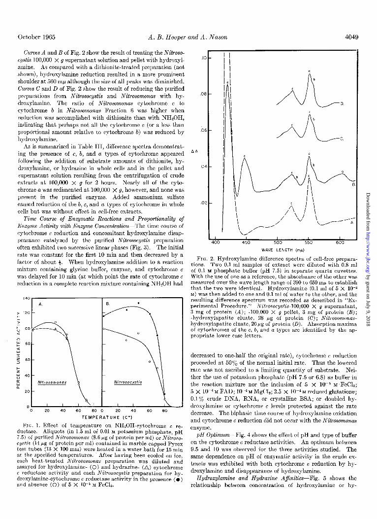
Curves A and B of Fig. 2 show the result of treating the Nitroso- cystis 100,000 X g supernatant solution and pellet with hydroxyl- amine. As compared with a dithionite-treated preparation (not shown), hydroxylamine reduction resulted in a more prominent shoulder at 560 rnp although the size of all peaks was diminished. Curves C and D of Fig. 2 show the result of reducing the purified preparations from Nitrosocystis and Nitrosomonas with hy- droxylamine. The ratio of Nitrosomonas cytochrome c to cytochrome 6 in Nitrosomonas Fraction 6 was higher when reduction was accomplished with dithionite than with NH,OH, indicating that perhaps not all the cytochrome c (or a less than proporbional amount relative to cytochrome b) was reduced by hydroxylamine.
As is summarized in Table III, difference spectra demonstrat- ing the presence of c, b, and a types of cytochrome appeared following the addition of substrate amounts of dithionite, hy- droxylamine, or hydrazine in whole cells and in the pellet and supernatant solution resulting from the centrifugation of crude extracts at 100,000 x g for 2 hours. Nearly all of the cyto- chrome a was sedimented at 100,000 x g, however, and none was present in the purified enzyme. Added ammonium sulfate caused reduction of the b, c, and a types of cytochrome in whole cells but was without effect in cell-free extracts.
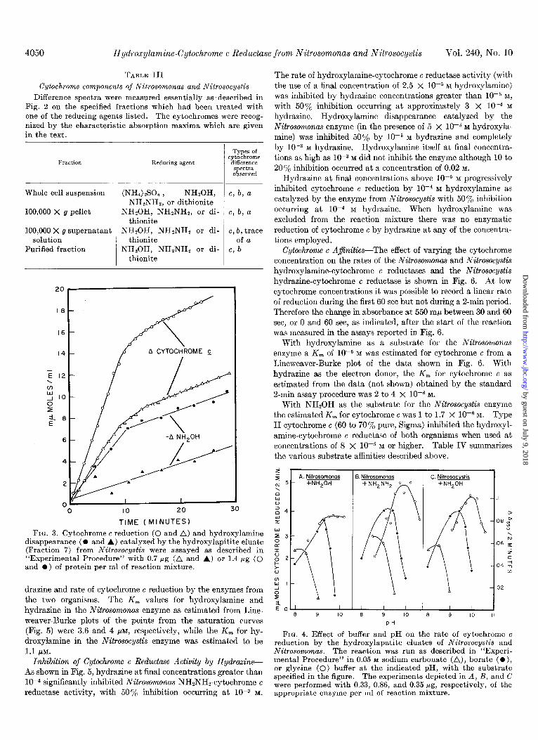
Time Course of Enzymatic Reactions and Proportionality of Enzyme Activity with Enzyme Concentration-The time course of cytochrome c reduction and concomitant hydroxylamine disap- pearance catalyzed by the purified Nitrosocystis preparation often exhibited two successive linear phases (Fig. 3). The initial rate was constant for the first 10 min and then decreased by a factor of about +. When hydroxylamine addition to a reaction mixture containing glycine buffer, enzyme, and cytochrome c was delayed for 10 min (at which point the rate of cytochrome c reduction in a complete reaction mixture containing NHzOH had
140
A. EL .
* tzo- +
z 60 - 3
Nitrosocystis n
20 -
I
0 20 40 60 80 0 20 40 60 80
TEMPERATURE (Co)
FIG. 1. Effect of temperature on NHZOH-cytochrome e re- ductase. Aliquots (in 1.5 ml of 0.01 M potassium phosphate, pH 7.5) of purified Nitrosomonas (8.6 fig of protein per ml) or Nitroso- cystis (14 pg of protein per ml) contained in marble-capped Pyrex test tubes (13 X 100 mm) were heated in a water bath for 15 min at the specified temperatures. After having been cooled on ice, each heat-treated Nitrosomonas preparation was diluted and assayed for hydroxylamine- (0) and hydrazine- (A) cytochrome c reductase activity and each Nitrosocystis preparation for hy- droxylamine-cytochrome c reductase activity in the presence (0) and absence (0) of 5 X 10m5 M FeCL.
I ’ I I I I 400 450 500 550 600
WAVE LENGTH (m,u)
FIG. 2. Hydroxylamine difference spectra of cell-free prepara- tions. Two 0.1.ml samples of extract were diluted with 0.8 ml of 0.1 M phosphate buffer (pH 7.5) in separate quartz cuvettes. With the use of one as a reference, the absorbance of the other was measured over the wave length range of 390 to 650 rnp to establish that the two were identical. Hydroxylamine (0.1 ml of 5 X low3 M) was then added to one and 0.1 ml of water to the other, and the resulting difference spectrum was recorded as described in “Ex- perimental Procedure.” Nitrosocystis-100,000 X g supernatant, 3 mg of protein (A) ; -100,000 X g pellet, 3 mg of protein (I?); -hydroxylapatite eluate, 28 fig of protein (C); Nitrosomonas- hydroxylapatite eluate, 26 pg of protein (II). Absorption maxima of cytochromes of the c, b, and a types are identified by the ap- propriate lower case letters.
decreased to one-half the original rate), cytochrome c reduction proceeded at 50% of the normal initial rate. Thus the lowered rate was not ascribed to a limiting quantity of substrate. Nei- ther the use of potassium phosphate (pH 7.5 or 6.8) as buffer in the reaction mixture nor the inclusion of 5 X 1OP M “FeC18; 5 X lop4 M FAD; 1O-4 M MgC&; 2.5 X 10e4 M reduced glutatione; 0.1% crude DNA, RNA, or crystalline BSA; or doubled hy- droxylamine or cytochrome c levels protected against the rate decrease. The biphasic time course of hydroxylamine oxidation and cytochrome c reduction did not occur with the Nitrosomonas enzyme.
pH Optimum-Fig. 4 shows the effect of pH and type of buffer on the cytochrome c reductase activities. An optimum between 9.5 and 10 was observed for the three activities studied. The same dependence on pH of enzymatic activity in the crude ex- tracts was exhibited with both cytochrome c reduction by hy- droxylamine and disappearance of hydroxylamine.
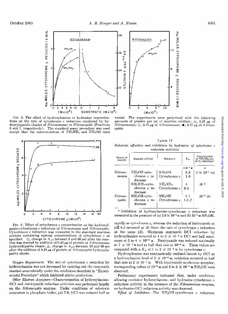
Hydroxylamine and Hydrazine AfinitiesFig. 5 shows the relationship between concentration of hydroxylamine or hy-
by guest on July 9, 2018http://w
ww
.jbc.org/D
ownloaded from
4050 Hydroxylamine-Cytochrome c Reductase from Nitrosomonas and Nitrosocystis Vol. 240, No. 10
TABLE III Cytochrome components of Nitrosomonas and Nitrosocystis
Difference spectra were measured essentially as’described in Fig. 2 on the specified fractions which had been treated with one of the reducing agents listed. The cytochromes were recog- nized by the characteristic absorption maxima which are given in the text.
Fraction Reducing agent
Types of cytochrome difference
spectra observed
Whole cell suspension
100,000 X g pellet
100,000 X g supernatant solution
Purified fraction
(NW 30, , NH20H, NHpNHs, or dithionite
NHsOH, NH2NH2, or di- thionite
NH?OH, NHzNH2 or di- thionite
NH20H, NH2NH2 or di- thionite
c, b, a
c, b, a
c, b, trace of a
c, b
The rate of hydroxylamine-cytochrome c reductase activity (with the use of a final concentration of 2.5 X 10F5 M hydroxylamine) was inhibited by hydrazine concentrations greater than lop5 M,
with 50% inhibition occurring at approximately 3 X lo+ M hydrazine. Hydroxylamine disappearance catalyzed by the Nitrosomonas enzyme (in the presence of 5 X 10m5 M hydroxyla- mine) was inhibited 50% by low5 M hydrasine and completely by lo+ M hydrazine. Hydroxylamine itself at final concentra- tions as high as lop3 M did not inhibit the enzyme although 10 to 20 y0 inhibition occurred at a concentration of 0.02 M.
Hydrazine at final concentrations above 10e5 M progressively inhibited cytochrome c reduction by 1O-4 M hydroxylamine as catalyzed by the enzyme from Nitrosocystis with 50% inhibition occurring at lop4 M hydrazine. When hydroxylamine was excluded from the reaction mixture there was no enzymatic reduction of cytochrome c by hydrazine at any of the concentra- tions employed.
Cytochrome c AfinitiesThe effect of varying the cytochrome concentration on the rates of the Nitrosomonas and Nitrosocystis hydroxylamine-cytochrome c reductases and the Nitrosocystis hydrazine-cytochrome c reductase is shown in Fig. 6. At low cytochronie concentrations it was possible to record a linear rate of reduction during the first 60 set but not during a 2-min period. Therefore the change in absorbance at 550 rnp between 30 and 60 set, or 0 and 60 set, as indicated, after the start of the reaction was measured in the assays reported in Fig. 6.
With hydroxylamine as a substrate for the Nitrosomonas enzyme a K, of low6 M was estimated for cytochrome c from a Lineweaver-Burke plot of the data shown in Fig. 6. With hydrazine as the electron donor, the K, for cytochrome c as estimated from the data (not shown) obtained by the standard 2-min assay procedure was 2 to 4 X lop6 M.
With NH,OH as the substrate for the Nitrosocystis enzyme the estimated K, for cytochrome c was 1 to 1.7 X 10e6 M. Type II cytochrome c (60 to 70% pure, Sigma) inhibited the hydroxyl- amine-cytochrome c reductase of both organisms when used at concentrations of 8 X 10M5 M or higher. Table IV summarizes the various substrate affinities described above.
IO 20 30
TIME ( MINUTES) E t
FIG. 3. Cytochrome e reduction (0 and A) and hydroxylamine disappearance (O and A) catalyzed by the hydroxylapitite eluate (Fraction 7) from Nitrosocystis were assayed as described in “Experimental Procedure” with 0.7 Mg (A and A) or 1.4 pg (0 and l ) of protein per ml of reaction mixture.
drazine and rate of cytochrome c reduction by the enzymes from the two organisms. The K, values for hydroxylamine and hydrazine in the Nitrosomonas enzyme as estimated from Line- weaver-Burke plots of the points from the saturation curves
3. Nitrosomonos
- _I
P -.os p
0 ;;
-.06 x 5 c
-.04 ;;I w
- .02
(Fig. 5) were 3.6 and 4 PM, respectively, while the K, for hy- droxylamine in the Nitrosocystis enzyme was estimated to be
FIG. 4. Effect of buffer and pH on the rate of cytochrome c reduction by the hydroxylapatite eluates of Nitrosocystis and
1.1 ILM. Nitrosomonas. The reaction was run as described in “Experi- Inhibition of Cytochrorrw G Reductase Activity by Hydrazine- mental Procedure” in 0.05 M sodium carbonate (A), borate -(O),
As shown in Fig. 5, hydrazine at final concentrations greater than or glycine (0) buffer at the indicated pH, with the substrate
lop4 significantly inhibited Nitrosomonas NHzNHp-cytochrome c specified in the figure. The experiments depicted in A, B, and C
reductase activity, with 50% inhibition occurring at 10-S M. were performed with 0.33, 0.86, and 0.35 rg, respectively, of the appropriate enzyme per ml of reaction mixture.
by guest on July 9, 2018http://w
ww
.jbc.org/D
ownloaded from
October 1965 A. B. Hooper and A. Nason 4051
t r N Nitrosomonos \
I I I I 2 ‘e
(M x IO61 SUBSTRATE (M ~10~)
I I 2 4 2’
(Mx 103)
FIG. 5. The effect of hydroxylamine or hydraaine concentra- tions on the rate of cytochrome c reduction catalyzed by hy- droxylapatite eluates of Nitrosomonas or Nitrosocystis (Fractions 6 and 7, respectively). The standard assay procedure was used except that the concentrations of NHzNHz and NHSOH were
varied. The experiments were performed with the following amounts of protein per ml of reaction mixture: A, 0.43 pg of Nitrosomonas; 0, 0.43 pg of Nitrosomonas; l , 0.17 pg of Nitroso- cystis.
I I I I I ‘/4
0 2 4 6 8 IO 12 14 16 80
CYTOCHROME c(Mx106)
FIG. 6. Effect of cytochrome c concentration on the hydroxyl- amine-cytochrome c reductase of Nitrosomonas and Nitrosocystis. Cytochrome c reduction was measured in the standard reaction mixture containing various concentrations of cytochrome c as specified. 0, change in A,,0 between 0 and 60 set after the reac- tion was started by addition of 0.43 pg of protein of Nitrosomonas hydroxylapatite eluate; A, change in 4550 between 30 and 60 set after the addition of 0.11 pg of protein of Nitrosocystis hydroxyla- patite eluate.
Oxygen Requirement-The rate of cytochrome c reduction by hydroxylamine was not decreased by carrying out the enzymatic reaction anaerobically under the conditions described in “Experi- mental Procedure” which inhibited nitrite production.
Other Electron Acceptors-Characterization of hydroxylamine- DC1 and -ferricyanide reductase activities was performed largely on the Nitrosocystis enzyme. Under conditions of substrate saturation in phosphate buffer, pH 7.0, DCI was reduced half as
-L
,
TABLE IV
Substrate a&uities and inhibition by hydrazine of cytochrome c reductase activities
Source of enzyme Enzyme activity
Nitroso- NHZOH-cyto- monas chrome c re-
ductase NHzNHz-cyto-
chrome c re- ductase
Nitroso- NHtOH-cyto- cystis chrome c re-
ductase
Substrate Gi
NHzOH Cytochrome c
NHsNHz Cytochrome c
NH,OH Cytochrome c
x10-6 M
3.6 1.0
Concentration of NHzNHz for
50 Y. inhibition*
M
3 X 10e4 (a)
10-s
1O-4 (6)
* Inhibition of hydroxylamine-cytochrome c reductase was measured in the presence of (a) 2.5 X low5 M and (b) lo+ M NH20H.
rapidly as cytochrome c, whereas the reduction of ferricyanide at pH 8.5 occurred at 20 times the rate of cytochrome c reduction at the same pH. Maximum enzymatic DC1 reduction by hydroxylamine occurred at 1 to 2 X low4 M DC1 and half maxi- mum at 3 to 4 X 10e5 M. Ferricyanide was reduced maximally at 7 X 1O-4 M and at half that rate at lop4 M. These values are compared with a K, of 1 to 2 X 1OV M for cytochrome c.
Hydroxylamine was enzymatically oxidized fastest by DC1 at a hydroxylamine level of 5 x lop4 M; oxidation occurred at half that rate at 2 x 10e5 M. With ferricyanide as electron acceptor, corresponding values of lop4 M and 2 to 3 X lo+ M NHzOH were observed.
Preliminary experiments indicated that, under conditions allowing extensive hydroxylamine- and hydrazine-cytochrome c reductase activity in the presence of the Nitrosomonas enzyme, no hydrazine-DC1 reductase activity was observed.
E$ect of Inhibitors-The NHZOH-cytochrome c reductase
by guest on July 9, 2018http://w
ww
.jbc.org/D
ownloaded from
4052 Hydroxylamine-Cytochrome c Reductase from Nitrosomonas and Nitrosocystis Vol. 240, No. 10
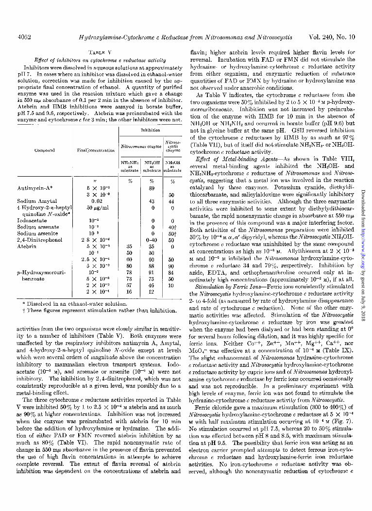
TABLE V Effect of inhibitors on cytochrome c reductase activity
Inhibitors were dissolved in aqueous solutions at approximately pH 7. In cases where an inhibitor was dissolved in ethanol-water solution, correction was made for inhibition caused by the ap- propriate final concentration of ethanol. A quantity of purified enzyme was used in the reaction mixture which gave a change in 550 rnp absorbance of 0.1 per 2 min in the absence of inhibitor. Atebrin and HMB inhibitions were assayed in borate buffer, pH 7.5 and 9.6, respectively. Atebrin was preincubated with the enzyme and cytochrome c for 3 min; the other inhibitors were not.
Compound Final:concentration
Antimycin-A*
Sodium Amytal 4-Hydroxy-2-n-hepty
quinoline N-oxide* Iodoacetate Sodium arsenate Sodium arsenite 2,4-Dinitrophenol Atebrin
p-Hydroxymercuri- benzoate
M
5 x 10⁻³
3 x 10-a 0.02
50 fig/ml
10-s 10-Z 10-z
2-8 x 10-d 5 x 10-S
10-4
2.5 X lo-’ 5 x 10-s 10-Z
5 x 10⁻³
2 x 10-s 2 x 10⁻⁴
-
Inhibition T
NHzNH: as
;ubstrate
70
35 50 60 80 78 73 57 16
: !
-
NH20H QS
jubstratc
%
89
43 0
0 0 0
O-40 35 50 60 88 91 73 46 12
! e
--
1
: s1 --
Yitroso- cystis
enzyme
NHaOH as
lbstrate
%
50 44 0
0 Jot 5@t 50 0
50 90 84 50 10
* Dissolved in an ethanol-water solution. t These figures represent stimulation rather than inhibition.
activities from the two organisms were closely similar in sensitiv- ity to a number of inhibitors (Table V). Both enzymes were unaffected by the respiratory inhibitors antimycin A, Amytal, and 4-hydroxy-2-n-heptyl quinoline N-oxide except at levels which were several orders of magnitude above the concentration inhibitory to mammalian electron transport systems. Iodo- acetate (lo+ M), and arsenate or arsenite (1OW M) were not inhibitory. The inhibition by 2,4-dinitrophenol, which was not consistently reproducible at a given level, was possibly due to a metal-binding effect.
The three cytochrome c reductase activities reported in Table V were inhibited 50% by 1 to 2.5 X lop4 M atebrin and as much as 90% at higher concentrations. Inhibition was not increased when the enzyme was preincubated with atebrin for 10 min before the addition of hydroxylamine or hydrazine. The addi- tion of either FAD or FMN reversed atebrin inhibition by as much as 80% (Table VI). The rapid nonenzymatic rate of change in 550 rnp absorbance in the presence of flavin prevented the use of high flavin concentrations in attempts to achieve complete reversal. The extent of flavin reversal of atebrin inhibition was dependent on the concentrations of atebrin and
flavin; higher atebrin levels required higher flavin levels for reversal. Incubation with FAD or FMN did not stimulate the hydrazine- or hydroxylamine-cytochrome c reductase activity from either organism, and enzymatic reduction of substrate quantities of FAD or FMN by hydrazine or hydroxylamine was not observed under anaerobic conditions.
As Table V indicates, the cytochrome c reductases from the two organisms were 50% inhibited by 2 to 5 x lop3 M p-hydroxy- mercuribenzoate. Inhibition was not increased by preincuba- tion of the enzyme with HMB for 10 min in the absence of NHzOH or NHzNHB and occurred in borate buffer (pH 9.6) but not in glycine buffer at the same pH. GSH reversed inhibition of the cytochrome c reductases by HMB by as much as 97% (Table VII), but of itself did not stimulate NH2NH2- or NH20H- cytochrome c reductase activity.
Effect of Metal-binding Agents-As shown in Table VIII, several metal-binding agents inhibited the NH20H- and NHtNHz-cytochrome c reductase of Nitrosomonas and Nitroso- cystis, suggesting that a metal ion was involved in the reaction catalyzed by these enzymes. Potassium cyanide, diethyldi- thiocarbamate, and salicylaldoxime were significantly inhibitory to all three enzymatic activities. Although the three enzymatic activities were inhibited to some extent by diethylydithiocar- bamate, the rapid nonenzymatic change in absorbance at 550 rnp in the presence of this compound was a major interfering factor. Both activities of the Nitrosomonas preparation were inhibited 50 % by 1O-4 M o(, a’-dipyridyl, whereas the Nitrosocystis NHSOH- cytochrome c reductase was uninhibited by the same compound at concentrations as high as lo+ M. Allylthiourea at 2 X 1OW M and lo-$ M inhibited the Nitrosomonas hydroxylamine-cyto- chrome c reductase 34 and 79%, respectively. Inhibition by azide, EDTA, and orthophenanthroline occurred only at in- ordinately high concentrations (approximately lop2 M), if at all.
Xtimulation by Ferric Ions-Ferric ions consistently stimulated the Nitrosocystis hydroxylamine-cytochrome c reductase activity 2- to 4-fold (as measured by rate of hydroxylamine disappearance and rate of cytochrome c reduction). None of the other enzy- matic activities was affected. Stimulation of the Nitrosocystis hydroxylamine-cytochrome c reductase by iron was greatest when the enzyme had been dialyzed or had been standing at 0” for several hours following dilution, and it was highly specific for ferric ions. Neither Co++, Zn*+, Mn++, Mg++, Ca++, nor Mood- was effective at a concentration of 10e4 M (Table IX). The slight enhancement of Nitrosomonas hydrazine-cytochrome c reductase activity and Nitrosocystis hydroxylamine-cytochrome c reductase activity by cupric ions and of Nitrosomonas hydroxyl- amine-cytochrome c reductase by ferric ions occurred occasionally and was not reproducible. In a preliminary experiment with high levels of enzyme, ferric ion was not found to stimulate the hydrazine-cytochrome c reductase activity from Nitrosocystis.
Ferric chloride gave a maximum stimulation (300 to 400%) of Nitrosocystis hydroxylamine-cytochrome c reductase at 5 X 1O-4 M with half maximum stimulation occurring at lop4 M (Fig. 7). No stimulation occurred at pH 7.5, whereas 20 to 50% stimula- tion was effected between pH 8 and 8.5, with maximum stimula- tion at pH 9.5. The possibility that ferric iron was acting as an electron carrier prompted attempts to detect ferrous iron-cyto- chrome c reductase and hydroxylamine-ferric iron reductase activities. No iron-cytochrome c reductase activity was ob- served, although the nonenzymatic reduction of cytochrome c
by guest on July 9, 2018http://w
ww
.jbc.org/D
ownloaded from
October 1965 A. B. Hooper and A. Nason 4053
TABLE VI
Reversal by Jlavins of atebrin inhibition of cytochrome c reduction
A standard reaction mixture (with borate buffer, pH 7.5, since atebrin and flavin precipitated at a higher pH1 was preincubated for 3 min in the presence of the indicated concentration of atebrin and the absence of substrate (NHzNHQ or NH,OH). Substrate and flavin were then added, and the rate of cytochrome c reduction was measured in the standard manner. The rate for the mixture treated in this way was as reported in the column headed “Reversed.” A second reaction mixture (column headed “Inhibited”) was simultaneously treated in an identical manner except that no flavin was added. A third reaction mixture (column headed “Control”) was preincubated in the absence of atebrin and assayed in the standard manner without flavin. Each rate value was corrected for the change in absorbance occurring in an identically treated reaction mixture which contained no enzyme.
Source of enzyme
Nitrosomonas
Nitrosocystis
M
NHzNHz 2.5 X 10⁻⁴ 2.5 X 10⁻⁴
NHzOH 10-s 10-a
NH,OH 10-z 0.143 0.013 0.068 FAD 10-a 0.062 0.014 0.046 FMN
0.095 0.076 0.140 0.140
A.&so/z min
0.031 0.025 0.031 0.035
0.048 FAD 0.059 FMN 0.077 FAD 0.081 FMN
Flavin concentration
M
10-a 10-Z
2 x 10-s 2 x 10-S
2 x 10-s 2 x 10-a
TABLE VII TABLE VIII
GSH reversal of HMB inhibition of cytochrome c reductase
The experiments were run essentially as described in Table Inhibition of cytochrome c reductase activity by metal-binding agents
VI (with borate buffer, pH 9.6) except that HMB and GSH were The rate of cytochrome c reduction assayed in the st.andard
used in place of atebrin and flavin, respectively. manner was compared with the rate in identical reaction mixtures containing 0.3 to 0.7 pg of enzyme and specified levels of inhibi-
1 Substrate lconc~~ti.,! ET ‘ILi-IIy lc,,,,&;i:,..
tor. In cases in which an inhibitor w& dissolved in aqueous Enzyme ethanol solution, correction was made for the inhibitory effect
Control Inhibited Reversed of the appropriate final concentration of ethanol.
Nitroso- NHtOH monas NHzNHz
Nitroso- NHzOH cystis
18X10-“~0.134~0.021~0.130~ 8x10⁻³
by ferrous ions (10e4 M) was rapid enough to have possibly KCN obscured any enzymatic activity. Moreover reproducible enzymatic hydroxylamine disappearance could not be detected or+‘-Dipyridyl in a standard reaction mixture lacking cytochrome c but con- taining in its place FeC13 (final concentration, 10-S M) as an electron acceptor and cu,cu’-dipyridyl to form a complex with Diethyldithiocarba-
ferrous ion. mate
Ferric chloride did not stimulate the enzymatic reduction of
DC1 or ferricyanide by hydroxylamine. Salicylaldoxime
Stoichiometry of Reaction-The data of Table X show that in the presence of the Nitrosomonas enzyme an average of 2.6 moles
Allylthiourea
of mammalian cytochrome c were reduced per mole of hydroxyla- mine oxidized at pH 9.6, whereas the ratio fell to 1.5 at pH 6.8. NaN3t Consistent measurements of the quantity of hydroxylamine oxidized were made difficult because (a) the intensity of color EDTA
development per mole of hydroxylamine was found to vary in a standard hydroxylamine curve determined with each experiment Orthophenanthro-
and (b) hydroxylamine disappeared nonenzymatical1.y at a rate linet
. -
comprising as much as 30% of the total hydroxylamine disap- * I, inhibitory.
Concentration
M % 2 x 10-S 44
10-3 90 10-J 48
10-4 1*
6 X 1O-3
10-Z 2 x 10-S
0.02
0.015
0.02
47
0
95
--
-
Inhibition
NHzNHz NHzOH NHzOH
% %
49 57
90
65
I
0
I
41
79 34
0
65
78
0
50
42
pearance. With the purified Nitrosocystis preparation the ratio of cyto-
t Certain inhibitors were incubated with the enzyme for 3 min before the substrate was added. Inhibitors were in aqueous
chrome c reduced to hydroxylamine oxidized (cytochrome solutions at approximately pH 7.
by guest on July 9, 2018http://w
ww
.jbc.org/D
ownloaded from
4054 Hydroxylamine-Cytochrome c Reductase from Nitrosomonas and Nitrosocystis Vol. 240, No. 10
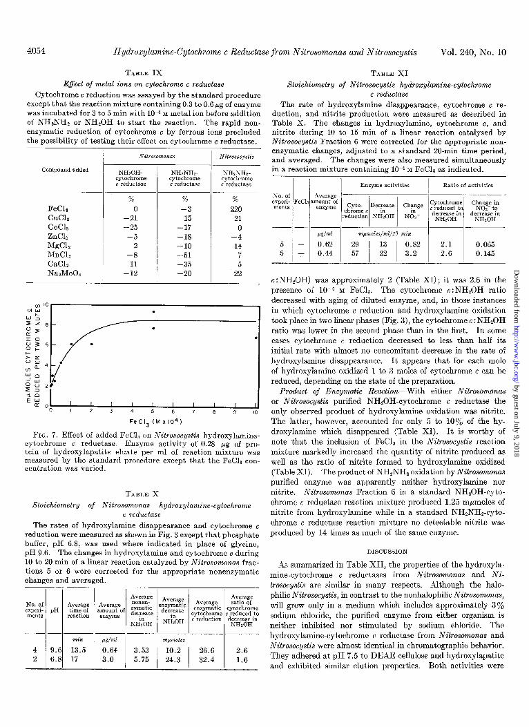
TABLE IX TABLE XI
Effect of metal ions on cytochrome c reductase Cytochrome c reduction was assayed by the standard procedure
except that the reaction mixture containing 0.3 to 0.6pg of enzyme was incubated for 3 to 5 min with 10V4 ivr metal ion before addition of NH2NH2 or NHSOH to start the reaction. The rapid non- enzymatic reduction of cytochrome c by ferrous ions precluded the possibility of testing their effect on cgtochrome c reductase.
Stoichiometry of Nitrosocystis hydroxylamine-cytochrome c reductase
The rate of hydroxylamine disappearance, cytochrome c re- duction, and nitrite production were measured as described in Table X. The changes in hydroxylamine, cytochrome c, and nitrite during 10 to 15 min of a linear reaction catalyzed by Nitrosocystis Fraction 6 were corrected for the appropriate non- enzymatic changes, adjusted to a standard 20-min time period, and averaged. The changes were also measured simultaneously in a reaction mixture containing 1O-4 M FeC18 as indicated. Compound added
FeCla CuClz CoClz ZnClp M&L MnCL CaClz NaPMoO
NHzOH- NHINHP cytochrome c reductase
cytochrome G reductase
% % % 0 -3 220
-21 15 21 -25 -17 0
-5 -18 -4 2 -10 14
-8 -51 7 11 -35 5
-12 -20 22
-- Nitrosocystis
NHzNHz- cytochrome t reductase
FeC13 (Mx104)
FIG. 7. Effect of added FeCh on Nitrosocystis hydroxylamine- cytochrome c reductase. Enzyme activity of 0.28 pg of pro- tein of hydroxylapatite eluate per ml of reaction mixture was measured by the standard procedure except that the FeCh con- centration was varied.
TABLE X
Stoichiometry of Nitrosomonas hydroxylamine-cytochrome c reductase
The rates of hydroxylamine disappearance and cytochrome c reduction were measured as shown in Fig. 3 except that phosphate buffer, pH 6.8, was used where indicated in place of glycine, pH 9.6. The changes in hydroxylamine and cytochrome c during 10 to 20 min of a linear reaction catalyzed by Nitrosomonas frac- tions 5 or 6 were corrected for the appropriate nonenzymatic changes and averaged.
Pg/+~l 4 0.64 2 3.0
Average :nzymatic Average %Z? decfease enzymatic cytochrome
NEOH cytochrome c reduced to c reduction decrease in
NHzOH
m~moles 10.2 26.6 2.6 24.3 I 32.4 I 1.6
/ / ] Enzyme activities 1 Ratio of activities
---I I I adml m~moles/??%l/Z0 min
5 - 0.62 29 13 5 + 0.44 57 22 3.2
I I
0.82 2.1 0.065 2.6 0.145
c :NH,OH) was approximately 2 (Table XI) ; it was 2.6 in the presence of 10e4 M FeC13. The cytochrome c:NH*OH ratio decreased with aging of diluted enzyme, and, in those instances in which cytochrome c reduction and hydroxylamine oxidation took place in two linear phases (Fig. 3), the cytochrome c : NHzOH ratio was lower in the second phase than in the first. In some cases cytochrome c reduction decreased to less than half its initial rate with almost no concomitant decrease in the rate of hydroxylamine disappearance. It appears that for each mole of hydroxylamine oxidized 1 to 3 moles of cytochrome c can be reduced, depending on the state of the preparation.
Product of Enzymatic Reaction--With either Nitrosomonas or Nitrosocystis purified NHzOH-cytochrome c reductase the only observed product of hydroxylamine oxidation was nitrite. The latter, however, accounted for only 5 to 10% of the hy- droxylamine which disappeared (Table XI). It is worthy of note that the inclusion of FeC13 in the Nitrosocystis reaction mixture markedly increased the quantity of nitrite produced as well as the ratio of nitrite formed to hydroxylamine oxidized (Table XI). The product of NHzNHz oxidation by Nitrosomonas purified enzyme was apparently neither hydroxylamine nor nitrite. Nitrosomonas Fraction 6 in a standard NHzOH-cyto- chrome c reductase reaction mixture produced 1.25 mnmoles of nitrite from hydroxylamine while in a standard NH2NHz-cyto- chrome c reductase reaction mixture no detectable nitrite was produced by 14 times as much of the same enzyme.
DISCUSSION
As summarized in Table XII, the properties of the hydroxyla- mine-cytochrome c reductases from Nitrosomonas and Ni- trosocystis are similar in many respects. Although the halo- philic Nitrosocystis, in contrast to the nonhalophilic Nitrosomonas, will grow only in a medium which includes approximately 3% sodium chloride, the purified enzyme from either organism is neither inhibited nor stimulated by sodium chloride. The hydroxylamine-cytochrome c reductase from Nitrosomonas and Nitrosocystis were almost identical in chromatographic behavior. They adhered at pH 7.5 to DEAE-cellulose and hydroxylapatite and exhibited similar elution properties. Both activities were
by guest on July 9, 2018http://w
ww
.jbc.org/D
ownloaded from
October 1965 A. B. Hooper and A. Nason
TABLE XII
4055
Comparison of Nitrosomonas NHZOH-cytochrome c reductase with Nitrosocystis NHSOH-cytochrome c reductase
Properties
Physical state
Heat sensitivity
Components of electron transport chain
pH optimum
K, for NHzOH
K,,, for cytochrome c
Moles of cytoohrome c reduced per min per mole of enzyme
Other electron acceptors
Inhibitors
Metal ion stimulators None
Stoichiometry of reaction (ratio of cyto- chrome c reduced to NHsOH oxidized)
Product
NHzNHz-cytochrome c reductase activity
Nilrosomonas NHzOH-cytochrome c reductase
Mol. wt. approximately l-2 X 10⁵ estimated from be- havior on Sephadex
In supernatant solution of sonic extracts centrifuged for 2 hr at 100,000 X g
Adheres to DEAE-cellulose at pH 7.5, eluted by 0.25 M KC1
Adheres to hydroxylapatite at pH 7.5 eluted by 0.2-0.3 M potassium phosphate
Stable to heating for 15 min at 50’; all activity lost at 7o”
Cytochrome 6 and c and possibly flavin
9.5-10 in glycine buffer
3.6 X 10-M
lo-’ M
2,850
DCI, ferricyanide (only two tried)
Insensitive to electron transport inhibitors antimycin- A, Amytal, 4-hydroxy-2-n-heptyl quinoline N-oxide, iodoacetate, arsenite, and arsenate
Sensitive to atebrin, HMB, KCN, a,a’-dipyridyl, di- ethyldithiocarbamate, salicylaldoxime, allylthiourer
2.6
Unidentified intermediate of probable oxidation state between NHzOH and Not-; little or no NOz-
V max 50% of NHzOH-cytochrome c reduotase
eluted from Sephadex G-200 in a manner characteristic of a molecule with a molecular weight of 100,000 to 200,000.
Because of (a) its large size, (b) the association of at least two types of cytochrome (b and c types), (c) the apparent involve- ment of flavin and iron, and (d) the obvious similarity, from a comparative biochemical viewpoint, of this system to other established electron transport chains such as DPNH- and suc- cinate-cytochrome c reductases (see, for example, Reference 16), it seems likely that each enzyme is in reality a complex of as- sociated proteins, activators, and possibly lipids rather than a single simple protein. Difference spectra of whole cells or crude extracts oxidizing NHzOH indicate the involvement of cyto- chromes of the b, c, and a types. The terminal cytochrome oxidase (cytochrome a), however, is apparently separated during
Nifrosocystis NHzOIkytochrome L reductase
Same
Same
Same, elutes at approximately 0.3 M KC1
Same
Stable to heating for 15 min at 70”; all activity lost at 85”
Same
Same
1.1 x lo+ M
1-1.7 x lo- M
1,300 (2,6003,900 with ferric ions)
Same
Same
Same
Ferric ion specifically activates 2- to 4-fold
1.1 (2.6 with ferric ion)
same
41most entirely absent, Vmsx 05yo of NHtOH-cytochrome c reductase
the course of purification of the NHQOH-cytochrome c reductase. It can be speculated that the natural sequence of electron trans- port in NHzOH-cytochrome c reductase involves the following:
NHzOH -+ flavin (Fe+++) -+ cytochrome b + cytochrome c
The present evidence for the involvement of flavin is the inhibition of enzymatic activity by the flavin analogue atebrin and the reversal of this effect by either FMN or FAD. A similar effect was reported by Falcone et aZ. (5) for hydroxylamine reduc- tion of cytochrome c catalyzed by crude extracts of Nitrosomonas. Treatment of the partially purified enzyme preparation used in the present investigation with acid-ammonium sulfate in order to dissociate flavin from apoenzyme failed to result in a flavin requirement by the NHSOH-cytochrome c reductase. Although
by guest on July 9, 2018http://w
ww
.jbc.org/D
ownloaded from
4056 Hydroxylamine-Cytochrome c Reductase from Nitrosomonas and Nitrosocystis Vol. 240, No. 10
Falcone et al. (5) reported that spectral changes characteristic of flavin reduction can be induced by adding hydroxylamine to a preparation similar to the crude fractions used in this study, it is not certain that similar spectral changes observed in the present study were due to flavin reduction. Attempts to detect the presence of flavin in the purified enzyme by fluorescence assay were not successful. The unusual stimulation by atebrin of hydroxylamine-DC1 activity and the reversal of this effect by flavin are probably explained in terms of nonenzymatic reactions giving absorbance changes at 600 mp.
The Nitrosocystis enzyme is stimulated specifically by ferric iron, especially following dialysis, dilution, or aging. Although the same effect could not be demonstrated with the Nitrosomonas enzyme, it is possible that iron is also involved in the hydroxyla- mine-cyt’ochrome c reductase from this organism but is more tightly bound. Iron might function in these systems (a) as an electron carrier undergoing alternate oxidation and reduction, (6) by stabilization of the structural organization of the complex, (c) by promotion of substrate or cofactor binding, or (d) in some other indirect manner. It should be noted that at pH 9.6, which was the optimum pH for ferric chloride stimulation, most or all ferric iron was found as the insoluble ferric hydroxide. In addition to stimulating the Nitrosocystis NHzOH-cytochrome c reductase, iron stimulated nitrite production in both organisms.
It is quite clear that nitrite is not the immediate product of enzymatic hydroxylamine oxidation since the purified hydroxyla- mine cytochrome c reductase preparations used in the present study resulted in little or no nitrite formation whereas substantial quantities of hydroxylamine disappeared. The direct product of hydroxylamine oxidation catalyzed by NHzOH-cytochrome c reductase is not known but is presumably a substance with nitro- gen in an oxidation state between that of hydroxylamine ( -1) and nitrite (+3).
I f the hydroxylamine dehydrogenase reaction involves the transfer from hydroxylamine to cytochrome c of either 1 or 2 electrons, then the nitrogen atom in the resulting product would be expected to be in the zero oxidation state (as in Nz) or +l state (as in N,O, H2N202, or HNO), respectively. The fl state appears more probable in view of the value of 2 moles of cyto- chrome reduced per mole of hydroxylamine oxidized reported by Falcone et al. (5). That ratio is based on the assumption that all of the hydroxylamine in their reaction mixture was enzy- matically oxidized. In addition, they report that NzO, which is a spontaneous breakdown product of HzNzOz and HNO, is liberated by the reaction in place of nitrite.
It is possible that an intermediate of the +1 state can be further oxidized either (a) by the same enzyme, (b) by a separate enzyme, or (c) nonenzymatically to the +2 oxidation state of nitrogen (perhaps to NO or NzOg). In fact, the presently ob- served stoichiometry ranging from 2.1 to 2.6 moles of cytochrome c reduced per mole of NH,OH oxidized could indicate that hy- droxylamine can be oxidized by these extracts to a compound containing nitrogen in either the +1 or +2 oxidation state. The above values are subject to the limitations due to the difficulty encountered in measuring the enzymatic rate of hydroxylamine disappearance. The possible involvement of NO as an inter- mediate compound in hydroxylamine oxidation to nitrite is consistent with the demonstration by Anderson (6) of cytochrome c-stimulated uptake of NO in the presence of a particulate Nitrosomonas extract with ferricyanide as an electron acceptor. It is also interesting that the ratio of cytochrome c to NHzOH
varied among experiments and occasionally decreased to a value of less than 2 with aging of the preparation.
Aleem and Lees (17) have suggested that the biological con- version of hydroxylamine to nitrite takes place via a nitroxyl intermediate. It was then proposed that nitroxyl combines with nitrous acid to form “nitrohydroxylamine,” which could be subsequently ouidized to nit,rite by molecular oxygen.
The pH optimum (9.5 to 10) for hydroxylamine-cytochrome c reductase is higher than that (8.5) of a similar system in Nitro- somonas reported by Aleem and Lees (17) due, most likely, to their use only of carbonate buffer at higher pH values, which is shown here to be inhibitory. Nicholas and Jones (4) reported that the pH optimum for nitrite production from hydroxylamine by extracts of Nitrosomonas was between 7.0 and 8.6, and Engel and Alexander (18) observed approximately the same pH opti- mum for hydroxylamine oxidation to nitrite by resting Ni- trosomonas cells.
The Nitrosomonas hydrazine-cytochrome c reductase activity is the same as that observed by Nicholas and Jones (4). It resulted in the production of neither hydroxylamine nor nitrite and probably accounts for the inhibitory effect of hydrazine on NOz production from hydroxylamine by intact cells first reported by Hofman and Lees (2). The hydrazine- and hy- droxylamine-cytochrome c reductase activities are similar in several properties, including heat stability, pH optimum, sub- strate affinity, and sensitivity to a number of inhibitors. This information, together with results of a number of experiments reported in the present paper, suggests that hydrasine possibly interacts with cytochrome c reductase in essentially two ways: (a) at final concentrations of 10m6 to 1O-5 M as an electron donor and competitive inhibitor of hydroxylamine oxidation and (b) at final concentrations of lo-* to lop3 M as a noncompetitive inhibitor, (perhaps as a metal- or carbonyl-binding agent) of the enzyme.
The present data are not sufficient to establish the positions at which DC1 or ferricyanide act to accept electrons in the electron transport chain. The Nitrosocystis DC1 and ferricyanide re- ductase activities differ from the cytochrome reductase in that they are not stimulated by Fe+++, suggesting that both electron acceptors act at some point in the pathway prior to the involve- ment of iron.
While this work was in progress, several independent reports on similar systems in Nitrosomonas were published (5, 17). The preparation of Aleem and Lees (17) differs from the purified enzyme described here in that (a) the specific activity is only 5% as great, (b) the ability to produce nitrite from hydroxyla- mine was retained, (c) cytochrome c reduction was inhibited by 5 X 1O-4 M hydroxylamine, completely by 0.01 M, and (d) the K, (1.3 x low4 M) for cytochrome c was considerably higher than that determined in the present work (1 to 2 X lOF6 M).
Falcone et al. (5) have reported a particulate nitrite-producing system containing NHzOH-cytochrome c reductase activity as well as a NADPH-, and succinate-cytochrome c reductase and cytochrome oxidase activity.
SUMMARY
Hydroxylamine-cytochrome c reductases from Nitrosomonas and Nitrosocystis were purified from the supernatant solution resulting from a 2-hour centrifugation at 100,000 X g of extracts prepared by sonic oscillation. The Nitrosomonas enzyme was purified 14-fold with approximately 25% recovery by adsorption
by guest on July 9, 2018http://w
ww
.jbc.org/D
ownloaded from
October 1965 A. B. Hooper and A. Nason
on calcium phosphate gel, diethylaminoethyl cellulose chro- matography, ammonium sulfate precipitation, and chro- matography on Sephadex G-200 and hydroxylapatite. The Nitrosocystis enzyme was purified 30-fold with 17% recovery by fractionation with heat treatment; pH and ammonium sulfate precipitation; and chromatography on Sephadex G-200, DEAE-
cellulose and hydroxylapatite. The Nitrosomonas and Nitro- socystis preparations reduced 2,850 and 1,300 moles of cytochrome c per min per mole of enzyme, respectively.
The purified enzyme contained neither reduced nicotinamide adenine dinucleotide-, NADPH-, nitrite-, ammonia-, nor suc- cinate-cytochrome c reductase activity. Cytochrome c, 2,6- dichlorophenolindophenol, and ferricyanide were suitable electron acceptors while NAD, NADP, flavin mononucleotide, and flavin adenine dinucleotide were not enzymatically reduced by hy- droxylamine. The Nitrosomonas enzyme contained hydrazine- cytochrome c reductase activity which was similar to the NH,OH- cytochrome c reductase in almost all its properties. Enzymatic nitrite production from hydroxylamine accounted for only 10% or less of the hydroxylamine consumed. Neither nitrite nor hydroxylamine was an enzymatic product of hydrazine oxidation.
Although reduced spectra of cytochromes of the b, c, and a type appeared when whole cells or sonic extracts were treated with dithionite, hydrazine, or hydroxylamine, only cytochromes b and c appeared when the partially purified fractions were treated in the same way. Both enzymes are stable for months at -4” and are completely inactivated by heating at 80-85” for 15 min. Ferric ions completely restored the activity of Nitrosocystis enzyme, which had been 5Oa/, inactivated at 70”. Both enzymes showed a fairly narrow pH optimum (pH 9.5 to 10 in borate or glycine buffer) and were inhibited by carbonate above pH 8.5.
Hydroxylamine had a K, of 3.6 X lO-‘j M and 1 X 10-G M
for the NHzOH-cytochrome c reductase of Nitrosomonas and Nitrosocystis, respectively, while the corresponding K, values for cytochrome c were lop6 M and 1.7 X 10-e M. For the Nitro- somonas hydrazine-cytochrome c reductase, hydrazine and cyto- chrome c had K, values of 4 X lo+ M and 2 to 4 X 10-G M, re- spectively. Hydrazine inhibited NHzOH-cytochrome c reduc- tase of Nitrosomonas and Nitrosocystis 50% at 1 to 3 X lop4 M and inhibited Nitrosomonas NHzNHrcytochrome c reductase 50 y0 at 10-S M. Hydroxylamine inhibited the hydroxylamine-cyto- chrome c reductase from either organism 10 to 20yo at 0.02 M.
Neither of the hydroxylamine-cytochrome c reductases re- quired the presence of oxygen. The enzymes were inhibited by antimycin-A, Amytal, 4 hydroxy-2-n-heptyl quinoline N-oxide, iodoacetate, arsenite, and arsenate only at high levels, if at all. Inhibition of the two enzymes and Nitrosomonas NH2NH2- cytochrome c reductase by either atebrin or p-hydroxymercuri- benzoate was reversed by flavin (FAD or FMN) or glutathione, respectively. None of the enzymatic activities was stimulated
by flavin or glutathione. Several metal-binding agents, includ- ing potassium cyanide, a!, cu’-dipyridyl, diethyldithiocarbamate, and salicylaldoxime, inhibited both NHzOH-cytochrome c reductases.
Nitrosocystis hydroxylamine-cytochrome c reductase was stimulated specifically by ferric ions and not by cobalt, zinc, manganese, magnesium copper, calcium, or molybdate ions. The stimulation was greatest at a concentration of 4 X 10m4 M
FeC13 and was favored when the pH was in the vicinity of 9.6. For each mole of hydroxylamine oxidized by the Nitrosomonas
NHzOH-cytochrome c reductase 2.6 moles of cytochrome c were reduced enzymatically. The ratio of cytochrome c to NHzOH with Nitrosocystis was 2.1 and 2.6 in the absence and presence of ferric ion, respectively.
The transfer of electrons from NHzOH to oxygen is postulated to occur via NHZOH-cytochrome c reductase and cytochrome oxidase as follows.
NHZOH + flavin (Fe-) -+ cytochrome b + cytochrome c -+ cytochrome a + Oz
REFERENCES
1. LEES, H., Biochem. J., 62, 134 (1952). 2. HOFMAN, T., AND LEES, H., Biochem. J., 64, 579 (1953). 3. ENGEL, M. S., AND ALEXANDER, M., J. Bacterial., 76,217 (1958). 4. NICHOLAS, D. J. D., AND JONES, 0. T. G., Nature, 186, 512
(1960). 5.
;:
8.
9. 10.
11.
12.
13.
14.
15. 16.
17.
18.
FA&o&, A. B., SHUG, A. L., AND NICHOLAS, D. J. D., Bio- chim. et Biophys. Acta, 77, 199 (1963).
ANDERSON, J. H., Biochem. J., 91, 8 (1964). BURGE, W. D., MALAVOLTA, E., AND DELWICHE, C. C., J.
Bacterial., 86, 106 (1963). HOOPER, A. B., WATSON,. S. W., AND NASON, A., Federation
Proc., 22, 467 (1963). MURRAY, R. G. E‘., AN& WATSON, S. W., Nature, 197,211 (1963). SMITH, L., in S. P. COLOWICK AND N. 0. KAPLAN (Editors)
Methods in enzymology, Vol. II, Academic Press, Inc., New York, 1955, p. 735.
KEILIN, D., AND HARTREE, E. F., Proc. Roy. Sot. (London), Ser. B, 134, 397 (1938).
FREAR, D. S., AND BURRELL, R. C., Anal. Chem., 27, 1664 (1955).
NICHOLAS, D. J. D., AND NASON, A., in S. P. COLOWICK AND N. 0. KAPLAN (Editors), Methods in enzymology, Vol. III, Academic Press, Inc., New York, 1957, p. 983.
LAYNE, E., in S. P. COLOWICH AND N. 0. KAPLAN (Editors), Methods in enzymology, Vol. III, Academic Press, Inc., New York, 1957, p. 448.
MASSEY, V., Biochim. et Biophys. Acta, 34, 255 (1959). MASSEY, V., AND VEEGER, C., Ann. Rev. Biochem., 32, 579
(1963). ALEEM, M. I. H., AND LEES, H., Can. J. Biochem. and Physiol.,
41, 763 (1963). ENGEL, M. S.,. AND ALEXANDER, M., Proc. Soil Science SOC.
U. S., 24, 48 (1960).
by guest on July 9, 2018http://w
ww
.jbc.org/D
ownloaded from
Alan B. Hooper and Alvin NasonNitrosocystis oceanus and Nitrosomonas europaeaChemoautotrophs
Reductase from thecCharacterization of Hydroxylamine-Cytochrome
1965, 240:4044-4057.J. Biol. Chem.
http://www.jbc.org/content/240/10/4044.citation
Access the most updated version of this article at
Alerts:
When a correction for this article is posted•
When this article is cited•
to choose from all of JBC's e-mail alertsClick here
http://www.jbc.org/content/240/10/4044.citation.full.html#ref-list-1
This article cites 0 references, 0 of which can be accessed free at
by guest on July 9, 2018http://w
ww
.jbc.org/D
ownloaded from