characterization of no-induced nitrosative status in human...
TRANSCRIPT
Research ArticleCharacterization of NO-Induced Nitrosative Statusin Human Placenta from Pregnant Women with GestationalDiabetes Mellitus
Francisco Visiedo,1 Celeste Santos-Rosendo,1 Rosa M. Mateos-Bernal,1
M. del Mar Gil-Sánchez,2 Fernando Bugatto,3 Manuel Aguilar-Diosdado,4
Carmen Segundo,5 and Cristina López-Tinoco3
1Research Unit, Puerta del Mar University Hospital, Cadiz, Spain2Department of Genetic, Faculty of Biology, University of Seville, Seville, Spain3Department of Obstetrics and Gynecology, Puerta del Mar University Hospital, Cadiz, Spain4Department of Endocrinology and Nutrition, Puerta del Mar University Hospital, Cadiz, Spain5Salus Infirmorum, Faculty of Nursing, University of Cadiz, Cadiz, Spain
Correspondence should be addressed to Cristina Lopez-Tinoco; [email protected]
Received 3 August 2016; Revised 2 December 2016; Accepted 15 December 2016; Published 16 March 2017
Academic Editor: Giuseppe Filomeni
Copyright © 2017 Francisco Visiedo et al. This is an open access article distributed under the Creative Commons AttributionLicense, which permits unrestricted use, distribution, and reproduction in any medium, provided the original work is properlycited.
Dysregulation of NO production is implicated in pregnancy-related diseases, including gestational diabetes mellitus (GDM).The role of NO and its placental targets in GDM pregnancies has yet to be determined. S-Nitrosylation is the NO-derivedposttranslational protein modification that can modulate biological functions by forming NO-derived complexes with longer half-life, termed S-nitrosothiol (SNO). Our aim was to examine the presence of endogenous S-nitrosylated proteins in cysteine residuesin relation to antioxidant defense, apoptosis, and cellular signal transduction in placental tissue from control (𝑛 = 8) and GDM(𝑛 = 8) pregnancies. S-Nitrosylation was measured using the biotin-switch assay, while the expression and protein activity wereassessed by immunoblotting and colorimetricmethods, respectively. Results indicated that catalase and peroxiredoxin nitrosylationlevels were greater in GDM placentas, and that was accompanied by reduced catalase activity. S-Nitrosylation of ERK1/2 and AKTwas increased in GDM placentas, and their activities were inhibited. Activities of caspase-3 and caspase-9 were increased, withthe latter also showing diminished nitrosylation levels. These findings suggest that S-nitrosylation is a little-known, but critical,mechanism by which NO directly modulates key placental proteins in women with GDM and, as a consequence, maternal and fetalanomalies during pregnancy can occur.
1. Introduction
Gestational diabetes mellitus (GDM) is a glucose intoleranceof varying severity with onset at, or first recognition during,pregnancy and prevalence of around 5% of all pregnancies[1]. GDM increases perinatal morbidity and mortality aswell as subsequent diabetes mellitus type 2 (DMT2) in themother [2]. The pathophysiology of GDM remains unclear,although oxidative/nitrosative stress induced by NO andoxygen radical presence in addition to proinflammatorystatus have been reported as key factors.
S-Nitrosylation is a well-documentedmechanism of NO-induced proteinmodification, which is relatedwithmodifica-tions of processes such as cell proliferation, cell survival, andapoptosis [3, 4].
A common feature of pregnancy is inflammation, whichis aggravated by obesity and GDM. One of the main featuresin the inflammatory response is NO production, which cau-ses molecular damage in a process termed nitrosative stress.This feature, in addition to maternal hyperglycemia andaltered cytokine profile, constitutes a main set of pathogenicfactors that induce measurable alterations in GDM placenta
HindawiOxidative Medicine and Cellular LongevityVolume 2017, Article ID 5629341, 10 pageshttps://doi.org/10.1155/2017/5629341

2 Oxidative Medicine and Cellular Longevity
[5]. These factors are associated with an increased risk ofadverse perinatal outcomes and metabolic diseases in themother as well as in the offspring later in life [6, 7]. Thesechanges occur alongside those of normal pregnancy which isa high-energy demanded state characterized by high utiliza-tion of oxygen. Both these features lead to increased oxidativestress. Also, changes in the prooxidant and antioxidant defe-nses are implicit in the pregnancy process [8].
In pathological situations such as GDM, oxidative as wellas nitrosative stress can exert synergistic effects. For example,S-nitrosylation has been reported as a possible chemical pro-cess through which antioxidant enzyme activity is regulated[9]. Structural alterations in GDM full-term placentas havebeen described. These include increased villous immaturityand increased angiogenesis [10]. Also, molecules involvedin placental function are altered in GDM, for example,ERK1/2 and AKT/PI3-K signaling pathways in trophoblastproliferation, and differentiation processes are affected inGDM [11].
Further, an altered balance between cell proliferation andapoptosis can be observed as part ofGDMpathogenic processwhich, potentially, can result in larger placenta size [12].Caspase activity alteration (oxidative or nitrosative stress-mediated) could explain this phenomenon [4].
We investigated the presence of NO-mediated nitrosativemodifications of proteins in human placenta from womenwith GDM. We compared the findings in control individualswith the hypothesis that placental nitrosative status is sig-nificantly altered in GDM pregnancies. This alteration maybe verified via the detection of S-nitrosothiol (SNO) groupsin proteins related to antioxidant defense, cell survival, andapoptosis.
2. Methods
2.1. Reagents and Antibodies. All chemical reagents requiredfor this study were obtained from Sigma (St. Louis, MO,USA) and Bio-Rad (Hercules, CA, USA). Antibodies (anti-p-ERK1,2, anti-ERK total, anti-caspase 3, anti-peroxiredoxin-1, anti-catalase, and 𝛽-actin) were purchased from CellSignaling Technology (Danvers, MA, USA), anti-caspase 9was purchased from MBL (Nagoya, Japan), and iNOS waspurchased from R&D Systems (Minneapolis, MN, USA).
2.2. Study Population and Tissue Collection. This study wasperformed on placentas from control and GDM pregnancies.GDMwas diagnosed according to the criteria of the NationalDiabetes Data Group [13] and validated by the GrupoEspanol de Diabetes y Embarazo (Spanish Group of Diabetesand Pregnancy) [14]. Informed consent was obtained fromall patients in accordance with the Hospital UniversitarioPuerta del Mar (HUPM) Ethics Committee requirementsand those of the Declaration of Helsinki. Full-term placentaswere collected immediately after elective Cesarean sectionat the Department of Obstetrics and Gynecology, HUPM(Cadiz).The indications for elective caesarean section at termwere breech presentation, placenta previa, and a previousCaesarean section. None of the pregnant women recruited
into this study had history of chronic diseases, hypertension,preeclampsia, fetal anomalies, smoking habit, and infections.
Placentas were placed on ice and processed within 10 minof delivery. Decidua tissue and large vessels were removedfrom villous placenta by blunt dissection under asepticculture conditions. Small fragments (∼100mg wet wt.) wereremoved from the central region of the placenta near theumbilical cord, rinsed in cold PBS to eliminate attachedblood, and stored at −80∘C until being required for analyses.
2.3. SDS-Western Blotting for the Measurement of TotalProtein Expression. Frozen placenta samples were thawedand homogenized in lysis buffer containing 20mM Tris-HCl (pH 7.5), 150mM NaCl, 1mM Na2EDTA, 1mMEGTA, 1% Triton, 2.5mM sodium pyrophosphate, 1mMbeta-glycerophosphate, 1mM Na3VO4, 2 𝜇M leupeptin plus1mM phenylmethylsulfonyl fluoride (Sigma), and proteaseinhibitors (Protease Inhibitor Cocktail, Sigma). After 20min-utes on ice, the homogenates were sonicated and centrifugedat 11000×g for 5 minutes at 4∘C. Quantification of protein inthe supernatant was determined using the bicinchoninic acid(BCA) assay (Thermo Scientific, Madrid, Spain).
Protein extracts (40 𝜇g/sample) were heat-denatured at100∘C for 5 minutes in Laemmli sample buffer containing0.5M Tris (pH 6), 10% SDS, 20% glycerol, and 0.5% (w/v)bromophenol blue dye. Samples of the protein extracts wereseparated on 10% SDS-PAGE, transferred to polyvinylidenefluoride membrane, and blotted with antibodies directedagainst the proteins under study (diluted at 1 : 500–1 : 1000).Secondary antibodies were used at a dilution of 1 : 5000.Signals were detected by chemiluminescence (Immun-StarKit, Bio-Rad) and band densitometry was quantified withImageJ software.
2.4. Detection of S-Nitrosylated Proteins Using the Biotin-Switch Technique. The biotin-switch assay was performed asdescribed by Jaffrey and Snyder with a few modifications[15]. Essentially, placental tissue was homogenized with HENbuffer (100mM HEPES-NaOH, pH 7.2, 1mM EDTA, and0.1mM neocuproine pH 7.2) containing 1% Triton X-100and 0.1% SDS. The protein concentration was determinedusing the BCA protein assay (see above). An aliquot of 40 𝜇gprotein was mixed with 400𝜇L blocking buffer (25% SDSand 0.1% MMTS in HEN buffer) at 55∘C for 30 minutes inthe dark with agitation to enable S-methylthiolation of eachcysteine thiol with S-methyl methanethiosulfonate (MMTS).After blocking, the extracts were precipitated with coldacetone and resuspended in 100 𝜇L HENS buffer (HEN plus1% SDS) to prevent biotinylation of primary amines. Thiolgroups were then reduced with 50mM sodium ascorbate(Sigma) and biotinylated with 33𝜇L reducing buffer (4mMBiotin-HPDP; Pierce, Waltham, MA, USA) for 1 h at roomtemperature. Proteins were reprecipitated using acetone andresuspended in HNES buffer. For purification of biotiny-lated proteins, samples were diluted with neutralizing buffer(20mM HEPES-NaOH, pH 7.4, 100mM NaCl, 1mM EDTA,and 0.5% Triton X-100). The proteins were drawn out ofsuspension using NeutrAvidin Agarose resin beads (ThermoScientific) and incubated overnight at 4∘C. Finally, the beads
Oxidative Medicine and Cellular Longevity 3
Table 1: Anthropometric and clinical characteristics of GDM and control patients and their offspring (placental samples).
Characteristics Control (𝑛 = 8) GDM (𝑛 = 8) 𝑝 valueDelivery mode Cesarean section Cesarean sectionMaternal age (years) 30.52 ± 4.5 31.43 ± 4.4 0.4Parity 1.36 ± 0.7 1.45 ± 0.7 0.6Gestational age at partum 38.9 ± 1.8 39.2 ± 3.05 0.6Newborn weight (g) 3270 ± 500 3319 ± 457 0.7Maternal pregravid BMI (Kg/m2) 23.31 ± 4.2 27.13 ± 4.6 0.001Placental weight (g) 549.2 ± 39.9 612.3 ± 82.6 0.17Data represent mean ± SEM. BMI: body mass index. ∗𝑝 < 0.05 versus control group by Mann–Whitney 𝑈 test.
were washed (×3) with neutralizing buffer and proteins wereelutedwith SDS sample buffer for 5min at 95∘C and subjectedto Western blot analysis.
2.5. Enzyme Activity Determinations. Catalase activity wasdetermined by spectrophotometric absorbance at 240 nm ofhydrogen peroxide breakdown, as described in the methodby Aebi [16].
2.6. Proliferation Assays. 10 𝜇m cryosections from placentawere fixed with acetone 100% for 15min at 4∘C, washed withPBS, and incubated for 30min with 0.1% Triton X-100 inPBS for tissue permeabilization and incubated for 30minwith 4% BSA blocking solution in PBS at room temperature.Double immunostaining was performed overnight at 4∘Cusing polyclonal rabbit anti-Ki-67 (Abcam, Cambridge CB4OFL, UK) and monoclonal mouse cytotrophoblast specificanti-desmosomal protein (Sigma-Aldrich, St. Louis, MO,USA) antibodies according to the manufacturers’ instruc-tions. Antibody staining was revealed using anti-rabbit IgGAlexa 488 conjugated and anti-mouse IgG Alexa 546 conju-gated antibodies (Molecular Probes, Inc., Eugene, OR, USA).Double positive cells and placenta areas were quantified andresults were expressed as number of desmosome+/Ki-67+cells/mm2 placenta.
2.7. Detection of Apoptosis. Cytotrophoblast apoptosis wasdetermined using the DeadEnd Fluorometric TUNEL Sys-tem (Promega, Madison, WI, USA) according to themanufacturer’s instructions, simultaneously with cytotro-phoblast immunostaining (using monoclonal mouse anti-desmosomal protein; Sigma-Aldrich, St. Louis, MO, USA).Quantification was as above, and the results were expressedas number of desm+/TUNEL+ cells/mm2 of placenta.
2.8. Statistical Analysis. The significance tests used were theMann–Whitney𝑈 test and Student’s t-test.𝑝 < 0.05was acce-pted as a significant difference between variables compared.
3. Results
3.1. Clinical and Anthropometric Characteristics of the Preg-nant Participants. As shown in Table 1, control and GDMgroups had similar anthropometric variables, except for
iNOS
Actin
0
1
2
3
iNO
S
∗
Control GDM
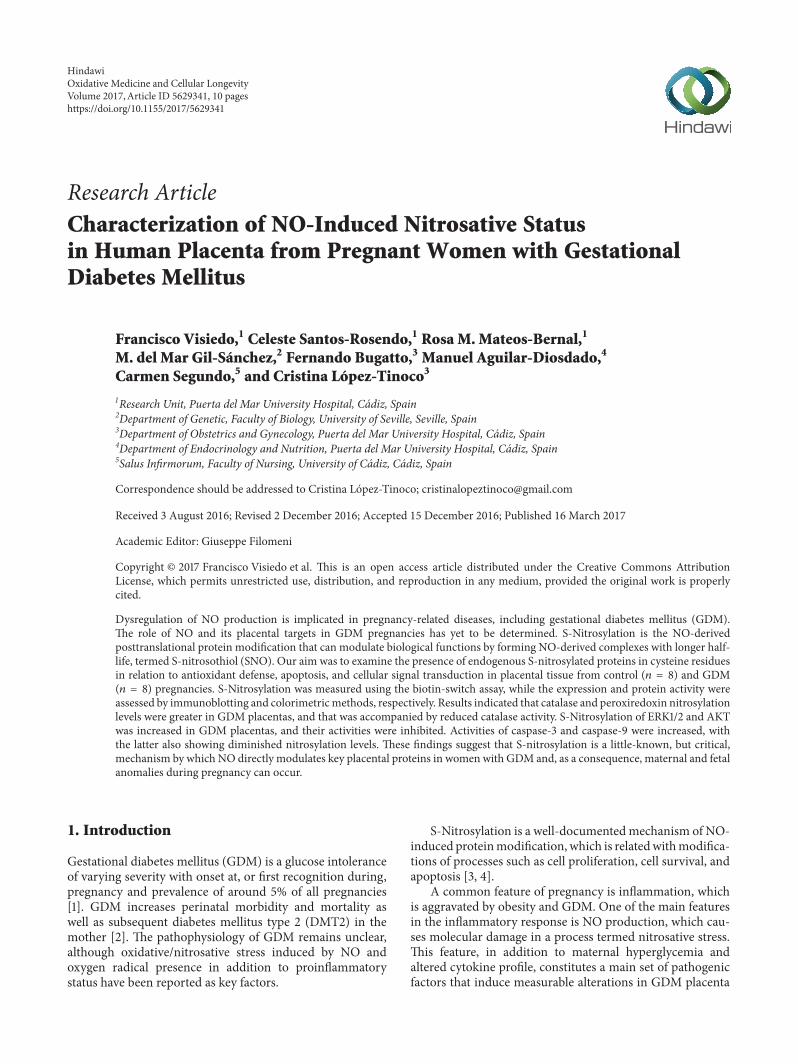
Figure 1: iNOS expression in GDM. Notes. iNOS expressionquantified byWestern blot of placental tissue obtained from controland GDM pregnancies. Results are expressed as means ± SEM ofiNOS to actin ratio measured by densitometry (𝑛 = 3). Lower panelis a representative image from an experiment. Units on bar chart 𝑦-axis are arbitrary. ∗𝑝 < 0.05, GDM versus control.
significantly higher maternal pregestational body mass index(BMI) in GDM versus control group (𝑝 = 0.001).
3.2. iNOS Expression in GDM Placenta. iNOS expression inplacental tissue from the GDM group compared to control(depicted in Figure 1) shows an increment in iNOS expressionin placenta fromGDMdemonstrating that inducible isoformof NOS is activated under pathological conditions.
3.3. Identification of Placenta S-Nitrosylated Target EnzymesRelated to Antioxidant Defense. Protein S-nitrosylation is a
4 Oxidative Medicine and Cellular Longevity
SNO-catalase
Input
0
1
2
3SN
O-c
atal
ase
∗
Control GDM
(a)
SNO-Prdx-1
Input
0
1
2
3
SNO
-Prd
x-1
∗
Control GDM
(b)
Control GDM0
0.1
0.2
0.3
Cata
lase
activ
ity
∗
(c)
0
1
2
3
Cata
lase
/act
in
Catalase
Actin
∗
Control GDM
(d)
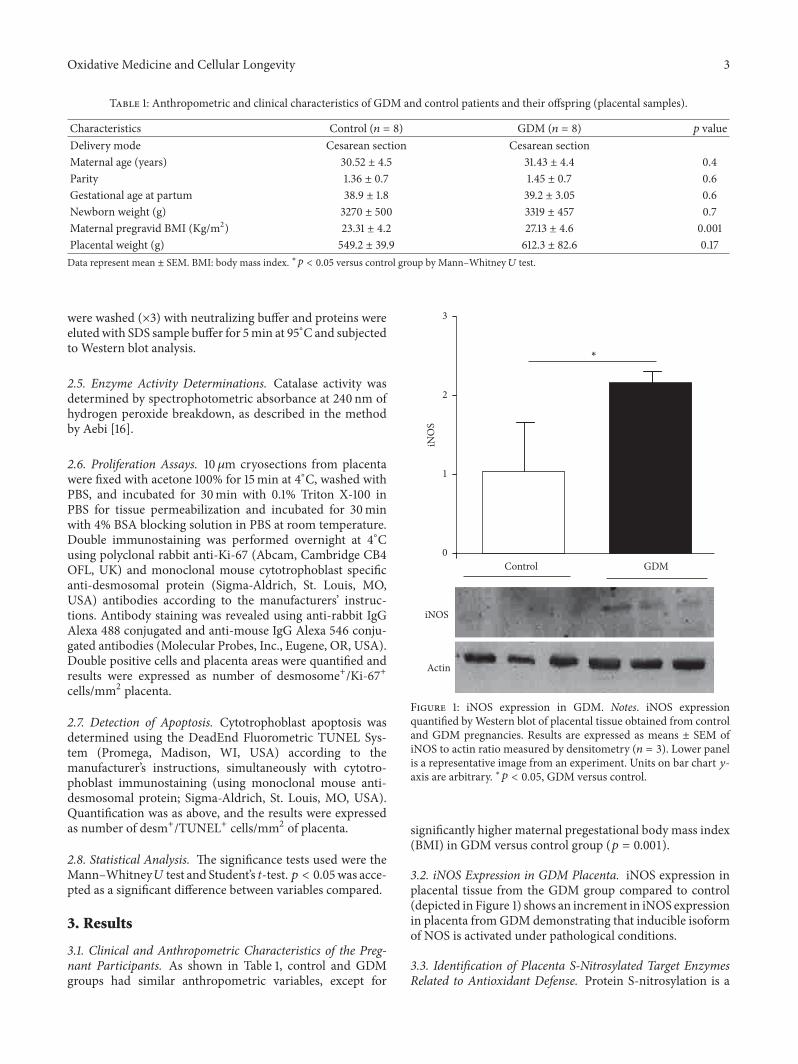
Figure 2: S-Nitrosylation in antioxidant enzymes. Notes. ((a) and (b)) S-Nitrosylation measured in placental tissue obtained from controland GDM pregnancies. Methods used were biotin-switch technique for catalase (a) and peroxiredoxin-1 (b). Results in bar charts are mean± SEM of SNO-protein (𝑛 = 6) relative to input (total quantity of studied protein present in each sample). Units on bar chart 𝑦-axes arearbitrary. Representative images of immunoblotted SNO-proteins are shown under each graph. Catalase activity (c) and expression (d)were quantified by spectrophotometric analysis and immunoblotting, respectively. Placental tissues were obtained from control and GDMpregnancies. Results reported as mean ± SEM of enzymatic activity (𝑛 = 8) and catalase to actin ratio measured by densitometry (𝑛 = 3);lower panel is a representative image of immunoblotted proteins. ∗𝑝 < 0.05, GDM versus control.
reversible posttranslational modification that regulates thefunction of many target proteins (including enzymes) viathe action of nitric oxide (NO). To assess whether full-term placentas from women with GDM had a characteristicnitrosative profile of enzymes associated with protectionagainst oxidative stress-induced cell death, overall levelsof NO-mediated S-nitrosylation were measured as covalentattachments to cysteine residues in the main antioxidantproteins. Increased levels of S-nitrosylation were exhibitedby catalase (Figure 2(a)) and peroxiredoxin (detoxifyingenzymes of hydrogen peroxide; Figure 2(b)) in GDM placen-tas compared to the control group. S-Nitrosylation levels of
other antioxidant enzymes such as Cu/Zn-superoxide dis-mutase (Cu/Zn-SOD-1) and glutathione peroxidase (GPx-1)remained unchanged (data not shown). With regard tocatalase, total amount of protein and enzymatic activitywere determined. An increment of catalase total amount(Figure 2(c)) accompanied by an inhibition of its activity(Figure 2(d)) was observed in placentas from women withGDM in comparison with control group. These observationssuggest that NO-induced S-nitrosylation inhibits enzymaticactivity of key endogenous antioxidants that have a protectivefunction against oxidative stress in placenta in pregnantwomen with GDM.
Oxidative Medicine and Cellular Longevity 5
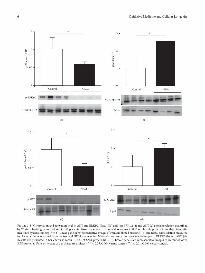
3.4. Identification of Placental S-Nitrosylated Target EnzymesRelated to Cell Survival. Activation of cell survival-associatedERK1/2 pathway is regulated by mechanisms related to post-translational modifications, including downstream scaffoldsand phosphorylation of threonine and tyrosine residues.Hence, we quantified the level of placental ERK1/2 phospho-rylation in control and GDM pregnancies. As depicted inFigure 3(a), phosphorylated ERK1/2 decreased significantlyin GDM placentas compared to control placentas. Moreover,the levels of placental ERK1/2-SNO were measured in bothgroups by biotin-switch assay, and a significant increase wasfound in placental tissue from pregnant women with GDM(Figure 3(b)).
AKT is another signaling pathway involved in placentalfunction and the main proteins of the pathway are alsoregulated by posttranslational modifications. We quantifiedAKT phosphorylation in GDM and control placental tissueand observed a significant increase in GDM (Figure 3(c)). Inaddition,AKTS-nitrosylationwas determined and the resultsshowed that the GDM group had higher levels of this post-translational modification than control group (Figure 3(d)).
These results highlight S-nitrosylation of ERK1/2 andAKT as crucial mechanisms by which NO directly regulatesproteins linked to several biological pathways associatedwith cell survival and function in placental tissue duringpregnancy.
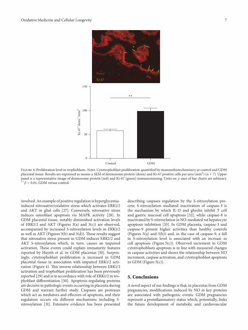
3.5. Determination of Cytotrophoblast Proliferation. GDM-induced alteration of above-described enzyme activities canbe related with a disrupted balance between apoptosis andproliferation in placental cytotrophoblast.We quantified pro-liferating cytotrophoblasts in placental tissue from pregnantwomen with GDM and observed a significant increase inGDM placentas compared with control group (Figure 4).
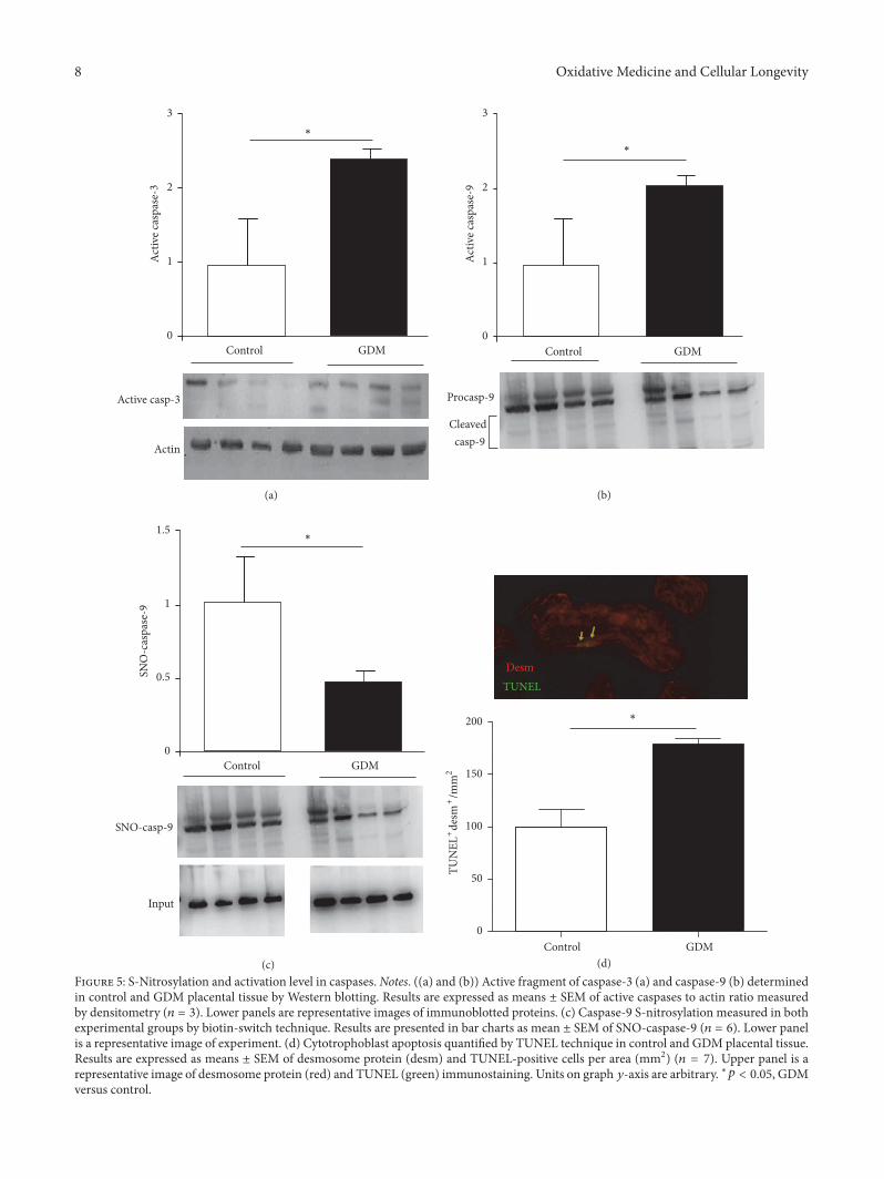
3.6. Apoptosis Quantification and Identification of PlacentalS-Nitrosylated Caspases. NO can regulate this programmedcell death via S-nitrosylation of active caspases. We ana-lyzed caspase-9 and caspase-3 activation and observed anincrement in placental tissue from pregnant women withGDM (Figures 5(a) and 5(b)). S-Nitrosylation was measuredto evaluate the regulatory effect of NO effect on caspase-9,and the results showed a significant decrease in caspase-9SNO levels in GDM placentas relative to the control group(Figure 5(c)).
In addition, cytotrophoblast apoptosis was higher inGDM placental tissue than in control (Figure 5(d)).
4. Discussion
In the present study, we highlight that NO-mediatednitrosative status is altered in placental tissue from GDMpregnancies. Our findings are based on the direct evaluationof SNO-proteins involved in various cellular physiologicalprocesses including antioxidant defense, apoptosis, and cellu-lar signaling transduction, all of which combine in providinga mechanism for redox-based regulation of standard placen-tal function.
NO is a pleiotropic signaling molecule which is synthe-sized by the enzyme NOS and regulates essential cellularprocesses such as muscle relaxation and blood pressureregulation [17]. Overproduction of NO by aberrant iNOSinduction is implicated in the pathogenesis of many disor-ders including neurodegenerative diseases, inflammatory andautoimmune diseases, cardiovascular diseases, and cancer[18]. In the case of GDM, inflammatory status and maternalhyperglycemia are involved in the generation of nitrosativestress resulting from induction of iNOS gene expression[19]. This is in addition to the described increased NOduring normal pregnancy [20]. The principal target of thesealterations is the placenta.
Posttranslational protein modifications are some of mec-hanisms by which NO alters signaling pathways leading topathologic events. Recently, protein S-nitrosylation has beenrecognized as an important, reversible, posttranslationalmodification consisting of a covalent attachment of a nitrogenmonoxide group to the thiol side-chain of cysteine. Undernitrosative stress conditions, S-nitrosylation can result inaltered protein activity and adverse biological consequences[21].
Our data (Figure 1) demonstrate that GDM causesincreased iNOS expression in full-term placental tissue,which is in accordance with other reports [22]. Of noteis the fact that we had used only placentas obtained fromelective cesarean deliveries so as to rule out potential effects oflabor on placental oxidative and nitrosative stress. Frequently,oxidative/nitrosative stress occurs in pathological situations.In this regard, our group had described GDM in whichoxidative stress markers and antioxidants status occurred[23]. Both oxidative and nitrosative stress processes inducespecific effects on biological systems, although crosstalk canbe observed between them. There is considerable evidencefrom plants of NO-induced regulation of enzymes associatedwith oxidative stress [24]. These observations are morelimited in mammals. In rat liver, where NO signaling iscritical, peroxisome-localized catalase has been identified asone of potential substrates for S-nitrosylation [25]. How-ever, there is a lack of physiological evidence, implicatingS-nitrosylated catalase in physiopathological models. NO-mediated activity regulation of other antioxidant enzymes hasbeen described in animal models and includes inhibition ofSOD activity associated with increased NO and peroxynitritein acute coronary syndrome [26], while modulation by S-nitrosylation of peroxiredoxin has been described in celllines [9]. In GDM placental tissue, S-nitrosylation was notobserved in SOD (data no shown). However, it is present inother antioxidant enzymes such as catalase (Figure 2(a)) andperoxiredoxin (Figure 2(b)). A lower activity of catalase hasbeen identified in relation to this posttranslational modifica-tion (Figure 2(c)).
Other proteins in which NO-mediated regulation couldplay a key role under pathological conditions in GDM arethose related to placenta cell survival mechanisms and func-tion. MAPK and AKT/PI3-K signaling pathways are crucialin maintaining placental processes by inducing cell replica-tion and trophoblast differentiation [22]. The role of NO intheir regulation depends on the specific biological system
6 Oxidative Medicine and Cellular Longevity
p-ERK1/2
Total ERK1/2
0
0.5
1
1.5
p-ER
K/to
tal E
RK
∗
Control GDM
(a)
SNO-ERK1/2
Input
∗∗
0
1
2
3
SNO
-ERK
1/2
Control GDM
(b)
p-AKT
Total AKT
0
0.5
1
1.5
p-A
KT/to
tal A
KT
∗
Control GDM
(c)
SNO-AKT
Input
∗
0
1
2
3
SNO
-AKT
Control GDM
(d)
Figure 3: S-Nitrosylation and activation level in AKT and ERK1/2. Notes. ((a) and (c)) ERK1/2 (a) and AKT (c) phosphorylation quantifiedby Western blotting in control and GDM placental tissue. Results are expressed as means ± SEM of phosphoprotein to total-protein ratiomeasured by densitometry (𝑛 = 6). Lower panels are representative images of immunoblotted proteins. ((b) and (d)) S-Nitrosylationmeasuredin placental tissue obtained from control and GDM pregnancies. Methods used were biotin-switch technique in ERK1/2 (b) and AKT (d).Results are presented in bar charts as mean ± SEM of SNO-protein (𝑛 = 6). Lower panels are representative images of immunoblottedSNO-proteins. Units on 𝑦-axes of bar charts are arbitrary. ∗𝑝 < 0.05, GDM versus control; ∗∗𝑝 < 0.01, GDM versus control.
Oxidative Medicine and Cellular Longevity 7
Control GDM
Desm
Ki-67
∗∗
0
50
100
150
Ki-6
7+de
sm+
/mm
2
Figure 4: Proliferation level in trophoblasts. Notes. Cytotrophoblast proliferation quantified by immunohistochemistry in control and GDMplacental tissue. Results are expressed as means ± SEM of desmosome protein (desm) and Ki-67 positive cells per area (mm2) (𝑛 = 7). Upperpanel is a representative image of desmosome protein (red) and Ki-67 (green) immunostaining. Units on 𝑦-axes of bar charts are arbitrary.∗∗𝑝 < 0.01, GDM versus control.
involved.An example of positive regulation is hyperglycemia-induced nitrosative/oxidative stress which activates ERK1/2and AKT in glial cells [27]. Conversely, nitrosative stressinduces osteoblast apoptosis via MAPK activity [28]. InGDM placental tissue, notably diminished activation levelsof ERK1/2 and AKT (Figures 3(a) and 3(c)) are observed,accompanied by increased S-nitrosylation levels in ERK1/2as well as AKT (Figures 3(b) and 3(d)). These results suggestthat nitrosative stress present in GDM induces ERK1/2 andAKT S-nitrosylation which, in turn, causes an impairedactivation. These events could explain immaturity featuresreported by Huynh et al. in GDM placentas [10]. Surpris-ingly, cytotrophoblast proliferation is increased in GDMplacental tissue in association with impaired ERK1/2 acti-vation (Figure 4). This inverse relationship between ERK1/2activation and trophoblast proliferation has been previouslyreported [29] and is in accordance with role of ERK1/2 in tro-phoblast differentiation [30]. Apoptosis-regulating proteinsare decisive in pathologic events occurring in placenta duringGDM and warrant further study. Caspases are proteaseswhich act as mediators and effectors of apoptosis, and theirregulation occurs via different mechanisms including S-nitrosylation [31]. Extensive evidence has been presented
describing caspases regulation by the S-nitrosylation pro-cess; S-nitrosylation mediated inactivation of caspase-3 isthe mechanism by which IL-15 and ghrelin inhibit T celland gastric mucosal cell apoptosis [32], while caspase-8 isinactivated by S-nitrosylation inNO-mediated rat hepatocyteapoptosis inhibition [33]. In GDM placenta, caspase-3 andcaspase-9 present higher activities than healthy controls(Figures 5(a) and 5(b)) and, in the case of caspase-9, a fallin S-nitrosylation level is associated with an increase incell apoptosis (Figure 5(c)). Observed increment in GDMcytotrophoblasts apoptosis is in line with measured changesin caspase activities and shows the relationship between NOincrement, caspase activation, and cytotrophoblast apoptosisin GDM (Figure 5(c)).
5. Conclusions
A novel aspect of our findings is that, in placentas fromGDMpregnancies, modifications induced by NO in key proteinsare associated with pathogenic events. GDM pregnanciesrepresent a proinflammatory status which, potentially, linksthe future development of metabolic and cardiovascular
8 Oxidative Medicine and Cellular Longevity
Active casp-3
Actin
Control GDM0
1
2
3
Activ
e cas
pase
-3
∗
(a)
Cleavedcasp-9
Procasp-9
Control GDM0
1
2
3
Activ
e cas
pase
-9
∗
(b)
SNO-casp-9
Input
0
0.5
1
1.5
SNO
-cas
pase
-9
Control GDM
∗
(c)Control GDM
DesmTUNEL
0
50
100
150
200
TUN
EL+
desm
+/m
m2
∗
(d)Figure 5: S-Nitrosylation and activation level in caspases.Notes. ((a) and (b)) Active fragment of caspase-3 (a) and caspase-9 (b) determinedin control and GDM placental tissue by Western blotting. Results are expressed as means ± SEM of active caspases to actin ratio measuredby densitometry (𝑛 = 3). Lower panels are representative images of immunoblotted proteins. (c) Caspase-9 S-nitrosylation measured in bothexperimental groups by biotin-switch technique. Results are presented in bar charts as mean ± SEM of SNO-caspase-9 (𝑛 = 6). Lower panelis a representative image of experiment. (d) Cytotrophoblast apoptosis quantified by TUNEL technique in control and GDM placental tissue.Results are expressed as means ± SEM of desmosome protein (desm) and TUNEL-positive cells per area (mm2) (𝑛 = 7). Upper panel is arepresentative image of desmosome protein (red) and TUNEL (green) immunostaining. Units on graph 𝑦-axis are arbitrary. ∗𝑝 < 0.05, GDMversus control.
Oxidative Medicine and Cellular Longevity 9
diseases in both mother and child [34]. The described NO-mediated protein modifications could participate in thisprocess and their presence could be used as biomarkersto predict nitrosative damage and future complications inmother and child.
Abbreviations
GDM: Gestational diabetes mellitusiNOS: Inducible nitric oxide synthaseIR: Insulin resistanceROS: Reactive oxygen speciesRNS: Reactive nitrogen speciesNO: Nitric oxideSOD: Superoxide dismutase.
Conflicts of Interest
The authors declare that there are no conflicts of interest thatwould prejudice the impartiality of this scientific work.
Authors’ Contributions
Francisco Visiedo, Celeste Santos-Rosendo, Carmen Segu-ndo, and Cristina Lopez-Tinoco contributed equally to thismanuscript.
Acknowledgments
This study was financed by grants from the AndalusiaDepartment of Health (PI-0525-2012) and PAIDI (CTS-368).Editorial assistance was by Dr. Peter R. Turner.
References
[1] A. Ben-Haroush, Y. Yogev, and M. Hod, “Epidemiology ofgestational diabetes mellitus and its association with Type 2diabetes,” Diabetic Medicine, vol. 21, no. 2, pp. 103–113, 2004.
[2] M. Albareda, A. Caballero, G. Badell et al., “Diabetes and abnor-mal glucose tolerance in women with previous gestationaldiabetes,” Diabetes Care, vol. 26, no. 4, pp. 1199–1205, 2003.
[3] S. Moncada, R. M. J. Palmer, and E. A. Higgs, “Nitric oxide:physiology, pathophysiology, and pharmacology,” Pharmaco-logical Reviews, vol. 43, no. 2, pp. 109–142, 1991.
[4] M. L. Circu and T. Y. Aw, “Reactive oxygen species, cellu-lar redox systems, and apoptosis,” Free Radical Biology andMedicine, vol. 48, no. 6, pp. 749–762, 2010.
[5] L. Myatt, “Review: reactive oxygen and nitrogen species andfunctional adaptation of the placenta,” Placenta, vol. 31, supple-ment, pp. S66–S69, 2010.
[6] J. W. Baynes and S. R. Thorpe, “Role of oxidative stress indiabetic complications: a new perspective on an old paradigm,”Diabetes, vol. 48, no. 1, pp. 1–9, 1999.
[7] M. Lappas, U. Hiden, G. Desoye, J. Froehlich, S. H.-D. Mouzon,and A. Jawerbaum, “The role of oxidative stress in the patho-physiology of gestational diabetes mellitus,” Antioxidants andRedox Signaling, vol. 15, no. 12, pp. 3061–3100, 2011.
[8] C. A.M. Leal, M. R. C. Schetinger, D. B. R. Leal et al., “Oxidativestress and antioxidant defenses in pregnant women,” RedoxReport, vol. 16, no. 6, pp. 230–236, 2011.
[9] R. Engelman, P. Weisman-Shomer, T. Ziv, J. Xu, E. S. J. Arner,and M. Benhar, “Multilevel regulation of 2-Cys peroxiredoxinreaction cycle by S-nitrosylation,” Journal of Biological Chem-istry, vol. 288, no. 16, pp. 11312–11324, 2013.
[10] J. Huynh, D. Dawson, D. Roberts, and R. Bentley-Lewis, “Asystematic review of placental pathology in maternal diabetesmellitus,” Placenta, vol. 36, no. 2, pp. 101–114, 2015.
[11] A. Ozmen, G. Unek, D. Kipmen-Korgun, and E. T. Korgun,“The PI3K/Akt andMAPK-ERK1/2 pathways are altered in STZinduced diabetic rat placentas,” Histology and Histopathology,vol. 29, no. 6, pp. 743–756, 2014.
[12] T. R. Magee, M. G. Ross, L. Wedekind, M. Desai, S. Kjos, and L.Belkacemi, “Gestational diabetes mellitus alters apoptotic andinflammatory gene expression of trophobasts fromhuman termplacenta,” Journal of Diabetes and Its Complications, vol. 28, no.4, pp. 448–459, 2014.
[13] National Diabetes Data Group, “Classification and diagnosis ofdiabetes mellitus and other categories of glucose intolerance,”Diabetes, vol. 28, no. 12, pp. 1039–1057, 1979.
[14] R. Corcoy, B. Lumbreras, J. L. Bartha, andW. Ricart, “New diag-nostic criteria for gestational diabetes mellitus after the HAPOstudy. Are they valid in our environment?” Endocrinologıa yNutricion, vol. 57, pp. 277–280, 2010.
[15] S. R. Jaffrey and S. H. Snyder, “The biotin switch method for thedetection of S-nitrosylated proteins,” Science’s STKE, vol. 2001,no. 86, p. pl1, 2001.
[16] H. Aebi, “Catalase in vitro,”Methods in Enzymology, vol. 105, pp.121–126, 1984.
[17] L. J. Ignarro, G. M. Buga, K. S. Wood, R. E. Byrns, and G.Chaudhuri, “Endothelium-derived relaxing factor producedand released from artery and vein is nitric oxide,” Proceedings ofthe National Academy of Sciences of the United States of America,vol. 84, no. 24, pp. 9265–9269, 1987.
[18] J. N. Sharma, A. Al-Omran, and S. S. Parvathy, “Role of nitricoxide in inflammatory diseases,” Inflammopharmacology, vol.15, no. 6, pp. 252–259, 2007.
[19] P. Yang, Y. Cao, and H. Li, “Hyperglycemia induces induciblenitric oxide synthase gene expression and consequent nitro-sative stress via c-Jun N-terminal kinase activation,” AmericanJournal of Obstetrics & Gynecology, vol. 203, no. 2, pp. 185.e5–185.e11, 2010.
[20] S. M. Sladek, R. R. Magness, and K. P. Conrad, “Nitric oxideand pregnancy,” American Journal of Physiology—RegulatoryIntegrative and Comparative Physiology, vol. 272, no. 2, pp.R441–R463, 1997.
[21] D. T. Hess, A. Matsumoto, S.-O. Kim, H. E. Marshall, and J.S. Stamler, “Protein S-nitrosylation: purview and parameters,”Nature ReviewsMolecular Cell Biology, vol. 6, no. 2, pp. 150–166,2005.
[22] G. Schonfelder, M. John, H. Hopp, N. Fuhr, M. van Der Giet,and M. Paul, “Expression of inducible nitric oxide synthasein placenta of women with gestational diabetes,” The FASEBJournal, vol. 10, no. 7, pp. 777–784, 1996.
[23] C. Lopez-Tinoco, M. Roca, A. Garcıa-Valero et al., “Oxidativestress and antioxidant status in patients with late-onset gesta-tional diabetes mellitus,” Acta Diabetologica, vol. 50, no. 2, pp.201–208, 2013.
[24] J. C. Begara-Morales, B. Sanchez-Calvo,M. Chaki et al., “Differ-ential molecular response of monodehydroascorbate reductaseand glutathione reductase by nitration and S-nitrosylation,”Journal of Experimental Botany, vol. 66, no. 19, pp. 5983–5996,2015.
10 Oxidative Medicine and Cellular Longevity
[25] M. W. Foster and J. S. Stamler, “New Insights into protein S-nitrosylation mitochondria as a model system,” The Journal ofBiological Chemistry, vol. 279, no. 24, pp. 25891–25897, 2004.
[26] S. Gheddouchi, N. Mokhtari-Soulimane, H. Merzouk et al.,“Low SOD activity is associated with overproduction of perox-ynitrite and nitric oxide in patients with acute coronary synd-rome,”Nitric Oxide—Biology and Chemistry, vol. 49, pp. 40–46,2015.
[27] P. Kumar, G. N. Rao, B. B. Pal, and A. Pal, “Hyperglycemia-induced oxidative stress induces apoptosis by inhibiting PI3-kinase/Akt and ERK1/2 MAPK mediated signaling pathwaycausing downregulation of 8-oxoG-DNA glycosylase levels inglial cells,” International Journal of Biochemistry and Cell Biol-ogy, vol. 53, pp. 302–319, 2014.
[28] T. L. Chen, G. J. Wu, C. S. Hsu, T. H. Fong, and R. M.Chen, “Nitrosative stress induces osteoblast apoptosis throughdownregulating MAPK-mediated NFkappaB/AP-1 activationand subsequent Bcl-X(L) expression,” Chemico-Biological Inter-actions, vol. 184, pp. 359–365, 2010.
[29] L.-J. Shih, T.-F. Chen, C.-K. Lin, H.-S. Liu, and Y.-H. Kao,“Green tea (–)-epigallocatechin gallate inhibits the growth ofhuman villous trophoblasts via the ERK, p38, AMP-activatedprotein kinase, and protein kinase B pathways,” American Jou-rnal of Physiology—Cell Physiology, vol. 311, no. 2, pp. C308–C321, 2016.
[30] G. Daoud, M. Amyot, E. Rassart, A. Masse, L. Simoneau, and J.Lafond, “ERK1/2 and p38 regulate trophoblasts differentiationin human term placenta,” Journal of Physiology, vol. 566, no. 2,pp. 409–423, 2005.
[31] J. B. Mannick, C. Schonhoff, N. Papeta et al., “S-nitrosylation ofmitochondrial caspases,” Journal of Cell Biology, vol. 154, no. 6,pp. 1111–1116, 2001.
[32] P. T. Saligrama, K. A. Fortner, M. A. Secinaro, C. C. Collins, J. Q.Russell, and R. C. Budd, “IL-15 maintains T-cell survival via S-nitrosylation-mediated inhibition of caspase-3,” Cell Death andDifferentiation, vol. 21, no. 6, pp. 904–914, 2014.
[33] Y.-M. Kim, T.-H. Kim, H.-T. Chung, R. V. Talanian, X.-M.Yin, and T. R. Billiar, “Nitric oxide prevents tumor necrosisfactor 𝛼–induced rat hepatocyte apoptosis by the interruptionofmitochondrial apoptotic signaling through S-nitrosylation ofcaspase-8,” Hepatology, vol. 32, no. 4, pp. 770–778, 2000.
[34] C. Lopez-Tinoco,M. Roca, A. Fernandez-Deudero et al., “Cyto-kine profile, metabolic syndrome and cardiovascular diseaserisk in women with late-onset gestational diabetes mellitus,”Cytokine, vol. 58, no. 1, pp. 14–19, 2012.

Submit your manuscripts athttps://www.hindawi.com
Stem CellsInternational
Hindawi Publishing Corporationhttp://www.hindawi.com Volume 2014
Hindawi Publishing Corporationhttp://www.hindawi.com Volume 2014
MEDIATORSINFLAMMATION
of
Hindawi Publishing Corporationhttp://www.hindawi.com Volume 2014
Behavioural Neurology
EndocrinologyInternational Journal of
Hindawi Publishing Corporationhttp://www.hindawi.com Volume 2014
Hindawi Publishing Corporationhttp://www.hindawi.com Volume 2014
Disease Markers
Hindawi Publishing Corporationhttp://www.hindawi.com Volume 2014
BioMed Research International
OncologyJournal of
Hindawi Publishing Corporationhttp://www.hindawi.com Volume 2014
Hindawi Publishing Corporationhttp://www.hindawi.com Volume 2014
Oxidative Medicine and Cellular Longevity
Hindawi Publishing Corporationhttp://www.hindawi.com Volume 2014
PPAR Research
The Scientific World JournalHindawi Publishing Corporation http://www.hindawi.com Volume 2014
Immunology ResearchHindawi Publishing Corporationhttp://www.hindawi.com Volume 2014
Journal of
ObesityJournal of
Hindawi Publishing Corporationhttp://www.hindawi.com Volume 2014
Hindawi Publishing Corporationhttp://www.hindawi.com Volume 2014
Computational and Mathematical Methods in Medicine
OphthalmologyJournal of
Hindawi Publishing Corporationhttp://www.hindawi.com Volume 2014
Diabetes ResearchJournal of
Hindawi Publishing Corporationhttp://www.hindawi.com Volume 2014
Hindawi Publishing Corporationhttp://www.hindawi.com Volume 2014
Research and TreatmentAIDS
Hindawi Publishing Corporationhttp://www.hindawi.com Volume 2014
Gastroenterology Research and Practice
Hindawi Publishing Corporationhttp://www.hindawi.com Volume 2014
Parkinson’s Disease
Evidence-Based Complementary and Alternative Medicine
Volume 2014Hindawi Publishing Corporationhttp://www.hindawi.com