characterization of rat regions kenshi hayashi, reiko ... · volume 1 5 number 16 1 987 nucleic...
TRANSCRIPT
Volume 1 5 Number 16 1 987 Nucleic Acids Research
Characterization of rat c-myc and adjacent regions
Kenshi Hayashi, Reiko Makino, Hideki Kawamura, Akira Arisawa and Kenji Yoneda
Biochemistry Division, National Cancer Center Research Institute 1 - 1 , Tsukiji 5-Chome, Chuo-ku, Tokyo, Japan
Received May 13, 1987; Revised and Accepted July 5, 1987 Accession no. YO0396
ABSTRACT Rat genomic regions covering c - w were cloned from the
DNA of both normal liver and two lines of Morris hepatomas, one of which had c - w amplification. The three restriction maps showed perfect agreement within the overlapping regions. The 7 kb regions, which included the entire normal rat c - w and the region 2.2 kb upstream, and one from the hepatomas, were sequenced and found to be identical. The codins resions of exdns 2 and 3 were highly conserved between rat, mouse 2nd man, but some differences in amino acids were noted. Exon 1 and the non-coding region of exon 3 showed limited homology between the three species. Rat exon 1 contained several nonsense codons in each frame and no ATG codon, indicating there to be no coding capacity in this exon. The 2.2 kb upstream regions and the introns compared showed unusual conservation between the rat and human genes. Some motifs, previously proposed as having a functional role in human c - w , were also found in equivalent positions of the rat sequence.
Nucleas S1 protection mapping revealed the second promoter to be preferentially used in most tissues or in hepatoma cells, and the second poly A addition signal to be the only one functional in all the RNA sources examined.
INTRODUCTION
MYC was initially discovered in the form of a viral onco-
gene of an avian myelocytomatosis virus, MC 29 (11, and
subsequently identified in various vertebrate genomes in the
forms of its cellular counterpart, c-E, and transducing v - w
in several oncogenic retroviruses (2,3). The structure of
c - w is highly conserved through evolutionary stages ( 4 1 , and
the product, c-% protein, is known to be localized in the
nucleus ( 5 ) and to bind to DNA (6). Several cellular
functions, including growth competence or differentiation, seem
to require the expression or suppression of c-E, although the
O I RL Prss Limited, Oxford, England. 641 9
Nucleic Acids Research
precise role which the gene has in these functions is not yet
clear. Various types of cancer cells overexpress c-myc mRNA
(7), some of them as a result of rearrangement or amplification
(8). Such gene overexpression or deregulation is suspected to
be a cause of the unlimited growth of the cancer cells.
Expression of c-myc seems to be regulated in a complicated
manner involving transcription initiation (9), transcript
elongation (10, 11) and mRNA degradation (12, 13). Such
regulation requires sequences of functional significance to be
located upstream, and within exons and introns. Recent studies
using human c-E have suggested both positive and negative
regulatory sequences to be located upstream and within exon 1
and intron 1 (14, 15). Rearrangements resulting from
translocations or retroviral insertions far upstream of the
gene have been reported in some B-cell lymphomas (7),
suggesting the importance of remote sequence elements in the
regulation of the gene. One approach towards identifing such
elements is the search for evolutionally conserved sequences in
the gene and its neighboring regions. To date, an 8 kb
sequence of the entire human c-myc and its flanking regions has
been reported (16, 17, 18) . Another mammalian c - w sequence
available is that of the mouse (19, 20, 21) although, in this
case, information on the upstream region is limited and the
sequence of intron 2 is unavailable.
In the present papar we report the cloning of normal rat
c - w and that of the Morris hepatomas, 5123D and 7794A. Over-
expression of the c-= mRNA in both hepatomas, and
amplification of the gene in the latter, have been reported
previously (22). Approximately 7 kb of both normal rat and
Morris hepatoma 7794A, that including the gene and the 2.2 kb
upstream region, were sequenced. The sequences from the two
sources were exactly the same. Comparison between rat and
human sequences revealed remarkably conserved stretches
distributed throughout the 2.2 kb upstream region and both
introns, as well as in the exons.
It is shown that the second of the two promoters is very
much preferred as the mFWA initiation site in various tissues
and cultured hepatoma cells, and also that, in all tissues and
Nucleic Acids Research
cells examined, only the signal downstream of the dual
termination (poly A addition) is used.
MATERIALS AND METHODS
Cloning
A transplanted Morris hepatoma 7794A genomic library was
constructed by partial digestion of its DNA with Sau3A1,
ligation with arms of EMBL3a (23) and in vitro packaging (24).
The library was screened by plaque-hybridization (25) using the
3'-half of the v-myc sequence (26) as a probe, and three over-
lapping clones were obtained. A detailed restriction map was
constructed for one of the clones, XM77-5.
c-E clones from normal Buffalo rat liver (XB-14) and
Morris hepatoma 5123D (XD-5) were obtained by complete
digestion of their DNA with EcoRI, ligation and packaging into
phage Charon 4A, and screening with a probe identical to that
used for the isolation of XM77-5.
DNA sequencing of c-rnyc from Morris hepatoma 7794A
After appropriate subcloning of regions of XM77-5 into
plasmids, the PstI-PstI 4 kb fragment (5'-flank, exon 1 and
most of intron 1) or the Sacl-Hpal 3 kb fragment (half of
intron 1, exon 2, intron 2 and most of exon 3) were sonicated,
cloned into M13mplO using Deininger's method (27) and sequenced
by dideoxy chain termination (28) using an M13 sequencing kit
(Amersham) . The HpaI-HpaI 0.6 kb fragment (3'-end of exon 3
and its flank) was separately cloned into M13mplO and sequenced
from both ends.
DNA sequencing of c-myc from normal rat
A 2.1 kb HindIII-Hind111 fragment (5'-flank to 5'-end of
exon 1) and a HindIII-XbaI 4.3 kb fragment (5'-end of exon 1 to
end of coding region of exon 3) were cloned into M13mp18 or
mp19, progressively deleted by the method of Yanisch-Perron
al. (29) and se quenced. When necessary, 7-deaza-2'-dGTP was - used in place of dGTP in the sequencing reaction to alleviate
the compaction of bands in the GC-rich regions (30).
Computer analyses
Sequences from gel readings were assembled using the
GENIAS (Mitsui Knowledge Inc.) or GENETYX programs (Japan Soft-
Nucleic Acids Research
ware Development Company). All other computer analyses were
done by UWGCG (31) or IDEAS (32) programs run on the VAX11/750
of the Institute of Medical Science, Tokyo University.
Nuclease S1 protection assay(33)
Fragments spanning either promoter sites or poly A
addition sites were cloned into M13mp18. Strand specific
probes (lo8 cpm/pg) were synthesized using a sequencing primer
(Amersham) , and isolated by alkaline agarose gels after
digestion with appropriate enzymes (34). Probes (1-1.5 x 10 5
+ cpm) were hybridized with either total or poly A RNA in 0.3 M
NaC1, 10 mM Tris-HC1, pH 7.5, and 1 mM EDTA (3'-end) or in 80%
formamide, 0.4 M NaC1, 20 mM PIPES, pH 6.8, and 10 mM EDTA
(5'-end) at 45 OC overnight. The annealed mixture was digested
with nuclease S1 (Takara) at 37 OC for 30 min. The protected
fragments were run in 8 M urea, 4% acrylamide gels.
RESULTS
Structure of rat c-myc
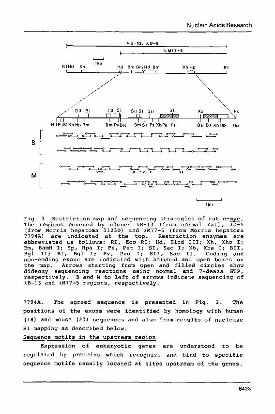
Fig 1 shows the restriction map of the rat c - w locus
cloned from three independent sources, normal Buffalo rat liver
and Morris hepatomas 5123D and 7794A. The three clones had
exactly the same restriction sites in the overlapping regions.
The approximate location and orientation of c - ware
determined by Southern hybridizations of appropriately
restricted fragments to the 5'- or 3'-half of v - w . There are
some discrepancies between the restriction map shown in Fig. 1
and that of the Lou/Wsl rat reported by Pear et &. (35),
especially at the exon 1 region. The difference in rat strain
may possibly explain this discrepancy. We examined the Hind111
and BamHI sites of the exon 1 region in both Sprague-Dawley and
Wistar rat strains, and the results agreed with the map
presented here.
The regions spanning approximately 7 kb of XM77-5 (from
Morris hepatoma 7794A with amplified c - w ) and XB-13 (from
normal Buffalo rat liver) were sequenced independently using
the strategy outlined in Fig. 1. The two sequences turned out
to be exactly the same, suggesting no structural change to have
occurred during c-E amplification in the Morris hepatoma,
Nucleic Acids Research
I AB-13, AD-5
I
I AM77-5 -
R l H d Xh ' kb Hd Bm Bm Hd Brn Xh Hp R I u I ICY J
I' I'
I' ,'
,'
'.\, Ps I '1
Hd PsSIXb Hp Bm B m P v B l l B m S l P s X b P s Ps BI I B I XhHp H p
Fig. 1 Restriction map and sequencing strategies of rat c-E. The regions covered by clones XB-13 (from normal rat), AD-5 (from Morris hepatoma 5123D) and AM77-5 (from Morris hepatoma 7794A) are indicated at the top. Restriction enzymes are abbreviated as follows: RI, Eco RI; Hd, Hind 111; Xh, Xho I; Bm, BamH I; Hp, Hpa I; Ps, Pst I; SI, Sac I; Xb, Xba I; BII, Bgl 11; BI, Bgl I; Pv, Pvu I; SII, Sac 11. Coding and non-coding exons are indicated with hatched and open boxes on the map. Arrows starting from open and filled circles show dideoxy sequencing reactions using normal and 7-deaza GTP, respectively. B and M to left of arrows indicate sequencing of AB-13 and AM77-5 regions, respectively.
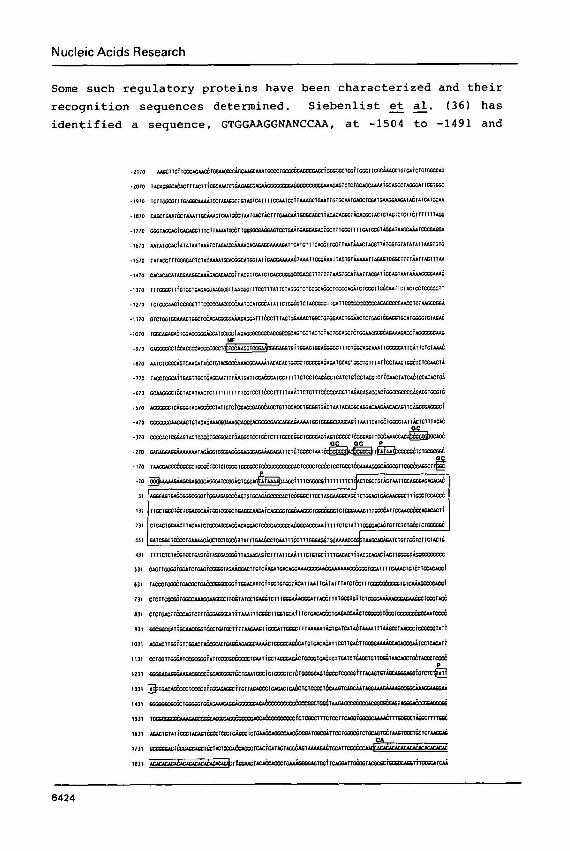
7794A. The agreed sequence is presented in Fig. 2. The
positions of the exons were identified by homology with human
(18) and mouse (20) sequences and also from results of nuclease
Sl mapping as described below.
Sequence motifs in the upstream region
Expression of eukaryotic genes are understood to be
regulated by proteins which recognize and bind to specific
sequence motifs usually located at sites upstream of the genes.
Nucleic Acids Research
Some such regulatory proteins have been characterized and their
recognition sequences determined. Siebenlist et al. ( 3 6 ) has
identified a sequence, GTGGAAGGNANCCAA, at -1504 to -1491 and
Nucleic Acids Research
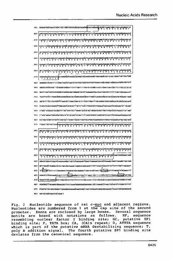
Fig. 2 Nucleotide sequence of rat c - w and adjacent regions. Nucleotides are numbered from 1 at the cap site of the second promoter. Exons are enclosed by large boxes. Several sequence motifs are boxed with notations as follows. NF, sequence resembling nuclear factor I binding site; GC, putative SP1 binding site; P, TATA box; CA, (CA)n repeat; D, ATTTA sequence which is part of the putative mRNA destabilizing sequence; T, poly A addition signal. The fourth putative SP1 binding site deviates from the canonical sequence.
Nucleic Acids Research
at -870 to -857 (the nucleotide adjacent to the 5'-side of the
second transcription initiation site is designated -1) of human
c-E, both of which have been suggested as the binding sites
of nuclear factor 1 (37). The exactly matched sequence does
not occur in the 2 kb upstream region of the rat sequence;
instead, a related sequence, GTGGAAGGTCCGAA, was found at -944
to -931. This, as well as the human sequence, bears some
resemblance to the enhancer core sequences of viral and
cellular genes.
Several putative Spl (38) binding sites, GGGCGG or its
complement, were found in proximity to the rat c - w promoter,
and most of these were also found in their corresponding
positions in the human gene.
No apparent CCAAT box was found within the region 0.5 kb
upstream of the gene. Three TATA boxes, two at the beginning
of exon 1 and one in intron 1, were found in the rat c - z .
These three promoters are also seen in the human and mouse
genes. In human and mouse c-E, the first two promoters are
known to function in various tissues and cells, the third
usually being cryptic and activated when the upstream promoters are removed by translocation in some B-cell lymphomas (19, 20) . We found the second promoter to be predominant in rats, as
shown in the following section.
Dual termination (poly A addition) signals, each
associated with TG motifs, are located at the end of exon 3
(39). Human and mouse c-myc also have dual terminators at
these sites (18, 20), suggesting this to have some, as yet
unknown, significance. So far, the second terminator is the
only one functional in the various rat tissues and cells
examined (data shown below). It is interesting to note that
each AATAAAA sequence was preceded by ATTTA and T-clusters
approximately 60 bases upstream. Recently, a similar sequence
was shown to be responsible for the instability of
granulocyte-macrophage colony stimulating factor (GM-CSF) mRNA
( 4 8 ) .
Another conspicuous feature of rat c-% is the presence
of in intron 1. A similar repeat, (CA)20, was found at
the corresponding position in the mouse (201, but not the
Nucleic Acids Research
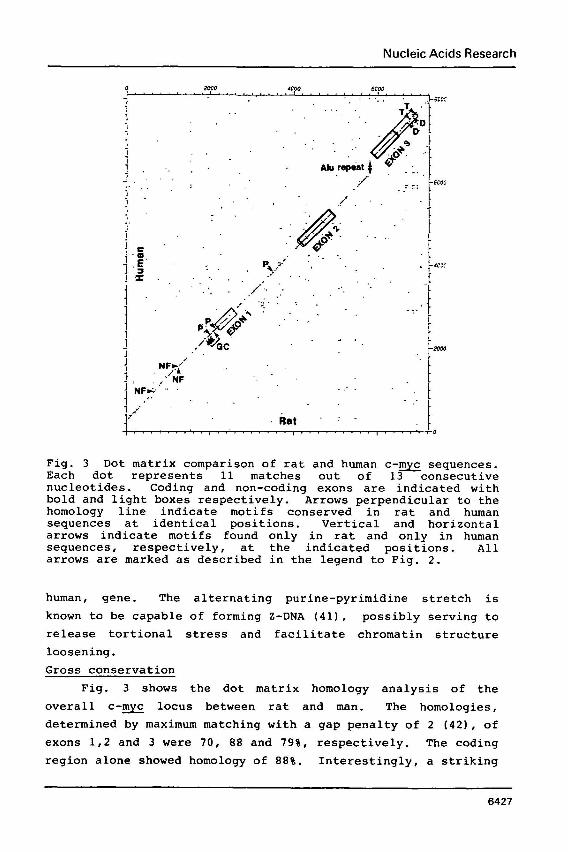
Fig. 3 Dot matrix comparison of rat and human c-- sequences. Each dot represents 11 matches out of 13 consecutive nucleotides. Coding and non-coding exons are indicated with bold and light boxes respectively. Arrows perpendicular to the homology line indicate motifs conserved in rat and human sequences at identical positions. Vertical and horizontal arrows indicate motifs found only in rat and only in human sequences, respectively, at the indicated positions. All arrows are marked as described in the legend to Fig. 2.
human, gene. The alternating purine-pyrimidine stretch is
known to be capable of forming 2-DNA (411, possibly serving to
release tortional stress and facilitate chromatin structure
loosening.
Gross conservation
Fig. 3 shows the dot matrix homology analysis of the
overall c - w locus between rat and man. The homologies,
determined by maximum matching with a gap penalty of 2 (421, of
exons 1,2 and 3 were 70, 88 and 79%, respectively. The coding
region alone showed homology of 888. Interestingly, a striking
Nucleic Acids Research
H R M
H R M
H R M
H R M
H R M
H R M
H R M
H R M
Fig. 4
T Y 0 MPLNVSFANR NYDLDYDSVQ PYFICDEEEN FYHOOOOSEL OPPAPSEDIW BKFELLPTPP 6 0
N T
- P L G N S V LSPSRRSGLC SPSYVAVATS FSPREDDDGG GGNFSTADOL EMMTELLGGD MVNOSFICDP 1 2 0
0
G PN DDETF lKN l l IODCMWSGFS AAAKLVSEKL ASYQAARKDS TSLSPARGHS VCSTSSLYLO 1 8 0
S A O S P OGS
P S
E A G PSAG HEETPPTTSS DSEEEODDEE EIDVVSVEKR OPPAKRSESG SSPSRGHSKP PHSPLVLKRC 300
E T F
V V R T V HVSTHOHNYA APPSTRKDYP AAKRAKLDSG RVLKOISNNR KCSSPRSSDT EENDKRRTHN 3 6 0
E O E VLERORRNEL KRSFFALRDQ IPELENNEKA PKVVILKKAT AYILSVOADE HKLISEKDLL 4 2 0
I T
C RKRREQLKHK LEOLRNSGA 4 3 9
Amino acid in human (above) a Underlined regions
sequence of rat c - w . Residues different .nd mouse (below) c - w are also indicated. indicate w boxes.
conservation outside exons can be seen clearly as a diagonal
line extending across the whole region. The overall homologies
of the 5'-flank (2.2 kb) , intron 1 and intron 2, were 68, 65 and 67%, respectively.
The interruption of the diagonal line by the insertion of
an Alu family sequence at the 3'-end of intron 2 should also be
noted (16, 18). The lack of such a middle repetitive sequence
in the rat sequence implies that this insertion has no
essential role in c - w function.
Coded protein
The putative amino acid sequence of rat c - w is presented
in Fig. 4, with those of mouse and human for comparison. The
discrepancies between rat and human c-myc numbered 38 out of
439 while those between rat and mouse c - w numbered 10 out of
439. The non-conservative changes among sequence from the
Nucleic Acids Research
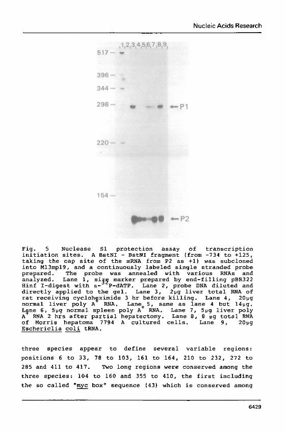
Fig. 5 Nuclease S1 protection assay of transcription initiation sites. A BstNI - BstNI fragment (from -734 to +125, taking the cap site of the mRNA from P2 as +1) was subcloned into M13mp19, and a continuously labeled single stranded probe prepared. The probe was annealed with various RNAs and analyzed. Lane 1, si marker prepared by end-filling pBR322 Hinf I-digest with a-"P-dATP . Lane 2, probe DNA diluted and directly applied to the gel. Lane 3, 2pg liver total RNA of rat receiving cycloh~ximide 3 hr before killing. Lane 4, 20pg normal liver poly A RNA. Lane+ 5 , same as lane 4 but 14pg. Lgne 6, 5pg normal spleen poly A RNA. Lane 7, 5pg liver poly A RNA 2 hrs after partial hepatectomy. Lane 8, 8 pg total RNA of Morris hepatoma 7794 A cultured cells. Lane 9, 20pg Eschericlia coli tRNA.
three species appear to define several variable regions:
positions 6 to 33, 78 to 103, 161 to 164, 210 to 232, 272 to
285 and 411 to 417. Two long regions were conserved among the
three species: 104 to 160 and 355 to 410, the first including
the so called "w boxn sequence (431 which is conserved among
Nucleic Acids Research
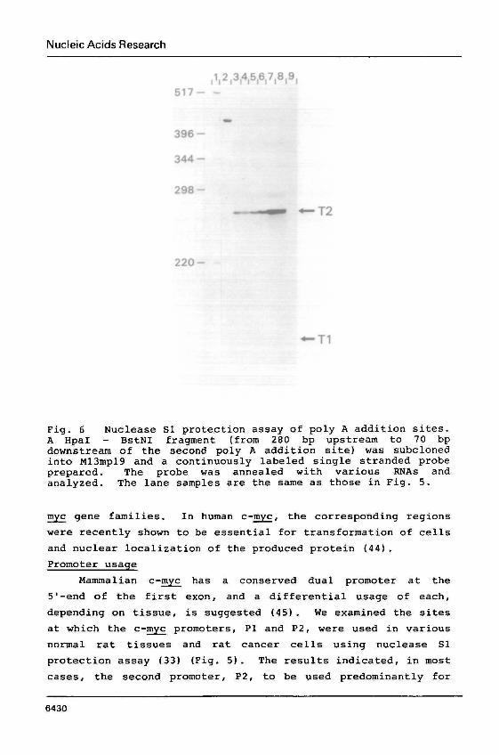
Fig. 6 Nuclease S1 protection assay of poly A addition sites. A HpaI - BstNI fragment (from 280 bp upstream to 70 bp downstream of the second poly A addition site) was subcloned into M13mp19 and a continuously labeled single stranded probe prepared. The probe was annealed with various RNAs and analyzed. The lane samples are the same as those in Fig. 5.
gene families. In human c-F, the corresponding regions
were recently shown to be essential for transformation of cells
and nuclear localization of the produced protein (44).
Promoter usage
Mammalian c - w has a conserved dual promoter at the
5'-end of the first exon, and a differential usage of each,
depending on tissue, is suggested (45) . We examined the sites
at which the c-myc promoters, P1 and P2, were used in various
normal rat tissues and rat cancer cells using nuclease S1
protection assay (33) (Fig. 5 ) . The results indicated, in most
cases, the second promoter, P2, to be used predominantly for
Nucleic Acids Research
c-myc transcription initiation. In the spleen, however, the
c-myc mRNA starting from P1 was also significant. Previous
reports have shown, in mouse spleen, P2 to be very much the
preferred initiation site ( 4 5 ) . There may be some species
difference, therefore, in promoter usage.
In the rat liver, the mRNA dominance starting from P2 did
not change upon induction of regeneration or after
cycloheximide administration. In either case, it is known that
there is a remarkable increase of c-myc mRNA (46) . It has been shown previously that inhibition of protein
synthesis by cylcloheximide stops c-myc mRNA turnover (12, 13).
The fact that the proportion with a P2 preference did not
change before and after cycloheximide administration indicates
the sequence between P1 and P2 not to contribute to the mRNA
stability.
Poly A addition site
The dual poly A addition signals are also conserved
rat, mouse and man. S1 mapping of the 3'-terminus of
mRNA from various sources indicated the downstream signal
among
c -myc to be
used exclusively in all cases examined (Fig. 6). The upstream
signal, however, may be functional under some circumstances, as
it has been suggested previously that poly A addition could
occur at the upstream signal of human c-myc, when it was
expressed in Xenopus oocytes (47).
The exact position of poly A addition in the mRNA seems,
however, to be heterogenous, as evidenced by several faint
bands observed near the major band in Fig. 6. The relative
amount of mRNA having these heterogeneous termini differed
neither between normal and cancer cells nor between resting and
proliferating cells.
DISCUSSION
We cloned the c-myc of a normal rat and two lines of
transplanted hepatomas, one of which contained the gene
amplified 10 fold (22). Restriction maps and the sequences
determined from the independent sources agreed, suggesting no
mutations nor rearrangements to have occurred in the amplified
gene and its flanking regions. This is in agreement with
Nucleic Acids Research
earlier notions of no significant structural changes being
found in the amplified c-E genes of some human malignant
cells ( 4 8 ) .
Apart from the exon sequences, homologous stretches
between rat and human c - w were found throughout the compared
region (Fig. 3). The evolutionary distance between rat and man
is estimated to be approximately 100 million years. Consider
ing the changing rate of sequences with no selective pressure
to be 0.66% per million years ( 4 9 ) , the "meaningless" sequences
should diverge to give almost no homology between the two
species. The remarkable conservation in the surroundins
regions of c - w , therefore, suggests: (1) there to be many
sites, in the region several kb upstream and in the introns of
c-T, where transacting factors bind and contribute to gene
regulation, (2) such regions to have some biological functions
not directly related to c - w . In this regard, it is of
interest that recent reports have suggested a new
transcriptional activity that seems to start from heterogeneous
sites in c-E and extend upstream of the gene (10, 11) . Gazin et al. (50) have suggested that human c - w exon 1
codes for a protein which reacts with antibodies raised to
synthetic peptides having amino acid sequences deduced from
regions of the nucleotide sequence of the exon. They assumed a
minor mRNA species starting upstream of the two major c - w
promoters, and the initiation codon provided by the upstream
sequence. In the corresponding region, we could find no amino
acid sequences possibly related to those used in raising the
antibodies. Such a protein cannot, therefore, be coded in the
rat c - w region.
It is known that c - w mRNA turns over rapidly (half life
15 - 30 min) in various cells e and 2 vitro (12,
authors' unpublished data). The short life time is one of the
characteristics of some mRNAs involved in cell cycling, e, e, those of c-fos (51, 52), c-m~& (53) and some growth factors.
These mRNAs have a region in common containing ATTTA and T
clusters. Shaw c. (40) have shown one such regions from GM-CSF mRNA to be responsible for the instability of the mRNA.
An RNase, possibly, specifically recognizing this sequence
Nucleic Acids Research
motif, could be involved in the metabolism of the short lived
mRNAs. One candidate for this enzyme is RNase L, which is
known to degrade U-rich sequences preferentially (54) . Accelerated degradation of c-myc mRNA by interferon (55) also
supports the idea of the enzyme's involvement, since interferon
causes a rapid synthesis of 2'-5' oligo A, an activator of this
RNase (56) . We found the non-coding region in exon 3 to be
responsible for the rapid turnover of rat c-myc mRNA
(manuscript in preparation), and this region to contain two
sets of ATTTA and T-clusters. Mouse and human c-myc also have
two motifs at identical positions in the non-coding regions of
exon 3. In all cases, these motifs were located approximately
60 nucleotides upstream of the poly A addition signals.
Inhibition of protein synthesis by cycloheximide is known
to stop c-E mRNA degradation (121, and result in mRNA
accumulation in various types of cell. One interpretation of
this effect of cycloheximide is that the inhibitor "freezesn
ribosomes on the mRNA, thus sterically protecting the mRNA from
RNase attack. The destabilizing sequence, which is likely to
be the site recognised by the involved RNase is, however,
located downstream of the termination codon and expected to be
exposed whether or not the ribosomes were frozen on the
upstream sequence of the mRNA. Perhaps the effect of
cycloheximide should be otherwise explained, such as the drug
inactivating the RNase.
Interestingly, poly A addition at the upstream signal, if
such occurs, removes one of the destabilizer sequences from
the mRNA. Such shortened mRNA may have a longer life and,
therefore, may accumulate even in the absence of
transcriptional activation. Although we have not yet found
such a shortened mRNA version, the conservation of the poly A
addition signal's duality hints at such a regulatory mechanism
being required at some stage of the life cycle.
Finally, we are tempted to emphasize that the clones and
the rat c - 9 sequence data could be useful in studying the
possible involvement of this gene in cancer development since
the rat is the most widely used aminal in experimental
carcinogenesis.
Nucleic Acids Research
ACKNOWLEDGEMENTS
We thank Dr. J. M. Bishop for supplying p v - w , Dr. M.
Yamamoto for allowing us to use Vax11/750 and Dr. S. Nishimura
for suggesting the use of 7-deaza GTP in the sequencing
reactions. This work was supported by a Grant-in-Aid from the
Ministry of Health and Welfare of Japan for a Comprehensive
10-Year Strategy for Cancer Control and Grants-in-Aid for
Cancer Research from the Ministry of Education,
Culture of Japan.
REFERENCES Duesberg, P.H., Bister, K. and Vogt. P. K. Nat. Acad. Sci. USA 74. 4320-4324. Kan, N. C., Flordellis, C. S., Garon, C. F., H. and Papas, T. S. (1983) Proc.. Nat. Acad. 6566-6570.
Science and
(1977) Proc.
Duesberg, P. Sci. USA 80,
Hayflick, J., Seeburg, P. H., Ohlsson, R., Pfeifer-Ohlsson, S., Watson, D., Papas, T. and Duesberg, P. H. (1985) Proc. Nat. Acad. Sci. USA 82, 2718-2722. Shilo, B.-Z. and Weinberg. R. A. (1981) Proc. Nat. Acad. Sci. USA 78, 6789-6792. Abrams, H. D., Rohrschneider, L. R. and Eisenman, R. N. (1982) Cell 29, 427-439. Watt, R., Shatzman, A. R. and Rosenberg, M. (1985) Mol. Cel. Biol. 5, 448-456. Klein, G. and Klein, E. (1985) Nature 315, 190-195. Bishop, J. M. (1983) Annu. Rev. Biochem. 52, 301-354. Greenberg, M. E. and Ziff. E. (1984) Nature 311, 433-438. Nepveu, A. and Marcu, K. (1986) EMBO J. 5, 2859-2865. Bentley, D. L. and Groudine, M. (1986) Nature 321, 702-706. Blanchard, J.-M., Piechaczyk, M., Dani, C., Chambard, J-C., Franchi, A., Pouyssegur, J. and Jeanteur, P. (1985) Nature 317, 443-445. Linial, M., Gunderson, N. and Groudine, M. (1985) Science 230, 1126-1132. Chung, J., Sinn, E., Reed, R. R. and Leder, P. (1986) Proc. Nat. Acad. Sci. USA 83, 7918-7922. Remmers, E., Yang, J.-Q. and Marcu, K. B. (1986) EMBO J. 5, 899-904. Battey, J., Moulding, C., Taub, R., Murphy, W., Stewart, T., Potter, H., Lenoir, G. and Leder, P. (1983) Cell 34, 779-787. Colby, W. W., Chen, E. Y., Smith, D. H. and Levinson, A. D. (1983) Nature 301, 722-725. Gazin, C., Dupont De Dinechin, S., Hampe, A., Masson, J-M., Martin, P., Stehelin, D. and Galibert, F. (1984) EMBO J. 3, 383-387. Stanton, L. W., Watt, R. and Marcu, K. B. (1983) Nature 303, 401-406. Bernard, O., Cory, S., Gerondakis, S., Webb, E. and Adams, J. M. (1983) EMBO J. 2, 2375-2383.
Nucleic Acids Research
Keath, E. J., Kelekar, A. and Cole, M. D. (1984) Cell 37, 521-528. Hayashi, K., Makino, R. and Sugimura, T. (1984) Gann 75, 475-478. Frischauf, A.-M., Lehrach, H., Poustka, A. and Murray, N. (1983) J. Mol. Biol. 170, 827-842. Enquist, L. and Sternberg, N. (1979) Methods in Enzymol. 68, 281-298. Benton, W. D. and Davis, R. W. (1977) Science 196, 180-182. vennstrom, B., Moscovici, C., Goodman, H. M. and Bishop, J. M. (1981) J. Virol. 39, 625-631. Deininger, P. L. (1983) Analyt. Biochem. 129, 216-223. Sanger, F., Nicklen, S. and Coulson, A. R. (1977) Proc. Nat. Acad. Sci. USA 74, 5463-5467. Yanisch-Perron, C., Vieira, J. and Messing, J. (1985) Gene 33, 103-119. Mizusawa, S., Nishimura, S. and Seela, F. (1986) Nuc. Acids Res. 14, 1319-1324. Devereux, J., Haeberli, P. and Smithies, 0. (1984) Nuc. Acids Res. 12, 387-395. Kanehisa, M. (1984) Nuc. Acids Res. 12, 417-428. Berk, A. J. and Sharp. P. (1977) Cell 12, 721- Davis, L. G., Dibner, M. D. and Battey, J. F. in Basic Methods in Molecular Biology, Elsevier N.Y. Pear, W. S., Ingvarsson, S., Steffen, D., ~unke, M., Francke, U., Bazin, H., Klein, G. and ~iimegi, J. (1986) Proc. Nat. Acad. Sci. USA 83, 7376-7380. Siebenlist, U., Hennighausen, L., Battey, J. and Leder, P. (1984) Cell 37, 381-391. Briggs, M. R., Kadonaga, J. T., Bell, S. P. and Tjian, R. (1986) Science 234, 47-52. Nagata, K., Guggenheimer, R. A. and Hurwitz, J. (1983) Proc. Nat. Acad. Sci. USA 80, 6177-6181. Birnstiel, M. L., Busslinger, M. and Strub, K. (1985) Cell 41, 349-359. Shaw, G. and Kamen, R. (1986) Cell 46, 659-667. Wang, A. H.-J., Quigley, G. J., Kolpak, F. J., Crawford, J. L., van Boom, J. H., van der Marel, G. and Rich, A. (1979) Nature 282, 680-686. Needleman, S. B. and Wunsch, C. D. (1970) J. Mol. Biol. 48, 443-453. Schwab, M., Alitalo, K., Klempnauer, K.-H., Varmus, H. E., Bishop, J. M., Gilbert, F., Brodeur, G., Goldstein, M. and Trent, J. (1983) Nature 305, 245-248. Stone, J., De Lange, T., Ramsay, G., Jabocits, E., Bishop, J. M., Varmus, H. and Lee, W. (1987) Mol. Cel. Biol. 7, 1697-1709. Stewart, T. a., Bellv6, A. R. and Leder, P. (1984) Science 226. 793-710. ~aklno, R., Hayashi, K. and Sugimura, T. (1984) Nature 310, 697-698. Nishikura, K., Goldflam, S. and Vuocolo, G. A. (1985) Mol. Cel. Biol. 5, 1434-1441. Shibuya, M. and Yamaguchi, S. (1986) in 5th International Workshop on Immune Deficient Animals (Karger), in press. Britten, J. (1986) Science 231, 1393-1398.
Nucleic Acids Research
50 . G a z i n , C . , R i g o l e t , M . , B r i a n d , J. P . , Van R e g e n m o r t e l , M.H.V. a n d G a l i b e r t , F . ( 1 9 8 6 ) EMBO J . 5 , 2241-2250.
51 . K r u i j e r , W . , Coope r , J. A., H u n t e r , T. a n d Verma, I . M. ( 1 9 8 4 ) N a t u r e 312 , 711-716.
52 . M e i j l i n k , F . , C u r r a n , T . , Mi l ler , A. D. a n d Verma, I. (1985 ) P r o c . N a t . Acad , S c i . USA 8 2 , 4987-4991.
53 . Thompson, C. B., C h a l l o n e r , P. B . , Neiman, P. E. a n d G r o u d i n e , M. ( 1986 ) N a t u r e 319 , 374-380.
54 . F loyd -Smi th , G . , S l a t t e r y , E. a n d L e n g y e l , P. ( 1981 ) S c i e n c e 212 , 1030-1032.
55 . E i n a t , M . , R e s n i t z k y , D. a n d K imch i , A. ( 1985 ) N a t u r e 313 , 597-600.
5 6 . W i l l i a m s , B. R. G., G o l g h e r , R. R. , Brown, R. E . , G i l b e r t , C. S . a n d K e r r , I. M. ( 1979 ) N a t u r e 282, 582-586.