characterization of two fsh inducible genes, a nuclear...
TRANSCRIPT

29Japanese Journal of Reproductive Endocrinology(2004)9:29-34
MINI REVIEW
Characterization of Two FSH Inducible Genes,a Nuclear Transcriptional Co-activator p120and a Novel Serine Protease-like Protein,in the Rat Ovarian Granulosa Cells
Miki YOSHINO1,2), Tetsuya MIZUTANI1,2), Kazuya YAMADA1,2), Takeshi ARIMA1,2), Takashi YAZAWA1,2),Hiroko OGATA-KAWATA1,2)
, Tetsuya MIZUTANI)
, Tetsuya MIZUTANI, Toshio SEKIGUCHI1,2)
, Kazuya YAMADA)
, Kazuya YAMADA, Takashi KAJITANI1,2) and Kaoru MIYAMOTO1,2)
1)Department of Biochemistry, Faculty of Medical Sciences, University of Fukui, Fukui 910-1193, Japan2)CREST, JST (Japan Science and Technology), Japan
Correspondence:Kaoru MIYAMOTO, Department of Biochemistry, Faculty of Medical Sciences, University of Fukui, Shimoaizuki, Matsuoka, Fukui 910-1193, JapanTEL: +81-776-61-8316FAX: +81-776-61-8102E-mail: [email protected]
Abstract
Ovarian follicular development is primarily depen-dent on pituitary gonadotropins. We report here the identification of two genes, a nuclear transcription co-activator p120, and a novel serine protease-like pro-tein, as FSH inducible genes in the rat granulosa cells. Characterization of the genes revealed that rat p120 may work together with PR in the granulosa cells of large antral follicles to promote the ovulation events, and that the novel serine protease-like protein may be involved in the proteolytic process during the follicular development and ovulation process.
Introduction
Follicular growth is primarily controlled by pituitary gonadotropins, LH and FSH. Especially FSH stimulates proliferation and differentiation of granulosa cells of small follicles in the ovary, and promotes their develop-ment to pre-ovulatory follicles [1]. Recently we identi-fied many gonadotropin inducible genes expressed in the ovary by subtraction cloning and DNA microarray analyses, and found that a transcriptional co-activator p120 and a novel serine protease-like protein were strongly induced in the rat cultured granulosa cells by
FSH. In this report, characterization of the co-activa-tor p120 and the novel serine protease-like protein in the ovary will be described.
Subtraction cloning and DNA microarray
The subtraction cloning and DNA microarray tech-nology are quite powerful methods for the comprehen-sive analysis of gene expression. In order to identify FSH-inducible genes in the rat granulosa cells, sub-traction cloning and DNA microarray analyses were performed. Cultured granulosa cells (5.0×10⁶ viable cells) were treated with or without 30 ng/ml FSH for 15 hours. Messenger RNAs from FSH treated cells as well as control cells were converted to double strand-ed cDNAs, and subtraction cloning was performed as previously reported [2]. Resulting cDNAs of size-range between 0.5-2.0 kbp long were subcloned to construct a subtraction plasmid cDNA library. About 400 clones in the plasmid library were randomly picked up and their nucleotide sequences were partially determined. For DNA microarray analysis, mRNAs from the cells treated with or without FSH were labeled with fluo-rescent dyes, Cye3- or Cye5, respectively. The labeled cDNA pools were hybridized to microarray glass slides containing 3200 cDNA elements. As listed in Table 1, hundreds of candidate genes were picked-up and 10 genes were confirmed as FSH inducible genes by the semi-quantitative RT-PCR analysis. Among these FSH-inducible genes, we describe here the expression and localization of the transcriptional co-activator p120, as well as those of the novel serine protease-like

30 Japanese Journal of Reproductive Endocrinology Vol. 9 2004
Miki YOSHINO et al.
protein in the ovary during the follicular development and the ovulation processes.
Characterization of rat p120, an FSH inducible transcriptional co-activator
In this study we identify rat homologue of p120, a nuclear transcription co-activator, as one of the FSH inducible genes in the rat granulosa cells. We then cloned a full-length cDNA encoding rat p120 and the nucleotide sequence was determined. Fig. 1 shows the deduced amino acid sequence of rat p120, which is compared with that of human p120. It contains pro-line-rich, acidic, and bromo-domains, respectively, and there are two LXXLL-motifs between proline-rich and acidic domains. The sequences are well conserved be-tween rat and human (94% identity at amino acid lev-el), except that rat p120 has 45 amino acid residues extended at N-terminus. The transcriptional co-activa-tor p120 was originally isolated as a novel nuclear co-activator for thyroid hormone receptor [3]. Further studies revealed that p120 also interacts with andro-gen receptor or 9-cis-retinoic acid receptor (RXR)
Fig. 1. Deduced amino acid sequence of rat p120 The proline-rich domain between 223 and 288 amino acid residues from the N-terminus is underlined, and acidic amino acid-rich domain
between 602 and 731 is shown by bold type. The bromodomain between 809 and 894 is boxed. The putative NR box (LXXLL motif), which is necessary for the interaction with nuclear hormone receptors, is boxed by colored background. Since there are high homology between rat and human p120 sequence (94% identity at amino acid level), rat p120 is expected to have similar functions.
Table 1. Induced genes in the rat granulosa cells stimulated with FSH for 15 hours
Accessionnumber Definition
AB180485 Human thyroid hormone receptor coactivating protein like protein (rat p120)
AB180912 Unknown serine protease like protein
NM_017290 Rat ATPase, Ca2+ transporting, cardiac muscle, slow twitch 2 (Atp2a2)
NM_022265 Rat programmed cell death 4 (Pdcd4)
M64393 Rat 3-α-hydroxysteroid dehydrogenase(3-α-HSD)
NM_019683 Mouse globin inducing factor, fetal (Gbif-pending)
NM_011462 Mouse spindlin (Spin)
U23776 Rat Eker rat-associated intracisternal-A particleelement
NM_012600 Rat Malic enzyme 1, soluble (Me1)
AY009092 Rat retrovirus SC1

31MINI REVIEW
Rat p120 and a Novel Serine Protease in the Ovary
functioning as a co-activator of these nuclear recep-tors [4]. The receptors belong to the nuclear receptor super-family proteins that activate the transcription of their target genes when the steroid hormones or li-gands bind to them. The nuclear hormone receptors generally bind to the respective hormone responsive elements (HREs) in the upstream regions of the tar-get genes, and activate the transcription. Deficiencies of estrogen receptors or progesterone receptors (PR) result in various defects of reproductive functions, such as abnormal follicular development, anovulation, and infertility [5-7]. Transcriptional co-activators are required to form active protein complexes with the hormone nuclear receptors and the basic transcription machinery proteins for the target gene transcription. Co-activators p160/SRC and CBP/p300 are known to interact with the nuclear hormone receptors at the C-terminal AF-2 region through the LXXLL-motif (NR box) [8-10]. Knock-out mice of steroid receptor co-activator 3 (SRC-3), a member of p160 family, ex-hibit infertility [11], and those of nuclear receptor inter-acting protein 1 (Nrip1/RIP140) are also infertile due to the blockage of the oocytes release from the Graaf-ian follicles by the ovulatory stimuli [12]. These reports indicate that co-activators are also important for the follicular development and ovulation. Therefore we ex-amined expression and localization of p120 gene in the ovary during the follicular development using imma-ture rats primed with gonadotropins. As shown in Fig. 2A, in situ hybridization revealed that the induction pattern of p120 was very similar to that of PR in the granulosa cells of large pre-ovulatory follicles. It is well established that PR participates in the ovulation process occurring in the pre-ovulatory large antral fol-licles. Ovulation is triggered by LH surge from the pi-tuitary. About 4 hours after the LH surge, expression of PR is rapidly induced in the granulosa cells of pre-ovulatory large antral follicles destined to ovulate [13, 14]. Progesterone binds to the induced receptor to acti-vate gene expression of specific proteases, ADAMTS-1 or Cathepsin L, which are thought to work on follicle rupture by degrading the ovulating follicle walls [15]. In addition, we examined co-activator functions of rat p120 by co-transfection experiments to show that p120 may promote the action of PR during the ovulation. As
shown in Fig. 2B, p120 enhanced the transcription ac-tivity of PR in the presence of the ligand progesterone. These observations suggest that p120 may work to-gether with PR in the granulosa cells of ovulatory folli-cles to promote the ovulation in the ovary.
A novel serine protease-like protein in the ovary
We also examined expression and localization of the novel FSH-inducible serine protease-like protein in the ovary during the follicular development, in order to check possible involvement of the gene in some ovarian proteolytic processes. The extracellular matrix (ECM) influences basic cellular processes such as pro-liferation, differentiation, migration and adhesion, and is fundamental to normal development. A tightly regu-lated proteolytic activity controls the remodeling and degradation of matrix components in these processes. In the ovary, many physiological and pathological pro-cesses including follicular development, ovulation and
Fig. 2. Localization and transcriptional co-activator function of rat p120
(A) Sections of ovaries from immature rats primed with hCG for 4 hours followed PMSG 48 hours were hybridized with p120 (a, b; antisense probe, c; sense probe) or PR (d, e; antisense probe, f; sense probe). Expression of these genes was com-pletely co-localized in the granulosa cells of large antral folli-cles, suggesting that p120 works together with PR in the ovary. (B) Transcriptional co-activator activity of rat p120 was exam-ined by co-transfection experiments. CV-1 cells were trans-fected with a luciferase reporter plasmid and the human PR ex-pression vector with or without 10-7M progesterone, in the presence or absence of p120. The luciferase activity of PR was upregulated about two fold in the presence of p120.

32 Japanese Journal of Reproductive Endocrinology Vol. 9 2004
Miki YOSHINO et al.
angiogenesis require a regulated turnover of ECM components. Although a lot of proteolytic enzymes in-cluding members of the plasminogen activator (PA) or matrix metalloproteinase (MMP) have been recog-nized to be involved in several ovarian proteolytic events, a definitive functional role for individual prote-ases has yet to be demonstrated [16]. Indeed, there ex-ist many deficient mouse strains that lack individual matrix degrading proteases, but these mice are fertile and don't have major defects of ovulation and female reproduction, as shown in knockout mice of PA [17]. In this context, we cloned a full-length cDNA encod-ing a novel FSH inducible ovarian serine protease-like protein and the nucleotide sequence was determined. Fig. 3 shows the nucleotide sequence of the serine pro-tease-like protein along with deduced amino acid se-quence. The gene encodes a protein of 406 amino acid
residues with a putative signal sequence, and putative active site Asp, His, and Ser residues that are con-served in serine protease family such as PA protein. As shown in Fig. 4, RT-PCR revealed that the serine protease was expressed in the brain (lane 1), adrenal (lane 4), intestine (lane 9), testis (lane 6), and ovary (lane 11). PMSG treatment increased the gene expres-sion in the ovary after 48 hours (lane 12). Northern blot analysis revealed that the serine pro-tease mRNA level was rapidly induced by the FSH treatment within 3 hours, and the levels were kept high throughout the treatment (Fig. 5A). As shown in Fig. 5B, the treatment of immature rats with PMSG in
Fig. 3. Nucleotide and deduced amino acid sequence of the serine protease-like protein
The amino acids are numbered starting from the initiation co-don. The deduced signal sequence between 1 and 17 residues from the N-terminus is underlined, and conserved residues be-tween 162 and 167 involving the putative active site histidine residue is boxed.
Fig. 5. Induction of the serine protease-like protein gene expression in the ovary
Northern blot analysis of the serine protease-like protein mRNA in cultured granulosa cells treated with FSH (A), in immature rat ovaries primed with PMSG (B), and in immature rat ovaries primed with PMSG followed by hCG (C).
Fig. 4. Expression of the serine protease-like protein gene in various tissues
RT-PCR was performed using various tissues. The samples are brain (lane 1), pituitary (lane 2), liver (lane 3), adrenal (lane 4), uterus (lane 5), testis (lane 6), kidney (lane 7), spleen (lane 8), intestine (lane 9), stomach (lane 10), untreated ovary (lane 11), ovary treated with PMSG for 48 hours (lane 12).
33MINI REVIEW
Rat p120 and a Novel Serine Protease in the Ovary
vivo also induced the protease gene expression in the ovary within 6 hours after the treatment, with a tran-sient peak at 24 hours after the treatment. Further hCG treatment also increased the gene expression with a peak at 2 hours as shown in Fig. 5C.
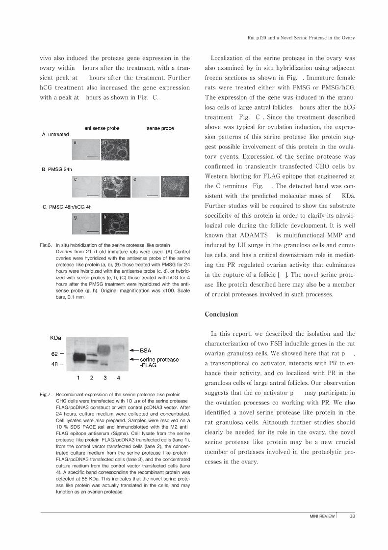
Localization of the serine protease in the ovary was also examined by in situ hybridization using adjacent frozen sections as shown in Fig. 6. Immature female rats were treated either with PMSG or PMSG/hCG. The expression of the gene was induced in the granu-losa cells of large antral follicles 4 hours after the hCG treatment (Fig. 6C). Since the treatment described above was typical for ovulation induction, the expres-sion patterns of this serine protease-like protein sug-gest possible involvement of this protein in the ovula-tory events. Expression of the serine protease was confirmed in transiently transfected CHO cells by Western blotting for FLAG epitope that engineered at the C terminus (Fig. 7). The detected band was con-sistent with the predicted molecular mass of 55 KDa. Further studies will be required to show the substrate specificity of this protein in order to clarify its physio-logical role during the follicle development. It is well known that ADAMTS-1 is multifunctional MMP and induced by LH surge in the granulosa cells and cumu-lus cells, and has a critical downstream role in mediat-ing the PR-regulated ovarian activity that culminates in the rupture of a follicle [15]. The novel serine prote-ase-like protein described here may also be a member of crucial proteases involved in such processes.
Conclusion
In this report, we described the isolation and the characterization of two FSH inducible genes in the rat ovarian granulosa cells. We showed here that rat p120, a transcriptional co-activator, interacts with PR to en-hance their activity, and co-localized with PR in the granulosa cells of large antral follicles. Our observation suggests that the co-activator p120 may participate in the ovulation processes co-working with PR. We also identified a novel serine protease-like protein in the rat granulosa cells. Although further studies should clearly be needed for its role in the ovary, the novel serine protease-like protein may be a new crucial member of proteases involved in the proteolytic pro-cesses in the ovary.
Fig.6. In situ hybridization of the serine protease-like protein Ovaries from 21-d-old immature rats were used. (A) Control
ovaries were hybridized with the antisense probe of the serine protease-like protein (a, b), (B) those treated with PMSG for 24 hours were hybridized with the antisense probe (c, d), or hybrid-ized with sense probes (e, f), (C) those treated with hCG for 4 hours after the PMSG treatment were hybridized with the anti-sense probe (g, h). Original magnification was x100. Scale bars, 0.1 mm.
Fig.7. Recombinant expression of the serine protease-like protein CHO cells were transfected with 10 μg of the serine protease-
FLAG/pcDNA3 construct or with control pcDNA3 vector. After 24 hours, culture medium were collected and concentrated. Cell lysates were also prepared. Samples were resolved on a 10 % SDS-PAGE gel and immunoblotted with the M2 anti-FLAG epitope antiserum (Sigma). Cell lysate from the serine protease-like protein-FLAG/pcDNA3 transfected cells (lane 1), from the control vector transfected cells (lane 2), the concen-trated culture medium from the serine protease-like protein-FLAG/pcDNA3 transfected cells (lane 3), and the concentrated culture medium from the control vector transfected cells (lane 4). A specific band corresponding the recombinant protein was detected at 55 KDa. This indicates that the novel serine prote-ase-like protein was actually translated in the cells, and may function as an ovarian protease.

34 Japanese Journal of Reproductive Endocrinology Vol. 9 2004
Miki YOSHINO et al.
Acknowledgement
This work was supported in part by a grant from the Smoking Research Foundation, and by 21st Century COE Program (Medical Science).
References
1. Richards JS (1994) Hormonal control of gene expression in the ovary. Endocr Rev 15, 725-751.
2. Mizutani T, Sonoda Y, Minegishi T, Wakabayashi K, Miya-moto K (1997) Cloning, characterization, and cellular distri-bution of rat scavenger receptor class B type I (SRBI) in the ovary. Biochem Biophys Res Commun 234, 499-505.
3. Monden T, Wondisford FE, Hollenberg AN (1997) Isolation and characterization of a novel ligand-dependent thyroid hormone receptor-coactivating protein. J Biol Chem 272, 29834-29841.
4. Monden T, Kishi M, Hosoya T, Satoh T, Wondisford FE, Hol-lenberg AN, Yamada M, Mori M (1999) p120 acts as a spe-cific coactivator for 9-cis-retinoic acid receptor (RXR) on peroxisome proliferator-activated receptor-gamma/RXR heterodimers. Mol Endocrinol 13, 1695-1703.
5. Lydon JP, DeMayo FJ, Funk CR, Mani SK, Hughes AR, Montgomery CAJ, Shyamala G, Conneely OM, O'Malley BW (1995) Mice lacking progesterone receptor exhibit pleiotro-pic reproductive abnormalities. Genes Dev 9, 2266-2278.
6. Couse JF, Hewitt SC, Bunch DO, Sar M, Walker VR, Davis BJ, Korach KS (1999) Postnatal sex reversal of the ovaries in mice lacking estrogen receptors alpha and beta. Science 286, 2328-2331.
7. Couse JF, Korach KS (1999) Estrogen receptor null mice: what have we learned and where will they lead us? Endocr Rev 20, 358-417.
8. Heery DM, Kalkhoven E, Hoare S, Parker MG (1997) A sig-nature motif in transcriptional co-activators mediates bind-ing to nuclear receptors. Nature 387, 733-736.
9. Torchia J, Rose DW, Inostroza J, Kamei Y, Westin S, Glass CK, Rosenfeld MG (1997) The transcriptional co-activator p/CIP binds CBP and mediates nuclear-receptor function. Nature 387, 677-684.
10. McInerney EM, Rose DW, Flynn SE, Westin S, Mullen TM, Krones A, Inostroza J, Torchia J, Nolte RT, Assa-Munt N, Milburn MV, Glass CK, Rosenfeld MG (1998) Determinants of coactivator LXXLL motif specificity in nuclear receptor transcriptional activation. Genes Dev 12, 3357-3368.
11. Xu J, Liao L, Ning G, Yoshida-Komiya H, Deng C, O'Malley BW (2000) The steroid receptor coactivator SRC-3 (p/CIP/RAC3/AIB1/ACTR/TRAM-1) is required for normal growth, puberty, female reproductive function, and mamma-ry gland development. Proc Natl Acad Sci USA 97 , 6379-6384.
12. White R, Leonardsson G, Rosewell I, Ann JM, Milligan S, Parker M (2000) The nuclear receptor co-repressor nrip1 (RIP140) is essential for female fertility. Nat Med 6 , 1368-1374.
13. Park OK, Mayo KE (1991) Transient expression of proges-terone receptor messenger RNA in ovarian granulosa cells after the preovulatory luteinizing hormone surge. Mol Endo-crinol 5, 967-978.
14. Natraj U, Richards JS (1993) Hormonal regulation, localiza-tion, and functional activity of the progesterone receptor in granulosa cells of rat preovulatory follicles. Endocrinology 133, 761-769.
15. Robker RL, Russell DL, Espey LL, Lydon JP, O'Malley BW, Richards JS (2000) Progesterone-regulated genes in the ovulation process: ADAMTS-1 and cathepsin L proteases. Proc Natl Acad Sci USA 97, 4689-4694.
16. Tsafriri A (1995) Ovulation as a tissue remodelling process. Proteolysis and cumulus expansion. Adv Exp Med Biol 377, 121-140.
17. Carmeliet P, Schoonjans L, Kieckens L, Ream B, Degen J, Bronson R, De Vos R, van den Oord JJ, Collen D, Mulligan RC (1994) Physiological consequences of loss of plasminogen activator gene function in mice. Nature 368, 419-424.