characterization of vibrio cholerae o1 el tor galu and gale mutants
TRANSCRIPT

INFECTION AND IMMUNITY,0019-9567/01/$04.0010 DOI: 10.1128/IAI.69.1.435–445.2001
Jan. 2001, p. 435–445 Vol. 69, No. 1
Copyright © 2001, American Society for Microbiology. All Rights Reserved.
Characterization of Vibrio cholerae O1 El Tor galU and galE Mutants:Influence on Lipopolysaccharide Structure, Colonization,
and Biofilm FormationJUTTA NESPER,1 CRYSTAL M. LAURIANO,2 KARL E. KLOSE,2 DAGMAR KAPFHAMMER,1
ANITA KRAIß,1 AND JOACHIM REIDL1*
Zentrum fur Infektionsforschung, Universitat Wurzburg, 97070 Wurzburg, Germany,1 andUniversity of Texas Health Science Center, San Antonio, Texas 78284-77582
Received 5 June 2000/Returned for modification 9 August 2000/Accepted 4 October 2000
Recently we described the isolation of spontaneous bacteriophage K139-resistant Vibrio cholerae O1 El Tormutants. In this study, we identified phage-resistant isolates with intact O antigen but altered core oligosac-charide which were also affected in galactose catabolism; this strains have mutations in the galU gene. Weinactivated another gal gene, galE, and the mutant was also found to be defective in the catabolism of exogenousgalactose but synthesized an apparently normal lipopolysaccharide (LPS). Both gal mutants as well as a roughLPS (R-LPS) mutant were investigated for the ability to colonize the mouse small intestine. The galU andR-LPS mutants, but not the galE mutant, were defective in colonization, a phenotype also associated withO-antigen-negative mutants. By investigating several parameters in vitro, we could show that galU and R-LPSmutants were more sensitive to short-chain organic acids, cationic antimicrobial peptides, the complementsystem, and bile salts as well as other hydrophobic agents, indicating that their outer membrane no longerprovides an effective barrier function. O-antigen-negative strains were found to be sensitive to complement andcationic peptides, but they displayed significant resistance to bile salts and short-chain organic acids. Fur-thermore, we found that galU and galE are essential for the formation of a biofilm in a spontaneous phage-resistant rugose variant, suggesting that the synthesis of UDP-galactose via UDP-glucose is necessary forbiosynthesis of the exopolysaccharide. In addition, we provide evidence that the production of exopolysaccha-ride limits the access of phage K139 to its receptor, the O antigen. In conclusion, our results indicateinvolvement of galU in V. cholerae virulence, correlated with the observed change in LPS structure, and a rolefor galU and galE in environmental survival of V. cholerae.
The causative agent of the intestinal disease cholera is Vibriocholerae, a gram-negative motile bacterium. Of the more than150 known serogroups, only the noncapsulated O1 and theencapsulated O139 serogroup have been found to be associ-ated with epidemic cholera. Epidemic O139 strains are relatedto and were derived from O1 El Tor strains after geneticalterations of the O-antigen biosynthesis gene cluster (16). Theongoing seventh pandemic, which began in 1961, is caused byO1 El Tor strains (3). V. cholerae is a natural inhabitant ofaquatic ecosystems and is known to attach to environmentalsurfaces such as plants, filamentous green algae, zooplankton,crustaceans, or insects (8). Recently, V. cholerae O1 El Tor wasfound to form a three-dimensional biofilm on abiotic surfaces(70). Biofilm formation may be important in the life cycle ofpathogenic V. cholerae strains, because they reside within nat-ural aquatic habitats during interepidemic periods. O1 El Torstrains are also able to switch to a rugose colony phenotype.This morphology correlates with the constitutively productionof an exopolysaccharide allowing biofilm formation on abioticsurfaces (65, 72). Such rugose variants are chlorine resistantand fully virulent in humans (43).
Important steps of the noninvasive disease process includeingestion of V. cholerae along with contaminated food or water,
passage through the gastric acid barrier of the stomach, adher-ence to and penetration through the intestinal mucus lining,adherence to intestinal epithelial cells, multiplication, andcholera toxin (CT) production (25). In the course of the tran-sition from the external environment to the human body, thebacteria are exposed to a series of changes, such as in temper-ature, acidity, and osmolarity, and must survive in the intestine,an environment which is enriched in growth-inhibitory sub-stances like bile salts and organic acids. They are also exposedto antimicrobial components of the innate immune system, likecomplement factors secreted by intestinal epithelial cells (2) ordefensins produced by Paneth cells (37). Several V. choleraegene products have been shown to be important for coloniza-tion of the small intestine. These include the toxin-coregulatedpili (TCP) (62), accessory colonization factors (49), regulatoryproteins (e.g., ToxR/ToxS, TcpP/TcpH, and ToxT) and outermembrane proteins (21, 55), metabolic factors, biotin, purinebiosynthetic genes, and the O antigen of the lipopolysaccharide(LPS) (6). Some factors are suggested to be involved in adhe-sion to the small intestine; however, no specific V. choleraeadhesins or specific mucosal receptors have been identified.
In gram-negative bacteria, LPS is the major integral compo-nent of the outer membrane with three general features:O antigen, core oligosaccharide, and lipid A. The O antigenof V. cholerae O1 consists of a homopolymer of approximately18 perosamine residues which are substituted with tetronate(30). Recently, the structure of the LPS core oligosaccharide
* Corresponding author: Mailing address: Zentrum fur Infektions-forschung, Universitat Wurzburg, Rontgenring 11, 97070 Wurzburg,Germany. Phone: (49) (0)931 312153. Fax: (49) (0)931 312578. E-mail:[email protected].
435
Dow
nloa
ded
from
http
s://j
ourn
als.
asm
.org
/jour
nal/i
ai o
n 17
Feb
ruar
y 20
22 b
y 12
1.14
3.17
3.21
6.
was reported for O1 and O139 mutants (33, 64); the resultsfrom these investigators are summarized in Fig. 1A. The site ofattachment for the O1 antigen is unknown, whereas the O139antigen is linked to the heptosyl III (HepIII) residue (33).Furthermore, the linkage of the carbohydrate quinovosamine,also detected in the LPS of O1 strains, is not known (59). Incontrast to the well-investigated O1-antigen biosynthesis genecluster (rfb) (59), little is known about the genes and corre-sponding enzymes involved in the biosynthesis of the coreoligosaccharide. In gram-negative bacteria, the major core oli-gosaccharide biosynthesis genes are clustered on the chromo-some in the waa (former rfa) locus, encoding specific trans-ferases and enzymes involved in the synthesis of activatedcarbohydrates (23). Some activated moieties like UDP-glucoseand UDP-galactose are often involved in the synthesis of dif-ferent surface structures, and hence enzymes controlling theirbiosynthesis are not necessarily genetically linked with LPSbiosynthesis gene clusters. Enzymes for the biosynthesis ofUDP-glucose and UDP-galactose are UDP-glucose-pyrophos-
phorylase, encoded by galU, and UDP-glucose-4-epimerase,encoded by galE (36). In some bacteria, GalE and GalU arealso important in the catabolism of exogenous galactose to-gether with enzymes encoded by galT and galK (36). In manygram-negative pathogens, mutations in galE or galU lead toattenuated virulence, mainly if changes in LPS or capsularstructures were observed (18). However, nothing is knownabout the role of galU and galE in V. cholerae O1 virulence.
Recently, temperate phage K139 (51) was found to use theO antigen of V. cholerae O1 as its receptor (46). A collection ofspontaneous phage-resistant O1 El Tor P27459 strains wereinvestigated according to their LPS pattern on silver-stainedpolyacrylamide (PAA) gels, and different groups of alteredLPS structures were described. These included O-antigen-neg-ative strains, where one mutant was characterized for the lossof O antigen due to transposition of IS1004 into wbeW, encod-ing a putative glycosyltransferase. Another class identifiedwere rough LPS (R-LPS) mutants comprising an altered coreoligosaccharide with no O antigen attached; however, the mu-tation(s) responsible remains unidentified. In this study, weinvestigated the genetic nature of a phage-resistant mutantwith intact O antigen but altered core oligosaccharide compo-sition and found that this strain contained a mutation in galU.We also compared galU with galE and LPS-defective (R-LPSand O-antigen-negative) mutants with regard to LPS synthesis,galactose catabolism, the ability to colonize the small intestineof mice, and the ability to survive in the presence of bacteri-cidal substances. Among our spontaneous phage-resistant mu-tants, we have also found rugose colony variants (unpublishedresults). In this work, we provide evidence that galU and galEare involved in rugose polysaccharide production.
MATERIALS AND METHODS
Bacterial strains, plasmids, and media. V. cholerae strains used in this studyare listed in Table 1. Escherichia coli K-12 strain LE392 (54) was used for allgenetic constructions, unless the vector being used was a derivative of pCVD442(13); in this case E. coli SM10lpir (42) was used. All strains were grown in Luriabroth (LB) at 37°C except as noted otherwise. Antibiotics were used at thefollowing concentrations: kanamycin, 50 mg/ml; and chloramphenicol, 2.5 mg/ml(V. cholerae) or 30 mg/ml (E. coli); ampicillin, 50 or 100 mg/ml; and streptomycin,100 mg/ml. Plasmids used in this study are listed in Table 1.
Oligonucleotides, PCR, and Southern hybridization. All oligonucleotides usedfor PCR are listed in Table 2. PCRs were performed as described by Mullis andFaloona (44). Southern blotting was performed according to Southern (56).Briefly, chromosomal DNA was prepared according to Grimberg et al. (19),digested with appropriate restriction enzymes, fractionated on a 0.7% agarosegel, and transferred to a Hybond N1 membrane (Amersham Pharmacia Biotech,Freiburg, Germany). Hybridization with horseradish peroxidase-labeled probeand detection of hybridizing bands were carried out according to the procedureprovided by the manufacturer of the ECL (enhanced chemiluminescence) system(Amersham Pharmacia Biotech, Freiburg, Germany).
Computer analysis. The publicly available sequences from the V. choleraegenome project (http://www.tigr.org) (22) were examined for the presence of galgenes. The amino acid sequences of E. coli GalE and GalU, obtained from thedatabase, were used as query sequences. Homology analysis was performed withBasic BLAST Search 2.0 (1).
Construction of plasmids. To construct complementing plasmids, gene-spe-cific oligonucleotides (Table 2) were designed to introduce PstI and FspI sites atthe 59 and 39 ends, respectively, of the genes galU and galE. Following PCR am-plification, the products were digested and ligated into the PstI- and FspI-digest-ed plasmid pACYC177. The resulting plasmids pACYCgalU and pACYCgalEexpress the corresponding genes from the bla promoter. To construct suicidevector pCVDgalU, an internal fragment of galU, obtained after PCR with prim-ers galU1 and -2, was ligated into the SacI/XbaI-digested plasmid pCVD442 (13);this plasmid was used to construct an insertion mutation in the galU gene. Suicide
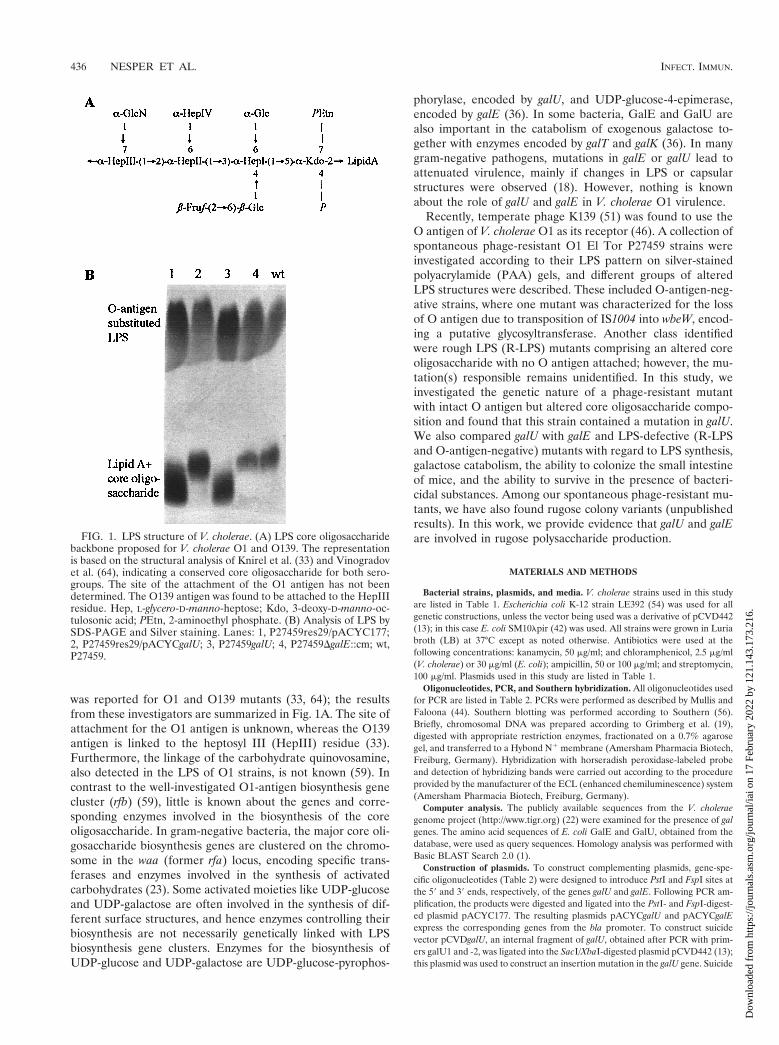
FIG. 1. LPS structure of V. cholerae. (A) LPS core oligosaccharidebackbone proposed for V. cholerae O1 and O139. The representationis based on the structural analysis of Knirel et al. (33) and Vinogradovet al. (64), indicating a conserved core oligosaccharide for both sero-groups. The site of the attachment of the O1 antigen has not beendetermined. The O139 antigen was found to be attached to the HepIIIresidue. Hep, L-glycero-D-manno-heptose; Kdo, 3-deoxy-D-manno-oc-tulosonic acid; PEtn, 2-aminoethyl phosphate. (B) Analysis of LPS bySDS-PAGE and Silver staining. Lanes: 1, P27459res29/pACYC177;2, P27459res29/pACYCgalU; 3, P27459galU; 4, P27459DgalE::cm; wt,P27459.
436 NESPER ET AL. INFECT. IMMUN.
Dow
nloa
ded
from
http
s://j
ourn
als.
asm
.org
/jour
nal/i
ai o
n 17
Feb
ruar
y 20
22 b
y 12
1.14
3.17
3.21
6.

plasmids to introduce chromosomal in-frame deletions of the genes wbeW andgalE were constructed in the same manner. DNA fragments of about 500 bpencompassing the regions upstream of the corresponding gene were PCR am-plified using primer pairs 5xSmaI and 5xSalI, x is the gene of interest (Table 2).The downstream DNA region of the corresponding gene was PCR amplifiedusing primer pairs 3xSalI and 3xApaI (Table 2). The PCR products were digestedwith SalI and ligated overnight (O/N). The ligation mix was digested with SmaIand ApaI, purified, and ligated into pKEK229 (9) that had been digested withSmaI and ApaI to give plasmids pKEKDgalE and pKEKDrfbW. In addition aPCR-derived cat fragment (with oligonucleotides cm1 and -2 [Table 2]) was di-gested with SalI and ligated together with the SalI-digested plasmid pKEKDgalEto give the chloramphenicol-resistant (Cmr) plasmid pKEKDgalE::cm.
Construction of bacterial strains. To construct strains containing a mutationin galU, plasmid pCVDgalU was mated by conjugation from E. coli SM10lpirinto V. cholerae P27459-S, P27459res105, or CO970, selecting for streptomycinand ampicillin resistance (Smr and Apr) (42). The resulting strains obtaineda chromosomal insertion caused by the integration of the plasmid throughhomologous recombination via the internal galU fragment. Strains harboringin-frame deletion mutations were constructed by allelic exchange (13). Plas-mid pKEKDwbeW was mated into P27459-S from E. coli SM10lpir by selectingfor Smr and Apr. Single colonies were grown for successive generations in LBwith no antibiotic selection and then plated on LB without NaCl but containing10% sucrose at 30°C. Several sucrose-resistant, Aps colonies were isolated andscreened for loss of wbeW in cross-streaking experiments using lytic phageK139.cm9 (45). Strain P27459DwbeW was furthermore shown to synthesizeno O antigen in Western blot analysis. O-antigen biosynthesis was restored inP27459DwbeW containing a plasmid carrying wbeW (46). The sucrose counter-selection method failed to introduce galE in-frame deletions. In this case, plas-mid pKEKDgalE::cm was mated from E. coli SM10lpir into V. cholerae P27459-Sand P27459res105, selecting for Smr and Cmr. Single colonies were tested forAps, indicating that the plasmid was absent and a double crossover had occurred.The correct chromosomal insertion for all mutants was confirmed by Southernblot analysis.
LPS analysis. The isolation, separation by sodium dodecyl sulfate-polyacryl-amide gel electrophoresis (SDS-PAGE), and silver staining of LPS were per-formed as described previously (46).
Serum resistance assay. Cells were grown to mid-logarithmic phase in LB,washed, and mixed to a final concentration of 20% with normal human serum(obtained and pooled from four donors) or 20% heat-inactivated normal humanserum in phosphate-buffered saline with 0.1% peptone. Approximately 108 CFUwas used in each assay. After incubation at 37°C for 1 h, the cells were harvested,washed, and resuspended in phosphate-buffered saline with 0.1% peptone. Vi-able cell counts were determined by plating serial dilutions onto LB agar.
Mouse colonization assays. The infant mouse colonization assay has beendescribed previously (32). Briefly, mutant strains (lac1) were mixed with thestrain P27459lacZ (lac) and given in a peroral inoculum ratio of approximately107 CFU mutant to 107 CFU of wild type to 5- to 6-day-old CD-1 suckling mice.After a 24-h period of colonization, intestinal homogenates were collected andthe ratio of mutant to wild type was determined by plating appropriate dilutionson LB agar containing streptomycin and X-Gal (5-bromo-4-chloro-3-indolyl-b-D-galactopyranoside).
CTX-KmF transduction and GM1-ELISA. For optimal CT and TCP expres-sion, the strains were incubated under inducing conditions. O/N cultures grownin AKI medium (27) were diluted 1:100 in AKI without NaHCO3, grown for 4 hat 37°C under static growth conditions, and then transferred to vigorous shakingfor 4 h at 37°C (40). For CT measurement, 1 ml of the culture was spun down,and the culture supernatant was filtered (0.2-mm-pore-size filter; Schleicher &Schuell, Dassel, Germany), and frozen at 220°C. The level of CT present in 200ml of the supernatant was measured by GM1-ganglioside enzyme-linked immu-nosorbent assay (GM1-ELISA) (61) using purified CT (Sigma, Deisenhofen,Germany) to generate a standard curve.
For phage transduction, 100 ml of the same culture was mixed with 100 ml ofphage CTX-KmF (66). After standing for 30 min at room temperature, themixture was diluted and plated onto LB agar containing kanamycin. The trans-duction frequency was calculated from the CFU of kanamycin-resistant (Kmr)transductants correlated with the optical density at 600 nm (OD600) of therecipient strain. An additional CTX-KmF transduction experiment was per-formed to enumerate the numbers of both transductants and potential recipients.Strain P27459DtoxR was used as a negative control for both assays. The DtoxR1mutation was introduced into the chromosome of P27459-S by plasmid pMD60,as previously described (31), to form strain P27459DtoxR. Phage CTX-KmF wasprepared from a 100-ml O/N culture (LB, 30°C) of strain O395pCTX-Km byfiltering the culture supernatant through a 0.2-mm-pore-size filter.
Determination of MICs antimicrobial cationic peptides. Standard MIC testingof susceptibility to polymyxin B (50 mg/ml to 780 ng/ml; Sigma) and protamine(675 to 10.5 mg/ml; Sigma) was done as described by Steinberg et al. (57). TheMIC was defined as the lowest concentration of drug which prevented visibleturbidity after incubation for 18 h at 37°C without shaking.
Sensitivity to hydrophobic agents. Sensitivity to SDS and novobiocin wasassessed by determination of the MIC. O/N cultures of strains to be tested werediluted in LB to give 1 3 105 to 5 3 105 CFU/ml. After 100-ml aliquots of thediluted cultures were dispensed into each well of a 96-well microtiter plate(Greiner, Frickenhausen, Germany), 11 ml of test compound was added. SDS(Merck, Hamburg, Germany) and novobiocin (Sigma) were dissolved in water at
TABLE 1. V. cholerae strains and plasmids used in this study
Strain orplasmid
Genotype ordescription
Source orreference
StrainsCO970 O1 El Tor Ogawa, India 1994,
Smr CmrJ. J. Mekalanos
CO970galU CO970 galU::pCVD442, Apr This studyO395pCTX-Km Kmr 66P27459-S Wild type (O1 El Tor, Inaba),
spontaneous Smr48
P27459DgalE::cm DgalE::cm in P27459-S, Cmr This studyP27459galU galU::pCVD442 in P27459-S, Apr This studyP27459lacZ lacZ::pMD13 in P27459-S, Apr 46P27459res29 Altered core oligosaccharide, intact
O antigen; mutant of P27459-S46
P27459res46 R-LPS mutant of P27459-S This studyP27459res105 Rugose colony variant of P27459-S This studyP27459res105DgalE::cm DgalE::cm in P27459res105, Cmr This studyP27459res105galU galU::pCVD442 in P27459res105, Apr This studyP27459res118 R-LPS mutant of P27459-S 46P27459DwbeW DwbeW in P27459-S, O-antigen
negativeThis study
PlasmidspACYC177 Apr Kmr 52pACYCgalE galE allele in bla of pACYC177; Kmr This studypACYCgalU galU allele in bla of pACYC177; Kmr This studypBR322 Apr Tcr 71pCVD442 oriR6K, mobRP4, sacB, Apr 13pCVDgalU 9galU9 in pCVD442; Apr This studypJNwbeW wbeW allele in bla of pACYC177; Kmr 46pKEK229 pCVD442 with alternative MCS; Apr 9pKEKDgalE::cm orf9 9galE-cm-9galE orf9 in pKEK229;
Apr CmrThis study
pKEKDwbeW orf359 9wbeW-9wbeW orf9 in pKEK229;Apr
This study
pTrckan Kmr 45
TABLE 2. Oligonucleotides used in this study
Primer Sequence (59-39)a
cm1 .............................GCAAGTCGACGGGCCCGCACCTCAAAAACACCATCATACAcm2 .............................AGTCGTCGACAGGCGTTTAAGGGCACCAATAACTGgalEPstI......................AAAACTGCAGAAAAGCCGTTACAATCGCCGgalEFspI.....................GAAAATGCGCAGGGCGTTTGTATAGCAACAG59galESmaI ................TCCCCCGGGCTATCGTACGGCTCATAACA59galESalI ..................TACGCGTCGACATACCCGCTTGGATCATCTG39galESalI ..................TACGCGTCGACCGTAATCTACACACAATGGC39galEApaI ................GTAAGGGCCCAATCGGCCACGGTGGAAATTgalUPstI .....................AACTGCAGCTAGCACACCATAGAGCAAgalUFspI ....................CCCTGCGCATGCATTGTTTAGATAGCGAATgalU1 ..........................AAGAGCTCAGCTATTGGGTGATATCCGTAgalU2 ..........................GATCTAGACTTCGATATAGCCTTCAACGC59wbeWSmaI .............TCCCCCGGGAATTCCCTTACCTCTTGGTGC59wbeWSalI ...............ACGGCTCGACCAGAAAATAGGGGAGCCAGT39wbeWSalI ...............ACGCGTCGACGACACCAGTAAAGCTTGCAG39wbeWApaI..............GAGGGCCCCGATGTGCGCATGAGTTAAG
a Underlined nucleotides are not exact matches to the sequence and wereintroduced to add restriction enzyme sites. Nucleotide sequences were derivedfrom the publicly available sequences from the V. cholerae genome project(http://www.tigr.org) (22) and accession no. X59954 and Y07788.
VOL. 69, 2001 V. CHOLERAE gal MUTANTS 437
Dow
nloa
ded
from
http
s://j
ourn
als.
asm
.org
/jour
nal/i
ai o
n 17
Feb
ruar
y 20
22 b
y 12
1.14
3.17
3.21
6.

10-fold the desired test concentration and then 2-fold serially diluted in water.The final concentrations tested ranged from 600 to 9.3 mg/ml for SDS and 1.5mg/ml to 23 ng/ml for novobiocin. The sensitivity to bile was tested on thiosulfatecitrate bile sucrose (TCBS) agar plates, prepared as instructed for the commer-cially available TCBS (Difco, Heidelberg), containing various amounts of bile(0 to 0.8%).
Biofilm formation. Quantitative biofilm measurement was performed in amicrotiter assay as described previously (50), with minor modifications. Briefly,bacteria were grown O/N in LB and diluted 1:100 in fresh LB containing appro-priate antibiotics; then 100-ml aliquots were placed in a 96-well microtiter plate(polystyrene; Greiner) and incubated for 24 h at room temperature. Bacterialcultures were poured out, washed three times with H2O, fixed with 2.5% glutar-aldehyde, washed twice, and stained with 200 ml of a 0.4% crystal violet solution.After solubilization of the crystal violet with 300 ml of ethanol-acetone (80:20),the OD was determined at 570 nm in an ELISA reader (Bio-Rad, Munich,Germany). Qualitative biofilm formation was assessed by incubating 1:100-di-luted O/N cultures (LB, with appropriate antibiotics) in borosilicate tubes for48 h without shaking at room temperature. Strains were noted as biofilm positivewhen floating pellicles with closely packed bacteria were formed; strains growingas a suspension were designated as biofilm negative. Cultures were then pouredout, and adherent bacteria were stained with crystal violet.
Acid tolerance response assay. The ability of cells to survive exposure toorganic acids (cocktail of 2.5 mM butyric acid, 8.7 mM acetic acid, and 3.7 mMpropionic acid) in LB (pH 4.4) was determined as described by Merrell andCamilli (41) except that no organic acids were added to the adaptation medium.
Plaque inhibition assay. The phage neutralizing capacity of purified LPS wasdetermined as described previously (46).
b-Lactamase detection by immunoblotting analysis. Cells were harvested atOD600 1.5 to 2.5 and diluted to give 2 ml of OD600 5 1. After centrifugation of1 ml of cell suspension, the cell pellets were resuspended in 100 ml of samplebuffer. The supernatants were filtered, and proteins were precipitated with 10%(final concentration) trichloracetic acid. The protein pellet was solubilized in 100ml of sample buffer. Samples (20 ml) were separated by SDS-PAGE (34), trans-ferred to a nitrocellulose membrane, and reacted with an antibody directedagainst BlaM (5 Prime33 Prime Inc., Boulder, Colo.).
RESULTS
Identification of a spontaneous galU mutant expressingan altered LPS core oligosaccharide. The recently describedspontaneous phage K139.cm9-resistant mutant P27459res29showed an altered LPS core oligosaccharide with an intact Oantigen (Fig. 1B, lane 1) (46). The observation that this straincould not grow on M9 medium containing galactose as a car-bon source and the fact that glucose is a component of the LPScore oligosaccharide (Fig. 1A) implicated a possible galU mu-tation in this strain. To test this hypothesis, we constructed aplasmid encoding galU, based on nucleotide sequence infor-mation obtained from the V. cholerae genome project (http://www.tigr.org) (22). Plasmid pACYCgalU complemented allobserved phenotypes in strain P27459res29, showing a normalLPS pattern on a silver-stained PAA gel (Fig. 1B, lane 2) aswell as restoring sensitivity against phage K139.cm9 andgrowth on M9 galactose plates (data not shown).
Construction and characterization of gal mutants. To de-termine whether a possible virulence effect of the galU mutantmight be caused by a general defect in galactose utilization,we constructed a V. cholerae strain with a deletion/insertion inanother galactose utilization gene, galE. Additionally, we gen-erated a galU strain by plasmid insertion (Fig. 2; Materials andMethods) and compared the phenotypes of the two strains.Neither mutant could grow on M9 galactose plates, confirmingthat galE is also involved in galactose utilization as predicted.Growth could be restored in trans by a plasmid encoding galEin strain P27459DgalE::cm and by a plasmid encoding galU instrain P27459galU (data not shown). In E. coli and Salmonella
enterica serotype Typhimurium, the genetic loss of galE andgalU causes bacteriolysis when growing cells are exposed tohigh concentrations of D-galactose (36). This property was test-ed in V. cholerae galE and galU mutants on LB plates contain-ing 2% galactose. The galE mutant could not grow on suchplates, whereas both galU mutants (spontaneous and con-structed) grew initially but eventually lysed. This phenotypecould be complemented by introduction of galU or galE intrans. The LPS patterns of both galE and galU mutants werethen analyzed by SDS-PAGE. In a silver-stained PAA gel, thegalU mutant showed an altered core with an apparent intact Oantigen (Fig. 1B, lane 3). In contrast, the LPS of the galEmutant migrated similarly to the LPS of the wild-type (wt)strain (Fig. 1B, lane 4), revealing that GalE does not affect LPSbiosynthesis. Another evidence for the presence of intact LPS,was the finding that the galE mutant is as sensitive as the wtstrain in cross-streak analysis versus phage K139.cm9 (data notshown).
Colonization of gal and LPS mutants in infant mice. Theability of gal mutants to colonize the small intestine was as-sessed in a competition assay using perorally infected CD-1suckling mice. To compare the colonization behaviors of thegal mutants and other LPS mutants, we also investigatedthe spontaneous R-LPS mutant P27459res46 and a definedO-antigen-negative strain (DwbeW; Materials and Methods).As shown in Table 3, strain P27459DwbeW was significantlyreduced in its ability to colonize the mouse small intestine,a result also found for other O-antigen-deficient V. choleraestrains (gmd [67] and manB [6]). No mutant bacteria wererecovered from mice inoculated with the R-LPS mutantP27459res46, harboring an altered LPS core oligosaccharidewithout attached O antigen. The core oligosaccharide-defec-tive galU mutants (P27459 res29, P27459 galU) were approxi-mately 50- to 100-fold attenuated in the ability to colonize,although they carry the O antigen. The colonization defect ofthe spontaneous mutant P27459res29 was fully complementedby the presence of a plasmid expressing galU, revealing that thecolonization defect of this strain is due to a mutation in galU.Importantly, the galE mutant with intact LPS colonized as wellas the wt strain, indicating that the colonization defect ofgalU-deficient strains is due to neither their galactose sensitiv-ity nor their defect in utilizing exogenous galactose. These
FIG. 2. Chromosomal organization of investigated gal genes andsurrounding ORFs. While this report was under review, the completegenomic sequence of V. cholerae was published (22). The representa-tion is therefore based on data from the V. cholerae genomic database(http://www.tigr.org). galE (VCA0774) is located on chromosome 2,whereas galU (VC0395) is located on chromosome 1. Based on ho-mology analysis, the surrounding ORFs seem not to be involved ingalactose-catabolism or LPS biosynthesis. Knockout constructions in-troduced into strain P27459-S are illustrated schematically (Materialsand Methods).
438 NESPER ET AL. INFECT. IMMUN.
Dow
nloa
ded
from
http
s://j
ourn
als.
asm
.org
/jour
nal/i
ai o
n 17
Feb
ruar
y 20
22 b
y 12
1.14
3.17
3.21
6.

results suggest that the core oligosaccharide defect in galUmutants attenuates V. cholerae intestinal colonization.
Outer membrane integrity of LPS mutants. Misassembly ofouter membrane protein multimers in LPS mutants, and thesignificance of LPS to transporters, has been described for somegram-negative bacteria (35, 68). In V. cholerae, the presence ofthe type IV pilus TCP during infection is important for colo-nization (62). We measured TCP expression by a transductionassay using CTX-KmF, a Kmr version of phage CTXF, whichis known to use TCP as its receptor (66). As indicated in Ta-ble 3, there were no significant differences in the frequency ofphage transduction in galU, wbeW, and R-LPS strains. Thesedata indicated that there is functional TCP on the surface ofall three LPS-deficient mutants, allowing at least an efficientphage CTXF transduction. In addition, we investigated thepresence of the main virulence factor CT and hemagglutinin-protease (HAP) in the extracellular medium, since bothenzymes are known to be secreted by the type II secretion ap-paratus eps (extracellular protein secretion) (53). CT concen-tration in the supernatant was measured by GM1-ELISA, andHAP activity was determined on agar plates containing 1%nonfat milk. As shown in Table 3, no significant differenceswere found between the LPS mutants and the wt strain.
Outer membrane integrity was further studied by investigat-ing the localization of the periplasmic enzyme b-lactamase,expressed from plasmid pBR322, in Western blot analysis.Very little b-lactamase was detected in culture supernatantsof the wt strain, (Fig. 3B, lane 2), the galU mutant exhibitedslightly more (lane 3), and the spontaneous R-LPS mutantproduced enhanced amounts (lane 4). The latter observationcorrelates with less b-lactamase in corresponding whole-cell
extracts (Fig. 3A, lane 4). However, no significant difference ofb-lactamase production was observed between wt and the galUmutant in whole-cell extracts (Fig. 3A, lane 2 and 3). We havefound no evidence that the increased level of b-lactamase inthe supernatant is due to enhanced cell lysis (e.g., by observingnormal growth abilities in liquid cultures and the inability todetect activity of the cytoplasmic enzyme LacZ in the super-natants [data not shown]). The results suggest that the core-deficient mutants are leaky for periplasmic proteins, enhancedfor the R-LPS and less leaky for the galU mutant.
Survival in the presence of organic acids. V. cholerae may beexposed to high concentrations of weak organic short-chainacids present in the gastrointestinal tracts of humans (12). It iswell known that weak organic acids can permeate the cytoplas-mic membrane in the protonated form (depending on environ-mental pH), deprotonate, and accumulate intracellularly in theharmful anionic form (17). We investigated the survival of thethree different types of LPS mutants after exposure to short-chain acids. The O-antigen mutant (DwbeW) survived afterexposure for 1 h to the shock medium (LB containing 2.5 mMbutyric acid, 8.7 mM acetic acid, and 3.7 mM propionic acid[pH 4.4]) as well as the wt strain (Table 4). In contrast, mutantstrains with an incomplete LPS core oligosaccharide are ap-parently more susceptible to organic acids. The galU mutant(res29) was about 75-fold attenuated, while the R-LPS mutant(res118) showed the most severe defect. Merrell and Camilli(41) reported that V. cholerae is capable of mounting an acidtolerance response which protects against such substances.Therefore, we tested the survival of the LPS core oligosaccha-ride mutants after adaptation for 1 h in LB (pH 5.7) beforeplacement in the shock medium. All mutants survived as wellas the wt strain after a 1-h exposure, but after longer exposure(.2 h), the R-LPS mutant was approximately 50-fold attenu-ated (data not shown). These results suggest that an intact LPScore region, in addition to the inducible acid tolerance re-sponse, provides protection against protonated weak acids.
Sensitivity to bile and other hydrophobic agents. The LPScore oligosaccaride region of gram-negative bacteria appearsto act as an effective outer membrane barrier against hydro-phobic agents with charged groups (47). To determine whetherthis is also true for V. cholerae, we tested growth on TCBSplates containing 0.8% bile. In the human intestine, the con-centration of bile salts is estimated to be ;1% (depending onnutrition status) (24). As shown in Fig. 4, galU (res29) andR-LPS (res118) mutants did not grow on these plates. How-ever, the R-LPS mutant also grew poorly on the same plates
FIG. 3. Effect of LPS alteration on the localization of b-lactamase.BlaM was immunodetected by ECL as indicated in Materials and Meth-ods. (A) Whole-cell extracts; (B) supernatant extraction. Lanes: 1, wtP27459 without plasmid; 2, wt P27459/pBR322; 3, spontaneous galUmutant SP27459res29/pBR322; 4, R-LPS mutant SP27459res118/pBR322. The experiment was performed in duplicate with indepen-dently growing cells. Samples were matched by equivalent OD600 units.
TABLE 3. Intestinal colonization, CTX-KmF phagetransduction, and CT release
Strain Mouse CIa
(no. of mice)CTX-KmF
transductionbCT pro-ductionc
HAPsecretiond
P27459-S (lac1) 1.6 (3) 1 1 1P27459galU 0.03 (5) 1 1 1P27459res29 0.01 (6) 1 1 1P27459res29/pACYC177 0.01 (6) NDe ND NDP27459res29/pACYCgalU 1.94 (5) ND ND NDP27459DgalE::cm 0.96 (6) 1 1 1P27459DwbeW 0.024 (5) 1 1 1P27459DwbeW/pJNwbeW 0.8 (4) ND ND NDP27459res46, P27459res118 f ,1024 (5) 1 1 1
a The competitive index (CI) for colonization is defined as the output ratio ofmutant to wt bacteria divided by the input ratio of mutant (lac1) to wt bacteria(lac). Strains P27459galU, P27459res29, P27459res29/pACYC177, P27459DwbeW,and P27459res46 had significant differences in colonization compared to the wtstrain (P27459) as determined by Student’s two-tailed t test (P , 0.01); all otherstrains showed less significant differences (P . 0.01). In vitro competition assayswere also done for galU mutants: P27459res29, CI 0.7; P27459res29/pACYC177,CI 4.7; P27459galU, CI 1. The in vitro CIs demonstrate that galU mutants had nohandicap or growth defect versus the wt under in vitro conditions.
b Determined in triplicate (Materials and Methods). The frequency of trans-duction ranged from 1024 to 1025, with no significant differences between thestrains tested.
c Measured in duplicate (Materials and Methods). The levels of CT presentfound in the culture supernatants were between 0.5 and 2 mg ml21/OD600, withno significant differences between the strains tested.
d Detected as a zone of clearing on 1% nonfat milk agar plates.e ND, not determined.f Mouse colonization assays were initially performed with strain P27459res46,
an R-LPS mutant which lacks O antigen and has an altered core, like P27459res118.All other experiments were done with strain P27459res118. To preserve mice, wedid not repeat the colonization assays with strain P27459res118.
VOL. 69, 2001 V. CHOLERAE gal MUTANTS 439
Dow
nloa
ded
from
http
s://j
ourn
als.
asm
.org
/jour
nal/i
ai o
n 17
Feb
ruar
y 20
22 b
y 12
1.14
3.17
3.21
6.

lacking bile, while the galU mutant grew similar to the wtstrain. O-antigen-negative (wbeW) mutants, galE mutants, andgalU mutants complemented with a galU expression plasmidwere able to grow on TCBS containing bile. A growth defect ofgalU and R-LPS mutants was also observed in LB containing.0.1% of the bile component deoxycholate (data not shown).In addition, the sensitivity of the galU and R-LPS mutantstrains to anionic hydrophobic agents was confirmed by deter-mining the MICs of SDS and novobiocin. As indicated in Table4, the R-LPS mutant (res118) and both galU mutants (res29,galU) were more sensitive to SDS and novobiocin than wt,O-antigen-negative, or galE strains. These results demonstratea role for the V. cholerae core oligosaccharide in resistance tobile and other hydrophobic agents.
Resistance to bactericidal substances of the innate immunesystem. Antimicrobial cationic peptides are widespread in na-ture and are produced by many organisms. For example, in thehuman small intestine, at least two such peptides (defensins)are expressed from Paneth cells (37). Cationic peptides havebeen shown to permeabilize the outer membrane of gram-negative bacteria followed by a depolarization of the cytoplas-mic membrane (20). Since cationic peptides possess similarfeatures and a similar mode of action, the role of V. choleraeLPS in resistance to two such peptides was investigated bydetermining the MICs of polymyxin B and protamine. As
shown in Table 4, sensitivity to these peptides decreased inthe following order: R-LPS mutant (res118) . galU mutants(res29, galU) . O-antigen-negative strain (DwbeW) . wt 5galE mutant 5 galU mutants containing plasmid pACYCgalU.These results indicate that the LPS core oligosaccharide ismore important than the O-antigen for resistance against cat-ionic peptides.
Recently, it was demonstrated that V. cholerae O-antigen-negative strains are sensitive to complement activity (5, 67), asdetermined by serum resistance assays. To analyze whether theLPS core oligosaccharide also contributes to serum resistance,the galU mutants (res29, galU) were investigated in a serumbactericidal assay (Materials and Methods). galU mutants ex-hibited markedly greater serum sensitivity than wt or galEstrains (Table 4). Survival was restored in galU mutants har-boring complementation plasmid pACYCgalU. Strains lackingthe O antigen (DwbeW, R-LPS) were about 100-fold moresensitive to human serum than galU mutants, indicating thatwhile an intact core oligosaccharide plays a critical role, the Oantigen is the most important factor in complement resistance.
Role of galE and galU in biofilm formation. Among ourspontaneous phage K139.cm9-resistant P27459 isolates, wefound strains with a rugose colony morphology. Such variantshave been described previously (72) to be constitutively syn-thesizing an exopolysaccharide (VPS) allowing the bacteria to
FIG. 4. Growth phenotypes of V. cholerae O1 El Tor and mutant strains on TCBS containing bile at the indicated concentrations.
TABLE 4. Resistance against organic acids, hydrophobic agents, antimicrobial peptides, and the complement
Strain Resistance to organic acids(% survival)a
MIC (mg/ml)bSerum resistance
(% survival)cSDS Novobiocin Polymyxin B Protamine
P27459-S 5.7 3 1023 6 2.9 3 1023 300 0.75 12.5 337 85 6 13 (5)P27459galU NDd 150 0.187 1.56 84 NDP27459galU/pTrcAkan ND 150 0.187 ND ND 0.21 (1)P27459galU/pACYCgalU ND 300 0.75 12.5 337 100 (3)P27459res29 7.6 3 1025 6 8.9 3 1025 150 0.187 1.56 84 0.6 6 1 (4)P27459res29/pACYC177 ND 150 0.187 ND ND 1.1 6 1.6 (2)P27459res29/pACYCgalU ND 300 0.75 12.5 337 100 (3)P27459DgalE::cm ND 300 0.75 12.5 337 81 6 26 (2)P27459DwbeW 5.8 3 1023 6 1.7 3 1023 300 0.75 6.25 168 0.006 6 0.0056 (2)P27459DwbeW/pACYC177 ND ND ND 6.25 168 0.001 6 0.0019 (2)P27459DwbeW/pJNwbeW ND ND ND 12.5 337 100 (2)P27459res118 0 150 0.047 0.78 21 0.0003 6 0.0004 (2)
a Survival of wt and mutant strains in response to short-chain organic acid challenge. Cells were grown in LB (pH 7) and subsequently shocked in LB (pH 4.4)containing 8.7 mM acetic acid, 2.5 mM butyric acid, and 3.7 mM propionic acid. Approximately 109 CFU/ml was used in each experiment. Percent survival at 60 minwas calculated by comparison with the initial number of cells measured at time zero. Data shown are the average from triplicate experiments 6 standard deviation.
b All experiments were performed in triplicate.c Calculated as 100 3 (number of CFU after cells were incubated for 1 h in 20% normal human serum/number of CFU after cells were incubated for 1 h in 20%
heat-inactivated normal human serum). Indicated is the mean 6 standard deviation. The numbers of independent experiments are given in parentheses.d ND, not determined.
440 NESPER ET AL. INFECT. IMMUN.
Dow
nloa
ded
from
http
s://j
ourn
als.
asm
.org
/jour
nal/i
ai o
n 17
Feb
ruar
y 20
22 b
y 12
1.14
3.17
3.21
6.

form a biofilm on abiotic surfaces. The composition of the VPSfrom two different V. cholerae O1 El Tor rugose isolates hasrecently been published. Wai et al. found a VPS consisting ofN-acetyl-D-glucosamine, D-mannose, 6-deoxy-D-galactose, andD-galactose (65), whereas Yildiz and Schoolnik described thecomposition of the VPS as glucose, galactose, N-acetylglucos-amine, mannose, and xylose (72). These findings prompted usto investigate a possible role of galE and galU in production ofthe VPS.
The parental wt strain P27459 was unable to form a biofilmon polystyrene or borosilicate glass, in contrast to the rugosevariant P27459res105. To study the influence of galU and galEmutations on biofilm formation, we introduced these muta-tions into the rugose variant P27459res105. Both mutationsyielded smooth colony forms, suggesting that galU and galEmutants are unable to synthesize the VPS. Introducing plas-mids carrying galU or galE restored the rugose colony mor-phology. The phenotypes were confirmed in a quantitativebiofilm measurement assay (Fig. 5A). To confirm the galUphenotype in a nonrugose wt V. cholerae O1 El Tor strain, wescreened several clinical isolates for the ability to produce abiofilm in polystyrene microtiter dishes. Strain CO970, whichformed a biofilm, was chosen for further investigation. Asshown in Fig. 5B, a galU mutant of strain CO970 was no longerable to form a biofilm on borosilicate glass tubes. Our results
indicate that UDP-glucose or UDP-galactose is necessary forbiosynthesis of the VPS.
Rugose phenotype and phage K139 accessibility. We alsowished to determine why spontaneous rugose variants of strainP27459 are phage resistant. Since the O antigen is the receptorof phage K139, we first analyzed the LPS structure of rugosestrain P27459res105 by SDS-PAGE analysis (silver stainingand Western blotting). Furthermore, the ability of the phage tobind to purified LPS from P27459res105 was determined in aplaque inhibition assay (46). Neither type of analysis revealeddifferences from the wt LPS, indicating that the phage couldpotentially bind to strain P27459res105 LPS. Second, we inves-tigated the sensitivity to phage K139.cm9 of the nonrugosemutant P27459res105DgalE::cm in cross-streaking and plaqueformation experiments. The nonrugose mutant is phage sensi-tive, in contrast to its parent rugose variant (data not shown).All results together provide evidence that production of theVPS limits the access of phage K139 to its receptor, the Oantigen, hence leading to attenuated phage susceptibility.
DISCUSSION
In this work, we identified a mutation in galU that caused thespontaneous bacteriophage K139.cm9-resistant phenotype ofstrain P27459res29. The O antigen of galU mutants is able tobe adsorbed by phage K139; nevertheless, these strains arephage resistant (46). The mechanism responsible for this phe-notype is unresolved. The structure of the core oligosaccharidein galU-deficient strains is not known; however, preliminaryevidence suggests that it contains no glucose. We predict thatgalU-deficient strains lack the a-Glc and b-Fru-b-Glc brancheson the HepI residue (Fig. 1A), but proof of this awaits struc-tural analysis.
We demonstrated that galU mutants were significantly re-duced in the ability to colonize the mouse small intestine. Thisphenotype could be caused by a pleiotropic effect, becausegalU mutants are predicted to be unable to synthesize UDP-glucose. Besides its function as a substrate for glucosyltrans-ferases resulting in glucosylated surface structures, UDP-glu-cose plays a well-established biochemical role as a glycosyldonor in the enzymatic biosynthesis of carbohydrates. Someexamples in gram-negative bacteria are synthesis of the osmo-protectants trehalose (under conditions of high osmolarity)and membrane-derived oligosaccharide (under conditions oflow osmolarity) in E. coli (11); synthesis of UDP-arabinose,which is incorporated into lipid A of S. enterica serovar Typhi-murium to protect against antimicrobial cationic peptides, es-pecially polymyxin B (14); and synthesis of UDP-galactose,which serves as a donor for several surface structures, includ-ing glycosylated pili in Neisseria meningitidis (58). It is less clearwhether UDP-glucose additionally acts as an intracellular sig-nal molecule. For one study, it was reported that UDP-glucosecontrols the expression of ss in E. coli (4).
Investigating both galU and galE mutants, we ruled out thepossibility that the observed colonization defect of V. choleraeO1 galU mutants is due to a conferred galactose sensitivity orto the inability to catabolize galactose, both of which are phe-notypes of galE and galU mutants (Fig. 6). Unlike the galUstrain, the galE mutant was found to colonize the mouse smallintestine as well as the wt strain. The most apparent difference
FIG. 5. Biofilm formation ability of V. cholerae and its mutants.(A) OD570 quantifies the amount of crystal violet associated withthe biofilm on polystyrene microtiter plates after staining. Columns:1, P27459 (wt); 2, rugose strain P27459res105; 3, P27459res105galU;4, P27459res105galU/pACYCgalU; 5, P27459res105DgalE::cm; 6,P27459res105DgalE::cm/pACYCgalE. Shown is the average of fourindependent experiments (each experiment performed in triplicate).Error bars represent standard deviations. Strains containing controlplasmids (columns 3 and 5) were measured separately, but the data aresummarized together with those for strains without control plasmids.(B) Biofilms on borosilicate glass tubes stained with crystal violet.Tubes: B1, CO970 (wt); B2, CO970galU; B3, CO970galU/pTrcAkan;B4, CO970galU/pACYCgalU.
VOL. 69, 2001 V. CHOLERAE gal MUTANTS 441
Dow
nloa
ded
from
http
s://j
ourn
als.
asm
.org
/jour
nal/i
ai o
n 17
Feb
ruar
y 20
22 b
y 12
1.14
3.17
3.21
6.
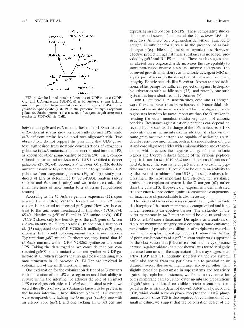
between the galE and galU mutants lies in their LPS structures.galE-deficient strains show an apparently normal LPS, whilegalU-deficient strains have altered core oligosaccharide. Twoobservations do not support the possibility that UDP-galac-tose, synthesized from nontoxic concentrations of exogenousgalactose in galE mutants, could be incorporated into the LPS,as known for other gram-negative bacteria (38). First, compo-sitional and structural analyses of O1 LPS have failed to detectgalactose (29, 30, 64). Second, a V. cholerae O1 galEK doublemutant, insensitive to galactose and unable to synthesize UDP-galactose from exogenous galactose (Fig. 6), apparently pro-duced wt LPS as determined by SDS-PAGE analysis (silverstaining and Western blotting) and was able to colonize thesmall intestines of mice similar to a wt strain (unpublishedresults).
According to the V. cholerae genome sequences (22), openreading frame (ORF) VC0262, located within the rfb genecluster, is annotated as a second galE gene. However, in con-trast to the galE gene investigated in this study (VCA0774;65.4% identity to galE of E. coli in 338 amino acids), ORFVC0262 shows only low homology to the galE gene of E. coli(26.6% identity in 169 amino acids). In addition, Fallarino etal. (15) suggested that ORF VC0262 is unlikely a galE gene,showing that it could not complement an S. enterica serovarTyphimurium galE mutant. Furthermore, they found that V.cholerae mutants within ORF VC0262 synthesize a normalLPS. Taking the data together, we conclude that our con-structed galEK double mutant could not synthesize UDP-ga-lactose at all, which suggests that no galactose-containing sur-face structures in V. cholerae O1 El Tor are involved incolonization of the small intestine in mice.
One explanation for the colonization defect of galU mutantsis that alteration of the LPS core region reduced their ability tosurvive within the intestine. To address the role of an intactLPS core oligosaccharide in V. cholerae intestinal survival, wetested the effects of several substances known to be present inthe human intestine. Three different types of LPS mutantswere compared: one lacking the O antigen (wbeW), one withan altered core (galU), and one lacking an O antigen and
expressing an altered core (R-LPS). These comparative studiesdemonstrated several functions of the V. cholerae LPS sub-structures. An intact core oligosaccharide, without attached Oantigen, is sufficient for survival in the presence of anionicdetergents (e.g., bile salts) and short organic acids. However,effective protection against these substances is no longer pro-vided by galU and R-LPS mutants. These results suggest thatan altered core oligosaccharide increases the susceptibility toshort protonated organic acids and anionic detergents. Theobserved growth inhibition seen in anionic detergent MIC as-says is probably due to the disruption of the inner membraneintegrity. Enteric bacteria like E. coli are known to need addi-tional efflux pumps for sufficient protection against hydropho-bic substances such as bile salts (73), and recently one suchsystem has been identified in V. cholerae (7).
Both V. cholerae LPS substructures, core and O antigen,were found to have roles in resistance to bactericidal sub-stances of the innate immune system. The core oligosaccharideregion was found to be more important than the O antigen inresisting the outer membrane-disturbing action of cationicpeptides. Resistance against cationic peptides can depend onseveral factors, such as the charge of the LPS molecules or LPSconcentration in the membrane. In addition, it is known thatsome gram-negative bacteria are capable of activating an in-ducible resistance mechanism, such as the modification of lipidA and core oligosaccharides with aminoarabinose and ethanol-amine, which reduces the negative charge of inner anionicregions and thereby reduces binding of the cationic peptides(14). It is not known if V. cholerae induces modifications oflipid A; hence, the sensitivity of galU mutants to cationic pep-tides such as polymyxin B could also be due to an inability tosynthesize aminoarabinose from UDP-glucose (see above). In-terestingly, the most important LPS structure for resistanceagainst the complement system is the O antigen (67) ratherthan the core LPS. However, our experiments demonstratedthat for effective protection against complement components,an intact core oligosaccharide is also necessary.
The results of the in vitro assays suggest that in galU mutantsthe integrity of the outer membrane is compromised and it nolonger represents an effective barrier. The instability of theouter membrane in galU mutants could be due to weakenedLPS core-LPS core interactions. Disruption or alterations ofthe outer membrane integrity could eventually cause enhancedpenetration of proteins and diffusion of periplasmic material,resulting in periplasmic leakage (47, 63). Evidence for the lossof periplasmic proteins of a galU mutant strain was supportedby the observation that b-lactamase, but not the cytoplasmicenzyme b-galactosidase (data not shown), was found in slightlyincreased amounts in the supernatant. This may suggest thatactive HAP and CT, normally secreted via the eps system,could also escape from the periplasm due to penetration ordiffusion across the outer membrane. However, other thanslightly increased b-lactamase in supernatants and sensitivityagainst hydrophobic substances, we found no evidence forouter membrane damage, since outer membrane preparationsof galU strains indicated no visible protein alterations com-pared to the wt strain (data not shown). Additionally, we foundTCP, which could serve as phage receptor for CTXF phagetransduction. Since TCP is also required for colonization of thesmall intestine, we suggest that the colonization defect of the
FIG. 6. Synthesis and possible functions of UDP-glucose (UDP-Glc) and UDP-galactose (UDP-Gal) in V. cholerae. Strains lackinggalE are predicted to accumulate the toxic products UDP-Gal andgalactose-1-phosphate (Gal-1P) in the presence of high exogenousgalactose. Strains grown in the absence of exogenous galactose mustsynthesize UDP-Gal via GalE.
442 NESPER ET AL. INFECT. IMMUN.
Dow
nloa
ded
from
http
s://j
ourn
als.
asm
.org
/jour
nal/i
ai o
n 17
Feb
ruar
y 20
22 b
y 12
1.14
3.17
3.21
6.
galU mutants is not due to alterations of TCP. However, itcould also be possible that the quantity or function of TCP isaltered in the mutant strain within the harsh environment ofthe small intestine. The secretion of virulence factors and theassembly of TCP in galU mutants remain to be addressed.
The spontaneous R-LPS mutants showed the most severedefect in colonization and survival in in vitro assays. The mu-tation in strain P27459res118 remains unidentified, but wespeculate that it is affected in the HepII-transferase (Fig. 1A),since it has been hypothesized that V. cholerae O1 heptoselessmutants are nonviable (60). The alteration in the core oligo-saccharide in this R-LPS mutant (res118) was found to have amore drastic effect on outer membrane integrity. It was ex-pected that this type of mutant could not colonize the smallintestine because it lacks the O antigen. The exact role of theLPS O antigen in colonization is unknown. An early reporthypothesized that a lack of the pilus TCP leads to attenuatedcolonization (26); however, recently it was reported that O-antigen-negative strains express TCP (5). We can also demon-strate that TCP function as the CTXF phage receptor is main-tained in the O-antigen-negative strains, which is consistentwith the finding of Chiang and Mekalanos (5). One knownphenotype of O-antigen mutants is a remarkable sensitivityagainst the complement system (67). Our data in this reportshowed that O-antigen-negative strains are in addition slightlymore sensitive to cationic peptides. It was recently reportedthat the Helicobacter pylori O antigen is involved in resistanceto inorganic pH stress, a condition found in the stomach (39).However, we found no evidence that V. cholerae O-antigen-negative strains (or the core mutants) are more sensitive toinorganic acids (data not shown).
We reported also that galU and galE are involved in theproduction of the VPS necessary for biofilm formation onabiotic surfaces. Biofilm formation is likely to be important inthe life cycle of pathogenic V. cholerae strains, since thesestrains survive and persist in aquatic ecosystems (8, 16). Com-monly, bacteria in aquatic environments are rarely found in theplanktonic or free-swimming phase but instead are organizedin a three-dimensional biofilm on aquatic surfaces. This mayprovide an advantage for the bacteria since they are betterprotected against bactericidal agents, and nutrients may beavailable (10). Recently, it was reported that V. cholerae O1 ElTor strains develop a three-dimensional biofilm on abioticsurfaces. Furthermore, it was shown that for accelerated at-tachment to abiotic surfaces, V. cholerae needs a functionalflagellum as well as the type IV mannose-sensitive hemagglu-tinin pili, and that the development of the three-dimensionalbiofilm requires production of the VPS (69). The regulation ofthis complex process is not fully understood.
Constitutive expression of VPS by V. cholerae O1 El Torleads to a rugose colony phenotype, and these cells are able toform a biofilm on abiotic surfaces (72), probably without theneed of the regulatory cascade necessary in wt bacteria. Wecould enrich for rugose strains by exposure of the motile,biofilm-negative O1 El Tor strain P27459 to the lytic phageK139.cm9. One nonmotile rugose isolate was further charac-terized, and our results suggested that production of the VPSis responsible for enhanced phage resistance, possibly becausethe phage cannot penetrate the VPS well enough to bind itsreceptor, the O antigen. The mutation(s) responsible for con-
stitutive VPS production and the underlying regulatory mech-anisms are only poorly understood. However, nonmotility ofstrain P27459res105 may be the cause of the rugose phenotype,because recent results suggest that the absence of a flagellumleads to constitutive VPS expression, at least in O139 strains(P. Watnick, C. M. Lauriano, K. E. Klose, L. Croal, and R.Kolter, submitted for publication). We discounted an alternatepossibility, namely, that the loss of HapR, the activator ofHAP, caused the rugose phenotype (28), since we could ob-serve HAP activity in this strain (data not shown).
Our data suggest that UDP-glucose is a precursor in thesynthesis of the VPS, since the introduction of a galU mutationinto a previously rugose strain led to a nonrugose phenotype,and this strain failed to form a biofilm. Also, the O1 El Torstrain CO970 with a galU mutation was shown to be biofilmnegative. The introduction of a mutation in galE into rugosestrains also renders them nonrugose, and they are unable toform biofilms on abiotic surfaces, suggesting that VPS synthe-sis requires UDP-galactose to be synthesized via UDP-glucosein the absence of exogenous galactose (Fig. 6). Thus, we iden-tified in this work two additional enzymes necessary for bio-synthesis of the VPS and the formation of V. cholerae biofilms.They are encoded by galU and galE and are required in addi-tion to those encoded by the vps (Vibrio polysaccharide syn-thesis) region (72).
ACKNOWLEDGMENTS
We thank J. Schmidt-Brauns for critical reading of the manuscript.For the clinical V. cholerae strain used in this study, we thank J. J.Mekalanos.
This work was funded by BMBF grant 01KI8906 and NIH grantAI43486 to K.E.K.
REFERENCES
1. Altschul, S. F., T. L. Madden, A. A. Schaffer, Z. J., Z. Zhang, W. Miller, andD. J. Lipman. 1997. Gapped BLAST and PSI-BLAST: a new generation ofprotein database search programs. Nucleic Acids Res. 25:3389–3402.
2. Andoh, A., Y. Fujiyama, H. Sakumoto, H. Uchihara, T. Kimura, S. Koyama,and T. Bamba. 1998. Detection of complement C3 and factor B gene ex-pression in normal colorectal mucosa, adenomas and carcinomas. Clin. Exp.Immunol. 111:477–483.
3. Blake, P. A. 1994. Historical perspectives on pandemic cholera, p. 293–295.In I. K. Wachsmuth, P. A. Blake, and O. R. Olsvik (ed.), Vibrio cholerae andcholera. American Society for Microbiology, Washington, D.C.
4. Bohringer, J., D. Fischer, G. Mosler, and R. Hengge-Aronis. 1995. UDP-glucose is a potential intracellular signal molecule in the control of expres-sion of ss-dependent genes in Escherichia coli. J. Bacteriol. 177:413–422.
5. Chiang, S. L., and J. J. Mekalanos. 1999. rfb mutations in Vibrio cholerae donot affect surface production of toxin-coregulated pili but still inhibit intes-tinal colonization. Infect. Immun. 67:976–980.
6. Chiang, S. L., and J. J. Mekalanos. 1998. Use of signature-tagged transposonmutagenesis to identify Vibrio cholerae genes critical for colonization. Mol.Microbiol. 27:797–805.
7. Colmer, J. A., J. A. Fralick, and A. N. Hamood. 1998. Isolation and charac-terization of a putative multidrug resistance pump from Vibrio cholerae. Mol.Microbiol. 27:63–72.
8. Colwell, R. R. 1996. Global climate and infectious disease: the choleraparadigm. Science 274:2025–2031.
9. Correa, N. E., C. M. Lauriano, R. McGee, and K. E. Klose. 2000. Phosphor-ylation of the flagellar regulatory protein FlrC is necessary for Vibrio choleraemotility and enhanced colonization. Mol. Microbiol. 35:743–755.
10. Costerton, J. W., Z. Lewandowski, D. E. Caldwell, D. R. Korber, and H. M.Lappin-Scott. 1995. Microbial biofilms. Annu. Rev. Microbiol. 49:711–745.
11. Csonka, L. N., and W. Epstein. 1996. Osmoregulation, p. 1210–1223. In F. C.Neidhardt, R. Curtiss III, J. L. Ingraham, E. C. C. Lin, K. B. Low, B.Magasanik, W. S. Reznikoff, M. Riley, M. Schaechter, and H. E. Umbarger(ed.), Escherichia coli and Salmonella: Cellular and molecular biology, 2nded. American Society for Microbiology, Washington, D.C.
12. Cummings, J. H., E. W. Pomare, W. J. Branch, C. P. E. Naylor, and G. T.Macfarlane. 1987. Short chain fatty acids in human large intestine, portal,
VOL. 69, 2001 V. CHOLERAE gal MUTANTS 443
Dow
nloa
ded
from
http
s://j
ourn
als.
asm
.org
/jour
nal/i
ai o
n 17
Feb
ruar
y 20
22 b
y 12
1.14
3.17
3.21
6.

hepatic and venous blood. Gut 28:1221–1227.13. Donnenberg, M. S., and J. B. Kaper. 1991. Construction of an eae deletion
mutant of enteropathogenic Escherichia coli by using a positive-selectionsuicide vector. Infect. Immun. 59:4310–4317.
14. Ernst, R. K., T. Guine, and S. I. Miller. 1999. How intracellular bacteriasurvive: surface modifications that promote resistance to host innate immuneresponses. J. Infect. Dis. 179(Suppl. 2):S326–S330.
15. Fallarino, A., C. Mavrangelos, U. H. Stroeher, and P. A. Manning. 1997.Identification of additional genes required for O-antigen biosynthesis inVibrio cholerae O1. J. Bacteriol. 179:2147–2153.
16. Faruque, S. M., M. J. Albert, and J. J. Mekalanos. 1998. Epidemiology,genetics, and ecology of toxigenic Vibrio cholerae. Microbiol. Mol. Biol. Rev.62:1301–1314.
17. Foster, J. W. 1999. When protons attack: microbial strategies of acid adap-tation. Curr. Opin. Microbiol. 2:170–174.
18. Fry, B. N., S. Feng, Y. Y. Chen, D. G. Newell, P. J. Coloe, and V. Korolik.2000. The galE gene of Campylobacter jejuni is involved in lipopolysaccharidesynthesis and virulence. Infect. Imm. 68:2594–2601.
19. Grimberg, J., S. Maguire, and L. Belluscio. 1989. A simple method for thepreparation of plasmid and chromosomal E. coli DNA. Nucleic Acids Res.17:8893.
20. Groisman, E. A. 1994. How bacteria resist killing by host-defense peptides.Trends Microbiol. 2:444–449.
21. Hase, C. C., and J. J. Mekalanos. 1998. TcpP protein is a positive regulatorof virulence gene expression in Vibrio cholerae. Proc. Natl. Acad. Sci. USA95:730–734.
22. Heidelberg, J. F., J. A. Eisen, W. C. Nelson, R. A. Clayton, M. L. Gwinn, R. J.Dodson, D. H. Haft, E. K. Hickey, J. D. Peterson, L. Umayam, S. R. Gill,K. E. Nelson, T. D. Read, H. Tettelin, D. Richardson, M. D. Ermolaeva, J.Vamathevan, S. Bass, H. Qin, I. Dragoi, P. Sellers, L. McDonald, T. Utter-back, R. D. Fleishmann, W. C. Nierman, O. White, S. L. Salzberg, H. O.Smith, R. R. Colwell, J. J. Mekalanos, J. C. Venter, and C. M. Fraser. 2000.DNA sequence of both chromosomes of the cholera pathogen Vibrio chol-erae. Nature 406:477–483.
23. Heinrichs, D. E., J. A. Yethon, and C. Whitfield. 1998. Molecular basis forstructural diversity in the core regions of the lipopolysaccharides of Esche-richia coli and Salmonella enterica. Mol. Microbiol. 30:221–232.
24. Hofmann, A. F. 1998. Bile secretion and the enterohepatic circulation of bileacids, p. 937–948. In M. Feldman, B. F. Scharschmidt, and M. H. Sleisenger(ed.), Gastrointestinal and liver disease. W. B. Saunders Co., Philadelphia,Pa.
25. Holmgren, J., and A. M. Svennerholm. 1977. Mechanisms of disease andimmunity in cholera: a review. J. Infect. Dis. 136(Suppl.):S105–S112.
26. Iredell, J. R., and P. A. Manning. 1997. Outer membrane translocation arrestof the TcpA pilin subunit in rfb mutants of Vibrio cholerae O1 strain 569B.J. Bacteriol. 179:2038–2046.
27. Iwanaga, M., K. Yamamoto, N. Higa, Y. Ichinose, N. Nakasone, and M.Tanabe. 1986. Culture conditions for stimulating cholera toxin production byVibrio cholerae O1 El Tor. Microbiol. Immunol. 30:1075–1083.
28. Jobling, M. G., and R. K. Holmes. 1997. Characterization of hapR, a positiveregulator of the Vibrio cholerae HA/protease gene hap, and its identificationas a functional homologue of the Vibrio harveyi luxR gene. Mol. Microbiol.26:1023–1034.
29. Kabir, S. 1982. Characterization of the lipopolysaccharide from Vibrio chol-erae O395 (Ogawa). Infect. Immun. 38:1263–1272.
30. Kenne, L., B. Lindberg, P. Unger, B. Gustafsson, and T. Holme. 1982.Structural studies of the Vibrio cholerae O-antigen. Carbohydr. Res. 100:341–349.
31. Klose, K. E., and J. J. Mekalanos. 1998. Differential regulation of multipleflagellins in Vibrio cholerae. J. Bacteriol. 180:303–316.
32. Klose, K. E., and J. J. Mekalanos. 1998. Distinct roles of an alternative sigmafactor during both free-swimming and colonizing phases of the Vibrio chol-erae pathogenic cycle. Mol. Microbiol. 28:501–520.
33. Knirel, Y. A., G. Widmalm, S. N. Senchenkova, P.-E. Jansson, and A. Wein-traub. 1997. Structural studies on the short-chain lipopolysaccharide ofVibrio cholerae O139 Bengal. Eur. J. Biochem. 247:402–410.
34. Laemmli, U. K. 1970. Cleavage of structural proteins during the assembly ofthe head of bacteriophage T4. Nature 227:680–685.
35. Larid, M. W., A. W. Kloser, and R. Misra. 1994. Assembly of LamB andOmpF in deep rough lipopolysaccharide mutants of Escherichia coli K-12.J. Bacteriol. 176:2259–2264.
36. Lin, E. C. C. 1996. Dissimilatory pathways for sugars, polyols, and carboxy-lates, p. 307–342. In F. C. Neidhardt, R. Curtiss III, J. L. Ingraham, E. C. C.Lin, K. B. Low, B. Magasanik, W. S. Reznikoff, M. Riley, M. Schaechter, andH. E. Umbarger (ed.), Escherichia coli and Salmonella: cellular and molec-ular biology, 2nd ed. American Society for Microbiology, Washington, D.C.
37. Mallow, E. B., A. Harris, N. Salzman, J. P. Russell, R. J. DeBerardinis, E.Ruchelli, and C. L. Bevins. 1996. Human enteric defensins. J. Biol. Chem.271:4038–4045.
38. Maskell, D. J., M. J. Szabo, M. E. Deadman, and E. R. Moxon. 1992. The gallocus from Haemophilus influenzae: cloning, sequencing and the use of galmutants to study lipopolysaccharide. Mol. Microbiol. 6:3051–3063.
39. McGowan, C. C., A. Necheva, S. A. Thompson, T. L. Cover, and M. J. Blaser.1998. Acid-induced expression of an LPS associated gene in Helicobacterpylori. Mol. Microbiol. 30:19–31.
40. Medrano, A. I., V. J. DiRita, G. Castillo, and J. Sanchez. 1999. Transienttranscriptional activation of the Vibrio cholerae El Tor virulence regulatorToxT in response to culture conditions. Infect. Immun. 67:2178–2183.
41. Merrell, S. D., and A. Camilli. 1999. The cadA gene of Vibrio cholerae isinduced during infection and plays a role in acid tolerance. Mol. Microbiol.34:836–849.
42. Miller, V. L., and J. J. Mekalanos. 1988. A novel suicide vector and its usein construction of insertion mutations: osmoregulation of outer membraneproteins and virulence determinants in Vibrio cholerae requires toxR. J. Bac-teriol. 170:2575–2583.
43. Morris, J. G., M. B. Sztein, E. W. Rice, J. P. Nataro, G. A. Losonsky, P.Panigrahi, C. O. Tacket, and J. A. Johnson. 1996. Vibrio cholerae O1 canassume a chlorine-resistant rugose survival form that is virulent for humans.J. Infect. Dis. 174:1364–1368.
44. Mullis, K. B., and F. Faloona. 1987. Specific synthesis of DNA in vitro via apolymerase chain reaction. Methods Enzymol. 155:335–340.
45. Nesper, J., J. Blab, M. Fountouloakis, and J. Reidl. 1999. Characterizationof the major control region of Vibrio cholerae bacteriophage K139: immunity,exclusion, and integration. J. Bacteriol. 181:2902–2913.
46. Nesper, J., D. Kapfhammer, K. E. Klose, H. Merkert, and J. Reidl. 2000.Characterization of Vibrio cholerae O1 antigen as bacteriophage K139 re-ceptor, and identification of IS1004 insertions aborting O1 antigen biosyn-thesis. J. Bacteriol. 182:5097–5104.
47. Nikaido, H. 1996. Outer membrane, p. 29–47. In F. C. Neidhardt, R. CurtissIII, J. L. Ingraham, E. C. C. Lin, K. B. Low, B. Magsanik, W. S. Reznikoff,M. Riley, M. Schaechter, and H. E. Umbarger (ed.), Escherichia coli andSalmonella: cellular and molecular biology, 2nd ed. American Society forMicrobiology, Washington, D.C.
48. Pearson, G. D. N., A. Woods, S. L. Chiang, and J. J. Mekalanos. 1993. CTXgenetic element encodes a site-specific recombination system and an intes-tinal colonization factor. Proc. Natl. Acad. Sci. USA 90:3750–3754.
49. Peterson, K. M., and J. J. Mekalanos. 1988. Characterization of the Vibriocholerae ToxR regulon: identification of novel genes involved in intestinalcolonization. Infect. Immun. 56:2822–2829.
50. Pratt, L. A., and R. Kolter. 1998. Genetic analysis of Escherichia coli biofilmformation: roles of flagella, motility, chemotaxis and type I pili. Mol. Micro-biol. 30:285–293.
51. Reidl, J., and J. J. Mekalanos. 1995. Characterization of Vibrio choleraebacteriophage K139 and use of a novel mini transposon to identify a phage-encoded virulence factor. Mol. Microbiol. 18:685–701.
52. Rose, R. E. 1988. The nucleotide sequence of pACYC177. Nucleic AcidsRes. 16:356.
53. Sandkvist, M., O. Michel, L. P. Hough, V. M. Morales, M. Bagdasarian, M.Koomey, V. J. DiRita, and M. Bagdasarian. 1997. General secretion pathway(eps) genes required for toxin secretion and outer membrane biogenesis inVibrio cholerae. J. Bacteriol. 179:6994–7003.
54. Silhavy, T. J., M. L. Berman, and L. W. Enquist. 1984. Experiments withgene fusions. Cold Spring Harbor Laboratory, Cold Spring Harbor, N.Y.
55. Skorupski, K., and R. K. Taylor. 1997. Control of the ToxR virulenceregulon in Vibrio cholerae by environmental stimuli. Mol. Microbiol. 25:1003–1009.
56. Southern, E. M. 1975. Detection of specific sequences among DNA frag-ments separated by gel electrophoresis. J. Mol. Biol. 51:503–517.
57. Steinberg, D. A., M. A. Hurst, C. A. Fujii, A. H. Kung, J. F. Ho, F.-C. Cheng,D. J. Loury, and J. C. Fiddes. 1997. Protegrin-1: a broad-spectrum, rapidlymicrobicidal peptide with in vivo activity. Antimicrob. Agents Chemother.41:1738–1742.
58. Stimson, E., M. Virji, K. Makepeace, A. Dell, H. R. Morris, G. Payne, J. R.Saunders, M. P. Jennings, S. Barker, M. Panico, et al. 1995. Meningococcalpilin: a glycoprotein substituted with digalactosyl 2,4-diacetamid-2,4,6-tride-oxyhexose. Mol. Microbiol. 17:1201–1214.
59. Stroeher, U. H., K. E. Jedani, and P. A. Manning. 1998. Genetic organizationof the regions associated with surface polysaccharide synthesis in Vibriocholerae O1, O139 and Vibrio anguillarum O1 and O2: a review. Gene 223:269–282.
60. Stroeher, U. H., L. E. Karageorgos, R. Morona, and P. A. Manning. 1995. InVibrio cholerae serogroup O1, rfaD is closely linked to the rfb operon. Gene155:67–72.
61. Svennerholm, A. M., and J. Holmgren. 1978. Identification of Escherichiacoli heat-labile enterotoxin by means of a ganglioside immunosorbant assay(GM1-ELISA) procedure. Curr. Microbiol. 1:19–23.
62. Taylor, R. K., V. L. Miller, D. B. Furlong, and J. J. Mekalanos. 1987. Use ofphoA gene fusions to identify a pilus colonization factor coordinately regu-lated with cholera toxin. Proc. Natl. Acad. Sci. USA 84:2833–2837.
63. Vaara, M. 1992. Agents that increase the permeability of the outer mem-brane. Microbiol. Rev. 56:395–411.
64. Vinogradov, E. V., K. Bock, O. Holst, and H. Brade. 1995. The structure ofthe lipid A-core region of the lipopolysaccharides from Vibrio cholerae O1
444 NESPER ET AL. INFECT. IMMUN.
Dow
nloa
ded
from
http
s://j
ourn
als.
asm
.org
/jour
nal/i
ai o
n 17
Feb
ruar
y 20
22 b
y 12
1.14
3.17
3.21
6.
smooth strain 569B (Inaba) and rough mutant strain 95R (Ogawa). Eur.J. Biochem. 233:152–158.
65. Wai, S. N., Y. Mizunoe, A. Takade, S.-I. Kawabata, and S.-I. Yoshida. 1998.Vibrio cholerae O1 strain TSI-4 produces the exopolysaccharide materialsthat determine colony morphology, stress resistance, and biofilm formation.Appl. Environ. Microbiol. 64:3648–3655.
66. Waldor, K. W., and J. J. Mekalanos. 1996. Lysogenic conversion by a fila-mentous phage encoding cholera toxin. Science 272:1910–1914.
67. Waldor, M. K., R. Colwell, and J. J. Mekalanos. 1994. The Vibrio choleraeO139 serogroup antigen includes an O-antigen capsule and lipopolysaccha-ride virulence determinants. Proc. Natl. Acad. Sci. USA 91:11388–11392.
68. Wandersman, C., and S. Letoffe. 1993. Involvement of lipopolysaccharide inthe secretion of Escherichia coli a-haemolysin and Erwinia chrysanthemiproteases. Mol. Microbiol. 7:141–150.
69. Watnick, P., and R. Kolter. 1999. Steps in the development of a Vibriocholerae El Tor biofilm. Mol. Microbiol. 34:586–595.
70. Watnick, P. I., K. J. Fullner, and R. Kolter. 1999. A role for the mannose-sensitive hemagglutinin in biofilm formation by Vibrio cholerae El Tor. J. Bac-teriol. 181:3606–3609.
71. Watson, N. 1988. A new revision of the sequence of plasmid pBR322. Gene70:399–403.
72. Yildiz, F. H., and G. K. Schoolnik. 1999. Vibrio cholerae O1 El Tor: identi-fication of a gene cluster required for the rugose colony type, exopolysac-charide production, chlorine resistance, and biofilm formation. Proc. Natl.Acad. Sci. USA 96:4028–4033.
73. Zgurskaya, H. I., and H. Nikaido. 1999. Bypassing the periplasm: reconsti-tution of the AcrAB multidrug efflux pump of Escherichia coli. Proc. Natl.Acad. Sci. USA 96:7190–7195.
Editor: V. J. DiRita
VOL. 69, 2001 V. CHOLERAE gal MUTANTS 445
Dow
nloa
ded
from
http
s://j
ourn
als.
asm
.org
/jour
nal/i
ai o
n 17
Feb
ruar
y 20
22 b
y 12
1.14
3.17
3.21
6.