characterizationofdisease-related5 -reductase(akr1d1 ... · ·...
TRANSCRIPT
Characterization of Disease-related 5�-Reductase (AKR1D1)Mutations Reveals Their Potential to Cause Bile AcidDeficiency*□S
Received for publication, March 26, 2010, and in revised form, June 2, 2010 Published, JBC Papers in Press, June 3, 2010, DOI 10.1074/jbc.M110.127779
Jason E. Drury‡1, Rebekka Mindnich‡1, and Trevor M. Penning‡§2
From the §Center of Excellence in Environmental Toxicology and ‡Department of Pharmacology, University of PennsylvaniaSchool of Medicine, Philadelphia, Pennsylvania 19104-6084
Bile acid deficiency is a serious syndrome in newborns thatcan result in death if untreated. 5�-Reductase deficiency is oneform of bile acid deficiency and is characterized by dramaticallydecreased levels of physiologically active 5�-reduced bile acids.AKR1D1 (aldo-keto reductase 1D1) is the only known humanenzyme that stereo-specifically reduces the �4 double bond in3-keto steroids and sterols to yield the 5�-hydrogenated prod-uct.Analysis of theAKR1D1 gene in five patientswith 5�-reduc-tase deficiency revealed five different mutations resulting in anamino acid substitution in the protein. To investigate a causalrole for these observedpointmutations inAKR1D1 in 5�-reduc-tase deficiency,we characterized their effect on enzymatic prop-erties. Attempts to purify mutant enzymes by overexpression inEscherichia coli only yielded sufficient amounts of the P133Rmutant for further characterization. This enzyme displayed ahighly reduced Km and Vmax reminiscent of uncompetitivekinetics with 4-cholesten-7�-ol-3-one as substrate. In addition,this mutant displayed no change in cofactor affinity but wasmore thermolabile in the absence of NADPH as judged by CDspectroscopy. All mutants were compared following expressionin HEK 293 cells. Although these enzymes were equallyexpressed based on mRNA levels, protein expression and func-tional activity were dramatically reduced. Cycloheximide treat-ment also revealed that several of the expressed mutants wereless stable. Our findings show that the reported mutations inAKR1D1 in patients with 5�-reductase lead to significantlydecreased levels of active enzyme and could be causal in thedevelopment of bile acid deficiency syndrome.
AKR1D1 (aldo-keto reductase 1D1) is implicated as one ofthe key enzymes in bile acid biosynthesis. The enzyme is theonly known human �4-3-ketosteroid 5�-reductase and cata-lyzes the reduction of the �4-3-ketosteroid to form the A/B cisring structure, utilizing NADPH as a cofactor (1). The �4-3-ketosteroid functionality is common to all steroid hormonesexcept the estrogens, and theC4-C5double bond can be further
reduced in a stereo-specificmanner. 5�-Reduction of testoster-one to 5�-dihydrotestosterone results in increased androgenreceptor activation (2, 3), whereas 5�-reduction of progester-one to 5�-pregnane-3,20-dione results in activation of thepregnane X receptor (4) and constitutive active/androstanereceptor (5). In bile acid biosynthesis, AKR1D1 reduces�4-cholesten-7�-ol-3-one and �4-cholesten-7�,12�-diol-3-one to their respective 5�-dihydrosteroid forms (6). The result-ing 5�-reduced structure contains a 90° bend in the steroidscaffold that is believed to generate the essential emulsificationcharacteristics of the resultant human bile acids.Human 5�-reductase deficiency (OMIM 604741) was first
diagnosed by Setchell et al. (7) in siblings with neonatal hepati-tis and cholestatis. Since then, more than 20 cases have beenreported, characterized by reduced primary bile acid biosyn-thesis and accumulation of hepatotoxic �4-3-oxo- and 5�-re-duced (allo-) bile acids (8–11). The deficiency can be treatedwith primary bile acids (12, 13), whichnormalize livermorphol-ogy and return liver function to normal. This treatment servestwo functions. First there is feedback repression of the 7�-hy-droxylase gene (CYP7A1), the key regulatory and rate-limitingstep in primary bile acid biosynthesis, and this prevents theaccumulation of the deleterious �4-3-one precursors and allo-bile acids (14). Second, the natural bile acids administeredlead to normal emulsification of fat and absorption of fat-soluble vitamins. Several point mutations in AKR1D1(L106F, P133R, P198L, G223E, and R261C) have beendetected in patients with bile acid deficiency; however, theeffects of the observed mutations on enzyme structure-func-tion and whether they are causal in the observed phenotypehave remained unclear (8, 10, 15).Recently, knowledge about AKR1D1 enzyme function
increased with the elucidation of its crystal structure in com-plexwith cofactor and different steroid substrates and products(16, 17). These studies revealed that AKR1D1 had an (�/�)8-barrel structure with three large loops (A, B, and C) at the backof the barrel and contained similar cofactor and steroid sub-strate binding sites compared with other AKR1C enzymes.These enzymes, AKR1C1–AKR1C4, act as 3-, 17-, and 20-ke-tosteroid reductases and have been thoroughly characterized(18–22). The amino acids of the aldo-keto reductase catalytictetrad, consisting of Asp50, Tyr55, Lys84, andHis117 (numberingaccording to rat AKR1C9 (3�-hydroxysteroid dehydrogenase))are highly conserved in AKR1D1. However, substitution of his-tidine by glutamatic acid translates into a functional switch
* This work was supported, in whole or in part, by National Institutes of HealthGrants R01-DK47015 and P30 ES013508 (to T. M. P.).
□S The on-line version of this article (available at http://www.jbc.org) containssupplemental Table S1 and Fig. S1.
1 Both authors contributed equally to this work.2 To whom all correspondence should be addressed: Dept. of Pharmacology,
University of Pennsylvania, 130C John Morgan Bldg., 3620 Hamilton Walk,Philadelphia, PA 19104-6084. Tel.: 215-898-9445; Fax: 215-573-2236;E-mail: [email protected].
THE JOURNAL OF BIOLOGICAL CHEMISTRY VOL. 285, NO. 32, pp. 24529 –24537, August 6, 2010© 2010 by The American Society for Biochemistry and Molecular Biology, Inc. Printed in the U.S.A.
AUGUST 6, 2010 • VOLUME 285 • NUMBER 32 JOURNAL OF BIOLOGICAL CHEMISTRY 24529
by guest on June 2, 2018http://w
ww
.jbc.org/D
ownloaded from
from ketosteroid reduction (in AKR1C enzymes) to doublebond reduction (in AKR1D1) (23).Examination of the AKR1D1 crystal structure allowed us to
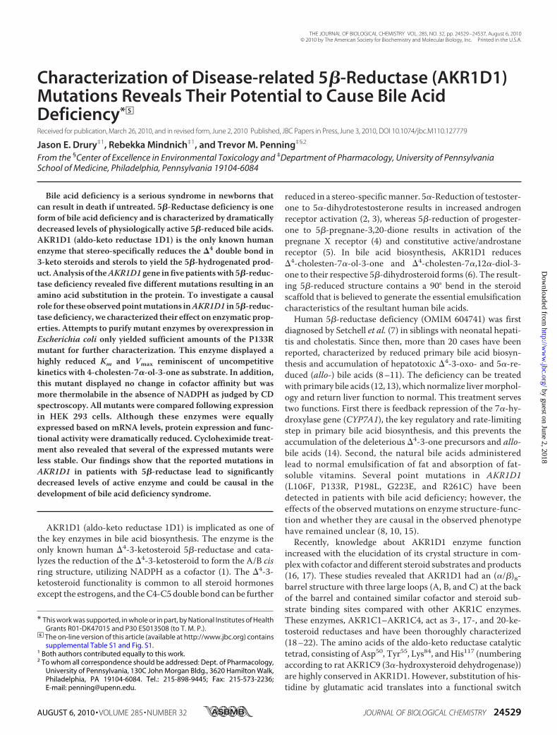
map the position of the reported point mutations associatedwith bile acid deficiency (Fig. 1A). We found that all mutationsexcept for P133R reside in areas highly conserved acrossAKR1D1 homologs in other mammalian species. However,none of the positions are in direct contact with the catalytictetrad, cofactor, or substrate binding sites, and therefore itremains unclear whether they could indeed be causal in thereported cases of bile acid deficiency.In this study, we examined the effects of the observed point
mutations on enzyme function following their introductioninto wild type AKR1D1.We report the expression, purification,and characterization of AKR1D1-P133R and compare it withthe wild type enzyme. Furthermore, although enzymes with thepointmutations L106F, P198L, G223E, and R261C could not bepurified, we were able to study these enzymes following expres-sion inmammalian cells. Our results indicate that all four pointmutations had severe effects on one or more of the following:steady state kinetic parameters, enzyme stability, and amountof soluble protein expressed in cells. In summary, althoughnone of the observed mutations affects catalysis in AKR1D1directly, all drastically reduced 5�-reductase activity in a bio-logical context. Hence, all AKR1D1 mutations reported inpatientswith bile acid deficiency and characterized in this studyhave the potential to cause the observed phenotype of 5�-re-ductase deficiency.
EXPERIMENTAL PROCEDURES
Materials—The vector pET-16b was purchased from Nova-gen. The GeneAmp RNA PCR Core kit was purchased fromPerkinElmer Life Sciences. Escherichia coli strain C41 (DE3)was provided by Dr. J. E. Walker (Medical Research CouncilLaboratory of Molecular Biology, Cambridge, UK). TheQuikChange II site-directed mutagenesis kit was purchasedfrom Stratagene. Restriction endonucleases were purchasedfrom New England Biolabs. Synthetic oligonucleotides wereobtained from Invitrogen. NADPH was obtained from RocheApplied Science. Steroids were purchased from Steraloids, Inc.[4-14C]Testosterone (50 mCi/mmol) was obtained fromPerkinElmer Life Sciences. Nickel-Sepharose 6 Fast Flow waspurchased from Amersham Biosciences. The bovine serumalbumin protein standard was purchased from Sigma. Bradfordreagent and Restore Western blot stripping buffer were pur-chased from Bio-Rad. Antibodies were obtained from GEHealthcare (ECL anti-mouse IgG from sheep), Sigma (anti-�-actin), or Santa Cruz Biotechnology, Inc. (Santa Cruz, CA)(horseradish peroxidase-conjugated anti-rabbit from mouse).All cell culture reagents except for fetal calf serum (ThermoScientific) were purchased from Invitrogen. HEK 293 cells wereobtained from ATCC. All other reagents were of ACS quality(i.e. they meet the specifications of the American ChemicalSociety) or higher.Construction of Expression Vectors—Previously, we reported
the expression of AKR1D1 using the prokaryotic expressionvectors pET16b and pET28a (16). For expression in HEK 293cells, the AKR1D1 coding sequence was subcloned into
pcDNA3.1 vector from a modified pET16b construct lackingthe His tag using the compatible XbaI and BamHI restrictionsites. Point mutations were subsequently introduced by site-directed mutagenesis using the QuikChange method and thefollowing forward and reverse primer pairs: L106F, 5�-dCCCTGG AGA GGA CAT TCA GGG TCC TCC AGC-3� and5�-dGCTGGAGGACCCTGAATGTCCTCTCCAGGG-3�;P133R, 5�-dGCC AGG AGA TGA AAT ATA CCG TAG AGATGAGAATGGC-3� and 5�-dGCCATTCTCATCTCTACGGTA TAT TTC ATC TCC TGG C-3�; P198L, 5�-dCCA GGTTGA GTG CCA TCT GTA TTT CAC CCA GCC-3� and5�-dGGC TGG GTG AAA TAC AGA TGG CAC TCA ACCTGG-3�; G223E, 5�-dGCA TAT AGC CCT TTG GAG ACCAGT AGG AAT CC-3� and 5�-dGGA TTC CTA CTG GTCTCC AAA GGG CTA TAT GC-3�; R261C, 5�-dGCA GCTCAA ATT GTT TTG TGT TTC AAC ATC CAG CGAGGG-3� and 5�-dCCC TCG CTG GAT GTT GAA ACA CAAAAC AAT TTG AGC TGC-3�. Mutated codons are indicatedby the underlined nucleotides. All constructs were verified bydideoxy sequencing.Expression and Purification of AKR1D1-P133R—Recombi-
nant AKR1D1-P133R was purified to homogeneity following aprotocol recently established for the wild type enzyme (16).AKR1D1-P133R was obtained in 16% yield and had a final spe-cific activity of 24 nmol of testosterone reducedmin�1 mg�1 ofpurified enzyme under standard radiometric assay conditionsas described recently (24). Homogeneity of theAKR1D1-P133Rmutant enzyme was checked by SDS-PAGE. Protein concen-trationwasmeasured using theBradford assay, according to themanufacturer’s instructions, using bovine serum albumin as astandard.Measurement of Steady State Kinetic Parameters by Spec-
trofluorimetric Assay—The kcat and Km values for testosteroneand cortisone were determined fluorimetrically as describedbefore (24) with steroid substrate concentration ranging from0.60 to 40 �M. Kinetic analyses of initial velocities obtainedwere performed by fitting the data using the programGraFit tothe Henri-Michaelis-Menten equation,
� � �Vmax � [S])�(Km � �S]) (Eq. 1)
where v is the initial velocity of the reaction, [S] is the molarconcentration of the substrate, andKm is theMichaelis-Mentenconstant for the substrate. Dividing Vmax by the molar concen-tration of the enzyme gave kcat. When substrate inhibition wasobserved, initial velocity data were fit in a similarmanner to thefollowing equation,
� � �Vmax � [S])�(Km � �S] � [S]2�Ki� (Eq. 2)
where variables were the same as in Equation 1 with the inclu-sion of Ki, which is the dissociation constant for substrate fromthe E�NADP� complex.Fluorescence Titration with NADPH—The dissociation con-
stant (Kd) of the enzyme for NADPH was determined by mon-itoring the quenching of the intrinsic protein fluorescenceupon the addition of cofactor as described (25) with the follow-ing changes. Titrations were performed in 10 mM potassiumphosphate (pH 6.0) in a 1.4-ml volume at 37 °C using 0.11 �M
Steroid 5�-Reductase Disease Mutants
24530 JOURNAL OF BIOLOGICAL CHEMISTRY VOLUME 285 • NUMBER 32 • AUGUST 6, 2010
by guest on June 2, 2018http://w
ww
.jbc.org/D
ownloaded from
AKR1D1-P133R. Emission spectraweremonitored from320 to500 nmon a fluorescence spectrophotometer F-4500 after exci-tation at 295 nm. The data were fitted to theMorrison equation(26) (using Equations 3 and 4) to compensate for the similarconcentration of enzyme and ligand in the titration and deter-mine the dissociation constant,
�E � NADPH] {(Kd � �E � [NADPH]) � ((Kd � �E
� �NADPH])2 � 4[E�NADPH])1/2}/2 (Eq. 3)
�E � NADPH]/[E � �F/�Fmax (Eq. 4)
where [E�NADPH] is the concentration of E�NADPH binarycomplex, [E] is the total concentration of enzyme, �F is thedifference in fluorescence in the presence and absence ofNADPH, and �Fmax is the maximum �F at the saturating con-centration of NADPH.Circular Dichroism Experiments—Circular dichroism spec-
tropolarimetry was performed with wild type (4.8 � 10�6 M)and AKR1D1-P133R (3.8 � 10�6 M) in 10 mM potassium phos-phate (pH 7.0). Protein spectra were recorded between a 260-and 190-nmwavelength at 22 °C inmillidegrees on a Jasco J810spectropolarimeter in a 0.1-cm cell. A background spectrumwas obtained with only buffer. Following the spectral scans,melt curves were performed on each of the samples at 222 nmby raising the temperature from 0 to 90 °C in increments of2 °C. A second protein spectrum was recorded for each sampleafter the melt curve was complete.Heat Stability Experiments—To determine the thermal sta-
bility of the enzymes, wild type and AKR1D1-P133R wereheated for 10 min at various increments from 25 to 50 °C in 10mM potassium phosphate (pH 7.0) and 1 mM EDTA. Sampleswere placed on ice, centrifuged for 1min to remove any precip-itate, and then assayed fluorimetrically.Cell Culture and Transient Transfection—HEK 293 cells
were maintained in 10-cm culture dishes at 37 °C and 5% CO2inDulbecco’smodified Eagle’smedium containingGlutaMAXTM
and supplemented with 10% heat-inactivated fetal calf serumand 1% penicillin, streptomycin. For experiments, 1 millioncells/well were seeded into 6-well plates and the followingday were transfected with either 1.5 �g of the respectiveAKR1D1 construct or empty vector per well using FuGene 6according to the manufacturer’s protocol. Forty-eight hourslater, cells were either harvested for RNA expression andanalysis of enzyme activity or subjected to cycloheximidetreatment.Cycloheximide Treatment—Cells were seeded and tran-
siently transfected as described above. Forty-eight hours fol-lowing transfection, cells were harvested by scraping (0 h timepoint), or cycloheximide (dissolved in water and sterile filtered)was added to the medium to a final concentration of 20 �g/ml,and cells were harvested 0, 6, and 24 h following treatment.Reverse Transcription-PCR—RNA was extracted from cells
using the RNeasy minikit from Qiagen according to the manu-facturer’s protocol. To obtain cDNA, 1 �g of RNA was reversetranscribed with random hexamer primers by use of theGeneAmp RNA PCR core kit (Applied Biosystems). Quality ofthe cDNA was monitored by PCR amplification of GAPDH
(primers 5�-dCAT CTC TGC CCC CTC TGC TGA-3� and5�-dGGA TGA CCT TGC CCA CAG CCT-3�). EndogenousAKR1D1 was amplified with a forward primer matching asequence in exon 7 (5�-dGGG GTG GTT GTC ATT CCTAA-3�) and a reverse primer matching a sequence in the 3�-un-translated region of the gene (5�-dGAC TAC CCA TTG CACCGT CT-3�). For exogenous AKR1D1 detection, the same for-ward primer was employed together with a reverse primer thatanneals in the cloning vector 3� of the insert (5�-dAAC TAGAAG GCA CAG TCG AG-3�).Polyclonal AKR1D1 Antibody—A rabbit polyclonal anti-
serum raised against full-length purified His-tagged AKR1D1was produced by ProSci, Inc. (Poway, CA). Antiserum prepara-tion from the first bleed showed the lowest cross-reactivity forthe detection of other human aldo-keto reductases and wassubjected to further purification to improve specificity againstAKR1D1. For this, 50–150�g of humanAKR1A1 andAKR1C1to AKR1C4 were blotted onto a strip of nitrocellulose mem-brane. Thismembranewas incubatedwith a 1:50 dilution of theantiserum in Tris-buffered saline (TBS)3 containing 0.1%Tween 20 (TBST) for 1 h at 4 °C. Diluted antiserum wasremoved, the membrane was stripped for 20 min at 42 °C andwashed three times 10min eachwithTBS at room temperature,and the diluted antiserum from before was reapplied. Thiscycle was repeated seven times, and the precleaned poly-clonal �-AKR1D1 antibodywas diluted 1:50withTBS and thenmixed 1:1 with glycerol and 0.02% sodium azide (final concen-tration) before aliquoting and storage at �20 °C. Enhancedspecificity of this antibody forAKR1D1 versus a panel of humanAKRs is documented in supplemental Fig. S1.Western Blot—Cell pellets were washed once in Dulbecco’s
phosphate-buffered saline and then lysed by incubation in lysisbuffer (20mMTris-HCl, pH 8.0, 150mMNaCl, 10% glycerol, 1%Nonidet P-40, 5 mM EDTA, 0.5 mM EGTA, 20 mM �-glycero-phosphate, 100 �M sodium orthovanadate, 1� proteinaseinhibitor mix (Roche Applied Science)) on ice for 30 min withintermittent agitation. Lysates were centrifuged, the solublefraction was removed, and the protein concentration was mea-sured by a Bradford assay. Eighty micrograms of total proteinper sample well were resolved by SDS-PAGE and blotted onto anitrocellulose membrane. For Western blot development, themembrane was blocked in 5% dry milk in TBST, the polyclonalanti-AKR1D1 antibody was diluted to 1:2000 in 3% dry milk inTBST, the secondary antibody (monoclonal mouse anti-rabbithorseradish peroxidase conjugate, Santa Cruz Biotechnology,Inc.) was diluted to 1:10,000 in 3% dry milk in TBST, and thesignals were visualized using the ECL technique (GE Health-care). To detect the mutants that were poorly expressed, thesecondary antibody was diluted 1:100,000 in 3% dry milk inTBST, and the signals were visualized using the SuperSignalWest Femto Kit (Thermo Scientific). For the subsequent detec-tion of �-actin, blots were stripped by incubating twice for 10min in 42 °C Western stripping buffer (Bio-Rad) and washingthree times for 10 min each in TBST, and the procedure wasthen completed as described above. The primary antibody
3 The abbreviations used are: TBS, Tris-buffered saline; WT, wild type.
Steroid 5�-Reductase Disease Mutants
AUGUST 6, 2010 • VOLUME 285 • NUMBER 32 JOURNAL OF BIOLOGICAL CHEMISTRY 24531
by guest on June 2, 2018http://w
ww
.jbc.org/D
ownloaded from
monoclonal mouse anti-�-actin was applied in a 1:1000 dilu-tion in 3% dry milk in TBST, and the secondary antibody sheepanti-mouse horseradish peroxidase conjugate was diluted1:5000 in 3% dry milk in TBST. Detection was achieved by useof the ECL technique (GE Healthcare).Detection of 5�-Reductase Activity in HEK 293 Cells—Cells
were seeded and transiently transfected as described above. At46 h after transfection, medium was replaced with 2 ml of Dul-becco’s modified Eagle’s medium containing 1% heat-inacti-vated fetal calf serum, GlutaMAXTM, and 1% penicillin, strep-tomycin perwell. Two hours later, testosteronewas added in 10�l of DMSO to yield final concentrations ranging from 0.5 to 20�M per well. Final concentrations below 10 �M contained 20nCi, and higher final concentrations contained 30 nCi of 14C-labeled testosterone, respectively. At time points, 200 �l ofmedium was removed and extracted twice with 2.5 volumes ofcold water-saturated ethyl acetate, and the combined organicphase was dried down. Recovery of radioactive material follow-ing extraction was �90%.Product Identification by Thin Layer Radiochromatography—
Dried samples were resuspended in ethyl acetate, applied toPartisil LK6D Silica TLC (thin layer chromatography) plates(Whatman International Ltd.), and developed twice in toluene/acetone (80:20 v/v). Radiochromatograms were scanned withan automatic TLC-linear analyzer (Bioscan Imaging ScannerSystem 200-IBM with AutoChanger 3000, Bioscan (Washing-ton, D. C.)), and the relative percentage of peaks was comparedand quantified as nmol of product formed using the specificradioactivity of the isotope assuming that each steroid wasrecovered with the same efficiency. Products were identified byco-migration with authentic standards applied to the sameplate following visualization by spraying with acetic acid/sulfu-ric acid/anisaldehyde (100:2:1, v/v/v) and heat.
RESULTS
Expression and Purification of Recombinant Disease-relatedAKR1D1 Mutants—To examine whether the disease-relatedAKR1D1mutants could account for bile acid deficiency, we setout to purify the L106F, P133R, P198L, andR261C enzymes andcompare their properties with wild type AKR1D1. Based onSDS-polyacrylamide gels, all four mutants could be clearlydetected in bacterial sonicates following overnight induction ofthe expression of the His-tagged proteins in E. coli (data notshown). However, the majority of the expressed mutants accu-mulated in inclusion bodies. Attempts to purify the disease-related mutants resulted in extremely low yields, and only theAKR1D1-P133R mutant could be purified in milligramamounts to homogeneity for further analysis (supplementalTable S1). Subsequent to this portion of the study, theAKR1D1-G223E mutant was described (15), and this mutantwas compared in mammalian transfection studies with theother disease-based mutants (see below).Comparison of Kinetic Constants between Wild Type and
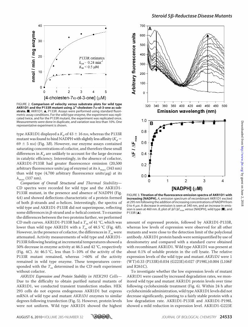
AKR1D1-P133R—The reported P133R mutation is present inloopA of the enzyme and is not directly involved in forming theprotein scaffold or the catalytic tetrad (16, 17) (Fig. 1A). Wedetermined steady state kinetic constants for the wild type andAKR1D1-P133R mutant. The specific activity of the homoge-
nous recombinantmutant enzymewith 10�M testosteronewas24 nmol min�1 mg�1 and was 3-fold lower than wild typeenzyme. The catalytic efficiency toward testosterone (0.21min�1 �M�1) was found to be 12-fold less as reflected by anincrease in Km (12.7 �M) and a decrease in kcat (2.7 min�1)(Table 1). Interestingly, the substrate inhibition of wild typeenzyme by testosterone was not observed with the P133Rmutant. By contrast, �4-3-ketosteroids with longer C17 sidechains (e.g. cortisone and �4-cholesten-7�-ol-3-one) showedlarge decreases in kcat and Km. The kcat of cortisone wasdecreased 16-fold (0.6 min�1), and Km was decreased 12-fold(1.3 �M). The same pattern was observed with �4-cholesten-7�-ol-3-one; however, it was not possible to accuratelymeasurethe Km for this substrate because the enzyme was essentiallysaturated at the lowest concentrations of substrate at which areaction rate could be reliably measured (Fig. 2). At saturation,kcat for �4-cholesten-7�-ol-3-one was depressed 7-fold. Bycontrast, Km is significantly lower than 0.8 �M, the value forwild type enzyme. Thus, the effect of this mutation was achange from a low affinity, high capacity enzyme to a high affin-ity, low capacity enzyme.NADPH Binding and Kd Determination—We investigated
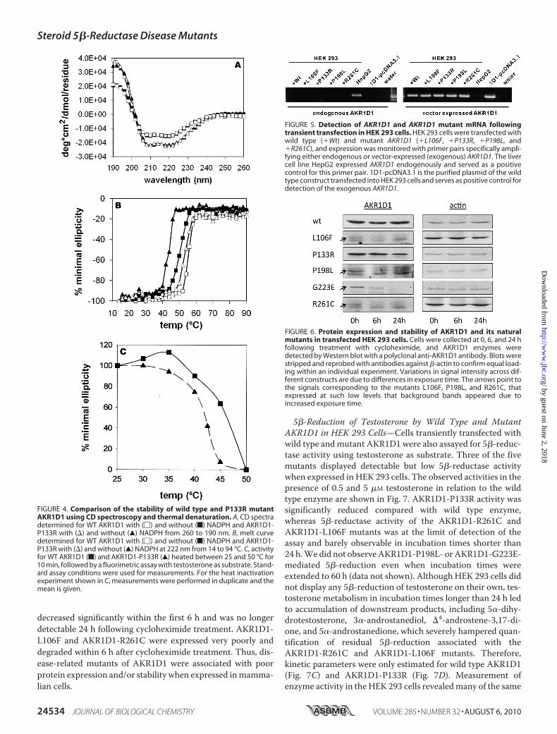
whether the P133Rmutation affects cofactor binding. AKR1D1contains 5 tryptophan residues and has intrinsic fluorescencewhen excited at 295 nm. Incremental addition of NADPHquenched the fluorescence emission signal at 340 nm and gen-erated an energy transfer band at 460 nm (Fig. 3A). This energytransfer band probably results from the interaction of the nic-otinamide ring with Trp89 based on identical experiments withAKR1C2 and structural homology considerations (27). Plots of�F/�Fmax versus [NADPH] were fitted to the Morrison equa-tion to obtain the dissociation constant (Kd) of NADPH. Wild
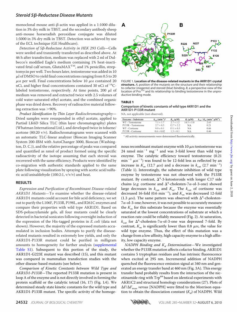
FIGURE 1. Location of the disease-related mutants in the AKR1D1 crystalstructure. A, position of the mutants on the structure and their relationshipto cofactor (magenta) and steroid (blue) binding. B, a perspective view of thelocation of Pro133 and its relationship to binding testosterone in the unpro-ductive binding mode.
TABLE 1Comparison of kinetic constants of wild type AKR1D1 and theAKR1D1-P133R mutantNA, not applicable (not observed).
Enzyme - Substrate kcat (min-1)* Km (μM) Ki (μM) kcat / Km (min-1 μM-1) WT - Testosterone 7.1 ± 1.7 2.7 ± 1.2 14.5 ± 5.8 2.63 P133R - Testosterone 2.7 ± 0.3 12.7 ± 2.2 NA 0.21 WT - Cortisone 9.9 ± 0.1 15.1 ± 0.3 NA 0.66 P133R - Cortisone 0.6 ± 0.02 1.3 ± 0.1 NA 0.46
* All activity measurements were determined fluorimetrically.
Steroid 5�-Reductase Disease Mutants
24532 JOURNAL OF BIOLOGICAL CHEMISTRY VOLUME 285 • NUMBER 32 • AUGUST 6, 2010
by guest on June 2, 2018http://w
ww
.jbc.org/D
ownloaded from
type AKR1D1 displayed aKd of 43 � 16 nM, whereas the P133Rmutantwas found tobindNADPHwith slightly less affinity (Kd69 � 5 nM) (Fig. 3B). However, our enzyme assays containedsaturating concentrations of cofactor, and therefore these smalldifferences in Kd are unlikely to account for the large decreasein catalytic efficiency. Interestingly, in the absence of cofactor,AKR1D1-P133R had greater fluorescence emission (20,500arbitrary fluorescence units/�g of enzyme) at its �max (343 nm)than wild type (4,700 arbitrary fluorescence units/�g) at its�max (337 nm).Comparison of Overall Structural and Thermal Stability—
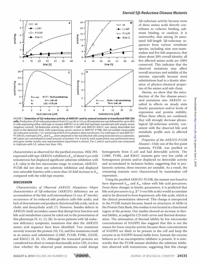
CD spectra were recorded for wild type and the AKR1D1-P133R mutant, in the presence and absence of NADPH (Fig.4A) and showed deflections characteristic of a protein formedof both �-strands and �-helices. Interestingly, the spectra ofwild type and AKR1D1-P133R did not superimpose, reflectingsomedifferences in�-strand and�-helical content. To examinethe differences between the two proteins further, we performedCDmelt curves. AKR1D1-P133R had a Tm of 41 °C, which waslower than wild type AKR1D1 with a Tm of 48.5 °C (Fig. 4B).However, in the presence of cofactor, the differences inTmwereattenuated. Activity measurements of wild type and AKR1D1-P133R following heating at incremental temperatures showed a50% decrease in enzyme activity at 46.5 and 42 °C, respectively(Fig. 4C). At 46.5 °C, less than 5–10% of the activity of theP133R mutant remained, whereas �60% of the activityremained in wild type enzyme. These temperatures corre-sponded with the Tm determined in the CD melt experimentwithout cofactor.AKR1D1 Expression and Protein Stability in HEK293 Cells—
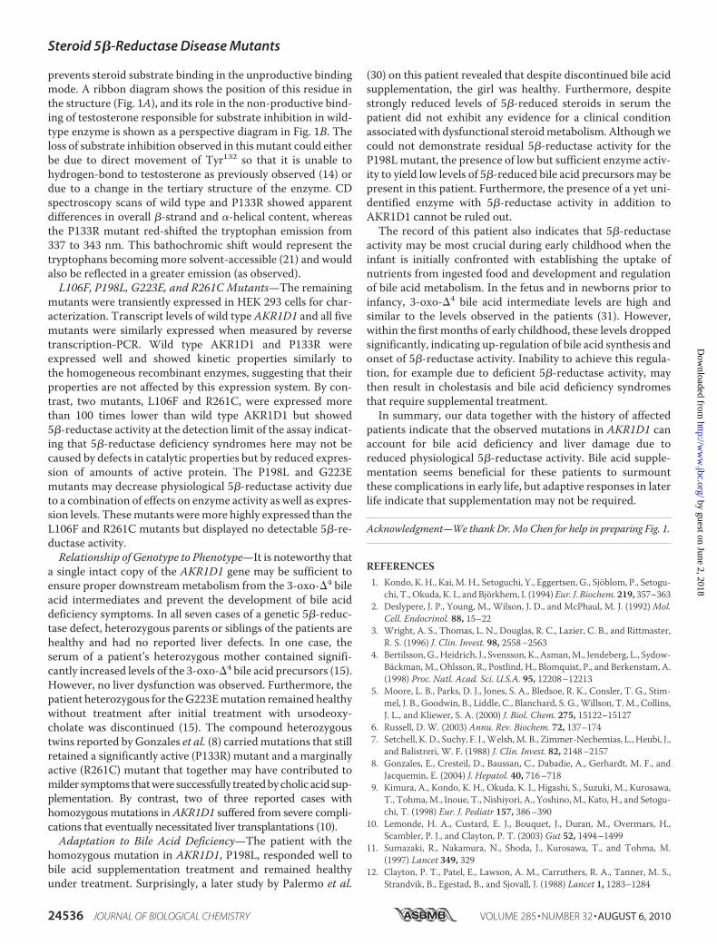
Due to the difficulty to obtain purified natural mutants ofAKR1D1, we conducted transient transfection studies. HEK293 cells do not express endogenous AKR1D1 but expressmRNA of wild type and mutant AKR1D1 enzymes to similardegrees following transfection (Fig. 5). However, protein levelswere not uniform. Wild type AKR1D1 showed the highest
amount of expressed protein, followed by AKR1D1-P133R,whereas low levels of expression were observed for all othermutants and were close to the detection limit of the polyclonalantibody. AKR1D1protein bandswere semiquantified by use ofdensitometry and compared with a standard curve obtainedwith recombinant AKR1D1.Wild type AKR1D1 was present atabout 0.1% of soluble protein in the cell lysate. The relativeexpression levels of the wild type and mutant AKR1D1 were 1(WT):0.33 (P133R):0.034 (G223E):0.027 (P198L):0.004 (L106Fand R261C).To investigate whether the low expression levels of mutant
AKR1D1 were caused by increased degradation rates, we mon-itored wild type and mutant AKR1D1 protein levels over timefollowing cycloheximide treatment (Fig. 6). Within 24 h aftercycloheximide administration,wild typeAKR1D1 levels did notdecrease significantly, pointing to a fairly stable protein with alow degradation rate. AKR1D1-P133R and AKR1D1-P198Lshowed a mild reduction in expression level. AKR1D1-G223E
FIGURE 2. Comparison of velocity versus substrate plots for wild typeAKR1D1 and the P133R mutant using �4-cholesten-7�-ol-3-one as sub-strate. f, AKR1D1; Œ, P133R. Assays were performed using standard fluori-metric assay conditions. For the wild type enzyme, the experiment was repli-cated twice, and for the P133R mutant, the experiment was replicated once.Measurements were done in duplicate, and variation was less than 10%. Onerepresentative experiment is shown.
FIGURE 3. Titration of the fluorescence emission spectra of AKR1D1 withincreasing [NADPH]. A, emission spectrum of recombinant AKR1D1 excitedat 295 nm following the addition of increasing concentrations of NADPH from0 to 4 �M. A decrease in emission is seen at 340 nm, and an increase in emis-sion is seen at 460 nm. B, plot of �F/�Fmax versus [NADPH], wild type (f), andP133R (Œ).
Steroid 5�-Reductase Disease Mutants
AUGUST 6, 2010 • VOLUME 285 • NUMBER 32 JOURNAL OF BIOLOGICAL CHEMISTRY 24533
by guest on June 2, 2018http://w
ww
.jbc.org/D
ownloaded from
decreased significantly within the first 6 h and was no longerdetectable 24 h following cycloheximide treatment. AKR1D1-L106F and AKR1D1-R261C were expressed very poorly anddegraded within 6 h after cycloheximide treatment. Thus, dis-ease-related mutants of AKR1D1 were associated with poorprotein expression and/or stability when expressed inmamma-lian cells.
5�-Reduction of Testosterone by Wild Type and MutantAKR1D1 in HEK 293 Cells—Cells transiently transfected withwild type andmutant AKR1D1 were also assayed for 5�-reduc-tase activity using testosterone as substrate. Three of the fivemutants displayed detectable but low 5�-reductase activitywhen expressed inHEK 293 cells. The observed activities in thepresence of 0.5 and 5 �M testosterone in relation to the wildtype enzyme are shown in Fig. 7. AKR1D1-P133R activity wassignificantly reduced compared with wild type enzyme,whereas 5�-reductase activity of the AKR1D1-R261C andAKR1D1-L106F mutants was at the limit of detection of theassay and barely observable in incubation times shorter than24 h.Wedid not observeAKR1D1-P198L- orAKR1D1-G223E-mediated 5�-reduction even when incubation times wereextended to 60 h (data not shown). Although HEK 293 cells didnot display any 5�-reduction of testosterone on their own, tes-tosterone metabolism in incubation times longer than 24 h ledto accumulation of downstream products, including 5�-dihy-drotestosterone, 3�-androstanediol, �4-androstene-3,17-di-one, and 5�-androstanedione, which severely hampered quan-tification of residual 5�-reduction associated with theAKR1D1-R261C and AKR1D1-L106F mutants. Therefore,kinetic parameters were only estimated for wild type AKR1D1(Fig. 7C) and AKR1D1-P133R (Fig. 7D). Measurement ofenzyme activity in the HEK 293 cells revealedmany of the same
FIGURE 4. Comparison of the stability of wild type and P133R mutantAKR1D1 using CD spectroscopy and thermal denaturation. A, CD spectradetermined for WT AKR1D1 with (�) and without (f) NADPH and AKR1D1-P133R with (�) and without (Œ) NADPH from 260 to 190 nm. B, melt curvedetermined for WT AKR1D1 with (�) and without (f) NADPH and AKR1D1-P133R with (�) and without (Œ) NADPH at 222 nm from 14 to 94 °C. C, activityfor WT AKR1D1 (f) and AKR1D1-P133R (Œ) heated between 25 and 50 °C for10 min, followed by a fluorimetric assay with testosterone as substrate. Stand-ard assay conditions were used for measurements. For the heat inactivationexperiment shown in C, measurements were performed in duplicate and themean is given.
FIGURE 5. Detection of AKR1D1 and AKR1D1 mutant mRNA followingtransient transfection in HEK 293 cells. HEK 293 cells were transfected withwild type (�Wt) and mutant AKR1D1 (�L106F, �P133R, �P198L, and�R261C), and expression was monitored with primer pairs specifically ampli-fying either endogenous or vector-expressed (exogenous) AKR1D1. The livercell line HepG2 expressed AKR1D1 endogenously and served as a positivecontrol for this primer pair. 1D1-pcDNA3.1 is the purified plasmid of the wildtype construct transfected into HEK 293 cells and serves as positive control fordetection of the exogenous AKR1D1.
FIGURE 6. Protein expression and stability of AKR1D1 and its naturalmutants in transfected HEK 293 cells. Cells were collected at 0, 6, and 24 hfollowing treatment with cycloheximide, and AKR1D1 enzymes weredetected by Western blot with a polyclonal anti-AKR1D1 antibody. Blots werestripped and reprobed with antibodies against �-actin to confirm equal load-ing within an individual experiment. Variations in signal intensity across dif-ferent constructs are due to differences in exposure time. The arrows point tothe signals corresponding to the mutants L106F, P198L, and R261C, thatexpressed at such low levels that background bands appeared due toincreased exposure time.
Steroid 5�-Reductase Disease Mutants
24534 JOURNAL OF BIOLOGICAL CHEMISTRY VOLUME 285 • NUMBER 32 • AUGUST 6, 2010
by guest on June 2, 2018http://w
ww
.jbc.org/D
ownloaded from
characteristics as observed for the purified enzymes. HEK 293-expressedwild typeAKR1D1 exhibited aKm of about 4�Mwithtestosterone but displayed significant substrate inhibition witha Ki value in the low micromolar range. In contrast, AKR1D1-P133R did not show any substrate inhibition and displayednon-saturable kinetics with a more than 10-fold increase in Kmcompared with the wild type enzyme.
DISCUSSION
Characteristics of Observed AKR1D1 Mutations—Majorcharacteristics of 5�-reductase (AKR1D1) deficiency are anaccumulation of the bile acid intermediates (3-oxo-�4 sterols),occurrence of 5�-reduced side products (allo-bile acids), andlack of downstream end products (functional bile acids, such ascholic and deoxycholic acid) (7). However, besides defects inAKR1D1 itself, secondary causes that disrupt liver function andbile acid metabolism cannot be ruled out in the presentation ofthis phenotype (9, 11, 12, 28). In seven patients with 5�-reduc-tase deficiency symptoms, mutations that alter the AKR1D1amino acid sequence have been identified. Two mutationsseverely truncate the protein (10, 15), and five mutations resultin an amino acid substitution (L106F, P133R, P198L, G223E,and R261C) (8, 10, 15). Although the truncated proteins areconsidered too short to remain functionally active (29), it is lessclear whether the observed point mutations could disrupt
5�-reductase activity because noneof these amino acids directly con-tributes to cofactor binding, sub-strate binding, or catalysis. It isnoteworthy, that among 24 anno-tated full-length 5�-reductase se-quences from various vertebratespecies, including nine non-mam-malian and five fish sequences, thatshare about 30% overall identity, allfive affected amino acids are 100%conserved. This indicates that theobserved mutations may affectoverall structure and stability of theenzyme, especially because mostsubstitutions lead to a drastic alter-ation of physico-chemical proper-ties of the amino acid side chain.Herein, we show that the intro-
duction of the five disease-associ-ated mutations into AKR1D1 re-sulted in effects on steady statekinetic parameters and/or levels ofexpression and protein stability.When these effects are combined,they will strongly decrease physio-logical 5�-reductase activity, con-sistent with the observed bile acidmetabolic profile seen in affectedpatients.Properties of the AKR1D1-P133R
Mutant—Only one of the five pointmutants, P133R, was purified to
homogeneity from E. coli and had detectable activity. TheL106F, P198L, and R261C mutants gave very low yields ofhomogenous protein and/or displayed no detectable activityand accumulated in inclusion bodies, suggesting that in pro-karyotic systems, these enzymes are unstable. As a result, theremaining mutants were characterized by mammalian cellexpression.Using the purified AKR1D1-P133R, themutant was found to
have depressed kcat and Km values with bile acid precursors.From these changes in kinetic parameters, it is predicted thatbile acid precursors (e.g.�4-3-oxo bile acids) would accumulateand/or be directed to form hepatotoxic allo-bile acids, which isthe clinical presentation observed. This change is unexpectedin the P133R mutant because, based on structures of AKRs inthe ProteinData Bank, this residue is not located in a functionalregion of the protein. Our studies showed an increase in ther-mal lability, as judged by CDmelt curves and thermal denatur-ation. The attenuation of thermal lability by low micromolarconcentrations of NADPH also suggests that this is not thereason for lower enzyme activity because these concentrationsof NADPH are likely to be present in the cell and keep theenzyme in its NADPH-bound stable form. Instead, the enzymebehaves as if an uncompetitive inhibitor is present. It is note-worthy that the P133R mutant abolishes the substrate inhibi-tion observed with testosterone, suggesting that this change
FIGURE 7. Detection of 5�-reductase activity of AKR1D1 and its natural mutants in transfected HEK 293cells. Production of 5�-reduced products from 0.5 �M (A) or 5.0 �M (B) testosterone was followed for up to 48 hin cells expressing either wild type or mutant AKR1D1 or in cells that had been transfected with empty vector(negative control). 5�-Reductase activity for AKR1D1-L106F and AKR1D1-R261C was clearly detectable butclose to the detection limit. Cells expressing vector control or AKR1D1-P198L did not exhibit measurable5�-reduction activity ( 0.1 pmol/mg at 60 h of incubation; data not shown). For wild type (C) and AKR1D1-P133R (D), estimates of Km and Vmax were obtained in the transfected cells using testosterone as substrate.All values are normalized to total amount of protein. For A and B, each experiment was performed twiceand gave similar results; one representative experiment is shown. For C and D, each point was measuredin triplicate with S.E. values less than 10%.
Steroid 5�-Reductase Disease Mutants
AUGUST 6, 2010 • VOLUME 285 • NUMBER 32 JOURNAL OF BIOLOGICAL CHEMISTRY 24535
by guest on June 2, 2018http://w
ww
.jbc.org/D
ownloaded from
prevents steroid substrate binding in the unproductive bindingmode. A ribbon diagram shows the position of this residue inthe structure (Fig. 1A), and its role in the non-productive bind-ing of testosterone responsible for substrate inhibition in wild-type enzyme is shown as a perspective diagram in Fig. 1B. Theloss of substrate inhibition observed in thismutant could eitherbe due to direct movement of Tyr132 so that it is unable tohydrogen-bond to testosterone as previously observed (14) ordue to a change in the tertiary structure of the enzyme. CDspectroscopy scans of wild type and P133R showed apparentdifferences in overall �-strand and �-helical content, whereasthe P133R mutant red-shifted the tryptophan emission from337 to 343 nm. This bathochromic shift would represent thetryptophans becoming more solvent-accessible (21) and wouldalso be reflected in a greater emission (as observed).L106F, P198L, G223E, and R261CMutants—The remaining
mutants were transiently expressed in HEK 293 cells for char-acterization. Transcript levels of wild type AKR1D1 and all fivemutants were similarly expressed when measured by reversetranscription-PCR. Wild type AKR1D1 and P133R wereexpressed well and showed kinetic properties similarly tothe homogeneous recombinant enzymes, suggesting that theirproperties are not affected by this expression system. By con-trast, two mutants, L106F and R261C, were expressed morethan 100 times lower than wild type AKR1D1 but showed5�-reductase activity at the detection limit of the assay indicat-ing that 5�-reductase deficiency syndromes here may not becaused by defects in catalytic properties but by reduced expres-sion of amounts of active protein. The P198L and G223Emutants may decrease physiological 5�-reductase activity dueto a combination of effects on enzyme activity as well as expres-sion levels. Thesemutants weremore highly expressed than theL106F and R261C mutants but displayed no detectable 5�-re-ductase activity.Relationship of Genotype to Phenotype—It is noteworthy that
a single intact copy of the AKR1D1 gene may be sufficient toensure proper downstreammetabolism from the 3-oxo-�4 bileacid intermediates and prevent the development of bile aciddeficiency symptoms. In all seven cases of a genetic 5�-reduc-tase defect, heterozygous parents or siblings of the patients arehealthy and had no reported liver defects. In one case, theserum of a patient’s heterozygous mother contained signifi-cantly increased levels of the 3-oxo-�4 bile acid precursors (15).However, no liver dysfunction was observed. Furthermore, thepatient heterozygous for theG223Emutation remained healthywithout treatment after initial treatment with ursodeoxy-cholate was discontinued (15). The compound heterozygoustwins reported by Gonzales et al. (8) carriedmutations that stillretained a significantly active (P133R) mutant and a marginallyactive (R261C) mutant that together may have contributed tomilder symptoms thatwere successfully treatedbycholic acid sup-plementation. By contrast, two of three reported cases withhomozygous mutations in AKR1D1 suffered from severe compli-cations that eventually necessitated liver transplantations (10).Adaptation to Bile Acid Deficiency—The patient with the
homozygous mutation in AKR1D1, P198L, responded well tobile acid supplementation treatment and remained healthyunder treatment. Surprisingly, a later study by Palermo et al.
(30) on this patient revealed that despite discontinued bile acidsupplementation, the girl was healthy. Furthermore, despitestrongly reduced levels of 5�-reduced steroids in serum thepatient did not exhibit any evidence for a clinical conditionassociatedwith dysfunctional steroidmetabolism.Althoughwecould not demonstrate residual 5�-reductase activity for theP198Lmutant, the presence of low but sufficient enzyme activ-ity to yield low levels of 5�-reduced bile acid precursors may bepresent in this patient. Furthermore, the presence of a yet uni-dentified enzyme with 5�-reductase activity in addition toAKR1D1 cannot be ruled out.The record of this patient also indicates that 5�-reductase
activity may be most crucial during early childhood when theinfant is initially confronted with establishing the uptake ofnutrients from ingested food and development and regulationof bile acid metabolism. In the fetus and in newborns prior toinfancy, 3-oxo-�4 bile acid intermediate levels are high andsimilar to the levels observed in the patients (31). However,within the first months of early childhood, these levels droppedsignificantly, indicating up-regulation of bile acid synthesis andonset of 5�-reductase activity. Inability to achieve this regula-tion, for example due to deficient 5�-reductase activity, maythen result in cholestasis and bile acid deficiency syndromesthat require supplemental treatment.In summary, our data together with the history of affected
patients indicate that the observed mutations in AKR1D1 canaccount for bile acid deficiency and liver damage due toreduced physiological 5�-reductase activity. Bile acid supple-mentation seems beneficial for these patients to surmountthese complications in early life, but adaptive responses in laterlife indicate that supplementation may not be required.
Acknowledgment—We thank Dr. Mo Chen for help in preparing Fig. 1.
REFERENCES1. Kondo, K. H., Kai,M. H., Setoguchi, Y., Eggertsen, G., Sjoblom, P., Setogu-
chi, T., Okuda, K. I., and Bjorkhem, I. (1994) Eur. J. Biochem. 219, 357–3632. Deslypere, J. P., Young, M., Wilson, J. D., and McPhaul, M. J. (1992)Mol.
Cell. Endocrinol. 88, 15–223. Wright, A. S., Thomas, L. N., Douglas, R. C., Lazier, C. B., and Rittmaster,
R. S. (1996) J. Clin. Invest. 98, 2558–25634. Bertilsson,G., Heidrich, J., Svensson, K., Asman,M., Jendeberg, L., Sydow-
Backman,M., Ohlsson, R., Postlind, H., Blomquist, P., and Berkenstam, A.(1998) Proc. Natl. Acad. Sci. U.S.A. 95, 12208–12213
5. Moore, L. B., Parks, D. J., Jones, S. A., Bledsoe, R. K., Consler, T. G., Stim-mel, J. B., Goodwin, B., Liddle, C., Blanchard, S. G.,Willson, T.M., Collins,J. L., and Kliewer, S. A. (2000) J. Biol. Chem. 275, 15122–15127
6. Russell, D. W. (2003) Annu. Rev. Biochem. 72, 137–1747. Setchell, K. D., Suchy, F. J.,Welsh,M. B., Zimmer-Nechemias, L., Heubi, J.,
and Balistreri, W. F. (1988) J. Clin. Invest. 82, 2148–21578. Gonzales, E., Cresteil, D., Baussan, C., Dabadie, A., Gerhardt, M. F., and
Jacquemin, E. (2004) J. Hepatol. 40, 716–7189. Kimura, A., Kondo, K. H., Okuda, K. I., Higashi, S., Suzuki, M., Kurosawa,
T., Tohma,M., Inoue, T., Nishiyori, A., Yoshino,M., Kato, H., and Setogu-chi, T. (1998) Eur. J. Pediatr 157, 386–390
10. Lemonde, H. A., Custard, E. J., Bouquet, J., Duran, M., Overmars, H.,Scambler, P. J., and Clayton, P. T. (2003) Gut 52, 1494–1499
11. Sumazaki, R., Nakamura, N., Shoda, J., Kurosawa, T., and Tohma, M.(1997) Lancet 349, 329
12. Clayton, P. T., Patel, E., Lawson, A. M., Carruthers, R. A., Tanner, M. S.,Strandvik, B., Egestad, B., and Sjovall, J. (1988) Lancet 1, 1283–1284
Steroid 5�-Reductase Disease Mutants
24536 JOURNAL OF BIOLOGICAL CHEMISTRY VOLUME 285 • NUMBER 32 • AUGUST 6, 2010
by guest on June 2, 2018http://w
ww
.jbc.org/D
ownloaded from
13. Ichimiya, H., Egestad, B., Nazer, H., Baginski, E. S., Clayton, P. T., andSjovall, J. (1991) J. Lipid Res. 32, 829–841
14. Russell, D. W., and Setchell, K. D. (1992) Biochemistry 31, 4737–474915. Ueki, I., Kimura, A., Chen, H. L., Yorifuji, T., Mori, J., Itoh, S., Maruyama,
K., Ishige, T., Takei, H., Nittono, H., Kurosawa, T., Kage, M., and Matsu-ishi, T. (2009) J. Gastroenterol. Hepatol. 24, 776–785
16. Di Costanzo, L., Drury, J. E., Penning, T. M., and Christianson, D. W.(2008) J. Biol. Chem. 283, 16830–16839
17. Faucher, F., Cantin, L., Luu-The, V., Labrie, F., and Breton, R. (2008)Biochemistry 47, 8261–8270
18. Penning, T.M., Burczynski,M. E., Jez, J.M., Hung, C. F., Lin, H. K.,Ma, H.,Moore, M., Palackal, N., and Ratnam, K. (2000) Biochem. J. 351, 67–77
19. Steckelbroeck, S., Jin, Y., Gopishetty, S., Oyesanmi, B., and Penning, T. M.(2004) J. Biol. Chem. 279, 10784–10795
20. Jin, Y., Stayrook, S. E., Albert, R. H., Palackal, N. T., Penning, T. M., andLewis, M. (2001) Biochemistry 40, 10161–10168
21. Komoto, J., Yamada, T., Watanabe, K., and Takusagawa, F. (2004) Bio-
chemistry 43, 2188–219822. Qiu, W., Zhou, M., Labrie, F., and Lin, S. X. (2004) Mol. Endocrinol. 18,
1798–180723. Jez, J. M., and Penning, T. M. (1998) Biochemistry 37, 9695–970324. Drury, J. E., Di Costanzo, L., Penning, T. M., and Christianson, D. W.
(2009) J. Biol. Chem. 284, 19786–1979025. Ratnam, K., Ma, H., and Penning, T. M. (1999) Biochemistry 38,
7856–786426. Morrison, J. F. (1969) Biochim. Biophys. Acta. 185, 269–28627. Jin, Y., and Penning, T. M. (2006) Biochemistry 45, 13054–1306328. Clayton, P. T. (1991) J. Inherit. Metab. Dis. 14, 478–49629. Jez, J. M., Bennett, M. J., Schlegel, B. P., Lewis, M., and Penning, T. M.
(1997) Biochem. J. 326,, 625–63630. Palermo, M., Marazzi, M. G., Hughes, B. A., Stewart, P. M., Clayton, P. T.,
and Shackleton, C. H. (2008) Steroids 73, 417–42331. Inoue, T., Kimura, A., Aoki, K., Tohma, M., and Kato, H. (1997)Arch. Dis.
Child Fetal Neonatal Ed. 77, F52–F56
Steroid 5�-Reductase Disease Mutants
AUGUST 6, 2010 • VOLUME 285 • NUMBER 32 JOURNAL OF BIOLOGICAL CHEMISTRY 24537
by guest on June 2, 2018http://w
ww
.jbc.org/D
ownloaded from
Jason E. Drury, Rebekka Mindnich and Trevor M. PenningTheir Potential to Cause Bile Acid Deficiency
-Reductase (AKR1D1) Mutations RevealsβCharacterization of Disease-related 5
doi: 10.1074/jbc.M110.127779 originally published online June 3, 20102010, 285:24529-24537.J. Biol. Chem.
10.1074/jbc.M110.127779Access the most updated version of this article at doi:
Alerts:
When a correction for this article is posted•
When this article is cited•
to choose from all of JBC's e-mail alertsClick here
Supplemental material:
http://www.jbc.org/content/suppl/2010/06/03/M110.127779.DC1
http://www.jbc.org/content/285/32/24529.full.html#ref-list-1
This article cites 31 references, 9 of which can be accessed free at
by guest on June 2, 2018http://w
ww
.jbc.org/D
ownloaded from