chemical approaches to targeted protein degradation through modulation … · ·...
TRANSCRIPT

Review Article
Chemical approaches to targeted proteindegradation through modulation of theubiquitin–proteasome pathwayIan Collins, Hannah Wang, John J. Caldwell and Raj ChopraCancer Research UK Cancer Therapeutics Unit, The Institute of Cancer Research, London SM2 5NG, U.K.
Correspondence: Raj Chopra; [email protected]
Manipulation of the ubiquitin–proteasome system to achieve targeted degradation ofproteins within cells using chemical tools and drugs has the potential to transformpharmacological and therapeutic approaches in cancer and other diseases. An increasedunderstanding of the molecular mechanism of thalidomide and its analogues followingtheir clinical use has unlocked small-molecule modulation of the substrate specificity ofthe E3 ligase cereblon (CRBN), which in turn has resulted in the advancement of newimmunomodulatory drugs (IMiDs) into the clinic. The degradation of multiple context-specific proteins by these pleiotropic small molecules provides a means to uncover newcell biology and to generate future drug molecules against currently undruggable targets.In parallel, the development of larger bifunctional molecules that bring together highlyspecific protein targets in complexes with CRBN, von Hippel–Lindau, or other E3 ligasesto promote ubiquitin-dependent degradation has progressed to generate selective chem-ical compounds with potent effects in cells and in vivo models, providing valuable toolsfor biological target validation and with future potential for therapeutic use. In this review,we survey recent breakthroughs achieved in these two complementary methods and thediscovery of new modes of direct and indirect engagement of target proteins with theproteasome. We discuss the experimental characterisation that validates the use of mole-cules that promote protein degradation as chemical tools, the preclinical and clinicalexamples disclosed to date, and the future prospects for this exciting area of chemicalbiology.
IntroductionThe process by which proteins are systematically degraded in a timely manner represents a fundamen-tal mechanism for maintaining protein and cellular homeostasis. Protein homeostasis is mainlyregulated by the ubiquitin–proteasome pathway, which was initially discovered by studying the deg-radation of denatured globin in reticulocyte lysates by the seminal work of Ciehanover, Hershko, Roseand colleagues [11,25]. Their work showed that proteins were targeted for degradation in anATP-dependent manner by covalent conjugation of multiple molecules of an ATP-dependent proteo-lytic factor, APF-1, later identified as ubiquitin. Subsequent work showed that ubiquitin-mediatedprotein degradation occurs in a stepwise manner through an enzymatic cascade starting with activa-tion of ubiquitin by the E1 ubiquitin ligase enzymes (UBEs), ubiquitin-like modifier-activatingenzymes (UBAs) 1 and 6. Activated ubiquitin is then transferred to a ubiquitin-conjugating enzymeE2, of which there are ∼30 examples. Subsequently, the members of the E3 ligase family (consisting of∼600 enzymes) transfer ubiquitin to the target protein substrate [4].Polyubiquitination occurs via the creation of isopeptide bonds between the C-terminal glycine of
ubiquitin and the N-terminal methionine or one of several lysine residues in the substrate ubiquitin.Attachment of multiple ubiquitin molecules through conjugation to lysine-48 residues is associated
Version of Record published:15 March 2017
Received: 30 November 2016Revised: 4 January 2017Accepted: 16 January 2017
© 2017 The Author(s). This is an open access article published by Portland Press Limited on behalf of the Biochemical Society and distributed under the Creative Commons Attribution License 4.0 (CC BY). 1127
Biochemical Journal (2017) 474 1127–1147DOI: 10.1042/BCJ20160762

with protein degradation via the proteasome. On the other hand, polyubiquitination via lysine-63 is associatedwith creating scaffolds for cell signalling and other critical biological processes [32]. Ubiquitination is reversedby ∼100 deubiquitinase enzymes (DUBs) that are proteases consisting of five subfamilies: ubiquitin-specificproteases (USP), ubiquitin carboxyl-terminal hydrolases (UCH), ovarian tumour-like (OTUs), Machado-Josephdisease (MJD) protein domain proteases and the JAMM ( JAB1/MPN/MOV34) metalloprotease family [49].The E3 ligases consist of four different classes of enzymes [51]. These include the really interesting new gene
(RING) domains that recognise target proteins and mediate transfer of ubiquitin from E2; the homologous tothe E6-AP carboxyl terminus (HECT) domains, which transfer ubiquitin to their own cysteine residues andthen onto the target substrate protein; the u/box E3 ligases that function like RING E3 ligases, having aRING-like domain but lacking the cysteine and histidine zinc co-ordination sites; and finally, the recentlydescribed RING-in-between-RING (RBR) family, which behaves as a hybrid between RING and HECTenzymes. The largest family of E3 ligases is the cullin (CUL)-RING ligases (CRLs), which play a role in manydiverse cellular processes [62]. Most CRL E3 ligases are assembled in a modular form [63], where the CULprotein acts as a molecular scaffold that assembles the multisubunit CRL complexes. The functional complexesconsist of four subunits — the CUL protein, which engages a receptor protein for substrate recognition, anadaptor protein, and a RING finger E3 ligase that binds to a ubiquitin-charged E2 that catalyses the transfer ofubiquitin to specific substrates. A major part of this review concerns the CRL4 sub-family, and Figure 1 showsthe key components of an important CUL4 complex. Cereblon (CRBN) is the substrate receptor for this CRL4complex, with DNA damage-binding protein 1 (DDB1) serving as the adapter protein and CUL4 as the scaf-fold. Substrate receptors interacting with DDB1 are also known as DDB1- and CUL4-associated factors(DCAFs) [1]. There are a large number of DCAFs, including CRBN, that are involved in the targeting of awide range of substrates for ubiquitination, resulting in the regulation of a broad range of essential cellular pro-cesses including DNA repair, DNA replication, and chromatin remodelling [38]. Roc1 (also called Rbx1) is theRING finger E3 ligase that is part of the CRL4CRBN complex.Given the critical role of the ubiquitin–proteasome system in cellular physiology and its dysregulation in many
diseases including cancer, neurodegenerative diseases, infections, metabolic disorders, and inflammation, there isa great deal of recent endeavour to target components of this system for drug discovery and development.Indeed, it has been suggested that the current efforts mirror the enthusiasm for targeting the kinases in the late1990s and 2000s [13]. The aim of this review is to critically evaluate the current work and convey the excitementfor using chemical approaches for targeting protein degradation through modulation of the ubiquitin–proteasomepathway. A key early exemplar of success in this area was the development of proteasome inhibitors such as bor-tezimib and carfilzomib [50]. Prior to that targeting of neddylation, which is related to the ubiquitin–proteasomepathway, has also shown promising results [17]. There is also a great deal of effort to target the DUBs [49].In this review, we will focus on two complementary approaches to achieve targeted protein degradation
(Figure 2A). First, we consider how small molecules can be used to directly modulate the CRL4CRBN complex tochange its specificities for substrate binding and thus redirect the spectrum of target degradation. This has alreadyled to small-molecule drugs effective in haematological cancers [76]. Second, we survey the substantial recent dataand examples demonstrating how CRBN and other E3 ligases may be hijacked by bifunctional molecules designedto deliver specific target proteins to the complex for ubiquitination and degradation [71,16,46,74,36]. Thisapproach has already delivered chemical tools that allow target depletion in cells or in vivo to be studied usingpharmacological agents as an alternative to RNA interference or other genetic methods. Furthermore, we discusshow some of these approaches may enable the drugging of hitherto difficult to drug proteins.
Direct modulation of CRBN E3 ligase substrate specificityby small moleculesIn the early 1960s, thalidomide was infamously withdrawn from the market after reports of severe birthdeformities in infants born to women who took the anti-morning sickness drug during pregnancy. Since thenthalidomide has been demonstrated to be effective in treating a complication of leprosy and also multiplemyeloma (MM), owing to its antiangiogenic, immunomodulatory, and anti-inflammatory properties[47,60,69,3]. However, thalidomide’s molecular target remained unknown until 2010 when Hiroshi Handa’slaboratory in Tokyo discovered through a series of affinity purification assays that thalidomide directly binds toCRBN and inhibits its ubiquitination [28]. Notably, Ito et al. [28] showed that the phthalimide portion of the
1128 © 2017 The Author(s). This is an open access article published by Portland Press Limited on behalf of the Biochemical Society and distributed under the Creative Commons Attribution License 4.0 (CC BY).
Biochemical Journal (2017) 474 1127–1147DOI: 10.1042/BCJ20160762

thalidomide structure did not bind to CRBN and that CRBN is the protein target of thalidomide responsiblefor thalidomide-mediated teratogenesis in zebrafish.The revival of thalidomide’s clinical utility has since led to the development of more potent and less toxic
analogues known as immunomodulatory drugs (IMiDs), such as lenalidomide and pomalidomide (Figure 2B).CRBN was also identified as the target of lenalidomide and pomalidomide, and is responsible for the immuno-modulatory and antiproliferative activities of these agents in MM [44]. It was hypothesised that IMiDs alter theabundance, localisation, and activity of CRLCRBN E3 ligase substrates (Figure 3A,B) [78,44]. Three studies in2014 by the Ebert, Kaelin, and Chopra groups [22,33,45] showed that these changes arise from the ability ofIMiDs to alter CRBN’s E3 ligase substrate preference, resulting in the ubiquitination and degradation of thetranscription factors Ikaros (IKZF1) and Aiolos (IKZF3). The data from these and subsequent studies alsodemonstrated that Ikaros and Aiolos degradation was dependent on the presence of IMiDs and therefore repre-sents drug-induced neomorphic activity, with Ikaros and Aiolos identified as neosubstrates of the CRBN E3ligase complex [39]. It was subsequently shown that, in MM, proteasomal degradation of Ikaros and Aiolosresulted in the down-regulation of c-MYC followed by a decrease in interferon regulatory factor 4 (IRF4)expression, and that this was associated with growth inhibition and apoptosis. These results suggested a func-tional link between Ikaros and Aiolos, and the pathological deregulation of c-MYC and IRF4 in MM, whichhad hitherto not been described [6,24]. Furthermore, the degradation of Aiolos and Ikaros in T-cells [22]explained, at least in part, the activation of the immune system seen in patients receiving IMiD compounds[21]. Both Aiolos and Ikaros were shown to be repressors of T-cell function and their degradation results ininterleukin 2 production and T-cell activation. Of note, lenalidomide and known existing thalidomide analo-gues do not affect Aiolos and c-MYC in epithelial tumours, suggesting that in this context Aiolos and c-MYCare regulated through different mechanisms from those in B-cell malignancies.Recently, the enzyme casein kinase 1α (CK1α) was discovered as a target of ubiquitin-mediated degradation
in the presence of lenalidomide. Treatment with lenalidomide resulted in almost complete loss of CK1α inprimary human stem cells derived from patients with myelodysplastic syndrome [del (5q) MDS] [34]. The genethat encodes CK1α, CSNK1A1, is on the long arm of chromosome 5, and is haploinsufficient in del (5q) MDS.CK1α regulates the activity of multiple proteins; for example, it negatively regulates TP53, a tumour-suppressorprotein [66]; therefore, degradation of CK1α by lenalidomide restores TP53 and leads to clonal extinction ofthe del (5q) clone. This demonstrates that thalidomide analogues that change E3 ligase substrate specificityhave the ability to unearth novel biology and, furthermore, to modulate a hitherto difficult to drug pathway. Inthe present study, thalidomide, pomalidomide, and CC-122, a novel CRBN-binding molecule (Figure 2B), didnot degrade CK1α, highlighting that even though these compounds have similar chemical structures, with ashared glutarimide ring, they can have both unique and common substrate-modifying specificities.The difference in CRBN E3 ligase substrate specificity was recently explained by elucidation of the crystal struc-
tures of the DDB1–CRBN complex bound to thalidomide, lenalidomide, and pomalidomide, reported by boththe Celgene and Thoma groups [10,19]. These structures establish that CRBN is a substrate receptor within theCRL4CRBN complex that enantioselectively binds IMiD compounds. The glutarimide ring of thalidomide and itsanalogues binds into a hydrophobic tritrytophan pocket, termed the thalidomide-binding domain (TBD), whichis evolutionarily conserved from Archea to higher mammals [5,34]. The phthalimide ring is exposed on thesurface of the CRBN protein and alters the surface of the E3 ligase substrate receptor to enable interaction withnew substrates. More recently, the Thoma group extended the work on CK1α by elucidating a highly informative
Figure 1. An example of an E3 ligase complex.
Components of the CUL-RING ligase 4 (CRL4). CUL4, Cullin-RING ligase 4; DCAF, DDB1- and CUL4-associated factor; DDB1,
DNA damage-binding protein 1; ROC1, RING-box protein 1.
© 2017 The Author(s). This is an open access article published by Portland Press Limited on behalf of the Biochemical Society and distributed under the Creative Commons Attribution License 4.0 (CC BY). 1129
Biochemical Journal (2017) 474 1127–1147DOI: 10.1042/BCJ20160762
2.45 Å crystal structure of DDB1–CRBN bound to lenalidomide and CK1α [55]. In this structure, CRBN andlenalidomide jointly provide the binding interface for a CK1α β-hairpin loop located in the kinase N-lobe.Interestingly, the structure reveals that CK1α binding to CRL4CRBN is strictly dependent on the presence of anIMiD [55]. Moreover, the presence of Gly40 in the β-hairpin loop is important for mediating the degradation of
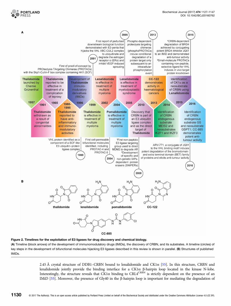
Figure 2. Timelines for the exploitation of E3 ligases for drug discovery and chemical biology.
(A) Timeline (block arrows) of the development of immunomodulatory drugs (IMiDs), the discovery of CRBN, and its substrates. A timeline (circles) of
key steps in the development of bifunctional molecules hijacking E3 ligases described in this review is shown in parallel. (B) Structures of published
IMiDs.
1130 © 2017 The Author(s). This is an open access article published by Portland Press Limited on behalf of the Biochemical Society and distributed under the Creative Commons Attribution License 4.0 (CC BY).
Biochemical Journal (2017) 474 1127–1147DOI: 10.1042/BCJ20160762

target substrates, implying that this residue may form part of the protein degradation motif (degron) recognisedby the IMiD–CRBN complex. Furthermore, the binding of Ikaros to CRBN similarly requires the presence of anIMiD compound, and both protein substrates, Ikaros and CK1α, adopt a related binding mode. These importanthigh-affinity protein–protein interactions, which are specifically induced by small molecules, will provide oppor-tunities for future drug discovery, particularly for targeted protein degradation.It is expected that the binding of IMiDs to CRBN will displace endogenous substrates, of which little is known,
and promote the recruitment of neosubstrates (Figure 3A,B). Recently, the homeobox gene, MEIS2, was identifiedas an endogenous substrate of CRBN, whereby MEIS2 ubiquitination is inhibited by the IMiDs, resulting inincreased protein levels. These data indicate that IMiDs modulate ubiquitination; in some instances, creating aneomorph for substrate degradation, as for Aiolos and Ikaros, and in others, competing out endogenous sub-strates, such as MEIS2, thereby leading to a decrease in their degradation [19]. More recently, glutamine synthe-tase (GS) has also been shown to be an endogenous substrate of CRL4CRBN [53]. GS recognition by CRBN leadsto its polyubiquitination by CRL4CRBN in response to high glutamine levels. Contrary to the case of MEIS2,IMiDs enhance GS binding to CRBN. Furthermore, two lysine resides (K11 and K14) in the N-terminus of GSare acetylated by p300/CBP in response to high glutamine concentrations. These acetylation marks serve as adegron to allow CRBN-binding and CRL4CRBN-mediated ubiquitination. These data suggest that, so far, theredoes not appear to be an identifiable, universally conserved degron motif and that CRL4CRBN-mediated degrad-ation of substrates is likely to be dependent on both the cellular context and metabolic state of the cell.CC-122 and CC-885 are the most recently developed IMiDs (Figure 2B). CC-122 is a pleiotropic pathway
modifier that also binds CRBN and promotes degradation of Aiolos and Ikaros in diffuse large B-cell lymph-oma (DLBCL) T-cells in in vitro and in vivo models and in patients, resulting in both cell autonomous andimmune-stimulatory effects. In DLBCL cell lines, both CC-122-induced degradation and short hairpinRNA-mediated knockdown of Aiolos and Ikaros correlate with increased transcription of interferon(IFN)-stimulated genes; this is independent of IFN-α, -β, and -γ production and/or secretion, and results inapoptosis in both activated B-cell (ABC) and germinal centre B-cell DLBCL cell lines [24]. This drug is nowentering Phase II/III studies for poor-risk lymphoma patients.CC-885 is the first IMiD to demonstrate potent antitumour activity in both haematological and epithelial
cancers [48]. In addition to inducing CRBN-mediated degradation of Ikaros as seen with other thalidomideanalogues, CC-885 promotes the degradation of the translation termination factor GSPT1, resulting in cytotox-icity. Lenalidomide and pomalidomide do not degrade GSPT1, possibly due to their lack of the extended ureamoiety of CC-885 that enables additional interactions with CRBN and GSPT1. Intriguingly, structural studiesof CRBN–DDB1 with CC-885 and GSPT1 revealed that CC-885 creates a hotspot on the CRBN surface fordirect interaction with GSPT1 and this interaction is not determined by a peptide sequence but rather by thegeometric arrangement of three hydrogen bond acceptors on the GSPT1 backbone and the precise position ofits glycine residue.It is clear that the ability of IMiDs to direct CRL4CRBN to degrade several different proteins opens up the
potential to discover new effects of protein degradation and target hitherto undruggable proteins and pathways.This approach will be even more powerful if we are able to predict and select which proteins are degraded. Oneavenue is to use sequence and structural homologies in putative CRL4CRBN degrons to define potential substrateproteins and to design small-molecule CRL4CRBN binders that can selectively direct the degradation of theseproteins. This rational design approach complements the empirical phenotypic screening employed to discoverthe existing IMiDs. Currently, defined degron sequences are cell context-dependent and vary according to theIMiD, with small changes in chemical structure leading to altered substrate specificity. Thus, further under-standing is required to inform such a rational degron-based approach.
Hijacking E3 ligases for specific target degradation usingbifunctional moleculesHijacking the von Hippel–Lindau E3 ligaseAn alternative approach to achieve targeted protein degradation with chemical compounds involves the use oflarger bifunctional molecules consisting of distinct substrate-binding and E3 ligase-binding groups conjugatedby a linker; as first demonstrated for the recruitment of the Skp1–CUL–F-box complex containing Hrt1 (SCF)ubiquitin ligase to degrade methionine aminopeptidase-2 (MetAP-2) [61]. The conjugate molecule serves toassemble a ternary complex between the E3 ligase, target protein, and probe molecule, allowing the E3 ligase
© 2017 The Author(s). This is an open access article published by Portland Press Limited on behalf of the Biochemical Society and distributed under the Creative Commons Attribution License 4.0 (CC BY). 1131
Biochemical Journal (2017) 474 1127–1147DOI: 10.1042/BCJ20160762

complex to ubiquitinate the non-natural substrate and promote proteasome-dependent degradation [illustratedfor the von Hippel–Lindau (VHL) E3 ligase in Figure 4A]. Whereas binding of the low molecular mass IMiDmolecules discussed above results in a subtle variation in the E3 ligase receptor-binding surface, changing theaffinity and specificity for protein–protein interactions with substrates, the bifunctional molecule approachcreates a spatially distinct small-molecule binding site for protein substrates proximal to, but separate from, theE3 ligase itself. For a productive ternary species to be formed, the bifunctional molecule must contain a select-ive ligand for the target protein of interest with a suitable position for attachment of a recognition group forthe E3 ligase via a linker group, without substantial loss of target affinity. In addition, the proximity and orien-tation of the E3 ligase and target protein in the ternary complex must be permissive for target proteinubiquitination.Recruitment of the VHL E3 ligase to induce degradation of targeted proteins is so far the most extensively
explored approach to hijacking the ubiquitin–proteasome system, following the first demonstration ofproof-of-concept by PROteolysis TArgeting Chimeras (PROTACs) from Deshaies, Crews and colleagues ([61];reviewed in refs [57,9,71,36]). Initially, small peptide chains were used to mimic the protein–protein interactionbetween the VHL E3 ubiquitin ligase complex and the endogenous substrate, hypoxia-inducible factor 1α(HIF1α). The minimal recognition domain amino acid sequence ALAPYIP [27] contains a key central prolinemotif, analogous to P564 in HIF1α, that becomes hydroxylated in cells ultimately leading to ubiquitination anddegradation of HIF1α under normoxic conditions [18].The first cell-permeable bifunctional molecules included PROTAC-4 (Figure 4B), which was designed to
target the F36V mutation of FK506-binding protein (FKBP12) [64]. The VHL-interacting seven amino acidpeptide sequence was appended to the ligand AP21998, which targets a mutant FKBP12. To confer cell perme-ability and a degree of stability to proteolysis, a poly-D-arginine tag was also introduced to PROTAC-4.Consequently, F36V mutant FKBP12 protein fused to a green fluorescent protein (GFP) tag expressed in HeLacells was shown in a fluorescence assay to be degraded by treatment with PROTAC-4 at a concentration of25 mM over 2.5 h. As further proof that the bifunctional molecules could be effective at inducing selectiveprotein degradation in cells, PROTAC-5 (Figure 4B) was designed using the same seven amino acid and polyar-ginine peptide. In this instance, the peptide motif was conjugated to dihydrotestosterone to target the androgenreceptor (AR). Fluorescence analysis of GFP-labelled AR in HEK293 cells showed protein degradation after 1 h
Figure 3. The consequences of perturbation of CUL4CRBN E3 ligase function.
(A) The role of CRL4CRBN in mediating protein homeostasis of endogenous substrates (Meis2 and glutamine synthetase); the
conserved tritryptophan pocket that binds IMiDs is highlighted. (B) Known protein substrates of consequence (for example,
Aiolos, Ikaros, CK1α, and GSPT1) whose rate of degradation is altered in response to the binding of thalidomide to the
tritryptophan pocket of CRL4CRBN; as yet undiscovered substrates are denoted by X and Y.
1132 © 2017 The Author(s). This is an open access article published by Portland Press Limited on behalf of the Biochemical Society and distributed under the Creative Commons Attribution License 4.0 (CC BY).
Biochemical Journal (2017) 474 1127–1147DOI: 10.1042/BCJ20160762

Figure 4. Selected bifunctional molecules hijacking the VHL E3 ligase using peptide motifs to target VHL. Part 1 of 2
(A) Cartoon showing the complexes involved in VHL-dependent ubiquitination (ELBC, elongin B–elongin C heterodimer; CUL2,
© 2017 The Author(s). This is an open access article published by Portland Press Limited on behalf of the Biochemical Society and distributed under the Creative Commons Attribution License 4.0 (CC BY). 1133
Biochemical Journal (2017) 474 1127–1147DOI: 10.1042/BCJ20160762

exposure to PROTAC-5 at concentrations of 25 mM and above. Additionally, pretreatment with the proteasomeinhibitor epoxomicin prevented degradation of the GFP–AR protein, as did treatment alone with either testos-terone or the truncated peptide–polyarginine construct.The bifunctional molecule E2-penta (Figure 4B) was used to hijack the VHL VBC–CUL2 complex to ubiqui-
tinate and degrade estrogen receptor α (ERα) in a potential approach to antiangiogenesis [2]. Endogenous 17βoestradiol (E2) promotes angiogenesis via ERα by direct endothelial cell proliferation, migration, andup-regulation of basic fibroblast growth factor and vascular endothelial growth factor (VEGF) and their recep-tors. Introduction of the pre-hydroxylated proline motif, the key recognition factor in the degradation ofHIF1α, allowed the chain to be shortened to a pentapeptide that showed improved potency over longer peptideanalogues, presumably due to better cell permeability. Inhibition of VEGF-induced sprouting of human umbil-ical vein endothelial cells occurred following incubation with 2 mM concentrations of E2-penta over 24 h. Thiswas the first report of using PROTACs to perturb downstream biological function and provided proof ofconcept for using such chemical probes to degrade target proteins and study the biological consequences.The aryl hydrocarbon receptor (AHR) is present in the cytoplasm as part of a chaperone complex and has
been implicated in tumour promotion and progression. Apigenin-Protac (Figure 4B), containing the hydroxy-lated pentapeptide VHL-recognition domain, was found to bind significantly less strongly to AHR than theunconjugated AHR ligand apigenin (Apigenin-Protac IC50 = 4 mM, compared with Apigenin IC50 = 0.3 mM)[56]. Despite this 10-fold decrease in binding affinity, Apigenin-Protac was found to degrade AHR proteinlevels in neonatal primary human keratinocytes at concentrations of 10 mM after 12 h incubation, in additionto inhibiting expression of 2,3,7,8-tetrachlorodibenzo-p-dioxin-induced cytochrome P450 1A1 (CYP1A1)protein, a biomarker of AHR function. Usefully, a negative control bifunctional molecule was also reported,whereby the hydroxyproline amino acid critical for VHL recognition was replaced with alanine, and was shownto have no effect on AHR degradation.In a recent example, the hydroxylated pentapeptide VHL-recognition motif was used to target SMAD family
member 3 (SMAD3), a key signalling protein in renal fibrosis, for degradation [72]. In the absence of a suitablesmall-molecule SMAD3 inhibitor, an in silico docking screen of a commercial library was performed. Of thehits thought possibly suitable as PROTAC components and confirmed by surface plasmon resonance asSMAD3 ligands, a benzofuran ligand with moderate binding affinity (Kd = 45 mM) and slow off-rate wasselected for inclusion in the bifunctional conjugate Smad3-Protac (Figure 4B). The molecule was found todecrease basal SMAD3 protein expression in both renal fibroblast (NRK-49F) and renal mesangial cells (HMC)at a 200 mg/ml concentration, representing an approximate 100-fold reduction in potency compared with theactivity observed in human renal carcinoma ACHN cell lysates, and illustrating the difficulty of routinelyobtaining high cell permeability with large bifunctional molecules containing a significant peptidic character.A two-headed approach to the design of PROTAC-type conjugates was explored in the context of ER degrad-
ation [14]. The two-headed PROTAC (Figure 4B) was found to have a 3-fold improved binding affinity for ERin comparison with two control PROTAC molecules, each containing a single ER-targeting head group ateither the N- or C-terminus of the VHL-targeting pentapeptide. The linker length between the ER ligand andthe VHL-targeting peptide in the PROTAC was found to be important for activity [15]. Although observed tohave poorer solubility than the monomeric counterparts, two-headed PROTAC had improved ability to induceER degradation at 10 mM over 48 h incubation, with a greater than 5-fold enhancement of potency over mono-meric counterparts. Importantly, to demonstrate the mechanism of action, the control compound where thecentral hydroxyproline VHL-recognition amino acid was replaced with norleucine did not cause ER degrad-ation under the same conditions, nor did degradation occur with two-headed PROTAC in the presence of theproteasome-specific inhibitor epoxomicin.
Figure 4. Selected bifunctional molecules hijacking the VHL E3 ligase using peptide motifs to target VHL. Part 2 of 2
cullin2; E2, E2 ubiquitin ligase; Ub, ubiquitin). (B) Structures of bifunctional molecules showing the affinity groups targeting
proteins for degradation (red), linker motifs (black), and peptide motifs targeting the VHL E3 ligase (blue). Y denotes sites of
intracellular phosphorylation. POH, hydroxyproline. (C) Proteins targeted for degradation by selected bifunctional molecules and
the concentrations used in cellular assays where maximal target depletion was observed (AHR, aryl hydrocarbon receptor; AR,
androgen receptor; ER, estrogen receptor; FKBP12, FK506-binding protein 12; FRS2α, fibroblast growth factor receptor
substrate 2; PI3K, phosphatidylinositol-4,5-bisphosphate 3-kinase; SMAD3, SMAD family member 3).
1134 © 2017 The Author(s). This is an open access article published by Portland Press Limited on behalf of the Biochemical Society and distributed under the Creative Commons Attribution License 4.0 (CC BY).
Biochemical Journal (2017) 474 1127–1147DOI: 10.1042/BCJ20160762

An exciting extension of the bifunctional conjugate approach whereby the conditional degradation of aprotein target occurs only subsequent to an intracellular phosphorylation event has been developed [26]. As anexample, the tyrosine residue highlighted in TrkAPPFRS2α (Figure 4B) must first be phosphorylated by thekinase, tropomyosin receptor kinase A (TrkA), before degradation of the target, fibroblast growth factor recep-tor substrate 2 (FRS2α), is observed. In the presence of nerve growth factor (NGF), TrkA undergoesNGF-induced dimerisation and trans-autophosphorylation. The fully active phosphorylated tyrosine kinase can,in turn, bind and phosphorylate many substrates, including FRS2α. TrkAPPFRS2α was designed to incorporate a10 amino acid recognition sequence around the tyrosine residue phosphorylated in TrkA, a seven amino acidrecognition sequence to bind to VHL, and a polyarginine sequence to improve cell permeability. In the pres-ence of TrkAPPFRS2α, NGF-treated PC12 cells incorporated radiolabelled 32P phosphate into the TrkAPPFRS2αconjugate, a phenomenon not observed in the absence of NGF. Incubating PC12 cells in the presence of NGFfor 7 h showed an increase in phosphorylation of FRS2α with no loss of protein levels. However, including40 mM TrkAPPFRS2α gave a 50% reduction in FRS2α protein levels and loss of downstream extracellular signal-regulated kinase (ERK)1/2 phosphorylation, without any associated decrease in overall ERK1/2 protein levels.Pretreatment of PC12 cells with epoxomicin was used to confirm dependence on the ubiquitin-proteasomepathway and indeed caused accumulation of FRS2α and higher molecular mass FRS2α–ubiquitin conjugates inthe presence of NGF and TrkAPPFRS2α. The requirement for TrkA phosphorylation of TrkAPPFRS2α to inducedegradation was supported by use of a Phe/Tyr residue swap in a control bifunctional molecule, thus removingthe site of potential phosphorylation, and giving no FRS2α degradation or reduction in ERK1/2 phosphoryl-ation in the presence of NGF.A further example of the conditional degradation strategy was described using ErbB2PPPI3K (Figure 4B) to
deplete phosphatidylinositol-3-kinase (PI3K). ErbB3 phosphorylation in a heterodimer complex between ErbB2and ErbB3 epidermal growth factor receptor tyrosine kinases at the cell membrane results in recruitment ofPI3K through binding to phospho-ErbB3. The design of the phosphoPROTAC ErbB2PPPI3K therefore containeda 24 amino acid sequence taken from the PI3K-binding segment of ErbB3. Both the highlighted tyrosine resi-dues in Figure 4B are phosphorylated by ErbB2, leading to recruitment of PI3K for degradation. Accordingly,ErbB2PPPI3K induced depletion of PI3K expression in MCF-7 cells at concentrations of 40 mM and above.Replacing the Tyr residues in ErbB2PPPI3K with Phe as before confirmed the requirement for tyrosine phosphor-ylation to occur before observing any reduction in PI3K protein levels. Additionally, a mouse study using10 mg/kg i.p. dosing of ErbB2PPPI3K in OVCAR8 subcutaneous xenograft tumours demonstrated a 40% reduc-tion in tumour weight relative to control.While cellular and in vivo efficacy has been shown in peptide-derived VHL affinity groups as discussed
above, the use of a non-peptide, selective small-molecule VHL-recognition motif could offer many advantagesin regard to improved potency, metabolic stability, and permeability. The rational design of small-moleculeVHL ligands such as cmpd 15 (Figure 5A) and determination of its crystal structure bound to VHL [8] has ledto the development of submicromolar VHL ligands with reduced peptide character [20]. Incorporation of at-butyl group in the VHL-binding component, as in Protac_ER-related receptor-α (ERRα; Figure 5A), has beenfound to give high affinity for VHL [7]. Protac_ERRα gave 50% degradation (DC50) of ERRα in MCF-7 cells at100 nM, whereas the epimeric proline alcohol, used as a negative control as it no longer binds to VHL, gave∼20% degradation at the same concentration. In addition, Protac_ERRα showed depletion of ERRα in vivowhen dosed at 100 mg/kg i.p., three times per day, to mice bearing MDA-MB-231 xenograft tumours.Similarly, Protac_RIPK2 (Figure 5A) was shown to be particularly potent in inducing degradation of RIPK2,with a DC50 of 1.4 nM in human THP-1 monocytes and complete depletion at 10 nM. Of note was a biphasicresponse at high concentrations of Protac_RIPK2, where protein levels of RIPK2 recovered to basal levels, aneffect attributed to an inability to form the proposed ternary complex in the presence of excess Protac_RIPK2concentrations, which increases binary complex formation and thus stops degradation. The epimeric prolinealcohol of Protac_RIPK2 made as a negative control did not show any degradation at concentrations up to10 mM. Both active Protac_RIPK2 and the inactive epimeric alcohol were used to provide evidence to supportthe formation of a ternary complex through chemoproteomic pull-down and immunoprecipitation experi-ments. Moreover, Protac_RIPK2 was found to act catalytically in vitro, inducing super-stoichiometric ubiquiti-nation of RIPK2. For both Protac_ERRα and Protac_RIPK2, a dependence on proteasomal degradation in cellswas confirmed by pretreatment with the proteasome inhibitor epoxomicin, resulting in blockade of the degrad-ation of ERRα and RIPK2. The specificities of Protac_ERRα and Protac_RIPK2 towards degradation of theirintended target proteins were assessed by cellular expression proteomics in cancer cells, monitoring ∼7600
© 2017 The Author(s). This is an open access article published by Portland Press Limited on behalf of the Biochemical Society and distributed under the Creative Commons Attribution License 4.0 (CC BY). 1135
Biochemical Journal (2017) 474 1127–1147DOI: 10.1042/BCJ20160762

proteins. For Protac_ERRα, only degradation of ERRα and (after prolonged exposure) breakpoint cluster region(BCR) protein were observed, while Protac_RIPK2 showed a similar specificity with RIPK2 and the kinaseMAPKAPK3 as the only proteins degraded.The treatment of chronic myelogenous leukaemia (CML) by inhibition of the oncogenic protein kinase
BCR-ABL using small-molecule drugs such as imatinib, dasatinib, or bosutinib has been highly effective. Thereis, however, a need for lifelong treatment with the drug, speculated to be due to the ability of BCR-ABL to actas a scaffolding protein in a compensatory pathway that produces leukaemic stem cells in spite of kinase inhib-ition. Degradation of BCR-ABL therefore presents an alternative approach to modulation of this importanttarget [35]. Of the VHL-binding conjugates synthesised with varying linker group lengths, DAS-6-2-2-6-VHL(Figure 5A) showed degradation of the wild-type protein Abelson tyrosine kinase (c-ABL) at 1 mM in K562human CML cells over 24 h. However, no associated degradation of the oncogenic BCR-ABL fusion proteinwas observed, despite potent binding of dasatinib to both proteins, and in contrast with the effects of relatedconjugates designed to recruit the CRBN E3 ligase complex, discussed in detail in ‘Bifunctional moleculeshijacking the MDM2, cIAP, and CRBN E3 ligases’. Thus, a clear need to optimise not only the target-binding
Figure 5. Selected bifunctional molecules hijacking the VHL E3 ligase using small-molecule VHL inhibitors.
(A) Structures of bifunctional molecules hijacking the VHL E3 ligase, showing the affinity groups targeting proteins for degradation (red), linker motifs
(black), and VHL E3 ligase-targeting motif (blue). (B) Proteins targeted for degradation by selected bifunctional molecules and the concentrations
used in cellular assays where maximal target depletion was observed (BET, bromodomain and extra-terminal family of proteins; BRD4,
bromodomain-containing protein 4, member of the BET family; c-ABL, Abelson tyrosine kinase; RIPK, receptor-interacting serine/threonine-protein
kinase 1; ERRα, estrogen-related receptor α).
1136 © 2017 The Author(s). This is an open access article published by Portland Press Limited on behalf of the Biochemical Society and distributed under the Creative Commons Attribution License 4.0 (CC BY).
Biochemical Journal (2017) 474 1127–1147DOI: 10.1042/BCJ20160762

motif and linker length in the bifunctional molecules, but also the choice of E3 ligase for recruitment in agiven degradation target context, has been demonstrated.The bifunctional molecule ARV-771 (Figure 5A) has been found to induce potent degradation of the bromo-
domain and extra-terminal domain (BET) family of proteins, potentially of great interest in the treatment ofprostate cancer, and castration-resistant prostate cancer in particular [59]. ARV-771 was constructed by conju-gating JQ-1, a small-molecule BET inhibitor, to a hydroxyproline-based VHL-binding domain via an optimisedlinker group. Degradation of the BET family of proteins, bromodomains (BRD)2, 3, and 4, by ARV-771 wasobserved in 22Rv1, VCaP, and LnCaP95 human prostate cancer cell lines at concentrations of 5–10 nM. Lossof expression of c-MYC, a downstream effector of BET, was also observed at both the protein and mRNAlevels at <10 nM concentrations for ARV-771. An inactive control diastereoisomer ARV-766, containing theepimeric proline alcohol unable to bind VHL, was found to have comparable binding affinity for the BET pro-teins to that seen for ARV-771 and JQ-1. ARV-766 showed a negligible effect on c-MYC mRNA levels at 1 mMdespite being a potent BET inhibitor in its own right, suggesting poor permeability. Given that the active conju-gate ARV-771 may be expected to have comparable cell permeability, the low nanomolar cellular activityobserved for ARV-771 has been hypothesised to be due to the potential catalytic action of the bifunctionalspecies. Additionally, ARV-771 was shown to induce BRD4 and c-MYC degradation in 22Rv1 tumour xeno-grafts in mice, and more importantly led to tumour regression in the 22Rv1 model following daily subcutane-ous dosing of 30 mg/kg.Variation in the linker length between JQ-1 and the VHL-binding motif has led to the discovery of linker-
dependent selectivity within the BET family of proteins [77]. MZ1 (Figure 5A) and JQ-1 were shown to havecomparable binding affinities for the BET family of proteins; however, MZ1 was found to preferentially degradeBRD4 over BRD2 and BRD3 at a concentration of 1 mM in HeLa cells over 24 h. In the U2OS cell line, MZ1demonstrated dose- and time-dependent selective degradation of BRD4 over BRD2 and BRD3. Furthermore,MZ1 was shown to have no effect on endogenous levels of VHL or HIF1α, an important factor in the potentialuse of such bifunctional molecules as drug therapies.
Bifunctional molecules hijacking the MDM2, cIAP, and CRBN E3 ligasesE3 ligases other than VHL have been successfully hijacked using bifunctional molecules. The first non-peptidicE3 ligase-targeting group was used to direct MDM2, whose natural substrates include the tumour-suppressorTP53, to degrade the AR (Figure 6A) [65]. A selective androgen receptor modulator (SARM) with nanomolaraffinity for AR was conjugated to the MDM2–TP53 interaction inhibitor nutlin to generate the SARM-nutlinPROTAC, which decreased AR expression in HeLa cells in a proteasome-dependent manner at a 10 mM con-centration (Figure 6B,C). However, interpretation of the cellular activity of the SARM-nutlin PROTAC is com-plicated as AR is known to be a direct substrate of MDM2 [40], and nutlin itself induces ubiquitination anddegradation of AR in cancer cells [41], raising the possibility of a direct modulatory effect of the SARM-nutlinbifunctional molecule on AR degradation independent of ternary complex formation [31,70].While the cellular potency for SARM-nutlin PROTAC was similar to that seen for VHL-targeting peptidic
PROTACs, subsequent iterations using small-molecule E3 ligase-binding groups have achieved breakthroughsin cell activity to the nanomolar range, notably for non-peptidic molecules hijacking VHL (Figure 5A,B) andCRBN (Figure 6B,C). A suite of bifunctional molecules, termed specific and non-genetic IAP-dependentprotein erasers (SNIPERs), was developed by Hashimoto and colleagues to redirect the activity of the E3 ligasecIAP1 that degrades caspase proteins and is overexpressed in many cancer cells (Figure 6A). The conjugatesmake use of derivatives of bestatin methyl ester, a cell permeable small molecule that binds to cIAP1 topromote autoubiquitination and degradation [67]. Proof-of-concept was achieved with SNIPER cmpd 4(Figure 6B) that linked a bestatin ester moiety to all-trans retinoic acid to capture the cellular retinoic acid-binding proteins (CRABP-I and -II) [29]. Proteasome-dependent degradation of CRABP-I/II was observed inthe range of 1–10 mM in cells. Interestingly, varying the linker length in SNIPER cmpd 4 changed the relativeefficiency of degradation of CRABP-I and -II, with CRABP-I better depleted by a conjugate with longer linker.Reduced expression of CRABP-II in IMR-32 human neuroblastoma cells by SNIPER cmpd 4 gave a dose-dependent reduction in cell motility.To avoid the concomitant autoubiquitination and degradation of cIAP promoted by bestatin ester derivatives,
the amide-linked SNIPER cmpd 6 was developed to retain cIAP binding but abolish autodegradation [30].SNIPER cmpd 6 promoted depletion of CRABP-II from 1 mM in cells with no effect on cIAP levels or apparentinhibition of cIAP endogenous function. A comparison of the effects of SNIPER cmpd 4 and cmpd 6 in
© 2017 The Author(s). This is an open access article published by Portland Press Limited on behalf of the Biochemical Society and distributed under the Creative Commons Attribution License 4.0 (CC BY). 1137
Biochemical Journal (2017) 474 1127–1147DOI: 10.1042/BCJ20160762
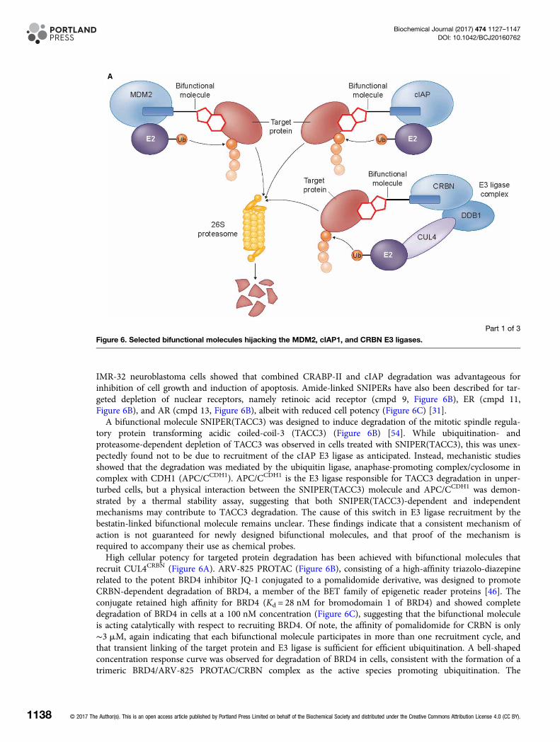
IMR-32 neuroblastoma cells showed that combined CRABP-II and cIAP degradation was advantageous forinhibition of cell growth and induction of apoptosis. Amide-linked SNIPERs have also been described for tar-geted depletion of nuclear receptors, namely retinoic acid receptor (cmpd 9, Figure 6B), ER (cmpd 11,Figure 6B), and AR (cmpd 13, Figure 6B), albeit with reduced cell potency (Figure 6C) [31].A bifunctional molecule SNIPER(TACC3) was designed to induce degradation of the mitotic spindle regula-
tory protein transforming acidic coiled-coil-3 (TACC3) (Figure 6B) [54]. While ubiquitination- andproteasome-dependent depletion of TACC3 was observed in cells treated with SNIPER(TACC3), this was unex-pectedly found not to be due to recruitment of the cIAP E3 ligase as anticipated. Instead, mechanistic studiesshowed that the degradation was mediated by the ubiquitin ligase, anaphase-promoting complex/cyclosome incomplex with CDH1 (APC/CCDH1). APC/CCDH1 is the E3 ligase responsible for TACC3 degradation in unper-turbed cells, but a physical interaction between the SNIPER(TACC3) molecule and APC/CCDH1 was demon-strated by a thermal stability assay, suggesting that both SNIPER(TACC3)-dependent and independentmechanisms may contribute to TACC3 degradation. The cause of this switch in E3 ligase recruitment by thebestatin-linked bifunctional molecule remains unclear. These findings indicate that a consistent mechanism ofaction is not guaranteed for newly designed bifunctional molecules, and that proof of the mechanism isrequired to accompany their use as chemical probes.High cellular potency for targeted protein degradation has been achieved with bifunctional molecules that
recruit CUL4CRBN (Figure 6A). ARV-825 PROTAC (Figure 6B), consisting of a high-affinity triazolo-diazepinerelated to the potent BRD4 inhibitor JQ-1 conjugated to a pomalidomide derivative, was designed to promoteCRBN-dependent degradation of BRD4, a member of the BET family of epigenetic reader proteins [46]. Theconjugate retained high affinity for BRD4 (Kd = 28 nM for bromodomain 1 of BRD4) and showed completedegradation of BRD4 in cells at a 100 nM concentration (Figure 6C), suggesting that the bifunctional moleculeis acting catalytically with respect to recruiting BRD4. Of note, the affinity of pomalidomide for CRBN is only∼3 mM, again indicating that each bifunctional molecule participates in more than one recruitment cycle, andthat transient linking of the target protein and E3 ligase is sufficient for efficient ubiquitination. A bell-shapedconcentration response curve was observed for degradation of BRD4 in cells, consistent with the formation of atrimeric BRD4/ARV-825 PROTAC/CRBN complex as the active species promoting ubiquitination. The
Part 1 of 3
Figure 6. Selected bifunctional molecules hijacking the MDM2, cIAP1, and CRBN E3 ligases.
1138 © 2017 The Author(s). This is an open access article published by Portland Press Limited on behalf of the Biochemical Society and distributed under the Creative Commons Attribution License 4.0 (CC BY).
Biochemical Journal (2017) 474 1127–1147DOI: 10.1042/BCJ20160762

observed sustained protein degradation was in contrast with the effects of unconjugated BRD4 ligands that leadto hyperaccumulation of BRD4 on prolonged exposure. As a result, increased downstream effects on suppres-sion of the c-MYC protein, inhibition of B-cell proliferation, and induction of apoptosis were seen with theconjugate compared to the effects of the unconjugated BRD4 ligands.In a parallel approach, the conjugation of the BRD4 inhibitor JQ-1 to a pomalidomide/thalidomide hybrid
gave the bifunctional molecule dBET1 (Figure 6B) that retained the selectivity of JQ-1 for BRD4 bindingwithin the BET family [74]. Taking advantage of the crystal structures available for both BRD4 and CRBN withligands bound, the linker length for dBET1 was designed based on in silico modelling of the ternary complex.Extensive depletion (>85%) of BRD4 by dBET1 was seen at 100–250 nM in human cells for up to 18 h
Part 2 of 3
Figure 6. Selected bifunctional molecules hijacking the MDM2, cIAP1, and CRBN E3 ligases.
© 2017 The Author(s). This is an open access article published by Portland Press Limited on behalf of the Biochemical Society and distributed under the Creative Commons Attribution License 4.0 (CC BY). 1139
Biochemical Journal (2017) 474 1127–1147DOI: 10.1042/BCJ20160762

(Figure 6C). Some recovery of protein levels was seen on longer exposure, suggestive of chemical instability ofthe bifunctional molecule. Proteomic assessment of the effects of dBET1 and the unconjugated BRD4 inhibitorJQ-1 showed highly similar, selective effects of both molecules. Out of 7429 proteins monitored, the bifunc-tional molecule dBET1 elicited depletion of MYC and PIM1 as expected based on the downstream effects ofdepletion of BRD4 and only three other proteins (BRD2, BRD3, and BRD4), consistent with the specificity ofthe JQ-1 ligand for the BET family. However, enhanced apoptotic effects were seen for dBET1 in cancer celllines and primary human acute myeloid leukaemia cells. The effects of targeted degradation of BRD4 wereinvestigated in vivo following intraperitoneal dosing of dBET1 to mice bearing xenograft human MV4-11 leu-kaemia cells. Inhibition of tumour growth relative to untreated controls was observed, with pharmacodynamicevidence of BRD4 depletion in treated tumours seen. A head-to-head comparison of equimolar amounts ofdBET1 and JQ-1 dosed in a model of disseminated leukaemia showed a ∼3-fold increase in antitumour activityfor the bifunctional molecule over the simple BRD4 ligand.Conjugation to CRBN-recruiting groups has also been demonstrated for the FKBP12 ligand steel factor
(SLF), giving the potent promoter of FKBP12 degradation, dFKBP-1 (Figure 6B) [74]. CRBN-recruiting groupswere also linked to the BCR-ABL receptor tyrosine kinase inhibitors bosutinib and dasatinib, to giveBOS-6-2-2-6-CRBN and DAS-6-2-2-6-CRBN, respectively (Figure 6B) [35]. In the latter study, Crews and col-leagues adopted a modular approach to vary the affinity group (kinase inhibitor), linker structure, and E3 ligaserecruiting group of the PROTAC molecules. This allowed a direct comparison between recruitment of VHLand CRBN E3 ligases for the same targets. The crystal structures of ligand-bound c-ABL were used to selectthe attachment points for the linkers, and the derived bifunctional molecules maintained nanomolar affinity forBCR-ABL despite a general fall in potency. Intriguingly, none of the conjugates made that targeted VHL led toBCR-ABL degradation in cells (see ‘Hijacking the von Hippel-Lindau E3 ligase’). This was not due to lack ofBCR-ABL binding and was speculatively attributed to an unproductive orientation of the VHL E3 ligase in the
Part 3 of 3
Figure 6. Selected bifunctional molecules hijacking the MDM2, cIAP1, and CRBN E3 ligases.
(A) Cartoon showing the complexes involved in MDM2-, cIAP-, and CRBN-dependent ubiquitination (cIAP, cellular inhibitor of
apoptosis protein; CRBN, cereblon; CUL4, cullin 4; DDB1, DNA damage-binding protein 1; E2, E2 ubiquitin ligase; MDM2,
mouse double minute 2 homologue; Ub, ubiquitin). (B) Structures of bifunctional molecules showing the affinity groups
targeting proteins for degradation (red), linker motifs (black), and small-molecule motifs that recruit the E3 ligases (blue). (C)
Proteins targeted for degradation by selected bifunctional molecules, the E3 ligases recruited, and the concentrations used in
cellular assays where maximal target depletion was observed (APC/CCDH1, anaphase-promoting complex/cyclosome in
complex with CDH1; AR, androgen receptor; BCR-ABL, breakpoint cluster region — Abelson kinase fusion; BRD4,
bromodomain 4; c-ABL, Abelson murine leukaemia viral oncogene cellular homologue; CRBP, cellular retinoic acid-binding
protein; ER, estrogen receptor; FKBP12, FK506-binding protein 12; RAR, retinoic acid receptor; TACC3, transforming acidic
coiled-coil-3).
1140 © 2017 The Author(s). This is an open access article published by Portland Press Limited on behalf of the Biochemical Society and distributed under the Creative Commons Attribution License 4.0 (CC BY).
Biochemical Journal (2017) 474 1127–1147DOI: 10.1042/BCJ20160762

trimeric complexes. In contrast, the conjugates recruiting CRBN (e.g. BOS-6-2-2-6-CRBN andDAS-6-2-2-6-CRBN) elicited potent degradation of BCR-ABL in cells, showing that the oncogenic tyrosinekinase has varying levels of susceptibility to modification by different hijacked E3 ligases. As the efficiency andselectivity of target degradation was also found to depend on the kinase inhibitor moiety, the authors suggestthat a modular approach to an array of bifunctional molecule designs, to optimise empirically the best combin-ation of target and E3 ligase-binding functionalities, may be advantageous when seeking new probes for tar-geted protein degradation. Such studies require rapid syntheses of the components of the bifunctionalmolecules, such as that recently demonstrated for amine-substituted phthalimide derivatives [42].A recent publication shows how high molecular mass bifunctional molecules can be self-assembled in situ in
cells from smaller components using bio-orthogonal ‘click’ chemistry to link separate precursors containing theCRBN-binding and protein-targeting functionalities [37]. These click-formed proteolysis-targeting chimeras(CLIPTACs) recruiting CRBN were prepared by conjugation of thalidomide to JQ-1 ( JQ1-CLIPTAC) or to acovalent ERK1/2 inhibitor (ERK1/2-CLIPTAC) to achieve depletion of BRD4 or ERK 1 and 2, respectively. Apotential advantage for achieving cell penetration with smaller components was demonstrated by the observa-tion that preformed CLIPTACs gave no target degradation, while combination of the smaller click-enabled thal-idomide moiety (10 mM) with click-enabled JQ-1 (3 mM) gave complete BRD4 depletion after 18 h.
Targeted protein degradation through recruitment of HSP70 molecularchaperones or direct binding to the 20S proteasome
The bifunctional molecules described above affect protein degradation by direct binding to E3 ligase complexes.Protein ubiquitination and degradation can also be achieved through hijacking the unfolded protein responseusing very lipophilic small-molecule tags to recruit molecular chaperones, such as HSP70 family members thatrecognise the exposed hydrophobic cores of unfolded proteins (Figure 7Ai). HSP70 and co-chaperone bindingdirect the tagged protein for E3 ligase-mediated ubiquitination and degradation as though it was an unfoldedclient. First demonstrated for Halotag proteins [52] using an adamantyl hydrophobic tag, this approach wasextended to bifunctional adamantyl derivatives including the selective androgen receptor degrader SARD279(Figure 7B) [23]. Conjugation of the high-affinity AR agonist RU59063 to the adamantyl group reduced thebinding affinity for AR by 37-fold, but led to degradation of AR in LNCaP human prostate cancer cells at lowmicromolar concentrations, while no AR degradation was seen upon treatment with unconjugated RU59063.Selective degradation of AR over the glucocorticoid receptor (GR) was induced by SARD279, consistent withthe selectivity of the RU59063 affinity group for AR over GR. Increasing the cellular expression of HSP70isoforms using the HSP90 inhibitor geldanamycin enhanced the SARD279-dependent AR degradation, suggest-ing a role for HSP70 in mediating degradation of the AR–SARD279 complex. SARD279 showed more potentinhibition of AR-dependent gene expression (IC50 = 156 nM) than that for AR degradation, indicating a dualmode of activity through competitive inhibition of AR transactivation and AR depletion. Importantly, in con-trast with competitive AR antagonists, eliminating AR protein with SARD279 was found to be antiproliferativein AR-dependent prostate cancer cell lines and also in castration-resistant prostate cell lines, whether resistanceto antiandrogens resulted from increased androgen levels or from the F876L AR mutation that converts antago-nists into agonists [23].A distinct approach to targeted protein degradation was achieved using ligands linked to arginine triply-
protected with the bulky, lipophilic tert-butyloxycarbonyl (Boc) group [43]. Thus, the Boc3-protected arginine(B3A) conjugate of the covalent glutathione S-transferase (GST) inhibitor ethacrynic acid (EA-B3A, Figure 7B)induced degradation of GST-fusion proteins in lysates from HeLa cancer cells and of endogenous GST-π aswell as ectopically expressed GST-fusion proteins in Cos-1 or HeLa cells. Covalent attachment of the affinitygroup to the target protein appeared to enhance the degradation potency of the bifunctional molecules. Theconjugate TMP-B3A (Figure 7B) based on trimethoprim, a reversible, non-covalent inhibitor of bacterial dihy-drofolate reductase (eDHFR), was tested head-to-head with EA-B3A for the ability to promote degradation ofan ectopically expressed eDHFR–HA–GST-α1 fusion protein in HeLa cells. While 80 mM EA-B3A gave com-plete depletion of the fusion protein in under 2 h, only 25% removal of protein was achieved by 80 mMTMP-B3A after 5 h.In contrast with other targeted degradation approaches, the mechanism of B3A-promoted degradation was
found not to require ubiquitination of the target protein, nor the involvement of the 26S proteasome [43,68].Neither did binding of B3A conjugates intrinsically destabilise the target proteins and induce unfolding.
© 2017 The Author(s). This is an open access article published by Portland Press Limited on behalf of the Biochemical Society and distributed under the Creative Commons Attribution License 4.0 (CC BY). 1141
Biochemical Journal (2017) 474 1127–1147DOI: 10.1042/BCJ20160762

Instead, a direct non-covalent interaction of the B3A group with the 20S proteasome was uncovered, and puri-fied 20S proteasome was found to be sufficient for target protein degradation in cell-free systems. Thus, thisrepresents the first example of using a bifunctional small molecule for direct targeting of a protein to the 20S
Figure 7. Selected bifunctional molecules directing target degradation through binding of HSP70 or the 20S proteasome.
(A) Cartoons showing the complexes involved in (i) HSP70-dependent protein degradation mediated by a hydrophobic adamantyl tag (HSP70, heat
shock protein 70; Ub, ubiquitin) and (ii) direct recruitment of the 20S proteasome by Boc3Arg tags. (B) Structures of bifunctional molecules that
direct target protein degradation through binding of HSP70 (SARD279) or the 20S proteasome (EA-B3A, TMP-B3A) showing the affinity groups
targeting proteins for degradation (red), linker motifs (black), and small-molecule motifs targeting degradation machinery (blue). (C) Mode of action
and proteins targeted for degradation, and the concentrations used in cellular assays where maximal target depletion was observed (AR, androgen
receptor; eDHFR, E. coli dihydrofolate reductase; GST-α1, glutathione S-transferase).
1142 © 2017 The Author(s). This is an open access article published by Portland Press Limited on behalf of the Biochemical Society and distributed under the Creative Commons Attribution License 4.0 (CC BY).
Biochemical Journal (2017) 474 1127–1147DOI: 10.1042/BCJ20160762

proteasome for degradation (Figure 7Aii) and is an exciting addition to the growing repertoire of pharmaco-logical techniques to control protein degradation.The two approaches outlined above offer complimentary alternatives to the hijacking of specific E3 ligases to
achieve targeted protein degradation. The hydrophobic and B3A tags do not have the same potential to alterendogenous E3 ligase substrate specificity as may be seen with VHL- or CRBN-directing tags, but there areother potentially interfering biological outputs from the functional groups in the conjugate molecules that maycomplicate interpretation of cellular experiments. HaloTag protein modification with adamantyl-derived groups
Table 1 Common experimental approaches to characterising bifunctional modulators of E3 ligase activity for theirsuitability as chemical tools
Characterisation Experimental approaches
Evidence of target degradation • Cell-based assessment of target protein expression• Dose-dependent depletion of target protein and
quantification of potency (DC50, DC90, or similar)
Evidence of binding to target protein • Biochemical (cell-free) assay of binding/inhibition by thebifunctional molecule
• Cell-based assay for inhibition of function of the targetprotein by the bifunctional molecule
• Reduction in target degradation in cells by competitionwith the unconjugated affinity group and/or an alternativesmall molecule targeting the same binding site
Evidence for binding and recruitment of an E3 ligase • Reduction in target degradation in cells by competitionwith the unconjugated recruitment motif for the E3 ligase(e.g. pomalidomide for CRBN)
• Target degradation abolished in ligase-deficient cell lines
Evidence for ubiquitin-dependent degradation • Assay for ubiquitination of the target protein followingimmunoprecipitation from cells treated with the bifunctionalmolecule and a proteasome inhibitor (e.g. MG132)
Evidence for 26S proteasome-dependent degradation • Inhibition of probe-induced target degradation in thepresence of a proteasome inhibitor (e.g. MG132,carfilzomib, and epoxomicin)
Evidence of a trimeric complex formation (E3 ligase —
bifunctional molecule — target protein) mediating theobserved effects
• Bell-shaped concentration–response for target proteindegradation in cells (may only be seen for potent probes)or in a cell-free proximity assay (e.g. AlphaScreen)
• Confirmation that target degradation induced by thebifunctional molecule is not induced by derivatives of theunconjugated affinity group or ligase recruitment groupalone, or a mixture of the two.
• Recovery of the target protein followingimmunoprecipitation of the E3 ligase in cells treated withthe bifunctional molecule
• Cell-free proximity assay using labelled ligase and targetprotein
Evidence of specificity for target degradation • Biochemical (cell-free) profiling of bifunctional molecule forbinding/inhibition based on activities of the affinity group
• Cell-based assay for degradation of known/potentialoff-targets based on biochemical profiling of thebifunctional molecule or its unconjugated affinity group
• Cell-based assay for effects on the degradation of knownsubstrates of the E3 ligase hijacked
• Differentiation of degradation promoted by the bifunctionalmolecule compared with negative control compoundswhere either the target affinity or ligase recruitment groupsare replaced by structurally related, non-binding analogues(e.g. epimeric derivatives)
• Cellular expression proteomic profiling to determine effectson degradation.
© 2017 The Author(s). This is an open access article published by Portland Press Limited on behalf of the Biochemical Society and distributed under the Creative Commons Attribution License 4.0 (CC BY). 1143
Biochemical Journal (2017) 474 1127–1147DOI: 10.1042/BCJ20160762

can induce a transient unfolded protein response [58]. While it is not demonstrated that this applies for thereversible, bifunctional molecules, the activation of HSP70 isoforms could complicate the phenotype seen ontargeted degradation. On the other hand, the simple B3A-containing molecule Cbz-B3A (Figure 7B), whichlacks a specific affinity group, has been shown to block eIF4E-binding protein 1-dependent translation throughan as-yet-uncharacterised interaction with ubiquilins [12]. These inevitable caveats of reagent selectivity not-withstanding, pharmacological degradation of putative targets by more than one of the complementaryapproaches available would help to rule out off-target effects.
Experimental approaches to characterising bifunctional modulators of E3ligase activityThe discovery and validation of new bifunctional molecules to hijack E3 ligases requires characterisation oftheir mode of action, especially if they are to be used effectively as tools to explore the biological consequencesof specific protein depletion. This mirrors the validation of classical small-molecule chemical probes [75]. Thecommon experimental approaches applied to characterise PROTACs, SNIPERs and other bifunctional mole-cules are summarised in Table 1. These typically provide evidence for engagement of the target protein(s) andE3 ligase, trimeric complex formation, ubiquitin- and proteasome-dependent degradation of the target, specifi-city for the target, and differentiation of the bifunctional compound from the component binding groups. Inmost cases, these experiments are supported by the parallel discovery and profiling of negative control com-pounds, typically bifunctional molecules where one of the binding groups has been rendered ineffective, as wellas the use of competition experiments between the bifunctional probe and the small-molecule componentbinding groups. Details of these approaches are presented in many of the publications surveyed in ‘HijackingE3 ligases for specific target degradation using bifunctional molecules’.
Conclusions and future perspectivesTwo major recent breakthroughs have been achieved in the field of targeted protein degradation promoted bysmall molecules. One is the increased cell potency now routinely achievable using non-peptidic functionality inbifunctional molecules that engage E3 ligases to promote highly specific protein depletion [16]. This hasenabled multiple proof-of-concept demonstrations of activity in animals with compounds promoting degrad-ation of BET proteins or ERRα [74,7,59]. Importantly, two studies targeting BET protein degradation usingbifunctional molecules that contain a BRD4-binding group showed antitumour activity in animal models, con-comitant with the targeted protein degradation in vivo [74,59]. Optimising the physicochemical properties ofhigh molecular mass bifunctional molecules to render them routinely suitable for human administrationremains difficult [73], but these promising demonstrations of in vivo efficacy give impetus to solving thischallenge.The second breakthrough, which already addresses achieving drug-like physicochemical properties with
small molecules that redirect E3 ligase activity, comes from understanding and exploiting the IMiD class ofsmall-molecule modulators of CRBN substrate specificity. Here, the challenge is to learn how to predict andcontrol the selectivity for neosubstrate degradation and to discern what limitations may exist to specificity. It isvery encouraging that effective drug molecules are already in clinical use from this approach, and this indicatesthat the pleiotropic effects of multiple target protein degradation can be successfully used for therapeuticbenefit.The two chemical approaches to targeted protein degradation through modulation of the ubiquitin–prote-
asome pathway described in the present study are highly complementary, both in terms of their current useand future prospects. There are common considerations, for example the possible consequences of competitionwith endogenous substrates of the particular E3 ligase hijacked, and the need to carefully validate the mechan-ism of action of the chemical probes. With bifunctional molecules, the biological effect of proteasome-mediateddepletion of the target protein needs to be differentiated from any direct effect of the affinity group on thetarget protein, especially when this is derived from a potent inhibitor or modulator in its own right. However,it is already clear that target depletion can have a more sustained activity than direct target inhibition and canovercome intrinsic feedback activation or overexpression of the target [46]. Exploiting the CRL4CRBN-mediateddegradation of target proteins with small molecules that modulate the receptor surface will require understand-ing of the target degron sequence, biological context, and the development of chemical libraries that bind thetritryptophan cage in CRBN. Binding of substrates to ‘hotspot’ interaction sites on CRBN and the ability to
1144 © 2017 The Author(s). This is an open access article published by Portland Press Limited on behalf of the Biochemical Society and distributed under the Creative Commons Attribution License 4.0 (CC BY).
Biochemical Journal (2017) 474 1127–1147DOI: 10.1042/BCJ20160762

affect degradation requires small molecules with low molecular mass and cell permeability that is a distinctadvantage for developing drug-like molecules. On the other hand, there is limited further scope for furtherchemistry optimisation without perturbing the substrate specificity. For the bifunctional targeting approach,understanding the biology of the E3 ligases recruited, the optimal type of linker and how to assemble thebifunctional compounds is important, as well as the availability of specific binders for target proteins in thefirst place. Both complementary approaches therefore have advantages and disadvantages, but both will have anincreasing role to play in the discovery of chemical probes for interrogating biological systems and the develop-ment of novel therapeutics through targeted protein degradation.
AbbreviationsAHR, aryl hydrocarbon receptor; AR, androgen receptor; BCR, breakpoint cluster region; BRD, bromodomain;BET, bromodomain and extra-terminal domain; Boc, tert-butyloxycarbonyl; c-ABL, Abelson tyrosine kinase;CK1α, casein kinase 1α; CLIPTACs, click-formed proteolysis-targeting chimeras; CML, chronic myelogenousleukaemia; CRABP, cellular retinoic acid-binding protein; CRBN, cereblon; CRLs, cullin-RING ligases; CUL,cullin; CUL4, cullin 4; DCAFs, DDB1- and CUL4-associated factors; DDB1, DNA damage-binding protein 1;DLBCL, diffuse large B-cell lymphoma; DUBs, deubiquitinase enzymes; EA-B3A, ethacrynic acid; ERK,extracellular signal-regulated kinase; ERRα, ER-related receptor-α; FKBP12, FK506-binding protein; FRS2α,fibroblast growth factor receptor substrate 2; GFP, green fluorescent protein; GR, glucocorticoid receptor; GS,glutamine synthetase; GST, glutathione S-transferase; HECT, homologous to the E6-AP carboxyl terminus;HIF1α, hypoxia-inducible factor 1α; HMC, human mast cell; IAP, inhibitor of apoptosis protein; IFN, interferon;IRF4, interferon regulatory factor 4; MDS, myelodysplastic syndrome; MEIS2, myeloid ecotropic viral integrationsite 1 homolog 2; MM, multiple myeloma; NGF, nerve growth factor; PI3K, phosphatidylinositol-3-kinase;PROTACs, PROteolysis TArgeting Chimeras; RING, really interesting new gene; SARM, selective androgenreceptor modulator; SMAD3, SMAD family member 3; SNIPERs, specific and non-genetic IAP-dependentprotein erasers; TACC3, transforming acidic coiled-coil-3; TBD, thalidomide-binding domain; TrkA, tropomyosinreceptor kinase A; VBC, VHL/Elongin B,C/CUL2 protein complex; VEGF, vascular endothelial growth factor; VHL,von Hippel–Lindau.
Competing InterestsRaj Chopra is a former employee of Celgene Corporation which has a commercial interest in IMiDs.
References1 Angers, S., Li, T., Yi, X., MacCoss, M.J., Moon, R.T. and Zheng, N. (2006) Molecular architecture and assembly of the DDB1-CUL4A ubiquitin ligase
machinery. Nature 443, 590–593 doi:10.1038/nature051752 Bargagna-Mohan, P., Baek, S.-H., Lee, H., Kim, K. and Mohan, R. (2005) Use of PROTACS as molecular probes of angiogenesis. Bioorg. Med. Chem.
Lett. 15, 2724–2727 doi:10.1016/j.bmcl.2005.04.0083 Bartlett, J.B., Dredge, K. and Dalgleish, A.G. (2004) The evolution of thalidomide and its IMiD derivatives as anticancer agents. Nat. Rev. Cancer 4,
314–322 doi:10.1038/nrc13234 Bett, J.S. (2016) Proteostasis regulation by the ubiquitin system. Essays Biochem. 60, 143–151 doi:10.1042/EBC201600015 Bhogaraju, S., and Dikic, I. (2014) A peek into the atomic details of thalidomide’s clinical effects. Nat. Struct. Mol. Biol. 21, 739–740 doi:10.1038/
nsmb.28826 Bjorklund, C.C., Lu, L., Kang, J., Hagner, P.R., Havens, C.G., Amatangelo, M. et al. (2015) Rate of CRL4(CRBN) substrate Ikaros and Aiolos degradation
underlies differential activity of lenalidomide and pomalidomide in multiple myeloma cells by regulation of c-MYC and IRF4. Blood Cancer J. 5, e354doi:10.1038/bcj.2015.66
7 Bondeson, D.P., Mares, A., Smith, I.E.D., Ko, E., Campos, S., Miah, A.H. et al. (2015) Catalytic in vivo protein knockdown by small-molecule PROTACs.Nat. Chem. Biol. 11, 611–617 doi:10.1038/nchembio.1858
8 Buckley, D.L., Van Molle, I., Gareiss, P.C., Tae, H.S., Michel, J., Noblin, D.J. et al. (2012) Targeting the von Hippel–Lindau E3 ubiquitin ligase usingsmall molecules to disrupt the VHL/HIF-1α interaction. J. Am. Chem. Soc. 134, 4465–4468 doi:10.1021/ja209924v
9 Buckley, D.L. and Crews, C.M. (2014) Small-molecule control of intracellular protein levels through modulation of the ubiquitin proteasome system.Angew. Chem. Int. Ed. 53, 2312–2330 doi:10.1002/anie.201307761
10 Chamberlain, P.P., Lopez-Girona, A., Miller, K., Carmel, G., Pagarigan, B., Chie-Leon, B. et al. (2014) Structure of the human cereblon–DDB1–lenalidomide complex reveals basis for responsiveness to thalidomide analogs. Nat. Struct. Mol. Biol. 21, 803–809 doi:10.1038/nsmb.2874
11 Ciehanover, A., Hod, Y. and Hershko, A. (1978) A heat-stable polypeptide component of an ATP-dependent proteolytic system from reticulocytes.Biochem. Biophys. Res. Commun. 81, 1100–1105 doi:10.1016/0006-291X(78)91249-4
12 Coffey, R.T., Shi, Y., Long, M.J., Marr, II, M.T. and Hedstrom, L. (2016) Ubiquilin-mediated small molecule inhibition of mammalian target of rapamycincomplex 1 (mTORC1) signaling. J. Biol. Chem. 291, 5221–5233 doi:10.1074/jbc.M115.691584
13 Cohen, P. and Alessi, D.R. (2013) Kinase drug discovery — what’s next in the field? ACS Chem. Biol. 8, 96–104 doi:10.1021/cb300610s
© 2017 The Author(s). This is an open access article published by Portland Press Limited on behalf of the Biochemical Society and distributed under the Creative Commons Attribution License 4.0 (CC BY). 1145
Biochemical Journal (2017) 474 1127–1147DOI: 10.1042/BCJ20160762

14 Cyrus, K., Wehenkel, M., Choi, E.-Y., Swanson, H. and Kim, K.-B. (2010) Two-headed PROTAC: an effective new tool for targeted protein degradation.ChemBioChem 11, 1531–1534 doi:10.1002/cbic.201000222
15 Cyrus, K., Wehenkel, M., Choi, E.-Y., Han, H.-J., Lee, H., Swanson, H. et al. (2011) Impact of linker length on the activity of PROTACs. Mol. Biosyst. 7,359–364 doi:10.1039/C0MB00074D
16 Deshaies, R.J. (2015) Protein degradation: prime time for PROTACs. Nat. Chem. Biol. 11, 634–635 doi:10.1038/nchembio.188717 Duncan, K., Schäfer, G., Vava, A., Parker, M.I. and Zerbini, L.F. (2012) Targeting neddylation in cancer therapy. Future Oncol. 8, 1461–1470 doi:10.
2217/fon.12.13118 Epstein, A.C.R., Gleadle, J.M., McNeill, L.A., Hewitson, K.S., O’Rourke, J., Mole, D.R. et al. (2001) C. elegans EGL-9 and mammalian homologs define
a family of dioxygenases that regulate HIF by prolyl hydroxylation. Cell 107, 43–54 doi:10.1016/S0092-8674(01)00507-419 Fischer, E.S., Böhm, K., Lydeard, J.R., Yang, H., Stadler, M.B., Cavadini, S. et al. (2014) Structure of the DDB1–CRBN E3 ubiquitin ligase in complex
with thalidomide. Nature 512, 49–53 doi:10.1038/nature1352720 Galdeano, C., Gadd, M.S., Soares, P., Scaffidi, S., Van Molle, I., Birced, I. et al. (2014) Structure-guided design and optimization of small molecules
targeting the protein–protein interaction between the von Hippel–Lindau (VHL) E3 ubiquitin ligase and the hypoxia inducible factor (HIF) alpha subunitwith in vitro nanomolar affinities. J. Med. Chem. 57, 8657–8663 doi:10.1021/jm5011258
21 Gandhi, A.K., Shi, T., Li, M., Jungnelius, U., Romano, A., Tabernero, J. et al. (2013) Immunomodulatory effects in a phase II study of lenalidomidecombined with cetuximab in refractory KRAS-mutant metastatic colorectal cancer patients. PLoS ONE 8, e80437 doi:10.1371/journal.pone.0080437
22 Gandhi, A.K., Kang, J., Havens, C.G., Conklin, T., Ning, Y., Wu, L. et al. (2014) Immunomodulatory agents lenalidomide and pomalidomide co-stimulateT cells by inducing degradation of T cell repressors Ikaros and Aiolos via modulation of the E3 ubiquitin ligase complex CRL4CRBN. Br. J. Haematol. 164,811–821 doi:10.1111/bjh.12708
23 Gustafson, J.L., Neklesa, T.K., Cox, C.S., Roth, A.G., Buckley, D.L., Tae, H.S. et al. (2015) Small-molecule-mediated degradation of the androgenreceptor through hydrophobic tagging. Angew. Chem. Int. Ed. Engl. 54, 9659–9662 doi:10.1002/anie.201503720
24 Hagner, P.R., Man, H.-W., Fontanillo, C., Wang, M., Couto, S., Breider, M. et al. (2015) CC-122, a pleiotropic pathway modifier, mimics an interferonresponse and has antitumor activity in DLBCL. Blood 126, 779–789 doi:10.1182/blood-2015-02-628669
25 Hershko, A., Ciechanover, A. and Rose, I.A. (1979) Resolution of the ATP-dependent proteolytic system from reticulocytes: a component that interactswith ATP. Proc. Natl Acad. Sci. U.S.A. 76, 3107–3110 doi:10.1073/pnas.76.7.3107
26 Hines, J., Gough, J.D., Corson, T.W. and Crews, C.M. (2013) Posttranslational protein knockdown coupled to receptor tyrosine kinase activation withphosphoPROTACs. Proc. Natl Acad. Sci. U.S.A. 110, 8942–8947 doi:10.1073/pnas.1217206110
27 Hon, W.-C., Wilson, M.I., Harlos, K., Claridge, T.D.W., Schofield, C.J., Pugh, C.W. et al. (2002) Structural basis for the recognition of hydroxyproline inHIF-1 alpha by pVHL. Nature 417, 975–978 doi:10.1038/nature00767
28 Ito, T., Ando, H., Suzuki, T., Ogura, T., Hotta, K., Imamura, Y. et al. (2010) Identification of a primary target of thalidomide teratogenicity. Science 327,1345–1350 doi:10.1126/science.1177319
29 Itoh, Y., Ishikawa, M., Naito, M. and Hashimoto, Y. (2010) Protein knockdown using methyl bestatin–ligand hybrid molecules: design and synthesis ofinducers of ubiquitination-mediated degradation of cellular retinoic acid-binding proteins. J. Am. Chem. Soc. 132, 5820–5826 doi:10.1021/ja100691p
30 Itoh, Y., Ishikawa, M., Kitaguchi, R., Sato, S., Naito, M. and Hashimoto, Y. (2011) Development of target protein-selective degradation inducer for proteinknockdown. Bioorg. Med. Chem. 19, 3229–3241 doi:10.1016/j.bmc.2011.03.057
31 Itoh, Y., Kitaguchi, R., Ishikawa, M., Naito, M. and Hashimoto, Y. (2011) Design, synthesis and biological evaluation of nuclear receptor-degradationinducers. Bioorg. Med. Chem. 19, 6768–6778 doi:10.1016/j.bmc.2011.09.041
32 Komander, D. and Rape, M. (2012) The ubiquitin code. Ann. Rev. Biochem. 81, 203–229 doi:10.1146/annurev-biochem-060310-17032833 Krönke, J., Udeshi, N.D., Narla, A., Grauman, P., Hurst, S.N., McConkey, M. et al. (2014) Lenalidomide causes selective degradation of IKZF1 and
IKZF3 in multiple myeloma cells. Science 343, 301–305 doi:10.1126/science.124485134 Krönke, J., Fink, E.C., Hollenbach, P.W., MacBeth, K.J., Hurst, S.N., Udeshi, N.D. et al. (2015) Lenalidomide induces ubiquitination and degradation of
CK1α in del(5q) MDS. Nature 523, 183–188 doi:10.1038/nature1461035 Lai, A.C., Toure, M., Hellerschmied, D., Salami, J., Jaime-Figueroa, S., Ko, E. et al. (2016) Modular PROTAC design for the degradation of oncogenic
BCR-ABL. Angew. Chem. Int. Ed. Engl. 55, 807–810 doi:10.1002/anie.20150763436 Lai, A.C. and Crews, C.M. (2016) Induced protein degradation: an emerging drug discovery paradigm. Nat. Rev. Drug Discov. doi:10.1038/nrd.2016.21137 Lebraud, H., Wright, D.J., Johnson, C.N. and Heightman, T.D. (2016) Protein degradation by in-cell self-assembly of proteolysis targeting chimeras. ACS
Cent. Sci. 2, 927–934 doi:10.1021/acscentsci.6b0028038 Lee, J. and Zhou, P. (2007) DCAFs, the missing link of the CUL4-DDB1 ubiquitin ligase. Mol. Cell 26, 775–780 doi:10.1016/j.molcel.2007.06.00139 Licht, J.D., Shortt, J. and Johnstone, R. (2014) From anecdote to targeted therapy: the curious case of thalidomide in multiple myeloma. Cancer Cell
25, 9–11 doi:10.1016/j.ccr.2013.12.01940 Lin, H.-K., Wang, L., Hu, Y.C., Altuwaijri, S. and Chang, C. (2002) Phosphorylation-dependent ubiquitylation and degradation of androgen receptor by
Akt require Mdm2 E3 ligase. EMBO J. 21, 4037–4048 doi:10.1093/emboj/cdf40641 Logan, I.R., McNeill, H.V., Cook, S., Lu, X., Lunec, J. and Robson, C.N. (2007) Analysis of the MDM2 antagonist nutlin-3 in human prostate cancer
cells. Prostate 67, 900–906 doi:10.1002/pros.2056842 Lohbeck, J. and Miller, A.K. (2016) Practical synthesis of a phthalimide-based cereblon ligand to enable PROTAC development. Bioorg. Med. Chem.
Lett. 26, 5260–5262 doi:10.1016/j.bmcl.2016.09.04843 Long, M.J.C., Gollapalli, D.R. and Hedstrom, L. (2012) Inhibitor mediated protein degradation. Chem. Biol. 19, 629–637 doi:10.1016/j.chembiol.2012.04.00844 Lopez-Girona, A., Mendy, D., Ito, T., Miller, K., Gandhi, A.K., Kang, J. et al. (2012) Cereblon is a direct protein target for immunomodulatory and
antiproliferative activities of lenalidomide and pomalidomide. Leukemia 26, 2326–2335 doi:10.1038/leu.2012.11945 Lu, G., Middleton, R.E., Sun, H., Naniong, M., Ott, C.J., Mitsiades, C.S. et al. (2014) The myeloma drug lenalidomide promotes the cereblon-dependent
destruction of Ikaros proteins. Science 343, 305–309 doi:10.1126/science.124491746 Lu, J., Qian, Y., Altieri, M., Dong, H., Wang, J., Raina, K. et al. (2015) Hijacking the E3 ubiquitin ligase cereblon to efficiently target BRD4. Chem. Biol.
22, 755–763 doi:10.1016/j.chembiol.2015.05.009
1146 © 2017 The Author(s). This is an open access article published by Portland Press Limited on behalf of the Biochemical Society and distributed under the Creative Commons Attribution License 4.0 (CC BY).
Biochemical Journal (2017) 474 1127–1147DOI: 10.1042/BCJ20160762

47 Marriott, J.B., Muller, G. and Dalgleish, A.G. (1999) Thalidomide as an emerging immunotherapeutic agent. Immunol. Today 20, 538–540doi:10.1016/S0167-5699(99)01531-5
48 Matyskiela, M.E., Lu, G., Ito, T., Pagarigan, B., Lu, C.-C., Miller, K. et al. (2016) A novel cereblon modulator recruits GSPT1 to the CRL4(CRBN)ubiquitin ligase. Nature 535, 252–257 doi:10.1038/nature18611
49 McClurg, U.L. and Robson, C.N. (2015) Deubiquitinating enzymes as oncotargets. Oncotarget 6, 9657–9668 doi:10.18632/oncotarget.392250 Mina, R., Cerrato, C., Bernardini, A., Aghemo, E. and Palumbo, A. (2016) New pharmacotherapy options for multiple myeloma. Expert Opin.
Pharmacother. 17, 181–192 doi:10.1517/14656566.2016.111501651 Nakayama, K.I. and Nakayama, K. (2006) Ubiquitin ligases: cell-cycle control and cancer. Nat. Rev. Cancer 6, 369–381 doi:10.1038/nrc188152 Neklesa, T.K., Tae, H.S., Schneekloth, A.R., Stulberg, M.J., Corson, T.W., Sundberg, T.B. et al. (2011) Small-molecule hydrophobic tagging-induced
degradation of HaloTag fusion proteins. Nat. Chem. Biol. 7, 538–543 doi:10.1038/nchembio.59753 Nguyen, T.V., Lee, J.E., Sweredoski, M.J., Yang, S.-J., Jeon, S.-J., Harrison, J.S. et al. (2016) Glutamine triggers acetylation-dependent degradation of
glutamine synthetase via the thalidomide receptor cereblon. Mol. Cell 61, 809–820 doi:10.1016/j.molcel.2016.02.03254 Ohoka, N., Nagai, K., Hattori, T., Okuhira, K., Shibata, N., Cho, N. et al. (2014) Cancer cell death induced by novel small molecules degrading the
TACC3 protein via the ubiquitin-proteasome pathway. Cell Death Dis. 5, e1513 doi:10.1038/cddis.2014.47155 Petzold, G., Fischer, E.S. and Thomä, N.H. (2016) Structural basis of lenalidomide-induced CK1α degradation by the CRL4CRBN ubiquitin ligase. Nature
532, 127–130 doi:10.1038/nature1697956 Puppala, D., Lee, H., Kim, K.B. and Swanson, H.I. (2008) Development of an aryl hydrocarbon receptor antagonist using the proteolysis-targeting
chimeric molecules approach: a potential tool for chemoprevention. Mol. Pharmacol. 73, 1064–1071 doi:10.1124/mol.107.04084057 Raina, K. and Crews, C.M. (2010) Chemical inducers of targeted protein degradation. J. Biol. Chem. 285, 11057–11060
doi:10.1074/jbc.R109.07810558 Raina, K., Noblin, D.J., Serebrenik, Y.V., Adams, A., Zhao, C. and Crews, C.M. (2014) Targeted protein destabilization reveals an estrogen-mediated ER
stress response. Nat. Chem. Biol. 10, 957–962 doi:10.1038/nchembio.163859 Raina, K., Lu, J., Qian, Y., Altieri, M., Gordon, D., Rossi, A.M.K. et al. (2016) PROTAC-induced BET protein degradation as a therapy for
castration-resistant prostate cancer. Proc. Natl Acad. Sci. U.S.A. 113, 7124–7129 doi:10.1073/pnas.152173811360 Raje, N. and Anderson, K. (1999) Thalidomide — a revival story. N. Engl. J. Med. 341, 1606–1609 doi:10.1056/NEJM19991118341211061 Sakamoto, K.M., Kim, K.B., Kumagai, A., Mercurio, F., Crews, C.M. and Deshaies, R.J. (2001) Protacs: chimeric molecules that target proteins to the
Skp1-Cullin-F box complex for ubiquitination and degradation. Proc. Natl Acad. Sci. U.S.A. 98, 8554–8559 doi:10.1073/pnas.14123079862 Sang, Y., Yan, F. and Ren, X. (2015) The role and mechanism of CRL4 E3 ubiquitin ligase in cancer and its potential therapy implications. Oncotarget 6,
42590–42602 doi:10.18632/oncotarget.605263 Sarikas, A., Hartmann, T. and Pan, Z.-Q. (2011) The cullin protein family. Genome Biol. 12, 220 doi:10.1186/gb-2011-12-4-22064 Schneekloth, Jr, J.S., Fonseca, F.N., Koldobskiy, M., Mandal, A., Deshaies, R., Sakamoto, K. et al. (2004) Chemical genetic control of protein levels:
selective in vivo targeted degradation. J. Am. Chem. Soc. 126, 3748–3754 doi:10.1021/ja039025z65 Schneekloth, A.R., Pucheault, M., Tae, H.S. and Crews, C.M. (2008) Targeted intracellular protein degradation induced by a small molecule: en route to
chemical proteomics. Bioorg. Med. Chem. Lett. 18, 5904–5908 doi:10.1016/j.bmcl.2008.07.11466 Schneider, R.K., Ademà, V., Heckl, D., Järås, M., Mallo, M., Lord, A.M. et al. (2014) Role of casein kinase 1A1 in the biology and targeted therapy of
del(5q) MDS. Cancer Cell 26, 509–520 doi:10.1016/j.ccr.2014.08.00167 Sekine, K., Takubo, K., Kikuchi, R., Nishimoto, M., Kitagawa, M., Abe, F. et al. (2008) Small molecules destabilize cIAP1 by activating
auto-ubiquitylation. J. Biol. Chem. 283, 8961–8968 doi:10.1074/jbc.M70952520068 Shi, Y., Long, M.J.C., Rosenberg, M.M., Li, S., Kobjack, A., Lessans, P. et al. (2016) Boc3Arg-linked ligands induce degradation by localizing target
proteins to the 20S proteasome. ACS Chem. Biol. 11, 3328–3337 doi:10.1021/acschembio.6b0065669 Stephens, T.D., Bunde, C.J.W. and Fillmore, B.J. (2000) Mechanism of action in thalidomide teratogenesis. Biochem. Pharmacol. 59, 1489–1499
doi:10.1016/S0006-2952(99)00388-370 Tinworth, C.P., Lithgow, H. and Churcher, I. (2016) Small molecule-mediated protein knockdown as a new approach to drug discovery. Med. Chem.
Commun. 7, 2206–2216 doi:10.1039/C6MD00347H71 Toure, M. and Crews, C.M. (2016) Small-molecule PROTACS: new approaches to protein degradation. Angew. Chem. Int. Ed. Engl. 55, 1966–1973
doi:10.1002/anie.20150797872 Wang, X., Feng, S., Fan, J., Li, X., Wen, Q. and Luo, N. (2016) New strategy for renal fibrosis: targeting Smad3 proteins for ubiquitination and
degradation. Biochem. Pharmacol. 116, 200–209 doi:10.1016/j.bcp.2016.07.01773 Whitty, A. and Zhou, L. (2015) Horses for courses: reaching outside drug-like chemical space for inhibitors of challenging drug targets. Future Med.
Chem. 7, 1093–1095 doi:10.4155/fmc.15.5674 Winter, G.E., Buckley, D.L., Paulk, J., Roberts, J.M., Souza, A., Dhe-Paganon, S. et al. (2015) Phthalimide conjugation as a strategy for in vivo target
protein degradation. Science 348, 1376–1381 doi:10.1126/science.aab143375 Workman, P. and Collins, I. (2010) Probing the probes: fitness factors for small molecule tools. Chem. Biol. 17, 561–577 doi:10.1016/j.chembiol.2010.05.01376 Zeldis, J.B., Knight, R., Hussein, M., Chopra, R. and Muller, G. (2011) A review of the history, properties, and use of the immunomodulatory compound
lenalidomide. Ann. N. Y. Acad. Sci. 1222, 76–82 doi:10.1111/j.1749-6632.2011.05974.x77 Zengerle, M., Chan, K.-H. and Ciulli, A. (2015) Selective small molecule induced degradation of the BET bromodomain protein BRD4. ACS Chem. Biol.
10, 1770–1777 doi:10.1021/acschembio.5b0021678 Zhu, Y.X., Braggio, E., Shi, C.-X., Bruins, L.A., Schmidt, J.E., Van Wier, S. et al. (2011) Cereblon expression is required for the antimyeloma activity of
lenalidomide and pomalidomide. Blood 118, 4771–4779 doi:10.1182/blood-2011-05-356063
© 2017 The Author(s). This is an open access article published by Portland Press Limited on behalf of the Biochemical Society and distributed under the Creative Commons Attribution License 4.0 (CC BY). 1147
Biochemical Journal (2017) 474 1127–1147DOI: 10.1042/BCJ20160762