chemical, biological, radiological and nuclear cbrn ... · chemical biological radiological and...
TRANSCRIPT
Chemical, biological, radiological and nuclear incidents: clinical management
and health protection
Protecting and improving the nation’s health

Chemical, biological, radiological and nuclear incidents handbook2
Chemical, biological, radiological and nuclear incidents handbook 3
Chemical, biological, radiological and nuclear incidents: clinical
management and health protection
Gent N, & Milton R, editors.CRBN incidents: clinical management & health protection.
2nd ed. London: Public Health England; 2018.PHE Publications gateway number 2018080

Chemical, biological, radiological and nuclear incidents handbook4
Executive summary 7
Incident Management Principles Immediate incident management for first responders 10
Step 1, 2, 3+ incident scene triggers for emergency personnel . . . . . . . . . . . . . . . . . . . 10Medical Emergency Response Incident Teams (MERIT) and Hazardous Area Response Teams (HART) . . . . . . . . . . . . . . . . . . . . . . . . . . . . . . . . . . . . . . . . . . . . 11Initial triage . . . . . . . . . . . . . . . . . . . . . . . . . . . . . . . . . . . . . . . . . . . . . . . . . . . . . . . . . . 12Incident scene priorities . . . . . . . . . . . . . . . . . . . . . . . . . . . . . . . . . . . . . . . . . . . . . . . . . 12Personal protective equipment (PPE) . . . . . . . . . . . . . . . . . . . . . . . . . . . . . . . . . . . . . . . 13Decontamination of casualties . . . . . . . . . . . . . . . . . . . . . . . . . . . . . . . . . . . . . . . . . . . . 14
Infection control 16Standard precautions – prevention of contact transmission . . . . . . . . . . . . . . . . . . . . . . 16Droplet spread disease precautions . . . . . . . . . . . . . . . . . . . . . . . . . . . . . . . . . . . . . . . . 17Aerosol spread disease precautions . . . . . . . . . . . . . . . . . . . . . . . . . . . . . . . . . . . . . . . . 18Suspect packages and parcels . . . . . . . . . . . . . . . . . . . . . . . . . . . . . . . . . . . . . . . . . . . 20
Emergency contacts template 22
Incident management records 24
Chain of evidence documentation 26
Chemical ThreatsDiagnosis and early management in chemical incidents 30
Recognising the release of a chemical . . . . . . . . . . . . . . . . . . . . . . . . . . . . . . . . . . . . . . 30Clinical response if you know, or strongly suspect, that your patient has been involved in a chemical incident . . . . . . . . . . . . . . . . . . . . . . . . . . . . . . . . . . . . . . . . 30Public health response if you know, or strongly suspect, that your patient has been involved in a chemical incident . . . . . . . . . . . . . . . . . . . . . . . . . . . . . . . . . . . . 30Evaluating rapidly evolving chemical exposure syndromes . . . . . . . . . . . . . . . . . . . . . . . 32
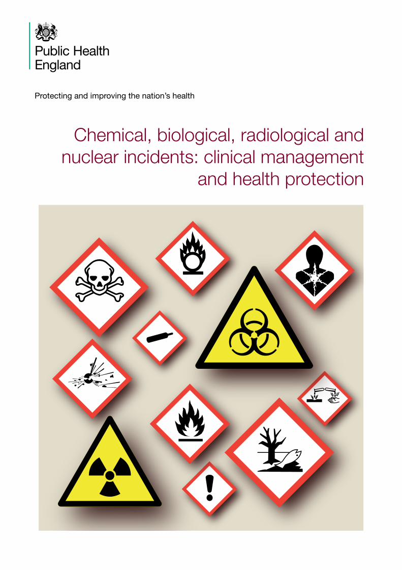
Understanding chemical hazard labels 33
Exposure Limit Values 37
Recognition and Management of Specific Chemicals and Toxidromes 39Nerve agent (organophosphate poisons) . . . . . . . . . . . . . . . . . . . . . . . . . . . . . . . . . . . . 39Toxic industrial chemicals – Chlorine (Cl2) and other irritant gasses . . . . . . . . . . . . . . . . 42Incapacitating agents (anticholinergics / volatile anaesthetics / opioids including fentanyls / psychotropics) . . . . . . . . . . . . . . . . . . . . . . . . . . . . . . . . . . 44Riot control agents (tear gas / CS gas / pepper spray / mace) . . . . . . . . . . . . . . . . . . . . 46
Contents
Chemical, biological, radiological and nuclear incidents handbook 5
Carbon Monoxide (CO) . . . . . . . . . . . . . . . . . . . . . . . . . . . . . . . . . . . . . . . . . . . . . . . . . . 49Hydrogen cyanide (HCN) and cyanide salts . . . . . . . . . . . . . . . . . . . . . . . . . . . . . . . . . . 51Hydrogen fluoride (HF) / hydrofluoric acid . . . . . . . . . . . . . . . . . . . . . . . . . . . . . . . . . . . 54Hydrogen Sulphide (H2S) . . . . . . . . . . . . . . . . . . . . . . . . . . . . . . . . . . . . . . . . . . . . . . . . 56Phosgene (COCl2) . . . . . . . . . . . . . . . . . . . . . . . . . . . . . . . . . . . . . . . . . . . . . . . . . . . . . 58Phosphine (PH3) . . . . . . . . . . . . . . . . . . . . . . . . . . . . . . . . . . . . . . . . . . . . . . . . . . . . . . 60Sulphur mustard . . . . . . . . . . . . . . . . . . . . . . . . . . . . . . . . . . . . . . . . . . . . . . . . . . . . . . 62Toxins (ricin and abrin) . . . . . . . . . . . . . . . . . . . . . . . . . . . . . . . . . . . . . . . . . . . . . . . . . . 64
Biological threats Biological agents: syndromes and differential diagnosis 68
Deliberate release incidents, recognition & response. . . . . . . . . . . . . . . . . . . . . . . . . . . . 68Differential diagnosis of unusual infections . . . . . . . . . . . . . . . . . . . . . . . . . . . . . . . . . . . 71Microbiological testing . . . . . . . . . . . . . . . . . . . . . . . . . . . . . . . . . . . . . . . . . . . . . . . . . . 73Pre and post exposure prophylaxis . . . . . . . . . . . . . . . . . . . . . . . . . . . . . . . . . . . . . . . . . 74
Recognition and Management of Specific Biological Agents 77Anthrax . . . . . . . . . . . . . . . . . . . . . . . . . . . . . . . . . . . . . . . . . . . . . . . . . . . . . . . . . . . . . 77Botulism . . . . . . . . . . . . . . . . . . . . . . . . . . . . . . . . . . . . . . . . . . . . . . . . . . . . . . . . . . . . . 80Brucellosis . . . . . . . . . . . . . . . . . . . . . . . . . . . . . . . . . . . . . . . . . . . . . . . . . . . . . . . . . . . 82Glanders . . . . . . . . . . . . . . . . . . . . . . . . . . . . . . . . . . . . . . . . . . . . . . . . . . . . . . . . . . . . 84Plague . . . . . . . . . . . . . . . . . . . . . . . . . . . . . . . . . . . . . . . . . . . . . . . . . . . . . . . . . . . . . . 86Q Fever . . . . . . . . . . . . . . . . . . . . . . . . . . . . . . . . . . . . . . . . . . . . . . . . . . . . . . . . . . . . . 88Coronavirus infections, severe acute respiratory syndrome (SARS) and Middle Eastern respiratory syndrome (MERS) . . . . . . . . . . . . . . . . . . . . . . . . . . . . . . . . . 90Shiga-toxin producing Escherichia coli (STEC) . . . . . . . . . . . . . . . . . . . . . . . . . . . . . . . . 93Smallpox . . . . . . . . . . . . . . . . . . . . . . . . . . . . . . . . . . . . . . . . . . . . . . . . . . . . . . . . . . . . 95Tularemia . . . . . . . . . . . . . . . . . . . . . . . . . . . . . . . . . . . . . . . . . . . . . . . . . . . . . . . . . . . . 98Venezuelan equine encephalitis (VEE) . . . . . . . . . . . . . . . . . . . . . . . . . . . . . . . . . . . . . . 101Viral haemorrhagic fever (VHF) . . . . . . . . . . . . . . . . . . . . . . . . . . . . . . . . . . . . . . . . . . . 103
Radiation threatsRadiation Facts 108
Ionising radiation . . . . . . . . . . . . . . . . . . . . . . . . . . . . . . . . . . . . . . . . . . . . . . . . . . . . . 108Exposure and contamination . . . . . . . . . . . . . . . . . . . . . . . . . . . . . . . . . . . . . . . . . . . . 109Measuring radioactivity and radiation . . . . . . . . . . . . . . . . . . . . . . . . . . . . . . . . . . . . . . 109Radiation doses and dose limits . . . . . . . . . . . . . . . . . . . . . . . . . . . . . . . . . . . . . . . . . . 110
Chemical, biological, radiological and nuclear incidents handbook6
Radiation injuries 111Management of acute radiation injuries. . . . . . . . . . . . . . . . . . . . . . . . . . . . . . . . . . . . . 112Post recovery management . . . . . . . . . . . . . . . . . . . . . . . . . . . . . . . . . . . . . . . . . . . . . 113Staff safety and patient management priorities during a radiation incident . . . . . . . . . . 113
Recognition and management of graded ionising radiation injuries 115European Society for Blood and Marrow Transplantation (EBMT) clinical scoring guide . . . . . . . . . . . . . . . . . . . . . . . . . . . . . . . . . . . . . . . . . . . . . . . . . . 115Assessment of large groups of individuals exposed to an ionising radiation source . . . . . . . . . . . . . . . . . . . . . . . . . . . . . . . . . . . . . . . . . . . . . . . . . . . . . . 116Managing EBMT score I patients . . . . . . . . . . . . . . . . . . . . . . . . . . . . . . . . . . . . . . . . . 117Managing EBMT score II patients . . . . . . . . . . . . . . . . . . . . . . . . . . . . . . . . . . . . . . . . . 118Managing EBMT score III patients . . . . . . . . . . . . . . . . . . . . . . . . . . . . . . . . . . . . . . . . 120
Mass Casualty GuidelinesManagement of blood-borne virus risk in victims of significant blast or multiple victim attacks creating penetrating injuries (hepatitis B, C and HIV) 123
Antibiotic prophylaxis guidance for bomb blast victims 127
Antimicrobial prophylaxis for suspected exposure to deliberate release of a bacterial agent (anthrax, plague, tularemia) 135
Management of potential injury to hearing following an attack involving blast 139
Management of potential brain injury following an attack Involving blast 143
MRI following an attack involving bomb blast or bullet wounds 145
Chemical, biological, radiological and nuclear incidents handbook 7
Executive summary
This is the first full revision of a suite of advice originally published in 2006 by the Health Protection Agency, a predecessor body of Public Health England, under the title ‘CBRN incidents: clinical management and health protection’.
This new edition updates the chemical and biological guidance given in the 2006 edition and adds additional material on a range of new and emerging threats in these areas.
The radiation incident response section has been completely re-written to integrate the well-established European Society for Blood and Marrow Transplantation’s rapid clinical assessment tool with care pathways derived from the WHO global consensus guidelines on radiation injury.
Additional materials have also been included concerning the health protection elements of response to mass casualty incidents including hearing loss, blood-borne virus transmission, antibiotic prophylaxis for bomb injury wounds and screening for cognitive impairment.
The audience for this publication remains to be first responders, emergency departments and public health and health protection professionals.
We have retained the system of keeping the disease, syndrome, or agent specific advice sheets as being capable of being used as standalone items that can be printed out and used to inform staff responding to identified threats.
This publication is the product of the advice and guidance offered by very many colleagues from the United Kingdom’s National Health Service, Defence Medical Services, and Public Health England. The guidelines on the management of chemical casualties was reviewed by colleagues from the UK’s National Poisons Information Service; and the contents of the whole document approved by National Health Service England’s Emergency Preparedness and Response Clinical Reference Group.
The editors and principal authors wish to thank all of these colleagues, too numerous to list individually, for all their help and support.

Chemical, biological, radiological and nuclear incidents handbook8
Incident Management Principles
Chemical, biological, radiological and nuclear incidents handbook10
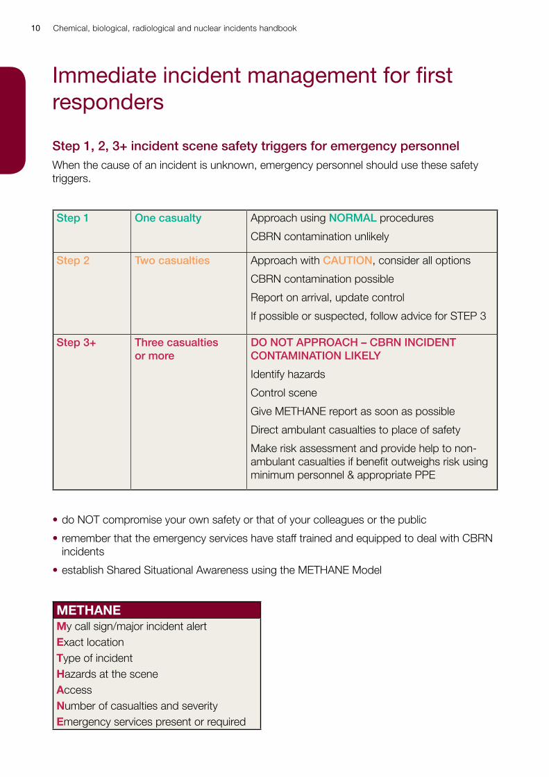
Immediate incident management for first responders
Step 1, 2, 3+ incident scene safety triggers for emergency personnelWhen the cause of an incident is unknown, emergency personnel should use these safety triggers.
Step 1 One casualty Approach using NORMAL procedures
CBRN contamination unlikely
Step 2 Two casualties Approach with CAUTION, consider all options
CBRN contamination possible
Report on arrival, update control
If possible or suspected, follow advice for STEP 3
Step 3+ Three casualties or more
DO NOT APPROACH – CBRN INCIDENT CONTAMINATION LIKELY
Identify hazards
Control scene
Give METHANE report as soon as possible
Direct ambulant casualties to place of safety
Make risk assessment and provide help to non-ambulant casualties if benefit outweighs risk using minimum personnel & appropriate PPE
• do NOT compromise your own safety or that of your colleagues or the public
• remember that the emergency services have staff trained and equipped to deal with CBRN incidents
• establish Shared Situational Awareness using the METHANE Model
METHANEMy call sign/major incident alertExact locationType of incidentHazards at the sceneAccessNumber of casualties and severityEmergency services present or required
Chemical, biological, radiological and nuclear incidents handbook 11
Medical Emergency Response Incident Teams (MERIT) and Hazardous Area Response Teams (HART)MERIT teams are deployed where ambulance personnel at scene attending an incident identify a benefit of having specialist or advanced clinical care at scene. Examples include:
• trauma requiring advanced management of pain; advanced airway management; fracture manipulation; specialist extrication including amputation; or entrapment over an extended period
• advanced triage including management of deteriorating situations
• critical care including specialised patient monitoring
• chemical, biological, radiological, nuclear (CBRN) contamination or suspected contamination
MERIT teams will normally be transported to the site by the Ambulance Service. On arrival at an incident MERITs should report to the medical incident officer (MIO), or in their absence, the ambulance incident officer (AIO) for briefing. At an incident:
• always follow instructions from the MIO, AIO, and other emergency service personnel on site
• channel all requests and queries on site through the MIO
• protect yourself – do not put your own life or health at risk to save others:
- ensure that you are wearing appropriate PPE before entering the hot zone/inner cordon or approaching any casualty
- ensure that you are clearly and appropriately identifiable - enter any inner cordon only through the inner cordon access point, where your entry will
be logged and you will be briefed about hazards - leave any inner cordon only through the inner cordon access point, so that you can be
debriefed and your departure can be logged
HART teams are deployed to support:
• incident response in hazardous areas (inner cordon) where working in specialised personal protective equipment is needed
• urban search and rescue (USAR)
• inland waterway operations (IWO)
• tactical medicine operations (TMO) where firearms may be used or public disorder is present
Chemical, biological, radiological and nuclear incidents handbook12
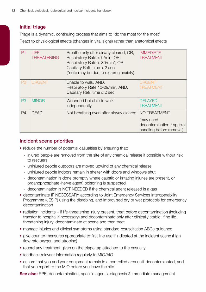
Initial triageTriage is a dynamic, continuing process that aims to ‘do the most for the most’
React to physiological effects (changes in vital signs) rather than anatomical effects
P1 LIFE THREATENING
Breathe only after airway cleared, OR, Respiratory Rate < 9/min, OR,Respiratory Rate > 30/min*, OR,Capillary Refill time > 2 sec(*note may be due to extreme anxiety)
IMMEDIATE TREATMENT
P2 URGENT Unable to walk, AND,Respiratory Rate 10-29/min, AND,Capillary Refill time ≤ 2 sec
URGENT TREATMENT
P3 MINOR Wounded but able to walk independently
DELAYED TREATMENT
P4 DEAD Not breathing even after airway cleared NO TREATMENT
(may need decontamination / special handling before removal)
Incident scene priorities• reduce the number of potential casualties by ensuring that:
- injured people are removed from the site of any chemical release if possible without risk to rescuers
- uninjured people outdoors are moved upwind of any chemical release - uninjured people indoors remain in shelter with doors and windows shut - decontamination is done promptly where caustic or irritating injuries are present, or
organophosphate (nerve agent) poisoning is suspected - decontamination is NOT NEEDED if the chemical agent released is a gas
• decontaminate IF NECESSARY according to Joint Emergency Services Interoperability Programme (JESIP) using the disrobing, and improvised dry or wet protocols for emergency decontamination
• radiation incidents – if life-threatening injury present, treat before decontamination (including transfer to hospital if necessary) and decontaminate only after clinically stable; if no life-threatening injury, decontaminate at scene and then treat
• manage injuries and clinical symptoms using standard resuscitation ABCs guidance
• give counter-measures appropriate to first line use if indicated at the incident scene (high flow rate oxygen and atropine)
• record any treatment given on the triage tag attached to the casualty
• feedback relevant information regularly to MIO/AIO
• ensure that you and your equipment remain in a controlled area until decontaminated, and that you report to the MIO before you leave the site
See also: PPE, decontamination, specific agents, diagnosis & immediate management
Chemical, biological, radiological and nuclear incidents handbook 13
Personal protective equipment (PPE)
Overview:• appropriate PPE will protect you, the patient and other patients and colleagues from
infection and from other hazards, but only if selected, worn, and discarded correctly. The algorithm below is designed to help you select PPE appropriate to the task
• don and remove PPE as you have been instructed in training.
• for advice on choosing and using PPE contact your infection control team (infection hazards) or for chemicals/radiation your local Health Protection Team who will liaise with national health protection experts as necessary
Yes/ maybe
Yes/ maybe
Yes/ maybe
Yes/ maybe
Yes/ maybe
NO
NO
NO
NO
NO
Has the patient been EXPOSED to a CHEMICAL;Is decontamination needed and NOT yet been undertaken?
WEAR FULL NHS CHEMICAL PPEBody, clothes, skin: chemical resistant coverall & bootsNose, mouth, lungs: chemical respirator (integral to PPE)Eyes: chemical resistant coverall with integral hoodHands: chemical resistant gloves
Could this be smallpox, a viral haemorrhagic fever (VHF), or other infection (eg TB) requiring airborne infection isolation?
WEAR PPE for AEROSOL SPREAD DISEASESBody, clothes, skin: full length impermeable gown, apron, hair & foot coveringsNose, mouth, lungs: FP3 respirator (fit tested/checked)Eyes: face shield, visor or gogglesHands: single disposable gloves
Will/might you be exposed to patient’s respiratory secretions (eg patient with cough, URTI, ‘flu symptoms) or are you about to do a cough-provoking procedure (eg suction, intubation, NG tube, bronchoscopy)?
WEAR PPE for DROPLET SPREAD DISEASESBody, clothes, skin: full-length gown or disposable plastic apronNose, mouth, lungs: surgical mask IF coronavirus suspected FP3 respirator (fit tested/checked)Eyes: face shield, visor or gogglesHands: single disposable gloves
Will/might you have contact with patient’s blood, body fluids, secretions, excretions, or a wound, mucosal surface, or sterile site?
WEAR PPE for STANDARD PRECAUTIONBody, clothes, skin: disposable plastic apronNose, mouth, lungs: surgical mask (if not wearing full-face shieldEyes: face shield, visor or gogglesHands: single disposable gloves
Could the patient have been contaminated by radioactive material and not been fully decontaminated?
AT ALL TIMES AND FOR ALL PATIENTS ALWAYS USE STANDARD PRECAUTIONS
WEAR PPE for STANDARD PRECAUTIONS PLUS DOUBLE GLOVES
SEEKEXPERTADVICE
See also: Decontamination, standard precautions, respiratory precautions, airborne infection isolation, and agent-specific handsheets
Chemical, biological, radiological and nuclear incidents handbook14
Decontamination of casualties
Overview: Following an overt release of hazardous materials, decontamination may be needed to reduce the risk of harm to the patient, to others, or to the wider environment.
Casualties should ideally be decontaminated at the scene, but it should be expected that contaminated casualties may also self-present to emergency department.
The first indication of a CBRN incident may be the arrival of contaminated or symptomatic patients at your emergency department or urgent care centre.
Prompt decontamination after chemical exposure is needed if caustic or irritating injuries are present, or organophosphate (nerve agent) poisoning is suspected.
Decontamination is NOT NEEDED if the chemical agent released is a gas.
In a radiation incident, treat and stabilise life-threatening injury before decontamination
Be alert to the unusual, the unexpected, and the unexplained – and if in doubt, seek expert advice.
Decontamination methods to be preferred are:
• disrobing
• improvised dry decontamination
• improvised wet decontamination
Standard Fire and Rescue Service frontline decontamination systems should only normally be used for planned and structured decontamination.
NHS secondary care decontamination facilities should be used to manage any casualties self-presenting at hospitals or where contaminated casualties have been transported directly to hospital and the nature of the contaminant may pose a risk to the secondary care environment if decontamination is not performed before admitting.
Disrobing:Casualty disrobing/undressing is a critical step in the decontamination process and is highly effective at reducing exposure to CBRN materials.
Undressing should be systematic and consistent with the steps outlined in the disrobe procedure.
Consideration must be given to ensuring the welfare and dignity of casualties as far as possible.
Improvised decontamination:Improvised decontamination is the use of an immediately available method of decontamination.
DRY decontamination is the default decontamination method in the UK - primarily for non-caustic chemical incidents.
Chemical, biological, radiological and nuclear incidents handbook 15
• perform by using use any available dry, paper tissue (eg ‘blue roll’), kitchen towel, toilet roll or paper tissues, towels and clean rags or strips of blanket or sheeting to blot the exposed skin
• sufficient absorbent material should be used to avoid transferring contamination from one part of the body to another – rubbing or blotting should not be too aggressive
• all waste material arising from decontamination should be bagged and left in a safe well ventilated space for disposal at a later stage
WET decontamination – to be used if signs and symptoms of caustic substance - is the use of water from any available source such as taps, showers, hose-reels, sprinklers, etc
• perform using any available source of water such as taps, showers, fixed installation hose-reels, sprinklers, etc
• when using water optimal decontamination takes 90 seconds and, ideally, uses a washing aid such as a cloth or sponge and soap / detergent
• the ‘RINSE-WIPE-RINSE’ Method of improvised wet decontamination should be used with preferably clean warm water and warm water containing detergent (5ml of detergent per litre of water or about three squirts of liquid detergent into a bucket of water), a sponge or soft brush
• do NOT use bleach
• self-decontamination by casualties is the best approach to take, when possible, with emergency service or emergency department personnel supervising and assisting
• RINSE the affected areas of a disrobed casualty with clean water (no detergent) using showerheads or water from buckets. RINSE from the highest point downward. RINSE only contaminated areas of skin, to avoid spread to uncontaminated areas
• WIPE the affected areas of skin with absorbent material carefully
• RINSE the decontaminated casualty with clean warm water (no detergent) to remove the detergent and any residual chemicals and dry the skin with a clean towel
• hearing aids should be removed, but should not be immersed in water: either wipe thoroughly with saline-moistened gauze, place in clear plastic specimen bag and keep with patient if patient cannot hear without them, or place with other personal effects
• eyes: if contact lenses present, remove if possible without harm; use topical anaesthetic if needed; flush eyes copiously with 0.9% saline
• If contaminated with radioactive material, survey for residual contamination and if more than 3 x background level, repeat decontamination process
• contain waste water where possible: if not possible do not delay or defer decontamination, seek advice, and inform Environmental Protection Authorities and local utility companies
See also: Home Office guidance (The decontamination of people exposed to chemical, biological, radiological or nuclear (CBRN) substances or material. Strategic National Guidance. [2nd edition, revised 2004]), see www.cabinetoffice.gov.uk, PPE, emergency contacts, radiation facts, specific agents, incident management record form.
Joint Emergency Services Interoperability Programme (JESIP) initial operational response to a CBRN incident doctrine, Home Office, 2013. www.jesip.org.uk/home
Chemical, biological, radiological and nuclear incidents handbook16
Infection control
OverviewInfection control is intended to prevent transmission of infection between patients, from patients to health care workers, and from health care workers to patients. Training in basic infection control and local policies should be provided as part of your orientation or induction. If you are in doubt about any aspect of infection control, or need training, seek help from your infection control team.
Infection control includes adopting safe behaviours and working practices (eg hand hygiene) that reduce transmission of infection; choice and use of personal protective equipment (PPE: gloves, gowns, eye/mouth/face protection, masks); patient placement (eg protective isolation for immunosuppressed patients, isolation rooms, cohort nursing); pre and post exposure prophylaxis (eg HBV immunisation); environmental measures (eg cleaning, laundering, safe disposal of clinical waste); design and engineering controls (eg auto-destruct syringes, laminar air flow), and organisational culture – working in an organisation where patient and worker safety is highly valued.
Infection control methods are used to prevent contact, droplet and aerosol transmission.
STANDARD PRECAUTIONS are applied by ALL STAFF in ALL HEALTH CARE SETTINGS to ALL PATIENTS, regardless of the patient’s diagnosis or presumed infection status, ALL THE TIME.
Any high consequence infectious diseases spread by droplet or aerosol routes should preferably be assessed initially at a designated infectious disease assessment centre where available.
Standard precautions – prevention of contact transmissionPractice good basic hygiene with regular hand cleaning.
Cover wounds or skin lesions with waterproof dressings.
Never touch your eyes, nose, mouth or face, or adjust PPE, with contaminated hands or gloves: you risk infecting yourself.
Limit your contact with items in the patient’s immediate environment to the minimum necessary for patient care and select PPE for a task according to the anticipated risks (splash, spray, splatter, touch, infection, chemical, radiation).
Wear gloves (single use disposable vinyl or nitrile) for: all invasive procedures; contact with sterile sites (including wound care and dressing changes); contact with mucous membranes, and all tasks assessed as carrying a risk of exposure to patients’ blood or body fluids.
Don gloves immediately before starting the task, remove and discard them safely on completion, and clean your hands before moving to another patient.
Work from ‘clean’ to ‘dirty’; change gloves during a procedure if you have to move from a ‘dirty’ body site to a ‘clean’ one.
If your gloves get torn or become heavily soiled during a procedure, remove them, discard them safely, clean your hands, and don a new pair.
Chemical, biological, radiological and nuclear incidents handbook 17
Wear a disposable single use plastic apron for any task where there is a risk that your clothing or uniform may be exposed to the patient’s body fluids or become wet; discard the apron safely when you complete the task and clean your hands before moving to another patient.
Wear a full-body, fluid-impermeable, gown for tasks where there is a risk of extensive splashing of body fluids or contamination of your skin.
Wear eye and face protection for tasks where there is a risk of splashes or spray to your face, eyes, nose or mouth.
Avoid using sharps if possible, and know how to use and discard sharps safely.
Do not re-sheath needles; discard used needles and syringes as a single unit into a sharps bin placed at point of use; do not overfill sharps bins.
Know what to do if there is a sharps injury or blood splash incident.
Always clear up blood spillages promptly and safely.
Never re-use single use disposable equipment (including single use ambu bags, laryngoscope blades/handles, suction equipment), and ensure that re-usable equipment is correctly decontaminated (eg by being sent to cssd) after use and before being used on another patient.
Always dispose of contaminated waste safely, and know how to deal with soiled linen.
Clean, disinfect and sterilise equipment, and decontaminate the environment as appropriate.
If you are in doubt, or unsure about any aspect of infection control, ask your infection control team for advice.
Droplet spread disease precautionsDroplets are particles (> 5 micrometres) generated when a patient coughs, sneezes or talks, and during cough-provoking procedures (eg bronchoscopy, chest physiotherapy, suctioning, intubation, nasogastric tube insertion, nebuliser therapy, non-invasive ventilation, CPAP).
Droplets expelled by an infected patient can travel for short distances through the air and, if deposited on the mucosal surfaces of the eyes, nose or mouth (or subsequently transferred there by hand-face contact) can infect anyone nearby (traditionally, within 1 metre, but possibly, at greater distances).
Diseases that are transmissible by droplet spread include: coronaviruses, influenza, pneumonic plague, monkeypox, smallpox, Mycoplasma pneumoniae, adenovirus, RSV, whooping cough, group A streptococcal infections and meningococcal meningitis (Neisseria meningitidis).
Smallpox and SARS may also be transmissible from person to person by airborne spread: airborne isolation infection precautions are required.
Hygiene measures, applied as part of standard infection control, will help to prevent transmission of these infections.
Chemical, biological, radiological and nuclear incidents handbook18
If you know or suspect that a patient has an infection transmissible by droplet spread or when the patient has syndromic signs and symptoms of an infection transmissible by droplet spread (eg URTI or flu-like illness; meningitis with petechial or ecchymotic rash; bronchiolitis in children):
• examine the patient in a single room or cubicle
• if the patient needs admission and a single room is not available, discuss patient placement with your infection control team
• encourage the patient to wear a surgical mask, provided that they can tolerate this medically
• encourage anyone accompanying or visiting the patient to wear a surgical mask
• limit patient movement outside the room to what is medically necessary
• if the patient has to be moved from the room (eg to go to X-ray), they should wear a surgical mask until they return to the room; those transporting or accompanying the patient do not need to wear a mask
• maintain droplet spread disease precautions until the suspected diagnosis has been excluded or, for bacterial infections, until 24 hours (meningococcal infection) or 72 hours (pneumonic plague) after the start of antibiotic therapy or, for viral infections, until symptoms resolve – but discuss discontinuation with your infection control team
Droplet spread disease PPE includes standard precautions PLUS a surgical mask or FP3 respirator AND eye protection during tasks that might produce splash/spray of blood or body fluids.
Don PPE in order: gown, mask, face shield or goggles, gloves.
Remove PPE in order determined by local protocol.
Note you should use an FP3 mask if the patient fulfils the case definition for coronavirus or for novel influenza until these diagnoses have been excluded.
Aerosol spread disease precautionsAirborne spread follows the inhalation of small (< 5 micrometres) particles containing an infectious agent.
These small particles may be formed after evaporation of droplets expelled from the respiratory tract (droplet nuclei) of an infected patient, or from dust particles containing microorganisms.
Small particles less than 5 micrometres can remain suspended in air, travel for longer distances in air than larger particles, and may be dispersed widely in air currents and through shared ventilation systems, so close contact (within 1-2 metres) with an infected person is not required for transmission of infection, although close contact may make transmission more likely.
Infections that may be transmissible from person to person by the airborne route include TB, chickenpox, measles, smallpox, coronaviruses, and potentially some viral haemorrhagic fevers (VHFs).
Chemical, biological, radiological and nuclear incidents handbook 19
Smallpox is most often transmitted by droplet spread or by contact, but airborne transmission from person to person has been documented.
Basic hygiene measures, applied as part of standard infection control, also help to prevent transmission of these infections.
If you know or suspect that a patient has an aerosol spread disease and has syndromic signs and symptoms of infection (eg fever & generalised vesicular rash; or fever & repetitive dry cough):
• immediately put a surgical (non-valved) mask on the patient and maintain this until patient has either been admitted to a negative pressure isolation room or assessed and the diagnosis of an infection transmissible by the airborne route excluded
• immediately place the patient in a single room/side room, close the door, and restrict entry to essential personnel
• all persons entering the room to don appropriate PPE before entering the room
• infection specialist assesses the patient. If the diagnosis of serious airborne infection cannot be excluded, arrange urgent further assessment and management in a designated specialist unit
Aerosol spread disease PPE must include gown, face shield or goggles, and FF3 (fit-checked and fit-tested) respirator donned before entry.
PPE is to be removed and safely discarded according to local protocol.
Note: surgical masks do not protect against the infection following inhalation of small (< 5 micrometres) particles. If you know or suspect that the patient has smallpox, a viral haemorrhagic fever, or other serious infection that may be transmissible by airborne infectious particles, you should wear an FF3 respirator.
See also: for detailed guidance on the management of smallpox, coronaviruses, and VHFs, see agent specific section. More detailed information available at: www.phe.org.uk and www.england.nhs.uk/ourwork/eprr/hm/
Chemical, biological, radiological and nuclear incidents handbook20
Suspect packages and parcels
Remember:• if you are EVER in ANY doubt about a package, letter or parcel
• DO NOT OPEN IT, HANDLE IT, OR MOVE IT
• CALL THE POLICE ON 999
Signs that might trigger suspicion include:
• any envelope or package with a suspicious or threatening message written on it or contained inside
• oily stains, strange odours
• envelopes that are lopsided, rigid, bulky, discoloured, or feel as though they contain powder
• unexpected envelopes or packages from foreign countries
• no postage stamp, no franking, no cancellation of the postage stamp, excessive postage
• incorrect spelling of common names, places or titles
• handwritten envelopes/packages from an unknown source particularly if addressed to an individual and marked ‘personal’ or ‘addressee only’
• symptoms (runny nose, streaming eyes, cough, skin irritation) in exposed persons
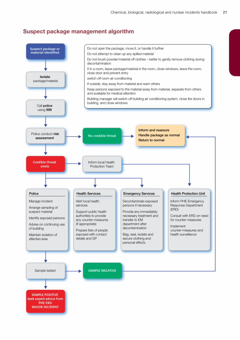
See suspect package management algorithm overleaf.
Chemical, biological, radiological and nuclear incidents handbook 21
Suspect package management algorithm
Isolate package/material
Call police using 999
Sample tested
Police conduct riskassessment
Police Health Services Emergency Services Health Protection Unit
Inform and reassureHandle package as normal
Return to normal
Inform local Health Protection Team
Suspect package ormaterial identified
Credible threat exists
SAMPLE POSITIVESeek expert advice from
PHE ERDMAJOR INCIDENT
No credible threat
SAMPLE NEGATIVE
Do not open the package, move it, or handle it further
Do not attempt to clean up any spilled material
Do not brush powder/material off clothes – better to gently remove clothing during decontamination
If in a room, leave package/material in the room, close windows, leave the room, close door and prevent entry
switch off room air conditioning
If outside, stay away from material and warn others
Keep persons exposed to the material away from material, separate from others and available for medical attention
Building manager will switch off building air conditioning system, close fire doors in building, and close windows
Manage incident
Arrange sampling of suspect material
Identify exposed persons
Advise on continuing use of building
Maintain isolation of affected area
Alert local health services
Support public health authorities to provide any counter-measures (if appropriate)
Prepare lists of people exposed with contact details and GP
Decontaminate exposed persons if necessary
Provide any immediately necessary treatment and transfer to EM department after decontamination
Bag, seal, isolate and secure clothing and personal effects
Inform PHE Emergency Response Department (ERD)
Consult with ERD on need for counter-measures
Implement counter-measures and health surveillance
Chemical, biological, radiological and nuclear incidents handbook22
Emergency contacts template
• emergency contact details should be included in your major incident plan, and should be checked and updated regularly (eg every six months and after every drill or exercise, with the task designated to a post – not a person – in the department)
• you may use this list as a template or use it to review and amend your own emergency plans
Useful internal department extension numbersEmergency department reception Chemical pathology laboratoryAdmissions Microbiology laboratoryPharmacy Haematology laboratoryEmergency theatres Blood bankMain theatres Emergency department X-rayITU Main X-rayCoronary care unit PortersPICU SecurityCSSD/sterile supplies MortuaryMajor incident control room CanteenEmergency medicine incident room Ambulance Liaison OfficerMajor incident press office Police Documentation Office
Local contacts (Internal)Contact Name Extension Bleep MobileTrust / Hospital Chief ExecutiveTrust / Hospital Senior Nurse ManagerTrust / Hospital Medical DirectorAccountable emergency OfficerConsultant chemical pathologistConsultant microbiologistConsultant haematologistConsultant infectious disease physicianInfection control leadOccupational Health leadRadiation protection/safety officerEmergency Planning Liaison OfficerHead pharmacistEmergency admissions/beds managerDuty managerChaplainsVoluntary services organiserSwitchboard supervisorDuty engineerSocial services emergency duty teamSenior Security ManagerCatering Manager
Chemical, biological, radiological and nuclear incidents handbook 23
Local contacts (External)Organisation Name Daytime
contactOut-of-hours
NHS EnglandHM Coroner / Procurator FiscalHealth Protection TeamLocal Authority Public Health TeamInfectious Disease UnitBurns UnitPoliceFire and Rescue ServiceAmbulance control
National supportNational Poisons Information Service UK (NPIS): 0344 892 0111 Ireland (NPIC): (01) 809 2566PHE Centre for Radiation, Chemicals and the Environment 0344 892 0555PHE Centre for Infections 0208 200 4400National Imported Fever Service 0844 778 8990PHE Emergency Response Department (ask for the ERD duty-officer) 300 303 3493
Chemical, biological, radiological and nuclear incidents handbook24
Incident management records
Overview:• many, if not all, major incidents, accidents or outbreaks will be followed by an investigation,
it is therefore very important that your records are comprehensive, contemporary, and legible
• incident management records should include the details of ALL advice given or received, and ALL actions taken to protect yourself, staff, patients or the public, or to inform others
• all records must be timed, dated and signed, preferably in a perfect bound log book with numbered pages. Records should be contemporaneous and any corrections or amendments made according to accepted best practice directions
• the form on the next page may be helpful, it can be freely copied. It may not cover everything, so please amend it as necessary

Chemical, biological, radiological and nuclear incidents handbook 25
Incident advice formUnit: Dept: Date:
Type of incident Place of incident No. of casualties
Task Advice received or action taken
(sign, date, time)
Source of advice
(name, date, time)
Contact details
Staff protection / PPE & safe system of working issues
Security of site
Air-conditioning system actions
Patient containment / contact tracing
Decontamination
Clinical investigations
Post-exposure treatment
Environmental sampling
Who informed?
Chemical, biological, radiological and nuclear incidents handbook26
Chain of evidence documentation
Overview:• if a deliberate release is suspected or there are other forensic considerations, chain of
evidence (sometimes called ‘chain of custody’) documentation will be needed for samples
• chain of evidence forms are intended to provide a complete record of the ‘life’ of a sample – from obtaining the sample, through testing (perhaps in two or three different laboratories), to storage
• any break in the chain of documentation may compromise the evidential value of the sample
• samples from a single patient to a single destination (eg microbiology, toxicology laboratory) can be grouped together on the same form
• every transfer of a sample must be documented. If you use the form below, which may be freely copied or used as a template for your own form, you will need to complete a new form for each transfer (eg from the person who took the sample to the porter who will take the sample to the laboratory; from porter to scientist; from laboratory to courier service; from courier service to scientist in reference laboratory). All the forms in this chain must be numbered in sequence
• keep all the forms for one set of samples together – and keep the originals carefully: photocopies cannot usually be used as evidence
• the consultant in charge of the case should authorise the transfer of the sample(s) to the laboratory. To prevent delay, particularly for specimens critical to patient care (eg group and save, cross match, ABGs), authorisation may be given verbally – but the consultant must sign the form as soon as practicable thereafter
• the pro-forma on the next page is illustrative and can be used as a template for the ED unit. It can be used, as an adobe form format. The Chain of Evidence Form needs to link to any specimens take
Chemical, biological, radiological and nuclear incidents handbook 27
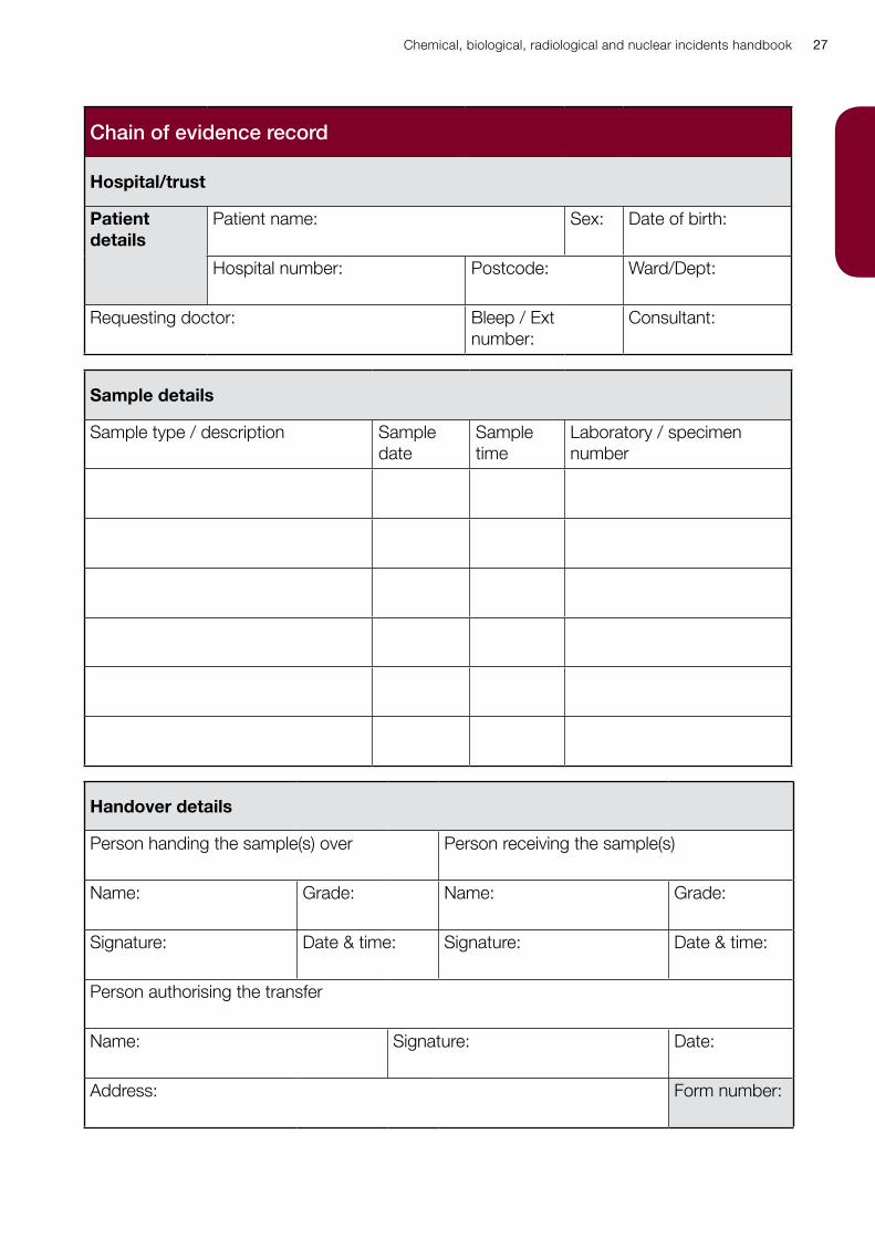
Chain of evidence record
Hospital/trust
Patient details
Patient name: Sex: Date of birth:
Hospital number: Postcode: Ward/Dept:
Requesting doctor: Bleep / Ext number:
Consultant:
Sample details
Sample type / description Sample date
Sample time
Laboratory / specimen number
Handover details
Person handing the sample(s) over Person receiving the sample(s)
Name: Grade: Name: Grade:
Signature: Date & time: Signature: Date & time:
Person authorising the transfer
Name: Signature: Date:
Address: Form number:
Chemical, biological, radiological and nuclear incidents handbook28
Further reading and other resources Important UK national sources of advice include:
Public Health England www.gov.uk/topic/health-protectionNHSE Emergency Preparedness, Resilience and Response www.england.nhs.uk/ourwork/eprrDepartment of Health www.dh.gov.uk Home Office www.homeoffice.gov.uk/counter-terrorism UK Resilience www.cabinetoffice.gov.uk/ukresilience
UK toxicology and pharmacology resources:
TOXBASE www.toxbase.org (registration required)British National Formulary www.medicinescomplete.com/mc/bnf/current (registration required)Chemical Incident Management Handbook, The Stationery Office, 2000, ISBN 0113222521
UK professional organisations for emergency and immediate care providers include:
BASICS (British Association for Immediate Care) www.basics.org.uk Royal College of Emergency Medicine www.rcem.ac.ukResuscitation Council (UK) www.resus.org.uk Advanced Life Support Group www.alsg.org.uk
Important international sources of advice include:Centers for Disease Control and Prevention, Atlanta, Emergency Preparedness and Response website www.bt.cdc.gov International Atomic Energy Authority www.iaea.org International Programme on Chemical Safety www.inchem.org International Commission on Radiological Protection www.icrp.org
Sources of expert telephone advice
PHE Chemical Hazards and Poisons Division 0844 892 0555PHE Centre for Emergency Preparedness and Response 01980 612 100National Imported Fever Service 0844 778 8990PHE Centre for Infections 020 8200 4400PHE National Poisons Information Service 0344 892 0111PHE Radiation Protection Division, office hours 01235 831600 or non office hours 01235 834590NAIR (National Arrangements for Incidents involving Radioactivity) RADSAFE 0800 834 153
Chemical, biological, radiological and nuclear incidents handbook 29
Chemical Threats
Chemical, biological, radiological and nuclear incidents handbook30
Diagnosis and early management in chemical incidents
Recognising the release of a chemicalVisual indicators of a CBRN event may include all or some of the following:
• step 1, 2, 3 plus triggers
• dead or distressed people and animals
• individuals showing unexplained signs of skin, eye or airway irritation, breathing difficulties, nausea, vomiting, sweating, blurred painful vision, disorientation, fitting, or unconsciousness
• the obvious presence of hazardous materials (smell, taste or appearance) or unusual materials/equipment
• unexplained vapour, mist clouds, oily droplets or films on surfaces or water
Clinical response if you know, or strongly suspect, that your patient has been involved in a chemical incident• use the Initial Operational Response method to coordinate emergency service actions
https://www.england.nhs.uk/ourwork/eprr/hm/#ior
• ensure that you are wearing appropriate personal protective equipment (PPE)
• decontaminate patient if needed and if this has not already been done (at scene, or outside accident and emergency department in designated NHS decontamination facilities/decontamination area)
• stabilise using standard guidelines (eg ABCDEs):
- airway (stabilise using standard guidelines (ABCs)) - supraglottic airways such as iGel preferred to intubation
- breathing (high flow rate oxygen by mask; ventilate if needed) - control any haemorrhage, set up IV access and provide fluid resuscitation if needed
• assess cause, give specific clinical counter-measures if appropriate, reassess, alert relevant Health Protection Teams within Public Health Centres, and seek expert advice from the National Poisons Information Service (PHE NPIS) and PHE Centre for Radiation, Chemicals and Environmental Hazards (CRCE)
Public health response if you know, or strongly suspect, that your patient has been involved in a chemical incident• assess the plausibility/credibility that a chemical agent has been used
• determine the immediate primary public health countermeasures – especially the need for shelter or evacuation
Chemical, biological, radiological and nuclear incidents handbook 31
• work with joint command structures to provide advice on safety of the public, specific public protection and clinical public health counter-measures that are likely to be required
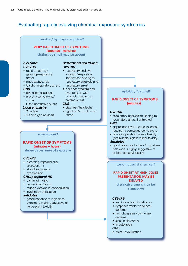
• the guideline ‘evaluating rapidly evolving chemical exposure syndromes’ may help to support your actions
• determine whether decontamination is needed and give advice on urgency and method – note that decontamination IS NOT REQUIRED for exposure to a gas
• ensure that appropriate support is available from PHE Emergency Response Division (ERD) Emergency Coordination of Scientific Advice (ECOSA Service), PHE CRCE and PHE NPIS
Chemical, biological, radiological and nuclear incidents handbook32
Evaluating rapidly evolving chemical exposure syndromes
VERY RAPID ONSET OF SYMPTOMS(seconds – minutes)
RAPID ONSET OF SYMPTOMS
RAPID ONSET OF SYMPTOMS(minutes)
CYANIDECVS /RS• rapid breathing /
gasping / respiratory arrest
• sinus tachycardia• Cardio- respiratory arrestCNS• dizziness / headache • anxiety / convulsions /
coma• Fixed unreactive pupilsblood chemistry• lactate• anion gap acidosis
CVS /RS• breathing impaired due
secretions++• sinus bradycardia• hypotensionCNS/peripheral NS• painful dim vision• convulsions / coma• muscle weakness / fasciculation• involuntary defacationantidotes• good response to high dose
atropine is highly suggestive of nerve-agent toxicity
CVS/RS• respiratory depression leading to
respiratory arrest if untreatedCNS• depressed level of consciousness
leading to coma and convulsions• pin-point pupils in severe toxicity
(not reliable sign in milder toxicity )Antidotes• good response to trial of high dose
naloxone is highly suggestive of opioid / fentanyl toxicity
CVS/RS• respiratory tract irritation ++• dyspnoea / stidor / laryngeal
oedema• bronchospasm / pulmonary
oedema• sinus tachycardia• hypotensionother• painful eye irritation
HYDROGEN SULPHIDECVS /RS• respiratory and eye
irritation / respiratory impairment leading to respiratory paralysis and respiratory arrest
• sinus tachycardia and hypotension with cyanosis – leading to cardiac arrest
CNS• dizziness / headache• agitation / convulsions /
coma
evaluating rapidly evolving chemical exposure syndromes
Chemical, biological, radiological and nuclear incidents handbook 33
Understanding chemical hazard labels
Identifying chemicals involved
CAS Registry NumbersThe CAS Register is maintained by a section of the American Chemical Society and is an authoritative collection of disclosed chemical substance information. CAS Numbers are listed for all chemicals referred to in this guidance as each chemical listed in the CAS Register is, unlike the UN number, assigned a unique numeric identifier that designates only one substance and is an accepted international method to enable searches to be made concerning specific chemical substances.
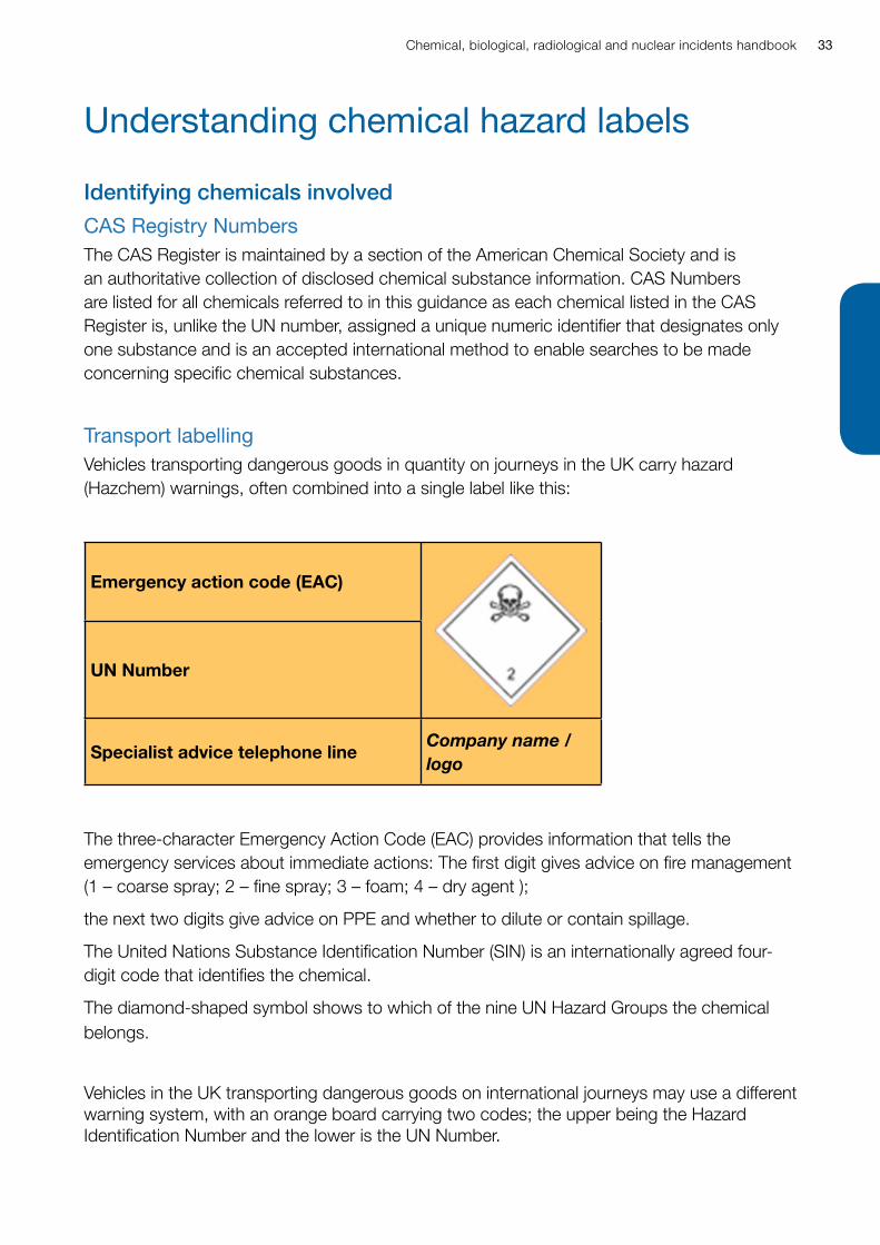
Transport labellingVehicles transporting dangerous goods in quantity on journeys in the UK carry hazard (Hazchem) warnings, often combined into a single label like this:
Emergency action code (EAC)
UN Number
Specialist advice telephone lineCompany name / logo
The three-character Emergency Action Code (EAC) provides information that tells the emergency services about immediate actions: The first digit gives advice on fire management (1 – coarse spray; 2 – fine spray; 3 – foam; 4 – dry agent );
the next two digits give advice on PPE and whether to dilute or contain spillage.
The United Nations Substance Identification Number (SIN) is an internationally agreed four-digit code that identifies the chemical.
The diamond-shaped symbol shows to which of the nine UN Hazard Groups the chemical belongs.
Vehicles in the UK transporting dangerous goods on international journeys may use a different warning system, with an orange board carrying two codes; the upper being the Hazard Identification Number and the lower is the UN Number.
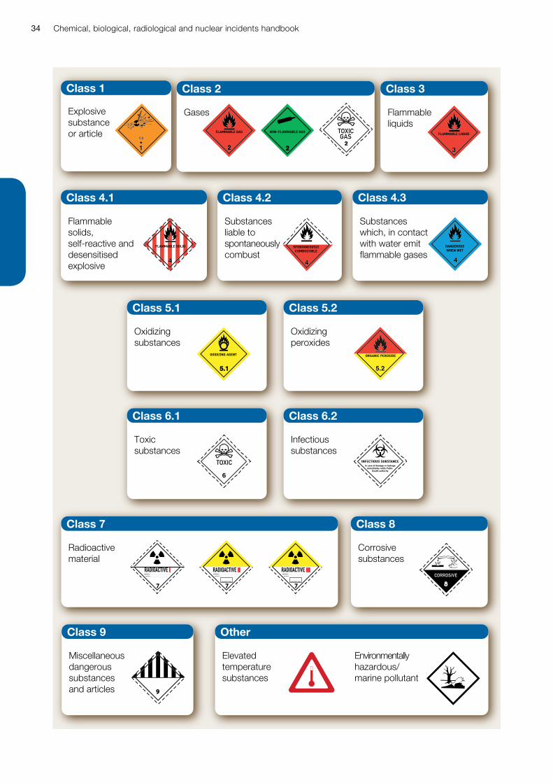
Chemical, biological, radiological and nuclear incidents handbook34
Class 1 Class 2 Class 3
Class 4.1 Class 4.2
Class 5.1 Class 5.2
Class 6.1 Class 6.2
Class 7 Class 8
OtherClass 9
Class 4.3
FLAMMABLE GAS NON-FLAMMABLE GAS TOXICGAS FLAMMABLE LIQUID
FLAMMABLE SOLID SPONTANEOUSLYCOMBUSTIBLE
DANGEROUSWHEN WET
OXIDIZING AGENT ORGANIC PEROXIDE
TOXIC INFECTIOUS SUBSTANCEIn case of damage or leakage
immediately notify Public Health authority
RADIOACTIVE I
7
CONTENTS ..................................................................ACTIVITY ............................................................
RADIOACTIVE II
7
CONTENTS ..................................................................ACTIVITY ............................................................
TRANSPORT INDEX
RADIOACTIVE III
7
CONTENTS ..................................................................ACTIVITY ............................................................
TRANSPORT INDEX
7CORROSIVE
9
Explosive substance or article
Gases Flammable liquids
Flammable solids, self-reactive and desensitised explosive
Substances liable to spontaneously combust
Oxidizing substances
Oxidizing peroxides
Toxic substances
Infectious substances
Radioactive material
Corrosive substances
Elevated temperature substances
Environmentally hazardous/ marine pollutant
Miscellaneous dangerous substances and articles
Substances which, in contact with water emit flammable gases
Chemical, biological, radiological and nuclear incidents handbook 35
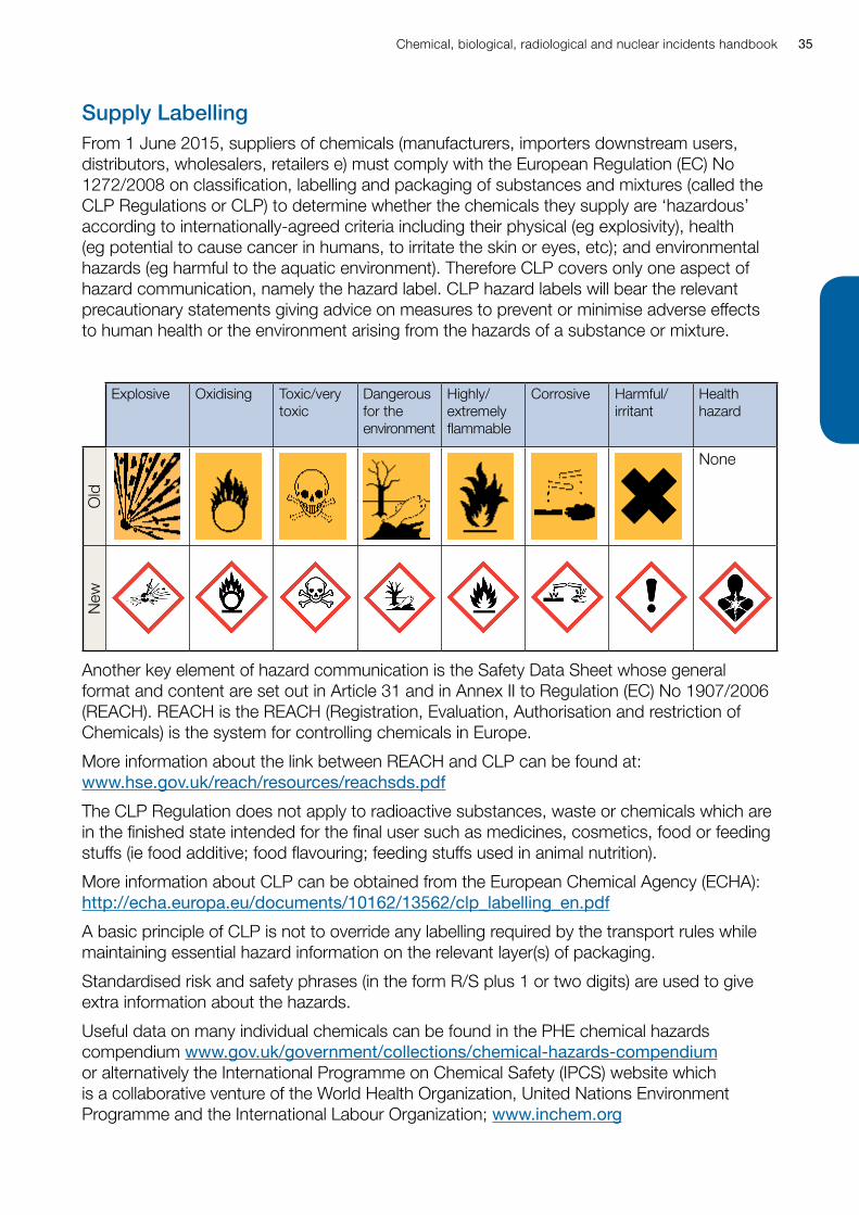
Supply LabellingFrom 1 June 2015, suppliers of chemicals (manufacturers, importers downstream users, distributors, wholesalers, retailers e) must comply with the European Regulation (EC) No 1272/2008 on classification, labelling and packaging of substances and mixtures (called the CLP Regulations or CLP) to determine whether the chemicals they supply are ‘hazardous’ according to internationally-agreed criteria including their physical (eg explosivity), health (eg potential to cause cancer in humans, to irritate the skin or eyes, etc); and environmental hazards (eg harmful to the aquatic environment). Therefore CLP covers only one aspect of hazard communication, namely the hazard label. CLP hazard labels will bear the relevant precautionary statements giving advice on measures to prevent or minimise adverse effects to human health or the environment arising from the hazards of a substance or mixture.
Explosive Oxidising Toxic/very toxic
Dangerous for the environment
Highly/ extremely flammable
Corrosive Harmful/ irritant
Health hazard
Old
None
New
Another key element of hazard communication is the Safety Data Sheet whose general format and content are set out in Article 31 and in Annex II to Regulation (EC) No 1907/2006 (REACH). REACH is the REACH (Registration, Evaluation, Authorisation and restriction of Chemicals) is the system for controlling chemicals in Europe.
More information about the link between REACH and CLP can be found at: www.hse.gov.uk/reach/resources/reachsds.pdf
The CLP Regulation does not apply to radioactive substances, waste or chemicals which are in the finished state intended for the final user such as medicines, cosmetics, food or feeding stuffs (ie food additive; food flavouring; feeding stuffs used in animal nutrition).
More information about CLP can be obtained from the European Chemical Agency (ECHA): http://echa.europa.eu/documents/10162/13562/clp_labelling_en.pdf
A basic principle of CLP is not to override any labelling required by the transport rules while maintaining essential hazard information on the relevant layer(s) of packaging.
Standardised risk and safety phrases (in the form R/S plus 1 or two digits) are used to give extra information about the hazards.
Useful data on many individual chemicals can be found in the PHE chemical hazards compendium www.gov.uk/government/collections/chemical-hazards-compendium or alternatively the International Programme on Chemical Safety (IPCS) website which is a collaborative venture of the World Health Organization, United Nations Environment Programme and the International Labour Organization; www.inchem.org
Chemical, biological, radiological and nuclear incidents handbook36
The Fire and Rescue Service will usually be able to provide information on chemical hazards from road accidents and other incidents.
TOXBASE (an on-line database, which requires pre-registration: www.toxbase.org) is the primary source of information on chemical poisoning for health care professionals in the UK.
For further expert advice, contact:
National Poisons Information Service 0344 8920111
PHE Chemicals on-call 0844 8920555
Chemical, biological, radiological and nuclear incidents handbook 37
Exposure Limit Values
In acute incidents most protective actions should be guided by the well established principles of shelter or evacuation and the need for clinical intervention guided by overt symptoms.
However, a number of publications give indicative values for actions based upon the concentration of chemicals in air (if the chemical is definitively identified, is uniformly distributed, or its maximum likely concentration can be calculated or measured).
In the United Kingdom the Health and Safety executives Workplace Exposure Limits (WELs) (listed in publication EH40) are relevant.
Internationally the Acute Exposure Guidance Levels (AEGLs) promulgated by the United States Environmental Protection Agency are widely influential.
Workplace Exposure Limits (WELs)WELs are designed for control of substances hazardous to health in the workplace. They are not emergency response reference levels / limits. However, the 15 minute reference period Short Term Exposure Limit (STEL values) and the 8-hr Time Weighted Average reference period Long-Term Exposure Limit (LTEL) values may be of help to health protection professionals when making decisions about the need, urgency and benefit of sheltering or evacuation responses.
Exceedance of STEL values may be persuasive in indicating that evacuation is required.
Levels indoors below the LTEL may be helpful in suggesting that sheltering is a safe protection method.
WEL values can be found at:
http://www.hse.gov.uk/pubns/priced/eh40.pdf
Acute Exposure Guideline Levels (AEGLs)AEGLs have been developed by the US Environmental Protection Agency to assist health protection agencies determine risk and priority for response to the accidental release of a wide range of chemical agents. They represent threshold exposure limits for the general public and are applicable to emergency exposure periods ranging from ten minutes to eight hours. These values are used in the UK to assess the severity of impact on public health whenever possible.
• AEGL-1 is the airborne concentration of a substance above which it is predicted that the general population, including susceptible individuals, could experience notable discomfort, irritation, or certain asymptomatic non-sensory effects – such effects are not disabling and are transient and reversible upon cessation of exposure
• AEGL-2 is the airborne concentration of a substance above which it is predicted that the general population, including susceptible individuals, could experience irreversible or other serious, long-lasting adverse health effects or an impaired ability to escape
• AEGL-3 is the airborne concentration of a substance above which it is predicted that the general population, including susceptible individuals, could experience life-threatening health effects or death
Chemical, biological, radiological and nuclear incidents handbook38
AEGLs are based on acute toxicology data and therefore do not reflect the effects that could result from frequent exposure. They are designed to protect the general population including the elderly and children and other vulnerable groups.
AEGL values can be found at:
www.epa.gov/aegl/access-acute-exposure-guideline-levels-aegls-values
Chemical, biological, radiological and nuclear incidents handbook 39
Chemical factsVolatile to varying degrees: sarin is much more volatile than tabun, whereas VX is non-volatile; some agents can be sprayed/aerosolised and therefore inhaled.
The vapour pressure is a measure of how quickly nerve agents evaporate and is increased by rises in ambient temperature; for example, the vapour pressure of sarin is 0.52 mmHg at 0°C and 2.9 mmHg at 25°C; nerve agents with a high vapour density compared to air (eg VX - 9.2) remain at ground level and tend to accumulate in low-lying areas.
Nerve agents, like organosphosphorus insecticides, inhibit acetylcholinesterase; acetylcholine therefore accumulates at nerve synapses and neuromuscular junctions, stimulating muscarinic and nicotinic receptors and central nervous system.
An additional reaction known as ‘aging’ also occurs as a consequence of the monodealkylation of the phosphorylated enzyme; the enzyme is then resistant to therapeutic reactivation by oximes – the time taken for aging to occur varies between different agents, but is very fast (minutes) in the case of soman.
Two deliberate releases of sarin in Japan in 1994 (Matsumoto) and 1995 (Tokyo subway) caused 18 deaths in total. Secondary effects occurred in health care workers without PPE treating un-decontaminated cases in emergency medicine departments where vapours from clothing increased in concentration in these enclosed spaces.
Summary• HIGHLY TOXIC chemical warfare agents: small drop on skin can be FATAL
• cause death by RESPIRATORY ARREST due to CNS depression and muscle paralysis by same mechanism as organophosphorus insecticides
• absorbed through skin (through clothing) and eyes, by inhalation, or by ingestion
• RAPID DRY DECONTAMINATION is essential following SKIN EXPOSURE; secondary cases can follow exposure to inadequately decontaminated primary cases
• clinical effects depend on agent, on dose, duration and route of exposure
• local effects are immediate
• SPECIFIC ANTIDOTES AVAILABLE AND CAN BE LIFE SAVING IF ADMINISTERED PROMPTLY
• seek expert advice from the National Poisons Information Service (NPIS)
• always treat as a deliberate release
Nerve agent (organophosphate poisons)
Chemical, biological, radiological and nuclear incidents handbook40
Acute effects of exposure Increased salivation, chest tightness, rhinorrhoea, bronchorrhoea and/or bronchospasm can occur within seconds or minutes of substantial inhalation of a nerve agent.
Pupils: miosis due to muscarinic effects, which may be painful and last for several days, occurs rapidly following ocular exposure to a nerve agent. It is a sensitive marker of exposure but not of severity; beware that mydriasis may be present where nicotinic effects predominate – best clinical summary is therefore presence of painful blurred vision with either miosis or mydriasis.
Skin contact with a nerve agent may produce localised sweating and fasciculation, which may spread to involve whole muscle groups.
Ingestion of food or water contaminated with nerve agents may cause abdominal pain, nausea, vomiting, diarrhoea, involuntary defecation.
All routes of exposure may result in systemic effects, including abdominal pain, nausea and vomiting, involuntary micturition and defecation, muscle weakness and fasciculation, tremor, restlessness, ataxia, coma and convulsions; bradycardia and hypotension, or tachycardia and hypertension, may occur, depending on whether muscarinic or nicotinic effects predominate; dysrhythmias may occur.
If exposure is substantial, death will occur from respiratory failure within minutes unless antidotes and ventilatory support are provided – individuals with mild or moderate exposure usually recover completely.
Late complications of poisoning may result from aspiration or hypoxic brain injury from early loss of consciousness and respiratory failure.
Management• if you suspect that your patient has been exposed to a nerve agent, ensure that you are
wearing appropriate PPE
• maintain airway, give supplemental oxygen, suction secretions
• remove patient’s clothing if not already done (double bag, seal, label, and store securely)
• for severe or moderate symptoms, establish IV access preferably, arrange assessment by anaesthetist.
• if the patient develops increased secretions, rhinorrhoea, bradycardia, hypotension, bronchorrhoea, and/or bronchospasm, administer atropine urgently
• give pralidoxime, or other available oxime, when effect atropinisation has been achieved
• control convulsions that are frequent or prolonged with diazepam, lorazepam or midazolam
• intubate and ventilate if apnoeic or severe respiratory distress (avoid succinyl choline); check ABGs, U&Es, glucose; monitor ECG, treat arrhythmias
• paralysis may mask seizures – consider EEG monitoring
• progression of symptoms suggests inadequate treatment; seek expert advice from the NPIS
• mild symptoms only (eye signs but no bronchospasm or bronchorrhoea or history of fits) observe for two hours post exposure, consider atropine or 0.5% tropicamide eye drops for painful/blurred vision, if no progression of symptoms, complete chemical exposure record form, discharge with information sheet
Chemical, biological, radiological and nuclear incidents handbook 41
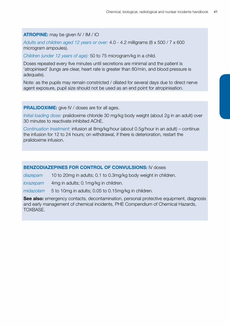
ATROPINE: may be given IV / IM / IO
Adults and children aged 12 years or over: 4.0 - 4.2 milligrams (8 x 500 / 7 x 600 microgram ampoules).
Children (under 12 years of age): 50 to 75 microgram/kg in a child.
Doses repeated every five minutes until secretions are minimal and the patient is ‘atropinised’ (lungs are clear, heart rate is greater than 80/min, and blood pressure is adequate).
Note: as the pupils may remain constricted / dilated for several days due to direct nerve agent exposure, pupil size should not be used as an end point for atropinisation.
PRALIDOXIME: give IV / doses are for all ages.
Initial loading dose: pralidoxime chloride 30 mg/kg body weight (about 2g in an adult) over 30 minutes to reactivate inhibited AChE.
Continuation treatment: infusion at 8mg/kg/hour (about 0.5g/hour in an adult) – continue the infusion for 12 to 24 hours; on withdrawal, if there is deterioration, restart the pralidoxime infusion.
BENZODIAZEPINES FOR CONTROL OF CONVULSIONS: IV doses
diazepam 10 to 20mg in adults; 0.1 to 0.3mg/kg body weight in children.
lorazepam 4mg in adults; 0.1mg/kg in children.
midazolam 5 to 10mg in adults; 0.05 to 0.15mg/kg in children.
See also: emergency contacts, decontamination, personal protective equipment, diagnosis and early management of chemical incidents, PHE Compendium of Chemical Hazards, TOXBASE.

Chemical, biological, radiological and nuclear incidents handbook42
EffectsInhalation Eyes Skincough, choking
wheeze/dyspnoea
tight chest/chest pain
nausea, vomiting
hypoxia
acid-base changes from respiratory impairment (hypercapnia respiratory acidosis and secondary renal compensation)
pneumonitis and non-cardiogenic pulmonary oedema (onset may be delayed for 12 to 24 hours following exposure)
cardiac arrest
watering, stinging
blepharospasm
frostbite after contact with compressed liquid gas
irritation
erythema or redness/chemical burns
burns or frostbite possible after contact with compressed liquid gas
Long term effects: rarely, reactive airway dysfunction syndrome with dyspnoea and increased bronchial resistance. Long-term decrease in residual volume has been described for some of these gases; those at greatest risk were older and had marked initial airflow obstruction.
Summary • IRRITANT and CORROSIVE
• exposure to high concentrations can be FATAL
• respiratory tract and eyes are main organs affected – dermal effects possible following contact with concentrated gas or pressurized liquid – those with existing respiratory disease are at greater risk (eg asthma, smokers)
• severity of effects depends on concentration and duration of exposure
• if exposure is suspected, discuss with NPIS
• where there is no clear evidence of industrial exposure or clear evidence of accidental release or self-harm (especially where two or more people are affected) then the incident must be treated as a potential deliberate release
Toxic industrial chemicals – Chlorine (Cl2) and other irritant gassesAgents with similar properties and management needs: Ammonia (NH3), Bromine (Br2), Chlorine Dioxide (ClO2), Hydrogen Chloride (HCl), Sulphur Dioxide (SO2).
See also sections on: Hydrogen Fluoride (HF), Fluorine (F2), Phosgene (COCl2), and the chemical suicide agents.
Chemical, biological, radiological and nuclear incidents handbook 43
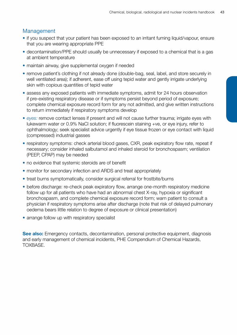
Management• if you suspect that your patient has been exposed to an irritant fuming liquid/vapour, ensure
that you are wearing appropriate PPE
• decontamination/PPE should usually be unnecessary if exposed to a chemical that is a gas at ambient temperature
• maintain airway, give supplemental oxygen if needed
• remove patient’s clothing if not already done (double-bag, seal, label, and store securely in well ventilated area); if adherent, ease off using tepid water and gently irrigate underlying skin with copious quantities of tepid water
• assess any exposed patients with immediate symptoms, admit for 24 hours observation if pre-existing respiratory disease or if symptoms persist beyond period of exposure; complete chemical exposure record form for any not admitted, and give written instructions to return immediately if respiratory symptoms develop
• eyes: remove contact lenses if present and will not cause further trauma; irrigate eyes with lukewarm water or 0.9% NaCl solution; if fluorescein staining +ve, or eye injury, refer to ophthalmology; seek specialist advice urgently if eye tissue frozen or eye contact with liquid (compressed) industrial gasses
• respiratory symptoms: check arterial blood gases, CXR, peak expiratory flow rate, repeat if necessary; consider inhaled salbutamol and inhaled steroid for bronchospasm; ventilation (PEEP, CPAP) may be needed
• no evidence that systemic steroids are of benefit
• monitor for secondary infection and ARDS and treat appropriately
• treat burns symptomatically, consider surgical referral for frostbite/burns
• before discharge: re-check peak expiratory flow, arrange one-month respiratory medicine follow up for all patients who have had an abnormal chest X-ray, hypoxia or significant bronchospasm, and complete chemical exposure record form; warn patient to consult a physician if respiratory symptoms arise after discharge (note that risk of delayed pulmonary oedema bears little relation to degree of exposure or clinical presentation)
• arrange follow up with respiratory specialist
See also: Emergency contacts, decontamination, personal protective equipment, diagnosis and early management of chemical incidents, PHE Compendium of Chemical Hazards, TOXBASE.
Chemical, biological, radiological and nuclear incidents handbook44
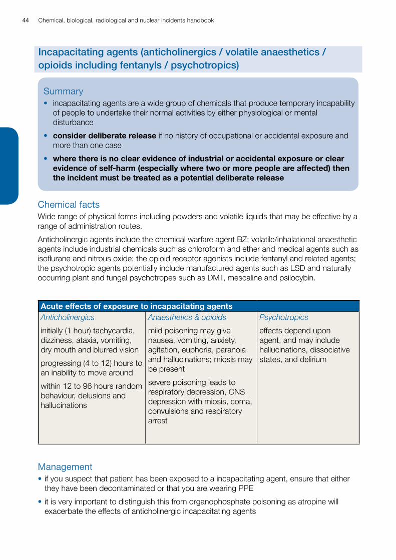
Chemical factsWide range of physical forms including powders and volatile liquids that may be effective by a range of administration routes.
Anticholinergic agents include the chemical warfare agent BZ; volatile/inhalational anaesthetic agents include industrial chemicals such as chloroform and ether and medical agents such as isoflurane and nitrous oxide; the opioid receptor agonists include fentanyl and related agents; the psychotropic agents potentially include manufactured agents such as LSD and naturally occurring plant and fungal psychotropes such as DMT, mescaline and psilocybin.
Acute effects of exposure to incapacitating agentsAnticholinergics
initially (1 hour) tachycardia, dizziness, ataxia, vomiting, dry mouth and blurred vision
progressing (4 to 12) hours to an inability to move around
within 12 to 96 hours random behaviour, delusions and hallucinations
Anaesthetics & opioids
mild poisoning may give nausea, vomiting, anxiety, agitation, euphoria, paranoia and hallucinations; miosis may be present
severe poisoning leads to respiratory depression, CNS depression with miosis, coma, convulsions and respiratory arrest
Psychotropics
effects depend upon agent, and may include hallucinations, dissociative states, and delirium
Management• if you suspect that patient has been exposed to a incapacitating agent, ensure that either
they have been decontaminated or that you are wearing PPE
• it is very important to distinguish this from organophosphate poisoning as atropine will exacerbate the effects of anticholinergic incapacitating agents
Summary• incapacitating agents are a wide group of chemicals that produce temporary incapability
of people to undertake their normal activities by either physiological or mental disturbance
• consider deliberate release if no history of occupational or accidental exposure and more than one case
• where there is no clear evidence of industrial or accidental exposure or clear evidence of self-harm (especially where two or more people are affected) then the incident must be treated as a potential deliberate release
Incapacitating agents (anticholinergics / volatile anaesthetics /opioids including fentanyls / psychotropics)
Chemical, biological, radiological and nuclear incidents handbook 45
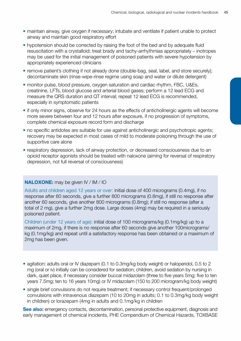
• maintain airway, give oxygen if necessary; intubate and ventilate if patient unable to protect airway and maintain good respiratory effort
• hypotension should be corrected by raising the foot of the bed and by adequate fluid resuscitation with a crystalloid; treat brady and tachy-arrhythmias appropriately – inotropes may be used for the initial management of poisoned patients with severe hypotension by appropriately experienced clinicians
• remove patient’s clothing if not already done (double-bag, seal, label, and store securely); decontaminate skin (rinse-wipe-rinse regime using soap and water or dilute detergent)
• monitor pulse, blood pressure, oxygen saturation and cardiac rhythm, FBC, U&Es, creatinine, LFTs, blood glucose and arterial blood gases; perform a 12 lead ECG and measure the QRS duration and QT interval; repeat 12 lead ECG is recommended, especially in symptomatic patients
• if only minor signs, observe for 24 hours as the effects of anticholinergic agents will become more severe between four and 12 hours after exposure, if no progression of symptoms, complete chemical exposure record form and discharge
• no specific antidotes are suitable for use against anticholinergic and psychotropic agents; recovery may be expected in most cases of mild to moderate poisoning through the use of supportive care alone
• respiratory depression, lack of airway protection, or decreased consciousness due to an opioid receptor agonists should be treated with naloxone (aiming for reversal of respiratory depression, not full reversal of consciousness)
NALOXONE: may be given IV / IM / IO
Adults and children aged 12 years or over: initial dose of 400 micrograms (0.4mg), if no response after 60 seconds, give a further 800 micrograms (0.8mg), if still no response after another 60 seconds, give another 800 micrograms (0.8mg); if still no response (after a total of 2 mg), give a further 2mg dose. Large doses (4mg) may be required in a seriously poisoned patient.
Children (under 12 years of age): initial dose of 100 micrograms/kg (0.1mg/kg) up to a maximum of 2mg, if there is no response after 60 seconds give another 100micrograms/kg (0.1mg/kg) and repeat until a satisfactory response has been obtained or a maximum of 2mg has been given.
• agitation: adults oral or IV diazepam (0.1 to 0.3mg/kg body weight) or haloperidol, 0.5 to 2 mg (oral or iv) initially can be considered for sedation; children, avoid sedation by nursing in dark, quiet place, if necessary consider buccal midazolam (three to five years 5mg; five to ten years 7.5mg; ten to 16 years 10mg) or IV midazolam (150 to 200 microgram/kg body weight)
• single brief convulsions do not require treatment; if necessary control frequent/prolonged convulsions with intravenous diazepam (10 to 20mg in adults; 0.1 to 0.3mg/kg body weight in children) or lorazepam (4mg in adults and 0.1mg/kg in children
See also: emergency contacts, decontamination, personal protective equipment, diagnosis and early management of chemical incidents, PHE Compendium of Chemical Hazards, TOXBASE
Chemical, biological, radiological and nuclear incidents handbook46
Chemical factsUsual forms of dispersal (as spray or as fine powder) result in inhalation, or skin contamination.
Effects increased by addition of hypochlorite: DO NOT use bleach in decontamination, use soap/detergent and water.
Fine powder may settle on clothes, furniture, floors, and be re-aerosolised by movement, causing secondary cases.
Clinical effects may also be caused by chemicals used in the dispersal system.
Symptoms should resolve 15 to 30 minutes after removal from exposure, particularly in the case of CS, although erythema may persist for an hour or longer.
Used by law enforcement, security forces and the military for crowd control and other purposes (eg training), and as a constituent in personal protection devices.
Summary• four agents are currently used in the UK as riot control agents: CN
(2-chloroacetophenone), CR (dibenzoxazepine), CS (2-chlorobenzylidene malononitrile) and pepper spray. Pepper spray is used by some police forces as an alternative to CS. Mace™ is a mixture of pepper spray and CS (predominantly the former)
• most UK police forces use a 5% w/v CS spray with the solvent methyl isobutyl ketone (MIBK) and nitrogen as a propellant
• these chemicals are designed to have short acting IRRITANT and INCAPACITANT effects
• main effects on EYES, RESPIRATORY SYSTEM, and, sometimes, SKIN
• onset IMMEDIATE within seconds-minutes of exposure
• clinical effects vary from mild to severe; severity increases with dose and duration of exposure
• no specific antidotes, treatment is supportive
• fatalities uncommon; those with pre-existing respiratory disease (eg asthma, smokers) may be at greater risk
• if exposure is suspected seek expert advice from the National Poisons Information Service (NPIS)
• where there is no clear evidence of industrial or accidental exposure or clear evidence of self-harm (especially where two or more people are affected) then the incident must be treated as a potential deliberate release
Riot control agents (tear gas / CS gas / pepper spray / mace)
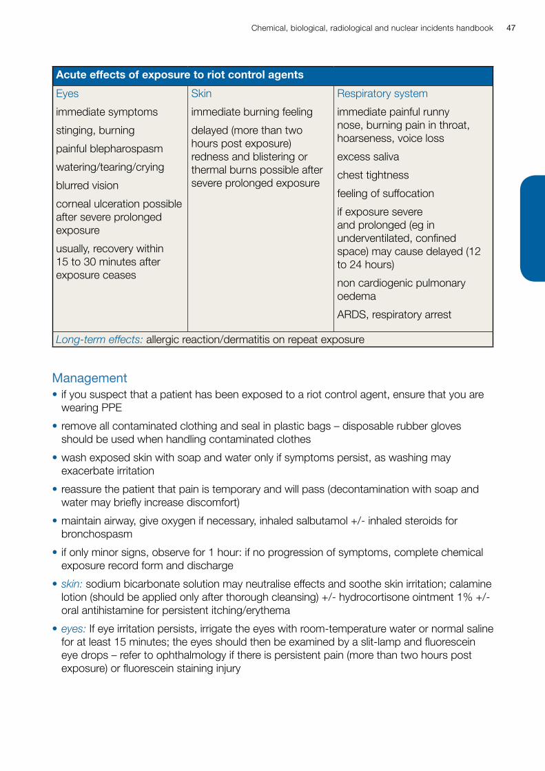
Chemical, biological, radiological and nuclear incidents handbook 47
Acute effects of exposure to riot control agents
Eyes
immediate symptoms
stinging, burning
painful blepharospasm
watering/tearing/crying
blurred vision
corneal ulceration possible after severe prolonged exposure
usually, recovery within 15 to 30 minutes after exposure ceases
Skin
immediate burning feeling
delayed (more than two hours post exposure) redness and blistering or thermal burns possible after severe prolonged exposure
Respiratory system
immediate painful runny nose, burning pain in throat, hoarseness, voice loss
excess saliva
chest tightness
feeling of suffocation
if exposure severe and prolonged (eg in underventilated, confined space) may cause delayed (12 to 24 hours)
non cardiogenic pulmonary oedema
ARDS, respiratory arrest
Long-term effects: allergic reaction/dermatitis on repeat exposure
Management• if you suspect that a patient has been exposed to a riot control agent, ensure that you are
wearing PPE
• remove all contaminated clothing and seal in plastic bags – disposable rubber gloves should be used when handling contaminated clothes
• wash exposed skin with soap and water only if symptoms persist, as washing may exacerbate irritation
• reassure the patient that pain is temporary and will pass (decontamination with soap and water may briefly increase discomfort)
• maintain airway, give oxygen if necessary, inhaled salbutamol +/- inhaled steroids for bronchospasm
• if only minor signs, observe for 1 hour: if no progression of symptoms, complete chemical exposure record form and discharge
• skin: sodium bicarbonate solution may neutralise effects and soothe skin irritation; calamine lotion (should be applied only after thorough cleansing) +/- hydrocortisone ointment 1% +/- oral antihistamine for persistent itching/erythema
• eyes: If eye irritation persists, irrigate the eyes with room-temperature water or normal saline for at least 15 minutes; the eyes should then be examined by a slit-lamp and fluorescein eye drops – refer to ophthalmology if there is persistent pain (more than two hours post exposure) or fluorescein staining injury
Chemical, biological, radiological and nuclear incidents handbook48
• admit patient if they develop severe respiratory symptoms or have not recovered completely in the two hours after exposure; observe for 24 hours. If no persistent respiratory symptoms and/or only minor eye or skin signs, complete chemical exposure record form, discharge with written information and follow-up appointment
See also: emergency contacts, decontamination, personal protective equipment, diagnosis and early management of chemical incidents, PHE Compendium of Chemical Hazards, TOXBASE.
Chemical, biological, radiological and nuclear incidents handbook 49
Clinical effectsFollowing acute exposure common signs and symptoms include headache, dizziness, confusion, disorientation, memory loss, fainting, and seizures.
Tachycardia, tachypnoea, hypotension, vasodilation, cyanosis, shock and cardiac arrest may be present.
In severe exposure cerebral oedema, coma and death may arise.
Long-term neurological effects may occur following an acute exposure, including cognitive and behavioural changes.
Management• remove from toxic atmosphere, maintain airway, give supplemental oxygen
• seek immediate advice from NPIS if symptoms are severe
• there are no specific counter-measures, for severe exposures hyperbaric oxygen should be considered in consultation with NPIS
• in symptomatic patients monitor pulse, blood pressure, oxygen saturation (note pulse oximeters are not reliable for measuring oxygen saturation in carbon monoxide poisoning) and cardiac rhythm FBC, U&Es, creatinine, LFTs, cardiac enzymes (if concomitant MI suspected), blood glucose, magnesium and arterial blood gases (including methaemoglobin concentration), perform a chest X-ray and a 12 lead ECG and measure the QRS duration and QT interval (repeat is recommended while symptoms persist)
• if the patient has clinical features of bronchospasm treat conventionally with nebulised bronchodilators and steroids
• pulmonary oedema: treat pulmonary oedema and/or acute lung injury with continuous positive airway pressure (CPAP) or in severe cases with IPPV and PEEP; the role of prophylactic corticosteroids (inhaled or systemic) and antibiotics is controversial
Summary• route of exposure is by inhalation
• exposure may be as a result of innapropriate use of petrol / diesel generators or pumps used in an indoor environment
• carbon monoxide poisoning may occur in house fires, improperly maintained gas or fossil fuel appliances, and carbon monoxide poisoning from use of charcoal barbeques in indoor / poorly ventilated environments have been recorded
• exposure may be as a result of deliberate self-harm; there is an increasing trend to use information from the internet to commit suicide using readily available household chemicals and materials and chemical suicides may occur in groups of people giving rise to suspicion of a deliberate release.
Carbon Monoxide (CO)

Chemical, biological, radiological and nuclear incidents handbook50
• hypotension should be corrected by raising the foot of the bed and by adequate fluid resuscitation with a crystalloid – treat brady and tachy-arrhythmias appropriately; inotropes may be used for the initial management of poisoned patients with severe hypotension by appropriately experienced clinicians
• metabolic acidosis should be corrected if persistent and severe with IV sodium bicarbonate or lactate
• before discharge: arrange three-month follow up, and complete chemical exposure record form
See also: emergency contacts, decontamination, personal protective equipment, diagnosis and early management of chemical incidents, chemical exposure record form, HPA Compendium of Chemical Hazards, TOXBASE.

Chemical, biological, radiological and nuclear incidents handbook 51
Chemical and clinical factsHydrogen cyanide is widely used in industry in the manufacture of plastics and nitrites; other cyanide compounds used in printing, dyeing, photography, metal cleaning and manufacturing of electronic equipment.
Cyanides are reversible cytochrome oxidase inhibitors which prevent cells from using oxygen.
Effects of cyanide• dizziness, headache
• nausea and vomiting
• rapid breathing
• sinus tachycardia
• anxiety and restlessness
• convulsions
• collapse, coma, respiratory arrest
• increased lactate concentration
• elevated anion gap acidosis
Summary• substantial exposure to HCN can lead to DEATH from respiratory or cardiac arrest
within seconds or minutes of exposure
• HCN is absorbed by inhalation, but liquid cyanide salts can be absorbed through skin, and ingestion is also possible
• severity of poisoning depends on concentration and duration of exposure; effects are less rapid after ingestion or dermal exposure
• DO NOT use mouth to mouth or mouth to nose resuscitation techniques (risk of secondary exposure)
• if exposure is suspected, discuss with NPIS
• SPECIFIC ANTIDOTES available, but SPEED CRITICAL
• where there is no clear evidence of industrial exposure or clear evidence of self-harm (especially where two or more people are affected) then the incident must be treated as a potential deliberate release
Hydrogen cyanide (HCN) and cyanide salts
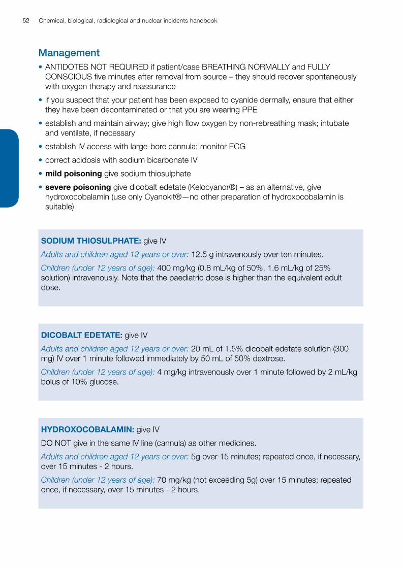
Chemical, biological, radiological and nuclear incidents handbook52
Management• ANTIDOTES NOT REQUIRED if patient/case BREATHING NORMALLY and FULLY
CONSCIOUS five minutes after removal from source – they should recover spontaneously with oxygen therapy and reassurance
• if you suspect that your patient has been exposed to cyanide dermally, ensure that either they have been decontaminated or that you are wearing PPE
• establish and maintain airway; give high flow oxygen by non-rebreathing mask; intubate and ventilate, if necessary
• establish IV access with large-bore cannula; monitor ECG
• correct acidosis with sodium bicarbonate IV
• mild poisoning give sodium thiosulphate
• severe poisoning give dicobalt edetate (Kelocyanor®) – as an alternative, give hydroxocobalamin (use only Cyanokit®—no other preparation of hydroxocobalamin is suitable)
SODIUM THIOSULPHATE: give IV
Adults and children aged 12 years or over: 12.5 g intravenously over ten minutes.
Children (under 12 years of age): 400 mg/kg (0.8 mL/kg of 50%, 1.6 mL/kg of 25% solution) intravenously. Note that the paediatric dose is higher than the equivalent adult dose.
DICOBALT EDETATE: give IV
Adults and children aged 12 years or over: 20 mL of 1.5% dicobalt edetate solution (300 mg) IV over 1 minute followed immediately by 50 mL of 50% dextrose.
Children (under 12 years of age): 4 mg/kg intravenously over 1 minute followed by 2 mL/kg bolus of 10% glucose.
HYDROXOCOBALAMIN: give IV
DO NOT give in the same IV line (cannula) as other medicines.
Adults and children aged 12 years or over: 5g over 15 minutes; repeated once, if necessary, over 15 minutes - 2 hours.
Children (under 12 years of age): 70 mg/kg (not exceeding 5g) over 15 minutes; repeated once, if necessary, over 15 minutes - 2 hours.
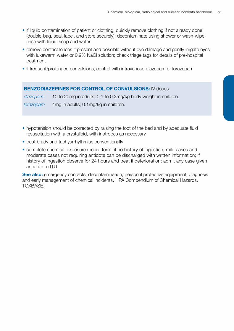
Chemical, biological, radiological and nuclear incidents handbook 53
• if liquid contamination of patient or clothing, quickly remove clothing if not already done (double-bag, seal, label, and store securely); decontaminate using shower or wash-wipe-rinse with liquid soap and water
• remove contact lenses if present and possible without eye damage and gently irrigate eyes with lukewarm water or 0.9% NaCl solution; check triage tags for details of pre-hospital treatment
• if frequent/prolonged convulsions, control with intravenous diazepam or lorazepam
BENZODIAZEPINES FOR CONTROL OF CONVULSIONS: IV doses
diazepam 10 to 20mg in adults; 0.1 to 0.3mg/kg body weight in children.
lorazepam 4mg in adults; 0.1mg/kg in children.
• hypotension should be corrected by raising the foot of the bed and by adequate fluid resuscitation with a crystalloid, with inotropes as necessary
• treat brady and tachyarrhythmias conventionally
• complete chemical exposure record form; if no history of ingestion, mild cases and moderate cases not requiring antidote can be discharged with written information; if history of ingestion observe for 24 hours and treat if deterioration; admit any case given antidote to ITU
See also: emergency contacts, decontamination, personal protective equipment, diagnosis and early management of chemical incidents, HPA Compendium of Chemical Hazards, TOXBASE.
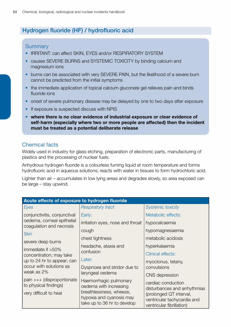
Chemical, biological, radiological and nuclear incidents handbook54
Chemical factsWidely used in industry for glass etching, preparation of electronic parts, manufacturing of plastics and the processing of nuclear fuels.
Anhydrous hydrogen fluoride is a colourless fuming liquid at room temperature and forms hydrofluoric acid in aqueous solutions; reacts with water in tissues to form hydrochloric acid.
Lighter than air – accumulates in low lying areas and degrades slowly, so area exposed can be large – stay upwind.
Acute effects of exposure to hydrogen fluorideEyes
conjunctivitis, conjunctival oedema, corneal epithelial coagulation and necrosis
Skin
severe deep burns
immediate if >50% concentration; may take up to 24 hr to appear; can occur with solutions as weak as 2%
pain +++ (disproportionate to physical findings)
very difficult to heal
Respiratory tract
Early:
irritation eyes, nose and throat
cough
chest tightness
headache, ataxia and confusion
Later:
Dyspnoea and stridor due to laryngeal oedema
Haemorrhagic pulmonary oedema with increasing breathlessness, wheeze, hypoxia and cyanosis may take up to 36 hr to develop
Systemic toxicity
Metabolic effects:
hypocalcaemia
hypomagnesaemia
metabolic acidosis
hyperkalaemia
Clinical effects:
myoclonus, tetany, convulsions
CNS depression
cardiac conduction disturbances and arrhythmias (prolonged QT interval, ventricular tachycardia and ventricular fibrillation)
Summary• IRRITANT: can affect SKIN, EYES and/or RESPIRATORY SYSTEM
• causes SEVERE BURNS and SYSTEMIC TOXICITY by binding calcium and magnesium ions
• burns can be associated with very SEVERE PAIN, but the likelihood of a severe burn cannot be predicted from the initial symptoms
• the immediate application of topical calcium gluconate gel relieves pain and binds fluoride ions
• onset of severe pulmonary disease may be delayed by one to two days after exposure
• If exposure is suspected discuss with NPIS
• where there is no clear evidence of industrial exposure or clear evidence of self-harm (especially where two or more people are affected) then the incident must be treated as a potential deliberate release
Hydrogen fluoride (HF) / hydrofluoric acid
Chemical, biological, radiological and nuclear incidents handbook 55
Management• if you suspect that your patient has been exposed to hydrofluoric acid, ensure that you are
wearing appropriate PPE
Skin exposure Remove contaminated clothing and irrigate the contaminated area with copious volumes of water as soon as possible and for one minute.
Apply calcium gluconate gel and massage into the burnt area wearing gloves appropriate to the level of decontamination – continue to massage while repeatedly applying gel until 15 minutes after the pain in the burnt area is relieved.
If skin contamination is extensive and clothing affected, be aware of the possibility of inhalation injury.
Opiate analgesia often required for pain.
Admit for 24 hours initial observation and bed rest as soon as possible after exposure even if minimal apparent clinical injury.
Check full blood count, urea and electrolytes, plasma calcium and magnesium urgently.
Treat hypovolaemic shock by replacing lost fluids and blood intravenously.
Urgently correct severe hypocalcaemia (proven or symptoms/signs suggestive of severe hypocalcaemia – tetany, arrhythmias, QTc prolongation, convulsions).
Correct hypmagnesaemia.
Treat hyperkalaemia and metabolic acidosis conventionally AFTER correcting hypocalcaemia.
Eye exposureflush the eyes with copious amounts of water or eye wash solution (sterile isotonic saline solution).
do not attempt to remove contact lenses.
there is some evidence from a single experimental study that if Hexafluorine® is applied to the eye within 25 seconds of exposure, and continued for 15 minutes, ocular damage is lessened.
Inhalationmaintain airway, give supplemental oxygen.
check full blood count, urea and electrolytes, plasma calcium and magnesium urgently; CXR.
supraglottic-epiglottic burn with erythema and oedema indicates that further oedema may occur and is an indication for early intubation or tracheostomy; treat bronchospasm conventionally with nebulised bronchodilators and steroids.
pulmonary oedema: may not appear for up to 36 hr. Treat with CPAP or IPPV/PEEP if severe; the role of prophylactic corticosteroids (inhaled or systemic) and antibiotics is controversial.
See also: emergency contacts, decontamination, personal protective equipment, diagnosis and early management of chemical incidents, PHE Compendium of Chemical Hazards, TOXBASE.
Chemical, biological, radiological and nuclear incidents handbook56
Chemical effectsSystemic effects include vomiting, diarrhoea, headache, nystagmus, dizziness, agitation, drowsiness, tremor, muscular weakness, seizures, tachycardia and hypotension.
Inhalation of high concentrations leads rapidly to collapse, respiratory paralysis, cyanosis, convulsions, coma, cardiac arrhythmias and death within minutes.
Dermal exposure causes discolouration, pain, itching, erythema and local frostbite if exposed to compressed liquid.
Ocular effects may be delayed.
Management• remove from toxic atmosphere, maintain airway, give supplemental oxygen if needed
• appropriate PPE should be worn and ensure that the scene is well ventilated
• if residual chemical materials present remove patient’s clothing if not already done (double-bag, seal, label, and store securely in well-ventilated area)
• seek immediate advice from NPIS if symptoms are severe
• there are no specific counter-measures; methaemoglobinaemia may be present in severe hydrogen sulphide and may require treatment with methylthioninium chloride (methylene blue); use high concentration oxygen (100% if possible)
• in symptomatic patients monitor pulse, blood pressure, oxygen saturation (note pulse oximeters are not reliable for measuring oxygen saturation in carbon monoxide poisoning) and cardiac rhythm FBC, U&Es, creatinine, LFTs, cardiac enzymes (if concomitant MI suspected), blood glucose, magnesium and arterial blood gases (including methaemoglobin concentration), perform a chest X-ray and a 12 lead ECG and measure the QRS duration and QT interval (repeat is recommended while symptoms persist)
• if the patient has clinical features of bronchospasm treat conventionally with nebulised bronchodilators and steroids
Summary• exposure may occur as a result of an industrial accident
• more commonly exposure may be as a result of deliberate self-harm; there is an increasing trend to use information from the internet to commit suicide using readily available household chemicals and materials and chemical suicides may occur in groups of people giving rise to suspicion of a deliberate release. individuals or groups may signpost chemical the risks to first responders
• if exposure is suspected, discuss with NPIS
• where there is no clear evidence of industrial exposure or clear evidence of self-harm (especially where two or more people are affected) then the incident must be treated as a potential deliberate release
Hydrogen Sulphide (H2S)
Chemical, biological, radiological and nuclear incidents handbook 57
• pulmonary oedema: treat pulmonary oedema and/or acute lung injury with continuous positive airway pressure (CPAP) or in severe cases with IPPV and PEEP; the role of prophylactic corticosteroids (inhaled or systemic) and antibiotics is controversial
• hypotension should be corrected by raising the foot of the bed and by adequate fluid resuscitation with a crystalloid – treat brady and tachy-arrhythmias appropriately; inotropes may be used for the initial management of poisoned patients with severe hypotension by appropriately experienced clinicians
• metabolic acidosis should be corrected if persistent and severe with IV sodium bicarbonate or lactate
• remove contact lenses if present and immediately irrigate the affected eye thoroughly with water or 0.9% saline for at least 10 to 15 minutes; arrange urgent ophthalmic opinion if corneal damage present
• before discharge: arrange three-month follow up, and complete chemical exposure record form
See also: emergency contacts, decontamination, personal protective equipment, diagnosis and early management of chemical incidents, chemical exposure record form, HPA Compendium of Chemical Hazards, TOXBASE.
Chemical, biological, radiological and nuclear incidents handbook58
Chemical and clinical factsWidely used in industry in manufacture of isocyanates, polyurethane and polycarbonate resins, pesticides, herbicides and dyes.
At room temperature, phosgene is a gas; with cooling and pressure, phosgene gas can be converted into a liquid so that it can be shipped and stored - when liquid phosgene is released, it quickly turns into a gas that stays close to the ground and spreads rapidly.
When combined with water in the body, phosgene produces hydrogen chloride and carbon dioxide, although as the gas is poorly soluble in water, only small amounts of hydrochloric acid are produced under normal physiological conditions; hydrogen chloride production is only relevant in causing mucus membrane irritation when phosgene is present at relatively high concentrations.
Phosgene produces direct damage to lung surfactant and peroxidation of lipids, including membrane phospholipids; and depending on the inhaled dose (rather than the exposure concentration) there may be a symptom free period of up to 48 hours following acute exposure.
Acute effects of exposure to phosgeneInitial (irritant phase)watering painful eyesblepharospasmcoughingtight chest/chest painwheeze, dyspnoealaryngospasmnausea and vomitingcontact burns/eye damage if exposed to liquidnon-cardiogenic pulmonary oedema
Latent period (not invariable)2-48 hoursno symptomspatient appears well
Delayed (oedema phase)non-cardiogenic pulmonary oedema/ARDSfrothy sputum, wheeze, cough, breathing difficultyhypotension, hypoxiatachycardiabronchial necrosissecondary pneumoniadeath
Chronic effects: Reactive airway dysfunction syndrome: dyspnoea and increased bronchial resistance for 3-6 months. Rarely, chronic bronchitis, emphysema, bronchiectasis, pulmonary fibrosis
Summary• IRRITANT: can affect SKIN, EYES or RESPIRATORY SYSTEM
• inhalation of phosgene can be fatal
• outcome cannot be predicted from initial symptoms and exercise will increase severity of symptoms
• if exposure is suspected, discuss with NPIS
• where there is no clear evidence of industrial exposure or clear evidence of self-harm (especially where two or more people are affected) then the incident must be treated as a potential deliberate release
Phosgene (COCl2)
Chemical, biological, radiological and nuclear incidents handbook 59
Management• in the circumstances of a large scale release, not all patients presenting will have been
exposed
• patients should not be treated if they are asymptomatic as there is no evidence that delaying therapy causes harm
• patients should be admitted for 24 hours initial observation and placed on bed rest as soon as possible after exposure
• if you suspect that a patient has been exposed to phosgene, ensure that either they have been decontaminated (particularly if exposure has been to liquid phosgene) or that you are wearing PPE
• remove patient’s clothing if not already done (double-bag, seal, label, and store securely); if adherent, ease off using tepid water and gently irrigate underlying skin with copious quantities of tepid water
• remove contact lenses if present; irrigate eyes with lukewarm water or 0.9% NaCl solution; if fluorescein staining injury, or overt eye injury, refer ophthalmology; seek urgent specialist advice if eye contact with liquid phosgene
• all patients should have regular physical examinations and measurement of peripheral oxygen saturations – check respiratory rate, pulse oximetry, ABG, CXR, peak expiratory flow rate
• once a patient’s oxygen saturation falls below 94%, treatment with the lowest concentration of oxygen required to maintain their oxygen saturations in the normal range should be started
• consider elective intubation early using the ARDSnet protective ventilation strategy as it lessens injury severity and significantly improves survival
• experimental evidence suggests that nebulised b2-agonists (eg salbutamol 5mg by nebuliser every four hours) may reduce lung inflammation if administered within one hour of exposure
• experimental evidence suggests that there is no benefit from nebulised steroid even when administered one hour after exposure, or high dose corticosteroid if administered intravenously ≥ six hours after exposure (it is not known if IV corticosteroids given earlier are of benefit)
• treat burns from contact with liquid phosgene symptomatically; may need surgical referral for frostbite
• before discharge (at 24 hours if asymptomatic): recheck lung function, arrange respiratory medicine review at three months, complete chemical exposure record form
See also: emergency contacts, decontamination, personal protective equipment, diagnosis and early management of chemical incidents, chemical exposure record form.
Chemical, biological, radiological and nuclear incidents handbook60
Chemical factsPhosphides (aluminium, zinc, magnesium) are used widely to protect grain held in stores, the holds of ships and in wagons transporting it by rail.
Phosphide interacts with moisture in the air between the grains to liberate phosphine.
Following ingestion of a metal phosphide, phosphine is liberated in the body.
Phosphine is available in cylinders, either alone or combined with carbon dioxide.
Clinical effectsPhosphine poisoning may occur following inhalation of phosphine or the ingestion of a phosphide.
Inhalation causes irritation to the mucous membranes of the nose, mouth, throat and respiratory tract; chest tightness, breathlessness, chest pain, palpitations, and severe retrosternal pain are common.
Nausea, vomiting, epigastric pain and diarrhoea may be so striking that a diagnosis of acute gastroenteritis is made.
Consciousness is usually only mildly depressed; headache, dizziness and staggering gait may ensue.
In more severe cases acute heart failure, pulmonary oedema (sometimes non-cardiogenic) and ventricular arrhythmias have been observed, particularly in children; cardiogenic shock results in metabolic acidosis, hyperlactataemia and acute renal failure.
Other less common features include disseminated intravascular coagulation and hepatic necrosis.
Summary• common routes of exposure are inhalation or ingestion (metal phosphides)
• exposure may be as a result of deliberate self-harm; there is an increasing trend to use information from the internet to commit suicide using readily available household chemicals and materials and chemical suicides may occur in groups of people giving rise to suspicion of a deliberate release. individuals or groups may signpost chemical the risks to first responders
• if exposure is suspected, discuss with NPIS
• where there is no clear evidence of industrial or accidental exposure or clear evidence of self-harm (especially where two or more people are affected) then the incident must be treated as a potential deliberate release
Phosphine (PH3)

Chemical, biological, radiological and nuclear incidents handbook 61
Management • remove from toxic atmosphere, maintain airway, give supplemental oxygen if needed
• if residual chemical materials present (especially metal phosphide rodenticide tablets) remove patient’s clothing if not already done (double-bag, seal, label, and store securely in well-ventilated area)
• remove contact lenses if present and immediately irrigate the affected eye thoroughly with water or 0.9% saline for at least 10 to 15 minutes; arrange urgent ophthalmic opinion if corneal damage present
• in symptomatic patients monitor pulse, blood pressure, oxygen saturation and cardiac rhythm, FBC, U&Es, creatinine, LFTs, cardiac enzymes, blood glucose, and arterial blood gases, perform a chest X-ray and a 12 lead ECG and measure the QRS duration and QT interval
• pulmonary oedema: treat pulmonary oedema and/or acute lung injury with continuous positive airway pressure (CPAP) or in severe cases with IPPV and PEEP
• hypotension should be corrected by raising the foot of the bed and by adequate fluid resuscitation with a crystalloid, with inotropes as necessary.
• treat brady and tachy-arrhythmias appropriately.
• metabolic acidosis should be corrected if persistent and severe with IV sodium bicarbonate
• before discharge: arrange a three-month follow up, and complete chemical exposure record form
See also: emergency contacts, decontamination, personal protective equipment, diagnosis and early management of chemical incidents, chemical exposure record form, HPA Compendium of Chemical Hazards, TOXBASE.
Chemical, biological, radiological and nuclear incidents handbook62
Chemical factsSulphur mustard, a chemical sometimes referred to as mustard gas, is a liquid which boils at 217°C and freezes at 13°C to 15°C, which explains its persistence in the environment.
Clinical effectsSeverity increases with dose and duration of exposure; and although tissue damage begins immediately on exposure, some clinical effects may be delayed and evolve over hours or days.
Skin exposure produces skin blisters and skin necrosis; erythema develops within a few hours of exposure; vesication usually begins on the second day after exposure and may progress for up to two weeks; necrosis of the epidermis and superficial dermis is complete four to six days after exposure and separation of necrotic slough then begins; scab formation begins within seven days; by 16 to 20 days, separation of slough is complete and re-epithelialization begins.
Eye exposure results in corneal damage with temporary blindness.
Inhalation causes nose bleeds, tracheobronchitis, acute respiratory distress syndrome (ARDS), and with time, tracheobronchial stenosis, chronic bronchitis, and bronchiolitis obliterans.
Long-term disability due to respiratory complications is common.
Sulphur mustard depresses bone marrow function which may lead to secondary infection.
Sulphur mustard is a recognized human carcinogen.
Summary• droplets of sulphur mustard can pose a risk to health from inhalation, ingestion of
contaminated food and water, as well as contact with the skin and eyes
• sulphur mustard may produce severely incapacitating eye, skin, respiratory tract, and possibly systemic, damage. It can be lethal at high concentrations
• rapidly absorbed through skin (can also penetrate clothing) and eyes, by inhalation, and (rarely) by ingestion
• RAPID WET DECONTAMINATION CRITICAL: secondary cases can follow exposure to inadequately decontaminated primary cases
• if exposure is suspected, discuss with NPIS
• always treat as deliberate release if the chemical exposure occurred in a public place, or anywhere other than at an industrial site where sulphur mustard is known to be used / stored
Sulphur mustard
Chemical, biological, radiological and nuclear incidents handbook 63
Management• give analgesia (may require opiates) for eye pain, erythema, blisters; AVOID topical
anaesthetic eye drops
• if you suspect that a patient has been exposed to sulphur mustard, ensure that either they have been decontaminated or that you are wearing PPE
• maintain airway, give oxygen if necessary, inhaled salbutamol for bronchospasm
• remove patient’s clothing if not already done (double-bag, seal, label, and store securely); shower or wash down or rinse-wipe-rinse with liquid soap and water, or dilute detergent;
• eyes: remove contact lenses if present, decontaminate eyes rapidly with lukewarm water or 0.9% NaCl solution; if blepharospasm develops seek urgent ophthalmology opinion; do not patch, but do prevent lids sticking together (sterile petroleum jelly, boric acid ointment 5%); use cycloplegic paralyses cillary muscle eye drops to prevent iris adherance to the cornea (anterior chamber synechiae) – atropine or homatropine tds
• skin: an oral antihistamine should be given and topical calamine solution should be applied for itching – dilute topical corticosteroids have proved beneficial in relieving irritation and reducing the attendant oedema at exposed sites, but its use had little or no effect on the subsequent rate of healing of the lesions
• debride ruptured blisters, clean with sterile 0.9% NaCl solution, cover small areas with petroleum gauze – topical bacteriostatic agents such as 1% silver sulphadiazine (Flamazine™) cream may reduce the incidence of secondary infection once the blisters have ruptured; intensive nursing care may be needed, especially if perineum or genitalia affected; seek early referral to plastic surgeon/burns unit; experimentally mechanical dermabrasion and laser debridement (“lasablation”) both produced an increased rate of wound healing and may be of benefit in a clinical context
• monitor FBC (WCC high initially, leucopaenia at three to five days, possible bone marrow depression later) – If bone marrow depression occurs consult a haematologist
• if no eye or skin signs develop within eight hours, complete chemical exposure record form and discharge with written information
• if only minor eye/skin signs occur by eight hours, observe for further 24 hours, then, if no progression and only minor erythema, small blisters, or minor eye irritation/conjunctivitis, complete chemical exposure record form, discharge with written information and follow-up appointment
See also: emergency contacts, decontamination, personal protective equipment, diagnosis and early management of chemical incidents, chemical exposure record form.
Chemical, biological, radiological and nuclear incidents handbook64
Acute effects of exposure to ricin/abrin (presentation variable, severity of initial symptoms may not be a good indicator of outcome)Likely after ingestion
abdominal pain, cramps
vomiting (often profuse)
diarrhoea (may be bloody)
gastrointestinal bleeding
hypovolaemic shock, DIC, multiple organ failure
Likely after inhalation
Principal toxic effects are confined to the lung
fever
cough
dyspnoea
tight chest
fibropurulent pneumonia
non cardiogenic pulmonary oedema/ARDS
respiratory failure
Chronic/long term effects: few data available. Animals surviving inhalational exposure have dose-dependent lung damage
Summary• ricin is present in, and can be extracted from beans (seeds) of the castor oil
plant, Ricinus communis – seed cases each contain three shiny red- brown-grey streaked seeds
• abrin is found in Abrus precatorius (‘rosary pea’, ‘jequirity bean’) – seeds are red-black or white-black
• one million tons of castor oil beans are processed each year: waste is 5% ricin by weight; there is no comparable industrial source of abrin
• accidental poisoning can occur after chewing castor beans or rosary peas, which are used to make necklaces, bracelets, prayer beads, and to fill maracas (one to three seeds may be fatal for child, eight may be fatal for adults, though adults have survived ingestion of ten to 30 seeds, and children four to ten)
• extremist groups in the US and UK are known to have planned to use ricin
• toxins may be inhaled (if aerosolised) or injected
• although highly toxic after injection, multiple cases unlikely
• cutaneous and systemic allergic responses to ricin exposure have been reported
• if exposure is suspected discuss with NPIS
• always treat any case as a potential deliberate release
Toxins (ricin and abrin)
Chemical, biological, radiological and nuclear incidents handbook 65
Management• if you suspect that a patient has been exposed to aerosolised ricin or abrin, ensure that you
are wearing PPE
• if patient is exposed to aerosolised ricin or abrin: remove patient’s clothing if not already done (double-bag, seal, label, and store securely)
• if not already done decontaminate skin (rinse-wipe-rinse regime using liquid soap and water, or dilute detergent)
• maintain airway, prevent aspiration of vomit, give supplemental oxygen if needed; do NOT give antispasmodics
• if contact lenses are present, remove if possible, and irrigate eyes with lukewarm water or 0.9% NaCl solution
• admit all symptomatic patients
• if the patient has no symptoms, but is thought to have been exposed to aerosolised or injected ricin or abrin, admit, observe, complete chemical exposure record form, and discharge with information sheet if still symptom free 24 hours later
• if the patient has no symptoms, but is thought to have ingested ricin or abrin, admit, observe, complete chemical exposure record form, and discharge with information sheet if still symptom free eight hours later
• if patient has ingested ricin or abrin: discuss whole bowel irrigation with NPIS
• if respiratory symptoms, check: arterial blood gases, CXR, peak expiratory flow rate, and repeat if necessary; consider inhaled salbutamol and inhaled steroids, ventilation (PEEP); monitor for secondary infection and ARDS and treat appropriately
• replace gastrointestinal fluid losses IV, and correct and maintain electrolyte balance
• seek expert advice from PHE CRCE or NPIS regarding analytical confirmation of ricin/abrin exposure
See also: emergency contacts, decontamination, personal protective equipment, diagnosis and early management of chemical incidents, PHE Compendium of Chemical Hazards, TOXBASE.

Chemical, biological, radiological and nuclear incidents handbook66

Chemical, biological, radiological and nuclear incidents handbook 67
Biological Threats
Chemical, biological, radiological and nuclear incidents handbook68
Biological agents: syndromes and differential diagnosis
Deliberate release incidentsA deliberate release may be overt (announced openly by perpetrators, eg the envelopes containing threatening notes and anthrax spores distributed through US Postal Service in 2001), or covert (unannounced, without any warning or indication of the organism involved) eg 1984 Salmonella typhimurium contamination of salad bars in restaurants in Dalles, Oregon, by followers of Baghwan Shree Rajneesh, when 751 people developed gastroenteritis.
Many different organisms could, in theory, be used deliberately and be distributed through food, water, or the air (by an explosive device, aerosol canister, or crop duster).
This manual focuses on organisms that could be aerosolised and/or would cause serious or fatal infections.
Recognition of release incidentsIntentional and naturally occurring outbreaks may be indistinguishable initially.
Symptoms of some forms of intentional or accidental chemical poisoning may mimic some infections (eg arsenic-contaminated coffee, Maine, 2003, and nicotine-contaminated minced meat, Michigan, 2003, both initially thought to be gastroenteritis; thallium poisoning, Florida, 1988, initially thought to be botulism).
Early recognition of a covert release of a biological agent will be achieved only if clinicians remain aware of the possibility, and are willing to alert and consult with their microbiologist, ID physician and Health Protection Team on suspicion, and before a definitive diagnosis has been reached.
Be alert to the unusual, the unexpected, and the case that ‘just doesn’t fit’:
• an unusual illness (eg sudden unexplained febrile death, critical illness or pneumonia death in a previously healthy young adult)
• an unusual number of patients with the same symptoms
• an illness unusual for the time of year (eg ‘flu’ in summer)
• an illness unusual for the patient’s age group (eg ‘chickenpox’ in a middle-aged adult)
• an illness in an unusual patient (eg cutaneous anthrax in a patient with no history of contact with animals, animal hides or products)
• an illness acquired in an unusual place (eg tularemia acquired in the UK)
• unusual clinical signs (eg mediastinal widening on CXR; sudden onset of symmetrical flaccid paralysis)
• unusual progression of an illness (eg lack of response to usually effective antibiotics; ‘chickenpox’ rash predominant on extremities)
Chemical, biological, radiological and nuclear incidents handbook 69
• any confirmed case of smallpox, plague, pulmonary anthrax, glanders, tularemia, Venezuelan equine encephalitis (VEE) or viral haemorrhagic fever (VHF) without history of travel to an endemic area in the UK should be assumed to be the result of a deliberate release until proven otherwise
Responding to a suspected release incident• use the action list that follows in conjunction with the handsheets on infection control
(standard precautions, respiratory precautions, airborne infection isolation) and PPE in the section on generic incident management, and those on microbiological testing and specific infections in this section
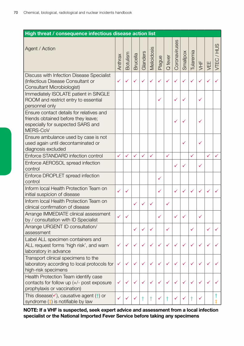
Chemical, biological, radiological and nuclear incidents handbook70
High threat / consequence infectious disease action list
Agent / Action
Ant
hrax
Bot
ulis
m
Bru
cella
Gla
nder
s
Mel
ioid
osis
Pla
gue
Q fe
ver
Cor
onav
iruse
s
Sm
allp
ox
Tula
rem
ia
VH
F
VE
E
VTE
C /
HU
S
Discuss with Infection Disease Specialist (Infectious Disease Consultant or Consultant Microbiologist)
ü ü ü ü ü ü ü ü ü ü ü ü ü
Immediately ISOLATE patient in SINGLE ROOM and restrict entry to essential personnel only
ü ü ü ü
Ensure contact details for relatives and friends obtained before they leave; especially for suspected SARS and MERS-CoV
ü ü ü
Ensure ambulance used by case is not used again until decontaminated or diagnosis excluded
ü ü
Enforce STANDARD infection control ü ü ü ü ü ü ü ü üEnforce AEROSOL spread infection control ü ü ü
Enforce DROPLET spread infection control ü
Inform local Health Protection Team on initial suspicion of disease ü ü ü ü ü ü ü ü ü
Inform local Health Protection Team on clinical confirmation of disease ü ü ü ü
Arrange IMMEDIATE clinical assessment by / consultation with ID Specialist ü ü ü ü ü ü
Arrange URGENT ID consultation/assessment ü ü ü ü ü ü ü
Label ALL specimen containers and ALL request forms ‘high risk’, and warn laboratory in advance
ü ü ü ü ü ü ü ü ü ü ü ü ü
Transport clinical specimens to the laboratory according to local protocols for high-risk specimens
ü ü ü ü ü ü ü ü ü ü ü ü ü
Health Protection Team identify case contacts for follow up (+/- post exposure prophylaxis or vaccination)
ü ü ü ü ü ü ü ü ü ü ü ü ü
This disease(ü), causative agent (†) or syndrome (‡) is notifiable by law
ü ü ü † † ü † ü ü † ü† ‡
NOTE: If a VHF is suspected, seek expert advice and assessment from a local infection specialist or the National Imported Fever Service before taking any specimens
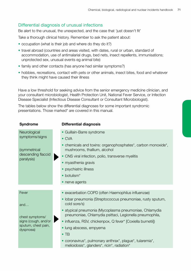
Chemical, biological, radiological and nuclear incidents handbook 71
Differential diagnosis of unusual infectionsBe alert to the unusual, the unexpected, and the case that ‘just doesn’t fit’
Take a thorough clinical history. Remember to ask the patient about:
• occupation (what is their job and where do they do it?)
• travel abroad (countries and areas visited, with dates, rural or urban, standard of accommodation, use of antimalarial drugs, bed nets, insect repellents, immunisations; unprotected sex, unusual events eg animal bite)
• family and other contacts (has anyone had similar symptoms?)
• hobbies, recreations, contact with pets or other animals, insect bites, food and whatever they think might have caused their illness
Have a low threshold for seeking advice from the senior emergency medicine clinician, and your consultant microbiologist, Health Protection Unit, National Fever Service, or Infection Disease Specialist (Infectious Disease Consultant or Consultant Microbiologist).
The tables below show the differential diagnoses for some important syndromic presentations. Those marked* are covered in this manual.
Syndrome Differential diagnosis
Neurological symptoms/signs
(symmetrical descending flaccid paralysis)
• Guillain-Barre syndrome
• CVA
• chemicals and toxins: organophosphates*, carbon monoxide*, mushrooms, thallium, alcohol
• CNS viral infection, polio, transverse myelitis
• myasthenia gravis
• psychiatric illness
• botulism*
• nerve agents
Fever
and…
chest symptoms/signs (cough, and/or sputum, chest pain, dyspnoea)
• exacerbation COPD (often Haemophilus influenzae)
• lobar pneumonia (Streptococcus pneumoniae, rusty sputum, cold sore/s)
• atypical pneumonia (Mycoplasma pneumoniae, Chlamydia pneumoniae, Chlamydia psittaci, Legionella pneumophila,
• influenza, RSV, chickenpox, Q fever* [Coxiella burnetii])
• lung abscess, empyema
• TB
• coronavirus*, pulmonary anthrax*, plague*, tularemia*, melioidosis*, glanders*, ricin*, radiation*

Chemical, biological, radiological and nuclear incidents handbook72
Fever
and…
generalised rash
• erythematous/maculopapular rash: rubella, measles, parvovirus B19, enteroviral infections, scarlet fever, typhoid (rose spots), dengue and arboviral infections, syphilis, and smallpox*
• vesicular/pustular rash: chickenpox, disseminated HSV, disseminated herpes zoster, hand foot and mouth disease, molluscum contagiosum, monkeypox, drug rash, impetigo, contact dermatitis, erythema multiforme, Stevens Johnson syndrome, scabies, acne, complications of smallpox vaccination, smallpox*
Fever
and…
localised skin signs +/- local lymphadenopathy
• impetigo, erysipelas
• fixed drug eruption, local reaction to vaccine/BCG
• orf, cowpox, necrotic recurrent herpes simplex virus (HSV) infection (cold sore)
• lymphogranuloma venereum, granuloma inguinale, chancroid
• bubonic plague*
• tick bite, spider bite, infected insect bite
• cutaneous anthrax*, tularemia*, glanders*, melioidosis*
Fever
and…
shock +/- bleeding tendency / DIC
• gram negative sepsis
• meningococcal infection (Neisseria meningitidis)
• toxic shock syndrome (Staphylococcus aureus)
• malaria, typhoid, leptospirosis, rickettsial infection (typhus, spotted fever), dengue
• viral haemorrhagic fevers*, anthrax*, plague*, tularemia*, glanders*
• melioidosis*, smallpox*, ricin*
• other causes of DIC, including leukaemia, solid tumour, intrauterine death, liver failure
Fever
and…
shock, plus acute diarrhoea, +/- blood in stool, +/- vomiting
• salmonella
• shigella
• campylobacter
• cryptosporidiosis
• VTEC / HUS*
• if with severe, continuous abdominal pain, also consider acute bowel ischaemia or complicated inflammatory bowel disease
Chemical, biological, radiological and nuclear incidents handbook 73
Microbiological testing
Taking SamplesAlways use standard precautions when taking any specimen.
Use additional PPE (eg double gloves, eye and face protection, FFP3 mask) appropriate to the task and infection, or if the aetiology is uncertain.
If you are uncertain about what PPE to use, or which specimens to collect, seek expert advice from a local infection specialist (infectious disease specialist/microbiologist) or consult the National Imported Fever Service.
Telephone the microbiology laboratory in advance to tell them to expect the specimens and the risk / differential diagnosis.
Label all specimens and forms as ‘high risk’ or ‘danger of infection’ (or otherwise identify them as high risk using locally agreed method).
If possible, take specimens for bacterial culture before starting antibiotic treatment. If antibiotics have already been given, mention this on the form.
Take at least four sets of blood cultures (two sets from each of two venepunctures at different sites at least 1 hour apart).
Put each specimen in a separate plastic specimen bag (ie three specimens, three specimen bags); seal specimen bags, with tape if necessary: do not use clips, staples or pins – this endangers the laboratory staff who open the bags.
Fill in all request forms fully and accurately, giving the working diagnosis and as much clinical information about the case as you can (‘? tularemia’ is helpful, but ‘fever, skin nodules, pneumonia, laboratory worker, on ampicillin ? tularemia’ much more so). Put each request form in a separate plastic bag. Never put a request form in the same bag as a specimen – use separate bags, then tape the bag containing the specimen and the bag containing the request form together (or use standard laboratory specimen bags with a separate compartment for the request form).
Complete a chain of evidence form if necessary.
Transport specimens to the local microbiology laboratory as soon as possible, using locally agreed procedures for high risk samples.
Do not use vacuum-tube specimen transport systems
Specimen packaging, labelling and transportation must comply with current national and international standards. See: Biological agents: managing the risks in laboratories and healthcare premises, ACDP/HSE 2005 www.hse.gov.uk
The table below shows the specimens that are important (or that may be helpful, if it is clinically appropriate to obtain them) in the laboratory diagnosis of high consequence pathogens – but it is not all-inclusive, and if you suspect that a patient has any of these illnesses, you should discuss the case with a senior clinician and with the consultant microbiologist.
Chemical, biological, radiological and nuclear incidents handbook74
Pre and post exposure prophylaxis
OverviewPost-exposure prophylaxis usually involves taking antibiotics for a period of time after exposure. For some toxins and organisms, such as botulinum toxin or smallpox, it may involve early administration of an immunoglobulin or vaccine. The decision to offer post-exposure prophylaxis after a deliberate or accidental release should be taken after a risk assessment has been made of the likelihood and extent of exposure. If a deliberate release occurs, advice about the use of prophylaxis will be provided through the local HPT. Groups likely to need prophylaxis include persons exposed at the incident scene (including first responders and handlers of contaminated clothing) and, for smallpox and pneumonic plague, contacts of cases, laboratory workers and others.
For exposure outside the context of deliberate release (eg accidental exposure during laboratory work; accidental inoculation of the live brucella vaccine that is used in animals), follow local occupational health protocols on reporting, care provision, counselling and follow up (including those on exposure to HBV, HCV and HIV), and seek expert advice if in doubt.
Before prescribing an antibiotic, check current recommendations for prophylaxis via PHE (www.phe.org.uk) and NHS England (www.england.nhs.uk) websites, and check drug dosages, contraindications and interactions in the BNF.
The table below shows the drug/s of first choice and alternatives (for use when the drug of first choice is contraindicated or is not available) in order of preference. It also includes alternatives for use when the organism is known to be sensitive to the drug (eg amoxicillin for anthrax): these alternatives, when appropriate, may be particularly useful for small children, pregnant women and babies. Except where specified, antibiotic prophylaxis should begin, if possible, within 24 hours of exposure.
NHS England has issued Patient Group Directions (PGDs) for use when members of the public may have been exposed to a biological agent (anthrax, tualremia orplague). These provide for initial (up to first 10 days) post exposure prophylaxis with ciprofloxacin and for completion of treatment with either doxycycline or ciprofloxacin.
Ciprofloxacin is licensed for use in children over 1 year of age for the prophylaxis and treatment of anthrax but not in pregnant women. There have been no formal studies of the use of ciprofloxacin in pregnancy, but it is unlikely to be associated with a high risk of abnormalities of foetal development. Ciprofloxacin does not enter breast milk in sufficient amounts to be harmful but the manufacturer advises avoidance. Systematic review of the use of fluoroquinolones in children suggests that the risk of arthropathy is relatively low and reversible with appropriate management. The risk of adverse effects of ciprofloxacin must be weighed against the risk of developing an infectious disease with significant morbidity and mortality. Doxycycline has adverse effects in children (deposition in growing bones and teeth, causing staining and, occasionally, dental hypoplasia), but may be used in a short course to initiate chemoprophylaxis, if ciprofloxacin and amoxicillin / co-amoxiclav are all contraindicated, until the most appropriate alternative antibiotic has been determined by sensitivity testing of the release strain. Doxycycline should not be given to breast feeding mothers; or if there is no alternative, breast feeding should be discontinued while treatment is given.
For patient information sheets, patient group directions, and additional information on ciprofloxacin and doxycycline, go to: www.england.nhs.uk/ourwork/eprr
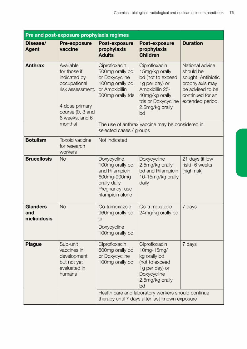
Chemical, biological, radiological and nuclear incidents handbook 75
Pre and post-exposure prophylaxis regimes
Disease/ Agent
Pre-exposure vaccine
Post-exposure prophylaxis Adults
Post-exposure prophylaxis Children
Duration
Anthrax Available for those if indicated by occupational risk assessment.
4 dose primary course (0, 3 and 6 weeks, and 6 months)
Ciprofloxacin 500mg orally bd or Doxycycline 100mg orally bd or Amoxicillin 500mg orally tds
Ciprofloxacin 15mg/kg orally bd (not to exceed 1g per day) or Amoxicillin 25-40mg/kg orally tds or Doxycycline 2.5mg/kg orally bd
National advice should be sought. Antibiotic prophylaxis may be advised to be continued for an extended period.
The use of anthrax vaccine may be considered in selected cases / groups
Botulism Toxoid vaccine for research workers
Not indicated
Brucellosis No Doxycycline 100mg orally bd and Rifampicin 600mg-900mg orally daily Pregnancy: use rifampicin alone
Doxycycline 2.5mg/kg orally bd and Rifampicin 10-15mg/kg orally daily
21 days (if low risk)- 6 weeks (high risk)
Glanders and melioidosis
No Co-trimoxazole 960mg orally bd or
Doxycycline 100mg orally bd
Co-trimoxazole 24mg/kg orally bd
7 days
Plague Sub-unit vaccines in development but not yet evaluated in humans
Ciprofloxacin 500mg orally bd or Doxycycline 100mg orally bd
Ciprofloxacin 10mg-15mg/kg orally bd (not to exceed 1g per day) or Doxycycline 2.5mg/kg orally bd
7 days
Health care and laboratory workers should continue therapy until 7 days after last known exposure
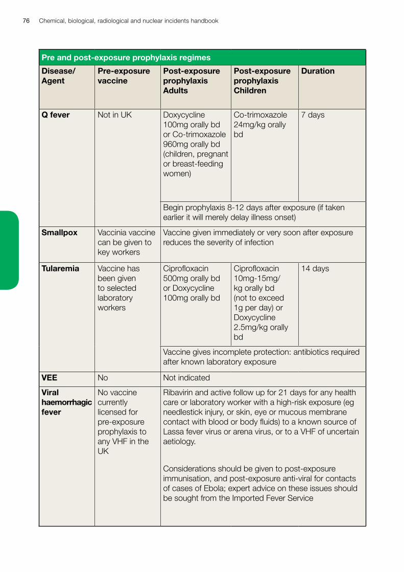
Chemical, biological, radiological and nuclear incidents handbook76
Pre and post-exposure prophylaxis regimes
Disease/ Agent
Pre-exposure vaccine
Post-exposure prophylaxis Adults
Post-exposure prophylaxis Children
Duration
Q fever Not in UK Doxycycline 100mg orally bd or Co-trimoxazole 960mg orally bd (children, pregnant or breast-feeding women)
Co-trimoxazole 24mg/kg orally bd
7 days
Begin prophylaxis 8-12 days after exposure (if taken earlier it will merely delay illness onset)
Smallpox Vaccinia vaccine can be given to key workers
Vaccine given immediately or very soon after exposure reduces the severity of infection
Tularemia Vaccine has been given to selected laboratory workers
Ciprofloxacin 500mg orally bd or Doxycycline 100mg orally bd
Ciprofloxacin 10mg-15mg/kg orally bd (not to exceed 1g per day) or Doxycycline 2.5mg/kg orally bd
14 days
Vaccine gives incomplete protection: antibiotics required after known laboratory exposure
VEE No Not indicated
Viral haemorrhagic fever
No vaccine currently licensed for pre-exposure prophylaxis to any VHF in the UK
Ribavirin and active follow up for 21 days for any health care or laboratory worker with a high-risk exposure (eg needlestick injury, or skin, eye or mucous membrane contact with blood or body fluids) to a known source of Lassa fever virus or arena virus, or to a VHF of uncertain aetiology.
Considerations should be given to post-exposure immunisation, and post-exposure anti-viral for contacts of cases of Ebola; expert advice on these issues should be sought from the Imported Fever Service
Chemical, biological, radiological and nuclear incidents handbook 77
Think of anthrax• rapid onset of severe febrile illness, sepsis or respiratory failure with wide mediastinum
on CXR
• painless black-scabbed ulcer on arm, neck or face with extensive local swelling
• gram positive rods (or Bacillus sp) in blood or CSF assessed not to be contaminants
• haemorrhagic meningitis
• unexplained febrile death
• inhalational anthrax is very rare indeed: a single confirmed case in the UK suggests deliberate release
• injectional anthrax may be linked to batches of contaminated drugs and is a public health emergency
Key factsCaused by Bacillus anthracis (Gram positive bacterium with hardy spore form that can survive in soil for decades).
Zoonotic; mainly from sheep, cattle, and goats.
Human anthrax now rare in UK (around 1 case/year) but still occurs in Africa, the Middle East and parts of rural Asia, the Americas and Europe.
Naturally acquired human anthrax is usually the result of contact with an infected animal, carcass or animal product.
Clinical features depend on route of exposure: contact with abraded skin causes cutaneous anthrax; breathing in the spores causes inhalational anthrax; eating undercooked anthrax-contaminated meat causes gastrointestinal anthrax.
Occupational risks: working with animals or animal hides, skins or hair, as in Hawick, Scotland in 2006 where there was one death, or working with the organism in the laboratory. Working in a postal sorting office or as mail handler was a risk in the 2001 outbreak, when deliberate release of letters containing anthrax spores via the US Postal Service caused 22 cases (five deaths).
Other risks: threatening letters or suspicious packages; contaminated heroin has been described as a source of necrotic injection-site infection and severe sepsis (Scotland / England / Germany 2009-2012).
Incubation period usually one to seven days (range < 24 hours to 60 days post exposure).
Anthrax
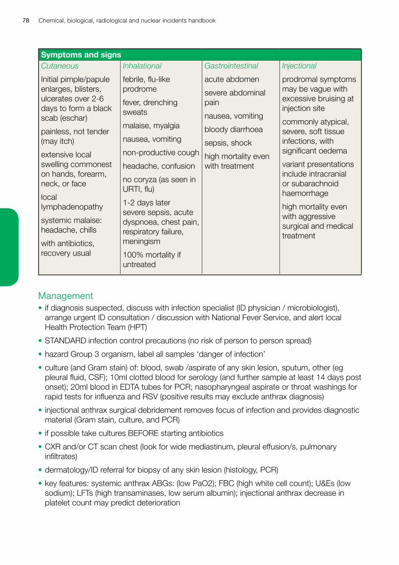
Chemical, biological, radiological and nuclear incidents handbook78
Symptoms and signsCutaneous
Initial pimple/papule enlarges, blisters, ulcerates over 2-6 days to form a black scab (eschar)
painless, not tender (may itch)
extensive local swelling commonest on hands, forearm, neck, or face
local lymphadenopathy
systemic malaise: headache, chills
with antibiotics, recovery usual
Inhalational
febrile, flu-like prodrome
fever, drenching sweats
malaise, myalgia
nausea, vomiting
non-productive cough
headache, confusion
no coryza (as seen in URTI, flu)
1-2 days later severe sepsis, acute dyspnoea, chest pain, respiratory failure, meningism
100% mortality if untreated
Gastrointestinal
acute abdomen
severe abdominal pain
nausea, vomiting
bloody diarrhoea
sepsis, shock
high mortality even with treatment
Injectional
prodromal symptoms may be vague with excessive bruising at injection site
commonly atypical, severe, soft tissue infections, with significant oedema
variant presentations include intracranial or subarachnoid haemorrhage
high mortality even with aggressive surgical and medical treatment
Management• if diagnosis suspected, discuss with infection specialist (ID physician / microbiologist),
arrange urgent ID consultation / discussion with National Fever Service, and alert local Health Protection Team (HPT)
• STANDARD infection control precautions (no risk of person to person spread)
• hazard Group 3 organism, label all samples ‘danger of infection’
• culture (and Gram stain) of: blood, swab /aspirate of any skin lesion, sputum, other (eg pleural fluid, CSF); 10ml clotted blood for serology (and further sample at least 14 days post onset); 20ml blood in EDTA tubes for PCR; nasopharyngeal aspirate or throat washings for rapid tests for influenza and RSV (positive results may exclude anthrax diagnosis)
• injectional anthrax surgical debridement removes focus of infection and provides diagnostic material (Gram stain, culture, and PCR)
• if possible take cultures BEFORE starting antibiotics
• CXR and/or CT scan chest (look for wide mediastinum, pleural effusion/s, pulmonary infiltrates)
• dermatology/ID referral for biopsy of any skin lesion (histology, PCR)
• key features: systemic anthrax ABGs: (low PaO2); FBC (high white cell count); U&Es (low sodium); LFTs (high transaminases, low serum albumin); injectional anthrax decrease in platelet count may predict deterioration
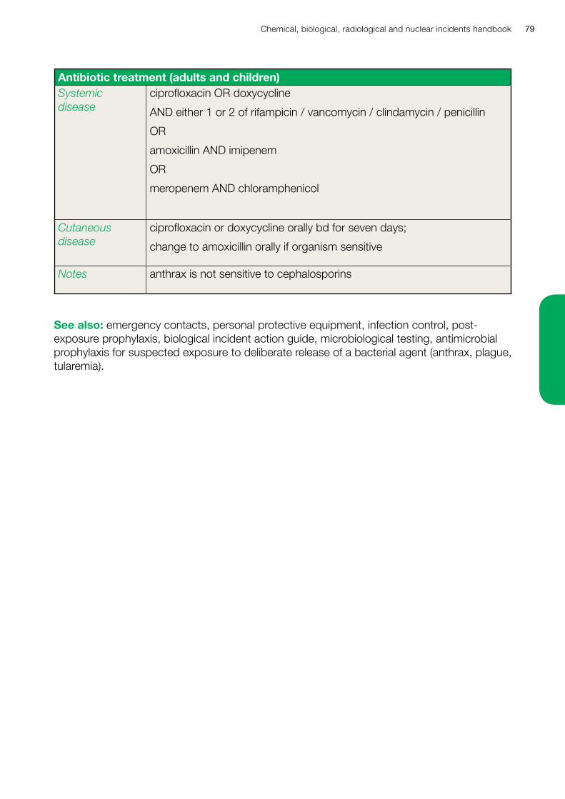
Chemical, biological, radiological and nuclear incidents handbook 79
Antibiotic treatment (adults and children)Systemic disease
ciprofloxacin OR doxycycline
AND either 1 or 2 of rifampicin / vancomycin / clindamycin / penicillin
OR
amoxicillin AND imipenem
OR
meropenem AND chloramphenicol
Cutaneous disease
ciprofloxacin or doxycycline orally bd for seven days;
change to amoxicillin orally if organism sensitive
Notes anthrax is not sensitive to cephalosporins
See also: emergency contacts, personal protective equipment, infection control, post-exposure prophylaxis, biological incident action guide, microbiological testing, antimicrobial prophylaxis for suspected exposure to deliberate release of a bacterial agent (anthrax, plague, tularemia).
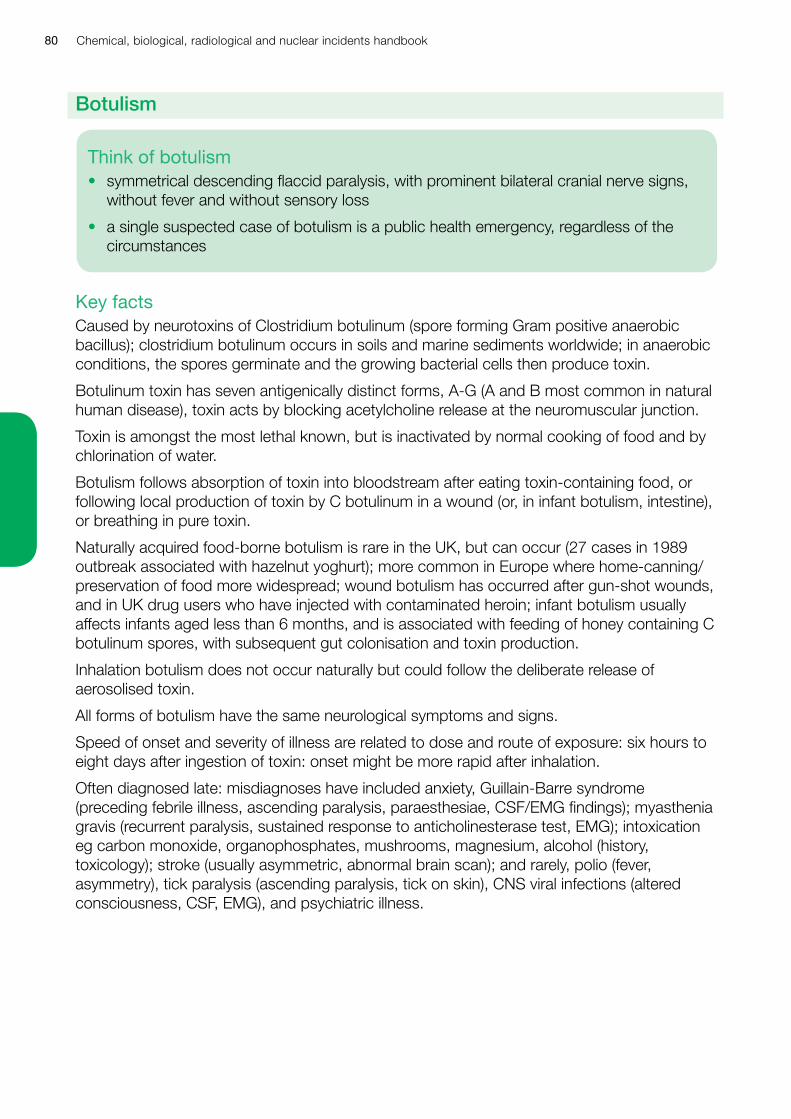
Chemical, biological, radiological and nuclear incidents handbook80
Key factsCaused by neurotoxins of Clostridium botulinum (spore forming Gram positive anaerobic bacillus); clostridium botulinum occurs in soils and marine sediments worldwide; in anaerobic conditions, the spores germinate and the growing bacterial cells then produce toxin.
Botulinum toxin has seven antigenically distinct forms, A-G (A and B most common in natural human disease), toxin acts by blocking acetylcholine release at the neuromuscular junction.
Toxin is amongst the most lethal known, but is inactivated by normal cooking of food and by chlorination of water.
Botulism follows absorption of toxin into bloodstream after eating toxin-containing food, or following local production of toxin by C botulinum in a wound (or, in infant botulism, intestine), or breathing in pure toxin.
Naturally acquired food-borne botulism is rare in the UK, but can occur (27 cases in 1989 outbreak associated with hazelnut yoghurt); more common in Europe where home-canning/preservation of food more widespread; wound botulism has occurred after gun-shot wounds, and in UK drug users who have injected with contaminated heroin; infant botulism usually affects infants aged less than 6 months, and is associated with feeding of honey containing C botulinum spores, with subsequent gut colonisation and toxin production.
Inhalation botulism does not occur naturally but could follow the deliberate release of aerosolised toxin.
All forms of botulism have the same neurological symptoms and signs.
Speed of onset and severity of illness are related to dose and route of exposure: six hours to eight days after ingestion of toxin: onset might be more rapid after inhalation.
Often diagnosed late: misdiagnoses have included anxiety, Guillain-Barre syndrome (preceding febrile illness, ascending paralysis, paraesthesiae, CSF/EMG findings); myasthenia gravis (recurrent paralysis, sustained response to anticholinesterase test, EMG); intoxication eg carbon monoxide, organophosphates, mushrooms, magnesium, alcohol (history, toxicology); stroke (usually asymmetric, abnormal brain scan); and rarely, polio (fever, asymmetry), tick paralysis (ascending paralysis, tick on skin), CNS viral infections (altered consciousness, CSF, EMG), and psychiatric illness.
Think of botulism• symmetrical descending flaccid paralysis, with prominent bilateral cranial nerve signs,
without fever and without sensory loss
• a single suspected case of botulism is a public health emergency, regardless of the circumstances
Botulism
Chemical, biological, radiological and nuclear incidents handbook 81
Symptoms and signsEarly symptoms and signs
no fever
facial weakness, ptosis, drooping eyelids
difficulty speaking, seeing, or swallowing
(the ‘four Ds’: dysphonia, dysarthria, diplopia, dysphagia)
dry mouth, pupils dilated and sluggishly reacting
normal sensation and alertness
nausea, vomiting and diarrhoea sometimes accompany food-borne botulism
Late symptoms and signs
neck weakness – loss of head control
descending weakness – pharynx, arms, accessory muscles of respiration, diaphragm, lower body
respiratory failure may be the first sign if onset is very rapid
loss of gag reflex and tendon reflexes
autonomic disturbance
death from airways obstruction and respiratory muscle paralysis
mortality reduced by early administration of antitoxin and good supportive care
Management• if diagnosis suspected, discuss with infection specialist (ID physician / microbiologist),
arrange urgent ID consultation / discussion with National Fever Service, and alert local Health Protection Team (HPT)
• no risk of person to person spread: use STANDARD precautions
• take a clear and detailed food history; obtain 10ml serum; 10g faeces (in sterile container) and other (gastric washings/lavage; bronchial washings/lavage; pus from abscess/wound; wound swab in transport medium) as appropriate, for urgent toxin detection by reference laboratory - obtain samples for toxin detection before giving any antitoxin
• tests that may help in excluding diagnosis include: brain scan, EMG, CSF examination, TensilonTM test
• ID physician will provide expert advice about further management, and about giving antitoxin (botulinum antitoxin is held in regional centres and Public Health England)
• decision to give antitoxin is made clinically, and not on laboratory test results
• antibiotics (penicillin with metronidazole) indicated only for wound botulism; if wound botulism, may also need surgical debridement
• monitor and support respiratory function: intubate, ventilate (possibly long term); treat secondary infection
See also: emergency contacts, personal protective equipment, post-exposure prophylaxis, infection control, microbiological testing, biological incident action guide.
Chemical, biological, radiological and nuclear incidents handbook82
Key factsCaused by Brucella abortus, Brucella melitensis, or Brucella suis (tiny Gram negative coccobacilli).
Zoonotic; affecting cows (Brucella abortus), sheep, goats and camels (B melitensis), pigs (B suis), and other mammals.
Animal disease is now rare in UK, but still common in some parts of Europe, M East, Africa, Asia, S and C America (including Mexico), and the Caribbean.
Naturally acquired human infection follows drinking unpasteurised milk or eating unpasteurised milk products from infected animals; breathing in the organism or directly contaminating the eyes, nose, mouth or abraded skin during close contact with infected animals, products of conception, or carcasses, or while working with the organism in the laboratory, and the accidental inoculation of live attenuated animal vaccine.
Human disease uncommon in UK (< 20 reported cases each year, usually acquired abroad).
B melitensis, B abortus and B suis cause similar human illnesses (B melitensis causes the most severe disease); clinical features do not depend on route of exposure.
Occupational risks for: animal handlers, vets, meat packers and abattoir workers exposed to infected animals, carcasses, or contaminated dust (eg when washing down buildings); laboratory workers.
Incubation period usually 1-3 weeks, but may be longer (up to 6 months).
Diagnosis easily missed as symptoms are variable and non-specific.
Think of brucellosis• fever of unknown origin
• endocarditis (culture negative)
• hepatitis (negative for HAV, HBV, HCV markers with granulomata on biopsy)
• a single confirmed case with no history of travel to endemic area or of occupational exposure suggests deliberate release
Brucellosis
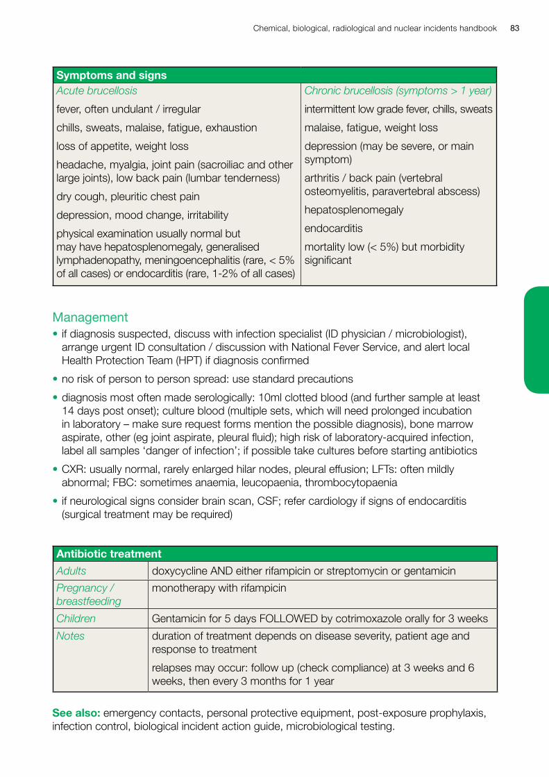
Chemical, biological, radiological and nuclear incidents handbook 83
Symptoms and signsAcute brucellosis
fever, often undulant / irregular
chills, sweats, malaise, fatigue, exhaustion
loss of appetite, weight loss
headache, myalgia, joint pain (sacroiliac and other large joints), low back pain (lumbar tenderness)
dry cough, pleuritic chest pain
depression, mood change, irritability
physical examination usually normal but may have hepatosplenomegaly, generalised lymphadenopathy, meningoencephalitis (rare, < 5% of all cases) or endocarditis (rare, 1-2% of all cases)
Chronic brucellosis (symptoms > 1 year)
intermittent low grade fever, chills, sweats
malaise, fatigue, weight loss
depression (may be severe, or main symptom)
arthritis / back pain (vertebral osteomyelitis, paravertebral abscess)
hepatosplenomegaly
endocarditis
mortality low (< 5%) but morbidity significant
Management• if diagnosis suspected, discuss with infection specialist (ID physician / microbiologist),
arrange urgent ID consultation / discussion with National Fever Service, and alert local Health Protection Team (HPT) if diagnosis confirmed
• no risk of person to person spread: use standard precautions
• diagnosis most often made serologically: 10ml clotted blood (and further sample at least 14 days post onset); culture blood (multiple sets, which will need prolonged incubation in laboratory – make sure request forms mention the possible diagnosis), bone marrow aspirate, other (eg joint aspirate, pleural fluid); high risk of laboratory-acquired infection, label all samples ‘danger of infection’; if possible take cultures before starting antibiotics
• CXR: usually normal, rarely enlarged hilar nodes, pleural effusion; LFTs: often mildly abnormal; FBC: sometimes anaemia, leucopaenia, thrombocytopaenia
• if neurological signs consider brain scan, CSF; refer cardiology if signs of endocarditis (surgical treatment may be required)
Antibiotic treatment
Adults doxycycline AND either rifampicin or streptomycin or gentamicin
Pregnancy / breastfeeding
monotherapy with rifampicin
Children Gentamicin for 5 days FOLLOWED by cotrimoxazole orally for 3 weeks
Notes duration of treatment depends on disease severity, patient age and response to treatment
relapses may occur: follow up (check compliance) at 3 weeks and 6 weeks, then every 3 months for 1 year
See also: emergency contacts, personal protective equipment, post-exposure prophylaxis, infection control, biological incident action guide, microbiological testing.
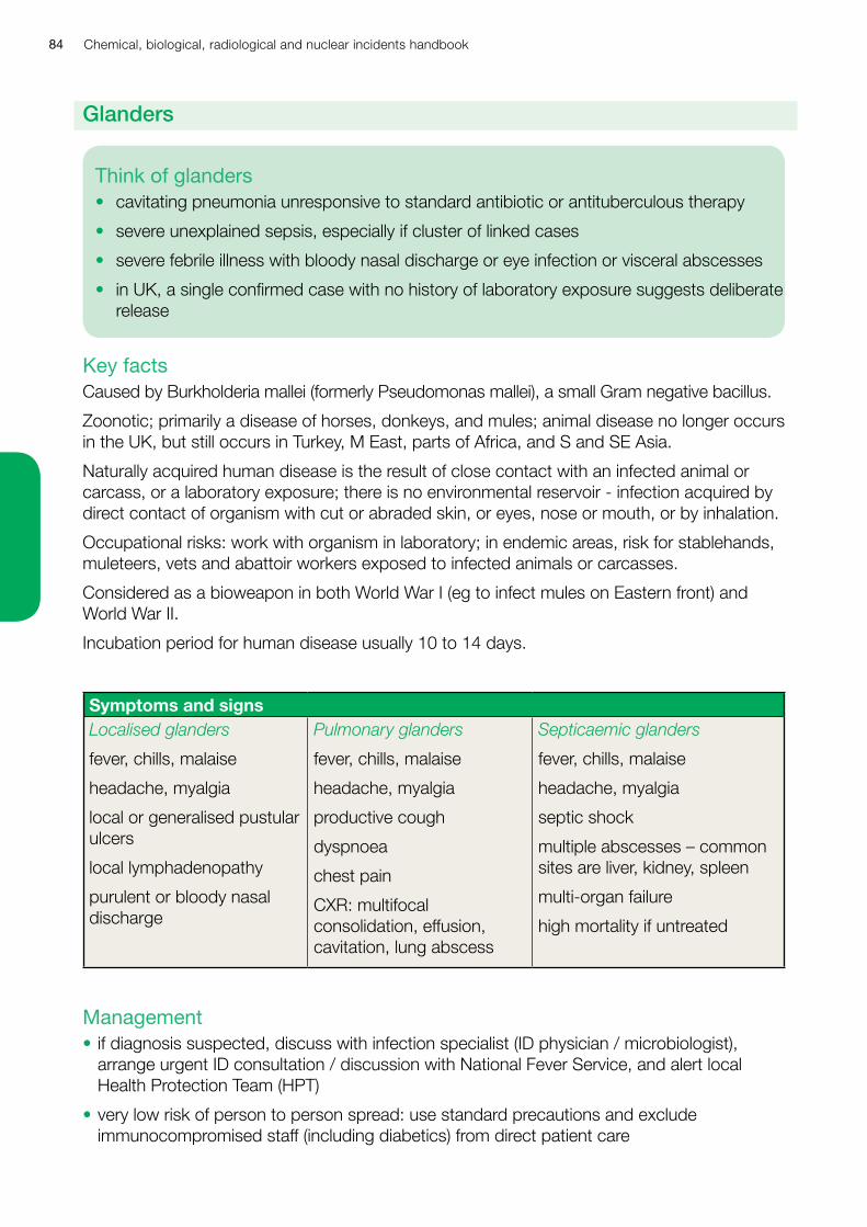
Chemical, biological, radiological and nuclear incidents handbook84
Key factsCaused by Burkholderia mallei (formerly Pseudomonas mallei), a small Gram negative bacillus.
Zoonotic; primarily a disease of horses, donkeys, and mules; animal disease no longer occurs in the UK, but still occurs in Turkey, M East, parts of Africa, and S and SE Asia.
Naturally acquired human disease is the result of close contact with an infected animal or carcass, or a laboratory exposure; there is no environmental reservoir - infection acquired by direct contact of organism with cut or abraded skin, or eyes, nose or mouth, or by inhalation.
Occupational risks: work with organism in laboratory; in endemic areas, risk for stablehands, muleteers, vets and abattoir workers exposed to infected animals or carcasses.
Considered as a bioweapon in both World War I (eg to infect mules on Eastern front) and World War II.
Incubation period for human disease usually 10 to 14 days.
Symptoms and signsLocalised glanders
fever, chills, malaise
headache, myalgia
local or generalised pustular ulcers
local lymphadenopathy
purulent or bloody nasal discharge
Pulmonary glanders
fever, chills, malaise
headache, myalgia
productive cough
dyspnoea
chest pain
CXR: multifocal consolidation, effusion, cavitation, lung abscess
Septicaemic glanders
fever, chills, malaise
headache, myalgia
septic shock
multiple abscesses – common sites are liver, kidney, spleen
multi-organ failure
high mortality if untreated
Management• if diagnosis suspected, discuss with infection specialist (ID physician / microbiologist),
arrange urgent ID consultation / discussion with National Fever Service, and alert local Health Protection Team (HPT)
• very low risk of person to person spread: use standard precautions and exclude immunocompromised staff (including diabetics) from direct patient care
Think of glanders• cavitating pneumonia unresponsive to standard antibiotic or antituberculous therapy
• severe unexplained sepsis, especially if cluster of linked cases
• severe febrile illness with bloody nasal discharge or eye infection or visceral abscesses
• in UK, a single confirmed case with no history of laboratory exposure suggests deliberate release
Glanders
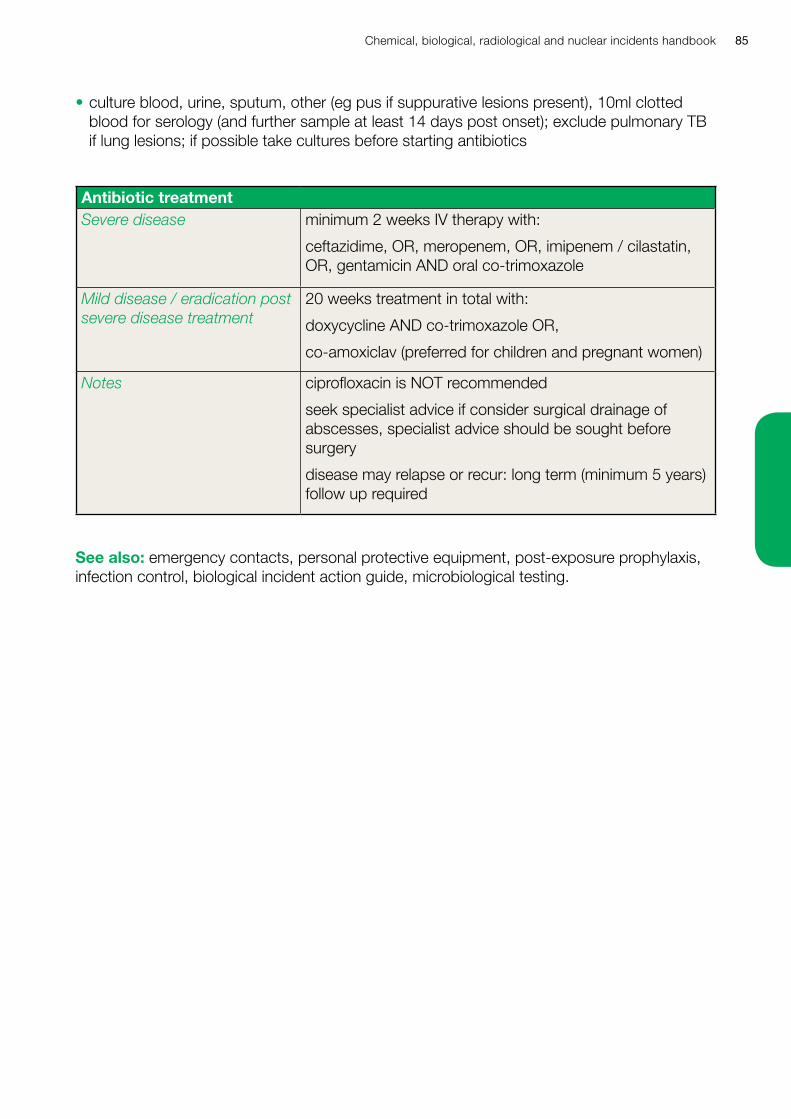
Chemical, biological, radiological and nuclear incidents handbook 85
• culture blood, urine, sputum, other (eg pus if suppurative lesions present), 10ml clotted blood for serology (and further sample at least 14 days post onset); exclude pulmonary TB if lung lesions; if possible take cultures before starting antibiotics
Antibiotic treatmentSevere disease minimum 2 weeks IV therapy with:
ceftazidime, OR, meropenem, OR, imipenem / cilastatin, OR, gentamicin AND oral co-trimoxazole
Mild disease / eradication post severe disease treatment
20 weeks treatment in total with:
doxycycline AND co-trimoxazole OR,
co-amoxiclav (preferred for children and pregnant women)
Notes ciprofloxacin is NOT recommended
seek specialist advice if consider surgical drainage of abscesses, specialist advice should be sought before surgery
disease may relapse or recur: long term (minimum 5 years) follow up required
See also: emergency contacts, personal protective equipment, post-exposure prophylaxis, infection control, biological incident action guide, microbiological testing.
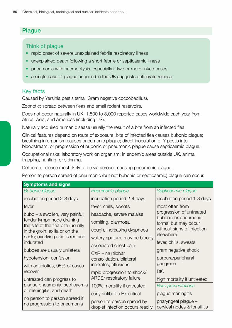
Chemical, biological, radiological and nuclear incidents handbook86
Key factsCaused by Yersinia pestis (small Gram negative coccobacillus).
Zoonotic; spread between fleas and small rodent reservoirs.
Does not occur naturally in UK, 1,500 to 3,000 reported cases worldwide each year from Africa, Asia, and Americas (including US).
Naturally acquired human disease usually the result of a bite from an infected flea.
Clinical features depend on route of exposure: bite of infected flea causes bubonic plague; breathing in organism causes pneumonic plague; direct inoculation of Y pestis into bloodstream, or progression of bubonic or pneumonic plague cause septicaemic plague.
Occupational risks: laboratory work on organism; in endemic areas outside UK, animal trapping, hunting, or skinning.
Deliberate release most likely to be via aerosol, causing pneumonic plague.
Person to person spread of pneumonic (but not bubonic or septicaemic) plague can occur.
Symptoms and signsBubonic plague
incubation period 2-8 days
fever
bubo – a swollen, very painful, tender lymph node draining the site of the flea bite (usually in the groin, axilla or on the neck); overlying skin is red and indurated
buboes are usually unilateral
hypotension, confusion
with antibiotics, 95% of cases recover
untreated can progress to plague pneumonia, septicaemia or meningitis, and death
no person to person spread if no progression to pneumonia
Pneumonic plague
incubation period 2-4 days
fever, chills, sweats
headache, severe malaise
vomiting, diarrhoea
cough, increasing dyspnoea
watery sputum, may be bloody
associated chest pain
CXR – multilobar consolidation, bilateral infiltrates, effusions
rapid progression to shock/ARDS/ respiratory failure
100% mortality if untreated
early antibiotic Rx critical
person to person spread by droplet infection occurs readily
Septicaemic plague
incubation period 1-8 days
most often from progression of untreated bubonic or pneumonic forms, but may occur without signs of infection elsewhere
fever, chills, sweats
gram negative shock
purpura/peripheral gangrene
DIC
high mortality if untreatedRare presentations
plague meningitis
pharyngeal plague – cervical nodes & tonsillitis
Think of plague• rapid onset of severe unexplained febrile respiratory illness
• unexplained death following a short febrile or septicaemic illness
• pneumonia with haemoptysis, especially if two or more linked cases
• a single case of plague acquired in the UK suggests deliberate release
Plague
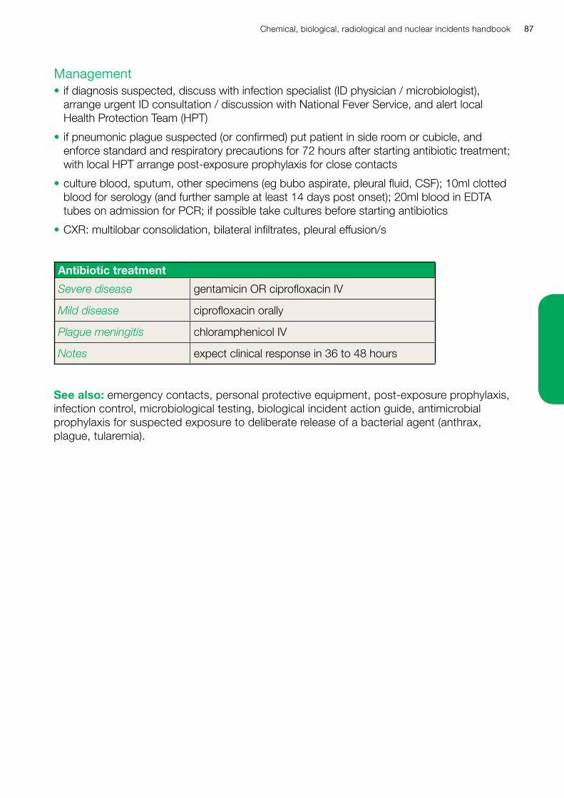
Chemical, biological, radiological and nuclear incidents handbook 87
Management• if diagnosis suspected, discuss with infection specialist (ID physician / microbiologist),
arrange urgent ID consultation / discussion with National Fever Service, and alert local Health Protection Team (HPT)
• if pneumonic plague suspected (or confirmed) put patient in side room or cubicle, and enforce standard and respiratory precautions for 72 hours after starting antibiotic treatment; with local HPT arrange post-exposure prophylaxis for close contacts
• culture blood, sputum, other specimens (eg bubo aspirate, pleural fluid, CSF); 10ml clotted blood for serology (and further sample at least 14 days post onset); 20ml blood in EDTA tubes on admission for PCR; if possible take cultures before starting antibiotics
• CXR: multilobar consolidation, bilateral infiltrates, pleural effusion/s
Antibiotic treatment
Severe disease gentamicin OR ciprofloxacin IV
Mild disease ciprofloxacin orally
Plague meningitis chloramphenicol IV
Notes expect clinical response in 36 to 48 hours
See also: emergency contacts, personal protective equipment, post-exposure prophylaxis, infection control, microbiological testing, biological incident action guide, antimicrobial prophylaxis for suspected exposure to deliberate release of a bacterial agent (anthrax, plague, tularemia).

Chemical, biological, radiological and nuclear incidents handbook88
Keys factsCaused by Coxiella burnetii (small Gram negative pleomorphic coccobacillus – difficult and dangerous to grow).
Zoonotic: worldwide distribution, with reservoirs in sheep, cattle, goats, and other mammals – infected animals usually asymptomatic but shed the organism in large numbers in placental tissue, amniotic fluid, milk, urine and faeces.
C burnetii is resistant to heat and drying, so survives well in the environment.
Infectious dose is very low.
Naturally acquired human infections usually caused by breathing in organism (eg when birthing infected animal, from contaminated dust, from aerosols in laboratory work); rarely, from eating or drinking unpasteurised milk or unpasteurised milk products.
50% human infections are asymptomatic; symptomatic infection may be more common in smokers.
In UK: 50-100 reported cases of human infection each year; 1% of all cases of community acquired pneumonia; most cases are sporadic, but outbreaks occur.
Incubation period of acute Q fever usually 18 to 21 days (range 4 to 40 days), may be shorter if large infective dose; chronic Q fever may occur years after untreated primary infection.
Symptoms and signsAcute Q fever
fever (often abrupt onset, high)
malaise, fatigue, sweats, headache, myalgia, dry cough (25% of symptomatic infections)
no rash
hepatitis (30% of symptomatic infections)
often self-limiting after one to two weeks
rarely, aseptic meningitis, endocarditis
Chronic Q fever
fever
weight loss, malaise, fatigue
aseptic meningitis / meningoencephalitis
endocarditis (75% aortic valve; usually affects prosthetic valve or damaged native valve)
needs prolonged (minimum two years) antibiotic treatment
Occupational risks for: farmers, shepherds, vets and abattoir workers exposed to infected animals, their body fluids or carcasses, or contaminated fomites, and laboratory workers.
Think of Q fever• community acquired pneumonia, especially if two or more linked cases
• endocarditis (culture negative)
• hepatitis (negative for HAV, HBV, and HCV markers, with granulomata on biopsy)
Q Fever
Chemical, biological, radiological and nuclear incidents handbook 89
Management• if diagnosis suspected, discuss with infection specialist (ID physician / microbiologist),
arrange urgent ID consultation / discussion with National Fever Service, and alert local Health Protection Team (HPT) if diagnosis confirmed
• no risk of person to person spread: use standard precautions
• dagnosis is usually serological: 10ml clotted blood for Q fever serology (and convalescent sample at least 28 days post onset); 4-5ml EDTA blood for PCR; histology/immunocytochemistry (eg of liver biopsy) sometimes also helpful.
• CXR: abnormal in 50% of symptomatic infections: patchy infiltrates, lobar consolidation, enlarged hilar nodes
• LFTs: raised (two to three times normal) transaminases, bilirubin usually normal
• FBC: normal, or raised WCC, sometimes thrombocytopaenia
• if neurological signs, consider brain scan, CSF; refer to cardiology if signs of endocarditis (surgical Rx may be required)
• although untreated infection is usually self-limiting, antibiotic treatment reduces the risk of chronic infection and speeds recovery
Antibiotic treatmentAdults doxycycline orally or IV, OR
tetracycline orally
Pregnancy / breast feeding co-trimoxazole 960mg orallyChildren co-trimoxazole 24 mg/kg orallyNotes Treatment response (usually within two to three days) indicated
by resolution of fever and of other symptoms
Treatment of chronic Q fever or Q fever endocarditis requires long term combined antibiotic therapy: seek expert advice
See also: emergency contacts, personal protective equipment, post-exposure prophylaxis, infection control, biological incident action guide, microbiological testing.
Chemical, biological, radiological and nuclear incidents handbook90
Key factsThe first highly pathogenic human coronavirus diseases emerged in 2003 (SARS coronavirus).
Human coronaviruses do not currently occur naturally in the UK.
SARS was first identified in Southern China in late 2002; with rapid person to person spreading causing outbreaks in Hong Kong SAR, Vietnam, Singapore and Canada in 2003, with more than 8000 cases in more than 30 countries; further cases in Singapore, Taiwan and China in late 2003 and 2004 were associated with laboratory-acquired infections. Mortality during SARS outbreaks 15%, higher in elderly and those with pre-existing illness; SARS in children less than ten years was mild and uncommon.
MERS was first identified in 2012 from a small cluster of cases emerging in the Middle East; cases suffered severe acute respiratory infection +/- acute renal failure; person to person transmission (patients and health care staff) in secondary care settings can be explosive (Republic of Korea, 2015). Mortality during MERS outbreaks has ranged from 20% to 40%; Most hospitalized MERS-CoV patients have had chronic co-morbidities.
Coronavirus infections are presumed to be usually acquired by droplet transmission (breathing in virus particles from respiratory secretions) during close contact with a symptomatic case, or by contamination of eyes, mouth or nose with respiratory secretions, body fluids, or faeces of a case.
Incubation period for SARS mean five days (range two to ten days); asymptomatic contacts are not infectious, and cases are non-infectious from 10 days after resolution of fever.
Incubation period for MERS mean five days (range two to fourteen days); asymptomatic contacts are not infectious, cases should have PCR evidence of clearance of virus before being considered none-infectious.
Health care workers caring for cases are at high risk of becoming infected if infection control is inadequate.
Think of coronavirus infectionsAny patient who meets this definition has a novel coronavirus/SARS/MERS until proven otherwise; fever more than 38°C and cough or dyspnoea or breathing difficulty and in the ten days before onset of illness, has either:
• travelled to/from a coronavirus affected area (check www.gov.uk/topic/health-protection)
• had close contact with other case(s) of severe respiratory illness from a coronavirus affected area
• prolonged face-to-face contact during the ten days before onset of illness with a confirmed case of novel coronavirus infection while the case was ill
• worked in a laboratory with possible exposure to pathogenic coronavirus
• is a health care worker caring for patients with severe acute respiratory infections in a secondary / tertiary health care setting
Novel coronavirus infections, severe acute respiratory syndrome(SARS) and Middle Eastern respiratory syndrome (MERS)
Chemical, biological, radiological and nuclear incidents handbook 91
Rapid detection and early isolation of cases, and early and effective infection control, are central to control of coronaviruses
Symptoms and signsInitial symptoms fever, chills, rigors
malaise, myalgia, headache
diarrhoea (sometimes)
At two to four days cough
breathing difficulty
respiratory failure, ARDS, death
rash, lymphadenopathy or CNS signs may make disease less likely
spectrum of disease – many cases will be relatively mild, and will not require hospital admission
Later renal failure / MOF (multi-organ failure)
Management• assess all patients with febrile respiratory illness charactereistic of a coronavirus infection in
an isolation room: patient to wear surgical (non-valved) mask; staff (including radiographer) to wear surgical mask, gown, gloves, pay scrupulous attention to handwashing and minimise hand-face or glove-face contact; restrict entry to essential staff and relatives
• determine the date of onset of symptoms and obtain a travel, occupational, and contact history for the ten days before onset. If patient does not fit case definition, unlikely to be SARS / MERS
• if diagnosis suspected, discuss with infection specialist (ID physician / microbiologist), arrange urgent ID consultation / discussion with National Fever Service, and alert local Health Protection Team (HPT)
• if patient satisfies case definition, and condition warrants admit to single isolation (ideally negative pressure) room, with airborne infection isolation precautions including P3 respirator (fit tested and checked)
• all specimens to be labelled ‘High Risk’
• CXR, pulse oximetry, ABG if O2 saturation on air <95%
• FBC and differential, U & Es, LFTs, creatinine, CK, LDH, CRP, blood cultures, clotted blood for acute serology (mycoplasma, legionella, chlamydiae, influenza A and B, adenovirus, RSV) plus 20ml reserve; second sample at 21 days post onset, sputum culture +/- Gram stain, respiratory sample for rapid tests for influenza A and B, and RSV, urine for legionella and pneumococcal antigens
• specialist investigations for SARS/MERS in liaison with local microbiologist and PHE reference laboratory

Chemical, biological, radiological and nuclear incidents handbook92
• minimise aerosol-provoking procedures (high risk to health care workers of infection); avoid high flow (6L/min, or more) oxygen
• antibiotics according to local treatment protocol for community acquired pneumonia
• no antiviral drug (eg ribavirin) or other drugs, have proven clinical effectiveness against novel coronavirus / SARS / MERS infection; interferons may have therapeutic activity; treatment is essentially supportive, the use of steroids unproven
• list patient’s close contacts (household, face-to-face – within 1 metre, health care workers, others) in ten days before onset
• re-assess at 48 hours: if CXR and clinical course consistent with SARS/MERS, and no alternative diagnosis, repeat specialist investigations for SARS/MERS, if not, remove from airborne infection isolation if appropriate, continue treatment and inform local HPT
See also: detailed public health guidance on MERS / SARS at: www.gov.uk/topic/health-protection/infectious-diseases, and guidelines on clinical management from the British Thoracic Society at: www.brit-thoracic.org.uk
Emergency contacts, personal protective equipment, infection control, microbiological testing, biological incident action guide, picture gallery.
NHS England guidance ‘Infectious respiratory viruses – the use of facemasks and respirators’ www.england.nhs.uk/ourwork/eprr/id
Chemical, biological, radiological and nuclear incidents handbook 93
Key factsCaused by shiga-toxin producing E.coli: usually of serotype O:157 in the UK.
Incubation period usually three to four days (range one to nine days).
Normal reservoir in farmyard animals and transmitted through faecal contamination of hands or foodstuffs.
Most common severe complication is HUS which occurs in 6% to 9% of cases and up to 15% of children; HUS may also be produced by other infective agents including Shigella dysenteria, Strep. pneumonia, coxsackievirus, echovirus, and adenovirus.
Acute bloody diarrhoea is also found in Shigella, Campylobacter, and Salmonella infections.
Symptoms and signsSTEC
History of bloody diarrhoea
Visibly bloody stools
Absence of fever
Leucocyte count >10x109/L
Abdominal tenderness
May also feature neurological events including seizures, coma and stroke; hepatomegaly
Sometimes: raised serum transaminases; transient diabetes; cardiac ischaemia
Rarely: pseudomembranous colitis in absence of C. difficile infection
HUS
Disease comprises triad of:
• Microangiopathic haemolytic anaemia
• Thrombocytopenia
• Acute renal injury
Prodromal abdominal pain, vomiting and diarrhoea for five to ten days prior to onset of HUS
Low Hb,WBC, and platelets, and raised LDH, may give early indication of onset of HUS
Haematuria/proteinuria appear early in the evolution of HUS
Think of STEC• acute bloody diarrhoea, especially if any of the following present:
- no reported fever - raised white blood cell count - abdominal tenderness - haemolytic-uraemic syndrome (HUS)
• outbreaks of STEC infection are a public health emergency
• outbreaks associated with rare/novel/non-enzootic strains should be considered as potential cases of deliberate release
Shiga-toxin producing Escherichia coli (STEC)(synonyms: Verotoxigenic E. coli/Shiga-toxin-producing or Shiga-like toxin-producing E. coli (STEC)/Enterohaemorrhagic E. coli (EHEC)).
Chemical, biological, radiological and nuclear incidents handbook94
Management• if diagnosis suspected, discuss with infection specialist (ID physician / microbiologist), and
alert local Health Protection Team (HPT)
• ensure good hygiene of cases to prevent secondary spread, with exclusion from school and workplaces if appropriate
• isolation/cohort nursing in secondary care for control of infection
• little risk of person to person spread in a health care setting: use standard precautions
• early fluid resuscitation, withhold opiate analgesia / anti-motility agents / non-steroidal anti-inflammatory drugs (NSAIDs)
• caution in the use of antibiotics which may be a risk factor for development of HUS; use only as indicated by needs other than the suspicion of enteric VTEC infection only
• where HUS develops: dialysis as indicated by standard criteria for acute kidney injury
• poor long term prognosis indicated by persistent oliguria, dehydration, WCC > 20,000 per mm3, Haematocrit < 23%, long duration of disease prior to onset of HUS (>14 days), and HUS associated with convulsion and hypertension
• plasmapheresis is not an established therapy for typical shiga-toxin associated disease
• beware ‘atypical HUS’, not associated with diarrhoeal illness, especially in adults: this is not associated with shiga-toxin associated disease and requires a different therapeutic approach
See also: emergency contacts, personal protective equipment, infection control, post-exposure prophylaxis, biological incident action guide, microbiological testing, PHE/RCPCH/RCGP ‘The management of acute bloody diarrhoea potentially caused by vero cytotoxin producing Escherichia coli in children’.
Chemical, biological, radiological and nuclear incidents handbook 95
Key factsCaused by a DNA orthopox virus.
Smallpox eradication was certified in 1980; only remaining smallpox virus is secured in two laboratories in US and Russian Federation; there is no current evidence of illicit stocks/undiscovered natural reservoirs of virus.
Only possible sources of infection now are an accidental release from a repository or deliberate release of a re-engineered virus.
Routine vaccination ceased in the 1971, and no population immunity can be assumed.
Commonest conditions occurring in the UK that are currently confused with potential cases of smallpox are Orf and varicella zoster (chicken pox); infections with other pox and herpes viruses may also mimic some aspects of smallpox.
May cause severe disease: mortality rate in outbreaks was 25% to 30%; highest in children less than one year, and the elderly.
Usually acquired by airborne route, but infection can follow direct contact of eyes, nose or mouth with vesicle fluid, respiratory secretions, saliva, or scabs and fomites (objects or materials which are likely to carry infection, such as clothes, utensils, and furniture).
Incubation period (from exposure to onset of illness) usually ten to 16 days (range 7 to 19 days, median 12 days).
Person to person spread occurs (secondary attack rate 10% to 25%); infectious dose low (probably 10-100 virions).
Cases are infectious to others from onset of fever until all scabs have separated.
Asymptomatic afebrile contacts are not infectious.
Outcome of any release will be determined by speed of diagnosis and management of initial cases and contacts.
Think of smallpox• abrupt onset of moderate fever and severe prostration
• a characteristic rash (begins on third day of illness, most dense on extremities and face, and with all pocks on any one part of body at the same stage of development)
• a single suspected case of smallpox is a public health emergency
Smallpox
Chemical, biological, radiological and nuclear incidents handbook96
Symptoms and signsClinical course of smallpox
febrile prodrome (days one to three): sudden onset fever, malaise, headache, backache, prostration, vomiting, abdominal pain – patients are usually anxious and poorly
erythematous rash (days 2 to 3); may become haemorrhagic
maculopapular rash (days 4 to 6)
vesicular rash becomes pustular (days 5 to 14+) as clear fluid in blisters
becomes cloudy and thickens; pustules are round, tense and deep in dermis, and feel like small hard peas in the skin
rash may affect palms and soles, and is densest on face and extremities
complications include haemorrhage, encephalitis, keratitis, multi-organ failure
scabs form (days 10 to 14), separate (days 14 to 28), heal with scarring
death can occur in first 48 hrs, before the rash develops
Clinical course of chickenpox
incubation period 14 to 21 days
no history of chickenpox
febrile prodrome usually mild or non- existent, patient usually not severely unwell
rash is densest on trunk, with relative sparing of face and extremities, not usually present on palms and soles
rash itches, evolves rapidly, lesions superficial, oval and appear in crops – macules, papules, vesicles and pustules at the same time on any one part of body
scabs form quickly (day 4 to 7), separate rapidly (before day 15)
caused by DNA herpes virus, can be distinguished from pox virus by electron microscopy
aciclovir effective in treatment
Differential diagnosis
febrile prodrome: influenza, malaria, meningitis, typhoid
erythematous stage: measles, rubella, parvovirus B19
papular stage: measles, chickenpox, Orf
later rash: chickenpox, monkeypox, disseminated herpes simplex, disseminated herpes zoster, drug rash, contact dermatitis, hand foot and mouth disease, Stevens Johnson syndrome, erythema multiforme, molluscum contagiosum, scabies, impetigo
Management• assess all patients with an illness characteristic of smallpox in an isolation room: patient
to wear surgical (non-valved) mask; staff (including radiographer) to wear surgical mask, gown, gloves, pay scrupulous attention to handwashing and minimise hand-face or glove-face contact; restrict entry to essential staff
• if diagnosis suspected, discuss with infection specialist (ID physician / microbiologist), arrange urgent ID consultation / discussion with National Fever Service, and alert local Health Protection Team (HPT)
• enforce standard and airborne infection control precautions (current advice is to switch off air conditioning – and leave it off until smallpox excluded or system decontaminated)

Chemical, biological, radiological and nuclear incidents handbook 97
• if infection specialist / National Fever Service suspects smallpox, they will alert a multi-agency clinical and public health team to assume responsibility for patient care and infection control, who will arrange further investigation, confirm diagnosis, undertake treatment planning and arrange any necessary vaccinations and other public health control measures
• if local infection specialist excludes smallpox, inform all those notified, stand down all action, arrange further patient management
See also: emergency contacts, personal protective equipment, post-exposure prophylaxis, infection control, biological incident action guide, microbiological testing.
Chemical, biological, radiological and nuclear incidents handbook98
Key factsCaused by Francisella tularensis (tiny Gram negative coccobacillus, several biovars, difficult and dangerous to grow).
Zoonotic; reservoirs in small mammals eg rabbit, lemming, vole, but does not occur naturally in UK. Common in parts of rural Europe, Asia, Americas and Australasia.
Naturally acquired human disease follows exposure by: bite of infected vector (tick, mosquito, deerfly); handling infected animal or carcass; breathing infected aerosol (from infected animal or carcass, contaminated hay, lawn mowing); eating contaminated food or water.
Clinical features depend on route of exposure: breathing in organism causes pneumonia; infection via bite or abraded skin causes ulcero/glandular disease; ingestion causes oropharyngeal disease; eye inoculation (eg by rubbing eyes with contaminated hands) causes oculoglandular disease.
Severity depends on infecting biovar and dose (type A most severe, < 10 organisms can infect).
Occupational risks: in UK, laboratory work; outside UK, in endemic areas, hunting, trapping, or farming.
Deliberate release most likely to be via aerosol, causing pneumonic tularemia.
Incubation period usually two to five days (range one to 14 days).
Think of tularemia• severe unexplained febrile illness or febrile death
• fever, single painful ulcer, with tender local lymphadenopathy
• cluster of cases of unexplained pneumonic or febrile illness
Tularemia
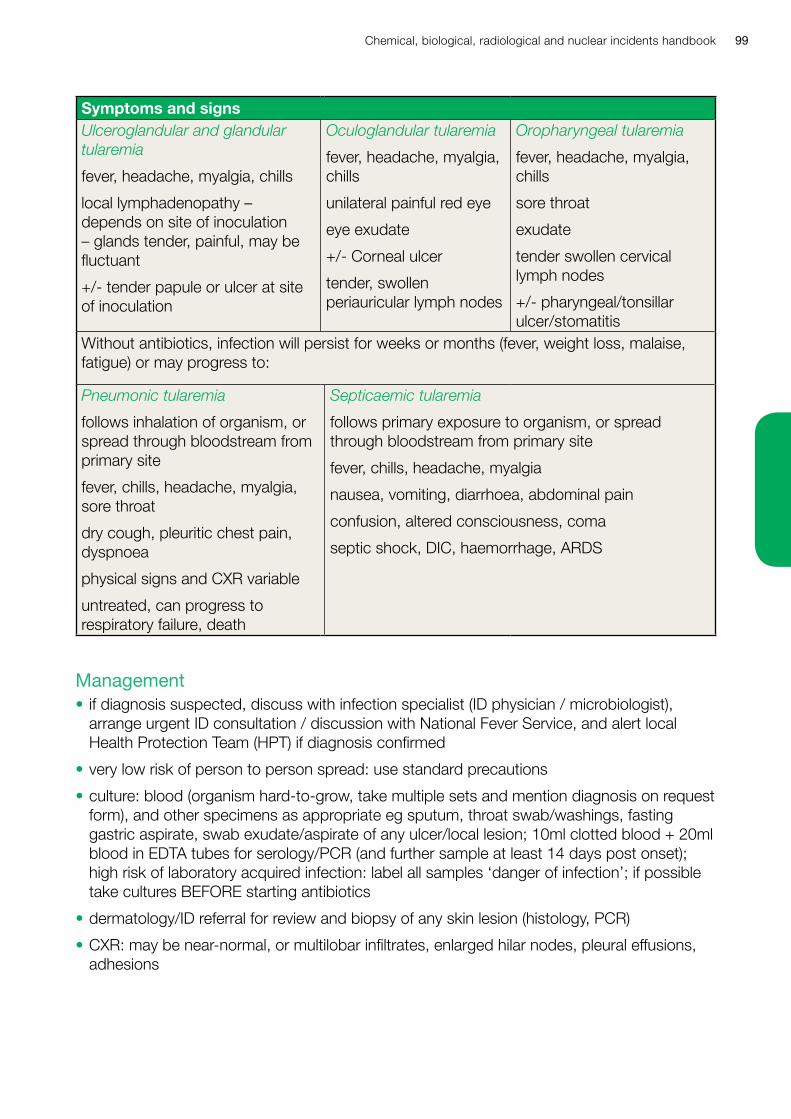
Chemical, biological, radiological and nuclear incidents handbook 99
Symptoms and signsUlceroglandular and glandular tularemia
fever, headache, myalgia, chills
local lymphadenopathy – depends on site of inoculation – glands tender, painful, may be fluctuant
+/- tender papule or ulcer at site of inoculation
Oculoglandular tularemia
fever, headache, myalgia, chills
unilateral painful red eye
eye exudate
+/- Corneal ulcer
tender, swollen periauricular lymph nodes
Oropharyngeal tularemia
fever, headache, myalgia, chills
sore throat
exudate
tender swollen cervical lymph nodes
+/- pharyngeal/tonsillar ulcer/stomatitis
Without antibiotics, infection will persist for weeks or months (fever, weight loss, malaise, fatigue) or may progress to:
Pneumonic tularemia
follows inhalation of organism, or spread through bloodstream from primary site
fever, chills, headache, myalgia, sore throat
dry cough, pleuritic chest pain, dyspnoea
physical signs and CXR variable
untreated, can progress to respiratory failure, death
Septicaemic tularemia
follows primary exposure to organism, or spread through bloodstream from primary site
fever, chills, headache, myalgia
nausea, vomiting, diarrhoea, abdominal pain
confusion, altered consciousness, coma
septic shock, DIC, haemorrhage, ARDS
Management• if diagnosis suspected, discuss with infection specialist (ID physician / microbiologist),
arrange urgent ID consultation / discussion with National Fever Service, and alert local Health Protection Team (HPT) if diagnosis confirmed
• very low risk of person to person spread: use standard precautions
• culture: blood (organism hard-to-grow, take multiple sets and mention diagnosis on request form), and other specimens as appropriate eg sputum, throat swab/washings, fasting gastric aspirate, swab exudate/aspirate of any ulcer/local lesion; 10ml clotted blood + 20ml blood in EDTA tubes for serology/PCR (and further sample at least 14 days post onset); high risk of laboratory acquired infection: label all samples ‘danger of infection’; if possible take cultures BEFORE starting antibiotics
• dermatology/ID referral for review and biopsy of any skin lesion (histology, PCR)
• CXR: may be near-normal, or multilobar infiltrates, enlarged hilar nodes, pleural effusions, adhesions
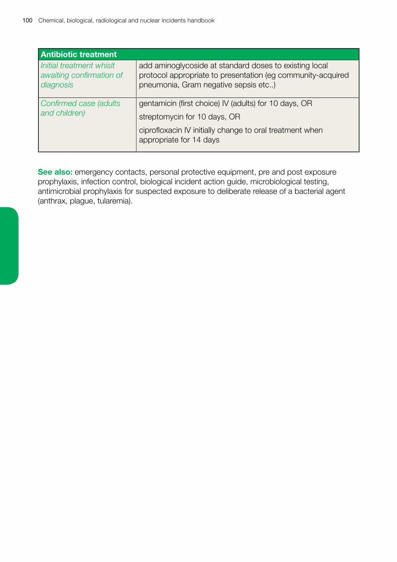
Chemical, biological, radiological and nuclear incidents handbook100
Antibiotic treatmentInitial treatment whislt awaiting confirmation of diagnosis
add aminoglycoside at standard doses to existing local protocol appropriate to presentation (eg community-acquired pneumonia, Gram negative sepsis etc..)
Confirmed case (adults and children)
gentamicin (first choice) IV (adults) for 10 days, OR
streptomycin for 10 days, OR
ciprofloxacin IV initially change to oral treatment when appropriate for 14 days
See also: emergency contacts, personal protective equipment, pre and post exposure prophylaxis, infection control, biological incident action guide, microbiological testing, antimicrobial prophylaxis for suspected exposure to deliberate release of a bacterial agent (anthrax, plague, tularemia).
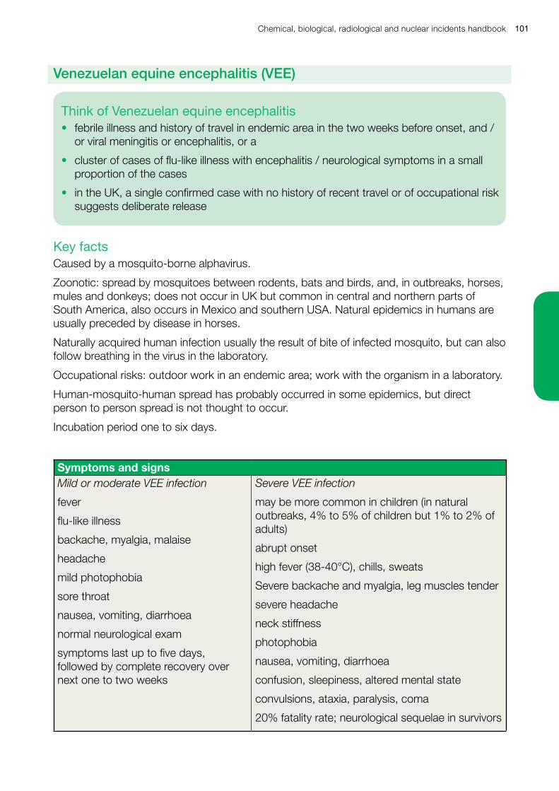
Chemical, biological, radiological and nuclear incidents handbook 101
Key factsCaused by a mosquito-borne alphavirus.
Zoonotic: spread by mosquitoes between rodents, bats and birds, and, in outbreaks, horses, mules and donkeys; does not occur in UK but common in central and northern parts of South America, also occurs in Mexico and southern USA. Natural epidemics in humans are usually preceded by disease in horses.
Naturally acquired human infection usually the result of bite of infected mosquito, but can also follow breathing in the virus in the laboratory.
Occupational risks: outdoor work in an endemic area; work with the organism in a laboratory.
Human-mosquito-human spread has probably occurred in some epidemics, but direct person to person spread is not thought to occur.
Incubation period one to six days.
Symptoms and signsMild or moderate VEE infection
fever
flu-like illness
backache, myalgia, malaise
headache
mild photophobia
sore throat
nausea, vomiting, diarrhoea
normal neurological exam
symptoms last up to five days, followed by complete recovery over next one to two weeks
Severe VEE infection
may be more common in children (in natural outbreaks, 4% to 5% of children but 1% to 2% of adults)
abrupt onset
high fever (38-40°C), chills, sweats
Severe backache and myalgia, leg muscles tender
severe headache
neck stiffness
photophobia
nausea, vomiting, diarrhoea
confusion, sleepiness, altered mental state
convulsions, ataxia, paralysis, coma
20% fatality rate; neurological sequelae in survivors
Think of Venezuelan equine encephalitis• febrile illness and history of travel in endemic area in the two weeks before onset, and /
or viral meningitis or encephalitis, or a
• cluster of cases of flu-like illness with encephalitis / neurological symptoms in a small proportion of the cases
• in the UK, a single confirmed case with no history of recent travel or of occupational risk suggests deliberate release
Venezuelan equine encephalitis (VEE)
Chemical, biological, radiological and nuclear incidents handbook102
Management• if diagnosis suspected, discuss with infection specialist (ID physician / microbiologist),
arrange urgent ID consultation / discussion with National Fever Service, and alert local Health Protection Team (HPT)
• very low risk of person to person spread: use standard precautions
• exclude malaria
• diagnosis clinical, confirmed by virus isolation/serology: 10ml clotted blood for serology (and further sample at least 14 days post onset); throat swab in virus transport medium; CSF if lumbar puncture performed (increased pressure, raised lymphocytes, mildly elevated protein)
• FBC: low WCC and lymphopaenia, and sometimes thrombocytopaenia early in disease; LFTs: mildly raised AST, LDH; CXR: normal
• if insect vectors (mosquitoes) are present, prevent any biting the patient (insecticides, insecticide-treated bed nets, screening)
• treatment is supportive:
- mild/moderate cases: analgesia and antipyretics; correct/maintain fluid balance as needed
- severe cases may need intensive supportive care: fluid balance, nutrition, ventilation, anticonvulsants, treatment of secondary infection, and long term follow up
See also: emergency contacts, personal protective equipment, post-exposure prophylaxis, infection control, biological incident action guide, microbiological testing.
Chemical, biological, radiological and nuclear incidents handbook 103
Key factsCaused by viruses from four different families: arenaviruses, filoviruses, bunyaviruses, and flaviviruses.
VHFs include Lassa fever, Junin (Argentinean haemorrhagic fever), Machupo (Bolivian haemorrhagic fever), Guanarito (Venezuelan haemorrhagic fever), Congo-Crimean haemorrhagic fever (CCHF), Rift Valley fever, Ebola, Marburg, yellow fever and dengue viruses.
All are zoonotic: distribution of natural disease is governed by the geographic distribution and ecology of the animal reservoir.
VHFs do not occur naturally in UK; imported cases are rare (<3 a year).
Route of infection varies: mosquito bite (dengue, yellow fever, Rift Valley fever); tick bite (CCHF); inhalation of dust contaminated with infected rodent droppings/urine (Lassa fever, hantaviruses); needlestick or direct contact of infected blood or body fluids with eyes, nose or mouth (Lassa, CCHF, Ebola, Marburg); most are infectious by droplet spread, but no evidence of naturally occurring airborne / aerosol spread.
For Lassa, Ebola, Marburg and CCHF, HIGH RISK of person to person spread and nosocomial infection (spread within health care settings to other patients and/or health care workers) by percutaneous or mucocutaneous exposure to blood or body fluids from febrile symptomatic patients; afebrile, asymptomatic contacts are not infectious.
Incubation periods disease-specific, vary from 1 - 21 days.
Illnesses range from mild to life threatening, but all VHFs have a febrile prodrome (fever, headache, malaise, myalgia, nausea, vomiting, collapse/lethargy?) of up to seven days, followed by signs of vascular involvement. In the second week of illness, cases tend either to recover or to deteriorate rapidly.
Differential diagnosis includes: malaria, typhoid, shigella, meningococcal sepsis, leptospirosis, other causes of DIC.
Think of viral haemorrhagic fever• fever of unknown origin and recent travel to endemic area or with flushed swollen face /
haemorrhage
• a single confirmed case in the UK, even if from endemic area, should be investigated to exclude deliberate release
Viral haemorrhagic fever (VHF)
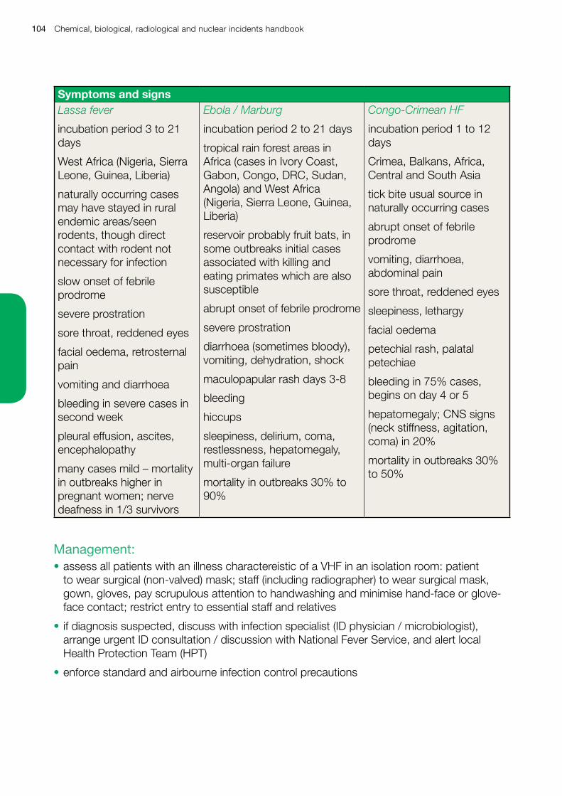
Chemical, biological, radiological and nuclear incidents handbook104
Symptoms and signsLassa fever
incubation period 3 to 21 days
West Africa (Nigeria, Sierra Leone, Guinea, Liberia)
naturally occurring cases may have stayed in rural endemic areas/seen rodents, though direct contact with rodent not necessary for infection
slow onset of febrile prodrome
severe prostration
sore throat, reddened eyes
facial oedema, retrosternal pain
vomiting and diarrhoea
bleeding in severe cases in second week
pleural effusion, ascites, encephalopathy
many cases mild – mortality in outbreaks higher in pregnant women; nerve deafness in 1/3 survivors
Ebola / Marburg
incubation period 2 to 21 days
tropical rain forest areas in Africa (cases in Ivory Coast, Gabon, Congo, DRC, Sudan, Angola) and West Africa (Nigeria, Sierra Leone, Guinea, Liberia)
reservoir probably fruit bats, in some outbreaks initial cases associated with killing and eating primates which are also susceptible
abrupt onset of febrile prodrome
severe prostration
diarrhoea (sometimes bloody), vomiting, dehydration, shock
maculopapular rash days 3-8
bleeding
hiccups
sleepiness, delirium, coma, restlessness, hepatomegaly, multi-organ failure
mortality in outbreaks 30% to 90%
Congo-Crimean HF
incubation period 1 to 12 days
Crimea, Balkans, Africa, Central and South Asia
tick bite usual source in naturally occurring cases
abrupt onset of febrile prodrome
vomiting, diarrhoea, abdominal pain
sore throat, reddened eyes
sleepiness, lethargy
facial oedema
petechial rash, palatal petechiae
bleeding in 75% cases, begins on day 4 or 5
hepatomegaly; CNS signs (neck stiffness, agitation, coma) in 20%
mortality in outbreaks 30% to 50%
Management:• assess all patients with an illness charactereistic of a VHF in an isolation room: patient
to wear surgical (non-valved) mask; staff (including radiographer) to wear surgical mask, gown, gloves, pay scrupulous attention to handwashing and minimise hand-face or glove-face contact; restrict entry to essential staff and relatives
• if diagnosis suspected, discuss with infection specialist (ID physician / microbiologist), arrange urgent ID consultation / discussion with National Fever Service, and alert local Health Protection Team (HPT)
• enforce standard and airbourne infection control precautions
Chemical, biological, radiological and nuclear incidents handbook 105
• local investigations including testing for malaria, full blood count, U&Es, LFTs, clotting screen, CRP, glucose, and blood cultures should be undertaken without waiting for results of VHF screen
• dagnosis of VHF is made by antigen detection/serology: seek advice from National Imported Fever Service/PHE Rare Imported Pathogens Laboratory, Porton Down on risk categorisation, sample transportation and samples needed from the patient (see also www.gov.uk/government/publications/viral-haemorrhagic-fever-sample-testing-advice)
• if the infection specialist / National Fever Service suspects VHF, they will alert a multi-agency clinical and public health team to assume responsibility for patient care and infection control, who will arrange further investigation, confirm diagnosis, undertake treatment planning and arrange any necessary vaccinations and other public health control measures
• if test positive national team will arrange admission to a designated Infectious Disease Unit using pre-planned ambulance transfer arrangements and provide interim treatment advice
• Ribavirin effective in Lassa fever and Congo-Crimean haemorrhagic fever but not for Marburg, Ebola, or flaviviruses:
• post-exposure immunisation of close contacts of cases of Ebola should be considered
See also: emergency contacts, personal protective equipment, post-exposure prophylaxis, infection control, microbiological testing, biological incident action guide; NHS England guidance ‘Infectious respiratory viruses – the use of facemasks and respirators’ www.england.nhs.uk/ourwork/eprr/id.

Chemical, biological, radiological and nuclear incidents handbook106
Chemical, biological, radiological and nuclear incidents handbook 107
Radiation Threats
Chemical, biological, radiological and nuclear incidents handbook108
Radiation Facts
Ionising radiation:Ionising radiation (invisible, odourless and tasteless) is a form of energy emitted spontaneously by radioactive materials and by certain electrically powered equipment such as x-ray sets.
Natural radiation is all around us: in air, from cosmic rays; in the earth and building materials; and in food and water – and all of this makes up the background radiation to which we are all exposed all the time, from conception to death.
Man-made sources of radiation and radioactive materials are used in medicine (diagnostic imaging, radiotherapy), research, throughout industry (nuclear power stations, mining, food irradiation), industrial radiography (eg of pipes, buildings, baggage), and for many other uses from measurement instrumentation to nuclear weapons.
Alpha particles, beta particles, gamma rays and x-rays are all forms of ionising radiation; neutrons, although not directly ionising, are classified as ionising radiation for protection purposes.
Alpha and beta particles, gamma rays and neutrons are produced as radioactive materials decay; X-rays are generally man-made using electrically powered equipment.
Alpha particles, in atomic terms, are relatively heavy and are doubly electrically charged; as such they interact strongly with atoms; as a result of this interaction, they lose momentum rapidly depositing their energy along a short track as they slow down, they travel only very short distances in air (a few cm) and do not penetrate further than the outer layers of human skin. Alpha emitters are hazardous only when inhaled, ingested, injected or absorbed (eg through a wound) where they can come into contact with ‘living’ bodily material unprotected by an outer layer of dead skin.
Beta particles are also electrically charged, but interact less strongly than alpha particles, so travel further and penetrate more: they can penetrate the dermis. Clothing, including standard PPE, provides some protection against them. They can cause radiation skin injury when exposures are large enough, but are hazardous to internal organs only when inhaled, ingested, injected or absorbed (eg through a wound). Eye protection (Perspex safety glasses) should be worn to protect the eyes in particular from beta radiation.
Gamma rays and x-rays are uncharged photons of electromagnetic radiation (light), so interact less strongly than beta particles, and travel many metres in air. They easily penetrate the human body, potentially causing organ damage. Gamma and x-rays can be attenuated by dense material such as concrete or lead shielding.
neutrons are uncharged particles, they can travel far and penetrate everything (except thick layers of concrete and water), and are highly damaging, but only likely to be present in the very early stages of a nuclear detonation or accident or in very particular industrial applications – note that neutron exposure can cause secondary, ionising, radiation in the other listed forms to be emitted by the exposed material.
Chemical, biological, radiological and nuclear incidents handbook 109
Exposure and contamination:An exposure occurs when all or part of the body is irradiated.
Three key factors affect exposure: duration, distance and shielding. If the exposure time to a specific source is halved, the radiation dose is halved. For gamma/x-ray ‘point’ sources, the inverse square law applies to distance: this states that doubling the distance between the source and the body reduces the dose by a factor of four; trebling the distance between the source and the body reduces the dose by a factor of nine, and so on. In general, more shielding between the source and the body reduces dose. The shielding material and the radiation type are important factors in the shielding effect.
A person is contaminated when radioactive material is deposited on skin and/or clothing (external contamination), or into the body (internal contamination) by inhalation, ingestion (hand-to-mouth, food, drink), or absorption via a wound.
In the same way that a patient who has had a CT scan or x-ray presents no risk to others, radiation safety precautions are not needed for patients who have been only been exposed to radiation and who are not contaminated with radioactive material.
If a patient is contaminated, internally or externally, then they will continue to be irradiated by that radioactive material until it is removed.
External contamination – usually dust or particulate matter – is usually readily removed by wet decontamination.
Even if a patient is contaminated, the risk of long term health effects for a health professional who uses standard precautions is likely to be tiny, if not trivial.
Radiation and radioactive contamination are readily detectable with the right equipment. Medical physics, nuclear medicine departments, and front line services have equipment for detecting beta and gamma radiation, and people trained to use it. Knowing the type of radioactive material involved is important in assessing how to manage it.
Measuring radioactivity and radiation:Radioactivity (and contamination by radioactive material) is measured in Bequerels (1Bq = 1 radioactive decay per second).
The absorbed dose of radiation (the amount of energy absorbed by per unit mass of tissue) is measured in Gray (Gy); 1Gy = 1 joule/kg of tissue.
Different types of radiation have different effects on human tissue (Gray for Gray, alpha particles and fast neutrons are more damaging than beta particles, gamma rays or x-rays in terms of the risks of cancer or of heritable genetic defects), so the absorbed dosage is multiplied by a radiation weighting factor to account for this. This gives the equivalent dose (of an organ or tissue), measured in Sieverts (Sv). For x-rays, gamma rays, and beta particles, the weighting factor = 1, so: 1 Gray = 1 Sievert = 1000 milli Sieverts.
Some organs are more sensitive to radiation exposure (radiosensitive) than others (eg bone marrow is more sensitive than thyroid), and exposures are rarely uniform. Weighting the equivalent doses received by different organs and tissues during an exposure to allow for each organ’s radiosensitivity, and then summing the results, gives the effective dose. The effective dose is also measured in sieverts (Sv). Summing all of the body’s organ weighting factors results in a weighting factor of 1, and so for whole body exposures the equivalent and effective dose are the same.
Chemical, biological, radiological and nuclear incidents handbook110
An estimate of the whole body dose is helpful in estimating long term health risk
Radiation doses and dose limits:Chest x-ray: 20 micro Sieverts.
Average annual background radiation in UK: 2.7 milli Sieverts (2,700 micro Sieverts).
Annual effective dose limit for member of the public: 1 milli Sievert (1,000 micro Sieverts).
Annual effective dose limit for radiation worker: 20 milli Sieverts (20,000 micro Sieverts).
Acute radiation injuries (whole body single dose): 1 Sievert and above.
LD50/60 (whole body dose killing 50% of those exposed within 60 days if untreated): ~4.5 Sieverts
Chemical, biological, radiological and nuclear incidents handbook 111
Radiation injuries
Think of radiation exposure• harm caused by exposure to ionising radiation may arise from either effects which kill
cells and cause harm to tissues and organs of the body (deterministic injury) or damage to genetic material that increases the long-term risks of developing cancer and hereditary effects (stochastic risk)
• deterministic injury should be suspected where:
- in any newly diagnosed acute bone marrow depression (leukopenia: infection; thrombocytopenia: bleeding gums, nosebleeds, bruising)
- ‘burns’, erythema, or bullae with no history of heat or chemical exposure - sudden, rapid, hair loss especially if there is a relevant occupational history or
unexplained nausea/vomiting +/- diarrhoea two to four weeks before onset - when dealing with any incident involving a bomb or other intentionally placed explosive
device• the presence of acute radiation injuries implies exposure to an effective dose >1Sv
• stochastic effects do not present acutely, but give an increased lifetime risk of developing cancer; for adult whole body exposures the increase in lifetime cancer risk, above the normal ever present risk, is approximately 5% per Sv effective dose of radiation
OverviewAll nuclear and other major sites in the UK have extensive emergency plans and exercise and update them regularly.
On average, there is one serious radiation incident – resulting in death or major radiation injury – in the world each year.
Incidents at major sites will be recognised and managed according to existing plans, however, in the last 50 years there have been more than 200 incidents involving lost, stolen or misused (‘orphan’) sources (eg Lilo, Georgia, 1996 to 1997, 11 trainee border guards exposed to 12 hidden, abandoned, sources had signs and symptoms of radiation injury, but the cause remained unrecognised by doctors for months). The first sign of a problem may be the presentation of a case to an emergency department.
Other concerns include the possibility of exposure from a ‘dirty bomb’ (conventional explosive used to disperse radioactive material), a low yield improvised nuclear device (IND), or a deliberately hidden source of radiation.
Acute Radiation InjuriesMost radiation accidents cause partial body injury (early erythema followed by bullae, and, if severe, ulceration and necrosis, often of the hands) and may not be associated with acute radiation syndrome.
Acute radiation injuries follow a large, usually external, relatively homogeneous exposure of all (or most) of the body to penetrating radiation (gamma rays, high-energy x-rays, neutrons) in a short time.
Chemical, biological, radiological and nuclear incidents handbook112
Symptoms of acute radiation injury usually occur in a four-phase sequence: prodromal phase latent period acute illness recovery/death.
As the radiation dose increases, the prodromal and latent periods shorten, and the severity of illness, and frequency of mortality, increase. Major trauma and radiation exposure interact synergistically on mortality.
Initial symptoms of acute radiation injury are non-specific, and rarely immediately life-threatening; the treatment of other injuries takes priority.
If, in the first six hours after a suspected exposure, there are no symptoms of exposure (eg nausea, vomiting), serious acute radiation injury is unlikely.
At doses up to 8Sv the only significant organ injury may be the haematopoietic system; above 6Sv gastrointestinal system damage is common; central nervous system (CNS ) and cardio-vascular system (CVS) injury usually only occur with doses of 20+ sieverts and are usually lethal.
Marked heterogeneity of the distribution of the dose may result in non-classical pattern of injuries ie a dose of 20+Sv to a limb may result only in local injury and may not be associated with haematopoietic or gastrointestinal effects.
It is not possible to generalise a figure for lethal whole body exposure, however, good outcomes of cytokine and supportive therapy in recent cases has suggested that survival may now be possible where whole body doses up to 12+Gy have occurred but this requires high level care and treatment.
Management of acute radiation injuriesManagement of acute radiation injuries are best undertaken from an early stage by a multidisciplinary team with expertise in radiation medicine, health physics (eg via nuclear medicine department), haematology, gastroenterology, bone marrow transplantation, plastic surgery, public health medicine and clinical toxicology.
For adults and children, the medical evaluation starts with the initial symptoms which may subsequently be supported by reconstructed or assessed doses. However, initial care should be based on overt clinical needs.
Bone marrow failure is the most significant treatable severe radiation injury that is likely to be survivable; serial WBCs are a sensitive indicator of incipient bone marrow failure; early cytokine treatment is indicated if the lymphocyte count falls to <1.5 x 10⁹/L.
In small scale incidents individual evaluation of likely dose, full bio-dosimetry and anticipatory care of the likely organ injuries can be made; however, where decisions need to be taken on the priority for care of a large numbers of patients affected by ionising radiation then a system of triaging radiological injury severity according to signs, symptoms and initial lymphocyte counts, such as the European Society for Blood and Marrow Transplantion (EBMT) system may be useful and an adapted clinical scoring guide, assessment algorithm, and associated clinical pathways are on the following page.
Chemical, biological, radiological and nuclear incidents handbook 113
Post recovery managementSome EBMT score I patients and all EBMT score II patients will require assessment and advice on severity of residual stochastic risk – the criteria for such for assessment will be dependent on the type and degree of exposure and will need to be determined by the appropriate national authority after the incident.
Where the received dose of radiation is calculable, or demonstrated through biological dosimetry techniques, a stochastic risk of approximately 5% per Sv effective dose of radiation increase in lifetime cancer risk, above the normal ever present risk will be present, however; no additional cancer screening assessments are appropriate apart from those national programme tests that are ordinarily provided for a person of that age and sex.
Staff safety and patient management priorities during radiation incidentsExcept in extreme cases of very high dose rate source fragments being present, patients who are contaminated present very low risk to healthcare staff if standard barrier precautions (gloves, aprons, etc) are used. Notwithstanding operational limitations, urgent medical or surgical care must not be delayed due to radioactive contamination.
If it is reported by the emergency services that radiation is present at an incident scene then:
• P1 casualties should be transported to the nearest appropriate Emergency Medicine (EM) Department without being first decontaminated; P2/3 casualties may be decontaminated before being transported to an appropriate care setting
• EM Departments should assume all patients are contaminated with radioactive material until proven otherwise
• triage and treat life and/or limb threatening problems before considering decontamination; if the patient’s clinical condition permits, decontaminate first, and then treat
• EM Department staff must wear standard precautions with surgical mask and double gloves; respirators and lead aprons are not required; individual personal dosemeters are not required – the health/medical physics department will monitor to reassure EM staff that they are not being exposed to harmful levels of radiation
• contact local medical and/or health physics teams and the local radiation safety officer urgently to arrange radiation screening of the patient, and provide continuing radiation protection support including ensuring that control zones are established, safety procedures are adhered to and who will also arrange for radiation dose estimation for the casualties
• isolate all patients in appropriate single rooms, or provide cohort care if a large number of casualties present. Forbid eating, drinking or smoking
• where medical/health physics team confirm the presence of radioactive material decontamination by removal of the outer layer of the patient’s clothing will usually be sufficient to effectively remove most external contamination; the medical/health physics team will undertake a repeat screen and direct you to remove any residual contamination if found
If trauma cases require lifesaving surgery, perform as soon as possible. Seek specialist advice through PHE.
Chemical, biological, radiological and nuclear incidents handbook114
See also: radiation facts, decontamination, personal protective equipment, emergency contacts, ‘TMT handbook’, ‘EBMT secondary triage pocket guide’, General Medical Council (GMC) ‘Good Medical Practice’, GMC ‘Treatment and care towards the end of life: good practice in decision making’, Nursing and Midwifery Council ‘The code: Standards of conduct, performance and ethics for nurses and midwives’.
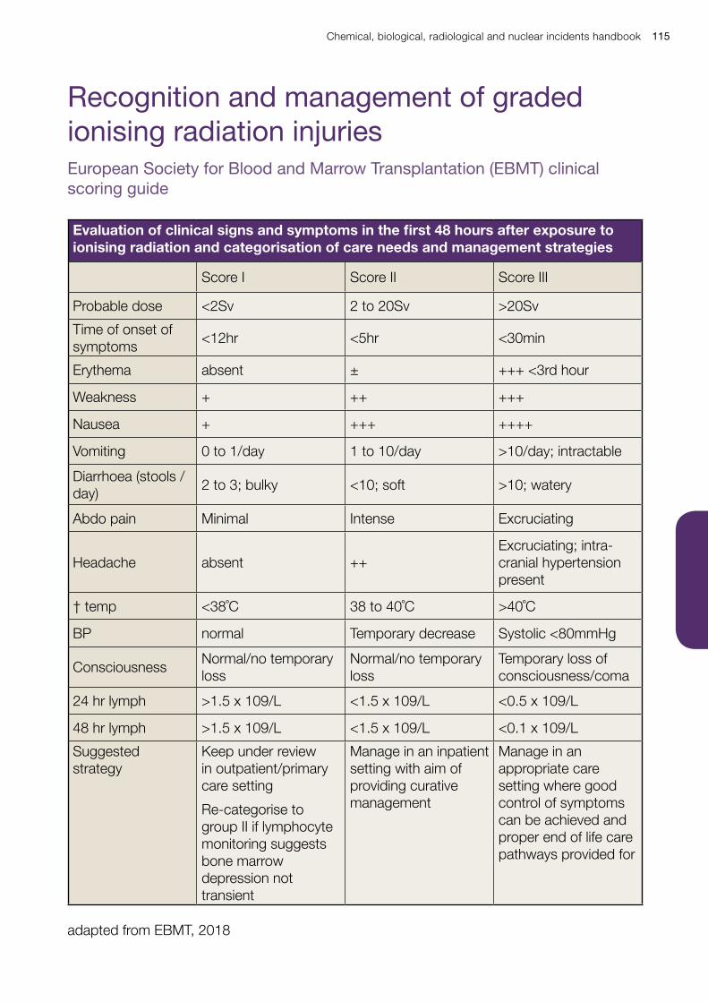
Chemical, biological, radiological and nuclear incidents handbook 115
European Society for Blood and Marrow Transplantation (EBMT) clinical scoring guide
Evaluation of clinical signs and symptoms in the first 48 hours after exposure to ionising radiation and categorisation of care needs and management strategies
Score I Score II Score III
Probable dose <2Sv 2 to 20Sv >20Sv
Time of onset of symptoms
<12hr <5hr <30min
Erythema absent ± +++ <3rd hour
Weakness + ++ +++
Nausea + +++ ++++
Vomiting 0 to 1/day 1 to 10/day >10/day; intractable
Diarrhoea (stools / day)
2 to 3; bulky <10; soft >10; watery
Abdo pain Minimal Intense Excruciating
Headache absent ++Excruciating; intra-cranial hypertension present
† temp <38˚C 38 to 40˚C >40˚C
BP normal Temporary decrease Systolic <80mmHg
ConsciousnessNormal/no temporary loss
Normal/no temporary loss
Temporary loss of consciousness/coma
24 hr lymph >1.5 x 10⁹/L <1.5 x 10⁹/L <0.5 x 10⁹/L
48 hr lymph >1.5 x 10⁹/L <1.5 x 10⁹/L <0.1 x 10⁹/L
Suggested strategy
Keep under review in outpatient/primary care setting
Re-categorise to group II if lymphocyte monitoring suggests bone marrow depression not transient
Manage in an inpatient setting with aim of providing curative management
Manage in an appropriate care setting where good control of symptoms can be achieved and proper end of life care pathways provided for
adapted from EBMT, 2018
Recognition and management of graded ionising radiation injuries
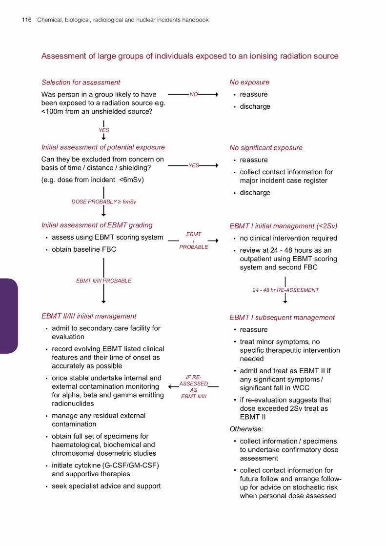
Chemical, biological, radiological and nuclear incidents handbook116
Selection for assessment
Was person in a group likely to have been exposed to a radiation source e.g. <100m from an unshielded source?
Initial assessment of potential exposure
Can they be excluded from concern on basis of time / distance / shielding?
(e.g. dose from incident <6mSv)
Initial assessment of EBMT grading
• assess using EBMT scoring system
• obtain baseline FBC
EBMT II/III initial management
• admit to secondary care facility for evaluation
• record evolving EBMT listed clinical features and their time of onset as accurately as possible
• once stable undertake internal and external contamination monitoring for alpha, beta and gamma emitting radionuclides
• manage any residual external contamination
• obtain full set of specimens for haematological, biochemical and chromosomal dosemetric studies
• initiate cytokine (G-CSF/GM-CSF) and supportive therapies
• seek specialist advice and support
EBMT I initial management (<2Sv)
• no clinical intervention required
• review at 24 - 48 hours as an outpatient using EBMT scoring system and second FBC
No significant exposure
• reassure
• collect contact information for major incident case register
• discharge
EBMT I subsequent management
• reassure
• treat minor symptoms, no specific therapeutic intervention needed
• admit and treat as EBMT II if any significant symptoms / significant fall in WCC
• if re-evaluation suggests that dose exceeded 2Sv treat as EBMT II
Otherwise:
• collect information / specimens to undertake confirmatory dose assessment
• collect contact information for future follow and arrange follow-up for advice on stochastic risk when personal dose assessed
YES
DOSE PROBABLY ≥ 6mSv
EBMT II/III PROBABLE
NO
YES
EBMTI
PROBABLE
24 - 48 hr RE-ASSESMENT
No exposure
• reassure
• discharge
IF RE-ASSESSED
ASEBMT II/III
Assessment of large groups of individuals exposed to an ionising radiation source
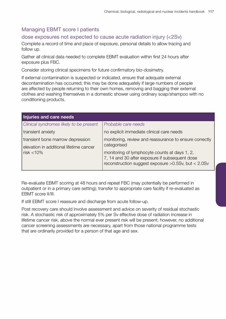
Chemical, biological, radiological and nuclear incidents handbook 117
Managing EBMT score I patientsdose exposures not expected to cause acute radiation injury (<2Sv)Complete a record of time and place of exposure, personal details to allow tracing and follow up.
Gather all clinical data needed to complete EBMT evaluation within first 24 hours after exposure plus FBC.
Consider storing clinical specimens for future confirmatory bio-dosimetry.
If external contamination is suspected or indicated, ensure that adequate external decontamination has occurred; this may be done adequately if large numbers of people are affected by people returning to their own homes, removing and bagging their external clothes and washing themselves in a domestic shower using ordinary soap/shampoo with no conditioning products.
Injuries and care needs
Clinical syndromes likely to be present
transient anxiety
transient bone marrow depression
elevation in additional lifetime cancer risk <10%
Probable care needs
no explicit immediate clinical care needs
monitoring, review and reassurance to ensure correctly categorised
monitoring of lymphocyte counts at days 1, 2, 7, 14 and 30 after exposure if subsequent dose reconstruction suggest exposure >0.5Sv, but < 2.0Sv
Re-evaluate EBMT scoring at 48 hours and repeat FBC (may potentially be performed in outpatient or in a primary care setting); transfer to appropriate care facility if re-evaluated as EBMT score II/III.
If still EBMT score I reassure and discharge from acute follow-up.
Post recovery care should involve assessment and advice on severity of residual stochastic risk. A stochastic risk of approximately 5% per Sv effective dose of radiation increase in lifetime cancer risk, above the normal ever present risk will be present; however, no additional cancer screening assessments are necessary, apart from those national programme tests that are ordinarily provided for a person of that age and sex.

Chemical, biological, radiological and nuclear incidents handbook118
Managing EBMT score II patientscurable radiation injuries (2 to 20Sv)Record clinical features and their time of onset as accurately as possible, particularly nausea, vomiting and diarrhoea, as this may give an indication of the radiation dose and prognosis.
The absorbed dose of radiation should be estimated based on clinical features such as time to emesis, laboratory results (especially lymphocyte and neutrophil counts) health physics advice and bioassay/biodosimetry measurements, including the EBMT / METROPOL guidelines.
When clinically stable undertake internal and external contamination monitoring for alpha, beta and gamma emitting radionuclides, if the patient has residual external contamination this should be removed unless fixed in the tissues.
Take blood samples for full blood count (FBC) with manual differential cell count, coagulation, urea and electrolytes (U&E), liver function test (LFT), thyroid function tests (TFT), amylase, human leukocyte antigen (HLA) typing and also a 10ml lithium heparin tube for chromosome analysis (refer to PHE Centre for Radiation, Chemical and Environmental Hazards); repeat the FBC including absolute lymphocyte count twice over the next 12 hours and daily thereafter.
Injuries and care needsClinical syndromes likely to be present
high likelihood of refractory haematopoietic syndrome if untreated – latent for up to six weeks
potential for gastrointestinal syndrome if dose estimated to be >5Sv – latent for up to 1 week
>10% elevation in additional lifetime cancer risk
Probable care needs
rescue of bone marrow failure (cytokines and haematopoietic stem cell transplantation)
symptomatic treatment of gastro-intestinal tract damage
reverse isolation
management of symptom
Monitor vital signs frequently, including pulse, blood pressure, urine output, oxygen saturations and temperature.
Conventional fluid and electrolyte replacement therapy and sedatives should be used where significant burns, hypervolemia, and/or shock is present.
Enteral nutrition is preferable to parenteral when possible.
Systemic steroids should not be used in the absence of a specific indication.
Seek expert haematological advice at an early stage for the management of pancytopenia. Where bone-marrow failure is established or imminent (lymphocyte count >1.5 x 10⁹/L at 24 to 48 hours) or the dose exposure is relatively homogeneous and probably exceeded 2Sv; then early therapy with granulocyte colony-stimulating factor (G-CSF) should be initiated; addition of erythropoiesis-stimulating agents may be appropriate.
Chemical, biological, radiological and nuclear incidents handbook 119
Where bone marrow failure is refractory to any other intervention (aplasia despite 14 to 21 days cytokine therapy) hematopoietic stem cell transplantation should be considered. Nursing should be provided in appropriate environment to minimise infection risk.
Nausea and vomiting should be managed with 5HT3-receptor antagonists eg ondansetron. Where significant gastro-intestinal tract damage is present or predicted consideration should be given to the use of fluoroquinolone prophylaxis, loperamide, and enteral nutrition.
Radiation burns may be managed with topical steroids, antibiotics, and antihistamines for radiation burns, ulcers, or blisters; excision and grafting of radiation ulcers or necrosis with intractable pain may be required.
Post recovery care should involve assessment and advice on severity of residual stochastic risk. A stochastic risk of approximately 5% per Sv effective dose of radiation increase in lifetime cancer risk, above the normal ever present risk will be present; however, no additional cancer screening assessments are necessary, apart from those national programme tests that are ordinarily provided for a person of that age and sex.
Chemical, biological, radiological and nuclear incidents handbook120
Managing EBMT score III patientsincurable radiation injuries (>20Sv)Record clinical features and their time of onset as accurately as possible, particularly nausea, vomiting and diarrhoea, as this may give an indication of the radiation dose and prognosis.
The absorbed dose of radiation should be estimated based on clinical features such as time to emesis, laboratory results (especially lymphocyte and neutrophil counts) health physics advice and bioassay/biodosimetry measurements, including the EBMT / METROPOL guidelines.
When clinically stable undertake internal and external contamination monitoring for alpha, beta and gamma emitting radionuclides, if the patient has residual external contamination this should be removed unless fixed in the tissues.
Take blood samples for full blood count (FBC) with manual differential cell count, coagulation, urea and electrolytes (U&E), liver function test (LFT), thyroid function tests (TFT), amylase, human leukocyte antigen (HLA) typing and also a 10ml lithium heparin tube for chromosome analysis (refer to PHE Centre for Radiation, Chemical and Environmental Hazards); repeat the FBC including absolute lymphocyte count twice over the next 12 hours and daily thereafter.
Injuries and care needsClinical syndromes likely to be present
Immediate effects (hours):
central nervous system syndrome
cardio-vascular system syndrome
if survival prolonged:
refractory haematopoietic syndrome
gastrointestinal syndrome
Probable care needs
multi-organ failure beyond curative measures
medical care needs are for symptom control only
death usually occurs within 1 to 3 days without very aggressive supportive care
Initially treat as EBMT score II cases until it is clear that the radiation inuries are not survivable.
Where clear evidence that inevitably lethal disease is present (especially if neuro-vascular or CNS syndrome are established indicating a whole body dose in excess of 20Sv is likely); consider the appropriate care pathway that will provide the best chance of palliating the patients suffering, in line with best ethical and professional standards and guidance.
If in doubt as to inevitability of progression to acutely fatal disease continue to manage as EBMT score II guidelines.
Chemical, biological, radiological and nuclear incidents handbook 121
Mass Casualty Guidelines
1 Management of blood-borne virus risk in victims of significant blast or multiple victim attacks creating penetrating injuries (hepatitis B, C and HIV) 123
2 Antibiotic prophylaxis guidance for bomb blast victims 127
3 Antimicrobial prophylaxis for suspected exposure to deliberate release of a bacterial agent (anthrax, plague, tularemia) 135
4 Management of potential injury to hearing following an attack involving blast 139
5 Management of potential brain injury following an attack Involving blast 143
6 MRI following an attack involving bomb blast or bullet wounds 145

Chemical, biological, radiological and nuclear incidents handbook122
Chemical, biological, radiological and nuclear incidents handbook 123
1 Management of blood-borne virus risk in victims of significant blast or multiple victim attacks creating penetrating injuries (hepatitis B, C and HIV)
Chemical, biological, radiological and nuclear incidents handbook124
BackgroundIt is a recognised complication of incidents, where traumatic injuries occur that result in transfer of human biological materials between the affected people, that these transfers create a potential risk of transmission of blood-borne virus infections (hepatitis B, hepatitis C and HIV).
This potential risk should be considered to be present for:
• incidents where two or more people have presented with significant blast injuries
• incidents where significant traumatic transfer of human biological materials may have taken place, such as serial attacks with bladed or other forms of penetrating weapons where multiple victims have been injured, that these injuries were incurred using the same, or, a small group of weapons, and the injuries incurred are of a nature that the transfer of significant amounts of blood or other body tissues may have occurred
Key factsThe risk of transmission of blood-borne viral infections in such circumstances is predicated on the prevalence of hepatitis B, C and HIV carriage in the UK population which is generally low and the transmissibility of these infections.
Current estimates suggest that the population prevalence of hepatitis B is <1%, and hepatitis C <0.5%.
For HIV the very high rate of viral suppression (giving negligible transmission risk) in those currently in treatment for HIV suggest a prevalence of transmissible infection in 4 in 10,000 people (0.04%).
The probability of transmission of these blood-borne viruses in such incidents is unknown; however, the usually accepted risks of transmission per incident following sharps injuries from known infected persons in clinical settings, which may be the closest natural model, is generally quoted as being 30% for hepatitis B, 3% for hepatitis C and 0.3% for HIV.
Immediate care and managementFor all patients with injuries that have breached the skin:
• all penetrating injuries should be radiographed and all foreign body implantations urgently removed if possible
• blood specimens should be taken before any specific post-exposure treatment is instituted, provided this does not delay post-exposure treatment, and tested for hepatitis B, hepatitis C and HIV. Consent should be sought to share the results of these tests with PHE to help inform and to review PHE’s assessment of risk to those involved
• a very rapid course of hepatitis B vaccination (day 0, day 7, and day 21) must be given starting within 72 hours of initial injury
• all patients should be followed up at 3 and 6 months to determine hepatitis B, C and HIV status (and then managed accordingly)
• HIV post-exposure prophylaxis is not recommended
Chemical, biological, radiological and nuclear incidents handbook 125
Local Health Protection Teams should be involved in managing blood-borne injury risks as soon as possible in order to identify, register and coordinate the long-term follow-up of affected people.
Post-exposure assessment, screening and careCollection of specimens from the scene, at post-mortems and from survivors to estimate risk of BBV infections is desirable to better evaluate risk to the affected people. However, it must be recognised that access to such specimens may be limited by legal and ethical considerations.
Serological markers of hepatitis B, hepatitis C and HIV infection should be sought three and six months after exposure.
Earlier testing using genomic methods that can reduce the elapsed time necessary for diagnosis may be considered.
Where an individual is found to be infected appropriate counselling, support and treatment must be given and PHE informed in order that the risk assessment for the incident can be re-evaluated and advice to all the affected persons and their treating clinicians modified if appropriate.
Lifestyle adviceAs the risk of infection from any of these blood-borne viruses is remote, or for the most likely risks effectively managed by post-exposure treatment, onward transmission to sexual partners, or to household contacts through shared hygiene settings / practices, is highly unlikely and no restrictions or precautions on personal lifestyle are advised (other than those always recommended to the general public to protect any individual against sexually transmitted infection).
Health care worker victimsThe additional risk of blood-borne virus infection to any health professional who is the victims of such a multiple trauma incidents is so low that no restrictions to performing exposure prone procedures are recommended. However, strict compliance with follow up and reporting of any adverse findings is mandatory.

Chemical, biological, radiological and nuclear incidents handbook126

Chemical, biological, radiological and nuclear incidents handbook 127
2 Antimicrobial Prophylaxis Guidance for Bomb Blast Victims
Chemical, biological, radiological and nuclear incidents handbook128
Principles of treatmentThis advice is suitable for ALL age groups of our patients.
This guidance is based on the best available evidence but its application must be modified by professional judgement and any knowledge of previous culture results. A dose and duration of treatment is suggested. In severe or recurrent cases consider a larger dose or longer course.
This guidance has been prepared mindful of best practice in the management of antimicrobial resistance issues.
In the event of any uncertainty clinicians are advised to contact their local microbiology department for advice.
1. Where multiple injuries have been sustained (eg bone fractures and eye injuries) it may be possible to rationalise antibiotic regimes following discussions with local microbiology departments
2. Blast injured patients will mount a brisk inflammatory response and so inevitably have pyrexia and a high CRP. If WCC is rising assess carefully for infection. Consider monitoring Procalcitonin (PCT) every other day to differentiate infection from an inflammatory response linked to Systemic Inflammatory Response Sydrome (SIRS) or trauma1
3. Local microbiology advice should be sought to consider possibility of nosocomial infection and take into account any prevalent organisms that have posed infection control risks
4. Consider linking patients in the laboratory using IT systems (such as LIMS)
5. To ensure continuity of prescribing it is advised that treating hospitals dispense sufficient medication to allow patients to complete antibiotic courses at home
6. MRI will show high marrow signal after blast injury which mimics osteomyelitis – these changes last for around six months, so bear in mind if investigating possible bone infection
7. Where receiving hospitals have had problems with resistant gram negative rods (especially carbapenemase producing enterobacteriaceae – CPE); affected patients will require isolation and screening as per PHE guidance2
8. Zygomycete infection has presented around 10 days post injury in military casualties where there was implantation of organic matter. This scenario is less likely following bomb injuries acquired in a civilian setting, but the microbiology of wounds should be monitored for invasive fungal infection
Tetanus prophylaxisALL bomb victims with injuries must have their tetanus immunisation status checked and treated according to the extant advice on management of patients with tetanus prone wounds in the ‘Green Book’3.
1 PCT may be expected to be elevated in the first 24-48 hours post trauma but fall quickly (Castillo GP et al, critical care medicine 2009, 37 (6):1845-9 Procalcitonin as a prognostic and diagnostic tool for septic complications after major trauma)
2 https://www.gov.uk/government/publications/carbapenemase-producing-enterobacteriaceae-early-detection-management-and-control-toolkit-for-acute-trusts
3 https://www.gov.uk/government/uploads/system/uploads/attachment_data/file/148506/Green-Book-Chapter-30-dh_103982.pdf
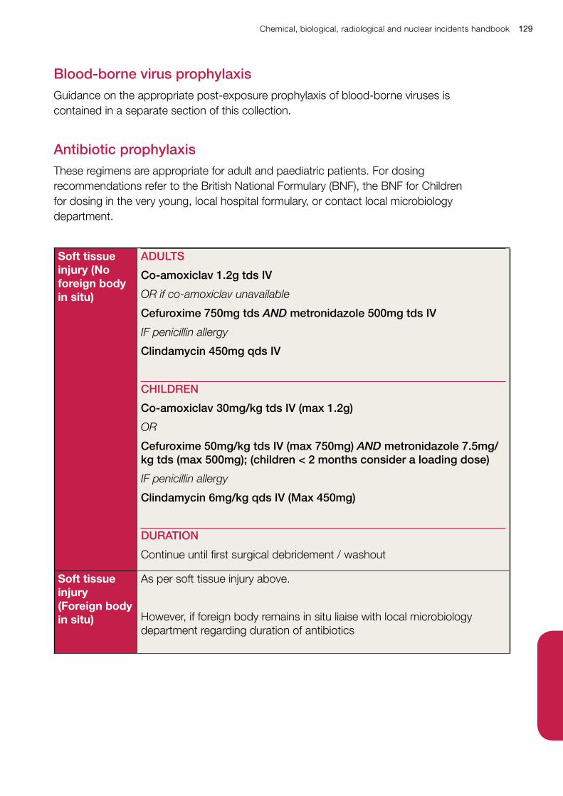
Chemical, biological, radiological and nuclear incidents handbook 129
Blood-borne virus prophylaxisGuidance on the appropriate post-exposure prophylaxis of blood-borne viruses is contained in a separate section of this collection.
Antibiotic prophylaxisThese regimens are appropriate for adult and paediatric patients. For dosing recommendations refer to the British National Formulary (BNF), the BNF for Children for dosing in the very young, local hospital formulary, or contact local microbiology department.
Soft tissue injury (No foreign body in situ)
ADULTS
Co-amoxiclav 1.2g tds IV
OR if co-amoxiclav unavailable
Cefuroxime 750mg tds AND metronidazole 500mg tds IV
IF penicillin allergy
Clindamycin 450mg qds IV
CHILDREN
Co-amoxiclav 30mg/kg tds IV (max 1.2g)
OR
Cefuroxime 50mg/kg tds IV (max 750mg) AND metronidazole 7.5mg/kg tds (max 500mg); (children < 2 months consider a loading dose)
IF penicillin allergy
Clindamycin 6mg/kg qds IV (Max 450mg)
DURATION
Continue until first surgical debridement / washout
Soft tissue injury (Foreign body in situ)
As per soft tissue injury above.
However, if foreign body remains in situ liaise with local microbiology department regarding duration of antibiotics
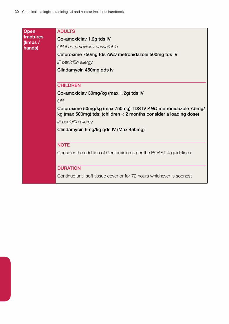
Chemical, biological, radiological and nuclear incidents handbook130
Open fractures (limbs / hands)
ADULTS
Co-amoxiclav 1.2g tds IV
OR if co-amoxiclav unavailable
Cefuroxime 750mg tds AND metronidazole 500mg tds IV
IF penicillin allergy
Clindamycin 450mg qds iv
CHILDREN
Co-amoxiclav 30mg/kg (max 1.2g) tds IV
OR
Cefuroxime 50mg/kg (max 750mg) TDS IV AND metronidazole 7.5mg/kg (max 500mg) tds; (children < 2 months consider a loading dose)
IF penicillin allergy
Clindamycin 6mg/kg qds IV (Max 450mg)
NOTE
Consider the addition of Gentamicin as per the BOAST 4 guidelines
DURATION
Continue until soft tissue cover or for 72 hours whichever is soonest
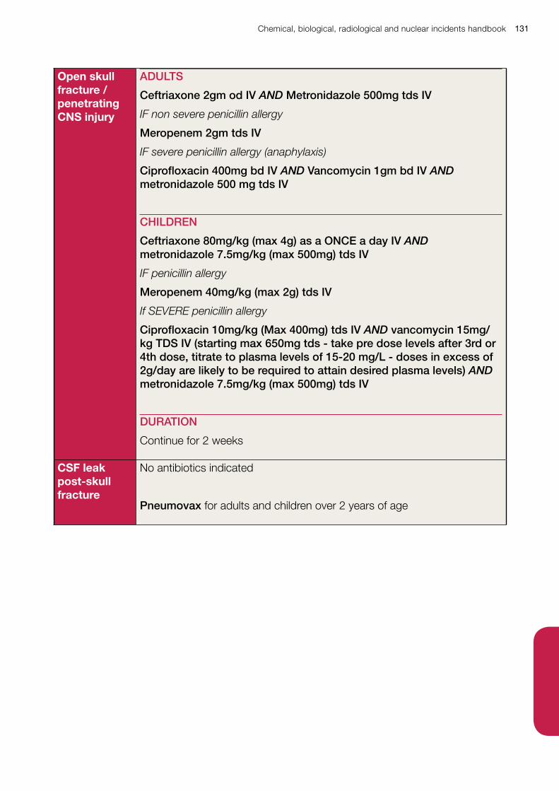
Chemical, biological, radiological and nuclear incidents handbook 131
Open skull fracture / penetrating CNS injury
ADULTS
Ceftriaxone 2gm od IV AND Metronidazole 500mg tds IV
IF non severe penicillin allergy
Meropenem 2gm tds IV
IF severe penicillin allergy (anaphylaxis)
Ciprofloxacin 400mg bd IV AND Vancomycin 1gm bd IV AND metronidazole 500 mg tds IV
CHILDREN
Ceftriaxone 80mg/kg (max 4g) as a ONCE a day IV AND metronidazole 7.5mg/kg (max 500mg) tds IV
IF penicillin allergy
Meropenem 40mg/kg (max 2g) tds IV
If SEVERE penicillin allergy
Ciprofloxacin 10mg/kg (Max 400mg) tds IV AND vancomycin 15mg/kg TDS IV (starting max 650mg tds - take pre dose levels after 3rd or 4th dose, titrate to plasma levels of 15-20 mg/L - doses in excess of 2g/day are likely to be required to attain desired plasma levels) AND metronidazole 7.5mg/kg (max 500mg) tds IV
DURATION
Continue for 2 weeks
CSF leak post-skull fracture
No antibiotics indicated
Pneumovax for adults and children over 2 years of age
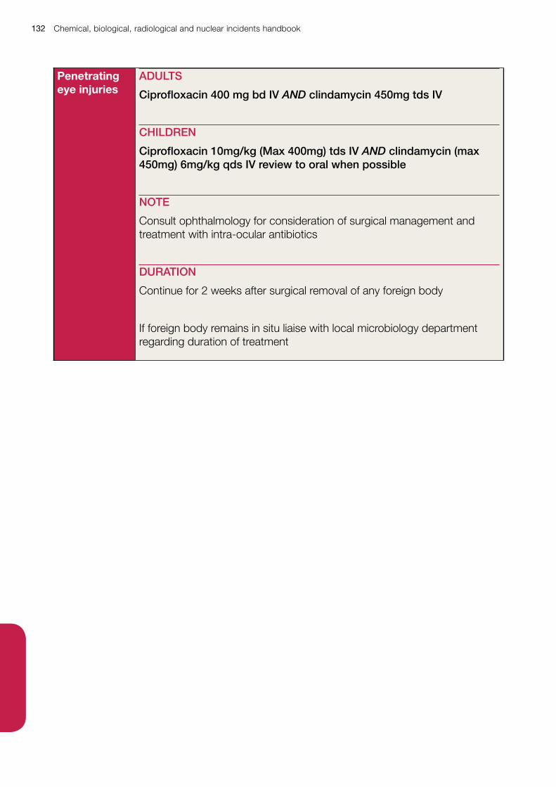
Chemical, biological, radiological and nuclear incidents handbook132
Penetrating eye injuries
ADULTS
Ciprofloxacin 400 mg bd IV AND clindamycin 450mg tds IV
CHILDREN
Ciprofloxacin 10mg/kg (Max 400mg) tds IV AND clindamycin (max 450mg) 6mg/kg qds IV review to oral when possible
NOTE
Consult ophthalmology for consideration of surgical management and treatment with intra-ocular antibiotics
DURATION
Continue for 2 weeks after surgical removal of any foreign body
If foreign body remains in situ liaise with local microbiology department regarding duration of treatment
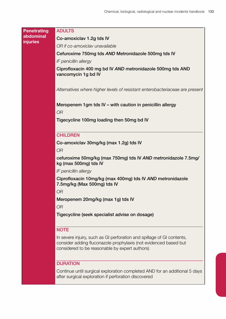
Chemical, biological, radiological and nuclear incidents handbook 133
Penetrating abdominal injuries
ADULTS
Co-amoxiclav 1.2g tds IV
OR if co-amoxiclav unavailable
Cefuroxime 750mg tds AND Metronidazole 500mg tds IV
IF penicillin allergy
Ciprofloxacin 400 mg bd IV AND metronidazole 500mg tds AND vancomycin 1g bd IV
Alternatives where higher levels of resistant enterobacteriaceae are present
Meropenem 1gm tds IV – with caution in penicillin allergy
OR
Tigecycline 100mg loading then 50mg bd IV
CHILDREN
Co-amoxiclav 30mg/kg (max 1.2g) tds IV
OR
cefuroxime 50mg/kg (max 750mg) tds IV AND metronidazole 7.5mg/kg (max 500mg) tds IV
IF penicillin allergy
Ciprofloxacin 10mg/kg (max 400mg) tds IV AND metronidazole 7.5mg/kg (Max 500mg) tds IV
OR
Meropenem 20mg/kg (max 1g) tds IV
OR
Tigecycline (seek specialist advise on dosage)
NOTE
In severe injuiry, such as GI perforation and spillage of GI contents, consider adding fluconazole prophylaxis (not evidenced based but considered to be reasonable by expert authors)
DURATION
Continue until surgical exploration completed AND for an additional 5 days after surgical exploration if perforation discovered
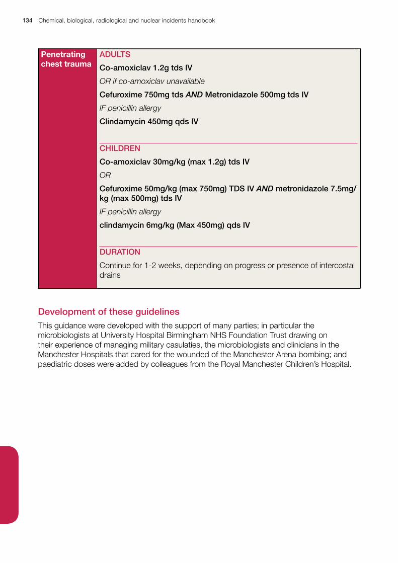
Chemical, biological, radiological and nuclear incidents handbook134
Penetrating chest trauma
ADULTS
Co-amoxiclav 1.2g tds IV
OR if co-amoxiclav unavailable
Cefuroxime 750mg tds AND Metronidazole 500mg tds IV
IF penicillin allergy
Clindamycin 450mg qds IV
CHILDREN
Co-amoxiclav 30mg/kg (max 1.2g) tds IV
OR
Cefuroxime 50mg/kg (max 750mg) TDS IV AND metronidazole 7.5mg/kg (max 500mg) tds IV
IF penicillin allergy
clindamycin 6mg/kg (Max 450mg) qds IV
DURATION
Continue for 1-2 weeks, depending on progress or presence of intercostal drains
Development of these guidelinesThis guidance were developed with the support of many parties; in particular the microbiologists at University Hospital Birmingham NHS Foundation Trust drawing on their experience of managing military casulaties, the microbiologists and clinicians in the Manchester Hospitals that cared for the wounded of the Manchester Arena bombing; and paediatric doses were added by colleagues from the Royal Manchester Children’s Hospital.
Chemical, biological, radiological and nuclear incidents handbook 135
3 Antimicrobial prophylaxis for suspected exposure to deliberate release of a bacterial agent (anthrax, plague, tularemia)
Chemical, biological, radiological and nuclear incidents handbook136
This advice supplements that given on the sections detailing the management of individual cases in the main body of the Biological Threats guidance of this collection.
It is intended as a quick reference sheet, or for use alongside national specimen patient group direction published by PHE.
RationaleEarly prophylactic (pre-symptomatic) antibiotics, using ciprofloxacin, doxycycline or amoxicillin / co-amoxiclav may be recommended by national public health authorities where deliberate release of anthrax, tularemia, plague, or other susceptible bacteria is suspected.
The use of antibiotic prophylaxis will usually be divided into two phases:
• initial treatment phase where simplicity and speed of initiation of prophylaxis is critical and uncertainty about the population at risk may lead to a relatively large group of people needing initial prophylaxis
• extended treatment phase where better information has allowed the population at risk to be more clearly identified and the most appropriate antibiotic to be used on the basis of age, pregnancy or other considerations
The initial treatment phase will use ciprofloxacin as the first choice antibiotic for all age groups.
An extended treatment phase, if required, will use doxycycline wherever possible, reserving continuity of prophylaxis with ciprofloxacin for children under 12 years of age.
For anthrax and plague amoxicillin or co-amoxiclav is available as a third-line medicine for people in whom both ciprofloxacin and doxycycline are contra-indicated. Tularemia is not sensitive to penicillins.
The recommended prophylaxis periods are:
• Anthrax up to 60 days – a shorter period may be recommended
• Tularemia 14 days
• Plague 7 days
Initial prophylaxis (10 day supply)• 1st line – ciprofloxacin unless there is an established history of severe allergic reaction
• 2nd line – doxycycline unless under 8 years of age, pregnant or breast feeding, or there is an established history of severe allergic reaction
• 3rd line (NOT tularemia) – amoxicillin / co-amoxiclav unless there is an established history of severe allergic reaction
If ciprofloxacin and amoxicillin / co-amoxiclav are contraindicated then doxycycline should be used at any age.
Extended prophylaxis beyond initial 10 day supplyUnder 8 years of age
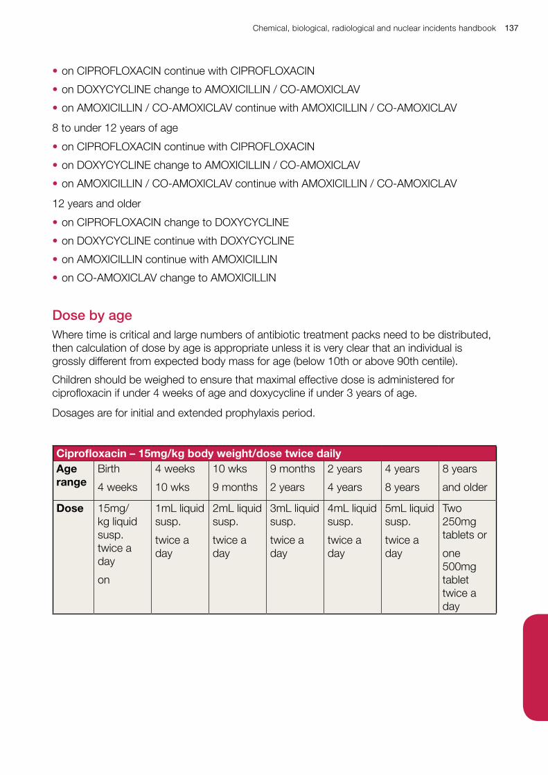
Chemical, biological, radiological and nuclear incidents handbook 137
• on CIPROFLOXACIN continue with CIPROFLOXACIN
• on DOXYCYCLINE change to AMOXICILLIN / CO-AMOXICLAV
• on AMOXICILLIN / CO-AMOXICLAV continue with AMOXICILLIN / CO-AMOXICLAV
8 to under 12 years of age
• on CIPROFLOXACIN continue with CIPROFLOXACIN
• on DOXYCYCLINE change to AMOXICILLIN / CO-AMOXICLAV
• on AMOXICILLIN / CO-AMOXICLAV continue with AMOXICILLIN / CO-AMOXICLAV
12 years and older
• on CIPROFLOXACIN change to DOXYCYCLINE
• on DOXYCYCLINE continue with DOXYCYCLINE
• on AMOXICILLIN continue with AMOXICILLIN
• on CO-AMOXICLAV change to AMOXICILLIN
Dose by ageWhere time is critical and large numbers of antibiotic treatment packs need to be distributed, then calculation of dose by age is appropriate unless it is very clear that an individual is grossly different from expected body mass for age (below 10th or above 90th centile).
Children should be weighed to ensure that maximal effective dose is administered for ciprofloxacin if under 4 weeks of age and doxycycline if under 3 years of age.
Dosages are for initial and extended prophylaxis period.
Ciprofloxacin – 15mg/kg body weight/dose twice dailyAge range
Birth
4 weeks
4 weeks
10 wks
10 wks
9 months
9 months
2 years
2 years
4 years
4 years
8 years
8 years
and older
Dose 15mg/kg liquid susp. twice a day
on
1mL liquid susp.
twice a day
2mL liquid susp.
twice a day
3mL liquid susp.
twice a day
4mL liquid susp.
twice a day
5mL liquid susp.
twice a day
Two 250mg tablets or
one 500mg tablet twice a day
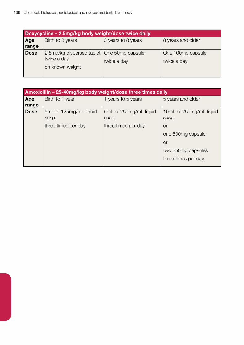
Chemical, biological, radiological and nuclear incidents handbook138
Doxycycline – 2.5mg/kg body weight/dose twice dailyAge range
Birth to 3 years 3 years to 8 years 8 years and older
Dose 2.5mg/kg dispersed tablet twice a day
on known weight
One 50mg capsule
twice a day
One 100mg capsule
twice a day
Amoxicillin – 25-40mg/kg body weight/dose three times dailyAge range
Birth to 1 year 1 years to 5 years 5 years and older
Dose 5mL of 125mg/mL liquid susp.
three times per day
5mL of 250mg/mL liquid susp.
three times per day
10mL of 250mg/mL liquid susp.
or
one 500mg capsule
or
two 250mg capsules
three times per day
Chemical, biological, radiological and nuclear incidents handbook 139
4 Management of potential injury to hearing following an attack involving blast
Chemical, biological, radiological and nuclear incidents handbook140
BackgroundMembers of the public who are exposed to a blast incident are at risk of multiple injuries. Primary blast injury is attributable to the interaction of the blast wave with the body by the passage of a high-pressure wave through body tissue4. The ear is the most commonly injured organ in the body following an explosion.5
Primary blast injuries of the auditory system cause significant morbidity, but can be overlooked, particularly in the presence of other injuries.
• the most common symptoms are hearing loss, otalgia and tinnitus
• many victims will suffer a short-lived but profound period of sensorineural deafness and tinnitus that resolves within hours
• a proportion of cases suffer permanent hearing loss which may be sensorineural, conductive or mixed6
• explosions occurring in confined spaces result in a higher incidence of primary blast injury, greater mortality, and greater injury severity7
• it is essential therefore that every patient that has been involved in a blast incident and is seen by a medical professional, is fully examined, has their examination findings recorded and has audiometric follow-up
Key FactsThe orientation of the auditory canal to the blast wave has a significant effect on the characteristics of the wave as it travels within the canal. Usually both ears are affected but the ear closest to the explosion is likely to be more severely affected.8
The presence of surrounding structures which affect the blast wave and normal biological variability result in apparently similar explosions producing a wide range of otological injuries.9, 10
Tympanic membrane perforation is the most common injury to the middle ear.
Human tympanic membranes begin to rupture at pressures as low as 5 psi (35 kPa) and 50% will fail at 15 psi (104 kPa).11
Depending on the explosion’s setting, between 2% and 32% of all people injured, and up to 94% of those with primary blast injuries will have a ruptured tympanic membranes.12
4 Okpala N. Management of blast ear injuries in mass casualty environments. Mil Med 2011 Nov;176(11): 1306-10
5 Cohen J et al. Blast injury of the ear in a confined space explosion: auditory and vestibular evaluation Isr Med Assosc J. 2002 Jul; 4(7):559-62
6 Cave KM , Cornish EM , Chandler DW : Blast injury of the ear: clinical update from the global war on terror . Mil Med 2007 ; 172 (7) : 726 – 30 .
7 Leibovici D , Gofrit ON , Shapira SC : Eardrum perforation in explosion survivors: is it a marker of pulmonary blast injury? Ann Emerg Med 1999 ; 34: 168 – 72 .
8 Fisher, T. Blast Injury of the Ear – Synopsis of Causation, Ministry of Defence Sep 2008
9 Garth RJN. Blast injury of the ear. In: Cooper GJ, Dudley HAF, Gann DS, et al, eds. Scientific Foundations of Trauma. Oxford, UK: Butterworth-Heinemann; 1997: p. 225-235.
10 Roberto M, Hamernik RP, Turrentine GA. Damage of the auditory system associated with acute blast trauma. Ann Otol Rhinol Laryngol 1989;98:23-34
11 Kerr AG. Blast injuries to the ear. Practitioner 1978; 221: 677 – 682.
12 Okpala N. Management of blast ear injuries in mass casualty environments. Mil Med 2011 Nov;176(11): 1306-10
Chemical, biological, radiological and nuclear incidents handbook 141
50-80% of tympanic membrane ruptures caused by acute acoustic trauma will heal spontaneously.13,14,15
Large and central perforations have the least tendency to heal and usually require surgical intervention which is normally performed when the tympanic membrane remains unhealed at 10-12 months.
Although intact tympanic membranes were once relied upon as a reliable marker of absence of exposure to significant overpressures, significant primary blast injuries may occur in its absence. In one study, 36.7% of patients with blast lung injury had intact tympanic membranes.16
A characteristic notched audiometric configuration is associated with noise-induced hearing loss, which sees a maximum reduction in sensitivity to stimulation in the range of 3 to 6 kHz, and recovery at 8 kHz.17
Cholesteatoma of the middle ear and mastoid air-cell system is a late complication occurring in up to 12% of blast induced perforations18 and presents up to 4 years after injury.19
There is little evidence that vasodilators, low molecular weight dextrans, anti-platelet drugs, vitamins or systemic steroids improve the outcome for patients with sensorineural hearing loss caused by blast.2021
Recommendations• for patients exposed to a blast incident and taken to hospital, ATLS guidelines must be
followed
• the secondary survey must include an otological assessment that is formally recorded in the patients’ notes. All patients, irrespective of reporting any deafness or hearing deficit should undergo audiometric follow-up22
• for those with tympanic membrane rupture, simple initial advice can be given to patients to avoid submersion in water, avoid probing the ear canal, and to seek review for antibiotics in the event of suspected infection
• attempts to reduce the degree of permanent hearing loss after blast injury by the use of corticosteroids, LMW dextran, magnesium and vasodilators have limited evidence in regards to their effectiveness and are not recommended as a universal measure
• patients presenting in Primary Care following blast exposure should undergo full otological examination, be referred for audiometry, and subsequent ENT follow up if required
13 Kerr AG, Byrne JE. Concussive effects of bomb blast on the ear. J Laryngol Otol 1975;89(2):131-43.
14 Walsh RM, Pracy JP, Huggon AM, Gleeson MJ. Bomb blast injuries to the ear: the London Bridge incident series. J Accid Emerg Med 1995;12(3):194-8.
15 Cohen JT, Ziv G, Bloom J et al. Blast injury of the ear in a confined space explosion: auditory and vestibular evaluation. Isr Med Assoc J. 2002;4(7):559-62.
16 Leibovici D, Gofrit ON, Shapira SC. Eardrum perforation in explosion survivors: is it a marker of pulmonary blast injury? Ann Emerg Med 1999; 34(2):168-172.
17 Okpala N. Management of blast ear injuries in mass casualty environments. Mil Med 2011 Nov;176(11): 1306-10
18 Seaman RW, Newell RC. Another etiology of middle ear cholesteatoma. Arch Otolaryngol. 1971; 94: 440 –442.
19 Kronenberg J, Ben-Shoshan J, Modan M, Leventon G. Blast injury and cholesteatoma. Am J Otol.1988; 9: 127– 130.
20 Horrocks, C.L. Blast injuries:biophysics, pathophysiology and management principles. J R Army Med Corps.2001 Feb 147 (1) 28-40.
21 Kellerhals B , Hippert F , Pfaltz CR : Treatment of acute acoustic trauma with low molecular weight dextran . Pract Otorhinolaryngol 1971 ; 33: 260 – 4 .
22 https://www.rcemlearning.co.uk/references/blast-injuries/
Chemical, biological, radiological and nuclear incidents handbook142
• in the event of a specific blast incident, PHE would consider a press release for Primary Care Practitioners who are likely to encounter patients affected by the incident. This should highlight the requirement for audiometry for patients presenting to their GP who were involved in the incident, and the need for increased subsequent monitoring in primary care to negate later potential complications such as cholesteatoma

Chemical, biological, radiological and nuclear incidents handbook 143
5 Management of potential brain injury following an attack involving a blast
Chemical, biological, radiological and nuclear incidents handbook144
BackgroundInjuries resulting from blasts are categorized into four groups:-
Primary blast injuries are caused by over- or under-pressurization from the blast and most commonly affect air-filled and fluid-encased organs within the body.
Secondary injuries occur from flying debris, including bomb fragments, and can affect any part of the body.
Tertiary injuries are the result of the individual being thrown by the blast pressure wave; this results in injuries such as fractures, blunt trauma injury, and amputations.
Quaternary injuries include all blast injuries not described in the aforementioned types, including inhalation of toxic fumes, crush injuries, burns and exacerbation of pre-existing medical conditions.
• the brain is most vulnerable to secondary and tertiary blast injury
• data for brain injury due to primary blast forces is limited
• most common types of traumatic brain injury are diffuse axonal injury, contusion and subdural haemorrhage
The relationships among blast-related traumatic brain injury, axonal injury and outcomes that include post-traumatic stress disorder are areas of active ongoing research.23
RecommendationsRisk of brain injury from a single blast incident is dependent on multiple factors. There is no current evidence to support any additional assessment for brain injury following a single blast exposure in addition to current ATLS guidelines. Further assessment or referral is dependent on the initial clinical assessment of individuals presenting with injuries following an exposure to a blast incident.
23 MacDonald C et al. Detection of Blast-Related Traumatic Brain Injury in U.S. Military Personnel The New England Journal of Medicine June 2 2011 Vol 363 No. 22 pgs2091-2100

Chemical, biological, radiological and nuclear incidents handbook 145
6 MRI following an attack involving bomb blast or bullet wounds
Chemical, biological, radiological and nuclear incidents handbook146
Background
Imaging studies using MRI following trauma from bomb or gunshot injuries need to be carefully considered if any of the fragments contain ferromagnetic materials.
Ferromagnetic materials (such as steel fragments) may exhibit point heating and movement, shifting to align with the magnetic field.
Clinical review of the Manchester Arena bomb blast incident suggest that where injuries due to deliberate inclusion of shrapnel materials are identified in some patients it should be presumed that fragments will be present in ALL patients with traumatic injuries from such incidents. Fragments from the building and ancillary furniture may also be present.
However, unjacketed firearm bullets will often be non-ferromagnetic, and at least one case series has shown that MRI examinations in such cases are usually safe.24
Fibrosis around fragments will stabilise movement in the magnetic field of an MRI scanner, usually by six weeks post-injury (although fragment movement has been reported at up to two years following injury). However, point heating remains a risk.25
No evidence has been found to show that metal detectors can effectively help to identify the presence of ferromagnetic fragments in the human body.
RecommendationsBomb blast injuries
• it must be assumed that all victims will have shrapnel injuries
• it must be assumed that the shrapnel fragments will be ferromagnetic
• metal detectors should not be used to determine composition of fragments, or as a guide to safety of acute MRI studies
• acute MRI studies are contraindicated until a clear understanding of injury patterns from CT traumagrams gives a picture of what fragments may be present
At 6 weeks there should be enough fibrosis around fragments to allow MRI to be undertaken safely, but noting that, if these fragments are close to nerve or vessels then extreme caution is still required. Patient consent must include discussion of potential fragment movement.
Patients should also be warned that there can be heating from the magnet and to report it during the scan, if felt, so that the imaging can be aborted.
Bullet injuries
Fragments will often be non-ferromagnetic and MRI studies should probably be undertaken if they will assist clinical management. However, patient consent must include discussion of potential fragment movement and heating.
24 Magnetic Resonance Imaging to Evaluate Cervical Spinal Cord Injury from Gunshot Wounds from Handguns. Justin Slavin, Narlin Beaty, Prashant Raghavan, Charles Sansur, Bizhan Aarabi. World Neurosurg. (2015) 84, 6:1916-1922.
25 A Practical Guide to MR Imaging Safety: What Radiologists Need to Know. Leo L. Tsai, Aaron K. Grant, Koenraad J. Mortele, Justin W. Kung, Martin P. Smith. RadioGraphics 2015; 35:1722–1737

© Crown copyright 2018
You may re-use this information (excluding logos) free of charge in any format or medium, under the terms of the Open Government Licence v3.0. To view this licence, visit OGL or email [email protected]. Where we have identified any third party copyright information you will need to obtain permission from the copyright holders concerned. Published May 2018 PHE publications gateway number: 2018080
Public Health England supports the UN Sustainable Development Goals
About Public Health EnglandPublic Health England exists to protect and improve the nation’s health and wellbeing, and reduce health inequalities. We do this through world-leading science, knowledge and intelligence, advocacy, partnerships and the delivery of specialist public health services. We are an executive agency of the Department of Health and Social Care, and a distinct delivery organisation with operational autonomy. We provide government, local government, the NHS, Parliament, industry and the public with evidence-based professional, scientific and delivery expertise and support.
Public Health EnglandWellington House 133-155 Waterloo RoadLondon SE1 8UGTel: 020 7654 8000www.gov.uk/phe Twitter: @PHE_ukFacebook: www.facebook.com/PublicHealthEngland